scientific cameras cooled ccd - thorlabs · compact scientific cameras these compact scientific...
TRANSCRIPT
Scie
ntifi
c C
am
era
s
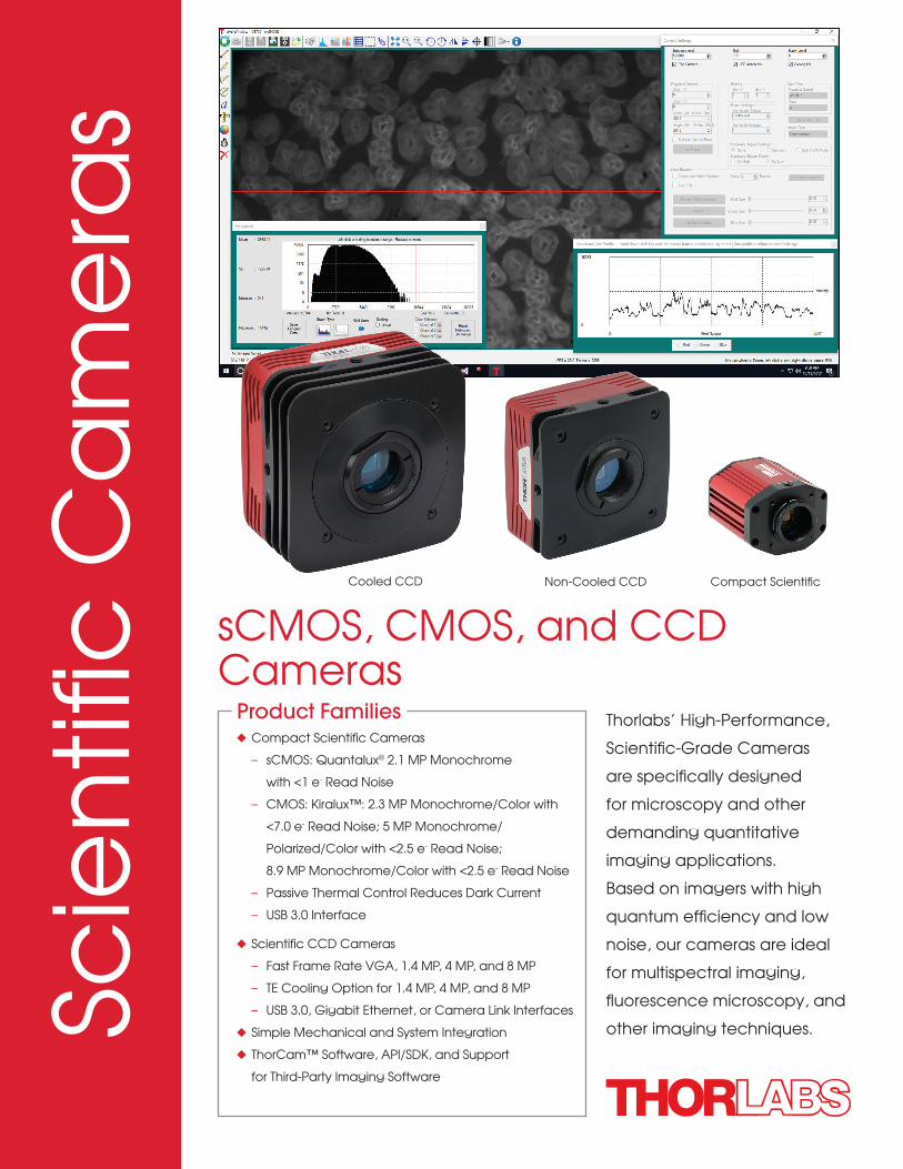
Non-Cooled CCDCooled CCD Compact Scientific
u Compact Scientific Cameras
– sCMOS: Quantalux® 2.1 MP Monochrome
with <1 e- Read Noise
– CMOS: Kiralux™: 2.3 MP Monochrome/Color with
<7.0 e- Read Noise; 5 MP Monochrome/
Polarized/Color with <2.5 e- Read Noise;
8.9 MP Monochrome/Color with <2.5 e- Read Noise
– Passive Thermal Control Reduces Dark Current
– USB 3.0 Interface
u Scientific CCD Cameras
– Fast Frame Rate VGA, 1.4 MP, 4 MP, and 8 MP
– TE Cooling Option for 1.4 MP, 4 MP, and 8 MP
– USB 3.0, Gigabit Ethernet, or Camera Link Interfaces
u Simple Mechanical and System Integration
u ThorCam™ Software, API/SDK, and Support
for Third-Party Imaging Software
Product Families Thorlabs’ High-Performance,
Scientific-Grade Cameras
are specifically designed
for microscopy and other
demanding quantitative
imaging applications.
Based on imagers with high
quantum efficiency and low
noise, our cameras are ideal
for multispectral imaging,
fluorescence microscopy, and
other imaging techniques.
sCMOS, CMOS, and CCD Cameras
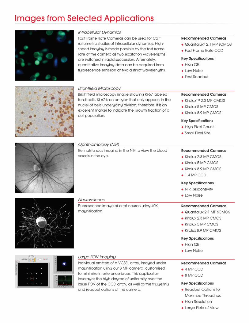
Images from Selected Applications
Intracellular Dynamics
Brightfield Microscopy
Ophthalmology (NIR)
Fast Frame Rate Cameras can be used for Ca2+
ratiometric studies of intracellular dynamics. High-speed imaging is made possible by the fast frame rate of the camera as two excitation wavelengths are switched in rapid succession. Alternately, quantitative imaging data can be acquired from fluorescence emission at two distinct wavelengths.
Brightfield microscopy image showing Ki-67 labeled tonsil cells. Ki-67 is an antigen that only appears in the nuclei of cells undergoing division; therefore, it is an excellent marker to indicate the growth fraction of a cell population.
Retinal/fundus imaging in the NIR to view the blood vessels in the eye.
Fluorescence image of a rat neuron using 40X magnification.
Individual emitters of a VCSEL array, imaged under magnification using our 8 MP camera, customized to minimize interference issues. This application leverages the high degree of uniformity over the large FOV of the CCD array, as well as the triggering and readout options of the camera.
Neuroscience
Large FOV Imaging
Recommended Cameras
u Quantalux® 2.1 MP sCMOS
u Fast Frame Rate CCD
Key Specifications
u High QE
u Low Noise
u Fast Readout
Recommended Cameras
u Kiralux™ 2.3 MP CMOS
u Kiralux 5 MP CMOS
u Kiralux 8.9 MP CMOS
Key Specifications
u High Pixel Count
u Small Pixel Size
Recommended Cameras
u Kiralux 2.3 MP CMOS
u Kiralux 5 MP CMOS
u Kiralux 8.9 MP CMOS
u 1.4 MP CCD
Key Specifications
u NIR Responsivity
u Low Noise
Recommended Cameras
u Quantalux 2.1 MP sCMOS
u Kiralux 2.3 MP CMOS
u Kiralux 5 MP CMOS
u Kiralux 8.9 MP CMOS
Key Specifications
u High QE
u Low Noise
Recommended Cameras
u 4 MP CCD
u 8 MP CCD
Key Specifications
u Readout Options to
Maximize Throughput
u High Resolution
u Large Field of View

Fluorescence Microscopy
Multispectral Imaging
Scanning Electron Microscopy
QA/Inspection of Optical Components Under Stress
Simultaneous NIR Dodt and Fluorescence
Merged triple emission fluorescence microscopy image.
The sample slide consists of multi-labeled bovine pulmonary
artery endothelial (BPAE) cells, showing at least one example of
a double nucleus.
Series of multispectral images taken with different passband wavelengths; the final stacked color image is shown. High QE scientific cameras are
especially beneficial for obtaining low-light, narrowband images. This image was acquired using
a Thorlabs KURIOS-WB1 Liquid Crystal Tunable Filter.
Scanning electron microscope (SEM) image of a nickel sample. Electron backscatter diffraction (EBSD) produces Kikuchi patterns that result from the interaction between
the electron beam and the sample material. Our high-QE, low-noise cameras make possible high-speed detection and analysis of these faint line patterns against relatively
high backgrounds.
False-color rendering of the degree of linear polarization (DoLP) image of an optical component under stress. (a)
No stress (b) low stress (c) medium stress (d) high stress. This image was acquired with the CS505MUP camera, which
has an array of pixel-sized wire grid polarizers between the microlenses and the light-sensitive pixels.
The image shows a live, simultaneous overlay of fluorescence and NIR Dodt contrast images of a 50 µm
brain section from a CX3CR1-GFP mouse, which has been immunostained for PECAM-1 with Alexa-687 to
highlight vasculature. Dodt contrast uses a gradient of light across a thick sample to reveal structural details. This
image was acquired using two cameras mounted on a 2SCM1-DC adapter.
Recommended Cameras
u Quantalux 2.1 MP sCMOS
u Kiralux 2.3 MP CMOS
u Kiralux 5 MP CMOS
u Kiralux 8.9 MP CMOS
Key Specifications
u High QE
u Low Noise
Recommended Cameras
u Quantalux 2.1 MP sCMOS
u Kiralux 2.3 MP CMOS
u Kiralux 5 MP CMOS
Key Specifications
u High QE
u Low Noise
Recommended Cameras
u 1.4 MP CCD
u 4 MP CCD
u Fast Frame Rate CCD
Key Specifications
u High QE
u ROI and Binning Modes
Recommended Camera
u CS505MUP
Key Specifications
u On-Chip Wire-Grid
Polarizer Array
Recommended Cameras
u Quantalux 2.1 MP sCMOS
u Kiralux 2.3 MP CMOS
u Kiralux 5 MP CMOS
u 1.4 MP CCD
Key Specifications
u NIR Responsivity
u High QE
u Low Noise
(Sample courtesy of Dr. Andrew Chojnacki, Department of Physiology and Pharmacology, Live Cell Imaging Facility, Snyder
Institute for Chronic Diseases, University of Calgary.)
a
c
b
d
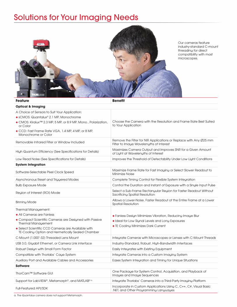
Solutions for Your Imaging Needs
Feature Benefit
Optical & Imaging
A Choice of Sensors to Suit Your Application:
u sCMOS: Quantalux® 2.1 MP, Monochrome
u CMOS: Kiralux™ 2.3 MP, 5 MP, or 8.9 MP, Mono., Polarization, or Color
u CCD: Fast Frame Rate VGA, 1.4 MP, 4 MP, or 8 MP, Monochrome or Color
Choose the Camera with the Resolution and Frame Rate Best Suited to Your Application
Removable Infrared Filter or Window Included Remove the Filter for NIR Applications or Replace with Any Ø25 mm Filter to Image Wavelengths of Interest
High Quantum Efficiency (See Specifications for Details) Maximizes Camera Output and Improves SNR for a Given Amount of Light at Wavelengths of Interest
Low Read Noise (See Specifications for Details) Improves the Threshold of Detectability Under Low Light Conditions
System Integration
Software-Selectable Pixel Clock Speed Maximize Frame Rate for Fast Imaging or Select Slower Readout to Minimize Noise
Asynchronous Reset and Triggered Modes Complete Timing Control for Flexible System Integration
Bulb Exposure Mode Control the Duration and Instant of Exposure with a Single Input Pulse
Region of Interest (ROI) Mode Select a Sub-Frame Rectangular Region for Faster Readout Without Sacrificing Spatial Resolution
Binning Mode Allows a Lower-Noise, Faster Readout of the Entire Frame at a Lower Spatial Resolution
Thermal Management:
u All Cameras are Fanless
u Compact Scientific Cameras are Designed with Passive Thermal Management
u Select Scientific CCD Cameras are Available with TE-Cooling Option and Hermetically Sealed Chamber
u Fanless Design Minimizes Vibration, Reducing Image Blur
u Ideal for Low Signal Levels and Long Exposures
u TE Cooling Minimizes Dark Current
C-Mount (1.000"-32) Threaded Lens Mount Integrate Cameras with Microscopes or Lenses with C-Mount Threads
USB 3.0, Gigabit Ethernet, or Camera Link interface Industry-Standard, Robust, High-Bandwidth Interfaces
Robust Design with Small Form Factor Easily Integrates with Existing Equipment
Compatible with Thorlabs’ Cage System Integrate Cameras into a Custom Imaging System
Auxiliary Port and Available Cables and Accessories Eases System Integration and Timing for Unique Situations
Software
ThorCam™ Software GUI One Package for System Control, Acquisition, and Playback of Images and Image Sequences
Support for LabVIEW®, Metamorph®, and MATLAB® a Integrate Thorlabs’ Cameras into a Third-Party Imaging Platform
Full-Featured API/SDK Incorporate in Custom Applications Using C, C++, C#, Visual Basic .NET, and Other Programming Languages
Our cameras feature industry-standard C-mount threading for direct compatibility with most microscopes.
a. The Quantalux camera does not support Metamorph.
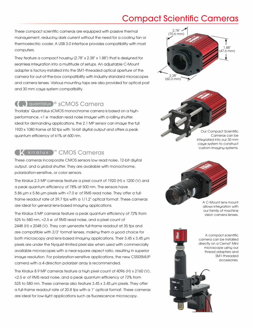
Compact Scientific CamerasThese compact scientific cameras are equipped with passive thermal
management, reducing dark current without the need for a cooling fan or
thermoelectric cooler. A USB 3.0 interface provides compatibility with most
computers.
They feature a compact housing (2.78" x 2.38" x 1.88") that is designed for
seamless integration into a multitude of setups. An adjustable C-Mount
adapter is factory-installed into the SM1-threaded optical aperture of the
camera for out-of-the-box compatibility with industry-standard microscopes
and camera lenses. Various mounting taps are also provided for optical post
and 30 mm cage system compatibility.
® sCMOS CameraThorlabs’ Quantalux sCMOS monochrome camera is based on a high-
performance, <1 e- median read noise imager with a rolling shutter.
Ideal for demanding applications, the 2.1 MP sensor can image the full
1920 x 1080 frame at 50 fps with 16-bit digital output and offers a peak
quantum efficiency of 61% at 600 nm.
™ CMOS CamerasThese cameras incorporate CMOS sensors low read noise, 12-bit digital
output, and a global shutter. They are available with monochrome,
polarization-sensitive, or color sensors.
The Kiralux 2.3 MP cameras feature a pixel count of 1920 (H) x 1200 (V) and
a peak quantum efficiency of 78% at 500 nm. The sensors have
5.86 µm x 5.86 µm pixels with <7.0 e- of RMS read noise. They offer a full-
frame readout rate of 39.7 fps with a 1/1.2" optical format. These cameras
are ideal for general lens-based imaging applications.
The Kiralux 5 MP cameras feature a peak quantum efficiency of 72% from
525 to 580 nm, <2.5 e- of RMS read noise, and a pixel count of
2448 (H) x 2048 (V). They can generate full-frame readout at 35 fps and
are compatible with 2/3" format lenses, making them a good choice for
both microscopy and lens-based imaging applications. Their 3.45 x 3.45 µm
pixels are under the Nyquist-limited pixel size when used with commercially
available microscopes with a near-square aspect ratio, resulting in superior
image resolution. For polarization-sensitive applications, the new CS505MUP
camera with a 4-direction polarizer array is recommended.
The Kiralux 8.9 MP cameras feature a high pixel count of 4096 (H) x 2160 (V),
<2.5 e- of RMS read noise, and a peak quantum efficiency of 72% from
525 to 580 nm. These cameras also feature 3.45 x 3.45 µm pixels. They offer
a full-frame readout rate of 20.8 fps with a 1" optical format. These cameras
are ideal for low-light applications such as fluorescence microscopy.
Our Compact Scientific Cameras can be
integrated into our 30 mm cage system to construct custom imaging systems.
A compact scientific camera can be installed directly on a Cerna® Mini
microscope using our thread adapters and
SM1-threaded accessories.
A C-Mount lens mount allows integration with our family of machine vision camera lenses.
1.88" (47.6 mm)
2.78" (70.6 mm)
2.38" (60.3 mm)
®
®
®
TM
TM
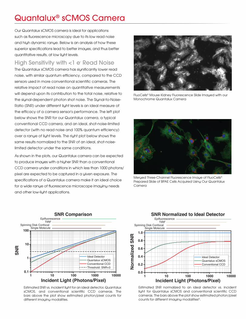
Quantalux® sCMOS CameraOur Quantalux sCMOS camera is ideal for applications
such as fluorescence microscopy due to its low read noise
and high dynamic range. Below is an analysis of how these
superior specifications lead to better images, and thus better
quantitative results, at low light levels.
High Sensitivity with <1 e- Read NoiseThe Quantalux sCMOS camera has significantly lower read
noise, with similar quantum efficiency, compared to the CCD
sensors used in more conventional scientific cameras. The
relative impact of read noise on quantitative measurements
will depend upon its contribution to the total noise, relative to
the signal-dependent photon shot noise. The Signal-to-Noise-
Ratio (SNR) under different light levels is an ideal measure of
the efficacy of a camera sensor's performance. The left plot
below shows the SNR for our Quantalux camera, a typical
conventional CCD camera, and an ideal, shot-noise-limited
detector (with no read noise and 100% quantum efficiency)
over a range of light levels. The right plot below shows the
same results normalized to the SNR of an ideal, shot-noise-
limited detector under the same conditions.
As shown in the plots, our Quantalux camera can be expected
to produce images with a higher SNR than a conventional
CCD camera under conditions in which less than 1000 photons/
pixel are expected to be captured in a given exposure. The
specifications of a Quantalux camera make it an ideal choice
for a wide range of fluorescence microscope imaging needs
and other low-light applications.
Estimated SNR vs. incident light for an ideal detector, Quantalux sCMOS, and conventional scientific CCD cameras. The bars above the plot show estimated photon/pixel counts for different imaging modalities.
Estimated SNR normalized to an ideal detector vs. incident light for Quantalux sCMOS and conventional scientific CCD cameras. The bars above the plot show estimated photon/pixel counts for different imaging modalities®.
1 10 100 1000 100000.1
1
10
100
EpifluorescenceTIRF
Spinning Disk Confocal
Ideal DetectorQuantalux sCMOS
Threshold: SNR=3
SNR Comparison
SNR
Incident Light (Photons/Pixel)
Single Molecule
Conventional CCD
1 10 100 1000 100000.0
0.2
0.4
0.6
0.8
1.0
EpifluorescenceTIRF
Spinning Disk Confocal
Ideal Detector
Conventional CCDQuantalux sCMOS
Nor
mal
ized
SN
R
Single Molecule
SNR Normalized to Ideal Detector
Incident Light (Photons/Pixel)
Merged Three-Channel Fluorescence Image of FluoCells®
Prepared Slide of BPAE Cells Acquired Using Our Quantalux Camera
FluoCells® Mouse Kidney Fluorescence Slide Imaged with our Monochrome Quantalux Camera
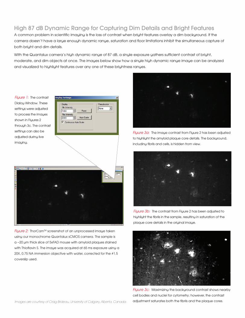
High 87 dB Dynamic Range for Capturing Dim Details and Bright FeaturesA common problem in scientific imaging is the loss of contrast when bright features overlay a dim background. If the
camera doesn’t have a large enough dynamic range, saturation and floor limitations inhibit the simultaneous capture of
both bright and dim details.
With the Quantalux camera’s high dynamic range of 87 dB, a single exposure gathers sufficient contrast of bright,
moderate, and dim objects at once. The images below show how a single high dynamic range image can be analyzed
and visualized to highlight features over any one of these brightness ranges.
Images are courtesy of Craig Brideau, University of Calgary, Alberta, Canada.
Figure 2: ThorCamTM screenshot of an unprocessed image taken
using our monochrome Quantalux sCMOS camera. The sample is
a ~20 µm thick slice of 5xFAD mouse with amyloid plaques stained
with Thioflavin S. The image was acquired at 65 ms exposure using a
20X, 0.75 NA immersion objective with water, corrected for the #1.5
coverslip used.
Figure 3a: The image contrast from Figure 2 has been adjusted
to highlight the amyloid plaque core details. The background,
including fibrils and cells, is hidden from view.
Figure 3b: The contrast from Figure 2 has been adjusted to
highlight the fibrils in the sample, resulting in saturation of the
plaque core details in the original image.
Figure 3c: Maximizing the background contrast shows nearby
cell bodies and nuclei for cytometry; however, the contrast
adjustment saturates both the fibrils and the plaque cores.
Figure 1: The contrast
Dialog Window. These
settings were adjusted
to process the images
shown in Figures 2
through 3c. The contrast
settings can also be
adjusted during live
imaging.
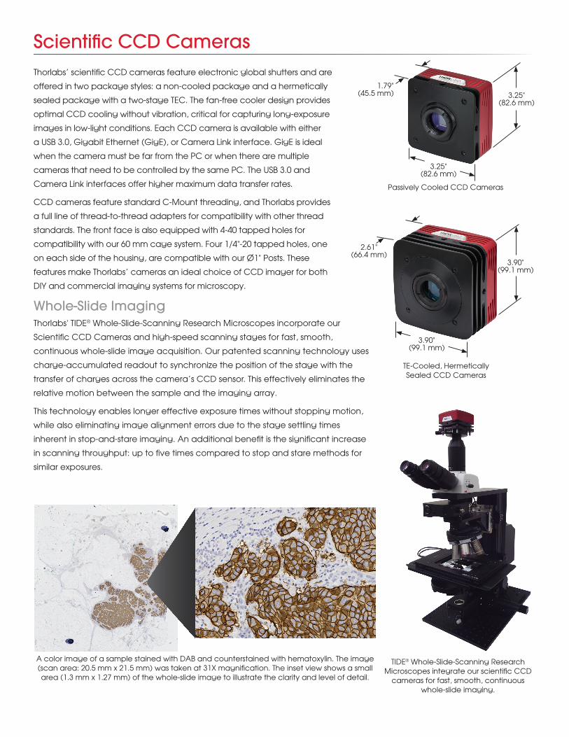
Whole-Slide ImagingThorlabs' TIDE® Whole-Slide-Scanning Research Microscopes incorporate our
Scientific CCD Cameras and high-speed scanning stages for fast, smooth,
continuous whole-slide image acquisition. Our patented scanning technology uses
charge-accumulated readout to synchronize the position of the stage with the
transfer of charges across the camera’s CCD sensor. This effectively eliminates the
relative motion between the sample and the imaging array.
This technology enables longer effective exposure times without stopping motion,
while also eliminating image alignment errors due to the stage settling times
inherent in stop-and-stare imaging. An additional benefit is the significant increase
in scanning throughput: up to five times compared to stop and stare methods for
similar exposures.
Scientific CCD CamerasThorlabs’ scientific CCD cameras feature electronic global shutters and are
offered in two package styles: a non-cooled package and a hermetically
sealed package with a two-stage TEC. The fan-free cooler design provides
optimal CCD cooling without vibration, critical for capturing long-exposure
images in low-light conditions. Each CCD camera is available with either
a USB 3.0, Gigabit Ethernet (GigE), or Camera Link interface. GigE is ideal
when the camera must be far from the PC or when there are multiple
cameras that need to be controlled by the same PC. The USB 3.0 and
Camera Link interfaces offer higher maximum data transfer rates.
CCD cameras feature standard C-Mount threading, and Thorlabs provides
a full line of thread-to-thread adapters for compatibility with other thread
standards. The front face is also equipped with 4-40 tapped holes for
compatibility with our 60 mm cage system. Four 1/4"-20 tapped holes, one
on each side of the housing, are compatible with our Ø1" Posts. These
features make Thorlabs’ cameras an ideal choice of CCD imager for both
DIY and commercial imaging systems for microscopy.
Passively Cooled CCD Cameras
3.25" (82.6 mm)
1.79" (45.5 mm)
3.25" (82.6 mm)
TE-Cooled, Hermetically Sealed CCD Cameras
2.61" (66.4 mm)
3.90" (99.1 mm)
3.90" (99.1 mm)
A color image of a sample stained with DAB and counterstained with hematoxylin. The image (scan area: 20.5 mm x 21.5 mm) was taken at 31X magnification. The inset view shows a small area (1.3 mm x 1.27 mm) of the whole-slide image to illustrate the clarity and level of detail.
TIDE® Whole-Slide-Scanning Research Microscopes integrate our scientific CCD
cameras for fast, smooth, continuous whole-slide imaging.
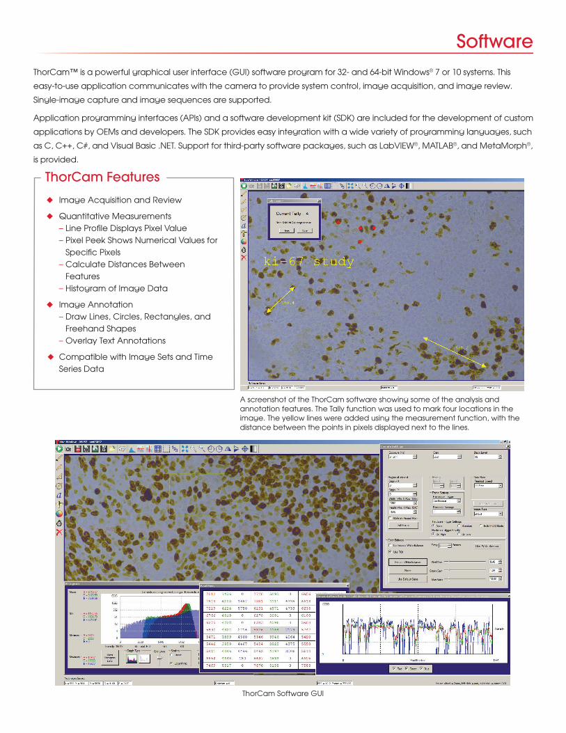
ThorCam Software GUI
ThorCam™ is a powerful graphical user interface (GUI) software program for 32- and 64-bit Windows® 7 or 10 systems. This
easy-to-use application communicates with the camera to provide system control, image acquisition, and image review.
Single-image capture and image sequences are supported.
Application programming interfaces (APIs) and a software development kit (SDK) are included for the development of custom
applications by OEMs and developers. The SDK provides easy integration with a wide variety of programming languages, such
as C, C++, C#, and Visual Basic .NET. Support for third-party software packages, such as LabVIEW®, MATLAB®, and MetaMorph®,
is provided.
Software
A screenshot of the ThorCam software showing some of the analysis and annotation features. The Tally function was used to mark four locations in the image. The yellow lines were added using the measurement function, with the distance between the points in pixels displayed next to the lines.
u Image Acquisition and Review
u Quantitative Measurements – Line Profile Displays Pixel Value – Pixel Peek Shows Numerical Values for Specific Pixels – Calculate Distances Between Features – Histogram of Image Data
u Image Annotation – Draw Lines, Circles, Rectangles, and Freehand Shapes – Overlay Text Annotations
u Compatible with Image Sets and Time Series Data
ThorCam Features
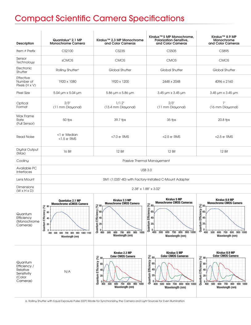
Compact Scientific Camera Specifications
DescriptionQuantalux® 2.1 MP
Monochrome CameraKiralux™ 2.3 MP Monochrome
and Color Cameras
Kiralux™ 5 MP Monochrome, Polarization-Sensitive, and Color Cameras
Kiralux™ 8.9 MP Monochrome
and Color CamerasFast Frame Rate, VGA
Monochrome Cameras1.4 MP Monochrome and Color Cameras
4 MP Monochrome and Color Cameras
8 MP Monochrome and Color Cameras
Item # Prefix CS2100 CS235 CS505 CS895 340 1501 4070 8051
Sensor Technology sCMOS CMOS CMOS CMOS Interline CCD Interline CCD Interline CCD Interline CCD
Electronic Shutter Rolling Shuttera Global Shutter Global Shutter Global Shutter Global Shutter Global Shutter Global Shutter Global Shutter
Effective Number of Pixels (H x V)
1920 x 1080 1920 x 1200 2448 x 2048 4096 x 2160 640 x 480 1392 x 1040 2048 x 2048 3296 x 2472
Pixel Size 5.04 µm x 5.04 µm 5.86 µm x 5.86 µm 3.45 µm x 3.45 µm 3.45 µm x 3.45 µm 7.4 µm x 7.4 µm 6.45 µm x 6.45 µm 7.4 µm x 7.4 µm 5.5 µm x 5.5 µm
Optical Format
2/3" (11 mm Diagonal)
1/1.2" (13.4 mm Diagonal)
2/3" (11 mm Diagonal)
1" (16 mm Diagonal)
1/3" (5.92 mm Diagonal)
2/3" (11 mm Diagonal)
4/3" (21.4 mm Diagonal)
4/3" (22 mm Diagonal)
Max Frame Rate (Full Sensor)
50 fps 39.7 fps 35 fps 20.8 fps200.7 fps (at 40 MHz Dual-Tap Readout)
23 fps (at 40 MHz Single-Tap Readout)
25.8 fps (at 40 MHz Quad-Tap Readout)b
17.1 fps (at 40 MHz Quad-Tap Readout)c
Read Noise<1 e- Median <1.5 e- RMS
<7.0 e- RMS <2.5 e- RMS <2.5 e- RMS <15 e- RMS at 20 MHz
<7 e- RMS at 20 MHz (Standard Models)
<6 e- RMS at 20 MHz (TE-Cooled Models)
<12 e- RMS at 20 MHz <10 e- RMS at 20 MHz
Digital Output (Max) 16 Bit 12 Bit 12 Bit 12 Bit 14 Bitd 14 Bit 14 Bitd 14 Bitd
Cooling Passive Thermal Management None TE-Cooled, Hermetically Sealed Versions Available
Available PC Interfaces USB 3.0 USB 3.0, Gigabit Ethernet, or Camera Link
Lens Mount SM1 (1.035"-40) with Factory-Installed C-Mount Adapter C-Mount (1.000"-32)
Dimensions (W x H x D) 2.38" x 1.88" x 3.02" 3.25" x 3.25" x 1.79" 3.25" x 3.25" x 1.79" (Standard); 3.90" x 3.90" x 2.61" (TE Cooled)
Quantum Efficiency (Monochrome Cameras)
Quantum Efficiency /Relative Sensitivity (Color Cameras)
N/A N/A
400 500 600 700 800 900 10000
20
40
60
80
Kiralux 5 MPColor CMOS Cameras
Quan
tum
Effi
cien
cy (%
)
Wavelength (nm)
400 500 600 700 800 900 10000
20
40
60
80
Kiralux 5 MPMonochrome CMOS Cameras
Quan
tum
Effi
cien
cy (%
)
Wavelength (nm)
400 500 600 700 800 900 10000
20
40
60
80
Kiralux 8.9 MPColor CMOS Camera
Quan
tum
Effi
cien
cy (%
)
Wavelength (nm)
400 500 600 700 800 900 10000
20
40
60
80
Kiralux 8.9 MPMonochrome CMOS Camera
Quan
tum
Effi
cien
cy (%
)
Wavelength (nm)400 500 600 700 800 900 1000 1100
0
20
40
60
80
Quantalux 2.1 MPMonochrome sCMOS Camera
Quan
tum
Effi
cien
cy (%
)
Wavelength (nm)
a. Rolling Shutter with Equal Exposure Pulse (EEP) Mode for Synchronizing the Camera and Light Sources for Even Illumination
400 500 600 700 800 900 10000
20
40
60
80
Kiralux 2.3 MPColor CMOS Camera
Quan
tum
Effi
cien
cy (%
)
Wavelength (nm)
400 500 600 700 800 900 10000
20
40
60
80
Kiralux 2.3 MPMonochrome CMOS Camera
Quan
tum
Effi
cien
cy (%
)
Wavelength (nm)
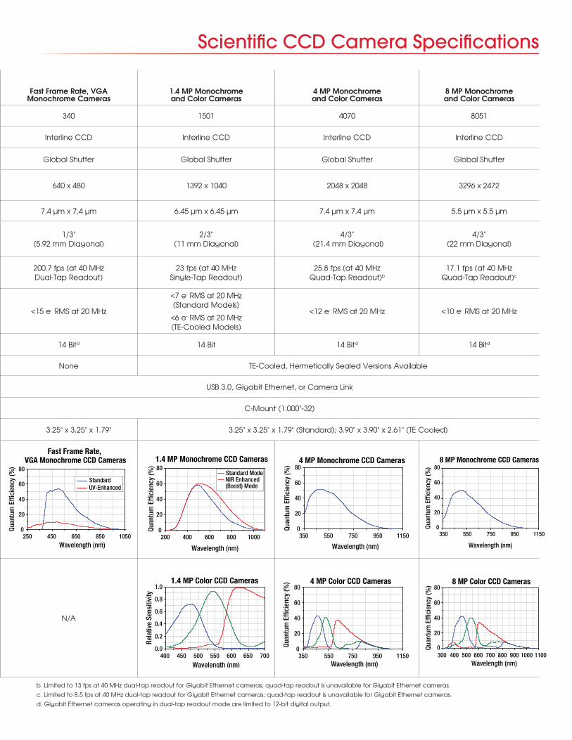
Scientific CCD Camera Specifications
DescriptionQuantalux® 2.1 MP
Monochrome CameraKiralux™ 2.3 MP Monochrome
and Color Cameras
Kiralux™ 5 MP Monochrome, Polarization-Sensitive, and Color Cameras
Kiralux™ 8.9 MP Monochrome
and Color CamerasFast Frame Rate, VGA
Monochrome Cameras1.4 MP Monochrome and Color Cameras
4 MP Monochrome and Color Cameras
8 MP Monochrome and Color Cameras
Item # Prefix CS2100 CS235 CS505 CS895 340 1501 4070 8051
Sensor Technology sCMOS CMOS CMOS CMOS Interline CCD Interline CCD Interline CCD Interline CCD
Electronic Shutter Rolling Shuttera Global Shutter Global Shutter Global Shutter Global Shutter Global Shutter Global Shutter Global Shutter
Effective Number of Pixels (H x V)
1920 x 1080 1920 x 1200 2448 x 2048 4096 x 2160 640 x 480 1392 x 1040 2048 x 2048 3296 x 2472
Pixel Size 5.04 µm x 5.04 µm 5.86 µm x 5.86 µm 3.45 µm x 3.45 µm 3.45 µm x 3.45 µm 7.4 µm x 7.4 µm 6.45 µm x 6.45 µm 7.4 µm x 7.4 µm 5.5 µm x 5.5 µm
Optical Format
2/3" (11 mm Diagonal)
1/1.2" (13.4 mm Diagonal)
2/3" (11 mm Diagonal)
1" (16 mm Diagonal)
1/3" (5.92 mm Diagonal)
2/3" (11 mm Diagonal)
4/3" (21.4 mm Diagonal)
4/3" (22 mm Diagonal)
Max Frame Rate (Full Sensor)
50 fps 39.7 fps 35 fps 20.8 fps200.7 fps (at 40 MHz Dual-Tap Readout)
23 fps (at 40 MHz Single-Tap Readout)
25.8 fps (at 40 MHz Quad-Tap Readout)b
17.1 fps (at 40 MHz Quad-Tap Readout)c
Read Noise<1 e- Median <1.5 e- RMS
<7.0 e- RMS <2.5 e- RMS <2.5 e- RMS <15 e- RMS at 20 MHz
<7 e- RMS at 20 MHz (Standard Models)
<6 e- RMS at 20 MHz (TE-Cooled Models)
<12 e- RMS at 20 MHz <10 e- RMS at 20 MHz
Digital Output (Max) 16 Bit 12 Bit 12 Bit 12 Bit 14 Bitd 14 Bit 14 Bitd 14 Bitd
Cooling Passive Thermal Management None TE-Cooled, Hermetically Sealed Versions Available
Available PC Interfaces USB 3.0 USB 3.0, Gigabit Ethernet, or Camera Link
Lens Mount SM1 (1.035"-40) with Factory-Installed C-Mount Adapter C-Mount (1.000"-32)
Dimensions (W x H x D) 2.38" x 1.88" x 3.02" 3.25" x 3.25" x 1.79" 3.25" x 3.25" x 1.79" (Standard); 3.90" x 3.90" x 2.61" (TE Cooled)
Quantum Efficiency (Monochrome Cameras)
Quantum Efficiency /Relative Sensitivity (Color Cameras)
N/A N/A
b. Limited to 13 fps at 40 MHz dual-tap readout for Gigabit Ethernet cameras; quad-tap readout is unavailable for Gigabit Ethernet cameras.
c. Limited to 8.5 fps at 40 MHz dual-tap readout for Gigabit Ethernet cameras; quad-tap readout is unavailable for Gigabit Ethernet cameras.
d. Gigabit Ethernet cameras operating in dual-tap readout mode are limited to 12-bit digital output.
200 400 600 800 10000
20
40
60
80
Standard ModeNIR Enhanced(Boost) Mode
1.4 MP Monochrome CCD Cameras
Quan
tum
Effi
cien
cy (%
)
Wavelength (nm)
400 450 500 550 600 650 7000.0
0.2
0.4
0.6
0.8
1.01.4 MP Color CCD Cameras
Rela
tive
Sens
itivi
ty
Wavelength (nm)350 550 750 950 1150
0
20
40
60
804 MP Color CCD Cameras
Quan
tum
Effi
cien
cy (%
)
Wavelength (nm)
350 550 750 950 11500
20
40
60
804 MP Monochrome CCD Cameras
Quan
tum
Effi
cien
cy (%
)
Wavelength (nm)
300 400 500 600 700 800 900 1000 11000
20
40
60
80
8 MP Color CCD Cameras
Quan
tum
Effi
cien
cy (%
)
Wavelength (nm)
0
20
40
60
80
250 450 650 850 10500
20
40
60
80
Standard UV-Enhanced
Fast Frame Rate, VGA Monochrome CCD Cameras
Quan
tum
Effi
cien
cy (%
)
Wavelength (nm)
350 550 750 950 11500
20
40
60
808 MP Monochrome CCD Cameras
Quan
tum
Effi
cien
cy (%
)
Wavelength (nm)

In addition to our large selection of standard scientific
cameras, we have the capability of building custom
cameras designed for unique scientific applications.
Options include high-performance board-level cameras,
custom camera housings, and software. If you have
special requirements, a custom application, or general
questions about our capabilities, please contact us at
[email protected]. We can help you evaluate your
application and budgetary requirements to create custom
cameras to satisfy your needs.
Our engineering team simplifies the customization process
by following the two-step process below. A scientific
camera can be customized by using one of our existing
standard cameras as a starting point, or it can be built to
the exact needs of an application. Small changes made
to our existing designs can be done efficiently.
Board-level photograph of the sensor and electronics in one of our scientific
CCD cameras.
Custom Cameras
USA | www.thorlabs.com Thorlabs Imaging Systems Phone: 1-703-651-1700
Thorlabs Scientific Imaging (TSI) Phone: 1-973-300-3000
Thorlabs Quantum Electronics (TQE) Phone: 1-973-300-3000
Thorlabs Ultrafast Optoelectronics (UFO) Phone: 1-973-300-3000
Thorlabs Vytran Division Phone: 1-973-300-3000
CANADA | www.thorlabs.com Thorlabs Canada (ULC) Phone: 1-973-300-3000
UK | www.thorlabs.de Phone: +44 (0)1353 654440
Thorlabs Vytran Europe Phone: +44 (0) 1392-445777
FRANCE | www.thorlabs.de Phone : +33 (0) 970 444 844
GERMANY | www.thorlabs.de Thorlabs GmbH Phone: +49 (0) 8131 5956-0
Thorlabs GmbH (Lübeck) Phone: +49 (0) 8131-5956-0
Thorlabs Elliptec GmbH Phone: +44 (0)1353 654440
CHINA | www.thorlabschina.cn Phone: +86 (0)21-60561122
SWEDEN | www.thorlabs.com Phone: +46 31 733 30 00 Polish Direct Line: +48 22 219 52 30
JAPAN | www.thorlabs.co.jp Phone: +81-3-6915-7701
BRAZIL | www.thorlabs.com Phone: +55 (16) 3413 7062
56 Sparta Avenue • Newton, New Jersey 07860Sales: 973.300.3000 • Fax: 973.300.3600 • www.thorlabs.com
Step 2: Configure a Solution
Imager Options u UV, Visible, or NIR Spectrum
u Quantalux® sCMOS, Kiralux™ CMOS, or CCD
I/O Options u Camera Link
u Gigabit Ethernet
u USB 3.0
Camera Body Options
u Standard Non-Cooled
u Hermetically Sealed with Two-Stage TEC
u Private Labeling
Electronics Modificationsu Customized Firmware
u Application-Specific Timing and Triggering Modes
Optics Mounting Options
u C-Mount Threading is Standard
Software
u Initial Evaluation Using ThorCam™ GUI for Cameras
u Algorithm Development Using Popular Third-Party Support Such as MATLAB® and LabVIEW®
u API / SDK Provided for Software Developers and OEMs
Supply Chain u Kanban Stocking Agreements
Step 1: Analyze Your Custom RequirementsImaging Specifications u Sensitivity
u Wavelength
System Requirements u Operating Environment
u Space Constraints
Application Space
u Compliance Issues
u Future Developments
u Resolution
u Speed
u Interfaces
u Software
u Logistics
400 500 600 700 800 900 1000 11000
10
20
30
40
50
60
70Alexa 750GFP
Quan
tum
Effi
cien
cy (%
)
Wavelength (nm)
DAPICy5
TRITC IR-DIC
Popular Fluorophore Emission Wavelengths Overlaid with the Quantum Efficiency Curve of
Our Quantalux sCMOS Camera
Our electronics boards can be easily reconfigured to fit various housing types.
V19-2