science. the microorganism causing the disease · · 2008-06-20the trophozoite is oblong and...
TRANSCRIPT
cycles, the relationship between environmentalconditions and degree of infection.
Disease associated with Haplosporidium
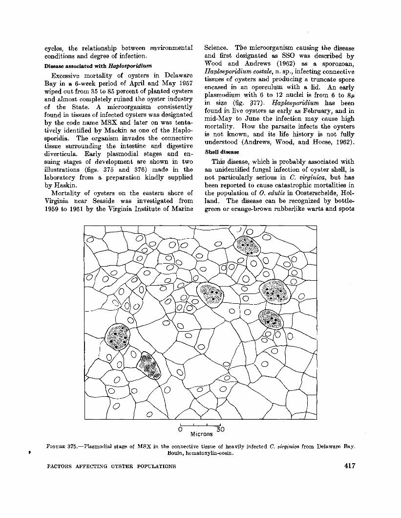
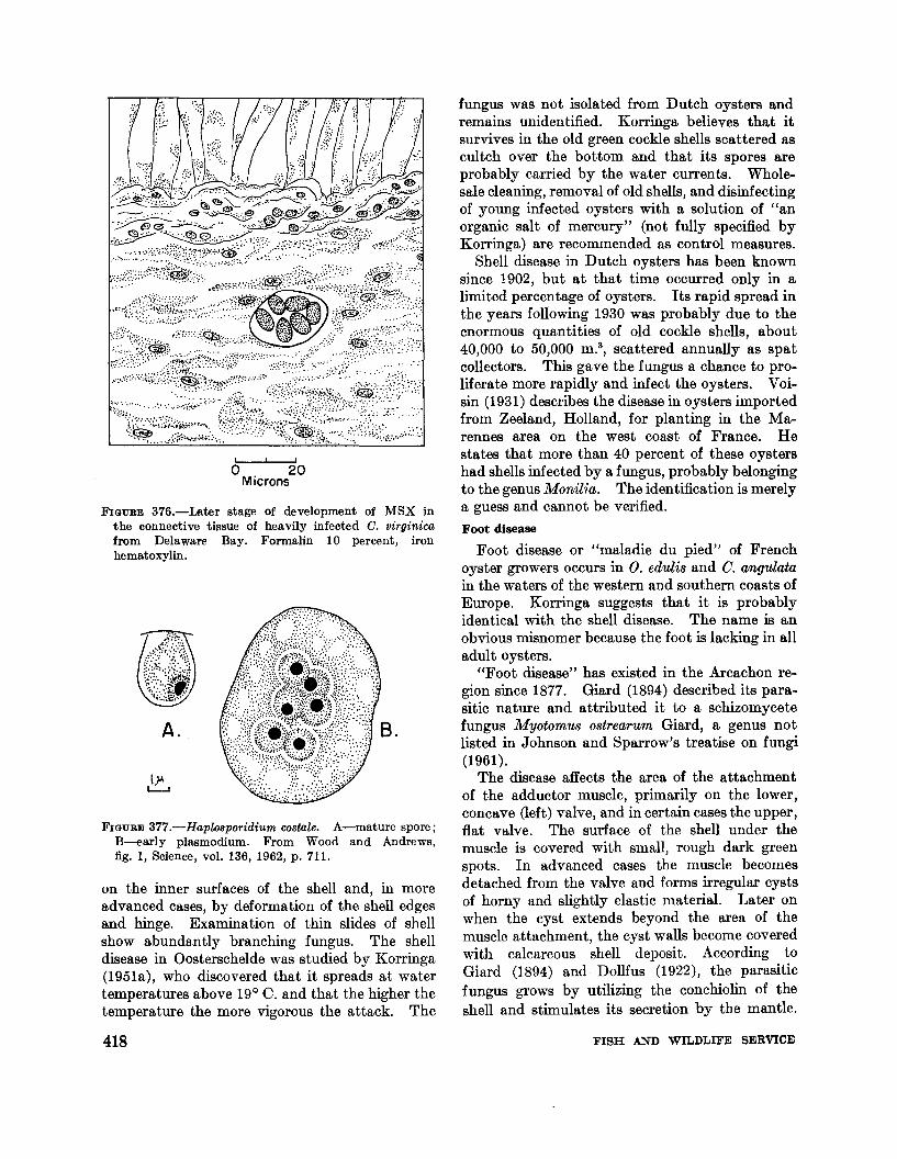
Excessive mortality of oysters in DelawareBay in a 6-week period of April and May 1957wiped out from 35 to 85 percent of planted oystersand almost completely ruined the oyster industryof the State. A microorganism consistentlyfound in tissues of infected oysters was designatedby the code name MSX and later on was tentatively identified by Mackin as one of the Haplosporidia. The organism invades the connectivetissue surrounding the intestine and digestivediverticula. Early plasmodial stages and ensuing stages of development are shown in twoillustrations (figs. 375 and 376) made in thelaboratory from a preparation kindly suppliedby Haskin.
Mortality of oysters on the eastern shore ofVirginia near Seaside was investigated from1959 to 1961 by the Virginia Institute of Marine
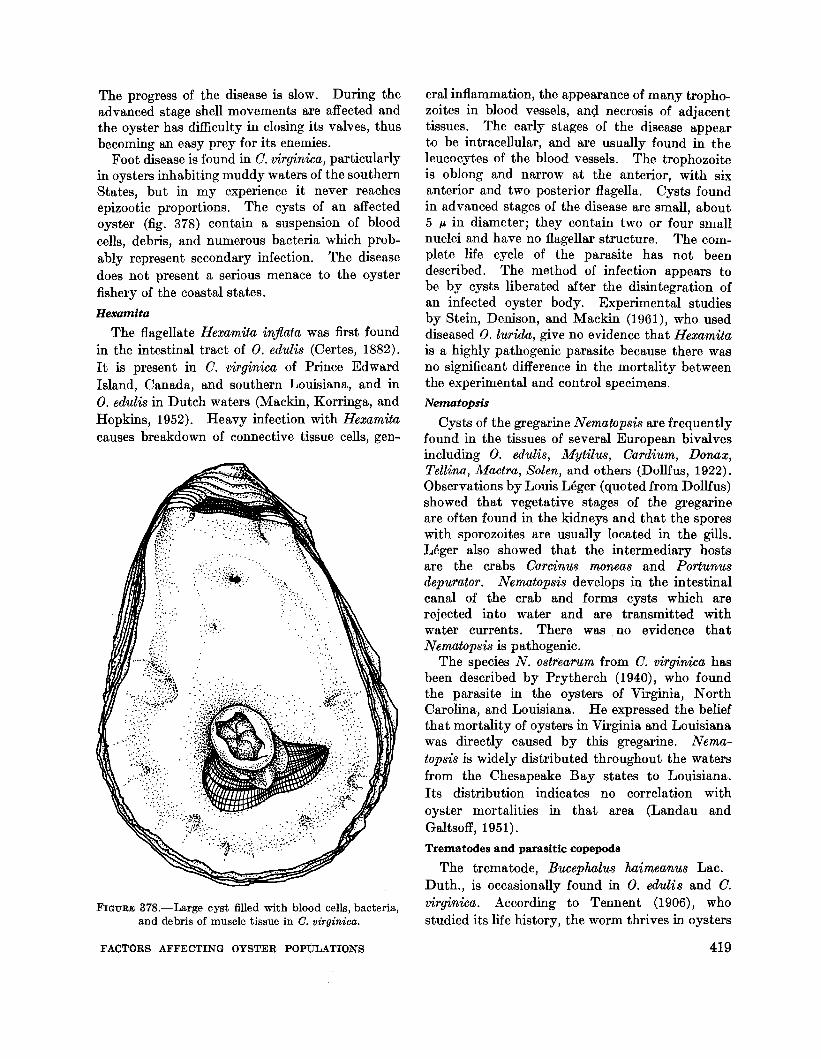
Science. The microorganism causing the diseaseand first designated as SSO was described byWood and Andrews (1962) as a sporozoan,Haplosporidium costale, n. sp., infecting connectivetissues of oysters and producing a truncate sporeencased in an operculum with a lid. An earlyplasmodium with 6 to 12 nuclei is from 6 to 8p,in size (fig. 377). Haplosporidium has beenfound in live oysters as early as February, and inmid-May to June the infection may cause highmortality. How the parasite infects the oystersis not known, and its life history is not fullyunderstood (Andrews, Wood, and Hoese, 1962).
Shell disease
This disease, which is probably associated withan unidentified fungal infection of oyster shell, isnot particularly serious in O. virginica, but hasbeen reported to cause catastrophic mortalities inthe population of O. edulis in Oosterschelde, Holland. The disease can be recognized by bottlegreen or orange-brown rubberlike warts and spots
30Microns
FIGURE 375.-Plasmodial stage of MSX in the connective tissue of heavily infected C. virginica from Delaware Bay., Bouin, hematoxylin-eosin.
FACTORS AFFECTING OYSTER POPULATIONS 417
~ .... , .'.
~ ..... , .::~~...
fungus was not isolated from Dutch oysters andremains unidentified. Korringa believes that itsurvives in the old green cockle shells scattered ascultch over the bottom and that its spores areprobably carried by the water currents. Wholesale cleaning, removal of old shells, and disinfectingof young infected oysters with a solution of "anorganic salt of mercury" (not fully specified byKorringa) are recommended as control measures.
Shell disease in Dutch oysters has been knownsince 1902, but at that time occurred only in alimited percentage of oysters. Its rapid spread inthe years following 1930 was probably due to theenormous quantities of old cockle shells, about40,000 to 50,000 m.a, scattered annually as spatcollectors. This gave the fungus a chance to proliferate more rapidly and infect the oysters. Voisin (1931) describes the disease in oysters importedfrom Zeeland, Holland, for planting in the Marennes area on the west coast of France. Hestates that more than 40 percent of these oystershad shells infected by a fungus, probably belongingto the genus MoniUa. The identification is merelya guess and cannot be verified.
Foot disease
Foot disease or "maladie du pied" of Frenchoyster growers occurs in O. edulis and O. angulatain the waters of the western and southern coasts ofEurope. Korringa suggests that it is probablyidentical with the shell disease. The name is anobvious misnomer because the foot is lacking in alladult oysters.
"Foot disease" has existed in the Arcachon region since 1877. Giard (1894) described its parasitic nature and attributed it to a schizomycetefungus Myotomus ostrearum Giard, a genus notlisted in Johnson and Sparrow's treatise on fungi(1961).
The disease affects the area of the attachmentof the adductor muscle, primarily on the lower,concave (left) valve, and in certain cases the upper,flat valve. The surface of the shell under themuscle is covered with small, rough dark greenspots. In advanced cases the muscle becomesdetached from the valve and forms irregular cystsof horny and slightly elastic material. Later onwhen the cyst extends beyond the area of themuscle attachment, the cyst walls become coveredwith calcareous shell deposit. According toGiard (1894) and Dollfus (1922), the parasiticfungus grows by utilizing the conchiolin of theshell and stimulates its secretion by the mantle.
FIGURE 377.-Haplosporidium costale. A-mature spore;B---early plasmodium. From Wood and Andrews,fig. 1, Science, vol. 136, 1962, p. 711.
A.
FIGURE 376.-Later stage of development of MSX inthe connective tissue of heavily infected C. virginicafrom Delaware Bay. Formalin 10 percent, ironhematoxylin.
I I
o 20Microns
on the inner surfaces of the shell and, in moreadvanced cases, by deformation of the shell edgesand hinge. Examination of thin slides of shellshow abundantly branching fungus. The shelldisease in Oosterschelde was studied by Korringa(1951a), who discovered that it spreads at watertemperatures above 19° O. and that the higher thetemperature the more vigorous the attack. The
418 FISH AND WILDLIFE SERVICE
The progress of the disease is slow. During theadvanced stage shell movements are affected andthe oyster has difficulty in closing its valves, thusbecoming an easy prey for its enemies.
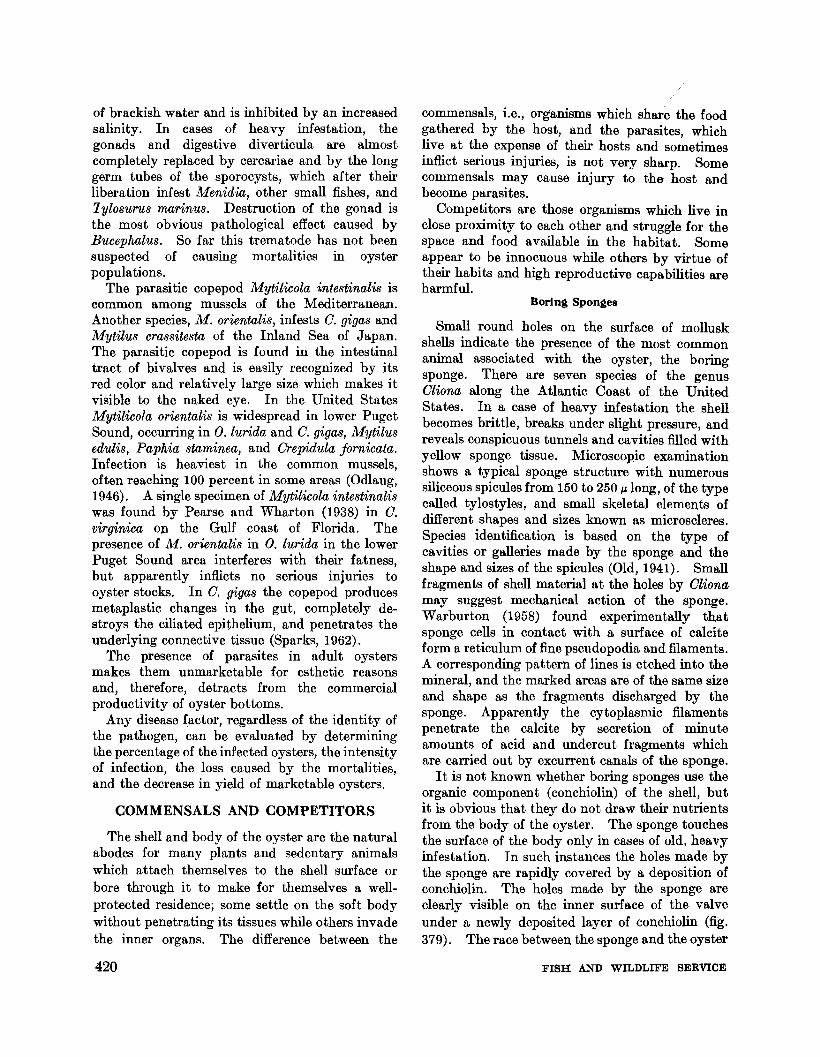
Foot disease is found in O. virginica, particularlyin oysters inhabiting muddy waters of the southernStates, but in my experience it never reachesepizootic proportions. The cysts of an affectedoyster (fig. 378) contain a suspension of bloodcells, debris, and numerous bacteria which probably represent secondary infection. The diseasedoes not present a serious menace to the oysterfishery of the coastal states.
Hexamita
The flagellate Hexamita injlata was first foundin the intestinal tract of O. edulis (Certes, 1882).It is present in O. virginica of Prince EdwardIsland, Canada, and southern Louisiana, and inO. edulis in Dutch waters (Mackin, Korringa, andHopkins, 1952). Heavy infection with Hexamitacauses breakdown of connective tissue cells, gen-
FIGURE 378.-Large cyst filled with blood cells, bacteria,and debris of muscle tissue in C. virginica.
FACTORS AFFECTING OYSTER POPULATIONS
eral inflammation, the appearance of many trophozoites in blood vessels, and necrosis of adjacenttissues. The early stages of the disease appearto be intracellular, and are usually found in theleucocytes of the blood vessels. The trophozoiteis oblong and narrow at the anterior, with sixanterior and two posterior flagella. Cysts foundin advanced stages of the disease are small, about5 p, in diameter; they contain two or four smallnuclei and have no flagellar structure. The complete life cycle of the parasite has not beendescribed. The method of infection appears tobe by cysts liberated after the disintegration ofan infected oyster body. Experimental studiesby Stein, Denison, and Mackin (1961), who useddiseased O. lurida, give no evidence that Hexamitais a highly pathogenic parasite because there wasno significant difference in the mortality betweenthe experimental and control specimens.
Nematopsis
Cysts of the gregarine Nematopsis are frequentlyfound in the tissues of several European bivalvesincluding O. edulis, Mytilus, Oardium, Donax,Tellina, Mactra, Solen, and others (Dollfus, 1922).Observations by Louis Leger (quoted from Dollfus)showed that vegetative stages of the gregarineare often found in the kidneys and that the sporeswith sporozoites are usually located in the gills.Leger also showed that the intermediary hostsare the crabs Oarcinu8 moneas and Portunusdepurator. Nemotopsis develops in the intestinalcanal of the crab and forms cysts which arerejected into water and are transmitted withwater currents. There was no evidence thatNematopsis is pathogenic.
The species N. ostrearum from O. virginica hasbeen described by Prytherch (1940), who foundthe parasite in the oysters of Virginia, NorthCarolina, and Louisiana. He expressed the beliefthat mortality of oysters in Virginia and Louisianawas directly caused by this gregarine. Nematopsis is widely distributed throughout the watersfrom the Chesapeake Bay states to Louisiana.Its distribution indicates no correlation withoyster mortalities in that area (Landau andGaltsoff, 1951).
Trematodes and parasitic copepods
The trematode, Bucephalus haimeanus Lac.Duth., is occasionally found in O. edulis and O.virginica. According to Tennent (1906), whostudied its life history, the worm thrives in oysters
419
of brackish water and is inhibited by an increasedsalinity. In cases of heavy infestation, thegonads and digestive diverticula are almostcompletely replaced by cercariae and by the longgerm tubes of the sporocysts, which after theirliberation infest Menidia, other small fishes, and1 ylosuTUS marinus. Destruction of the gonad isthe most obvious pathological effect caused byBucephalus. So far this trematode has not beensuspected of causing mortalities in oysterpopulations.
The parasitic copepod Mytilicola intestinalis iscommon among mussels of the Mediterranean.Another species, M. orientalis, infests O. gigas andMytilus crassitesta of the Inland Sea of Japan.The parasitic copepod is found in the intestinaltract of bivalves and is easily recognized by itsred color and relatively large size which makes itvisible to the naked eye. In the United StatesMytilicola orientalis is widespread in lower PugetSound, occurring in O. lurida and O. gigas, Mytilusedulis, Paphia staminea, and Orepidula fornicata.Infection is heaviest in the common mussels,often reaching 100 percent in some areas (Odlaug,1946). A single specimen of Mytilicola intestinaliswas found by Pearse and Wharton (1938) in O.virginica OJ} the Gulf coast of Florida. Thepresence of M. orientalis in O. lurida in the lowerPuget Sound area interferes with their fatness,but apparently inflicts no serious injuries tooyster stocks. In O. gigas the copepod producesmetaplastic changes in the gut, completely destroys the ciliated epithelium, and penetrates theunderlying connective tissue (Sparks, 1962).
The presence of parasites in adult oystersmakes them unmarketable for esthetic reasonsand, therefore, detracts from the commercialproductivity of oyster bottoms.
Any disease factor, regardless of the identity ofthe pathogen, can be evaluated by determiningthe percentage of the infected oysters, the intensityof infection, the loss caused by the mortalities,and the decrease in yield of marketable oysters.
COMMENSALS AND COMPETITORS
The shell and body of the oyster are the naturalabodes for many plants and sedentary animalswhich attach themselves to the shell surface orbore through it to make for themselves a wellprotected residence; some settle on the soft bodywithout penetrating its tissues while others invadethe inner organs. The difference between the
420
commensals, i.e., organisms which share the foodgathered by the host, and the parasites, whichlive at the expense of their hosts and sometimesinflict serious injuries, is not very sharp. Somecommensals may cause injury to the host andbecome parasites.
Competitors are those organisms which live inclose proximity to each other and struggle for thespace and food available in the habitat. Someappear to be innocuous while others by virtue oftheir habits and high reproductive capabilities areharmful.
Boring Sponges
Small round holes on the surface of molluskshells indicate the presence of the most commonanimal associated with the oyster, the boringsponge. There are seven species of the genusOliona along the Atlantic Coast of the UnitedStates. In a case of heavy infestation the shellbecomes brittle, breaks under slight pressure, andreveals conspicuous tunnels and cavities filled withyellow sponge tissue. Microscopic examinationshows a typical sponge structure with numeroussiliceous spicules from 150 to 250 IL10ng, of the typecalled tylostyles, and small skeletal elements ofdifferent shapes and sizes known as microscleres.Species identification is based on the type ofcavities or galleries made by the sponge and theshape and sizes of the spicules (Old, 1941). Smallfragments of shell material at the holes by Olionamay suggest mechanical action of the sponge.Warburton (1958) found experimentally thatsponge cells in contact with a surface of calciteform a reticulum of fine pseudopodia and filaments.A corresponding pattern of lines is etched into themineral, and the marked areas are of the same sizeand shape as the fragments discharged by thesponge. Apparently the cytoplasmic filamentspenetrate the calcite by secretion of minuteamounts of acid and undercut fragments whichare carried out by excurrent canals of the sponge.
It is not known whether boring sponges use theorganic component (conchiolin) of the shell, butit is obvious that they do not draw their nutrientsfrom the body of the oyster. The sponge touchesthe surface of the body only in cases of old, heavyinfestation. In such instances the holes made bythe sponge are rapidly covered by a deposition ofconchiolin. The holes made by the sponge areclearly visible on the inner surface of the valveunder a newly deposited layer of conchiolin (fig.379). The race between the sponge and the oyster
FISH AND WILDLIFE SERVICE

.;.~t(.:::':" .: .:~
/,~~;;~:;.:~: •.::.~::i;;i~'.:...:~~1i~,~
..
..• I.......;c
.. .' ...,
FIGURE 379.-Left valve of adult Crassostrea v~rg~ntca
heavily infested by boring sponge, Cliona celata. WoodsHole.
continues, and in most cases the oyster's protectivemeasures prevent direct contact between thesponge and the mantle. However, should thedeposition of shell material be delayed by adverseconditions, the sponge makes direct contact withthe mantle and produces lysis of the epitheliumand underlying connective tissue. Dark pigmented pustules form exactly opposite the holesin the shell. This extreme case observed inoysters kept for several months in the laboratoryis shown in fig. 380. The tissue of these oyst('rsis flabby, and the mantle is easily detached fromthe shell surface.
All oyster bottoms are, to a certain degree,infested by boring sponges which are found inboth live oysters and empty shells. There arecertain areas, however, where the infestation isparticularly heavy and the growth of the spongeis very rapid. After the death of an oyster thesponge continues to grow on the shell, forminglarge, irregular masses 2 or more feet wide andseveral inches thick. About 30 years ago suchlarge specimens were common in the bays andharbors of southern Cape Cod, but now they are
FACTORS AFFECTING OYSTER POPULATIONS
found only in deep offshore waters. The effect ofthe boring sponge can be estimated by determiningthe percentage of oysters with heavily infestedand brittle shells and by comparing their solidand glycogen contents with those of uninfestedoysters.
Boring clam
Oyster shells in the south Atlantic are ofteninfested with a boring clam, Diplothyra smithiiTryon of the family Pholadidae. Many paperson oyster biology refer to this clam as Martesiasp., but the taxonomy of the family revised byTurner (1955) corrects the nomenclature and restricts the name Martesia to wood-boring clams.
The boring clam D. smithii is about one-halfinch long. It is usually found inside the shellmaterial in a cavity which increases in size withthe growth of the clam. The range of distributionextends from northern Cape Cod (Provincetown,Mass.) south to the east and west coasts ofFlorida, Louisiana, and Texas. I have found nolive clams in oyster shells during my long-continued studies in New England waters, and onlya; few live specimens have been recovered fromdead oyster shells around Tangier Sound in theChesapeake Bay. In southern waters the boringclam is very common, particularly on some reefson the Texas coast. In 1926 oysters from Matagorda Bay, Tex., were found to be so heavilyinfested by Diplothyra that over 200 clams ofvarious sizes were found in a single adult (fig.381). In order to make this count the shell wasdissolved in hydrochloric acid and the bodies ofthe clams were collected.
As the cavity bored by the clam increases andapproaches the inner shell surface, the oyster protects itself by depositing layers of conchiolin overthe nearly perforated areas. Very rarely does onefind an oyster in which there is a direct contactbetween the clam and oyster mantle. On theouter surface of the shell the presence of clams isindicated by small holes. The weakening of theshell structure is the main effect of the boringclam on the oyster.
Mud worms
Of the several species of Polydora found in theintertidal zone of the Atlantic and Pacific coastsof the United States, only two, P. websteri Hartman and P. ligni Webster, are important tooyster ecology. P. websteri is found in oystershells and on the inner surfaces near the valve
421

/
f
./
FIGURE 380.-Black pustules on the surface of the visceral mass and mantle of C. virginica caused by contact with boringsponge, Cliona celata. Photograph of an oyster kept in the laboratory tanks at Woods Hole.
edges. The worm accumulates mud and buildsa V-shaped tube which is covered by semitransparent shell material secreted by the oyster.The formation is usually called a blister. P.ligni is abundant on tidal flats where it can befound living in small mud tubes or in crevicesof waterlogged wood structures and other submerged objects. The mud worm may be indirectly destructive to oysters, for when manyworms settle on shells they can smother an entireoyster population with their tubes. P. ciliata(Johnston) has been accused of extensive mortalities of oysters in New South Wales, Australia(Roughly, 1925). Frequent reports of findingthis species on the coast of eastern America arebased on erroneous identifications and probablyshould be referred to as P. websteri (Hartman,
422
1945). Korringa (1951b) finds no serious injuries by P. ciliata to oysters (0. edulis) in Dutchwaters and thinks that in many areas the damageswere caused by P. websteri and P. hoplura.
Knowledge of the life histories of P. websteriand P. ligni is incomplete. Both species layeggs in capsules attached to the inner walls ofthe tube in which the animal lives. The egglaying was noticed in the Woods Hole laboratorywhen P. ligni were placed in small glass tubingof appropriate length and diameter (fig. 382). Theprocess of egg laying has never been observed inspite of frequent examination of several tubes during both day and night (Mortensen and Galtsoff,1944). However, egg capsules were foundattached to the walls of the tubes shortly afterPolydora were left undisturbed in darkness.
FISH AND WILDLIFE SERVICE

The eggs develop within the capsule until thelarvae have acquired three pairs of setiferoussegments; then they leave the tube. At a temperature of 21 ° to 23° C. the development ofP. ligni under laboratory conditions varied from4 to 8 days. Larvae of P. websteri (fig. 383) alsohave three setiferous segments. According toHopkins (1958), planktonic larvae, presumablyP. websteri, occur in Louisiana waters througboutthe year; eggs were found in the tubes wbenwater temperature ranged between 12° and 18° C.
Tbe duration of the pelagic life of either speciesof Polyrkra is not known. Tbe planktonic larvaegrow and develop additional segments before theysettle on the substratum. Since the largestP. websteri worm found in plankton had 17 segments and the smallest found on oysters also had17 segments, it is probable that this species settlesat that age. The appearance of young P. Ugni atan early bottom stage is shown in fig. 384.
The larvae of P. websteri settle on the roughexterior surface of young oysters and make shoeshaped burrows near the extreme edge of thevalves. As the worm grows it enlarges its burrow.The process of excavation is probably chemical,apparently similar to that described by Wilson(1928) for P. hoplura and by Hannerz (1956) forP. ciliata.
The tubes of P. ligni are made of mud particlesbeld together by mucus secreted by the antennae
and the body surface. Ciliary motion alon!!: theten tacle grooves serves as an eflicien t mud-gathering device. Experimental evidence shows that ifthe lumps of mud are too large or if particles consist of the finest sand or foreign materials such ascorn starch or powdered glass, the ciliary motionis reversed and the material is rejected. Theselaboratory observations prove that the worm iscapable of selecting the substances needed for thebuilding of a soft tube.
The tube inhabited by the worm, whether Ushaped or straight, is lengthened by the worm atboth ends. To accomplish this P. ligni reversesits position in the tube by folding itself halfwayand sliding over its own ventral side. Theprocess, frequently observed in the Woods Holelaboratory, is accomplished with great speed andremarkable ease.
The amount of mud which P. ligni can accumulate in the formation of their tubes is astonishing.A sample collected on June 8, 1944 from the tidalflats of Delaware Bay contained about 430 closelypacked worm tubes per square inch of mud area.They all lay nearly perpendicular to the surface.A cubic inch of the washed and dried sampleweighed 20 g., of which 12.8 g. consisted of mudwith the balance made up of sand, empty shells,and organic matter. On this basis it is estimatedthat the worms gathered 4.9 pounds of dry mud
FIGURE 381.-Photograph of an adult C. virginica from Matagorda Bay, Tex., heavily infested with D. smithii. Theouter layer of the shell was chiseled off to expose the oavities.
FACTORS AFFECTING OYSTER POPULATIONS 423

FIGURE 382.-Photograph of live P. ligni lying quiescent inside a glass tube. Dorsal view.
FIGURE 384.-Young "bottom stage" of P. ligni Webster.Modified bristles from fifth segment at left, and ventralhooded crochet at right. From Fauvel, 1927.
I ,
o 10Microns
0.25o
I I
o 20Microns
observations, 20.9 percent of the oysters growingon the hard surface of tidal flats are infected, andthe percentage increases to 51.9 on soft, muddybottoms above low-water mark. There is no
oMicrons
per layer of surface 1 square foot in area and 1inch deep.
Since P. websteri is confined in oysters to mudblisters and does not come in direct contact withoyster tissues, it causes no visible injuries. Thisview is corroborated by the observations ofLoosanoff and Engle (1943), who found thatoysters heavily infested with P. websteri and grownin trays above the bottom were in excellentcondition.
However, personal observations made in Seaside,Va. and in Texas bays convinced me that oystersheavily infested by mud worms (fig. 385) areusually in poor condition. This opinion is sharedby Lunz (1940, 1941), who calls the mud worm apest in South Carolina oysters. According to his
FIGURE 383.-Drawing of newly emerged larva of P.websteri viewed alive from the dorsal side. FromHopkins, 1958.
424 FISH AND WILDLIFE SERVICE

FIGURE 385.-0yster shells with mud blisters made by P. websteri from the grounds at Assateague Island, Va.
evidence, however, that infestation by the mudworm constitutes a serious menace to the oysterpopulation.
Oyster Crab
Several species of the large family Pinnotheridae,commonly called oyster or pea crabs, are associatedwith oysters, mussels, and other bivalves. Theadult females have been known since ancienttimes and were first described by Aristotle. Themales of the American species, Pinnotheres ostreumSay, are much smaller than the females and arerarely seen. Usually one or two adult crabs peroyster can be found, and the percentage ofinfestation varies from zero in some New Englandwaters to about 77 percent in New Jersey. Thelatter figure, quoted from Christensen andMcDermott (1958), refers to the "invasion" of theoyster crab on certain grounds of Delaware Bay.The oyster crab is also abundant in Virginiawaters, where its life history has been studied bySandoz and Hopkins (1947). Some oysters contain a surprisingly large number of these crabs;the maximum reported in a seed oyster was 262(Stauber, 1945).
FACTORS AFFECTING OYSTER POPULATIONS
Larvae of the oyster crab are pelagic until latesummer. At this time larval development iscompleted, the first crab stage is reached, andthe small crabs invade the mantle cavities ofoysters. At this time the carapace width of theyoung crabs ranges from 0.59 to 0.73 mm.
The female crab may be found in various partsof the water-conducting system of the oyster, butsettles chiefly on the surface of the gills, in thepromyal and suprabranchial chambers, and growswith the growth of thp, host. The males are notpermanently attached to their host and mayleave to enter other oysters for copulation.
For many years the oyster crab has beenconsidered an innocuous commensal; however, thefemale crabs which have settled on the oystererode its gills and impair their function. Moreserious lesions may develop and cause leakage ofwater from the water tubes, which further reducesthe efficiency of the food collecting apparatus andof the gills. Rapid regeneration of the damagedgills probably saves many oysters from death,but interference with the normal gill functionscauses a relatively poor condition in many infestedoysters.
425

Spirochaetes
Tissues of oysters are often infected by spirochaetes which may be found in the stomach,crystalline style sac and in the gonads afterspawning. Dimitroff (1926) identified 10 speciesand found that 91 percent of the oysters sold inBaltimore, Md. were infected. He reported thefollowing species: Saprospira grandis Gross; S.lepta, S. puncta; Cristispira balbiani (Certes); C.anodontae Keysselitz; C. spiculifera Schellack; C.modiola Schellack; C. mina; C. tena; and Spirillumostrae Noguchi. The species are harmless tooysters and man.
Perforating algae
The empty shells of oysters and other mollusksfound on tidal flats and on the bottom are frequently perforated by various algae. Bornet andFlahault (1889) gave a detailed description andillustrations of several species, some of them alsofound in the carapaces of crabs. Live mollusksdo not escape the attacks of perforating algae.O. edulis of various ages living in the channel ofSaline de Cagliari, Italy, were found to be infestedby three species: Hyella caespitosa Bornet andFlahault; Mastigocoleus testarum Lagerheim; andGomontia polyrrhiza (Lagerheim) (Agostini, 1929).The algae penetrate the periostracum, then spreadacross the prismatic layer, and form branchingthreads in the inner layer of shell. Apparentlythe growing tips of the filaments dissolve thecalcium carbonate of the shell and make possiblethe expansion of algae which, in severe cases ofinfestation, spread through the entire valve andbecome noticeable by the greenish color of thevalve's inner surface. The color cannot be rubbedoff the surface since the alga is separated from theoyster and does not come in direct contact withits body. The algal filaments can be studied onfragments of shell or after decalcification in acid.
Gomontia polyrrhiza, continuously distributedalong the Atlantic coast, has been reported fromNorth Carolina and Connecticut, to NewBrunswick, Canada, growing in empty shells along theshores and occasionally found in live Spirorbisand barnacles (Taylor, 1937).
Live oysters infested with perforating algae areoccasionally found in shallow bays and estuariesof Cape Cod. The inner surfaces of the valvesare bluish-green. At Woods Hole I saw under amicroscope a network of perforating algae resembling Gomontia and probably mixed with other
426
species. The plants have not been positivelyidentified.
Perforating algae do not appear to be harmfulto oysters. Continuous growth in empty shellsaccelerates the disintegration of the shells andthe return of calcium salts to the sea.
Fouling organisms
Many sedentary marine organisms use oystershells as a convenient place to attach, either permanently or temporarily. They do not penetratethe shell nor do they inflict any direct injury onthe oyster, but they do compete with it for foodand space and sometimes smother the oyster bytheir accumulated mass. The most conspicuousamong them is the American species of slippershell, Orepidula jornicata (L.), which received international notoriety because of the havoc itcaused for oyster growers in Europe.
Various species of Orepidula are very commongastropods found attached to hard objects nearor below low water. O.jornicata does not presenta problem to oyster growers in the United States,although sometimes in certain estuaries, as inCotuit Bay, Mass., it becomes a nuisance becauseof its extraordinary abundance. Slipper shellssettle on oyster shells and tend to form a spirallycurved chain of individuals, the sexes of whichchange from female to male (fig. 386).
The lowest and, therefore, the oldest membersof the chain are always females. The uppermostare males, and those between the two extremesare hermaphrodites, which undergo changes fromfemale to male. To the biologist the species is ofinterest because the alteration of sex which takesplace in this mollusk offers an excellent opportunity for experimentation. Grounds heavily infested with Orepidula are, therefore, of great valueas a source of material for marine biological laboratories. Oyster growers do not share this enthusiasm because the presence of large numbersof unwanted slipper shells requires additionalwork in cleaning the oysters before delivery tomarket.
On many occasions O. jornicata has been introduced to Europe with the shipment of live oystersfrom the Uriited States. It has established itselfin Essex, Northumberland, Falmouth, England,and in South Wales. In 1929 the first specimensof C. jornicata were noticed in the Oosterschelde,Netherlands, and in 1932 to 1933, according toKorringa (1950), the situation became alarming.The mollusk spread to the German and Dutch
FISH AND WILDLIFE SERVICE