science example questions question 1: anatomy: femoral nerve block · question 1: anatomy: femoral...
TRANSCRIPT
Science – Example Questions
1
Question 1: Anatomy: Femoral nerve block
Opening Question(s): What methods of providing analgesia are there for a fractured
neck of femur?
Scientific principle(s) to be explored: Anatomy of the femoral triangle
Clinical applications(s) of the scientific principles: Relevance of the femoral triangle
to anaesthetic practice: regional anaesthesia, vascular access.
Pain ladder – simple oral analgesia (paracetamol 15mg/kg), +/- NSAIDS (caution
eGFR, hydration, aminoglycosides), mild opiods( caution worsens delirium)
Regional anaesthesia blocks – femoral, femoral 3-in-1, fascia iliaca, psoas
compartment
Splinting – leg traction for 6 weeks
Surgical fixation
Describe the anatomical features of the femoral triangle as relevant to performance
of a femoral nerve block
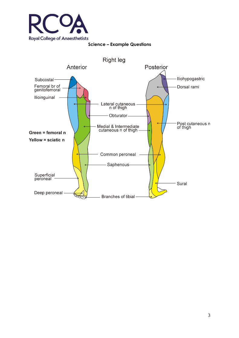
Boundaries: Laterally - medial border of sartorius
Medially – lateral border of adductor longus
Superior - inguinal ligament
Floor: Laterally - iliacus and psoas major
Medially - pectineus and adductor longus
Contents: Medial to lateral:
Femoral sheath - a downward prolongation of of transversalis fascia containing
connective tissue embedding the femoral canal, femoral vein, femoral artery
(overlying pectineus and psoas major)
Femoral nerve L2,3,4 (overlying iliacus)
Lateral femoral cutaneous nerve L2,3
Genitofemoral nerve L1,2,(femoral branch ) anterior to artery
Medial cutaneous nerve of the thigh L2,3 which crosses the femoral artery near the
apex
Femoral vein becomes posterior to artery in lower part of triangle.
Roof: Superficial fascia lata and fat, superficial inguinal lymph nodes
Describe how to conduct a suitable block for this fracture when ultrasound is not
available for use (i.e. using anatomy)
A. Femoral nerve block:
(L2-4 from lumbar plexus) for surgery of anterior thigh, knee or femoral fracture (med.
Lower leg).
Palpate femoral pulse below midpoint of inguinal ligament. Insert needle 1cm
laterally to pulse and 2 cm below ligament at 45 degree angle. 2 “pops” (fascia lata
and iliopectineal facia).
Nerve stimulator provokes contraction of quadriceps. 10-30ml local anaesthetic
injected.
Science – Example Questions
2
Sartorious stimulation due to direct muscle contact or anterior division of nerve.
unacceptable for good block.
Alternatives.
B. 3 in 1 block:
(femoral, lateral cutaneous nerve of the thigh, obturator) same approach, larger
volume of LA and application of distal pressure encouraging superior spread
C. Fascia iliaca block:
(femoral, lateral cutaneous nerve of the thigh, obturator) limb abducted, externally
rotated.
Needle inserted 0.5 – 1.0cm below inguinal ligament, at junction of lateral and
middle third of ligament. 2 “pops” as passes through fasia lata and fascia iliaca. 20-
30 ml LA.
What initiative is there to minimise errors and outline how it is conducted
Stop Before You Block (SBYB)
The WHO ‘sign in’ is performed as usual.
The patient identity, consent form and marking of the correct surgical site are
confirmed. IMMEDIATELY before needle insertion in the nerve block process the
correct site is confirmed again.
This involves:
Visualising the surgical arrow indicating site of surgery
Asking the patient to confirm the side of surgery (if conscious)
Double checking the consent form for operative side (if patient unconscious)
Filler question
Describe what is meant by the term “never event”
“serious incidents that are wholly preventable as guidance or safety
recommendations that provide strong systemic protective barriers are available at a
national level and should have been implemented by all healthcare providers”
discuss preventability and impact of the initiative
Science – Example Questions
3
Science – Example Questions
4
Question 2: Anatomy: Tracheostomy
Opening Question(s): What are the indications for tracheostomy in critical care?
Scientific principle(s) to be explored: Anatomy of the trachea and anterior triangle
of the neck.
Clinical applications(s) of the scientific principles: Appropriate patient selection;
preconditions and potential complications.
What are the indications for a tracheostomy in critical care?
1. To assist weaning from artificial ventilation
2. Facilitate withdrawal of sedation
3. Ongoing airway toilet/reduce secretion retention
4. Reduce oropharyngeal/tracheal complications of long-term intubation
5. Protect airway in neurological dysfunction
6. Longer term: to facilitate ICU discharge and on-going
management/domiciliary ventilation in patients with chronic conditions
(neuromuscular/respiratory)
What techniques are available?
Open (surgical) tracheostomy versus percutaneous techniques
1. Percutaneous dilatational with multiple dilators(Seldinger wire/Ciaglia)
2. Percutaneous dilatational with single dilator (Seldinger wire/Rhino)
3. Guidewire and dilating forceps (Grigg’s forceps)
4. Other (?PercuTwist)
What are the contraindications to bedside tracheostomy?
Absolute: local sepsis; tumour; patient refusal.
Relative: severe coagulopathy/anticoagulation; platelet
dysfunction/thrombocytopaenia; blood vessels crossing the surgical field; short fat or
immobile neck, unstable C-spine injury etc
Describe the anatomy relevant to performing a percutaneous tracheostomy starting
with the larynx.
The larynx is an organ of phonation also acting as a valve between upper and
lower airway
Innervated by branches of the vagus
SLN sensation of upper part and motor innervation of cricothyroid (adducts cords)
RLN sensation of lower larynx and motor to intrinsic muscles
Superiorly, strap muscles and epiglottis attach to hyoid.
Thyrohyoid membrane passes caudally to thyroid cartilage.
Vocal cords attach anteriorly to the thyroid cartilage and pass posteriorly to the
arytenoid cartilage articulating below with the cricoid cartilage. This has the shape
of a signet ring with the wider part posteriorly.
The cricothyroid membrane/ligament passes anteriorly between the thyroid and
cricoid cartilages.
Science – Example Questions
5
Cricotracheal ligament attaches the cricoid to the first tracheal ring.
Below the larynx, trachea passes behind the thyroid isthmus backwards and
downwards, anterior to the oesophagus to enter the thoracic cavity at the sternal
notch.
Its lateral relations include the RLNs, carotid arteries and jugular veins.
The aortic arch and innominate artery lie anteriorly in the thoracic inlet, where the
trachea passes medial to the
domes of the pleura.
How might a percutaneous tracheostomy be performed?
Informed consent/assent, monitoring (inc capnography), trained assistant, sterile
field.
Avoid operator/anaesthetist situations.
Reverse Trendelenburg. Increased FiO2. Bronchoscopic control (pros and cons?)
Below 2nd or 3rd tracheal rings. Palpation/ultrasound. Lidocaine/adrenaline. Incision
1-2cm; blunt dissection.
Needle puncture, aspiration of air bubbles; Seldinger wire; dilatation; insertion of
device.
Avoid damage to vessels/tracheal rings.
Confirm position of needle/guidewire/device bronchoscopically. Confirm CO2
trace.
Suction blood/secretions. Secure position. X-ray (Pneumothorax, airway collapse,
appropriate positioning above carina etc).
Complications
These relate to the procedure itself and anatomically related structures.
Immediate: Failure, loss of airway, bleeding, pneumothorax, oesophageal
perforation.
Early: Bleeding, dislodgement, obstruction.
Late: Erosion into vessels, tracheal dilatation, cuff-related stenosis, granulomata,
stenosis related to traumatic insertion, cord/laryngeal dysfunction.
Science – Example Questions
6
Question 3: Physiology: Stress Response
Opening Question(s): How can the stress response to surgery be blocked or
reduced ?
Scientific principle(s) to be explored: Responses of the neuroendocrine system to
surgical stimulation.
The consequences of these for the patient – what happens and why
Clinical applications(s) of the scientific principles: Regional blocks, their limitations,
do they improve outcomes?
Blocking the response
• Only with very large doses of opioids or regional anaesthesia.
• Can reduce by less invasive surgery e.g Laparoscopic surgery
• Intraoperative use of regional anaesthesia only delays the development of
the response.
• If wish to modify the response postop, need continual regional technique.
• Optimal duration of the use of RA unknown.
• More effective against stress produced during surgery to lower body than
upper abdomen, chest.
• Could be due to inadequate block of all afferent stimulation in this type of
surgery.
• The desirability of totally abolishing the response in unknown.
• Objective proof that morbidity and mortality are reduced is limited.
• Early nutrition
Describe the physiology of stress response
Afferent nerve stimulation initiates complex neuroendocrine changes which lead to:
• Catabolism
• Water and sodium retention
• Higher oxygen consumption
• Hyperglycaemia
• Hypercoagulability
• Pyrexia
Increased secretion of
• Beta-endorphins
• ACTH and cortisol (5-fold)
• Catecholamines
• Glucagon, growth hormone and prolactin
• ADH (up to 100-fold)
• Renin, and aldosterone
• Proinflammatory cytokines; interleukins, TNF-alpha, prostanoids
Science – Example Questions
7
Decreased secretion of
• insulin, testosterone, thyroxine
What are the consequences of the stress response?
• Breakdown of fat and protein (negative nitrogen balance), hyperglycaemia,
hyponatraemia, immunological suppression, increased coagulability,
decreasedfibrinolysis.
• Particularly marked in major surgery of the chest and abdomen.
• Correlates with the degree of surgical trauma.
Possible reasons for suppressing the response
• Causes muscle wasting, fatigue
• Increased demands on organs eg kidneys, heart
• Increased risk of thromboembolism
• Increased cardiac and respiratory complications
• Deterioration in blood glucose control particulary in diabetics
• Possible increased wound infection rates
• A degree of response is necassary to arespond to physiological insults e.g.
hypotension
What are the benefits of regional anaesthesia?
• Reduced opiate requirement
• Improved gut mobility/ reduced nausea and vomiting
• Less respiratory complications
• Avoid immunomodulatory effects opiates
• Improved analgesia
Science – Example Questions
8
Question 4: Physiology: Cerebral blood flow
Opening Question(s): What compromises cerebral blood flow (CBF) in a patient with
a head injury?
Scientific principle(s) to be explored: Understanding of the factors controlling CBF.
Clinical applications(s) of the scientific principles: Strategies for maintaining and
defending cerebral perfusion.
Compromise of cerebral blood flow in head injury: Reduced MAP
Increased ICP or both together
Strategies to maintain CBF
1. Improve Cerebral Perfusion Pressure (CPP) by maintaining MAP
• First 72 hr, loss autoregulation, CBF directly dependant on MAP, careful
control of BP, keep MAP >60mmHg.
• Hyperaemic phase(20-30% patients), may increase ICP, accept CPP 60 -
70mmHg.
• Vasospastic phase (10%), reduced CBF, hypometabolism, loss autoregulation.
Difficult to control.
2. Control of ICP
• Surgery: evacuate haematomas, decompressive craniotomy, excise
contusions, drainage of CSF
• Medical: diuretics, barbiturate coma, avoid hyponatraemia, treat pyrexias,
treat seizures, hypothermia - debatable, hyperventilation NOT recommended
• Mechanical: straining, high intathoracic pressure, venous congestion
Cerebral blood flow (CBF, normal 50ml/100g/min, or 700ml/min) influenced by >
1. Cerebral metabolism
• Cerebral metabolic rate for oxygen (CMRO2) constant at 3.5ml/100g/min
• Reduced by; hypothermia, at 27 degrees Celcius, CMRO2 & CBF halved,
coma, anaesthesia.
• Increased by pyrexia, seizures
2. Carbon dioxide and oxygen
• Linear relationship between PaCO2 and CBF between 2.7kPa (20mmHg) and
10.6kPa (80mmHg).
• Mediated by hydrogen ion conc in ECF surrounding cerebral vasculature
• Hyperoxia has little effect, max 10% increase in CBF
• Hypoxia PaO2 <6.7kPa (50mmHg) causes rapidly progressive increase in CBF,
secondary to acidosis.
3. Autoregulation
• Pressure difference across the vasculature, CPP=MAP-ICP
• Venous pressure difficult to measure so use ICP (venous pressure slightly
greater to remain patent)
• CBF constant over the CPP range 50-150mmHg.
Science – Example Questions
9
• Achieved by myogenic mechanism inducing vasoconstriction (increased
pressure), vasodilatation (reduced pressure).
• Lower limit represents maximal vasodilatation, below this CBF falls, “pressure
passive”
• Upper limit represents maximal vasoconstriction, above this disruption of BBB,
oedema and ischaemia (encephalopathy).
4. Autonomic nervous system
• Sympathetics cause vasoconstriction, serve to protect brain in hypertension
• Parasympathetics contribute to vasodilatation, maximal in post ischaemic
reperfusion and hypotension.
5. Blood viscosity
• Balance between reduced haematocrit improving flow and oxygen carrying
capacity, 30% optimal.
Monitoring CBF
No direct method available for use at bedside, therefore use ind
Jugular bulb oximetry: SjO2 inversely proportional to CBF for given metabolic
rate. Global value.
Near infrared spectroscopy: uses a scalp oximeter, problems with depth of
penetration, contamination by scalp flow. Very regional.
Transcranial Doppler: insonate large vessels, measures flow velocity.
Derived indices eg pulsatility index. Better at trends rather than absolute values.
Operator variability.
Science – Example Questions
10
Question 5: Pharmacology: Calcium Channel Blockers
Opening Question(s): What are the common indications for the use of calcium-
channel blocking drugs and which drugs are used?
Scientific principle(s) to be explored: Understand the basic pharmacology of this
group of drugs.
Clinical applications(s) of the scientific principles: Calcium-channel blockers in
anaesthetic practice.
General uses:
• Treatment of angina: e.g. diltiazem, amlodipine, verapamil
o As the primary treatment
o As adjunct to β-blockers or in patients in whom they are
contraindicated (eg. brittle asthmatics etc)
o Angina caused by coronary vasospasm
• Treatment of hypertension:
o Chronically e.g. amlodipine, nicardipine, lacidipine
(dihydropyridines)
o Acutely: nifedipine
• Antiarrhythmic: verapamil, diltiazem
• Vasospasm following SAH: nimodipine
• Preterm labour e.g. nifedipine, nicardipine
• Raynaud’s disease e.g. diltiazem, nifedipine
Acute use in anaesthetic practice
Management of SVT and AF
• Verapamil slows conduction in pacemaker tissue >↓ AV conduction and ↑
refractory period of AV tissue
• Risk of heart block if patient recently given β-blockers
• 5-10mg over 3 minutes with ECG monitoring
Management of cerebral vasospasm following SAH
• Cerebral vasospasm occurs 4-14 days post SAH in ~ 40-70% patients
• Results in progressive focal neurological deficit and even death (10-20%)
• Signs and symptoms: fever, neck stiffness, confusion, hemiplegia, altered
consciousness
• Nimodipine: given via central catheter, progressively increasing dose if BP
acceptable, or orally (4 hrly – high first pass metabolism). May reduce poor
outcome due to vasospasm after SAH by 40%
Anaesthetic implications: interactions of calcium-channel blockers with anaesthesia
• verapamil and β-blockers carry risk of AV block
• volatile anaesthetics – risk of exaggerated hypotension, MAC depressant
properties
Science – Example Questions
11
• digoxin – inhibits Cytochrome P450 with potential to increase digoxin levels
(↑ risk of AV block)
• non-depolarising NMB – verapamil and nifedipine have potential to
increase block
• reduction of lower oesophageal sphincter tone
• sudden withdrawal may be associated with an exacerbation of angina
Pharmacology of calcium-channel blockers
• Three major groups
o dihydropyridines e.g. nifedipine, amlodipine, nicardipine, nimodipine
o phenylalkylamines e.g. verapamil
o benzothiazepines e.g. diliazem
• Each group binds to a specific site on α-subunit on the L-type calcium
channel
• Inhibit the slow inward Ca current in smooth muscle and cardiac cells
• The reduced intracellular Ca causes:
o vasodilatation in vascular smooth muscle
o negative intotropic, chronotropic and dromotropic (slow conduction
velocity) properties on the heart
• Absorption is nearly complete after oral administration
• Bioavailability is reduced because of first-pass hepatic metabolism
• There is significant binding of all channel blockers to plasma proteins
(70-99%)
• Therapeutic effects are evident within 30-60 min after oral dose; peak
effects within 15 min i.v.
• They differ in their predilection for various sites of action, with disparate and
varied therapeutic effects.
Science – Example Questions
12
Question 6: Pharmacology: Pheochromocytoma
Opening Question(s): What is a phaeochromocytoma?
Scientific principle(s) to be explored: Pharmacology of endogenous and exogenous
adrenaline.
Clinical applications(s) of the scientific principles: Anaesthesia for excision of
phaeochromocytoma.
Phaeochromocytoma
• tumour of chromaffin tissue, 90% in adrenal medulla
• associated with multiple endocrine neoplasia (MEN) type 2,
neurofibromatosis, von Hippel-Lindau.
• diagnosis confirmed with 24-hr urinary met-Adr, nor-met-Adr.
• localised with CT or MRI.
Systemic actions of catecholamines:
• increase rate and force of myocardial contractility
• tachycardia when plasma level is about 50 pg/ml (about twice normal).
• increase myocardial excitability
• effects on diastolic perfusion of myocardium -effect modified by
baroreceptor stimulation (i.e. are greater in a denervated heart)
• dilates blood vessels in skeletal muscle and liver via alpha2
• constricts other blood vessels (alpha), but net effect is a drop in SVR
• systolic BP rises, diastolic BP falls, mean BP rises only slightly.
• endocrine effect
Termination of action
• following nerve stimulation reuptake of 75% NA (uptake-1 system)
• some removed via uptake-2 system into post-junctional and non-neuronal
tissue
• plasma catecholamines broken down in blood, liver, kidney and lungs by
MAO (in mitochondria) and COMT (in cytoplasm).
Management Preop investigations
• ECHO for asymptomatic cardiomyopathy.
• Alpha blockade: phenoxybenzamine for 7-10 days, longer if cardiomyopathy
or ECG changes.
• Aim to stabilize BP <140/90, HR< 100/min, symptoms reduced.
• May get reflex tachycardia requiring beta-blockade, drowsiness, dizziness,
blocked nose.
• Use of beta blockers (not as initial therapy: risk of hypertensive crisis. May be
added later for control of tachydysrhythmias).
Science – Example Questions
13
Anaesthesia
• Premedication - Anxiolytic premed, DVT prophylaxis, stop blockade evening
before surgery.
• Monitoring: ECG (CM5), IABP, CVP (PA catheter?), SpO2, EtCO2, urine output,
core temp.
• Laparotomy: GA plus epidural, laproscopy GA alone.
• Avoid histamine releasing drugs
• Role of Mg infusion as pre-surgical prophylaxis (inhibits catecholamine
release, causes arteriolar dilatation, antidysrhythmic effects).
• SNP to control hypertension, NA for hypotension, ADR to maintain CO, beta
blockers to control tachycardia
• Careful maintenance of temp as patient vasodilated.
• May need significant fluid volumes after removal of tumour.
Post op:
• ITU, BP stability, blood glucose control, hypoglycaemia common (increased
insulin secretion or decrease in lipolysis).
• Steroid replacement if bilateral adrenalectomy.
Science – Example Questions
14
Question 7: Clinical measurement: MRI
Opening Question(s): What are the problems facing the anaesthetist in the MRI
scanner?
Scientific principle(s) to be explored: Understanding of the basic principles of MR
scanners.
Clinical applications(s) of the scientific principles: How understanding the physical
principles allows safe anaesthesia in a hostile environment.
Guidance for Examiners: Candidates should demonstrate how to work safely in the
MRI Suite.
Isolation from main theatre, patient in magnet, difficult to observe and monitor
Noise in scanner – up to 95db, all patients need earplugs, even when anaesthetised
(risk cochlear damage)
Variable RF Magnetic field causes heating tissues, Burns from monitoring if wires
coiled, must avoid and closed loops e.g patients with crossed legs have been burnt
and any metal in clothing
Time variable magnetic field can cause muscle contraction and induced nerve
conduction some patients find unpleasant so request sedation/GA. Will cause VF at
high field manipulation.
Care with obese patients in industrial/ veterinary scanners
Helium escape need 1000L liquid helium to keep a superconducting magnet cool if
there is sudden shut down or spontaneous leak the helium will vapourise as it is
vented if the quench pipe malfunctions the oxygen level in MRI will plummet so
need monitoring system and door override to allow escape
What are the Contraindications for MRI?
• Patients with, spinal nerve stimulators, cochlear implants, shrapnel, intraocular
splinters, tattoos may heat up.
• Check compatability of pacemakers, vascular clips, IUCDs, aneurysm coils,
heart valves any orthopaedic prosthesis and stents
• Defibrillators can only be used outside the magnetic field
• Discuss safety of MRI in pregnancy. No evidence that it is teratogenic.
Recommendations are that ok to use if essential.
Equipment: need to have safety check at 5 G contour
• MR safe – no risk to patient or personnel under any condition
• MR conditional – safe, image not affected, function not affected as long as
follow MRI protocol
• MR Unsafe need to avoid all ferromagnetic materials, must be kept beyond
30-Gauss line.
Science – Example Questions
15
What are the principles of operation? (Physics)
• No ionising radiation
• Uses interaction between a static magnetic field and the field that arises from
atoms
• Patient placed within a strong magnetic field (0.5 – 3.0 Tesla, 1T = 104 Gauss,
earth’s field = 0.5 Gauss).
• Some atoms with odd numbers of protons (e.g.hydrogen) have an
asymmetrical net spin which results in atoms aligning either almost parallel or
almost opposed to an external magnetic field
• In addition they exibit precession (“wobble” about their axis of spin) in the
magnetic field at a frequency proportional to the field strength.
• Application of pulses of radiofrequency energy (microwaves) at the
frequency of the atoms precession
(64Mhz for H2) causes protons to “flip” from lower parallel to higher anti
parallel state.
• RF switched off, protons “flip” back and release radiowave energy. Detected
by a series of close fitting antennae or coils within a faraday cage to allow
the small signal to be received uncorrupted.
• Images are generated by perturbing the static field with small dynamic
gradient fields which allows spatial localisation by altering the precession
frequency
• Contrast between tissues results from:
A) Differences in hydrogen nuclei density between tissues
B) Physical and chemical differences in the tissues producing 2 distinct
relaxation patterns with time constants T1and T2 .
In T1 fat is bright and water dark and vice versa for T2. T2 is used to identify
tissue oedema andT1 white matter grey matter contrast
• Contrast (gadolinium) –alters relaxation rates hydrogen nuclei
• Renally cleared can cause nephrogenic systemic fibrosis in patients low GFR
or liver impairment
• 3 risk levels of contrast depending on propensity to release Gd
• Nausea and sickness fairly common but allergic reactions very rare
• Monitoring adaptions: most telemetric
All monitoring should be in control room (AAGBIguidelnes)
ECG – Monitor V5, V6 as furthest from aortic flow induced voltages causing artefacts,
ST segment and T waves, resembles pericarditis or hyperkalaemia. High impedance
graphite electrodes and short braided leads or telemetry to prevent induced
current. Electrodes close together to minimise impedance
LCD display not affected by magnet. Cardiac motion artefact reduced by ECG
gating. Risk of burns from heating effect on cables. Ensure no contact and that ECG
electrodes are correctly applied.
Pulse oximetry – burns with standard pulse oximeters.so fibreoptic cables used, signal
is distorted, filtered to improve signal.
Capnography – in line sampler over heats, use side-stream, but delays of up to 20
sec because of long sample tube.
Science – Example Questions
16
Blood pressure
- indirect methods available using nylon connectors.
- direct possible but transducer needs to be close to patient to avoid long lines
and damping. May cause artefact.
Anaesthetic equipment
• Non-MR compatible kept outside scanning room, or outside 50-G line,but long
tubes etc risk disconnection.
• MR-compatible with piped gases in scanner (no ferromagnetic material,
usually aluminium).
• All gas cylinders must be MR compatible.
• MR compatible laryngoscopes, ET tubes, LMAs, ventilators, infusion pumps,
suction, patient trolleys available.
Science – Example Questions
17
Question 8: Clinical measurement: Breathing Systems
Opening Question(s): What factors should you consider when selecting a breathing
system for a particular patient?
Scientific principle(s) to be explored: Breathing system design
Clinical applications(s) of the scientific principles: Problems associated with circle
breathing systems
[Clinical Measurement question]
Factors to be considered
• Size of patient - T-piece suggested, < ~ 15 kg, why?
• Emergency situation on the ward, giving 100% oxygen by hand - Mapleson C
circuit
• Economy of gas-flow with expensive volatile agents, reduce pollution - Circle
system
• Specialised circuit for CPAP etc in critical care
• Circuit to be used for SV or IPPV or ease of switching between SV and IPPV
• Long circuit needed - e.g. for patient in MR suite
• Ease of scavenging - easier for Lack/Bain than Magill/Mapleson A
Could you describe in detail the pathway taken by gas in a circle breathing system
Start with gas which has just been expired during spontaneous ventilation
Y-connector - expiratory limb tubing- expiratory one-way valve-reservoir bag with
spill valve-CO2 absorber fresh gas added-inspiratory valve-inspiratory tubing-Y-
connector
o fresh gas enters after absorber to prevent absorber drying, but gas not
humidified
o Reservoir bag and spill valve before absorber (inefficient to clean gas being
voided)
o Simple system for IPPV is bag/bellows in bottle but new machines more
complicated
Disadvantages
• Complex design with multiple connections
• Unidirectional valves may malfunction [check the candidate knows the
problem resulting]
• Slow response time at low fresh gas flow
• Need for volatile agent monitoring
• Breakdown of anaesthetic agents by soda-lime eg sevoflurane (is this
important?)
• Consumes absorber, may need changing intra-op
• Carbon monoxide accumulation (? significance)
• Multiple user inputs required to be used efficiently
Science – Example Questions
18
How is the problem of carbon monoxide production avoided
o Related to agent (worst with desflurane), broken down by KOH in soda-lime
o Related to type of absorber (worst with baralyme), its temperature and
dessication
o Modern soda-lime has no KOH, dessication resistant
How would you suspect carbon monoxide poisoning
• Pulse oximeter not helpful
• No reliable signs in anaesthetised patients beyond perhaps cardiac signs
cellular hypoxia
• Gas analyser may show erroneous volatile agent due to production of
trifluoromethane
• Analysis of arterial blood sample by co-oximeter to show COHb
• CO ~ 5-10% may be found in smokers, higher results in poisoning
• Neurological damage due presumably to cellular hypoxia
Advantages of circle breathing system
o Economy of fresh gas and volatile agent [how would you calculate cost
savings?]
o Maximum savings at very low flows for long procedures [what is lowest flow
possible?]
o Conserve heat and moisture in inhaled gases
o Reduced theatre pollution - working efficiency, long-term problems of
anaesthetic agents
o Reduced environmental pollution
- Nitrous oxide is a primary greenhouse gas and depletes ozone
- H-CFCs , and to a lesser extent desflurane and sevoflurane (no
chlorine atom) cause destruction of ozone layer