schwannoma with bone differentiation in a dog -...
TRANSCRIPT

Viott et al; Schwannoma with bone differentiation in a dog. Braz J Vet Pathol; 2008, 1(2): 52 - 55
Brazilian Journal of Veterinary Pathology. www.bjvp.org.br . All rights reserved 2007.
52
Case Report
Schwannoma with bone differentiation in a dog
Aline M. Viott1*; Adriano T. Ramos1; Alexandre Mazzanti2, Glaucia. D. Kommers1,
Dominguita. L. Graça1
1 Departamento de Patologia Veterinária. Universidade Federal de Santa Maria (UFSM), Centro de Ciências Rurais, Santa Maria, RS, Brasil.
2Departamento de Clínica e Cirurgia Veterinária, UFSM. *Corresponding author: Aline M. Viott, Centro de Ciências Rurais, Universidade Federal de Santa Maria
(UFSM), 97105-900 Santa Maria, RS, Brasil. [email protected].
Submitted February 10th 2008, Accepted May 5th 2008
Abstract
A schwannoma with bone differentiation was diagnosed in a seven-year-old German Shepherd female dog. Clinical signs were those of limping and proprioceptive and neurological deficits. Superficial and deep sensitivity were lower and muscle atrophy of the left forelimb was marked. Two subscapular masses were detected at examination. Microscopic analysis of the masses disclosed a pronounced proliferation of either palisading or loosely arranged within a myxoid tissue spindle neoplastic cells intermingled with bone tissue islands. The neoplasm was positive for vimentin, S100 protein and GFAP.
Key Words: Neoplasm, schwannoma, bone differentiation, immunohistochemistry.
In Veterinary Medicine schwannomas are the more common neoplasms of cranial, spinal and peripheral nerves. These neoplasms have been reported in many species, including cattle, horses and cats; yet, they are more prevalent in dogs (12). Cranial nerves, mainly the V pair, cervical spinal roots and the brachial plexus are the more affected sites (5).
Histologically schwannomas present with bent fascicles of spindle cells disposed in many directions. They may exhibit different morphological patterns: Antoni type A areas are composed of spindle cells arranged in palisades and Antoni type B areas are less cellular and organized, made of oval to round loosely arranged cells (9).
In humans many heterogeneous patterns have been described for peripheral nerve sheath tumors (PNST) which apply to schwannomas, i.e. epithelioid rhabdomyoblastic, cartilaginous, osseous, angiomatous, glandular and lipoblastic (11). Similar representations of
epithelioid, melanocytic, cartilaginous, osteogenic, glandular and epithelial patterns have been observed in dogs and other animal species (3). The goal of this article is to describe the clinical, histological and immunohistochemical aspects of a schwannoma with bone differentiation in a dog.
A seven-year-old German Shepherd female dog was presented to the Veterinary Hospital of the Universidade Federal de Santa Maria with limping of the left forelimb. The owner reported that the impaired gait was observed six months before and the dog had been treated with anti-inflammatory drugs with no clinical improvement. In the last two months the limping increased and the dog started to drag de limb. A radial nerve lesion was suspected.
On clinical examination diminished superficial and deep sensitivity, a deficient flexor reflex, and neurological and proprioceptive deficits were detected. Muscles of the scapular region of the affected limb were
Viott et al; Schwannoma with bone differentiation in a dog. Braz J Vet Pathol; 2008, 1(2): 52 - 55
Brazilian Journal of Veterinary Pathology. www.bjvp.org.br . All rights reserved 2007.
53
markedly atrophied. On palpation a mass on the subscapular region was found. The dog was sent to surgery. During surgery two neoplastic masses involving the brachial plexus and adjacent innervations were separated.
The masses were sent to the Laboratory of Veterinary Pathology for histological analysis. Fragments of the masses were processed routinely for light microscopy (paraffin embedding, 5 μm sections and HE histochemical method (4). Immunohistochemistry was performed with anti-vimentin, anti-S100 protein and anti-GFAP antibodies according to Viott et al (2007).
The smaller mass was white and firm, measured 5 x 4 x 3 cm and was whitish, smooth and shinny on the cutting surface. The larger mass measured 7 x 5 x 3 cm, was firm and reddish and was wrapped by a thin connective tissue capsule. On the cutting surface many areas of bonny consistency were observed.
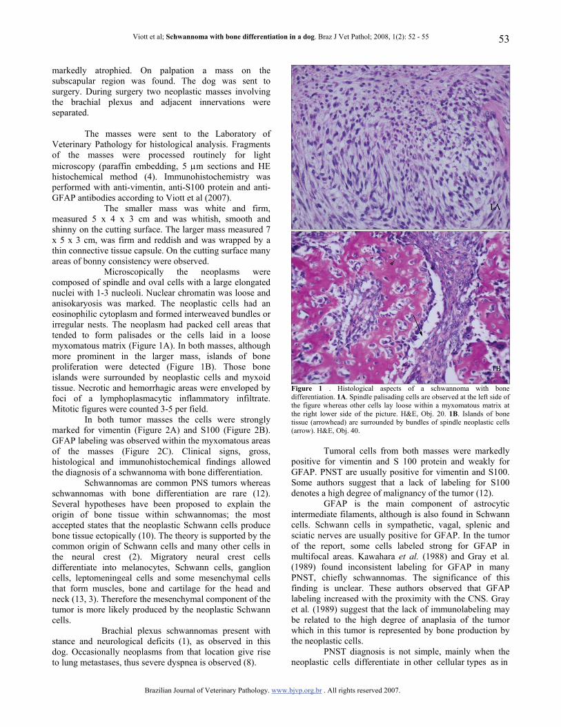
Microscopically the neoplasms were composed of spindle and oval cells with a large elongated nuclei with 1-3 nucleoli. Nuclear chromatin was loose and anisokaryosis was marked. The neoplastic cells had an eosinophilic cytoplasm and formed interweaved bundles or irregular nests. The neoplasm had packed cell areas that tended to form palisades or the cells laid in a loose myxomatous matrix (Figure 1A). In both masses, although more prominent in the larger mass, islands of bone proliferation were detected (Figure 1B). Those bone islands were surrounded by neoplastic cells and myxoid tissue. Necrotic and hemorrhagic areas were enveloped by foci of a lymphoplasmacytic inflammatory infiltrate. Mitotic figures were counted 3-5 per field.
In both tumor masses the cells were strongly marked for vimentin (Figure 2A) and S100 (Figure 2B). GFAP labeling was observed within the myxomatous areas of the masses (Figure 2C). Clinical signs, gross, histological and immunohistochemical findings allowed the diagnosis of a schwannoma with bone differentiation.
Schwannomas are common PNS tumors whereas schwannomas with bone differentiation are rare (12). Several hypotheses have been proposed to explain the origin of bone tissue within schwannomas; the most accepted states that the neoplastic Schwann cells produce bone tissue ectopically (10). The theory is supported by the common origin of Schwann cells and many other cells in the neural crest (2). Migratory neural crest cells differentiate into melanocytes, Schwann cells, ganglion cells, leptomeningeal cells and some mesenchymal cells that form muscles, bone and cartilage for the head and neck (13, 3). Therefore the mesenchymal component of the tumor is more likely produced by the neoplastic Schwann cells.
Brachial plexus schwannomas present with stance and neurological deficits (1), as observed in this dog. Occasionally neoplasms from that location give rise to lung metastases, thus severe dyspnea is observed (8).
Figure 1 . Histological aspects of a schwannoma with bone differentiation. 1A. Spindle palisading cells are observed at the left side of the figure whereas other cells lay loose within a myxomatous matrix at the right lower side of the picture. H&E, Obj. 20. 1B. Islands of bone tissue (arrowhead) are surrounded by bundles of spindle neoplastic cells (arrow). H&E, Obj. 40.
Tumoral cells from both masses were markedly positive for vimentin and S 100 protein and weakly for GFAP. PNST are usually positive for vimentin and S100. Some authors suggest that a lack of labeling for S100 denotes a high degree of malignancy of the tumor (12).
GFAP is the main component of astrocytic intermediate filaments, although is also found in Schwann cells. Schwann cells in sympathetic, vagal, splenic and sciatic nerves are usually positive for GFAP. In the tumor of the report, some cells labeled strong for GFAP in multifocal areas. Kawahara et al. (1988) and Gray et al. (1989) found inconsistent labeling for GFAP in many PNST, chiefly schwannomas. The significance of this finding is unclear. These authors observed that GFAP labeling increased with the proximity with the CNS. Gray et al. (1989) suggest that the lack of immunolabeling may be related to the high degree of anaplasia of the tumor which in this tumor is represented by bone production by the neoplastic cells.
PNST diagnosis is not simple, mainly when the neoplastic cells differentiate in other cellular types as in

Viott et al; Schwannoma with bone differentiation in a dog. Braz J Vet Pathol; 2008, 1(2): 52 - 55
Brazilian Journal of Veterinary Pathology. www.bjvp.org.br . All rights reserved 2007.
54
Figure 2. Immunohistochemical labelling of a schwannoma with bone differentiation. Neoplastic spindle cells and osteocytes label strongly with anti-vimentin (2A) and anti-S100 (2B) antibodies. IHQ, Obj. 40. The anti-GFAP antibody marks those cells immersed in a myxomatous matrix (2C). The bone matrix made up by tumoral cells is not immunolabelled (arrow). IHQ, Obj. 40 this case. The differential diagnosis of schwannomas from other neoplasms that arise from the common migratory neural crest cells may be very difficult, and only immunohistochemical studies may elucidate the situation (2). In the case reported here GFAP labeling of some cells
helped to establish the distinction between a schwannoma and an osteosarcoma. References 1. BROWER A., SALAMAT S., CRAWFORD J.,
MANLEY P. Unilateral limb enlargement in a dog with a malignant peripheral nerve sheath tumor. Vet. Pathol., 2005, 42, 353-6.
2. CHERNIE S., DORE M. Oral malignant Melanoma with Osteoid Formation in a Dog. Vet. Pathol. 1999, 36, 74-6.
3. CHIJIWA K., UCHIDA K., TATEYAMA S. Immunohistochemical evaluation of canine peripheral nerve sheath tumors and other soft tissue sarcomas. Vet. Pathol., 2004, 41, 307-18.
4. CULLING CFA., ALLISON RT., BARR WT. Cellular pathology technique. 4. ed. London: Butterworths,1985.
5. GRAÇA DL., FERNANDES CG., SOUZA MV., ILHA MRS. Schwannoma de V par craneano y ganglio em Pastor alemán. Relato de un caso. Med. Vet., 1998, 15, 652-6.
6. GRAY MH., ROSENBERG AE., DICKERSIN R., BHAN AK. Glial Fibrillary Acidic Protein and of Keratin Expression by Benign and Malignant Nerve Sheath Tumors. Hum. Pathol., 1989, 20, 1089-96.
7. KAWAHARA E., ODA Y., OOI A., KATSUDA S., NAKANISHI I. Expression of Glial Fibrillary Acidic Protein (GFAP) in Peripheral Nerve Sheath Tumors. Am. J. Surg. Pathol., 1988, 12, 115-20.
8. KOESTNER A., HIGGINS RJ. Tumors of the nervous system. MEUTEN DJ. Ed. Tumors of domestic animals. Iowa: Iowa State Press, 2002: 697-38
9. KUWAMURA M., YAMATE J., KOTANI T., TAKEUCHI T., SAKUMA S. Canine Peripheral Nerve Sheath Tumor with Eosinophilic Cytoplasmic Globules. Vet. Pathol., 1998, 35, 223-6.
10. PATNAIK AK., ERLANDSON RA., LIEBERMAN PH. Canine malignant melanotic schwannomas: A light and electron microscopic study of two cases. Vet. Pathol., 1984, 21, 483-8.
11. PLAZA JA., WAKELY JR. PE., SUSTER S. Lipoblastic Nerve Sheath Tumors Report of a Distinctive Variant of Neural Soft Tissue Neoplasm With Adipocytic Differentiation. Am. J. Sur. Pathol., 2006, 30, 331-44.
12. PUMAROLA M., ANÕR S., BORRÀZ D., FERRER I. Malignant epithelioid Schwannoma affecting the trigeminal nerve of a dog. Vet. Pathol., 1996, 33, 434-6.
13. SHAH NM., GROVES AK., ANDERSON DJ. Glial Growth Factor restricts mammalian neural crest stem cells to a glial fate. Cell., 1996, 77, 349-60.
14. VIOTT AM., RAMOS AT., INKELMANN MA., KOMMERS, GD., GRAÇA DL. 2007. Aspectos histoquímicos e imunoistoquímicos nos neoplasmas

Viott et al; Schwannoma with bone differentiation in a dog. Braz J Vet Pathol; 2008, 1(2): 52 - 55
Brazilian Journal of Veterinary Pathology. www.bjvp.org.br . All rights reserved 2007.
55
do sistema nervoso periférico. Arq. Bras. Med. Vet. Zootec., 2007, 59, 1145-53.