schistosomiasis japonica in the philippines: the long ... · 163 schistosomiasis japonica in the...
TRANSCRIPT
163
Schistosomiasis Japonica in the Philippines: The Long-Term Impact ofPopulation-Based Chemotherapy on Infection, Transmission, and Morbidity
Remigio M. Olveda, Bruce L. Daniel,*Bernadette D. L. Ramirez, Gemiliano D. L. Aligui,Luz P. Acosta, P. Fevidal, Edita Tiu, Federico de Veyra,Pierre A. Peters, Rodrigo Romulo, Ernesto Domingo,Peter M. Wiest,* and G. Richard Olds
Research Institute for Tropical Medicine, Department ofHealth. andUniversity of the Philippines, College of Medicine, Manila, and
Department ofHealth, Provincial Health Office, Leyte, Philippines;International Health Institute, Brown University, Providence,
Rhode Island
The long-term impact of annual case-finding and chemotherapy with praziquantel on schistosomiasis japonica was examined in an 8-year longitudinal study in the Philippines. The prevalence,incidence, and intensity of infection and schistosome-induced hepatomegaly significantly decreasedwithin 3-4 years of treatment and then stabilized despite continual population-based chemotherapy.Hepatomegaly rapidly developed in acutely infected persons, with 82% of subjects developing hepaticenlargement within 2 years of reinfection. These data suggest that abrupt discontinuation of currentcontrol measures in the Philippines may result in a rapid rebound in morbidity. Age-dependentacquired resistance to reinfection also developed in subjects chronically exposed to schistosomiasisjaponica, suggesting that a vaccine may represent an alternative approach for control of this parasiticinfection.
Schistosomiasis japonica is a major health problem in thePhilippines and China [1, 2]. Infection with Schistosoma japonicum leads to a significant delay in growth and developmentin children and to hepatosplenomegaly [3-5]. With time, schistosome-induced hepatic fibrosis develops, which leads to portalhypertension and esophageal varices [5]. Mortality due to schistosomiasis japonica has been estimated to be 1.8% per yearbefore the use of praziquantel [5]. The life cycle of S. japonicum occurs between the human host and a small amphibioussnail, Oncomelania species [5]. In contrast to the transmissionof other schistosome species, the transmission of S. japonicumin endemic areas is contributed to significantly by animal reservoirs, such as cattle and water buffaloes [5].
In the Philippines, 10 million people live in areas endemicfor schistosomiasis japonica, and > 500,000 are actively infected [3, 6]. The major foci for transmission are located on
Received 3 February 1995; revised 19 March 1996.Presented in part: XVII Pacific Congress, Honolulu, Hawaii, 31 May 1991.Informed consent was obtained from all patients or their parents or guardians.
Guidelines for human experimentation of the United States Department ofHealth and Human Services and Miriam Hospital, Brown University, werefollowed in the conduct of this clinical research.
Financial support: Edna McConnell Clark Foundation; Partnership in Research on Philippine Schistosomiasis funded by joint grant from WHO/TORand Rockefeller Foundation (project 880403); National Institutes of Health(AI-3060l); and Harvard Medical School Student Research Fund.
Reprints or correspondence (present address): Dr. G. Richard Olds, Dept.of Medicine, MetroHealth Medical Center, 2500 MetroHealth Dr., Cleveland,OH 44109-1998.
* Present affiliations: Department of Radiology, Stanford University Schoolof Medicine, Stanford, California (B.L.D.); Program in International Health,MetroHealth Medical Center, Case Western Reserve University, Cleveland(P.M.W.).
The Journal of Infectious Diseases 1996; 174:163-72© [996 by The University of Chicago. All rights reserved.0022-1899/96/7401-0021$01.00
the islands of Leyte, Samar, and Mindanao. Rice farming isthe predominant occupation in these endemic areas, whichallows for maximal contact between humans, animal reservoirs,and the freshwater snail intermediate host.
Praziquantel is currently the drug of choice for treatment ofS. japonicum infection. This drug achieves parasitologic curerates often >90%, with minimal side effects [7]. Praziquantelis expensive but it is the only acceptable and effective drugfor treatment of this species of schistosome [8]. Since praziquantel is safe and can be orally administered, it has becomethe basis of community-based chemotherapy programs for control of schistosomiasis japonica in the Philippines and China.
In 1981, a community-based treatment and morbidity studyin three villages in rural Leyte was initiated [9]. Villages werescreened annually for S. japonicum infection, and infected persons were treated with praziquantel. Because of the success ofthis approach, the Schistosomiasis Control Program, organizedby the Department of Health in the Philippines, began to surveyand treat all inhabitants on the islands of Leyte and Samarusing this strategy. This ambitious project managed to examineand treat almost 400,000 cases by 1984 [10].
In 1985, external funds were withdrawn and the scope ofthe program was severely reduced. Consequently, a subprojectof this study was initiated in 1986 to determine the impact ofincreasing the interval between community surveys for villagesfor which the prevalence of infection was < 10%. Since theseareas represented 60%-80% ofthe population at risk for schistosomiasis in the Philippines, this strategy would have alloweda more cost-effective allocation of limited domestic funds [10].
Here we describe the impact of this long-term communitybased program on infection and morbidity induced by schistosomiasis japonica in three villages on Leyte [9]. In addition,since this study followed> 5000 subjects over 8 years, we hadthe opportunity to examine these data for evidence of acquired

164 01veda et al. lID 1996; 174 (July)
resistance to reinfection with S. japonicum after chemotherapyand to examine the dynamics of the natural history ofS.japonicum-induced hepatomegaly in communities undergoing intensive chemotherapy with praziquantel.
Methods
Study population. The study population consisted of all persons :?= 1 year old in three rural villages in northeastern Leyte[9]. Chemotherapy-based schistosomiasis control was nonexistentbefore the initiation of this study. A census was done yearly toassess population movement and growth. Transmission of schistosomiasis occurs throughout the year, and malaria is not endemic.Rainfall is continuous throughout the year, with no distinct dryseasons. Rice farming is the primary occupational activity.
Study design. The study design was longitudinal over 8 consecutive years (1981-1989). Medical histories, quantitative stoolegg counts, and abdominal physical examinations were doneyearly. Stool samples from all subjects were examined at baselineand every 3 months during the first year of the study. Thereafter,annual physical and stool examinations were done. Basic demographic information, including date of birth, age, and sex, wereobtained from each subject on entry into the study or from parentsof subjects < 16 years of age. All infected subjects were treatedwith praziquantel (Bayer, Leverkusen, Germany), 50 mg/kg orallyin two divided doses 4 h apart [10]. Pregnant women were exemptfrom treatment until after delivery. In years 6 and 7, villages Aand B were screened, but treatment was intentionally deferred sothat the optimal interval between screening and treatment couldbe determined for villages with initial low prevalences of infection.
Clinical and parasitologic examinations. Annual parasitologicexaminations were done on a single stool sample obtained fromeach subject. Single stool samples were examined since, despitethe potential of missing infected persons, this project was a modelto evaluate the effectiveness of the national control program in thePhilippines. From each sample, duplicate 50-mg Kato-Katz thicksmears were prepared [11]. The presence of S. japonicum eggswas confirmed independently by 2 microscopists. An independentexpert microscopist randomly examined 10% of all slides to assessthe quality offield diagnosis, and >99% correlation was observedfor all years.
Liver size was determined by palpation and recorded as thedistance in centimeters below the costal margin in the right rnidclavicular line and below the xiphoid process in the midsternal line[9]. Significant hepatomegaly was defined as a liver edge >2 embelow the costal margin in the midclavicular line and > 3 ern inthe midsternal line. Subjects were also examined for the presenceof splenomegaly (unpublished data). Ten percent of all subjectswere examined blindly by 3 different examiners during each fieldsurvey to assess interobserver variability of physical examinationresults. More than 94% correlation was found on all occasions.Conflicting results were invariably due to the inability to palpateaccurately the liver in the midsternal line because of the muscularrectus muscle in young men and obese subjects.
Data analysis. Compliance with physical examination, stoolexamination, and chemotherapy and the average growth of thecommunity was determined annually. Each year, the prevalenceof infection was calculated among all subjects examined. The inci-
dence of infection was also estimated each year among subjectsat risk for infection. This estimate was calculated using all subjectswho were negative for S. japonicum eggs on Kato-Katz smears.In addition, all infected subjects who were treated with praziquantel were considered at risk for infection and included in the calculation, since the treatment efficacy with praziquantel was found tobe >98%. Some subjects had I or more years of missing databecause of noncompliance with annual stool examinations. Thesesubjects were assigned an equal probability of becoming infectedwithin each ofthe yearly intervals between their consecutive examinations. The X 2 test was used to compare proportions. Geometricmean egg counts of infected subjects between each year of thestudy were compared by Student's t test.
The rate of development ofhepatomegaly due to schistosomiasiswas determined using a failure-time analysis for subjects whoinitially did not have hepatomegaly on physical examination butsubsequently became infected with S. japonicum [12, 13]. Theprobability of developing hepatomegaly over time was estimated as1 minus the Kaplan-Meier estimate of survival without developingsignificant hepatomegaly. All infected subjects contributed to thisestimate. Time points beyond 1 year were generated from subjectsfor whom treatment was intentionally deferred (villages A and Bduring years 6 and 7) and from several hundred other subjectswho were infected with S. japonicum but were not treated at thetime of diagnosis since they did not return for therapy. Althoughnot randomly selected, this cohort was comparable to the studypopulation with regard to age, sex, and intensity of infection andthus allowed assessment of the probability of developing hepatomegaly over time.
Failure-time analysis was also used to examine the time intervalfor reversal ofhepatomegaly in subjects infected with S. japonicumwho had hepatomegaly at the time of treatment with praziquantel.Subjects who became reinfected before hepatomegaly resolvedwere excluded from this analysis. Furthermore, for subjects whohad multiple infections that were treated and were associated withresolution of hepatomegaly, only their initial infection and treatment was used for analysis.
The time to infection was determined using a failure-timemethod of analysis so that maximal use could be made of individual longitudinal data with missing interval data points [12, 13].Time to infection was defined as the interval between the entry ofa subject into the study and development of infection with S.japonicum. Subjects who entered the study were screened for thepresence of S. japonicum infection and, if infected, were treatedwith praziquantel. Thus, after the initial screening and treatmentin year 1, all subjects were considered uninfected. Time to infectionfor each case was calculated by subtracting the date of entry intothe study from the date of infection, which was estimated as midway between the last uninfected examination and the first positiveexamination as determined by the presence of S. japonicum eggsin the stool. For each subject, the first case of infection or reinfection after entry into the study was used for that subject in orderto avoid overrepresentation of subjects with multiple infectionswith S. japonicum over time. A few subjects who were initiallyinfected but inadvertently or intentionally not treated were omittedfrom analysis until they were subsequently shown to be treated.The length of participation in the study was determined for thosesubjects who never became infected.
JID 1996; 174 (July) Schistosomiasis Japonica in the Philippines 165
Time-to-infection curves were calculated for subpopulationsstratified by age and by village. Analysis stratified by villages wasdone when the incidence of infection stabilized after intensivescreening and chemotherapy. Years 1 and 2 after initiation of thestudy were excluded for villages A and B and years 1, 2, and 5for village C. Year 5 was excluded for village C since the incidenceof infection increased significantly after disruption of the watersupply. Since two distinct periods of stable prevalence of infectionwere observed in village C, these were analyzed separately anddesignated C-l for the first plateau phase, which included years 3and 4, and C-2 for the second phase for years 6 and 7. Comparisonsbetween time-to-infection curves were done using the log-ranktest.
50
Q) 40CJr::Q)
~ 30el:L
c 208..Q)
l:L 10
Prevalence
~ VillageA
Village B
__ VillageC
Results o 2 3 4 5 6 7
O-l-----r--,.--....-----.--....--....-....,..---...----,.--....-----.--....-----r-.....,
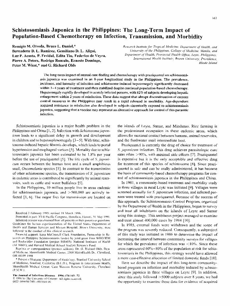
Figure 1. Prevalence and incidence of infection with Schistosomajaponicum after population-based chemotherapy in 3 villages in Leyte.Graphs summarize cumulative data on 5122 subjects followed from1981 through 1989. Incidence of infection is expressed by yearlyinterval since initiation of project in 1981. In year 5, typhoon destroyed water supply in village C (arrow). Villages A and B werescreened in years 6 and 7 (box), but treatment was deferred to determine optimal interval between community surveys and treatment invillages with low prevalence of infection.
and C, respectively (figure 1). The incidence of infection thendecreased over the next 2 years to 3% in village A, 5% in villageB, and 8% in village C and stabilized at these levels. Thesechanges paralleled the changes observed in the prevalence ofinfection for the community during this time period. Furthermore,the incidence of infection did not increase in villages A and Bwhen treatment was deferred for 2 years in years 6 and 7. However, the incidence of infection increased in village C to 18%21% despite yearly surveillance and treatment during the lastyears of the study coincident with the disruption of the pipedwater supply. Similar results were observed in the cohort ofsubjects followed for the entire study period.
76
____ Village A
Village B
__ VillageC
543
Years of the Control Program
2
Years Since the Start of the Control Program
Incidence
30
r::on.!! 20.5'0.s'"';. 10i:
'"Q)
>
Study population. A total of 5122 subjects were enrolledin the study over 8 consecutive years from November 1981through March 1989. Village populations at the beginning ofthe study were as follows: 341 in Santol (village A), 1008in Santa Rosa (village B), and 1241 in Macanip (village C).Compliance with providing stool samples for examination during the initial year of the study was 85%, 82%, and 90% invillages A, B, and C, respectively. In subsequent years, thecompliance rate remained similar or increased compared withthe first year of the study. For example, in 1989, compliancewas 92%, 90%, and 94% in villages A, B, and C, respectively.The growth rate of the study population was estimated to be4% per year, which included new births and immigrants aswell as deaths and emigration to other areas. The average fluxof villagers into and out of the study was 12% per year.
Infection with S. japonicum. The impact of annual screening and treatment of infected subjects with praziquantel isshown in figure 1. The prevalence of infection at the beginningof the study before treatment was 25%, 37%, and 44% forvillages A, B, and C, respectively [9]. Treatment of all infectedsubjects over a 3-year period was associated with a steadydecrease in the prevalence of S. japonicum infection. The prevalence of infection stabilized at 4%-6%, 7%-9%, and 11%13% in villages A, B, and C, respectively, in years 3 and 4(figure 1).
In village C, the prevalence of infection increased from 13%in year 4 to 24% in year 5 and stabilized at this level until theend of the study period in year 7, despite annual case findingand therapy (figure 1). This increase was associated with disruption of the piped water supply caused by a typhoon in year5. In contrast, when treatment was deferred for 2 years duringthe field surveys in years 6 and 7, the prevalence of infectionin villages A and B did not increase (figure I). When analysiswas restricted to the cohort of 396 subjects who participatedin the survey for all 8 years of the study, the change in theprevalence of infection was not significantly different from thatobserved for the community.
The incidence of infection determined at the first follow-upsurvey 1 year later was 12%, 9%, and 22% in villages A, B,
166 Olveda et al. JID 1996; 174 (July)
>-(ij AC'lQ)
E1.00
CiiQ.Q)
J: 0.8
-S.~
0.6-0J:
0.400
'0 0.2c:.~U«I
It 365 730 1095 1460Days Since Estimated Date of Initial Infection
>-(ij BC'lQ)
E1.00
CiiQ.Q)
J: 0.8
-S.~
0.6-0s:
0.400
'0 0.2c:0
U 0.0«I
It 0 365 730 1095 1460Days Since Treatment
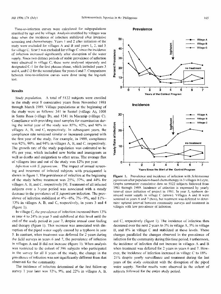
Figure 2. Changes in prevalence of hepatomegaly over time dueto Schistosoma japonicum in Leyte. A: Probability of developmentof hepatomegaly after infection with S. japonicum. All subjects inthis cohort had nonpalpable liver edges and were uninfected with S.japonicum at day O. For each subject, time of initial infection wasestimated to be midway between last negative and first positive stoolexamination. Time when hepatomegaly developed was estimated tobe midway between last normal physical examination and development of hepatic enlargement. Curve represents 1 minus Kaplan-Meierestimate of survival without developing significant hepatomegaly. B:Duration of significant hepatic enlargement following treatment ofsubjects infected with S. japonicum. All subjects had hepatomegalyat time of initial treatment with praziquantel. For all subjects, timeof reversal of hepatomegaly was estimated as midway between lastexamination with hepatomegaly and first examination without hepatomegaly. Kaplan-Meier estimates are shown.
reversal of hepatomegaly when the analysis was stratified byage or sex.
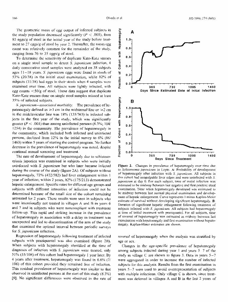
Changes in the age-specific prevalence of hepatomegalyamong subjects infected during year 1 and years 5- 7 of thestudy in village C are shown in figure 3. Data in years 5 - 7were aggregated in order to increase the number of infectedsubjects for this analysis. Results from the first examination inyears 5- 7 were used to avoid overrepresentation of subjectswith multiple infections. Only village C is shown, since treatment was deferred in villages A and B in the last 2 years of
The geometric mean of egg output of infected subjects inthe study population decreased significantly (P < .001), from85 eggs/g of stool in the initial year of the study before treatment to 27 eggs/g of stool by year 2. Thereafter, the mean eggcount was relatively constant for the remainder of the study,ranging from 26 to 35 eggs/g of stool.
To determine the sensitivity of duplicate Kato-Katz smearson a single stool sample to detect S. japonicum infection, 4daily consecutive stool samples were analyzed on 38 subjectsages 11-18 years. S. japonicum eggs were found in stools of53% (20/38) in the initial stool examination, while 82% ofsubjects (31/38) had eggs in their stools when 4 samples wereexamined over time. All subjects were lightly infected, withegg counts <50/g of stool. These data suggest that duplicateKato-Katz smears done on single stool samples missed at least35% of infected subjects.
S. japonicum - associated morbidity. The prevalence of hepatomegaly defined as ~3 em in the midsternal line or ~2 ernin the midclavicular line was 18% (135/763) in infected subjects in the first year of the study, which was significantlygreater (P < .001) than among uninfected persons (8.3%; 104/1254) in the community. The prevalence of hepatomegaly inthe community, which included both infected and uninfectedpersons, declined from 12% in the initial survey to 6% (86/1463) within 5 years of starting the control program. No furtherdecrease in the prevalence of hepatomegaly was noted, despitecontinual annual screening and treatment.
The rate of development of hepatomegaly due to schistosomiasis japonica was examined in subjects who were initiallyuninfected with S. japonicum but who later became infectedduring the course of the study (figure 2A). Of subjects withouthepatomegaly, 72% (422/582) had liver enlargement within 1year of infection; within 2 years, 82% (175/213) demonstratedhepatic enlargement. Specific rates for different age groups andsubjects with different intensities of infection could not bedetermined because of the small size of the cohort remaininguntreated for 2 years. These results were seen in subj ects whowere intentionally not treated in villages A and B in years 6and 7 and in subjects who were noncompliant with treatmentfollow-up. This rapid and striking increase in the prevalenceof hepatomegaly in association with a delay in treatment wasunexpected and led to discontinuation of the arm of the studythat examined the optimal interval between periodic surveysfor S. japonicum infection.
Regression of hepatomegaly following treatment of infectedsubjects with praziquantel was also examined (figure 2B).When subjects with hepatomegaly identified at the time ofdiagnosis of infection with S. japonicum were treated, only51% (55/108) of this cohort had hepatomegaly 1 year later. By4 years after treatment, hepatomegaly was found in 6.6% (7/106) of this cohort provided they remained free of infection.This residual prevalence of hepatomegaly was similar to thatobserved in uninfected persons at the start of this study (8.3%)[9]. No significant differences were observed in the rate of
JlD 1996; 174 (July) Schistosomiasis Japonica in thePhilippines
35
167
.1982
1111986, 1987, 1988Figure 3. Age-specific prevalence of hepatomegaly among subjects infected withSchistosoma japonicum in first year ofstudy (before use of community-based chemotherapy) and last 3 years of study (afterintensive yearly screening and chemotherapy) in villageC. No significant differences(X 2
, P < .05) were found between year 1and years 4-6.
30
25
>.
~ 20
'"Eo[ 15
'"J:
c:e10
'"Q.
5
°N= 71 100
0-9
106 128
10-14
34 63
15·19Age in Years
52 51
20-29
117 120
30·49
63 71
50+
the study . The prevalence of hepatomegaly peaked at 32%among 10- to l-t-year-clds and decreased in older subjects. Nosignificant differences were noted in the age-specific prevalences of hepatomegaly of infected subjects surveyed in yearI and the last 3 years of study . These data suggest that theprevalence of hepatic enlargement in the infected populationwas relatively unaffected by continuous screening and treatment. Thus, the impact of yearly treatment of infected subjectson the reduction in hepatomegaly in the community appearedto be almost entirely due to an overall decrease in the numberof cases of schistosomiasis.
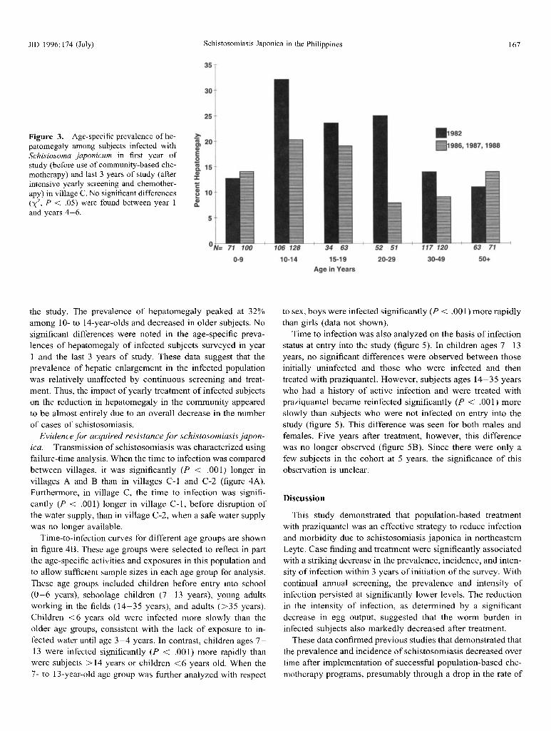
Evidence for acquired resistance for schistosomiasis japonica. Transmission of schistosomiasis was characterized usingfailure-time analysis . When the time to infection was comparedbetween villages , it was significantly (P < .001) longer invillages A and B than in villages Col and C-2 (figure 4A).Furthermore, in village C, the time to infection was significantly (P < .001) longer in village C-l, before disruption ofthe water supply, than in village C-2, when a safe water supplywas no longer available.
Time-to-infection curves for different age groups are shownin figure 4B. These age groups were selected to reflect in partthe age-specific activities and exposures in this population andto allow sufficient sample sizes in each age group for analysis.These age groups included children before entry into school(0-6 years), schoolage children (7 -13 years) , young adultsworking in the fields (14-35 years), and adults (>35 years) .Children < 6 years old were infected more slowly than theolder age groups, consistent with the lack of exposure to infected water until age 3-4 years . In contrast, children ages 713 were infected significantly (P < .00 I) more rapidly thanwere subjects> 14 years or children <6 years old. When the7- to l3-year-old age group was further analyzed with respect
to sex, boys were infected significantly (P < .00 1) more rapidlythan girls (data not shown).
Time to infection was also analyzed on the basis of infectionstatus at entry into the study (figure 5). In children ages 7-13years, no significant differences were observed between thoseinitially uninfected and those who were infected and thentreated with praziquantel. However, subjects ages 14-35 yearswho had a history of active infection and were treated withpraziquantel became reinfeeted significantly (P < .001) moreslowly than subjects who were not infected on entry into thestudy (figure 5). This difference was seen for both males andfemales. Five years after treatment, however, this differencewas no longer observed (figure 58). Since there were only afew subjects in the cohort at 5 years , the significance of thisobservation is unclear.
Discussion
This study demonstrated that population-based treatmentwith praziquantel was an effective strategy to reduce infectionand morbidity due to schistosomiasis japonica in northeasternLeyte. Case finding and treatment were significantly associatedwith a striking decrease in the prevalence, incidence, and intensity of infection within 3 years of initiation of the survey. Withcontinual annual screening, the prevalence and intensity ofinfection persisted at significantly lower levels. The reductionin the intensity of infection, as determined by a significantdecrease in egg output, suggested that the worm burden ininfected subjects also markedly decreased after treatment.
These data confirmed previous studies that demonstrated thatthe prevalence and incidence of schistosomiasis decreased overtime after implementation of successful population-based chemotherapy programs, presumably through a drop in the rate of

Figure 5. Time to infection with Schistosomajaponicum stratified byage and infection status. A: Children ages 7-13 years; B: ages 14-35years. Significant differences (P < .001) were seen in time to infectionin subjects ages 14-35 years who were infected at enrollment andtreated compared with subjects uninfected at time of enrollment. Numbers of subjects in each age group are in legend to figure 4.
the life cycle of S. japonicum in areas in which it is endemic[17]. These reservoirs consist of domestic animals, such aswater buffalo, dogs, and pigs, and wild animals [17, 18]. Treatment of these reservoirs is expensive, and the difficulty inidentifying infected animals has precluded inclusion of thisstrategy into the national control program in the Philippines.Other approaches, such as the use of public works projects andmolluscicides, are quite expensive and have been ineffectivein interrupting transmission in the Philippines [3]. Thus, for theforeseeable future, population-based chemotherapy will remainthe basic control strategy for schistosomiasis japonica in thePhilippines.
Another explanation for the limited impact on schistosomiasis japonica despite intensive chemotherapy is that Kato-Katzsmears missed significant numbers of infected persons. In thisstudy, duplicate Kato-Katz smears of single stool samples wereused to detect S. japonicum infection, since multiple stool examinations for each subject over this 8-year period would havebeen impractical and expensive. Furthermore, since the overall
3000
JID 1996;174 (July)
Uninfected -Age 7-13
Infected- Age 7-13
Uninfected- Age 14-35
Infected- Age 14-35
1000 2000Days Since Entering Study
1000 2000 3000Days Since Entering Study
c: A.2U.! 1.0-='50 0.8=~ 0.6mc:'S
0.4'iiiECDa: 0.2c:a
0.0UIVu: 0
Bl:
1.0.2t).!.s 0.8"50
= 0.6~me'c 0.4.c;ECDa: 0.2e.2t)
0.0IVu: 0
Olveda et al.
""""""'" Village A......"",'.", Village 8
Village C1
-- Village C2
-- 0-6" " 7-13
- 14-35- above 35
1000 2000 3000
Days Since Entering Study
1000 2000
Days Since Entering Study
reinfection [14-16]. It is assumed that this was due to a reduction in the number of infected humans contributing eggs intothe environment. The failure ofvillages A and B to demonstratea significant increase in the incidence of infection when treatment was deferred for 2 years suggested that this reduction isrelatively durable in villages for which the rate of new infectionwas relatively low.
Despite very high compliance by the local population andhigh cure rates with praziquantel, transmission of schistosomiasis japonica was clearly not eradicated. This is not surprisingsince, in contrast to the case with Schistosoma mansoni andSchistosoma haematobium, nonhuman reservoirs can maintain
Figure 4. Time to infection with Schistosoma japonicum as stratified by village (A) and age (B). Time to infection of 3243 subjectswas determined by failure-time analysis during period of stable prevalence and incidence of infection after annual screening and chemotherapy in villages A-C. Time to infection is expressed as curves ofinfection-free survival over time. Time to infection was significantly(P < .001) longer for villages A and B than for C-l and C-2. Inaddition, time to infection was significantly (P < .001) shorter forC-2 than for C-l. Children ages 7- 13 years were infected significantly(P < .001) more rapidly than adults> 14 years and children <7years. Initial number of subjects in each age group: 0-6 years, 870;7-13 years, 747; 14-35 years, 820; and >35 years, 806.
168
c A.2UCI) 1.0:E'0 0.8CI)CI)L-LL 0.6CI)CI)CI)C'lS 0.40
'00.2c
.2U
0.0C'lSL-
0LL
CB.2
UCI) 1.0:E'0 0.8CI)CI)
u: 0.6CI)CI)CI)C'lS 0.40
'00.2c
.2U
0.0C'lSL-
0LL
JID 1996; 174 (July) Schistosomiasis Japonica in the Philippines 169
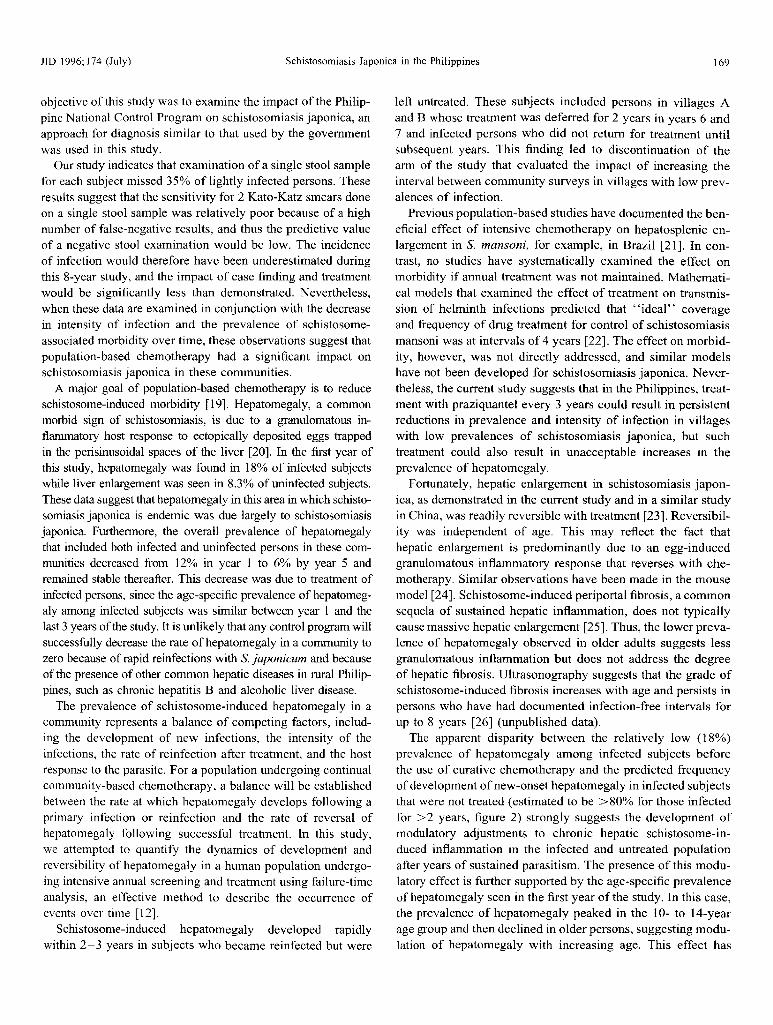
objective of this study was to examine the impact of the Philippine National Control Program on schistosomiasis japonica, anapproach for diagnosis similar to that used by the governmentwas used in this study.
Our study indicates that examination of a single stool samplefor each subject missed 35% of lightly infected persons. Theseresults suggest that the sensitivity for 2 Kato-Katz smears doneon a single stool sample was relatively poor because of a highnumber of false-negative results, and thus the predictive valueof a negative stool examination would be low. The incidenceof infection would therefore have been underestimated duringthis 8-year study, and the impact of case finding and treatmentwould be significantly less than demonstrated. Nevertheless,when these data are examined in conjunction with the decreasein intensity of infection and the prevalence of schistosomeassociated morbidity over time, these observations suggest thatpopulation-based chemotherapy had a significant impact onschistosomiasis japonica in these communities.
A major goal of population-based chemotherapy is to reduceschistosome-induced morbidity [19]. Hepatomegaly, a commonmorbid sign of schistosomiasis, is due to a granulomatous inflammatory host response to ectopically deposited eggs trappedin the perisinusoidal spaces of the liver [20]. In the first year ofthis study, hepatomegaly was found in 18% of infected subjectswhile liver enlargement was seen in 8.3% of uninfected subjects.These data suggest that hepatomegaly in this area in which schistosomiasis japonica is endemic was due largely to schistosomiasisjaponica. Furthermore, the overall prevalence of hepatomegalythat included both infected and uninfected persons in these communities decreased from 12% in year 1 to 6% by year 5 andremained stable thereafter. This decrease was due to treatment ofinfected persons, since the age-specific prevalence of hepatomegaly among infected subjects was similar between year 1 and thelast 3 years ofthe study. It is unlikely that any control program willsuccessfully decrease the rate of hepatomegaly in a community tozero because of rapid reinfections with S. japonicum and becauseof the presence of other common hepatic diseases in rural Philippines, such as chronic hepatitis B and alcoholic liver disease.
The prevalence of schistosome-induced hepatomegaly in acommunity represents a balance of competing factors, including the development of new infections, the intensity of theinfections, the rate of reinfection after treatment, and the hostresponse to the parasite. For a population undergoing continualcommunity-based chemotherapy, a balance will be establishedbetween the rate at which hepatomegaly develops following aprimary infection or reinfection and the rate of reversal ofhepatomegaly following successful treatment. In this study,we attempted to quantify the dynamics of development andreversibility of hepatomegaly in a human population undergoing intensive annual screening and treatment using failure-timeanalysis, an effective method to describe the occurrence ofevents over time [12].
Schistosome-induced hepatomegaly developed rapidlywithin 2-3 years in subjects who became reinfected but were
left untreated. These subjects included persons in villages Aand B whose treatment was deferred for 2 years in years 6 and7 and infected persons who did not return for treatment untilsubsequent years. This finding led to discontinuation of thearm of the study that evaluated the impact of increasing theinterval between community surveys in villages with low prevalences of infection.
Previous population-based studies have documented the beneficial effect of intensive chemotherapy on hepatosplenic enlargement in S. mansoni, for example, in Brazil [21]. In contrast, no studies have systematically examined the effect onmorbidity if annual treatment was not maintained. Mathematical models that examined the effect of treatment on transmission of helminth infections predicted that "ideal" coverageand frequency of drug treatment for control of schistosomiasismansoni was at intervals of 4 years [22]. The effect on morbidity, however, was not directly addressed, and similar modelshave not been developed for schistosomiasis japonica. Nevertheless, the current study suggests that in the Philippines, treatment with praziquantel every 3 years could result in persistentreductions in prevalence and intensity of infection in villageswith low prevalences of schistosomiasis japonica, but suchtreatment could also result in unacceptable increases in theprevalence of hepatomegaly.
Fortunately, hepatic enlargement in schistosomiasis japonica, as demonstrated in the current study and in a similar studyin China, was readily reversible with treatment [23]. Reversibility was independent of age. This may reflect the fact thathepatic enlargement is predominantly due to an egg-inducedgranulomatous inflammatory response that reverses with chemotherapy. Similar observations have been made in the mousemodel [24]. Schistosome-induced periportal fibrosis, a commonsequela of sustained hepatic inflammation, does not typicallycause massive hepatic enlargement [25]. Thus, the lower prevalence of hepatomegaly observed in older adults suggests lessgranulomatous inflammation but does not address the degreeof hepatic fibrosis. Ultrasonography suggests that the grade ofschistosome-induced fibrosis increases with age and persists inpersons who have had documented infection-free intervals forup to 8 years [26] (unpublished data).
The apparent disparity between the relatively low (18%)prevalence of hepatomegaly among infected subjects beforethe use of curative chemotherapy and the predicted frequencyof development ofnew-onset hepatomegaly in infected subjectsthat were not treated (estimated to be > 80% for those infectedfor >2 years, figure 2) strongly suggests the development ofmodulatory adjustments to chronic hepatic schistosome-induced inflammation in the infected and untreated populationafter years of sustained parasitism. The presence of this modulatory effect is further supported by the age-specific prevalenceof hepatomegaly seen in the first year of the study. In this case,the prevalence of hepatomegaly peaked in the 10- to 14-yearage group and then declined in older persons, suggesting modulation of hepatomegaly with increasing age. This effect has

170 Olveda et al. JIO 1996; 174 (July)
also been demonstrated in chronically infected mice and isbelieved to be immunologically mediated [24]. Such an immunologic adjustment in schistosome-induced disease would represent a beneficial adaptation for S. japonicum - infected hostsfaced with chronic parasitism [27, 28]. Furthermore, these datasuggest that modulation of schistosome-associated hepatic disease was dramatically altered following curative chemotherapy.Further studies are needed to determine the long-term clinicalimpact of new-onset hepatomegaly in infected persons.
Population-based chemotherapy is the primary approachused to control schistosomiasis japonica in rural Philippines.Our results suggest that this strategy is successful in controllingthe prevalence, incidence, and intensity of infection and, to alesser extent, morbidity due to the parasite S. japonicum. Theseresults also suggest a potential negative impact of populationbased chemotherapy. If infection cannot be eradicated fromthe Philippines and morbidity rapidly develops in reinfectedpersons within 2-3 years, intensive yearly chemotherapy mustbe continued without interruption. If annual treatment is discontinued, a rapid increase in the prevalence of hepatomegalywould occur, as demonstrated in villages A and B in 19871989. The long-term clinical impact of new-onset hepatic enlargement in infected persons is currently unknown but needsto be evaluated.
These data also allowed us to determine if there was epidemiologic evidence for the presence of acquired resistance to reinfection with S. japonicum in a human population. Resistanceto reinfection following curative chemotherapy has been demonstrated for S. mansoni and S. haematobium infections inhumans [29-31]. This is based on the observations that theintensity of reinfection after treatment with schistosomiasis isage-dependent [29-33]. Water contact has been shown to decline in older persons, but this decrease cannot account for themarked reduction in the intensity of infection and thereforesuggests development of resistance to reinfection over time.Furthermore, acquired resistance to reinfection is dependenton prior schistosome infections and therefore implies that themechanism of resistance to reinfection following parasitologiccure is immunologically based [32]. The cellular and humoralresponses for this acquired form of resistance in humans are,however, poorly defined [29].
Animal models have been helpful in the identification ofpotentially protective antigens and in the investigation of protective immune responses in experimental schistosomiasis [33].Most antigens tested so far have been identified in S. mansoni,but in recent years, similar studies for schistosomiasis japonicahave been done. For example, irradiated attenuated vaccinesinduced significant protection against S. japonicum infectionin the laboratory and in livestock in field trials in China [34,35]. Moreover, immunization with isolated antigens, such asparamyosin, induced significant immunity against infectionwith S. japonicum [36-38]. These studies are particularly important since S. japonicum is a zoonosis; immunization of bovines alone in countries in which the disease is endemic could
significantly reduce transmission of schistosomiasis japonicato humans.
In the present study, we provide evidence for the development of acquired resistance in a human population chronicallyexposed to S. japonicum in the Philippines. Our data suggestthat the risk of infection with S. japonicum was age-dependentand community-specific. Changes in the environment, such asthe destruction of the piped water supply in village C, wereassociated with a significant change in the time to reinfectionfor the community as a whole.
In all communities, the time to infection in children <6years of age was significantly longer than that observed forchildren ages 7-13. This may reflect less frequent exposure toinfected water among children, especially those <3 years old.Time to infection was also significantly more rapid in childrenages 7-13 years compared with older subjects. This suggeststhat if water exposure for both age groups was similar, thesepersons were more susceptible than older subjects to infection.This was the conclusion of a similar study of S. mansoni [29].
The most direct and compelling evidence for acquired resistance comes from the analysis of children stratified by age andinfection status. Our data demonstrated a significant decreasein the risk of reinfection with S. japonicum in older persons(14-35 years old) who were infected at the time of entry intothe study and treated compared with uninfected age-matchedpersons. This difference was clearly age-dependent, with resistance appearing to develop in subjects 14-35 years old but notin children < 14 years old. Thus, prior infection was associatedwith significant resistance to reinfection with S. japonicum.
The magnitude of this resistance to reinfection can be demonstrated by comparison of the Kaplan-Meier plots (figure 5).For example, 30% of the uninfected subjects ages 14-35 became infected within 702 days. In contrast, it took 1100 daysfor 30% of subjects of similar age to become reinfected, adelay of 1.1 years. Resistance to reinfection after treatment isclearly partial and apparently of short duration in that by 6years after entry in the study, the protection of prior infectionwas no longer evident.
These data suggest that children 7-13 years old were significantly more susceptible to infection with S. japonicum irrespective of a history of active infection. With increasing age,however, resistance to reinfection developed in subjects whowere previously infected and treated for S. japonicum comparedwith subjects never infected with this parasite. This study provides the first direct demonstration in humans of acquired resistance to reinfection conferred by active infection with thisschistosome species. It also suggests that resistance to reinfection may take as long as 10 years of chronic exposure andinfection to develop but is lost within 5 years after successfulchemotherapy.
The mechanism for the slow development of acquired resistance with age to S. japonicum is unknown. With S. mansoni,continued susceptibility to reinfection of young children is associated with high levels of "blocking" antibodies against

JID 1996; 174 (July) Schistosomiasis Japonica in the Philippines 171
carbohydrate epitopes expressed in eggs and young migratinglarvae [39]. With time, these antibodies decline with age,allowing development of a more effective immunologic response. In addition, IgE levels increase progressively with ageand significantly correlate with resistance to reinfection [40].Thus, IgE may have a protective role against S. mansoni inhumans. Similar mechanisms may also occur in S. japonicum.
Alternative explanations could explain these observations onreinfection. A decline in water contact with increasing age mayresult in a significant decrease in the rate of reinfection aftertreatment. We believe that reduced water contact is unlikely,since persons infected with S. japonicum at the time of entryinto the study would, if anything, be expected to have increasedexposure to infected water compared with uninfected subjects.Furthermore, water contact appears to be ubiquitous after theage of 4-6 years in these rice-farming communities. Studieson transmission of S. japonicum in which water contact risk ismeasured in terms of environmental endemicity and humanbehavior on an individual level are necessary to confirm theseresults and are currently ongoing.
Another explanation for these results is the differential sensitivity of Kato-Katz smears to detect S. japonicum infectionbetween the 7- to 14-year and 14- to 35-year age groups. However, there are no data from other studies to support this suggestion. Alternatively, resistance to infection may simply reflectan anatomic/mechanical blockage of migrating larvae due tohepatomegaly, as has been suggested in mice [41]. Whethersuch a process occurs in humans is unknown. However, ourdata suggest that the prevalence of hepatomegaly was greaterin the 10- to 14-year age group, who had higher levels ofinfection, than in older persons, who had reduced rates ofinfection and hepatomegaly [9]. These data make this hypothesis less likely.
In conclusion, our study suggests that the current strategyof the national schistosomiasis control program in the Philippines will dramatically decrease the prevalence, incidence, andintensity of schistosomiasis and will result in a modest reduction in morbidity. However, transmission will not be eradicated.We speculate that an unexpected result of population-basedchemotherapy will be to reduce the ability of the host to modulate egg-induced hepatic inflammation. This lack of modulationwill be ofno serious consequence to the health ofa community,provided that the control program is maintained. If the nationalcontrol program is ever stopped or dramatically curtailed, asoccurred in 1984 with the withdrawal of international funding,then a modest increase in the rate of infection and a markedrebound in short-term morbidity would be expected to occur.Thus, it is essential that international support be used to buildan infrastructure capable of maintaining intensive control efforts until newer approaches such as a vaccine in bovines orhumans become available. Our studies also suggest that acquired resistance to reinfection with S. japonicum occurs inhumans chronically exposed to this parasite in the Philippines.These epidemiologic observations in humans support experi-
mental studies in animal models that demonstrate that the successful induction of protective immunity against S. japonicuminfection is indeed possible [33 -38]. Thus, vaccine development for both bovines and humans is important in schistosomiasis japonica since, despite intensive population-based chemotherapy, S. japonicum infection cannot be eradicated in thePhilippines, so alternative approaches for control of this parasitic helminth are needed.
Acknowledgments
We gratefully acknowledge the encouragement of PrudencioOrtiz (Director, Region VII, Department of Health), Bayani BIas(Director, National Schistosomiasis Control Service), and LiliaArteche (Provincial Health Officer, Province of Leyte). We thankBerdan Aguilos, Mary Ann Lim, and Angie Nacorda for experttechnical assistance; Rosanna A. Castro for manuscript preparation; Joan H. Johnson for preparation of the graphs and analysis;and Stephen T. McGarvey and Theodore Sperdoff for critical reading. The staff of the Rural Health Unit of Macanip, Santa Rosa,and Santol played an essential role in the success of this project.
References
1. Bias BL, Velasco PF, Aliaiy OB, Erce ES, Basas JC, Bautista ES. Epidemiology and control of schistosomiasis in the Philippines: progress reportas of 1987. Mem Inst Oswaldo Cruz 1989;84:105-16.
2. Chen MA. Progress and problems in schistosomiasis control in China.Trop Med Parasitol 1989; 40: 174-6.
3. McGarvey ST, Aligui G, Daniel BL, Peters PA, Olveda R, Olds GR.Child growth and schistosomiasis japonica in northeastern Leyte, thePhilippines: cross-sectional results. Am J Trop Med Hyg 1992;46:57181.
4. McGarvey ST, Wu G, Zhang S, et al. Child growth, nutritional status, andschistosomiasis japonica in Jiangxi, People's Republic of China. Am JTrop Med Hyg 1993;48:571-81.
5. Gang CM. Schistosoma japonicum and S. japonicum-like infections: epidemiology, clinical, and pathological aspects. In: Jordan P, Webbe G,Sturrock RF, eds. Human schistosomiasis. Cambridge: CAB International, 1993:237-63.
6. Santos AT. The present status of schistosomiasis in the Philippines. Southeast Asian J Trop Med Public Health 1984; 15:439-45.
7. Pearson RP, Guerrant RC. Praziquantel: a major advance in anti-helminthictherapy. Ann Intern Med 1983;99:195-8.
8. Chen MG, Mott KE. Progress in assessment of morbidity due to schistosomiasis japonica infection: a review of recent literature. Trop Dis Bull1988; 85:RI-44.
9. Olveda RM, Tiu E, Fevidal P, De Veyra F, Icatlo FC, Domingo EO.Relationship of prevalence and intensity of infection to morbidity inschistosomiasis japonica: a study of three communities in Leyte, Philippines. Am J Trop Med Hyg 1983;32:1312-21.
10. Schistosomiasis Control Council. Annual report for 1984. Manila, Philippines: Department of Health, 1985: 1-126.
II. Peters PAS, Kazura JW. Update on diagnostic methods for schistosomiasis.Baillieres Clin Trap Med Commun Dis 1987;2:419-33.
12. Lee ET. Statistical methods for survival analysis. Belmont, CA: LifetimeLearning Publications, 1980.
13. Kaplan FL, Meier P. Non-parametric estimation from incomplete observations. J Am Stat Assoc 1958;53:457-65.
14. Jordan P. Schistosomiasis-research to control. Am J Trap Med Hyg1977;26:877-86.
172 Olveda et al. lID 1996; 174 (July)
15. Strickland GT. Schistosomiasis: eradication or control. Rev Infect Dis1982; 4: 1951-4.
16. World Health Organization. New strategy on schistosomiasis. SoutheastAsian J Trop Med Public Health 1984; 15:469~ 70.
17. Pesigan TP, Farooq M, Hairston NG, et al. Studies on schistosomiasisjaponicum infection in the Philippines. 1. General considerations andepidemiology. Bull WHO 1958; 18:345-55.
18. Santos AT. Current chemotherapy of schistosomiasis japonica in the Philippines. Southeast Asian J Trop Med Public Health 1976; 7:306-9.
19. Wilkins HA. Are measurements of intensity of infection or morbiditynecessary to evaluate schistosomiasis control within PHC? Trop MedHyg 1986; 37:223-5.
20. Boros DL. Immunopathology of Schistosoma mansoni infection. Clin Microbiol 1989;2:250-69.
21. Sleigh AC, Mort KE, HoffR, Maguire JH, Da Franca Silva JT. Manson'sschistosomiasis in Brazil: l l-year evaluation of successful disease control with oxamniquine. Lancet 1986; 1:635-7.
22. Warren KS, Bundy DAP, Anderson RM. Chapter 8. Tn: Jamison OJ, MoslyWH, eds. Helminth infections. Washington, DC: World Bank, Population Health and Nutrition Division, 1991.
23. Wiest PM, Wu G, Zhong S, et al. Impact of annual screening and chemotherapy with praziquantel on schistosomiasis japonica on Jishan Island,People's Republic of China. Am J Trop Med Hyg 1994:51:162-9.
24. Olds GR, Olveda R, Tracy JW, Mahmoud AAF. Adoptive transfer ofmodulation ofgranuloma formation and hepatosplenic disease in murineschistosomiasis japonica by serum from chronically infected animals. JImmunol 1982; 128:1393-7.
25. Sulit YSM, Domingo EO, Dalmacio-Cruz AE, De Peralta OS, ImperialES. Parasitic cirrhosis among Filipinos. Philippine Med Assoc J 1964;40:1021-8.
26. Wiest PM, Wu G, Zhang S, et a1. Schistosomiasis japonica on JishanIsland, Jiangxi Province, People's Republic of China: persistence ofhepatic fibrosis after reduction of the prevalence of infection with age.Trans R Soc Trop Med Hyg 1993; 87:290-4.
27. Colley DG. Immune responses and immunoregulation in experimental andclinical schistosomiasis. In: Mansfield JM, ed. Parasitic diseases. VolI. New York: Marcel-Dekker, 1981:1-83.
28. Phillips SM, Fox EG. The immunopathology of parasitic disease. ClinImmunol Allergy 1982; 2:667 - 703.
29. Butterworth AE, Corbett EL, Dunne DW, et al. Immunity and morbidityin human schistosomiasis. In: McAdams KPW1, ed. New strategies inparasitology. Edinburgh, UK: Churchill Livingstone, 1989:193-210.
30. Wilkins HA, Blumenthal UJ, Hagan P, Hayes RJ, Tulloch S. Resistanceto reinfection after treatment of urinary schistosomiasis. Trans R SocTrop Med 1987;81:29-35.
31. Butterworth AE. Immunology of schistosomiasis. In: Jordan P, WebberG, Sturrock RF, eds. Human schistosomiasis. Cambridge: CAB International, 1993:331-67.
32. Sturrock RF, Bensted Smith R, Butterworth AE, et al. Immunity aftertreatment of human schistosomiasis. Ill. Long-term effects of treatmentand retreatment. Trans R Soc Trop Med Hyg 1987;81:303-14.
33. Bergquist NR, Hall BF, lames S. Schistosomiasis vaccine development.Translating basic research into practical results. Immunologist 1994; 2:131-4.
34. Xu S, Shi F, Shen W, et al. Vaccination of bovines against schistosomiasisjaponica with cryopreserved-irradiated and freeze-thaw schistosomula.Vet Parasitol 1993;47:37-50.
35. Maloney NA, Bickle QD, Webbe G. The induction of specific immunityagainst Schistosomajaponicum by exposure ofmice to ultraviolet-attenuated cercariae. Parasitology 1985; 90:313-23.
36. Taylor MG. Schistosomiasis vaccines: farewell to the God of plague? 1Trop Med Hyg 1994; 97:257 -68.
37. Bashir M, Bickle Q, Bushara H, et al. Evaluation of defined antigenvaccines against Schistosoma bovis and S. japonicum in bovines. TropGeogr Med 1994;46:255-8.
38. Ramirez BL, Kurtis lD, Wiest PM, et al. Paramyosin: a candidate vaccineantigen against Schistosoma japonicum. Parasite Immunol 1996; 18:4952.
39. Butterworth AE, Fulford AJC, Dunne DW, Ouma JH, Sturrock RF. Longitudinal studies on human schistosomiasis. Philos Trans R Soc Lond BBioI Sci 1988;321:495-511.
40. Hagan P, Blumenthal UJ, Dunne 0, Simpson AJG, Wilkins HA. HumanTgE,IgG4, and resistance to reinfection with Schistosoma haematobium.
Nature 1991;349:243-5.41. Wilson RA, Coulson PS, McHugh SM. A significant part of the concomi
tant immunity of mice to Schistosoma mansoni is a consequence of aleaky hepatic portal system, not immune killing. Parasite Immunol1983;5:595-601.