scapular and humeral movement patterns of people with
TRANSCRIPT

Washington University School of MedicineDigital Commons@Becker
Physical Therapy Faculty Publications Program in Physical Therapy
3-2011
Scapular and humeral movement patterns ofpeople with stroke during range-of-motionexercisesDustin D. HardwickWashington University School of Medicine in St. Louis
Catherine E. LangWashington University School of Medicine in St. Louis
Follow this and additional works at: http://digitalcommons.wustl.edu/pt_facpubs
This Article is brought to you for free and open access by the Program in Physical Therapy at Digital Commons@Becker. It has been accepted forinclusion in Physical Therapy Faculty Publications by an authorized administrator of Digital Commons@Becker. For more information, please [email protected].
Recommended CitationHardwick, Dustin D. and Lang, Catherine E., "Scapular and humeral movement patterns of people with stroke during range-of-motionexercises" (2011). Physical Therapy Faculty Publications. Paper 20.http://digitalcommons.wustl.edu/pt_facpubs/20

1
Scapular and humeral movement patterns of people with stroke during range of motion exercises
Dustin D. Hardwick PT, PhD1, Catherine E. Lang PT, PhD1,2,3
1Program in Physical Therapy, 2Program in Occupational Therapy, 3Department of Neurology, Washington University School of Medicine, St. Louis, MO This work was supported by an American Heart Association Predoctoral Fellowship Award 0810101Z, Foundation for Physical Therapy PODS I, NIH HD047669, NIH HD007434. We thank Justin Beebe PT, PhD and Stacey DeJong PT for their assistance with data collection. We certify that no party having a direct interest in the results of the research supporting this article has or will confer a benefit on us or on any organization with which we are associated AND, if applicable, we certify that all financial and A portion of this work was presented at the following scientific meetings:
APTA Section on Research Retreat (August 2009) APTA Combined Sections Meeting (Febuary 2009)
Word Count: 3949
Corresponding Author:
Catherine E. Lang, PT, PhD Washington University in St. Louis - School of Medicine Program in Physical Therapy Campus Box 8502 4444 Forest Park Blvd. St Louis, MO 63108-2212 PH: 314-286-1945 FAX: 314-286-1410 Email address: [email protected]

2
Background and Purpose: Range-of-motion (ROM) exercises may contribute to hemiparetic
shoulder pain, but the mechanisms behind this are unknown. This study examined scapular and
humeral movement patterns in people with hemiparesis post stroke as they performed commonly
prescribed ROM exercises.
Methods: Using kinematic techniques, we studied 13 people with hemiparesis, both with and
without pain, as they performed three commonly prescribed ROM exercises: person-assisted
ROM, self-assisted ROM, and cane-assisted ROM. Their data were compared to 12 matched
controls performing scapular plane shoulder elevation using mixed model ANOVAs.
Correlation analyses were used to examine relationships between subjects’ ratings of pain and
kinematic data.
Results: The hemiparetic group had mild pain at rest that increased during the performance of
the exercises. Humeral external rotation in the hemiparetic group was decreased in all three
ROM exercises compared to shoulder elevation in the control group. Scapular upward rotation in
the hemiparetic group was decreased for the person-assisted ROM exercise only. No differences
in scapular tilt were found between groups. The extent of movement abnormalities was not
related to pain severity.
Discussion and Conclusions: People with hemiparesis had altered scapular and humeral
movement patterns and increased shoulder pain when performing the ROM exercises. These
data can assist clinicians in making decisions regarding which exercises to prescribe to preserve
shoulder motion and prevent contractures in this population.
3
Background and Purpose
Hemiparesis or hemiplegia, i.e. the loss of some or all voluntary muscle activation on one
side of the body, is a common impairment following stroke. The reduced ability to move leads to
prolonged periods of time spent immobile.1-3 A major concern for rehabilitation clinicians is the
time spent with the upper extremity resting in the lap; shoulder and arm muscles, particularly
shoulder internal rotators and extenders, and elbow flexors, are held in shortened positions,
potentially leading to loss of available motion and contractures. 4 To address this concern, people
with hemiparesis or hemiplegia are often prescribed range-of-motion (ROM) exercises. Data
supporting the effectiveness of ROM and stretching exercise in preventing loss of motion and
contractures after stroke are inconclusive.4-7
A related concern for rehabilitation clinicians is whether or not performing ROM
exercises contributes to hemiparetic shoulder pain.8,9 Hemiparetic shoulder pain is a disabling
condition with many possible etiologies 10-12 , affecting up to 72% of patients with
hemiparesis.11,13,14 ROM exercises could be one contributing factor to shoulder pain secondary
to altered scapular and humeral movement patterns. Precise scapulohumeral coupling is needed
to preserve the suprahumeral space and prevent impingement of the rotator cuff tendons. Proper
coupling includes upward rotation and posterior tilting of the scapula 15-18 and external rotation
of the humerus.15,16 Reduced voluntary neural drive from the stroke may disrupt the timing and
activation of scapulothoracic and rotator cuff muscles.19 As the arm is moved during an
exercise, the exercise may push the humerus into elevation angles higher than the subject can
actively produce without assistance. As a clinician, one needs to be concerned about prescribing
ROM exercise to preserve movement and avoid contractures while simultaneously avoiding
exercises that could contribute to the development or persistence of hemiparetic shoulder pain.
4
The purpose of this study was to examine the scapular and humeral movement patterns in
people with hemiparesis post stroke performing commonly prescribed ROM exercises: person-
assisted ROM, self-assisted ROM, and cane-assisted ROM. Their scapular and humeral
movement patterns during the exercises were compared to a group of neurologically intact
healthy controls performing scapular plane shoulder elevation, which was our best proxy for
normal shoulder motion. We hypothesized that people with hemiparesis would have abnormal
scapular and humeral movement patterns when performing the selected exercises. Additionally,
we hypothesized that the extent of movement abnormality would be related to the severity of
reported pain during that movement. A better understanding of the scapular and humeral
movement patterns associated with commonly prescribed ROM exercises may help clinicians
identify which exercises are to be avoided and how exercises may be modified to better replicate
the scapular and humeral movement patterns of normal shoulder motion.
Methods
Participants
This was a pilot sample of convenience. Thirteen subjects with hemiparesis were
recruited from a local rehabilitation hospital. Subjects with hemiparesis were included if they 1)
had a diagnosis of stroke, 2) onset of unilateral upper extremity weakness following stroke.
Subjects were excluded if they 1) had a history of shoulder pain and pathology prior to stroke, 2)
were unable to follow 2-step commands, 3) showed signs of hemi-neglect, 4) showed symptoms
consistent with referral from cervical or thoracic spine, 5) had any serious medical complications
that would prevent them from participating, and/or 6) were unable to provide informed consent.

5
Twelve healthy subjects were recruited from the community. The age and gender
composition of the control group was selected to match the age and gender composition of the
hemiparetic group. Control subjects were excluded if they 1) had a history of stroke, 2) history or
current complaints of shoulder pain or history of diagnosed shoulder pathology, 3) if they had
any serious medical conditions that would prevent them from participating, and/or 4) if they
failed to provide informed consent. The study was approved by the Washington University
Human Research Protection Office prior to recruitment and testing. All subjects signed informed
consent documents prior to participating.
Kinematic Measurements
Computer-based kinematic techniques were used to quantify movement of the
contralesional, more-involved shoulder, arm, and thorax.20 Three-dimensional movements of the
upper extremity were captured using an electromagnetic tracking system (Motion Monitor built
around Flock of Birds, Innovative Sports Training Inc., Chicago IL). Four sensors were attached
to the 1) trunk: mid-sternum, 2) the arm: proximal to the lateral epicondyle, bisecting the arm
mass, 3) the forearm: proximal to the mid-point between the radial and ulnar styloids on the
dorsum of the forearm, and 4) the scapula: distal flat aspect of the acromion (Figure 1).21 The
forearm sensor was initially included to monitor if subjects were moving with a flexed elbow;
since this did not occur, elbow sensor data are not included in this report. All sensors and
trailing wires were taped down and secured with Coban (3M, St. Paul MN) to prevent slippage
and arbitrary sensor movement. The hardware manufacturer reports a root mean square accuracy
of 0.5º for orientation and 1.8 mm for position for the sensors used. With arms relaxed, bony
landmarks on the thorax, scapula, and humerus were digitized with a custom probe to permit

6
transformation of sensor data into local segment coordinates using the accepted order of
internal/external rotation, upward/downward rotation, and posterior/anterior tilting, according to
the protocol recommended by the International Society of Biomechanics, Shoulder Group.20
Glenohumeral joint center was estimated using a least squares algorithm to find the point on the
humerus that moved least in respect to the scapula as it was moved through short arcs.
<<Insert Figure 1>>
Kinematic data were low-pass filtered at 6 Hz using a second-order Butterworth filter.
Motion Monitor software was used to calculate and extract segmental position and angle data
from the sensor data using standard rigid body methodology.20 The scapulothoracic angular data
extracted were scapular upward rotation and scapular tilt, and the glenohumeral angular data
were humeral elevation and humeral external rotation (Figure 2). Scapular internal/external
rotation data were also extracted but are not included in this report due to lack of consensus what
constitutes normal scapular internal and external rotation during humeral elevation with some
studies reporting scapular external rotation as the arm is elevated18,22,23 and some studies
demonstrating scapular internal rotation as the arm is elevated.24-26 Anatomical variations in the
shape and size of the thorax and ribs could also impact the relative internal and external rotation
of the scapula as it slides along the thorax. The plane of elevation for humeral elevation
depended on the exercise, but generally this elevation occurred between the sagital plane and the
scapular plane (approximately 30º anterior to the frontal plane by visual estimation). Scapular
upward rotation was rotation of the scapula in frontal plane about an anterior-posterior axis in
which the inferior angle moves laterally. Scapular posterior tilt was rotation of the scapula in the
sagital plane about a lateral axis in which the superior border of the scapula moves posteriorly.
Humeral external rotation was the spinning of the humerus on the glenoid laterally. All angular
7
data were calculated according to the recommended protocol.20 For ease of communication,
increasing the data for humeral external rotation and scapular upward rotation were multiplied by
-1. Custom-written software in MATLAB (The Mathworks Inc., Natick MA) was used for
subsequent analysis to find the above angles at the start of movement and at, 30º, 60º, 90º, and
120º of humeral elevation.
<<Insert Figure 2>>
Protocol
Testing began with subjects seated in a wooden chair with the upper limb dependent.
Care was taken to ensure that the tested upper extremity and scapula did not contact or were
otherwise obstructed by the chair. Subjects performed 3 trials of each exercise at a self-selected
pace and were given rest breaks as needed. All subjects were able to perform the exercise as
instructed, although some required several practice trials before movements were recorded.
Controls were tested using the same self-selected speed protocol as subjects. One examiner did
all the testing and digitizing.
The 3 commonly prescribed ROM exercises were: person-assisted ROM, self-assisted
ROM, and cane-assisted ROM. (Figure 3) They were all performed as active-assisted ROM, in
that the subject used their more-involved extremity as much as possible, and the assistance
provided further ROM beyond what they could do unassisted. Person-assisted ROM (Figure 3A)
was performed by a single tester. Assistance was given by another person under the middle
portion of the arm and under the mid-forearm as the subject performed humeral elevation. 7
Self-assisted ROM (Figure 3B) was performed with the subject supporting the elbow of the
more-involved extremity with the less-involved extremity as he or she performed humeral

8
elevation. 6 Person-assisted and self-assisted ROM occurred near the plane of flexion. Cane-
assisted ROM (Figure 3C) was performed using a plastic pipe that approximated the diameter
and length of a standard cane. The subject gripped the cane with an overhand grip with hands
slightly wider than shoulder width apart. They performed bilateral shoulder elevation, providing
assistance from the less-involved extremity through the cane to assist the more-involved
extremity. Cane-assisted ROM occurred in the scapular plane. As done clinically, the examiner
provided assistance with grasping the cane if needed. Once grasped, all subjects could produce
at least minimal forces to grip the cane.
<<Insert Figure 3>>
All exercises were compared to controls performing scapular plane shoulder elevation
because it represents the best proxy for normal scapular and humeral motion and it is often used
to examine shoulder motion in healthy controls and patient populations.18,21,23,27,28 We did not
compare scapular and humeral movements of the more-involved shoulder to the less-involved
shoulder because the less-involved shoulder has been found to have kinematic alterations 29 and
because the less-involved shoulder was assisting with 2 of 3 exercises. Comparisons were also
not made to controls performing the exercises because that would be a contrived situation, i.e.
people with healthy shoulders would not perform these exercises.
Clinical Measures
Shoulder pain at rest and during movement trials was recorded using a numeric pain
rating scale (0-10 points). Subjects rated their pain prior to testing and after each trial. This scale
has been shown to be a reliable and sensitive pain scale for use in older populations.30 It has
good reliability in subjects with orthopedic shoulder conditions 31 as well as subjects with
9
hemiparesis.32 The Stroke Impact Scale, Hand Function subscale was used to capture upper
extremity functional deficits in the sample.33 This reliable, valid, and quick measure agrees well
with the more time-consuming Fugl-Meyer Upper Extremity Motor subscale.34 Muscle tone at
the elbow and shoulder was assessed using the Modified Ashworth Scale.35
Data analysis
Statistica (StatSoft Inc., Tulsa OK) was used for statistical analyses and the criterion for
statistical significance was set at p < 0.05. A repeated measures ANOVA and post hoc t tests
were was used to compare pain at rest (prior to performing any movement) and pain during each
exercise, quantified by the average of numeric pain rating given during the 3 trials. Mixed-
model, repeated measures ANOVAs were used to test for significant differences in humeral
external rotation, scapular upward rotation, and scapular tilt between the hemiparetic group
performing each exercise and control group performing scapular plane shoulder elevation at start
of movement, 30º, 60º and 90º of humeral elevation. Averages of the three trials for each subject
were entered into the ANOVAs. Because we used a single control condition (control group
scapular plane shoulder elevation), we ran separate ANOVAs for each exercise vs. the control
condition. Post hoc comparisons using Fishers Least Significant Difference were used when
significant main or interaction effects were found. Protected t-tests with a more stringent
criterion of p < 0.01 were used to assess differences at 120º since many hemiparetic subjects did
not achieve these angles. This analysis strategy permitted the inclusion of all subjects in the
ANOVAs yet still examined the higher humeral elevation angles.
Since some of our subjects had shoulder pain and others did not, we used Spearman Rho
correlations to test if severity of pain during performance of each specific exercise was related to

10
scapular and humeral movement at various humeral angles during that same exercise. This would
provide an indication as to how pain might have influenced the recorded movements.
Results
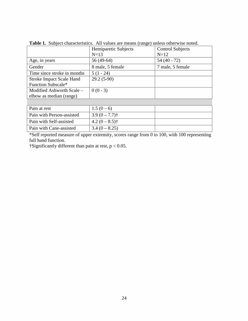
Characteristics of the 13 subjects with hemiparesis and 12 controls are provided in the
Table. Time since stroke for the hemiparetic subjects was variable, ranging from 1 month to 2
years. As expected, upper extremity function was decreased and average spasticity levels were
mild, as indicated by the Hand Function subscale of the Stroke Impact Scale and the Modified
Ashworth Scale, respectively.
<<Insert Table 1>>
Pain
Five hemiparetic subjects reported pain in their involved shoulder prior to testing. Of the
eight hemiparetic subjects who did not report pain prior to testing, four experienced some
shoulder pain during various exercises. On average, the hemiparetic group reported mild pain at
rest which increased during performance of the exercises (bottom of Table). Pain was increased
during the performance of the exercises compared to rest (within subjects main effect, F3,36 =
4.01, p = 0.015). Post hoc t-tests indicated that pain during the performance of person-assisted
ROM and self-assisted ROM were greater than pain at rest (p = 0.03, p = 0.02 respectively), and
pain during the performance of cane-assisted ROM showed a trend towards greater pain than
pain at rest, but did not reach significance (p = 0.08).
Scapular and humeral movement during the 3 exercises

11
Scapular and humeral movement data from the 3 exercises in the hemiparetic group and
from scapular plane shoulder elevation in the control group are shown in Figure 4. Here we
report the relevant main effects of group and group by angle interactions as they pertain to our
hypotheses. For the post hoc testing of group by angle interactions, we indicate the comparisons
where significant differences were not found. As expected, there were main effects of angle for
each exercise across the examined motions (p values < 0.05).
In the person-assisted ROM exercise (Figure 4, top row), the hemiparetic group had
decreased humeral external rotation (main effect of group, F1,23 = 14.2, p < 0.001; group x angle
interaction, F3,72 = 10.2, p < 0.001; post hoc testing yielded no significant difference at 0º, p =
0.30) and decreased scapular upward rotation (main effect of group, F1,23 = 4.4, p < 0.05; group x
angle interaction, F3,72 = 4.5, p < 0.006; post-hoc testing yielded no significant difference at 0º, p
= 0.70) compared to controls performing scapular plane shoulder elevation. Protected t-tests at
120º demonstrated decreased humeral external rotation (p < 0.01), but no difference in scapular
upward rotation (p = 0.42) in the hemiparetic group compared to controls. Scapular tilt was not
different between groups (main effect of group, F1,23 = 1.1, p = 0.32; at 120º protected t-test, p =
0.44).
In the self-assisted ROM exercise (Figure 4, middle row), the hemiparetic group had
decreased humeral external rotation (main effect of group, F1,23 = 29.4, p < 0.001; group x angle
interaction, F3,72 = 19.3, p < 0.001; post hoc testing yielded no significant difference at 0º, p =
0.86) compared to controls performing scapular plane shoulder elevation. Protected t-tests at
120º demonstrated decreased humeral external rotation (p < 0.001) in the hemiparetic group
compared to controls. Scapular upward rotation was not different between groups (main effect of
group, F1,23 = 1.9, p = 0.18), but showed a group x angle interaction (F3,72 = 5.1, p < .003; post

12
hoc testing yielded no significant difference at 0º, p = 0.90). Scapular upward rotation was not
different at 120º (protected t-test, p = 0.57). Scapular tilt was not different between groups (main
effect of group, F1,23 = 0.5, p = 0.49; at 120º protected t-test, p = 0.75).
In the cane-assisted ROM exercise (Figure 4, bottom row) the hemiparetic group had
decreased humeral external rotation (main effect of group, F1,23 = 15.5, p < 0.001; group x angle
interaction, F3,72 = 15.9, p < 0.001; post hoc testing yielded no significant difference at 0º, p =
0.31) compared to controls performing scapular plane shoulder elevation. Protected t-tests at
120º demonstrated decreased humeral external rotation (p < 0.001) in the hemiparetic group
compared to controls. No differences between groups were found for scapular upward rotation
(main effect of group, F1,23 < 0.01, p = 0.95; at 120° protected t-test p = 0.65) or scapular tilt
(main effect of group, F1,23 = 1.6, p = 0.22; at 120º protected t-test p = 0.73).
<<Insert Figure 4>>
Relationships between pain and movement
No relationships were found between reported pain during the performance of each
exercise and the scapular and humeral movement data. Spearman rho values ranged from -0.46
to + 0.36 (all p values > 0.05).
Discussion
The hemiparetic group had altered movement patterns during performance of the ROM
exercises compared to our proxy of normal shoulder motion. On average, the hemiparetic group
had mild pain at rest which increased during the performance of the exercises. Severity of pain
was not associated with scapular or humeral movement patterns during the exercises.

13
Our primary hypothesis was supported: people with hemiparesis had abnormal scapular
and humeral movement patterns when performing the tested exercises. The performance of
stretching and ROM exercises have been previously associated with shoulder pain in people with
hemiparesis.8,9 Our data build on these reports by describing abnormal scapular and humeral
movements that occurred during the performance of shoulder ROM exercises. Data from the
present study provide a biomechanical mechanism for how performing these exercises may
contribute to the development of shoulder pain post stroke.
The most salient finding during the performance of all three exercises was the decrease in
humeral external rotation. The lack of dynamic humeral external rotation found here is
compatible with literature showing an association between reduced passive humeral external
rotation and hemiparetic shoulder pain.32,36-38 Conditions that decrease humeral external rotation
increase rotator cuff compression particularly against the greater tuberosity; the compression
increases as the humerus is elevated.39-41 We speculate that performing these ROM exercises as
described could contribute to, or exacerbate, hemiparetic shoulder pain by repeatedly
compressing the rotator cuff tendons.
It is worth noting that the etiology and contributing factors of shoulder pain following are
multifactorial and poorly understood.11,12,36,42 43,44 It is likely that more than one factor plays is
responsible. These factors may overlap extensively and no single factor may be responsible for
pain in individual patients. These factors include shoulder subluxation, reflex sympathetic
dystrophy, and adhesive capsulitis. The resultant disruptions in movement patterns, regardless of
diagnosis, can lead to strain and tearing of rotator cuff muscles as well as impingement of the
rotator cuff tendons. It appears that performing ROM exercises as described may be promoting
14
these abnormal movement patterns and thus should be modified or avoided in this population
regardless of diagnosis.
Our secondary hypothesis was not supported: the extent of movement abnormalities were
not related to the extent of pain during the selected exercise. There are three possible
explanations for this. First, it is possible that severity of pain is not related to the extent of
movement abnormalities as seen in this sample. This possibility is consistent with the
understanding that feelings of pain are influenced by many factors.45-47 The extent of scapular
and humeral movement abnormalities might therefore be only one of many contributing factors.
Alternatively, it is possible that the relationship between pain and extent of movement
abnormalities is affected by time, i.e. performing many repetitions of these exercises over a long
period would create an association between pain severity and abnormal movement. In this
alternative scenario, rotator cuff compression incurred while performing these exercises would
accumulate. The eventual result might be microtrauma and pain, which in turn could lead to
more abnormal movement patterns.48 Since we did not investigate other factors that may have
contributed to the reported pain and we only tested three repetitions of each exercise, our data do
not permit us to distinguish between these possibilities. A third possibility for the lack of
relationship is the small sample size of this study (see suggestions for future studies under
Limitations below).
Clinical considerations for when prescribing specific exercises post stroke
Of the three exercises evaluated, person-assisted ROM of the hemiparetic shoulder had
the most differences in scapular and humeral motion compared to active ROM of the normal
shoulder. These differences, decreased humeral external rotation and scapular upward rotation,

15
may be attributed to the fact that the scapula and humerus were not monitored or controlled
during performance of this exercise. A skilled therapist performing this same exercise may be
much more likely to monitor and control these motions. It is often the case however, that a
therapist provides the initial instruction, and then this exercise is performed repeatedly with
assistance from a non-skilled caregiver. We sought to replicate this common method of
performance. The results of this study therefore highlight the importance of education to
caregivers who may be performing this person-assisted ROM exercise on people with
hemiparesis. Specific education on how to externally rotate the humerus and manually assist the
scapula into upward rotation may be needed to perform this exercise with more normalized
shoulder motions.
The self-assisted ROM exercise resulted in decreased humeral external rotation compared
to normal shoulder motion. Using the less-involved upper extremity to assist their more-involved
upper extremity naturally puts both arms into horizontal adduction and internal rotation; this is
particularly true for larger individuals with wide trunks. Based on these mechanical constraints,
therapists may want to avoid the self-assisted ROM exercise when considering options to
preserve movement and prevent contractures in people with hemiparesis post stroke.
The cane-assisted ROM exercise also resulted in decreased humeral external rotation
compared to normal shoulder motion. An overhand grip was used to grip the cane in the present
study. The overhand grip placed the forearm in pronation and likely contributed to a less
externally rotated humerus. One way to modify this exercise would be to switch to an underhand
grip. The underhand grip would position the forearm in supination and may help to promote
humeral external rotation. A challenge to making this modification is that people with stroke
might have more trouble maintaining an underhand grip than an overhand grip with the paretic

16
hand. This could be addressed with a strap or other individualized modification. We speculate
that, if modified to employ an underhand grip, the cane-assisted ROM exercise may be an
acceptable choice for preserving shoulder movement and preventing contractures in people with
hemiparesis post stroke. It should be noted however, that the clinical premise that contractures
can be prevent through ROM exercises is not fully supported by data at this time.49
Limitations
Three main limitations should be taken into account when interpreting the results of this
study. First, the sample size was small, limiting the ability to detect differences between groups
and relationships to pain and the ability to generalize our findings. Second, we studied only
three ROM exercises, each performed according to specific instructions. Other exercises and
their variations may have different effects on the movement patterns of the humerus and scapula.
Finally, our sample included people with hemiparesis both with and without shoulder pain.
While people with and without pain are prescribed ROM exercises during their rehabilitation,
grouping them together could have masked unique findings in one subgroup or the other. Future
longitudinal studies on this topic with larger sample sizes, more variations of exercises, and
grouping of subjects into subpopulations with respect to pain would greatly improve therapist
decision-making when choosing exercises.
Conclusions
Reduced humeral external rotation was the most common movement abnormality
observed during the performance of three commonly-prescribed shoulder ROM exercises by
people with hemiparesis post stroke. Our data can assist clinicians in making decisions

17
regarding which exercises to prescribe to preserve shoulder motion and prevent contractures in
this population.

18
References 1. Bernhardt J, Chitravas N, Meslo IL, Thrift AG, Indredavik B. Not all stroke units are the
same: a comparison of physical activity patterns in Melbourne, Australia, and Trondheim,
Norway. Stroke. Jul 2008;39(7):2059-2065.
2. Bernhardt J, Dewey H, Thrift A, Donnan G. Inactive and alone: physical activity within
the first 14 days of acute stroke unit care. Stroke. Apr 2004;35(4):1005-1009.
3. Lang CE, Wagner JM, Edwards DF, Dromerick AW. Upper Extremity Use in People
with Hemiparesis in the First Few Weeks After Stroke. J Neurol Phys Ther. Jun
2007;31(2):56-63.
4. Ada L, Goddard E, McCully J, Stavrinos T, Bampton J. Thirty minutes of positioning
reduces the development of shoulder external rotation contracture after stroke: A
randomized controlled trial. Arch Phys Med Rehabil. Feb 2005;86(2):230-234.
5. de Jong LD, Nieuwboer A, Aufdemkampe G. Contracture preventive positioning of the
hemiplegic arm in subacute stroke patients: a pilot randomized controlled trial. Clin
Rehabil. Aug 2006;20(8):656-667.
6. Lynch D, Ferraro M, Krol J, Trudell CM, Christos P, Volpe BT. Continuous passive
motion improves shoulder joint integrity following stroke. Clin Rehabil. Sep
2005;19(6):594-599.
7. Tseng CN, Chen CC, Wu SC, Lin LC. Effects of a range-of-motion exercise programme.
J Adv Nurs. Jan 2007;57(2):181-191.
8. Gustafsson L, McKenna K. A programme of static positional stretches does not reduce
hemiplegic shoulder pain or maintain shoulder range of motion--a randomized controlled
trial. Clin Rehabil. Apr 2006;20(4):277-286.

19
9. Kumar R, Metter EJ, Mehta AJ, Chew T. Shoulder pain in hemiplegia. The role of
exercise. Am J Phys Med Rehabil. Aug 1990;69(4):205-208.
10. Roy CW, Sands MR, Hill LD. Shoulder pain in acutely admitted hemiplegics. Clinical
Rehabilitation. 1994;8:334-340.
11. Wanklyn P, Forster A, Young J. Hemiplegic shoulder pain (HSP): natural history and
investigation of associated features. Disabil Rehabil. Oct 1996;18(10):497-501.
12. Roy CW, Sands MR, Hill LD, Harrison A, Marshall S. The effect of shoulder pain on
outcome of acute hemiplegia. Clinical Rehabilitation. 1995;9:21-27.
13. Joynt RL. The source of shoulder pain in hemiplegia. Arch Phys Med Rehabil. May
1992;73(5):409-413.
14. Van Ouwenaller C, Laplace PM, Chantraine A. Painful shoulder in hemiplegia. Arch
Phys Med Rehabil. Jan 1986;67(1):23-26.
15. Levangie PK, Norkin CC. Joint structure and function : a comprehensive analysis. 4th
ed. Philadelphia, PA: F.A. Davis Co.; 2005.
16. Poppen NK, Walker, P. S. Normal and abnormal motion of the shouler. Journal of Bone
and Joint Surgery. 1976;58(2):195-201.
17. Braman JP, Engel SC, Laprade RF, Ludewig PM. In vivo assessment of scapulohumeral
rhythm during unconstrained overhead reaching in asymptomatic subjects. J Shoulder
Elbow Surg. Apr 21 2009.
18. McClure PW, Michener LA, Sennett BJ, Karduna AR. Direct 3-dimensional
measurement of scapular kinematics during dynamic movements in vivo. J Shoulder
Elbow Surg. May-Jun 2001;10(3):269-277.

20
19. Twitchell TE. The restoration of motor function following hemiplegia in man. Brain.
1951;74:443-480.
20. Wu G, van der Helm FC, Veeger HE, et al. ISB recommendation on definitions of joint
coordinate systems of various joints for the reporting of human joint motion--Part II:
shoulder, elbow, wrist and hand. J Biomech. May 2005;38(5):981-992.
21. Ludewig PM, Cook TM. Alterations in shoulder kinematics and associated muscle
activity in people with symptoms of shoulder impingement. Phys Ther. Mar
2000;80(3):276-291.
22. Ludewig PM, Cook TM. The effect of head position on scapular orientation and muscle
activity during shoulder elevation. J Occup Rehabil. 1996;6(3):147-158.
23. Lukasiewicz AC, McClure P, Michener L, Pratt N, Sennett B. Comparison of 3-
dimensional scapular position and orientation between subjects with and without shoulder
impingement. J Orthop Sports Phys Ther. Oct 1999;29(10):574-583; discussion 584-576.
24. Braman JP, Engel SC, Laprade RF, Ludewig PM. In vivo assessment of scapulohumeral
rhythm during unconstrained overhead reaching in asymptomatic subjects. J Shoulder
Elbow Surg. Nov-Dec 2009;18(6):960-967.
25. Ludewig PM, Reynolds JF. The association of scapular kinematics and glenohumeral
joint pathologies. J Orthop Sports Phys Ther. Feb 2009;39(2):90-104.
26. Borstad JD, Ludewig PM. Comparison of scapular kinematics between elevation and
lowering of the arm in the scapular plane. Clin Biomech (Bristol, Avon). Nov-Dec
2002;17(9-10):650-659.
21
27. McClure PW, Michener LA, Karduna AR. Shoulder function and 3-dimensional scapular
kinematics in people with and without shoulder impingement syndrome. Phys Ther. Aug
2006;86(8):1075-1090.
28. Ludewig PM, Cook TM, Nawoczenski DA. Three-dimensional scapular orientation and
muscle activity at selected positions of humeral elevation. J Orthop Sports Phys Ther.
Aug 1996;24(2):57-65.
29. Meskers CG, Koppe PA, Konijnenbelt MH, Veeger DH, Janssen TW. Kinematic
alterations in the ipsilateral shoulder of patients with hemiplegia due to stroke. Am J Phys
Med Rehabil. Feb 2005;84(2):97-105.
30. Von Korff M, Jensen MP, Karoly P. Assessing global pain severity by self-report in
clinical and health services research. Spine (Phila Pa 1976). Dec 15 2000;25(24):3140-
3151.
31. Mintken PE, Glynn P, Cleland JA. Psychometric properties of the shortened disabilities
of the Arm, Shoulder, and Hand Questionnaire (QuickDASH) and Numeric Pain Rating
Scale in patients with shoulder pain. J Shoulder Elbow Surg. Mar 16 2009.
32. Rajaratnam BS, Venketasubramanian N, Kumar PV, Goh JC, Chan YH. Predictability of
simple clinical tests to identify shoulder pain after stroke. Arch Phys Med Rehabil. Aug
2007;88(8):1016-1021.
33. Duncan PW, Bode RK, Min Lai S, Perera S. Rasch analysis of a new stroke-specific
outcome scale: the Stroke Impact Scale. Arch Phys Med Rehabil. Jul 2003;84(7):950-963.
34. Duncan PW, Wallace D, Lai SM, Johnson D, Embretson S, Laster LJ. The stroke impact
scale version 2.0. Evaluation of reliability, validity, and sensitivity to change. Stroke. Oct
1999;30(10):2131-2140.

22
35. Bohannon RW, Smith MB. Interrater reliability of a modified Ashworth scale of muscle
spasticity. Phys Ther. Feb 1987;67(2):206-207.
36. Lo SF, Chen SY, Lin HC, Jim YF, Meng NH, Kao MJ. Arthrographic and clinical
findings in patients with hemiplegic shoulder pain. Arch Phys Med Rehabil. Dec
2003;84(12):1786-1791.
37. Andrew BW, Bohannon RW. Decrease shoulder range of motion on paretic side after
stroke. Phys Ther. 1989;69:768-772.
38. Niessen M, Janssen T, Meskers C, Koppe P, Konijnenbelt M, Veeger D. Kinematics of
the contralateral and ipsilateral shoulder: a possible relationship with post-stroke shoulder
pain. J Rehabil Med. Jun 2008;40(6):482-486.
39. Flatow EL, Soslowsky LJ, Ticker JB, et al. Excursion of the rotator cuff under the
acromion. Patterns of subacromial contact. Am J Sports Med. Nov-Dec 1994;22(6):779-
788.
40. An KN, Browne AO, Korinek S, Tanaka S, Morrey BF. Three-dimensional kinematics of
glenohumeral elevation. J Orthop Res. Jan 1991;9(1):143-149.
41. Brossmann J, Preidler KW, Pedowitz RA, White LM, Trudell D, Resnick D. Shoulder
impingement syndrome: influence of shoulder position on rotator cuff impingement--an
anatomic study. AJR Am J Roentgenol. Dec 1996;167(6):1511-1515.
42. Niessen MH, Veeger DH, Meskers CG, Koppe PA, Konijnenbelt MH, Janssen TW.
Relationship among shoulder proprioception, kinematics, and pain after stroke. Arch
Phys Med Rehabil. Sep 2009;90(9):1557-1564.

23
43. Aras MD, Gokkaya NK, Comert D, Kaya A, Cakci A. Shoulder pain in hemiplegia:
results from a national rehabilitation hospital in Turkey. Am J Phys Med Rehabil. Sep
2004;83(9):713-719.
44. Hakuno A, Sashika H, Ohkawa T, Itoh R. Arthrographic findings in hemiplegic
shoulders. Arch Phys Med Rehabil. Nov 1984;65(11):706-711.
45. Arntz A, Dreessen L, De Jong P. The influence of anxiety on pain: attentional and
attributional mediators. Pain. Mar 1994;56(3):307-314.
46. George SZ, Wallace MR, Wright TW, et al. Evidence for a biopsychosocial influence on
shoulder pain: pain catastrophizing and catechol-O-methyltransferase (COMT) diplotype
predict clinical pain ratings. Pain. May 2008;136(1-2):53-61.
47. Main CJ, Spanswick CC. Pain: psychological and psychiatric factors. Br Med Bull. Jul
1991;47(3):732-742.
48. Mueller MJ, Maluf KS. Tissue adaptation to physical stress: a proposed "Physical Stress
Theory" to guide physical therapist practice, education, and research. Phys Ther. Apr
2002;82(4):383-403.
49. Borisova Y, Bohannon RW. Positioning to prevent or reduce shoulder range of motion
impairments after stroke: a meta-analysis. Clin Rehabil. Aug 2009;23(8):681-686.
24
Table 1. Subject characteristics. All values are means (range) unless otherwise noted.
Hemiparetic Subjects N=13
Control Subjects N=12
Age, in years 56 (49-64) 54 (40 - 72) Gender 8 male, 5 female 7 male, 5 female Time since stroke in months 5 (1 - 24) Stroke Impact Scale Hand Function Subscale*
29.2 (5-90)
Modified Ashworth Scale – elbow as median (range)
0 (0 - 3)
Pain at rest 1.5 (0 – 6) Pain with Person-assisted 3.9 (0 – 7.7)† Pain with Self-assisted 4.2 (0 – 8.5)† Pain with Cane-assisted 3.4 (0 – 8.25) *Self reported measure of upper extremity, scores range from 0 to 100, with 100 representing full hand function. †Significantly different than pain at rest, p < 0.05.

25
Figure 1. Sensor placement for testing. Note that although not shown in picture, sensors and trailing wires were secured with tape and Coban to prevent slippage and arbitrary sensor movement.

26
Figure 2. Schematic illustration of rotations shown on a right sided scapula. A: The triangle represents the scapula and the bar represents the humerus as looking at it from behind the subject. Scapular upward rotation occurs when the inferior angle moves laterally as shown by the arrow. B: The small rectangle represents the scapula and the bar represents the humerus as looking at the subject from the side. Scapular posterior tilt occurs when the superior border of the scapula rotates posterior. Humeral external rotation occurs when the humerus spins on its long axis laterally.
Front
A B
Scapular Upward Rotation
Scapular Posterior Tilt
Humeral External Rotation

27
Figure 3. Photographs illustrating exercise performance. A: Person-assisted ROM; B: Self-assisted ROM; C: Cane-assisted ROM.
A
B
C

28
Figure 4. Group data. Values are means ± SEs of each data point.
Person-Assist ROM
Self-Assist ROM
Cane-Assist ROM
Humeral External Rotation
Scapular Upward Rotation
Scapular Tilt