sars: aetiology - who.int · sars: aetiology • jsm peiris the university of hong kong & queen...
TRANSCRIPT
SARS: Aetiology
• JSM Peiris The University of Hong Kong
& Queen Mary Hospital• WHO SARS Laboratory
Network• Hospital Authority and
Department of Health, HK

Early February 2003
• Surveillance of severe atypical pneumonia in Hospital Authority in Hong Kong
• Initiate contacts in Guangdong
Severe Atypical CAP in ICU
0
10
20
30
40
50
60
70
80
90
2001
-12
2002
-01
2002
-02
2002
-03
2002
-04
2002
-05
2002
-06
2002
-07
2002
-08
2002
-09
2002
-10
2002
-11
2002
-12
2003
-01
2003
-02
2003
-03
Case
s
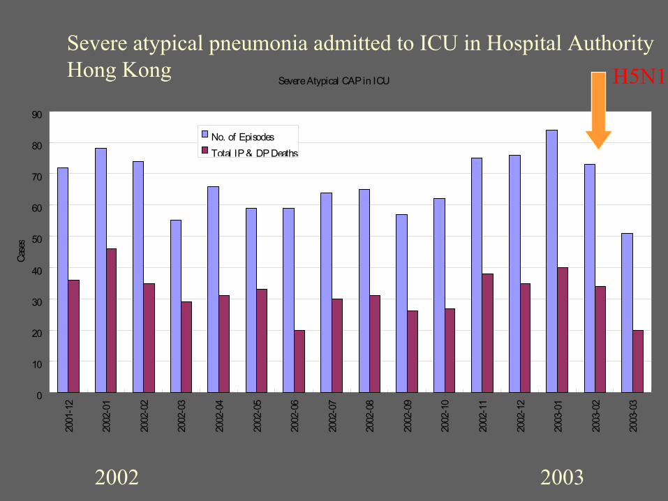
No. of EpisodesTotal IP & DP Deaths
Severe atypical pneumonia admitted to ICU in Hospital AuthorityHong Kong
2002 2003
H5N1
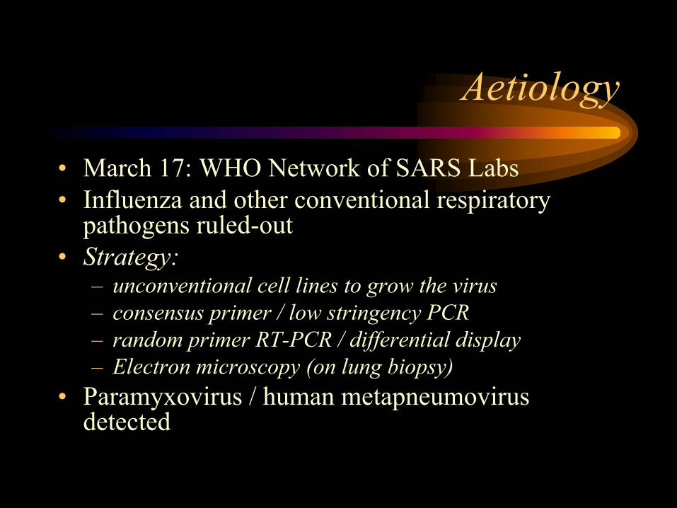
Aetiology
• March 17: WHO Network of SARS Labs• Influenza and other conventional respiratory
pathogens ruled-out• Strategy:
– unconventional cell lines to grow the virus– consensus primer / low stringency PCR– random primer RT-PCR / differential display– Electron microscopy (on lung biopsy)
• Paramyxovirus / human metapneumovirusdetected

A Novel coronavirus is associated with SARS

Two patients: Cytopathic effect in FRhK-4 cells
Uninfected Infected
Tested negative with reagents / PCR / RT/PCR for influenza A/B, adenovirus, RSV, parainfluenza, human metapneumovirus, enterovirus, rhinovirus, mycoplasma, chlamydia
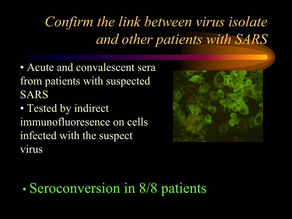
Confirm the link between virus isolate and other patients with SARS
• Seroconversion in 8/8 patients
• Acute and convalescent sera from patients with suspected SARS• Tested by indirect immunofluoresence on cells infected with the suspect virus

Negative stain Thin sect Direct EM Thin sect Cultured virus Cultured virus Lung Bx virus Cultured virus
Coronavirus-like agent is isolated
CPE in cell culture
Control cells Infected cells
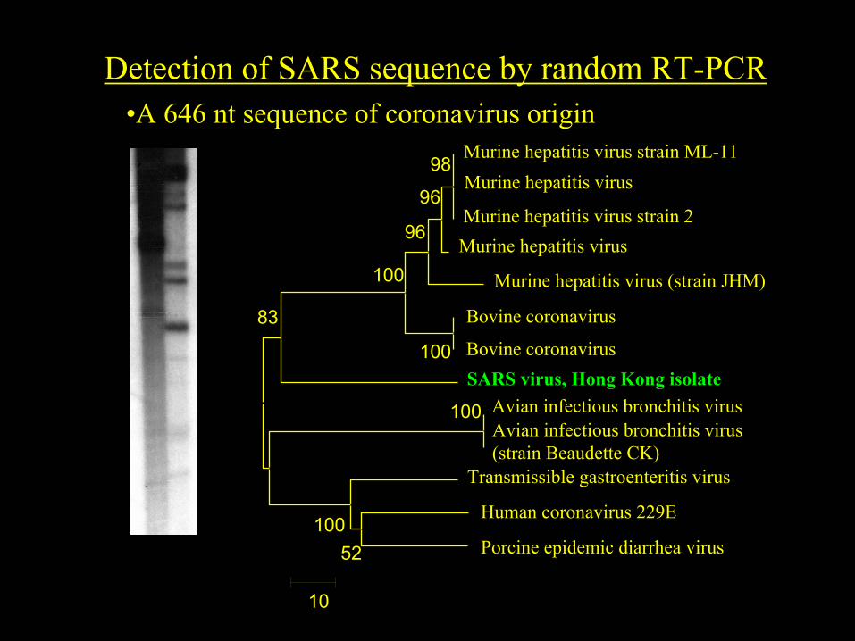
Detection of SARS sequence by random RT-PCR
100
52100
83
100
100
96
96
98
10
Avian infectious bronchitis virusAvian infectious bronchitis virus (strain Beaudette CK)
Human coronavirus 229E
Murine hepatitis virus
Murine hepatitis virus (strain JHM)
Murine hepatitis virus strain ML-11
Murine hepatitis virus
Porcine epidemic diarrhea virus
Bovine coronavirus
Bovine coronavirus
Transmissible gastroenteritis virus
SARS virus, Hong Kong isolate
Murine hepatitis virus strain 2
•A 646 nt sequence of coronavirus origin

Koch’s postulates: Association of microbe and disease
SEROLOGY:• 107 patients with clinically defined SARS
– Rising titre to coronavirus 104 / 107 (97%)– Rising IFA titre to human metapneumovirus
0 / 50 (0%)
• 45 paired sera from non-SARS patients: no antibody to CV
• 200 blood donors: no antibody

Sero-prevalence in blood donors
Indirect immunofluorescence
• Date tested No Positive / No Tested
• May 2003: 0 / 1,800
• March 2003: 0 / 200

SARS-coronavirus in macaques
Macaque # 4:
Severe multifocal pulmonary consolidation
Coronavirus detected in lung tissue
Severe interstitial pneumonia
Fouchier et al -Nature 2003, 423: 240.
Dual infections with SARS Coronavirus
Human metapneumovirusJ Tam et alF Plummer et al.

Dual infections with SARS Coronavirus
From ~ 800 SARS seroconverisonsMycoplasma 4 (+3)Adenovirus 5 (+5)Flu A 8 (+4)Flu B 3 (+3)Parainfluenza 7 (+3)Chlamydia 1 (+1)HSV 7Rotavirus 1Norwalk 2• W Lim, Department of Health
Human metapneumovirusJ Tam et alF Plummer et al.

Aetiology of SARS:
• SARS coronavirus (+/- host response) is necessary and sufficient to cause SARS
• We should now focus on SARS coronavirusrather than on SARS
• Co-factors (viruses, microbes or other) may play a role in explaining– Severity– “Super-spreading incidents”

Genome of the SARS-associated coronavirus
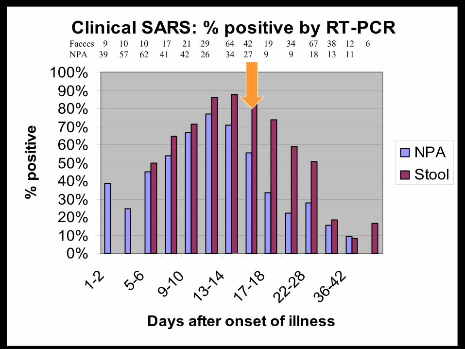
Clinical SARS: % positive by RT-PCR
0%10%20%30%40%50%60%70%80%90%
100%
1-2 5-6 9-10
13-14
17-18
22-28
36-42
Days after onset of illness
% p
ositi
ve
NPAStool
Faeces 9 10 10 17 21 29 64 42 19 34 67 38 12 6 NPA 39 57 62 41 42 26 34 27 9 9 18 13 11

Viral load in Nasopharyngeal Aspirate
Subsequent findings
• Laboratory diagnosis: Serology and RT-PCR
• Virus excretion in the faces• Transmission: more likely in later phase of
illness?• Virus stabililty in environment

WHO Network of Laboratories• Federal Laboratories for Health Canada, Winnipeg, Canada• Health Canada, Ottawa, Canada• Public Health Laboratory Centre, Hongkong SAR China• Prince of Wales Hospital, Hongkong SAR China• The University of Hongkong, Hong Kong SAR, China• Institut Pasteur, Paris, France• Bernhard-Nocht Institute, Hamburg and Johann Wolfgang
Goethe Universitat, Frankfurt, Germany• National Institute of Infectious Disease, Tokyo, Japan• Erasmus MC, Rotterdam, The Netherlands• Singapore General Hospital, Singapore• Central Public Health Laboratory, London, UK• Centers for Disease Control & Prevention, Atlanta, USA

Acknowledgements
• Y Guan, L Poon, KH Chan, JM Nicholls, FCC Leung, WH Seto, KY Yuen, The University of Hong Kong, Queen Mary Hospital
• W Lim, Department of Health• Hospital Authority and Department of Health• NIAID Research support • The “front line” health care workers in Hong
Kong