sars, a sara homolog repressible by agr, is an activator ... · however, the pathway by which the...
TRANSCRIPT

INFECTION AND IMMUNITY,0019-9567/01/$04.0010 DOI: 10.1128/IAI.69.4.2448–2455.2001
Apr. 2001, p. 2448–2455 Vol. 69, No. 4
Copyright © 2001, American Society for Microbiology. All Rights Reserved.
SarS, a SarA Homolog Repressible by agr, Is an Activator ofProtein A Synthesis in Staphylococcus aureus
AMBROSE L. CHEUNG,* KATHERINE SCHMIDT, BRIAN BATEMAN, AND ADHAR C. MANNA
Department of Microbiology, Dartmouth Medical School, Hanover, New Hampshire 03755
Received 20 November 2000/Returned for modification 18 December 2000/Accepted 8 January 2001
The expression of protein A (spa) is repressed by global regulatory loci sarA and agr. Although SarA maydirectly bind to the spa promoter to downregulate spa expression, the mechanism by which agr represses spaexpression is not clearly understood. In searching for SarA homologs in the partially released genome, wefound a SarA homolog, encoding a 250-amino-acid protein designated SarS, upstream of the spa gene. Theexpression of sarS was almost undetectable in parental strain RN6390 but was highly expressed in agr and sarAmutants, strains normally expressing high level of protein A. Interestingly, protein A expression was decreasedin a sarS mutant as detected in an immunoblot but returned to near-parental levels in a complemented sarSmutant. Transcriptional fusion studies with a 158- and a 491-bp spa promoter fragment linked to the xylEreporter gene disclosed that the transcription of the spa promoter was also downregulated in the sarS mutantcompared with the parental strain. Interestingly, the enhancement in spa expression in an agr mutant returnedto a near-parental level in the agr sarS double mutant but not in the sarA sarS double mutant. Correlating withthis divergent finding is the observation that enhanced sarS expression in an agr mutant was repressed by thesarA locus supplied in trans but not in a sarA mutant expressing RNAIII from a plasmid. Gel shift studies alsorevealed the specific binding of SarS to the 158-bp spa promoter. Taken together, these data indicated that theagr locus probably mediates spa repression by suppressing the transcription of sarS, an activator of spaexpression. However, the pathway by which the sarA locus downregulates spa expression is sarS independent.
Staphylococcus aureus is a versatile human pathogen that cancause a variety of infections ranging from minor wound infec-tions, pneumonia, and endocarditis to sepsis (3). The ability ofS. aureus to cause a multitude of diseases has been ascribed tothe array of extracellular and cell wall virulence determinantsproduced by this microorganism (27). The regulation of manyof these virulence determinants is controlled by global regula-tory loci such as sarA (previously designated as sar), agr, sae,and rot (8, 13–16, 23). These regulatory elements, in turn, exerttranscriptional control of target virulence genes.
The global regulatory locus agr encodes a two-componentquorum-sensing system that originates from the generation oftwo divergent transcripts, RNAII and RNAIII. RNAIII is theeffector molecule of the agr response, which entails upregula-tion of extracellular protein production (e.g., alpha-toxin)and downregulation of cell wall-associated protein synthesis(e.g., protein A and fibronectin-binding proteins) during thepostexponential phase (16). The RNAII transcript encodesa four-gene operon, agrBDCA, with AgrC and AgrA corre-sponding to the sensor and the activator proteins of a two-component regulatory system (16). Additionally, AgrD en-codes a 46-residue peptide which undergoes processing toform a quorum-sensing cyclic octapeptide, probably with theaid of the agrB gene product. Upon extracellular accumulationof a critical concentration of the cyclic octapeptide, the sensorprotein AgrC will become phosphorylated (19), thus leading toa second phosphorylation step of AgrA. Phosphorylated AgrA
will activate the transcription of RNAIII, the agr regulatorymolecule, to modulate target gene transcription (15, 23, 26).
In contrast to agr, the sarA locus upregulates the synthesis ofselected extracellular (e.g., a and b hemolysins) and cell wallproteins (e.g., fibronectin-binding protein A). Like the agr lo-cus, the sarA locus also represses the transcription of the pro-tein A gene (spa) (7). The sarA locus, contained within a 1.2-kbfragment, is composed of three overlapping transcripts, allencoding the major 372-bp sarA gene (1). DNA-binding stud-ies revealed that SarA, the major sarA regulatory molecule,binds to several target gene promoters, including those of agr,hla (a hemolysin gene), and spa. Accordingly, the binding ofSarA to a conserved binding site present in many target genepromoters leads to an upregulation in agr and hla transcription,as well as to a downregulation in spa transcription, thus impli-cating SarA to be a regulatory molecule that modulates targetgenes via both agr-dependent and agr-independent pathways(9).
Considering the fact that both sarA and agr repress spatranscription, it seems reasonable to predict the existence ofregulatory element(s) that counteracts this mode of regulation(i.e., activating spa). In searching for SarA homolog(s) in theS. aureus genome (The Institute for Genome Research [TIGR]),we came upon an open reading frame (ORF) upstream of thespa gene that shares homology with SarA. Transcriptionalanalysis indicated that the expression of this gene, designatedsarS for a gene supplemental to SarA, is enhanced in sarA andagr mutants, while the transcription of sarA and agr loci isunaltered in a sarS mutant. Inactivation of this gene leads to adecrease in protein A expression on immunoblots. Transcrip-tional analyses of sarA sarS and agr sarS double mutants indi-cated that the agr locus likely downregulates spa transcriptionby repressing sarS expression, whereas the sarA locus probably
* Corresponding author. Mailing address: Department of Microbi-ology, Vail 206, Dartmouth Medical School, Hanover, NH 03755.Phone: (603) 650-1340. Fax: (603) 650-1362. E-mail: [email protected].
2448
on March 15, 2019 by guest
http://iai.asm.org/
Dow
nloaded from

suppresses protein A expression via a different mechanism. Gelshift analysis revealed that purified SarS binds to the spa pro-moter in a dose-dependent fashion. In contrast to the suppres-sive effect of sarA and agr, these data suggested that sarSactivates protein A synthesis. The fact that sarS is repressibleby agr and not vice versa hints at the possibility that agr mayexert its effect on spa by repressing sarS expression.
MATERIALS AND METHODS
Bacterial strains, plasmids, and growth media. The bacterial strains andplasmids used in this study are listed in Table 1. CYGP, O3GL media (25), andtryptic soy broth were used for the growth of S. aureus strains, while Luria-Bertani medium was used to cultivate Escherichia coli. Antibiotics were used atthe following concentrations: erythromycin at 5 mg/ml, kanamycin at 75 mg/ml,tetracycline at 5 mg/ml, and ampicillin at 50 mg/ml.
Genetic manipulations in E. coli and S. aureus. Based on homology with sarA,the sarS gene was identified in contig 6207 in the TIGR S. aureus genomedatabase (www.TIGR.org). To construct a sarS mutant, part of the sarS gene,together with a part of flanking sequence, was amplified by PCR with theprimers 59-AGTTTTATGTTATAAACAATCGGA-39 and 59-GTTGTTTCTTGTTATTTTACGAA-39, using chromosomal DNA from strain RN6390 as thetemplate. The 1.8-kb PCR fragment (nucleotides [nt] 2925 to 1082 in contig
6207) was cloned into pUC18 in E. coli. Taking advantage of an internal EcoRIsite (nt 2616 to 2621) in the middle of the sarS coding region (nt 3098 to 2346),we cloned a ;1.4-kb ermC fragment into this site. The fragment containing anermC insertion into the sarS gene was cloned into the temperature-sensitiveshuttle vector pCL52.2 (18), which was then transformed into RN4220 by elec-troporation (28), followed by transduction into RN6390 with phage f11 asdescribed elsewhere (8). Transductants were selected at 30°C on erythromycin-and tetracycline-containing plates.
S. aureus RN6390 harboring the recombinant pCL52.2 was grown overnight at30°C in liquid medium in the presence of erythromycin, diluted 1:1,000 in freshmedia, and propagated at 42°C, a nonpermissive temperature for the replicationof pCL52.2. This cycle was repeated four times, and the cells were replicateplated onto O3GL plates containing erythromycin and erythromycin-tetracyclineto select for tetracycline-sensitive but erythromycin-resistant colonies, represent-ing mutants with double-crossovers. The mutations were confirmed by Southernhybridization with sarS and ermC probes. One clone, designated ALC1927, wasselected for further study.
To complement the sarS mutation in ALC1927, we introduced a 1.2-kb PCRfragment (nt 2925 to 1082 in contig 6207) encompassing the sarS gene into theshuttle plasmid pSK236. The recombinant shuttle plasmid was first electropo-rated into RN4220 and then into the sarS mutant ALC1927 (8). The presence ofthe recombinant plasmid was confirmed by restriction mapping. The presence ofthe sarS transcript in the complemented mutant was confirmed by Northern blotswith a sarS probe.
TABLE 1. Strains and plasmids used in this study
Strain orplasmid
Source orreference Comments
S. aureusRN4220 25 A mutant of 8325-4 that accepts foreign DNARN6390 25 Laboratory strain that maintains its hemolytic pattern when propagated on sheep erythrocyte agar
(parental strain)RN6911 26 An agr mutant of RN6390 with a Dagr::tetM mutationPC1839 4 8325-4 with a sarA::kan mutationALC184 7 sarA mutant of RN6390 with pRN6735 and pI524ALC865 7 RN6911 (agr mutant) with pALC862ALC1016 This study RN6390 with pALC1014ALC1342 This study A sarA mutant in which the sarA gene (nt 586 to 1107) (1) has been replaced by an ermC geneALC1794 9 RN6390 with pALC1639ALC1927 This study A sarS mutant of RN6390 with an ermC gene into the EcoRI site of the sarS geneALC2009 This study ALC1927 complemented with pALC2010ALC2033 This study RN6390 with Dagr::tetM and sarS::ermC mutationsALC2034 This study ALC1927 (sarS mutant) with pALC1639ALC2057 This study RN6390 with a sarA::kan mutationALC2067 This study RN6390 with sarS::ermC and sarA::kan mutationsALC2115 This study ALC1927 (sarS mutant) with pALC1014
E. coliXL1-Blue 21 A host strain for cloningBL21 21 A host strain for the pET14b expression vector
PlasmidspCR2.1 Invitrogen E. coli cloning vector for direct cloning of PCR productspUC18 21 E. coli cloning vectorpCL52.2 17 A temperature-sensitive E. coli-S. aureus shuttle vectorpET14b Novagen Expression vector for E. colipI524 16 S. aureus plasmid containing a b-lactamase repressorpLC4 31 A shuttle plasmid containing a promoterless xylE reporter genepRN6735 16 A derivative of pC194 containing the bla promoter and two-thirds of the blaZ gene followed by a
1.5-kb RNAIII fragment lacking its promoterpSK236 12 A shuttle vector containing pUC19 at the HindIII site of pC194pALC672 This study pCR2.1 with a 161-bp sarA P3 promoter fragmentpALC862 7 pSK236 containing the entire sarA locus with the sarA ORF and the triple promoter systempALC1014 This study pLC4 containing a 158-bp spa promoter fragment (nt 17 to 174) (20)pALC1639 9 pLC4 (transcriptional fusion vector) with a 491-bp spa promoter fragment (nt 1 to 174 plus 319 bp
upstream) (20)pALC1883 This study pUC18 containing a 1.8-kb sarS fragment (nt 2925 to 1082 in contig 6207)pALC1889 This study Temperature-sensitive shuttle plasmid pCL52.2 containing the ermC gene at the EcoRI site (nt
2616 to 2621) of the 1.8-kb sarS fragmentpALC2010 This study Shuttle plasmid pSK236 containing a 1.2-kb sarS fragment (nt 3459 to 2189 of contig 6207)pALC2040 This study pCR2.1 with a 1,562-bp spa structural gene (nt 219 to 1780) (20).pALC2043 This study pET14b containing the 750-bp sarS gene at the XhoI/BamHI site
VOL. 69, 2001 CHARACTERIZATION OF sarS IN S. AUREUS 2449
on March 15, 2019 by guest
http://iai.asm.org/
Dow
nloaded from

For the construction of the sarA sarS double mutant, we introduced thesarA::kan mutation into sarS mutant ALC1927 via a 80a lysate of a sarA insertionmutant PC1839 (with a sarA::kan mutation). As an additional control, we used asarA deletion mutant (ALC1342) in which the sarA gene has been replaced bythe ermC gene. Because of the ermC insertion, we were not able to construct asarA sarS mutation in the ALC1342 background. Likewise, an agr sarS mutantwas constructed by infecting the sarS mutant with a f11 lysate of the agr mutantRN6911. The authenticity of these double mutants was confirmed by Southernand Northern blots with sarA and agr probes (data not shown).
Analysis of hla and spa expression in the sarS mutant and its isogenic parents.To assess the phenotypes of the sarS mutant, we first evaluated the expression ofa hemolysin and protein A, two well-known virulence determinants in S. aureus.To determine a-hemolysin expression, equivalent amounts of extracellular pro-teins that had been harvested at stationary phase and concentrated by 10%trichloroacetic acid precipitation were blotted onto nitrocellulose, probed withrabbit anti-a-hemolysin antibody (a gift from B. Menzies, Nashville, Tenn.) di-luted 1:2,000, and then treated with the F(ab)2 fragment of goat anti-rabbit al-kaline phosphatase conjugate (Jackson Immunoresearch, West Grove, Pa.) asdescribed previously (5). Reactive bands were visualized as described by Blakeet al. (2).
To evaluate protein A production, cell wall-associated proteins were extractedfrom an equivalent number of S. aureus cells (from overnight cultures) withlysostaphin in a hypertonic medium (30% raffinose) to stabilize the protoplasts asdescribed previously (7). Equivalent volumes (1 to 2 ml each) of cell wall proteinextracts from 25 ml of cells (109 CFU/ml) were resolved on 10% sodium dodecylsulfate-polyacrylamide gels, blotted onto nitrocellulose, and probed with chickenanti-staphylococcal protein A antibody (Accurate Chemicals, Westbury, N.Y.) ata 1:3,000 dilution. Bound antibody was detected with a 1:5,000 dilution of F(ab)2
fragment of rabbit anti-chicken immunoglobulin G conjugated to alkaline phos-phatase (Jackson Immunoresearch), followed by the addition of developingsubstrates (2). The intensity of the protein A band was quantitated by densito-metric software (SigmaGel; Jandel Scientific). The data are presented as densi-tometric units.
Isolation of RNA and Northern blot hybridization. Overnight cultures of S.aureus were diluted 1:50 in CYGP and grown to mid-log (optical density at 650nm [OD650] 5 0.7), late-log (OD650 5 1.1), and early-postexponential (OD650 51.7) phases. The cells were pelleted and processed with a FastRNA isolation kit(Bio 101, Vista, Calif.) in combination with 0.1-mm-diameter zirconia-silicabeads in a FastPrep reciprocating shaker (Bio 101) as described earlier (6). Tenor twenty micrograms of each sample was electrophoresed through a 1.5%agarose–0.66 M formaldehyde gel in morpholinepropanesulfonic acid (MOPS)running buffer (20 mM MOPS, 10 mM sodium acetate, 2 mM EDTA; pH 7.0).Blotting of RNA onto Hybond N1 membranes (Amersham, Arlington Heights,Ill.) was performed with the Turboblotter alkaline transfer system (Schleicher &Schuell, Keene, N.H.). For the detection of specific transcripts (agr, sarA, sarS,spa, and hla), gel-purified DNA probes were radiolabeled with [a-32P]dCTP bythe random-primed method (Ready-To-Go Labeling Kit; Pharmacia) and hy-bridized under high-stringency conditions (5). The blots were subsequentlywashed and autoradiographed.
Preparation of cell extracts for detection of SarA. Cell extracts were preparedfor strains RN6390 and the corresponding sarS mutant. After pelleting, the cellswere resuspended in 1 ml of TEG buffer (25 mM Tris, 5 mM EGTA; pH 8), andcell extracts were prepared from lysostaphin-treated cells as described earlier(11). Cell extracts were immunoblotted onto nitrocellulose membranes as de-scribed above. For the detection of SarA, monoclonal antibody 1D1 (1:2,500dilution) was incubated with the immunoblot for 3 h, followed by another h ofincubation with a 1:10,000 dilution of goat anti-mouse alkaline phosphataseconjugate (Jackson Immunoresearch). Reactive bands were detected by devel-oping substrates as described previously (2).
Transcriptional fusion studies of spa promoter linked to the xylE reportergene. A 158-bp (nt 17 to 174) (20) and a 491-bp spa promoter fragment (9) withflanking EcoRI and HindIII sites were amplified by PCR using genomic DNA ofS. aureus RN6390 as the template and cloned into the TA cloning vector pCR2.1(Invitrogen, San Diego, Calif.). The EcoRI-HindIII fragments containing the spapromoter were then cloned into shuttle plasmid pLC4 (31), generating transcrip-tional fusions to the xylE reporter gene. The orientation and authenticity of thepromoter fragments were confirmed by restriction analysis and DNA sequencing.The recombinant plasmids were first introduced into S. aureus RN4220 by elec-troporation, according to the protocol of Schenk and Laddaga (28). Plasmidspurified from RN4220 transformants were then electroporated into RN6390 andits isogenic sarS mutant.
For enzymatic assays of the xylE gene product, overnight cultures were diluted1:50 or 1:100 in 250 ml of TSB containing appropriate antibiotics and shaken
at 37°C and 200 rpm. Starting after 3 h of growth, 10 to 50 ml of cell culturecorresponding to different OD600 values was serially removed, centrifuged, andwashed twice with 1 ml of ice-cold 20 mM potassium phosphate buffer (pH 7.2).The pellets were resuspended in 500 ml of 100 mM potassium phosphate buffer(pH 8.0) containing 10% acetone and 25 mg of lysostaphin per ml, incubated for15 min at 37°C, and then kept on ice for 5 min. Extracts were centrifuged at20,000 3 g for 50 min at 4°C to pellet cellular debris. The XylE (catechol2,3-dioxygenase) assays were determined spectrophotometrically at 30°C in atotal volume of 3 ml of 100 mM potassium phosphate buffer (pH 8.0) containing100 ml of cell extract and 0.2 mM catechol as described earlier (31). The reactionswere allowed to proceed for 25 min with an OD375 reading taken at the 25-mintime point. One milliunit is equivalent to the formation of 1.0 nmol of 2-hy-droxymuconic semialdehyde per min at 30°C. The specific activity is defined as amilliunit per milligram of cellular protein (31).
Overexpression and purification of SarS in a pET vector. The 750-bp sarSgene was amplified by PCR using the following oligonucleotides: 59-GCCG(CTCGAG)ATGAAATATAATAACCA-39 and 59-GCACTTTA(GGATCC)AGCACAC-39. The PCR product was digested with XhoI and BamHI (restrictionsites are indicated in parentheses), ligated into the expression vector pET14b(Novagen, Madison, Wis.), and transformed into the E. coli BL21(DE3).pLys.S.The resulting plasmid (pALC2043; see Table 1) contained the entire sarS codingregion in frame with a N-terminal His tag. Recombinant protein expression wasinduced by adding ITPG (isopropyl-b-D-thiogalactopyranoside; final concentra-tion, 1 mM) to a growing culture (30°C) at OD600 of 0.5. At 3 h after induction,the cells were harvested, resuspended in binding buffer (5 mM imidazole, 0.5 MNaCl, 20 mM Tris-HCl; pH 7.9), and sonicated on ice. Cellular debris wasremoved by centrifugation at 15,000 3 g for 15 min, and the clarified supernatantwas purified on a nickel affinity column (Novagen) according to the manufac-turer’s instructions. The protein was eluted with the elution buffer (1 M imid-azole, 0.5 M NaCl, 20 mM Tris-HCl; pH 7.9), followed by dialysis in the samebuffer lacking the imidazole. The authenticity of the purified SarS protein wasconfirmed by N-terminal sequencing, and the size of the recombinant protein wasverified by sodium dodecyl sulfate-gels stained with Coomassie blue.
Gel shift assays. To determine if the recombinant SarS protein binds to the spapromoter, DNA fragment (158 bp) was end labeled with [g-32P]ATP by using T4polynucleotide kinase. Labeled fragments were incubated at room temperaturefor 15 min with the indicated amount of purified protein in 25 ml of bindingbuffer (25 mM Tris-HCl, pH 7.5; 0.1 mM EDTA; 75 mM NaCl; 1 mM dithio-threitol; 10% glycerol) containing 0.5 mg of calf thymus DNA. The reactionmixtures were analyzed by nondenaturing polyacrylamide gel electrophoresis.The band shifts were detected by exposing dried gels to film.
RESULTS
Identification of the sarS gene. Predicated upon the SarAprotein sequence, we ran the BlastP program against the TIGRS. aureus genomic database. One of the matches is located up-stream of the spa gene (contig 6207, with the coding regionfrom nt 3098 to 2349). An identical gene, designated sarS, wasalso found in contig 773 at the University of Oklahoma ge-nome database. This gene, preceded by a typical Shine-Dal-garno sequence (AGGAGA) located 7 bp upstream of theinitiation codon, contains a 750-bp ORF encoding a 29.9-kDaprotein with a deduced pI of 9.36. A putative transcriptionterminal signal corresponding to a 13-bp inverted repeats (nt2345 to 2301 in contig 6207) is located 10 bp downstream of theTAA stop codon. About 33.2% of the residues are charged.Like that of SarA, the relatively small size, a predominance ofcharged residues, and a basic pI of SarS are features consistentwith regulatory proteins in prokaryotes (29). An alignment ofSarS with SarA revealed that SarS has two regions of identitywith SarA, with the first region (residues 1 to 125) having28.3% identity and the second region having 34.5% identity(Fig. 1). The extent of homology is relatively global in nature.A survey of the GenBank database indicated that SarS is iden-tical to the SarHI homolog recently reported by Tegmark et al.(30). Interestingly, SarS is also homologous to SarR (22), a
2450 CHEUNG ET AL. INFECT. IMMUN.
on March 15, 2019 by guest
http://iai.asm.org/
Dow
nloaded from

recently described SarA homolog that downregulates SarAprotein expression.
Expression of sarS in RN6390 and its isogenic sarA and agrmutants. To assess the role of sarS within the sarA/agr regula-tory cascade and its mode of control on virulence gene expres-sion, we proceeded to construct a sarS mutant by inserting anermC gene into the EcoRI site within the sarS gene in strainRN6390 (see Materials and Methods), thus resulting in a trun-cation of 91 residues from the C terminus. PCR with an ermC(59-ATGGTCTATTTCAATGGCAGTTAC) primer and asarS primer (59-AGGCTTTGGATGAAGCCGTTAC) outsidethe construct yielded a fragment consistent with the insertionof ermC into the sarS gene. Subsequent sequencing of the PCRproduct has verified the disruption of the sarS gene in themutant. This was also corroborated with Southern blots withselected ermC and sarS probes (data not shown).
A Northern blot with a sarS probe (nt 3098 to 2349 in contig6207), encompassing only the sarS coding region, revealed thatthe sarS gene is poorly transcribed in the parental strainRN6390, thus rendering the absence of the sarS gene difficultto decipher in the sarS mutant. Interestingly, the transcriptionof the sarS gene, sizing at 930 nt, was more prominent in bothagr and sarA mutants of RN6390 (Fig. 2A). In particular, in thesarA mutant, the sarS transcript was detected at late exponen-tial phase (OD650 5 1.1 using an 18-mm borosilicate glasstube) and was maximally transcribed during the postexponen-tial phase (OD650 5 1.7). The transcription of sarS was alsoincreased in the agr mutant, but the magnitude of the increasewas less than that of the sarA mutant (Fig. 2A). In contrast tothe sarA mutant, the agr mutant expressed the sarS transcriptmaximally during the late exponential phase. To assess therelative contributions of sarA and agr to sarS repression, we
assayed the sarS transcript level in an agr mutant comple-mented with a plasmid carrying the entire sarA locus (5), aswell as in a sarA mutant complemented with a fragment en-coding RNAIII, the agr regulatory molecule. Remarkably, thetranscription of sarS, augmented in an agr mutant, was re-pressed in the agr mutant clone expressing sarA in trans (Fig.2B). However, we were not able to detect transcriptional re-pression of sarS in a sarA mutant expressing RNAIII of agr,thus implying a differential role for sarA and agr in repressingsarS transcription.
We also examined the transcription of sarA and agr loci inthe sarS mutant. Northern analysis of RNAII and RNAIII didnot reveal any differences between the parental strain and theisogenic sarS mutant (data not shown). Likewise, the expres-sion of three sarA transcripts (designated sarA P1, P3, and P2transcripts) was similar between the two isogenic strains. SinceSarA is encoded by these transcripts (1), we also probed forSarA expression in an immunoblot of the cell extracts of theisogenic pair (25 mg of protein in each lane) with 1D1 anti-SarA monoclonal antibody (10). Our data showed that theexpression of SarA was comparable between RN6390 and itsisogenic sarS mutant (data not shown). Collectively, these dataimplied that the transcription of sarS is repressed by the sarAand agr gene products and not vice versa.
Assessment of hla and spa in a sarS mutant of S. aureus.Cognizant of the fact that both a-hemolysin and protein A areregulated by sarA and agr, two regulatory loci capable of re-pressing sarS expression, we proceeded to evaluate hla and spaexpression in the sarS mutant. In an immunoblot in whichequivalent amounts of extracellular proteins were blotted ontonitrocellulose and probed with rabbit anti-a-hemolysin anti-body (1:2,500 dilution), we found that a-hemolysin was syn-
FIG. 1. Sequence alignment of SarS, SarA, and SarR. SarS is 250 residues long. Based on regional homology, SarS can be divided into twodomains (S1 and S2) of 125 residues each. The homology with SarA is higher with the C-terminal domain (34.5%) than with the N-terminal domain(28.3%). The consensus residues are in black boxes. The consensus residue, designated as the majority at each position, is assigned when at leasthalf of the residues have the same amino acid.
VOL. 69, 2001 CHARACTERIZATION OF sarS IN S. AUREUS 2451
on March 15, 2019 by guest
http://iai.asm.org/
Dow
nloaded from

thesized in the sarS mutant at a level similar to that of theparental strain. Northern blotting with an hla probe also con-firmed comparable levels of gene expression between the twostrains (data not shown).
To ascertain the effect of a sarS mutation on spa expression,we first assayed for transcriptional activity of 491-bp (9) and158-bp (20) spa promoter fragments linked to the xylE reportergene in the isogenic sarS strains. Based on XylE assays, theactivity of the 491-bp spa promoter fragment was lower in thesarS mutant (29.6 6 0.06 and 24.0 6 0.28 mU /mg of cellularproteins at OD650 values of 1.1 and 1.7, respectively) than in its
isogenic parent (53.4 6 0.06 and 74 6 1.3 MU/mg of cellularproteins for OD650 values of 1.1 and 1.7, respectively). A sim-ilar expression pattern was also observed with the 158-bp spapromoter fragment, but the magnitude of the XylE activity wasmuch less in both isogenic strains (data not shown). We nextprobed an immunoblot, containing equivalent amounts of cellwall protein extracts of the mutant and complemented mutant,with affinity-purified chicken anti-protein A antibody (1:3,000dilution). As displayed in Fig. 3, the expression of protein Awas higher in the parental strain than in the sarS mutant.However, upon complementation with a plasmid expressingthe sarS gene, the expression of protein A was increased tonear parental level. These data implicated sarS to be involvedin the upregulation of spa expression in S. aureus.
Analysis of spa transcription in agr, sarA, agr sarS, and sarAsarS mutants. Since both sarA and agr repress sarS (see above)and spa transcription (7), we wanted to assess the relativecontribution of sarS, as mediated by sarA and agr, in spa re-pression. For this purpose, we compared spa transcription ofthe sarA sarS and agr sarS double mutants to single sarA andagr mutants in the RN6390 background. In a previous study ofthe effect of agr and sarA on spa transcription (7), we chosestrain RN6390 since this strain has a low basal level of spatranscription that can be accentuated by selective mutations.As shown in Fig. 4A, the transcription of spa, while enhancedin an agr mutant, was significantly reduced in the agr sarSdouble mutant, thus demonstrating that agr likely mediates sparepression by downregulating sarS. On the contrary, the up-regulation in spa expression in a sarA mutant (sarA::kan) wasmaintained in the sarS-sarA::kan double mutant. As an addi-tional control, the sarA deletion mutant ALC1342 also ex-pressed a high level of spa transcription. A similar expressionpattern was also observed in an immunoblot of cell wall proteinA for these strains (Fig. 4B), demonstrating repression in pro-
FIG. 2. (A) Northern blot of the sarS transcripts in sarA, agr, sarAsarS, and agr sarS mutants. A total of 20 mg of cellular RNA was loadedonto each lane. The intensity of the 16S and 23S RNA band was foundto be equivalent among lanes prior to transfer to Hybond N1 mem-brane. The blot was probed with a 750-bp sarS fragment (nt 2349 to3098 in contig 6207) labeled with [a-32P]dCTP, washed, and autora-diographed. Both sarA and sarA sarS mutants contained the sarA::kanmutation. The sarA deletion mutant ALC1342 in which the sarA genehas been replaced by an ermC gene was used as an additional control.(B) Northern blot of the sarS transcript in sarA mutant (ALC136) andagr mutant with agr and sarA provided in trans, respectively. The sarAmutant was complemented with pRN6735, yielding ALC184. The plas-mid pRN6735 was basally transcribed, yielding a low level of RNAIIItranscript (data not shown) even in the presence of a repressor plasmidpI524. The agr mutant RN6911 was complemented with pALC862, arecombinant pSK236 containing the entire sarA locus.
FIG. 3. Immunoblot of equivalent amounts of cell wall extractsof RN6390, sarS mutant, and complemented mutant. The blot wasprobed with affinity-purified chicken anti-protein A antibody at a1:3,000 dilution, followed by the addition of the appropriate conjugateand substrate. The portion of the blot labeled “5X” represents fivetimes as much cell wall extracts as the lanes labeled as “1X.” Purifiedprotein A (0.1 mg) was used as a positive control.
2452 CHEUNG ET AL. INFECT. IMMUN.
on March 15, 2019 by guest
http://iai.asm.org/
Dow
nloaded from

tein A expression in the agr sarS mutant (175 densitometricunits) compared with the single agr mutant (264 densitometricunits). However, the contribution of sarS to the sarA mutant(i.e., the sarA sarS double mutant) was more difficult to deci-pher since there were two major protein A bands of lowermolecular size in the sarA mutant (corresponding to densito-metric units of 314 and 131 for the upper and lower bands,respectively) compared with the sarS sarA double mutant (346densitometric units). The lower protein A bands may havebeen attributable to enhanced proteolytic activity in the sarAmutant, as has been previously reported (4, 8). Nevertheless, incomparing the intensity of the protein A band between thedouble sarS sarA mutant (346 densitometric U) and the agrmutant (264 densitometric U), we surmised that the expressionof protein A in the double mutant was not significantly lowerthan in the single sarA mutant (two bands at 314 and 131
densitometric U). Unlike the single sarA mutant, the sarS sarAdouble mutant did not exhibit a protein A band of smallermolecular size. Whether sarS plays a role in modulating pro-teolytic activity in the sarA mutant remains to be determined.Nevertheless, these data collectively supported the notion thatthe agr locus, in distinction to the sarA locus, likely mediatesspa repression via a sarS-dependent pathway.
To corroborate the view that agr and sarA mediate spa re-pression via different pathways, we determined spa transcrip-tion in an agr mutant complemented with a shuttle plasmidcarrying the entire sarA locus in trans, as well as in a sarAmutant with a plasmid expressing RNAIII. Although this ap-proach represented a higher gene dosage than at the physio-logic level, the result of this experiment, coupled with those ofsarS expression, provide additional evidence for the regulatorylinkage between agr and spa, using SarS as an intermediary.Accordingly, the transcription of spa in an agr mutant with sarAprovided in trans (Fig. 5), as with the expression of sarS (Fig.2B), was repressed compared to the agr mutant control. Incontrast, a sarA mutant with agr expressed in trans, while main-taining an elevated level of sarS transcription compared withthe parent (Fig. 2B), was still able to repress spa transcription(Fig. 5). Taken together, our results clearly indicated that sarAand agr repress spa transcription via divergent pathways, withagr being dependent on sarS, while the effect of sarA on spa issarS independent.
Gel shift assay of SarS with the spa promoter fragment.Recognizing that the sarS gene product may be an activator ofprotein A synthesis, we proceeded to evaluate the binding ofthe SarS protein to the spa promoter. For this experiment, wecloned the sarS gene into the pET14b expression vector (No-vagen, Madison, Wis.) in E. coli BL21. SarS was then overex-pressed in inducing conditions with 1 mM IPTG and purifiedwith a nickel affinity column from the crude cell lysate accord-ing to the manufacturer’s instructions. SarS, as eluted from thecolumn, was essentially homogeneous (.95%) (Fig. 6A). Us-ing purified SarS protein, we conducted gel shift assays of SarSwith a 158-bp spa promoter fragment (nt 17 to 174) (20). As
FIG. 4. (A) Northern blot of the spa transcript in agr, sarA, sarS agr,and sarS sarA mutants. The sarA and the sarS sarA mutants had thesarA::kan mutation. A total of 10 mg of RNA was applied to each lane.The blot was probed with a 1,562-bp spa fragment (nt 219 to 1780)(20). The parental strain RN6390 was a low protein A producer, witha very reduced level of spa transcription (7). The sarA deletion mutantALC1342 served as a positive control. (B) Immunoblot of cell wallextracts of agr, sarA, sarS agr, and sarS sarA mutants probed withchicken anti-protein A antibody. Equivalent amount of cell wall ex-tracts was applied to each lane. The positive control is purified proteinA (0.1 mg). The big arrow points to intact protein A, while the twosmaller arrows highlight degraded protein A fragments in the sarA::kan mutant (ALC2057).
FIG. 5. Northern blot of the spa transcript in sarA and agr mutants,with agr and sarA provided in trans, respectively. The strains used inthis blot are identical to those in Fig. 2B. The positive control is a plas-mid (pCR2.1) carrying a 1,562-bp spa fragment (nt 219 to 1780) (20).
VOL. 69, 2001 CHARACTERIZATION OF sarS IN S. AUREUS 2453
on March 15, 2019 by guest
http://iai.asm.org/
Dow
nloaded from
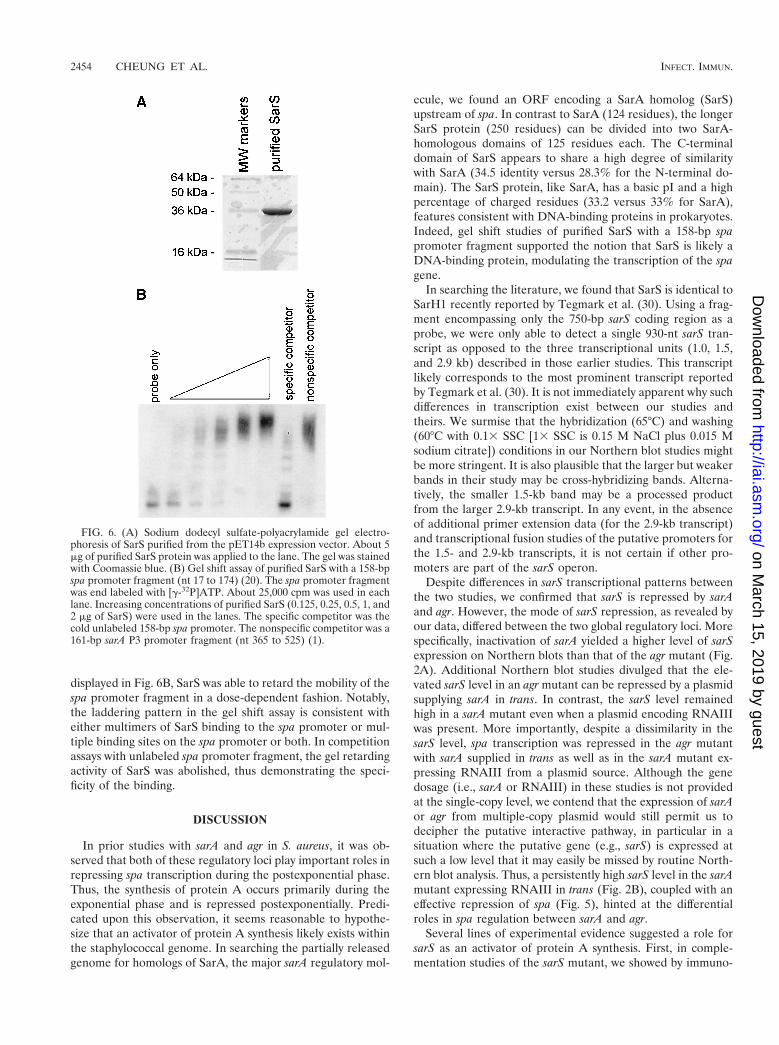
displayed in Fig. 6B, SarS was able to retard the mobility of thespa promoter fragment in a dose-dependent fashion. Notably,the laddering pattern in the gel shift assay is consistent witheither multimers of SarS binding to the spa promoter or mul-tiple binding sites on the spa promoter or both. In competitionassays with unlabeled spa promoter fragment, the gel retardingactivity of SarS was abolished, thus demonstrating the speci-ficity of the binding.
DISCUSSION
In prior studies with sarA and agr in S. aureus, it was ob-served that both of these regulatory loci play important roles inrepressing spa transcription during the postexponential phase.Thus, the synthesis of protein A occurs primarily during theexponential phase and is repressed postexponentially. Predi-cated upon this observation, it seems reasonable to hypothe-size that an activator of protein A synthesis likely exists withinthe staphylococcal genome. In searching the partially releasedgenome for homologs of SarA, the major sarA regulatory mol-
ecule, we found an ORF encoding a SarA homolog (SarS)upstream of spa. In contrast to SarA (124 residues), the longerSarS protein (250 residues) can be divided into two SarA-homologous domains of 125 residues each. The C-terminaldomain of SarS appears to share a high degree of similaritywith SarA (34.5 identity versus 28.3% for the N-terminal do-main). The SarS protein, like SarA, has a basic pI and a highpercentage of charged residues (33.2 versus 33% for SarA),features consistent with DNA-binding proteins in prokaryotes.Indeed, gel shift studies of purified SarS with a 158-bp spapromoter fragment supported the notion that SarS is likely aDNA-binding protein, modulating the transcription of the spagene.
In searching the literature, we found that SarS is identical toSarH1 recently reported by Tegmark et al. (30). Using a frag-ment encompassing only the 750-bp sarS coding region as aprobe, we were only able to detect a single 930-nt sarS tran-script as opposed to the three transcriptional units (1.0, 1.5,and 2.9 kb) described in those earlier studies. This transcriptlikely corresponds to the most prominent transcript reportedby Tegmark et al. (30). It is not immediately apparent why suchdifferences in transcription exist between our studies andtheirs. We surmise that the hybridization (65°C) and washing(60°C with 0.13 SSC [13 SSC is 0.15 M NaCl plus 0.015 Msodium citrate]) conditions in our Northern blot studies mightbe more stringent. It is also plausible that the larger but weakerbands in their study may be cross-hybridizing bands. Alterna-tively, the smaller 1.5-kb band may be a processed productfrom the larger 2.9-kb transcript. In any event, in the absenceof additional primer extension data (for the 2.9-kb transcript)and transcriptional fusion studies of the putative promoters forthe 1.5- and 2.9-kb transcripts, it is not certain if other pro-moters are part of the sarS operon.
Despite differences in sarS transcriptional patterns betweenthe two studies, we confirmed that sarS is repressed by sarAand agr. However, the mode of sarS repression, as revealed byour data, differed between the two global regulatory loci. Morespecifically, inactivation of sarA yielded a higher level of sarSexpression on Northern blots than that of the agr mutant (Fig.2A). Additional Northern blot studies divulged that the ele-vated sarS level in an agr mutant can be repressed by a plasmidsupplying sarA in trans. In contrast, the sarS level remainedhigh in a sarA mutant even when a plasmid encoding RNAIIIwas present. More importantly, despite a dissimilarity in thesarS level, spa transcription was repressed in the agr mutantwith sarA supplied in trans as well as in the sarA mutant ex-pressing RNAIII from a plasmid source. Although the genedosage (i.e., sarA or RNAIII) in these studies is not providedat the single-copy level, we contend that the expression of sarAor agr from multiple-copy plasmid would still permit us todecipher the putative interactive pathway, in particular in asituation where the putative gene (e.g., sarS) is expressed atsuch a low level that it may easily be missed by routine North-ern blot analysis. Thus, a persistently high sarS level in the sarAmutant expressing RNAIII in trans (Fig. 2B), coupled with aneffective repression of spa (Fig. 5), hinted at the differentialroles in spa regulation between sarA and agr.
Several lines of experimental evidence suggested a role forsarS as an activator of protein A synthesis. First, in comple-mentation studies of the sarS mutant, we showed by immuno-
FIG. 6. (A) Sodium dodecyl sulfate-polyacrylamide gel electro-phoresis of SarS purified from the pET14b expression vector. About 5mg of purified SarS protein was applied to the lane. The gel was stainedwith Coomassie blue. (B) Gel shift assay of purified SarS with a 158-bpspa promoter fragment (nt 17 to 174) (20). The spa promoter fragmentwas end labeled with [g-32P]ATP. About 25,000 cpm was used in eachlane. Increasing concentrations of purified SarS (0.125, 0.25, 0.5, 1, and2 mg of SarS) were used in the lanes. The specific competitor was thecold unlabeled 158-bp spa promoter. The nonspecific competitor was a161-bp sarA P3 promoter fragment (nt 365 to 525) (1).
2454 CHEUNG ET AL. INFECT. IMMUN.
on March 15, 2019 by guest
http://iai.asm.org/
Dow
nloaded from

blots that the diminution in protein A synthesis was restored bya shuttle plasmid carrying sarS. Second, we confirmed by tran-scriptional fusion studies of a spa promoter linked to the xylEreporter gene that spa promoter activity was indeed reduced ina sarS mutant. Third, gel shift studies have validated the no-tion that SarS can bind directly to the spa promoter in a dose-dependent fashion. Fourth, we extended our observation byNorthern analyses that the upregulation in spa transcription inan agr mutant, presumably mediated by a derepression of sarS,was abolished in an agr sarS double mutant, thus implicatingthe role of sarS in activating spa transcription in an agr mutant.However, contrary to the data of Tegmark et al., we found thatspa transcription in a sarA sarS double mutant, as with a singlesarA mutant, remained elevated. Thus, despite the experimen-tal observation that sarS is derepressed in a sarA mutant, thecontinued augmentation in spa transcription in a sarA sarSdouble mutant implied that the sarA locus likely repressesprotein A synthesis via a SarS-independent pathway. In thisregard, our recent finding that SarA, the major sarA regulatorymolecule, can directly bind to a consensus recognition se-quence upstream of spa promoter to downregulate spa tran-scription would provide an explanation for an alternativemechanism for direct SarA-mediated spa repression (9). Al-ternatively, other intermediate factor(s) controlled by sarA orother factors that act in conjunction with SarS may play a rolein sarA-mediated spa repression. It is also plausible that these“controlling factors” may be mediated via agr, since RNAIIIsupplied in trans in a significant gene dosage in a sarA mutantcould also suppress spa transcription (Fig. 5). Nonetheless, weare left to offer an explanation for the high level of sarS ex-pression in a sarA mutant. Perhaps, it may be reasonable tointerpret the upregulation in sarS in terms of hla repression,since Tegmark et al. found that a sarA sarH1 (i.e., sarA sarS)mutant, as opposed to a single sarA mutant, exhibited an up-regulation in hla transcription.
ACKNOWLEDGMENTS
We thank Simon Foster for strain PC1839 and Tom Kirn for assist-ing with protein alignment. Access to the S. aureus genome database atTIGR and at the University of Oklahoma is gratefully acknowledged.
This work was partially supported by NIH grant AI37142.
REFERENCES
1. Bayer, M. G., J. H. Heinrichs, and A. L. Cheung. 1996. The moleculararchitecture of the sar locus in Staphylococcus aureus. J. Bacteriol. 178:4563–4570.
2. Blake, M. S., K. H. Johnston, G. J. Russell-Jones, and E. C. Gotschlich.1984. A rapid sensitive method for detection of alkaline phosphatase-con-jugated anti-antibody on Western blots. Anal. Biochem. 136:175–179.
3. Boyce, J. M. 1997. Epidemiology and prevention of nosocomial infections,p. 309–329. In K. B. Crossley and G. L. Archer (ed.), The staphylococci inhuman disease. Churchill Livingstone, New York, N.Y.
4. Chan, P. F., and S. J. Foster. 1998. Role of SarA in virulence determinantproduction and environmental signal transduction in Staphylococcus aureus.J. Bacteriol. 180:6232–6241.
5. Cheung, A. L., M. G. Bayer, and J. H. Heinrichs. 1997. sar genetic determi-nants necessary for transcription of RNAII and RNAIII in the agr locus ofStaphylococcus aureus. J. Bacteriol. 179:3963–3971.
6. Cheung, A. L., K. Eberhardt, and V. A. Fischetti. 1994. A method to isolateRNA from gram-positive bacteria and mycobacteria. Anal. Biochem. 222:511–514.
7. Cheung, A. L., K. Eberhardt, and J. H. Heinrichs. 1997. Regulation ofprotein A synthesis by the sar and agr loci of Staphylococcus aureus. Infect.Immun. 2243–2249.
8. Cheung, A. L., J. M. Koomey, C. A. Butler, S. J. Projan, and V. A. Fischetti.1992. Regulation of exoprotein expression in Staphylococcus aureus by alocus (sar) distinct from agr. Proc. Natl. Acad. Sci. USA 89:6462–6466.
9. Chien, C.-T., A. C. Manna, S. J. Projan, and A. L. Cheung. 1999. SarA, aglobal regulator of virulence determinants in Staphylococcus aureus, binds toa conserved motif essential for sar dependent gene regulation. J. Biol. Chem.274:37169–37176.
10. Chien, Y. T., A. C. Manna, and A. L. Cheung. 1998. SarA level is a deter-minant of agr activation in Staphylococcus aureus. Mol. Microbiol. 31:991–1001.
11. Chien, Y., and A. L. Cheung. 1998. Molecular interactions between twoglobal regulators, sar and agr, in Staphylococcus aureus. J. Biol. Chem. 237:2645–2652.
12. Compagnone-Post, P., U. Malyankar, and S. A. Khan. 1991. Role of hostfactors in the regulation of the enterotoxin B gene. J. Bacteriol. 173:1827–1830.
13. Giraudo, A. T., A. L. Cheung, and R. Nagel. 1997. The sae locus of Staphy-lococcus aureus controls exoprotein synthesis at the transcriptional level.Arch. Microbiol. 168:53–58.
14. Giraudo, A. T., C. G. Raspanti, A. Calzolari, and R. Nagel. 1996. Charac-terization of a Tn551 mutant of Staphylococcus aureus defective in the pro-duction of several exoproteins. Can. J. Microbiol. 40:677–681.
15. Janzon, L., and S. Arvidson. 1990. The role of the d-hemolysin gene (hld) inthe regulation of virulence genes by the accessory gene regulator (agr) inStaphylococcus aureus. EMBO J. 9:1391–1399.
16. Kornblum, J., B. Kreiswirth, S. J. Projan, H. Ross, and R. P. Novick. 1990.Agr: a polycistronic locus regulating exoprotein synthesis in Staphylococcusaureus, p. 373–402. In R. P. Novick (ed.), Molecular biology of the staphy-lococci. VCH Publishers, New York, N.Y.
17. Lee, C. Y. 1992. Cloning of genes affecting capsule expression in Staphylo-coccus aureus strain M. Mol. Microbiol. 6:1515–1522.
18. Lin, W. S., T. Cunneen, and C. Y. Lee. 1994. Sequence analysis and molec-ular characterization of genes required for the biosynthesis of type 1 capsularpolysaccharide in Staphylococcus aureus. J. Bacteriol. 176:7005–7016.
19. Lina, G., S. Jarraud, G. Ji, T. Greenland, A. Pedraza, J. Etienne, R. P.Novick, and F. Vandenesch. 1998. Transmembrane topology and histidineprotein kinase activity of AgrC, the agr signal receptor in Staphylococcusaureus. Mol. Microbiol. 28:655–662.
20. Lofdahl, S., B. Guss, M. Uhlen, L. Philipson, and M. Lindberg. 1983. Genefor staphylococcal protein A. Proc. Natl. Acad. Sci. USA 80:697–701.
21. Maniatis, T., E. F. Fritsch, and J. Sambrook. 1989. Molecular cloning: alaboratory manual, 2nd ed. Cold Spring Harbor Laboratory, Cold SpringHarbor, N.Y.
22. Manna, A., and A. L. Cheung. 2001. Characterization of sarR, a modulator ofsar expression in Staphylococcus aureus. Infect. Immun. 69:885–896.
23. McNamara, P. J., K. C. Milligan-Monroe, S. Khalili, and R. A. Proctor.2000. Identification, cloning, and initial characterization of rot, a locus en-coding a regulator of virulence factor expression in Staphylococcus aureus.J. Bacteriol. 182:3197–3203.
24. Novick, R. P., S. J. Projan, J. Kornblum, H. F. Ross, G. Ji, B. Kreiswirth, F.Vandenesch, and S. Moghazeh. 1995. The agr P2 operon: an autocatalyticsensory transduction system in Staphylococcus aureus. Mol. Gen. Genet.248:446–458.
25. Novick, R. P. 1990. The staphylococcus as a molecular genetic system, p. 1–40. In R. P. Novick (ed.), Molecular biology of the staphylococci. VCHPublishers, New York, N.Y.
26. Novick, R. P., H. F. Ross, S. J. Projan, J. Kornblum, B. Kreiswirth, and S.Moghazeh. 1993. Synthesis of staphylococcal virulence factors is controlledby a regulatory RNA molecule. EMBO J. 12:3967–3977.
27. Projan, S. J., and R. P. Novick. 1997. The molecular basis of pathogenicity,p. 55–81. In K. B. Crossley and G. L. Archer (ed.), The staphylococci inhuman disease. Churchill Livingstone, New York, N.Y.
28. Schenk, S., and R. A. Laddaga. 1992. Improved method for electroporationof Staphylococcus aureus. FEMS Microbiol. Lett. 94:133–138.
29. Smith, I. 1993. Regulatory proteins that control late-growth development,p. 785–800. In A. L. Sonenshein, J. A. Hoch, and R. Losick (ed.), Bacillussubtilis and other gram-positive bacteria. ASM Press, Washington, D.C.
30. Tegmark, K., A. Karlsson, and S. Arvidson. 2000. Identification and char-acterization of SarH1, a new global regulator of virulence gene expression inStaphylococcus aureus. Mol. Microbiol. 37:398–409.
31. Zukowski, M. M., D. G. Gaffney, D. Speck, M. Kauffman, A. Findeli, A.Wisecup, and J. P. Lecocq. 1983. Chromogenic identification of geneticregulatory signals in Bacillus subtilis based on expression of a cloned Pseudo-monas gene. Proc. Natl. Acad. Sci. USA 80:1101–1105.
Editor: E. I. Tuomanen
VOL. 69, 2001 CHARACTERIZATION OF sarS IN S. AUREUS 2455
on March 15, 2019 by guest
http://iai.asm.org/
Dow
nloaded from