sample preparation of sem for plant smaples
TRANSCRIPT

ISSN:1369 7021 © Elsevier Ltd 2009VOLUME 12 – ELECTRON MICROSCOPY SPECIAL ISSUE32
Sample preparation for SEM of plant surfaces
Scanning electron microscopy (SEM) is an ideal technique for
examining plant surfaces at high resolution. Plant tissues must
be preserved by dehydration for observation in an electron
microscope because the coating system and the microscopes
operate under high vacuum and most specimens cannot withstand
water removal by the vacuum system without distortion1.
In order to examine the native structure of the sample, some
microscopes are designed to image frozen hydrated samples and
Plant tissues must be dehydrated for observation in most electron microscopes. Although a number of sample processing techniques have been developed for preserving plant tissues in their original form and structure, none of them are guaranteed artefact-free. The current paper reviews common scanning electron microscopy techniques and the sample preparation methods employed for visualisation of leaves under specific types of electron microscopes. Common artefacts introduced by specific techniques on different leaf types are discussed. Comparative examples are depicted from our lab using similar techniques; the pros and cons for specific techniques are discussed. New promising techniques and microscopes, which can alleviate some of the problems encountered in conventional methods of leaf sample processing and visualisation, are also discussed. It is concluded that the choice of technique for a specific leaf sample is dictated by the surface features that need to be preserved (such as trichomes, epidermal cells or wax microstructure), the resolution to be achieved, availability of the appropriate processing equipment and the technical capabilities of the available electron microscope.
A.K. Pathana*, J. Bondb and R.E. Gaskina
aPlant Protection Chemistry NZ, PO Box 6282, Rotorua, New ZealandbSCION, Private Bag 3020, Rotorua, New Zealand
*Email: [email protected]
MTElectronMicro_p32_43.indd 32 08/07/2010 11:52:16
Open access under CC BY-NC-ND license.

Sample preparation for SEM of plant surfaces REVIEW
VOLUME 12 – ELECTRON MICROSCOPY SPECIAL ISSUE 33
more recently environmental SEM microscopes have been developed
which can image the sample in their native-hydrated state. These
microscopes are specialised equipments and may not be available
in many labs. Hence, sample preparation by dehydration is still an
important consideration for observation in conventional microscopes.
For samples that necessitate dehydration, many techniques other
than just air-drying have been developed to remove water from the
sample, all aiming at minimal distortion of the cell and maximal
preservation of the original form and structure. These techniques
include freeze-drying, critical point drying, and various types of
chemical fixation treatments prior to dehydration of samples. However,
acceptable methods offer less than ideal preservation for some
plant species and may be inconsistent. The inconsistency is largely
due to diversity in tissue types, form, structure and composition of
plants. Inconsistencies also arise from variation in individual skills and
equipment used across different labs. Hence, new/modified techniques
are continually being tested and developed for the preparation of
specific plant tissues for visualisation under electron microscopes.
The paper reviews common techniques/methods used in the past
for leaf sample preparation for scanning electron microscopy. Selected
examples from our own work on common plant species (monocots and
dicots) of interest in pesticide research are presented and compared
with those from previous studies that have used similar techniques
for electron microscopy of plant tissues. Emphasis has been given to a
simple, but robust leaf sample preparation technique (simple air-drying),
which has proved highly effective for visualisation of plant waxes under
a field emission scanning electron microscope (FESEM) at low kV.
Approaches for sample preparation and visualisationSamples can be visualised in their native-hydrated state without
pre-treatment, frozen hydrated state or after removing liquids from
the samples using a variety of techniques. The choice of technique
will depend on the sample, the equipment available and the surface
features and structures that need to be visualised.
Hydrated samplesRapid observations of fresh hydrated samples can be made by
using an environmental scanning electron microscope (ESEM). The
technique has the potential to provide excellent low magnification
images of plant surfaces in their native-hydrated state. In addition, it
allows the flexibility to alter stage temperature and vapour pressure
in the specimen chamber. For example, leaf tissues can be examined
at high humidity in the chamber and minimise sample dehydration
during the imaging process. This technique can also be effectively used
to perform ‘dynamic’ experiments in wet mode to examine biological
events in developmental processes such as fungal growth on leaf
surfaces.
A FEI Quanta ESEM† (FEI Company, USA) was used at an
accelerating voltage of 10–20 kV, a stage temperature of 2 °C and a
chamber pressure of 6 Torr to visualise unprocessed chenopodium and
pea leaf surface in their native-hydrated state (Fig. 1a–d). Although the
‘true-to-life’ low magnification images of chenopodium leaves were
† Equipment used at Research Centre for Surface and Materials Science, University of Auckland, New Zealand.
Fig. 1 Leaf surfaces from unprepared and uncoated specimen visualised under an environmental scanning electron microscope: (a) chenopodium leaf surface showing intact epidermal cells and salt glands; (b) chenopodium salt gland at high magnification, note that waxes are not visible using this technique; (c) pea leaf surface showing intact epidermal cells, but waxes are not clearly visible; (d) epidermal cell collapse in pea leaf surface at high magnification. Waxes are not clearly visible.
(b)(a)
(c) (d)
MTElectronMicro_p32_43.indd 33 08/07/2010 11:52:18
REVIEW Sample preparation for SEM of plant surfaces
VOLUME 12 – ELECTRON MICROSCOPY SPECIAL ISSUE34
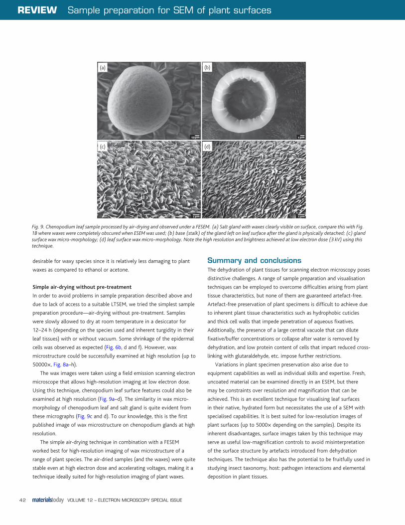
successfully obtained, the wax microstructure on the salt gland could
not be seen at all (compare Fig. 1 and Fig. 9) using this technique.
Similarly, the wax microstructure on pea leaf surface was not as clearly
visible as that from carefully air-dried samples (compare Fig. 1 and
Fig. 6). The sample also collapsed during imaging of the surface at high
magnification (5000×, Fig. 1d).
Despite having some limitations, the technique may allow
examination of some samples that could never be viewed in a
conventional SEM in their true-to-life form. This technique is increasingly
being used to study trichomes and glandular morphology and
function2-5. It is also being used to study elemental distribution in fresh
leaf samples using an appropriate energy-dispersive X-ray spectroscopy
(EDS) detector. Silicon accumulation in leaf tissues of sorghum, rice,
bamboo and orchard grass has been successfully studied using this
technique6-9. However, it is anticipated that the EDS scanning times
could be restricted to shorter lengths since the samples are relatively
delicate and unstable as compared to the dried samples that are robust
and suited for long time EDS examinations in a conventional SEM.
ESEM also provides a useful tool in taxonomic studies to classify
insects even at the species level10. The mechanism of leaf penetration
by fungi is not completely understood and may vary between
pathogens11. Studies with an ESEM may be useful to decipher the
fungal infection processes better. The technique can also be used to
study water droplet interactions on foliate surfaces. Cheng et al.12
successfully observed water condensation and evaporation processes on
lotus leaf surface using an ESEM and developed a geometric model for
liquid drops on rough surfaces.
Inherent limitations associated with ESEM include reduced field of
view and depth of focus, and in many cases, reduced resolution and
stability at high magnification since the sample is viewed without
coating with a heavy metal such as gold or chromium. Some samples
may move/change form during examination and necessitate rapid or
repeat image capture for a satisfactory micrograph. This equipment can
be considered as a SEM with added degrees of difficulty; however, the
art of imaging using an ESEM may be perfected for specific samples
using the right combination of accelerating voltage, stage temperature,
vapour pressure and working distance.
As an alternative to ESEM, a conventional SEM can be used to
view hydrated samples using special techniques such as QuantomiX
WetSEM™ technology that comprises a vacuum-tight capsule bounded
by a unique, electron-transparent and pressure-resistant membrane
(www.quantomix.com). This system allows visualisation of a wide
variety of fully hydrated samples in a conventional SEM by isolating the
sample from the vacuum while allowing penetration and reflection of
a scanning electron beam. A major disadvantage of this technique is its
relatively high cost due to the fact that a capsule can normally be used
only once for each sample.
Hydrated samples can also be visualised in a conventional SEM
if the liquids in the samples are substituted by glycerol13. Glycerol
evaporates very slowly under vacuum as compared to water, thereby
allowing the sample visualisation for short time in a conventional SEM
without causing malfunction of the vacuum system. An advantage of
the glycerol substitution method proposed by these authors is that the
leaf sample can be infiltrated from the underside by mounting them on
a piece of fabric soaked with glycerol, thus leaving the upper surface
untouched for visualisation. However, hydrophobic samples may prove
difficult to infiltrate with glycerol and develop shrinkage artefacts13.
This may not be a problem if the lower leaf surface is hydrophilic
and allows glycerol infiltration with ease. One good example of such
species is ryegrass, with the lower leaf surface much more wettable
than the upper leaf surface. A disadvantage of this technique is that the
longer visualisation times (greater than 30 min) may result in glycerol
accumulation in the oil of the vacuum pumps causing malfunction
of the equipment. In addition, satisfactory EDS analysis may not be
possible due to the scanning time restriction to ca. 30 min and the
possibility of changes in elemental form and distribution introduced by
the glycerol substitution. Although this technique has not been tested
in our lab due to intolerance of the FESEM to any kind of wet sample, it
was successfully used by Wagner et al.14 and Koch et al.15 to visualise
surface features of a range of plant species using a conventional SEM.
Recently a new TM-1000 tabletop microscope (Hitachi High-
Technologies Corporation, Japan)16 has been introduced that is a
relatively low-cost, portable, easy-to-use ESEM with a magnification
range of 20–10,000×. In addition, the TM-1000 detector is also
capable of showing contrast arising from differences in atomic number.
This instrument can be portable and carried to field sites for in situ
examination of plants, insects and microbes.
Frozen hydrated samplesFresh samples may be frozen (cryofixation) in their native-hydrated
state, coated and visualised on a cold-stage (cryo-chamber) attached
to the SEM. This technique, commonly called low temperature scanning
electron microscopy (LTSEM), has been widely used in the past for
examining plant surfaces. It is quick and is best suited to samples with
higher water content—samples that are usually very difficult to process
by conventional methods. This method is useful where preservation
of the natural ‘life-like’ morphology of cells and tissues is desired17.
Cryofixation is rapid and immobilises processes at a much faster rate
than chemical fixation18. This is a big advantage if dynamic biological
processes such as fungal spore discharge19 or fungal infection of a leaf20
need to be captured step-by-step in a ‘suspended animation’ form.
Another potential application of LTSEM is to trace elements on
plant surfaces or in freeze-fractures using EDS21-23. Some of these
elements may otherwise be lost to physiological processes (e.g.
translocation, leaching or biodegradation) or solubilised/moved
by solvents or chemicals if samples are processed by conventional
methods. Rapid freezing helps to retain them in their original form and
location.
MTElectronMicro_p32_43.indd 34 08/07/2010 11:52:19

Sample preparation for SEM of plant surfaces REVIEW
VOLUME 12 – ELECTRON MICROSCOPY SPECIAL ISSUE 35
A Philips SEM 505‡ coupled to a Hexland cryopreparation system
at a temperature of −140 °C to −120 °C was used at an accelerating
voltage of 4 kV to observe bean and wheat surfaces24. The samples
were attached to the stub and frozen by sample-holder contact with
the pre-chamber stage (−170 °C) in an atmosphere of dry nitrogen.
The samples were first viewed at low kV and ice contamination (if any)
was removed by raising the stage temperature to −60 °C prior to
coating with a gold layer of ca. 20-nm thickness. Excellent artefact-free
images of the leaf surface were obtained for both species with minimal
surface distortion as evident from intact epidermal cells and stomata
on bean and wheat leaves (Fig. 2a and b).
LTSEM can also be effectively used to observe the form of pesticide
deposit on plant surfaces25, since the deposits are cryo-fixed in their
original form and location without being exposed to chemical fixatives,
solvents or dehydrating forces as in conventional sample preparation
methods. The form and distribution of deposits remains unchanged
during visualisation as opposed to that under an ESEM in which it
may change due to introduction of additional moisture in the ‘wet’
mode. Using LTSEM, foliar deposits of the herbicide glyphosate with
and without adjuvants (spreading/penetrating and non-spreading/
non-penetrating) were observed on wheat leaf surfaces. The semi-
crystalline herbicide deposits and their modification by the adjuvants
on wheat leaf surface were effectively photographed in their original
form (Fig. 2c–f24).
LTSEM may also be used to examine internal structures by freeze-
fracture. However, the possibility of damage due to internal ice-crystal
growth needs to be considered. Rapid freezing (at several 1000 K/s)
of samples is required to minimise ice-crystal formation, or in ideal
Fig. 2. Leaf surfaces ((a) bean and (b) wheat) examined using a low-temperature scanning electron microscope (LTSEM). (c–f) Foliar deposits of herbicide glyphosate on wheat (with and without adjuvant) examined using a LTSEM in frozen hydrated state 1 h after application of the herbicide. (c) The herbicide deposit (without adjuvant) has formed a dense semi-crystalline film on top of epicuticular wax crystals (edge marked by arrows) and has failed to wet stomatal depressions; (d) higher magnification of the deposit edge of (c) showing irregular shaped semi-crystalline aggregates of herbicide (edge marked by arrows) formed on epidermal surface; (e) edge of herbicide deposit (marked by arrows) containing a spreading and penetrating adjuvant. Deposit is difficult to detect due to extensive spread and penetration caused by the adjuvant; (f) edge of herbicide deposit (marked by arrows) containing non-spreading and non-penetrating adjuvant. Deposit is clearly visible as an amorphous film lying on top of the epicuticular wax crystals.
‡ Equipment used at Long Ashton Research Station, Bristol, U.K.
(b)(a)
(c) (d)
(e) (f)
MTElectronMicro_p32_43.indd 35 08/07/2010 11:52:19

REVIEW Sample preparation for SEM of plant surfaces
VOLUME 12 – ELECTRON MICROSCOPY SPECIAL ISSUE36
conditions, to reduce water to a glassy state (vitreous ice). The faster
the cooling, the smaller the ice crystals will be. Crystal sizes of less
than 10 nm will do little damage to the samples. The rate of freezing
will also depend on the tissue thickness and composition. A number
of techniques can be utilised for rapid freezing, including plunge
freezing in liquid nitrogen (standard method), spray freezing with
propane, propane jet freezing, ‘slam’ freezing against a liquid helium or
nitrogen-cooled polished metal block, high pressure freezing (>2100
bar), etc.26,27. However, the depth of the specimen that is free from ice
crystal damage is normally limited to the outermost layers, typically
15–20 μm depending on the tissue type28.
LTSEM has been extensively used in the past and the subject has
been reviewed in detail by various authors17,29. It is well established that
LTSEM provides superior images but the technique is also not entirely
artefact-free. Artefacts in LTSEM may originate during cryofixation,
etching, freeze-fracturing, coating, specimen transfer and electron beam
irradiation17. Artefacts may also be introduced when frozen hydrated
samples have to be partially etched to remove ice contamination from
fracture faces. The frozen specimens are very beam sensitive and beam
damage may cause cracking of the specimen surface30. In order to
utilise this technique to its full potential, good operational skills are
required for artefact-free imaging of hydrated samples. In addition, the
need for a specialised cryo-chamber limits the use of this technique to
the few labs that can afford the additional cost involved.
Dried/dehydrated samplesStandard SEM procedures for biological samples involve chemical
fixation, drying/dehydration, mounting on a stub and coating with a
metal (e.g. chromium, gold, platinum, etc.) for examination under a
conventional SEM, often referred as ambient temperature scanning
electron microscopy (ATSEM). The fixation, drying/dehydrating steps
need to be done as carefully as possible to reduce shrinkage while
ensuring preservation of cell structures as close to the natural state as
possible.
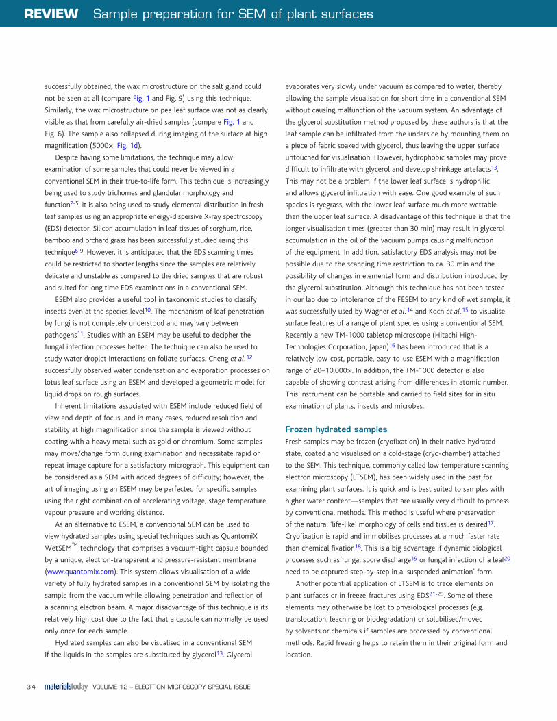
Fig. 3. Leaf surfaces from samples prepared using the CPD technique. (a) Bean leaf surface; (b) bean stomata; (c) chenopodium leaf surface; (d) chenopodium stomata; (e) broccoli leaf surface; (f) broccoli stomata. Note that some shrinkage of the epidermal cells is evident, especially around stomata. Waxes on chenopodium and broccoli appear to have been solubilised.
(b)(a)
(c) (d)
(e) (f)
MTElectronMicro_p32_43.indd 36 08/07/2010 11:52:21
Sample preparation for SEM of plant surfaces REVIEW
VOLUME 12 – ELECTRON MICROSCOPY SPECIAL ISSUE 37
There are several methods for drying/dehydrating leaf samples
for ATSEM, each having its own advantages and disadvantages. The
common drying techniques used in the past are (i) critical point
drying (CPD) (ii) freeze-drying (lyophilising) after prefreezing the
samples in liquid nitrogen-cooled liquid propane or Freon 22, and
then plunge freezing in liquid nitrogen31 and (iii) chemical fixation
in glutaraldehyde/osmium tetroxide before carrying out standard
dehydration in an organic solvent followed by CPD. To achieve an
acceptable preservation of plant tissues, these techniques have been
tried (with some variations) in different labs, including ours, with mixed
success32-37.
In our lab, the dehydrated samples were mounted on aluminium
stubs using aqueous conductive silver, and chromium coated (once
or twice) using an Emitech K575X peltier cooled turbo sputter coater
(Emitech Ltd., U.K.) prior to visualisation under a JEOL JSM-6700F field
emission scanning electron microscope (JEOL Ltd., Japan). The FESEM
is equipped with two secondary electron detectors: LEI (lower detector)
and SEI (upper detector). The SEI detector is higher in the column
and sees fewer shadows. Less charging is detected, and since it can be
used at a shorter working distance, the resolution is greater than with
the LEI. All surface wax images (at high magnifications) were taken
using the SEI detector since it gives comparatively high resolution. The
conditions used were an accelerating voltage 3–10 kV; illuminating
current 2.6 nA with a working distance 8–15 mm.
Critical point drying
Initially introduced by Anderson38 more than half-a-century ago, CPD
is the most commonly used dehydrating method for biological sample
preparation. This procedure removes liquids from the specimen and
avoids surface tension effects (drying artefacts) by never allowing a
liquid/gas interface to develop. The transition from liquid to gas at the
critical point takes place without an interface because the densities of
liquid and gas are equal at this point.
We used the standard CPD protocol for processing our samples.
After fixation with 2.5% glutaraldehyde in 0.2 M cacodylate and 2%
buffered osmium tetroxide, the samples were dehydrated through a
graded series of ethanol (10%, 20%, 30%, 50% and 70%—once for
10 min at each step), and then immersed in 100% acetone twice for
30 min each. The tissues were then transferred to an Emitech K850
critical point dryer (Emitech Ltd., U.K.) using liquefied carbon dioxide as
transitional fluid. We found that CPD gave an acceptable preservation
of bean, chenopodium and broccoli leaf surface (Fig. 3a, c and e,
respectively) but the organic solvents stripped-off epicuticular waxes
from chenopodium and broccoli (Fig. 3d and f, respectively). Some
shrinkage on leaf surface was also evident around stomata of bean
and chenopodium (Fig. 3b and d, respectively). Shrinking artefacts may
be introduced in the samples while permuting the specimens from
one solution to the next or into the CPD. To avoid these artefacts, it
is essential that the specimens are completely wet during the whole
preparation process. Although shrinking surface artefacts were observed
on some CPD processed samples, the internal structures (mesophyll
cells, etc.) were highly preserved in bean, chenopodium and broccoli
(Fig. 4a–c). A similar level of preservation of mesophyll cells was not
observed in a freeze-dried fracture of broccoli (Fig. 4d).
Bray et al.35 used CPD, Peldri II and hexamethyldisilazane (HMDS)
to determine a method that gave the best preservation of leaf tissues.
Fig. 4. Fractures from samples preservation using the CPD technique. (a) Bean; (b) chenopodium; (c) broccoli; (d) fracture of broccoli leaf surface preserved with the freeze-drying technique (liquid nitrogen) for comparison. Note the excellent level of preservation of mesophyll cells achieved using the CPD technique.
(b)(a)
(c) (d)
MTElectronMicro_p32_43.indd 37 08/07/2010 11:52:22
REVIEW Sample preparation for SEM of plant surfaces
VOLUME 12 – ELECTRON MICROSCOPY SPECIAL ISSUE38
Peldri II caused complete extraction of leaf epicuticular wax, while CPD
and HMDS showed minimal extraction compared with that of samples
air-dried directly from acetone. They also found that while all the three
methods showed signs of shrinkage, CPD provided relatively better
quality of tissue preservation.
CPD is the method of choice (with careful use) if fractures
and/or non-waxy epidermal surfaces (e.g. bean leaf surface) are
to be examined in an ATSEM. It is distinctively advantageous for
specific applications and has been used widely for biological sample
preparation. However, the technique necessitates the use of a
specific apparatus and the sample throughput is limited39. It may not
completely remove water from some tissues and may cause some
bulk shrinkage or generate violent bubbling40,41. The technique also
necessitates the use of organic solvents such as acetone that may
damage leaf epicuticular wax structures42. These structures are an
important part of the leaf surface micro-morphology and need to be
adequately preserved for scanning electron microscopy.
Freeze-drying
The first step in freeze-drying technique is to rapidly freeze the sample
to avoid ice-crystal formation. Rapidity of freezing is probably the most
influential factor on the final preservation quality of biological samples.
Ideally, samples should be directly plunged into liquid nitrogen (−196 °C),
but liquid nitrogen forms a gaseous/insulating layer (Leidenfrost effect)
thus losing contact with the sample. To avoid the Leidenfrost effect,
samples can be plunged in slush nitrogen (−210 °C) prepared by placing
a beaker filled with liquid nitrogen in a desiccator under vacuum.
Alternatively, rapid freezing of samples can be achieved by plunging
in nitrogen-cooled Freon 22 (−150 °C) or liquid propane (−178 °C),
subsequently freezing in liquid nitrogen and then freeze-drying
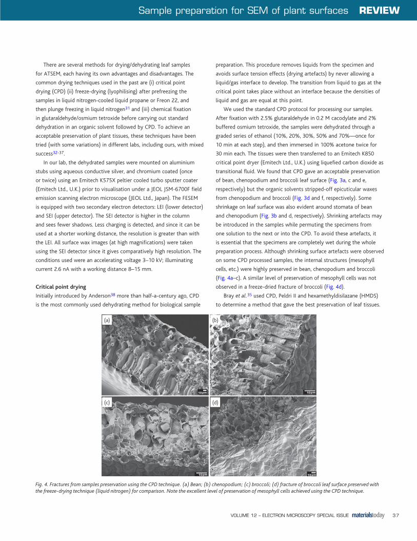
Fig. 5 Leaf surfaces from samples prepared using freeze-drying technique; (a–d) rapid freezing achieved by plunging in liquid nitrogen-cooled liquid Freon 22. (a) Some preservation of bean epidermal cells; (b) wheat waxes relatively more preserved as compared to those of broccoli; (c) wax dissolution apparent in waxy cabbage leaf; (d) waxes solubilised around broccoli stomata; (e) rapid freezing of bean leaf sample achieved using liquid nitrogen slush. All samples were freeze-dried in a basic freeze-drying system using dry-ice cake under vacuum.
(b)(a)
(c) (d)
(e)
MTElectronMicro_p32_43.indd 38 08/07/2010 11:52:23
Sample preparation for SEM of plant surfaces REVIEW
VOLUME 12 – ELECTRON MICROSCOPY SPECIAL ISSUE 39
under controlled conditions. Some labs recommend introduction
of a cryo-protectant (usually 20–30% glycerol) into the tissues
to minimise ice crystal size. However, this technique necessitates
pre-fixation of samples in glutaraldehyde which may not be desirable
if chemical fixatives need to be avoided. In addition, cryo-protectants
may not completely sublime away with water during the freeze-drying
step.
Freeze-drying necessitates availability of a good (turbo pumped)
vacuum system, an effective cold trap (−80 °C to −100 °C) for
sublimed water and liquid N2-based cooling. Sophisticated freeze-
drying equipment is available (e.g. EMITECH K750 and K775; Emitech
Ltd., U.K.) that provides adequate flexibility to manipulate conditions
for specific sample types. For good sample preservation, both freezing
(needs to be rapid) and freeze-drying processes need to be optimised
for specific samples.
If a suitable freeze-dryer is not accessible, a basic freeze-drying
system can be developed in-house. In our study, cryo-fixed (frozen
in liquid nitrogen/slush or liquid nitrogen-cooled Freon 22) samples
were freeze-dried using an in-house simple freeze-drier. The sample
was placed on a liquid N2-cooled brass metal holder under vacuum
with an effective cold trap and allowed to dry overnight. We placed
a brass metal holder on a cake of dry ice under vacuum to maintain
the drying temperature below −60 °C. The cake gradually decreases
in size over time while the brass metal holder gradually sinks-in and
remains in complete contact with dry ice all the time, thus providing
a constant low temperature for freeze-drying. We found acceptable
preservation of bean (non-waxy) and wheat (waxy) samples using this
technique after freezing samples in liquid nitrogen-cooled liquid Freon
22 (Fig. 5a and b). However, it did dissolve waxes of other species such
as cabbage and broccoli (Fig. 5c and d). In theory, freezing samples
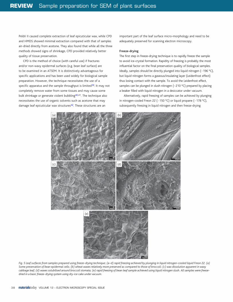
Fig. 6. Leaf samples prepared by chemical fixation with glutaraldehyde and osmium tetroxide followed by dehydration in ethanol and air-drying (left) and air-drying (right) techniques. (a and b) Barnyardgrass; (c and d) pea; (e and f) chenopodium. Note the relatively better preservation of leaf surfaces by simple air-drying as opposed to severe distortion by chemical fixation followed by dehydration in ethanol and then air-drying.
(b)(a)
(c) (d)
(e) (f)
MTElectronMicro_p32_43.indd 39 08/07/2010 11:52:24
REVIEW Sample preparation for SEM of plant surfaces
VOLUME 12 – ELECTRON MICROSCOPY SPECIAL ISSUE40
in liquid nitrogen slush should provide good freezing and acceptable
preservation of samples after freeze-drying without the need to use
liquid nitrogen-cooled liquid Freon 22. However, adequate preservation
of the majority of bean leaf surface was not achieved by using this
technique in our experiments (Fig. 5e).
In our view, the techniques described above can be further
improved and perfected for specific samples using liquid Freon 22/
liquid propane (for many non-waxy plant species) or liquid nitrogen
slush (for waxy plant species); freeze-drying using a dry ice cake may
provide adequate low temperature conditions required for the drying
process if sophisticated freeze-drying equipments are not accessible. A
limitation to this technique is that the use of liquid Freon 22 and liquid
propane could be restricted in many labs since the former is a powerful
greenhouse gas and the latter is highly flammable.
Chemical fixation
Chemical fixation involves soaking samples for various periods
of time (depending on their thickness and composition), usually
in glutaraldehyde and/or osmium tetroxide. The samples may be
subsequently dehydrated in a graded series of ethanol32, and then
dried by CPD or subjected to air-drying. Alternatively, the sample may
be transferred to tetramethylsilane (TMS) for 10–20 min before air-
drying34.
We fixed bean samples in 2.5% glutaraldehyde and 2% osmium
tetroxide followed by dehydration in a graded series of ethanol. The
samples were then dried by two different methods—CPD and simple air-
drying (in a desiccator under vacuum). CPD has been already described
in an earlier section. Although, acceptable preservation was achieved
by the CPD technique post-fixation with glutaraldehyde and osmium
tetroxide, epidermal cells appeared to be shrunken, especially around
stomata of bean and chenopodium (Fig. 3b and d). This shrinkage
of tissue probably occurred during dehydration rather than drying43.
Hardy et al.44 also failed to achieve acceptable surface preservation for
Dactylis glomerata and Elymus canadensis leaf samples using CPD and
TMS techniques after fixation in glutaraldehyde/acrolein mixture.
Simple air-drying after fixation in glutaraldehyde and osmium
tetroxide caused substantial alterations of the leaf surfaces of
barnyardgrass, pea and chenopodium species (Fig. 6a, c and e). This
was despite the expectation that fixing in glutaraldehyde and osmium
should strengthen the elastic cell walls by cross-linking polymers and
thus resist cell collapse due to dehydrating forces. Since comparatively
less surface distortion was achieved by CPD (post-fixation with
glutaraldehyde and osmium tetroxide, Fig. 3a–f) as well as simple air-
drying without fixation (Fig. 6b, d and f), it is clear that the fixatives
negatively influenced the drying step to cause substantial damage. Bray
et al.35 concluded that post-fixation with osmium tetroxide generally
resulted in poorer specimen preservation than using Karnovsky’s
fixative (mixture of glutaraldehyde and formaldehyde), especially for
dicotyledonous species such as Coleus blumei using the CPD method.
The authors suggested that the increased distortion of plant cell
surface structure following post-fixation with osmium tetroxide might
be due to (i) a build up of osmium molecules that could ultimately
inhibit infiltration of dehydrating agents (e.g. ethanol, acetone,
Freon 113) and transitional agents (Freon 13, Freon 23, carbon dioxide)
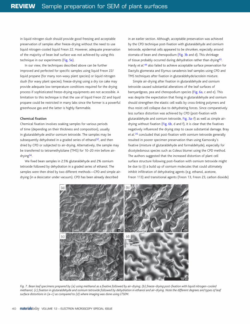
Fig. 7. Bean leaf specimens prepared by (a) using methanol as a fixative followed by air-drying; (b) freeze-drying post-fixation with liquid nitrogen-cooled methanol; (c) fixation in glutaraldehyde and osmium tetroxide followed by dehydration in ethanol and air-drying. Note the different degrees and types of leaf surface distortions in (a–c) as compared to (d) where imaging was done using LTSEM.
(a) (b)
(c) (d)
MTElectronMicro_p32_43.indd 40 08/07/2010 11:52:25
Sample preparation for SEM of plant surfaces REVIEW
VOLUME 12 – ELECTRON MICROSCOPY SPECIAL ISSUE 41
in the CPD technique and (ii) the drying solvents (e.g. HMDS, Peldri
II, TMS or dimethoxypropane—DMP) used as an alternative to the
CPD technique45-47. It is recommended that leaf samples should not
be fixed with glutaraldehyde and/or osmium tetroxide if post-fixation
drying is to be achieved by simple air-drying or with organic solvents.
Methanol was proposed as an alternative fixative/dehydrant that
could be used prior to CPD for preserving plant surfaces37. The use of
methanol instead of glutaraldehyde or osmium tetroxide was suggested
since it instantly fixes the elastically extended cell walls and can rapidly
penetrate inside plant cuticles and cell walls. The authors suggested that
this method resulted in improved fixation of cell wall dimension and is
deemed to be the most suitable for preserving plant epidermal surfaces.
They also achieved superior preservation of Silvina auriculata and
Verbascum arcturus trichomes as compared to conventional treatments.
We tested a variation of this technique by (i) immersing bean
samples in methanol for 20–40 s followed by simple air-drying (Fig. 7a)
or by (ii) immersing in liquid nitrogen-cooled methanol for 20–40 s,
re-immersing in liquid nitrogen followed by freeze-drying (Fig. 7b).
The techniques provided preservation of epidermal cell surface in
some areas and collapse in others, but the latter technique (Fig. 7b)
appeared to be relatively better in preservation quality. Although
the quality of preservation was far from ideal, it was still better than
that obtained from using glutaraldehyde and osmium tetroxide as
fixatives (Fig. 7c).
We expect that the methanol fixation of plant tissues to have
some potential for fine tuning for CPD, freeze-drying or simple air-
drying techniques and to have an application in preserving specific
plant tissues. Use of methanol as a fixative in these processes is also
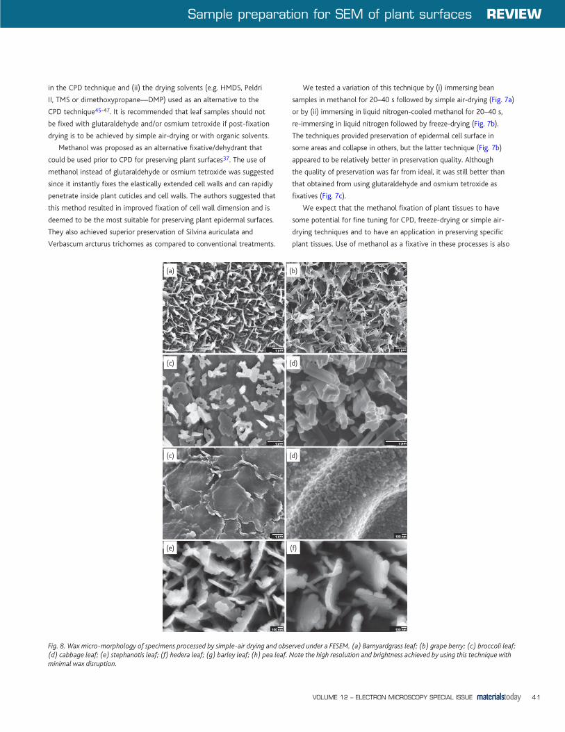
Fig. 8. Wax micro-morphology of specimens processed by simple-air drying and observed under a FESEM. (a) Barnyardgrass leaf; (b) grape berry; (c) broccoli leaf; (d) cabbage leaf; (e) stephanotis leaf; (f) hedera leaf; (g) barley leaf; (h) pea leaf. Note the high resolution and brightness achieved by using this technique with minimal wax disruption.
(b)(a)
(c) (d)
(c) (d)
(e) (f)
MTElectronMicro_p32_43.indd 41 08/07/2010 11:52:26
REVIEW Sample preparation for SEM of plant surfaces
VOLUME 12 – ELECTRON MICROSCOPY SPECIAL ISSUE42
desirable for waxy species since it is relatively less damaging to plant
waxes as compared to ethanol or acetone.
Simple air-drying without pre-treatment
In order to avoid problems in sample preparation described above and
due to lack of access to a suitable LTSEM, we tried the simplest sample
preparation procedure—air-drying without pre-treatment. Samples
were slowly allowed to dry at room temperature in a desiccator for
12–24 h (depending on the species used and inherent turgidity in their
leaf tissues) with or without vacuum. Some shrinkage of the epidermal
cells was observed as expected (Fig. 6b, d and f). However, wax
microstructure could be successfully examined at high resolution (up to
50000×, Fig. 8a–h).
The wax images were taken using a field emission scanning electron
microscope that allows high-resolution imaging at low electron dose.
Using this technique, chenopodium leaf surface features could also be
examined at high resolution (Fig. 9a–d). The similarity in wax micro-
morphology of chenopodium leaf and salt gland is quite evident from
these micrographs (Fig. 9c and d). To our knowledge, this is the first
published image of wax microstructure on chenopodium glands at high
resolution.
The simple air-drying technique in combination with a FESEM
worked best for high-resolution imaging of wax microstructure of a
range of plant species. The air-dried samples (and the waxes) were quite
stable even at high electron dose and accelerating voltages, making it a
technique ideally suited for high-resolution imaging of plant waxes.
Summary and conclusionsThe dehydration of plant tissues for scanning electron microscopy poses
distinctive challenges. A range of sample preparation and visualisation
techniques can be employed to overcome difficulties arising from plant
tissue characteristics, but none of them are guaranteed artefact-free.
Artefact-free preservation of plant specimens is difficult to achieve due
to inherent plant tissue characteristics such as hydrophobic cuticles
and thick cell walls that impede penetration of aqueous fixatives.
Additionally, the presence of a large central vacuole that can dilute
fixative/buffer concentrations or collapse after water is removed by
dehydration, and low protein content of cells that impart reduced cross-
linking with glutaraldehyde, etc. impose further restrictions.
Variations in plant specimen preservation also arise due to
equipment capabilities as well as individual skills and expertise. Fresh,
uncoated material can be examined directly in an ESEM, but there
may be constraints over resolution and magnification that can be
achieved. This is an excellent technique for visualising leaf surfaces
in their native, hydrated form but necessitates the use of a SEM with
specialised capabilities. It is best suited for low-resolution images of
plant surfaces (up to 5000× depending on the samples). Despite its
inherent disadvantages, surface images taken by this technique may
serve as useful low-magnification controls to avoid misinterpretation
of the surface structure by artefacts introduced from dehydration
techniques. The technique also has the potential to be fruitfully used in
studying insect taxonomy, host: pathogen interactions and elemental
deposition in plant tissues.
Fig. 9. Chenopodium leaf sample processed by air-drying and observed under a FESEM. (a) Salt gland with waxes clearly visible on surface, compare this with Fig. 1B where waxes were completely obscured when ESEM was used; (b) base (stalk) of the gland left on leaf surface after the gland is physically detached; (c) gland surface wax micro-morphology; (d) leaf surface wax micro-morphology. Note the high resolution and brightness achieved at low electron dose (3 kV) using this technique.
(a) (b)
(c) (d)
MTElectronMicro_p32_43.indd 42 08/07/2010 11:52:28
Sample preparation for SEM of plant surfaces REVIEW
VOLUME 12 – ELECTRON MICROSCOPY SPECIAL ISSUE 43
LTSEM followed by cryofixation of samples may prove useful as high
magnification controls, but this technique is not entirely artefact-free.
In addition the technique necessitates the use of a specialised cryo-
chamber attached to the microscope. If a cryo-stage is not available,
samples processed by simple air-drying and examined using appropriate
beam and probe current conditions may provide high-resolution
wax images (up to 50,000× magnifications) as demonstrated in the
current study. CPD following chemical fixation with glutaraldehyde
and/or osmium tetroxide gave excellent preservation of internal
leaf structures, but surface preservation was less than ideal for
many samples. Simple air-drying following chemical fixation (with
glutaraldehyde and osmium tetroxide) is not recommended at all for
sample preservation because the fixatives caused severe distortion of
leaf tissue by negatively influencing the drying process. Fixation with
methanol before carrying out the drying process provided some good
preservation, but the technique needs to be refined further for specific
plant species.
There is no universal method for plant tissue processing for scanning
electron microscopy. Plants vary in their tissue characteristics and are
relatively difficult to preserve in their original form as compared to
animal or insect tissues. Specific techniques need to be developed and
tested for a specific objective. The technique that can be employed
for a plant tissue is dictated by the surface features to be preserved,
the resolution and magnification to be achieved and availability of
processing equipment and the capabilities of electron microscope
available—a case of horses for courses.
AcknowledgementsThe project was funded by New Zealand Foundation for Research Science
and Technology. Thanks to Lloyd Donaldson and Adya Singh for useful
technical advice on microscopy issues and operations; Jerzy Zabkiewicz
for suggestions on freeze-drying techniques. The assistance provided by
Catherine Hobbis and Bryony James for the use of FEI Quanta ESEM is also
gratefully acknowledged.
REFERENCES
1. Holloway, P. J., and Baker, E. A., The aerial surfaces of higher plants. In: Hayat, M., (Editor), Principles and Techniques of Scanning Electron Microscopy, Van Norstrand Reinhold, New York (1974), pp 181–205.
2. Caissard, J. C., et al., American Journal of Botany (2004) 91, 1190.
3. Kolb, D., and Muller, M., Annals of Botany (2004) 94, 515.
4. Semerdjieva, S. I., et al., Physiologia Plantarum (2003) 117, 289.
5. Tattini, M., et al., Plant Biology (2007) 9, 411.
6. Lux, A., et al., Canadian Journal of Botany (1999) 77, 955.
7. Lux, A., et al., Physiologia Plantarum (2002) 115, 87.
8. Lux, A., et al., Plant and Soil (2003) 255, 85.
9. Laue, M., et al., Microchim Acta (2007) 156, 103.
10. Valdecasas, A., and Camacho, A., Invertebrate Biology (2005) 124, 66.
11. Kiesow, A., et al., Microscopy and Microanalysis (2003) 9, 488.
12. Cheng, Y. T., et al., Applied Physics Letters (2005) 87, 194112.
13. Ensikat, H. J., and Barthlott, W., Journal of Microscopy (1993) 172, 195.
14. Wagner, P., et al., Journal of Experimental Botany (2003) 54, 1295
15. Koch, K., et al., Environmental and Experimental Botany (2006) 56, 1.
16. Tools & Techniques Update, Tabletop instrument moves beyond optical microscopy, Nanotoday (2006) 1 (3), 54.
17. Read, N. D., and Jeffree, C. E., Journal of Microscopy (1991) 161, 59.
18. Beckett, A., and Read, N. D., Low-temperature scanning electron microscopy. In: Aldrich, C. H., and Todd, W. J., (eds.) Ultrastructural Techniques for Microorganisms, Plenum Publishing Corporation, New York (1986), pp 45–86.
19. Read, N. D., Low-temperature scanning electron microscopy of fungi and fungus–plant interactions. In: Mendgen, K., and Lesemann, D. E., (Editors), Electron Microscopy of Plant Pathogens, Springer-Verlag, Berlin (1990), pp 17–29.
20. Beckett, A., and Woods, A. M., Canadian Journal of Botany (1987) 65, 1998.
21. Goldstein, J. I., et al., Scanning Electron Microscopy and X-ray Microanalysis, Plenum Press, New York (1981).
22. Gupta, B. L., and Hall, T. A., Tissue and Cell (1981) 13, 623.
23. Echlin, P., and Taylor, S. E., Journal of Microscopy (1986) 141, 329.
24. Gaskin, R. E., Effects of physicochemical properties of some polyoxyethylene surfactants on the uptake of foliage-applied glyphosate. M.Sc. Thesis, University of Bristol, U.K.
25. Hart, C. A., and Young, B. W., Aspects of Applied Biology (1987) 14, 127.
26. Jeffree, C. E., et al., Planta (1987) 172, 20.
27. Muller, T., et al., Institute of Physics Conference (1988) 93 (3), 15.
28. Robards, A. W., The use of low temperature methods for structural and analytical studies of plant transport processes. In: Robards, A. W., (Editor), Botanical Microscopy, Oxford University Press, Oxford (1985), pp 39–64.
29. Sargent, J. A., Scanning Microscopy (1988) 2, 835.
30. Read, N. D., et al., Canadian Journal of Botany (1983) 61, 2059.
31. Nei, T., Cryotechniques. In: Hayat, M. A., (Editor), Principles and techniques of Scanning Electron Microscopy, Biological Applications vol. 1, Van Nostrand Reinhold Company, New York (1974), pp 113–124.
32. Hess, W. M., Stain Technology (1966) 41, 27.
33. Zachariah, K., and Pasternak, J., Stain Technology (1970) 45, 43.
34. Dey, S., et al., Journal of Microscopy (1989) 156, 259.
35. Bray, D. F., et al., Microscopy Research and Technique (1993) 26, 489.
36. Hardy, J. P., et al., American Journal of Botany (1995) 82, 1.
37. Neinhuis, C., and Edelmann, H. G., Journal of Microscopy (1996) 184, 14.
38. Anderson, T. F., Transactions of the New York Academy of Sciences (1951) 13, 130.
39. Araujo, F. C., et al., Journal of Electron Microscopy (2003) 52, 429.
40. Boyde, A., and Wood, C., Journal of Microscopy (1969) 90, 221.
41. Boyde, A., Biological specimen preparation for the scanning electron microscope: an overview. In: Johari, O., and Corvin, I., (eds.) Proceedings of the Fifth Annual Electron Microscopy Symposium Chicago (1972).
42. Juniper, B. E., and Jeffree, C. E., Plant Surfaces, Edward Arnold (Publishers) Limited, London (1983).
43. Boyde, A., and Maconnachie, E., Not quite critical point drying. In: Revel, J. P., et al., (eds.) The Science of Biological Specimen Preparation for Microscopy and Microanalysis, Scanning Electron Microscopy Inc., Chicago (1983), pp 71–75.
44. Hardy, J. P., Journal of Microscopy Society of America (1995) 1, 131.
45. Nation, J. L., Stain Technology (1983) 58, 347.
46. Kennedy, J. R., et al., Journal of Electron Microscopy Techniques (1989) 11, 117.
47. Weyda, F., Simple desiccation method for scanning electron microscopy using dimethoxypropane. In: Bailey, G. W., et al., (eds.) Proceedings of the 50th Annual Meeting of the Electron Microscopy Society of America San Francisco (1992).
Originally published in the Elsevier journal Micron. doi:10.1016/j.micron.2008.05.006
MTElectronMicro_p32_43.indd 43 08/07/2010 11:52:29