sample chapter 7 (14965.0k)
TRANSCRIPT

INTRODUCTION
CHONDROCRANIUM
Embryology
SPLANCHNOCRANIUM
EmbryologyOrigin of JawsTypes of Jaw Attachments
DERMATOCRANIUM
Parts of the DermatocraniumDermal Bone Series
OVERVIEW OF SKULL MORPHOLOGY
BraincaseJawsHyoid Apparatus
CRANIAL KINESIS
PHYLOGENY OF THE SKULL
AgnathansOstracodermsCyclostomes
GnathostomesFishesEarly TetrapodsPrimitive AmniotesModern ReptilesBirdsSynapsids
OVERVIEW OF SKULL FUNCTION
AND DESIGN
Prey CaptureFeeding in WaterFeeding in Air
Swallowing
OVERVIEW
Cranial Neural CrestEmergence of MammalsEvolutionary Modifications of Immature Forms:
Akinesis in MammalsComposite Skull
directly from the integument. Tissues contributing to theendoskeleton include fibrous connective tissue, bone, andcartilage.
During the course of vertebrate evolution, most bonesof the exoskeleton stay within the integument and protectsurface structures. Dermal armor of ostracoderms and bonyscales of fishes are examples. Other bones have sunkinward, merging with deeper bones and cartilaginous ele-ments of the endoskeleton to form composite structures. Asa practical matter, this makes it difficult to examine theexoskeleton and the endoskeleton separately. Parts of oneare often found in company with the other. Instead, weselect composite structural units and follow their evolution.This way of dividing the skeleton for study gives us two
The skeleton gives the vertebrate body shape, supports itsweight, offers a system of levers that together with musclesproduces movement, and protects soft parts such as nerves,blood vessels, and other viscera. Because it is hard, bits of theskeleton often survive fossilization better than does soft tis-sue anatomy; so our most direct contact with long-extinctanimals is often through their skeletons. The story of verte-brate function and evolution is written in the architecture ofthe skeleton.
The skeletal system is composed of an exoskeleton andan endoskeleton (figure 7.1a). The exoskeleton is formedfrom or within the integument, the dermis giving rise tobone and the epidermis to keratin. The endoskeleton formsdeep within the body from mesoderm and other sources, not
234
77C H A P T E R
Skeletal System:The Skull
kar28303_ch07.qxd 2/17/05 12:37 PM Page 234
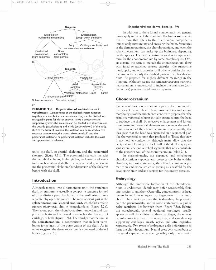
units: the skull, or cranial skeleton, and the postcranialskeleton (figure 7.1b). The postcranial skeleton includesthe vertebral column, limbs, girdles, and associated struc-tures, such as ribs and shells. In chapters 8 and 9, we exam-ine the postcranial skeleton. Our discussion of the skeletonbegins with the skull.
Introduction
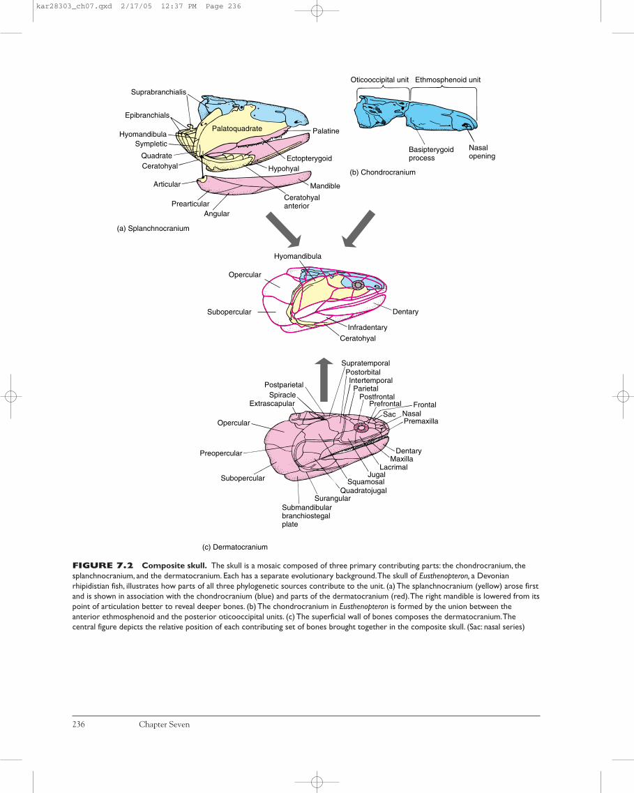
Although merged into a harmonious unit, the vertebrateskull, or cranium, is actually a composite structure formedof three distinct parts. Each part of the skull arises from aseparate phylogenetic source. The most ancient part is thesplanchnocranium (visceral cranium), which first arose tosupport pharyngeal slits in protochordates (figure 7.2a).The second part, the chondrocranium, underlies and sup-ports the brain and is formed of endochondral bone or ofcartilage, or both (figure 7.2b). The third part of the skull isthe dermatocranium, a contribution that in later verte-brates forms most of the outer casing of the skull. As itsname suggests, the dermatocranium is composed of dermalbones (figure 7.2c).
Endochondral and dermal bone (p. 179)
In addition to these formal components, two generalterms apply to parts of the cranium. The braincase is a col-lective term that refers to the fused cranial componentsimmediately surrounding and encasing the brain. Structuresof the dermatocranium, the chondrocranium, and even thesplanchnocranium can make up the braincase, dependingon the species. The neurocranium is used as an equivalentterm for the chondrocranium by some morphologists. Oth-ers expand the term to include the chondrocranium alongwith fused or attached sensory capsules—the supportivenasal, optic, and otic capsules. Still others consider the neu-rocranium to be only the ossified parts of the chondrocra-nium. Be prepared for slightly different meanings in theliterature. Although we use the term neurocranium sparingly,neurocranium is understood to include the braincase (ossi-fied or not) plus associated sensory capsules.
Chondrocranium
Elements of the chondrocranium appear to lie in series withthe bases of the vertebrae. This arrangement inspired severalmorphologists of the nineteenth century to propose that theprimitive vertebral column initially extended into the headto produce the skull. By selective enlargement and fusion,these intruding vertebral elements were seen as the evolu-tionary source of the chondrocranium. Consequently, theidea grew that the head was organized on a segmental planlike the vertebral column that produced it. Today this viewis not held as confidently, although many allow that theoccipital arch forming the back wall of the skull may repre-sent several ancient vertebral segments that now contributeto the posterior wall of the chondrocranium (table 7.1).
In elasmobranchs, the expanded and envelopingchondrocranium supports and protects the brain within.However, in most vertebrates, the chondrocranium is pri-marily an embryonic structure serving as a scaffold for thedeveloping brain and as a support for the sensory capsules.
EmbryologyAlthough the embryonic formation of the chondrocra-nium is understood, details may differ considerably fromone species to another. Generally, condensations of headmesenchyme form elongate cartilages next to the noto-chord. The anterior pair are the trabeculae, the posteriorpair the parachordals, and in some vertebrates, a pair ofpolar cartilages lies between them (figure 7.3a). Behindthe parachordals, several occipital cartilages usuallyappear as well. In addition to these cartilages, the sensorycapsules associated with the nose, eyes, and ears developsupporting cartilages: nasal, optic, and otic capsules,respectively. Two types of embryonic cells differentiate toform the chondrocranium. Neural crest cells contribute tothe nasal capsule, trabeculae (possibly only the anterior
Skeletal System: The Skull 235
Skeleton
Exoskeleton(within the integument)
Endoskeleton(deep, within the body)
Keratinizedexoskeleton
(from epidermis)
Bonyexoskeleton
(from dermis)
Bonyendoskeleton
Cartilaginousendoskeleton
Notochord
(a)
Skeleton
Cranial skeleton Postcranial
SplanchnocraniumChondrocranium
Dermatocranium
Axialskeleton
Vertebralcolumn
Notochord
Appendicularskeleton
GirdleLimbs
(b)
FIGURE 7.1 Organization of skeletal tissues invertebrates. Components of the skeletal system functiontogether as a unit but, as a convenience, they can be divided intomanageable parts for closer analysis. (a) As a protective andsupportive system, the skeleton can be divided into structures onthe outside (exoskeleton) and inside (endoskeleton) of the body.(b) On the basis of position, the skeleton can be treated as twoseparate components, the cranial skeleton (skull) and thepostcranial skeleton.The postcranial skeleton includes the axialand appendicular skeletons.
kar28303_ch07.qxd 2/17/05 12:37 PM Page 235
236 Chapter Seven
FIGURE 7.2 Composite skull. The skull is a mosaic composed of three primary contributing parts: the chondrocranium, thesplanchnocranium, and the dermatocranium. Each has a separate evolutionary background.The skull of Eusthenopteron, a Devonianrhipidistian fish, illustrates how parts of all three phylogenetic sources contribute to the unit. (a) The splanchnocranium (yellow) arose firstand is shown in association with the chondrocranium (blue) and parts of the dermatocranium (red).The right mandible is lowered from itspoint of articulation better to reveal deeper bones. (b) The chondrocranium in Eusthenopteron is formed by the union between theanterior ethmosphenoid and the posterior oticooccipital units. (c) The superficial wall of bones composes the dermatocranium.Thecentral figure depicts the relative position of each contributing set of bones brought together in the composite skull. (Sac: nasal series)
Dentary
Infradentary
Ceratohyal
SupratemporalPostorbital
ParietalPostfrontal
Prefrontal
Intertemporal
Sac NasalPremaxilla
Frontal
DentaryMaxilla
LacrimalJugal
SquamosalQuadratojugal
SurangularSubmandibularbranchiostegalplate
Subopercular
Preopercular
Postparietal
ExtrascapularSpiracle
Subopercular
Opercular
Hyomandibula
Basipterygoidprocess
Nasalopening
Oticooccipital unit Ethmosphenoid unit
Palatine
EctopterygoidHypohyal
Ceratohyalanterior
Mandible
AngularPrearticular
Articular
CeratohyalQuadrate
SympleticHyomandibula
Epibranchials
Suprabranchialis
Palatoquadrate
(a) Splanchnocranium
(b) Chondrocranium
(c) Dermatocranium
Opercular
kar28303_ch07.qxd 2/17/05 12:37 PM Page 236

part), and perhaps to part of the otic capsule (figure 7.4a).Mesenchyme of mesodermal origin contributes to the restof the chondrocranium (figure 7.4b). As development pro-ceeds, these cartilages fuse. The region between the nasalcapsules formed by the fusion of the anterior tips of the tra-beculae is the ethmoid plate. The parachordals growtogether across the midline to form the basal plate betweenthe otic capsules. The occipitals grow upward and around
the nerve cord to form the occipital arch (figure 7.3b).Collectively, all of these expanded and fused cartilagesconstitute the chondrocranium.
In elasmobranchs, the chondrocranium does notossify. Instead the cartilage grows still farther upward andover the brain to complete the protective walls and roof ofthe braincase. In most other vertebrates, the chondrocra-nium becomes partly or entirely ossified (figure 7.3c).
Skeletal System: The Skull 237
TABLE 7.1 Endochondral Contributions to the Chondrocranium
Endochondral FishesStructure (Teleost) Amphibians Reptiles/Birds Mammals
Occipital bones Supraoccipital Supraoccipital Supraoccipital SupraoccipitalExoccipital Exoccipital Exoccipital Exoccipital Occipital boneBasioccipital Basioccipital Basioccipital Basioccipital
Mesethmoid bone Mesethmoida Absent Absent Mesethmoid(internasal) (absent in primitive
mammals, ungulates) EthmoidEthmoid region Ossified Unossified Unossified Turbinals
(ethmo-, naso-, maxillo-)
Sphenoid bonesSphenethmoid Sphenethmoid Sphenethmoid Sphenethmoid PresphenoidOrbitosphenoid Orbitosphenoid Orbitosphenoid Orbitosphenoid Orbitosphenoid Sphenoidc
Basisphenoid [Basisphenoid]b Basisphenoid Basisphenoid BasisphenoidPleurosphenoid Pleurosphenoid ? Pleurosphenoid Absent
(crocodilians,amphisbaenians)
Laterosphenoid Laterosphenoid Absent(snakes)
Otic capsule Prootic Prootic Prootic Petrosal withPeriotic Epiotic Opisthotic Opisthotic mastoid process
Sphenotic Epiotic(absent in birds)
aThis bone is of dermal origin, so it is not strictly homologous to tetrapod mesethmoid.bThis bone is usually absent or reduced in some fishes.cAlisphenoid from the splanchnocranium contributes.
Basisphenoid
Parachordal
Nasal capsule
Trabecula
Otic capsule
Basalplate
Occipitalarch
Ethmoidplate
Opticcapsule
Polar cartilage
Sphenethnoid
Ethmoid
Basioccipital
Supraoccipital
Exoccipital
(c)(b)(a)
OccipitalsNotochord
FIGURE 7.3 Embryonic development of the chondrocranium. Cartilage (blue) appears first but in most vertebrates isreplaced by bone (white) later in development.The chondrocranium includes these cartilaginous elements that form the base and back ofthe skull together with the supportive capsules around sensory organs. Early condensation of mesenchymal cells differentiates intocartilage (a) that grows and fuses together to produce the basic ethmoid, basal, and occipital regions (b) that later ossify (c), forming basicbones and sensory capsules.
After deBeer.
∂
f•
kar28303_ch07.qxd 2/17/05 12:37 PM Page 237
Splanchnocranium
The splanchnocranium is an ancient chordate structure. Inamphioxus, the splanchnocranium, or at least its forerunner,is associated with the filter-feeding surfaces.
Among vertebrates, the splanchnocranium generallysupports the gills and offers attachment for the respiratorymuscles. Elements of the splanchnocranium contribute tothe jaws and hyoid apparatus of gnathostomes.
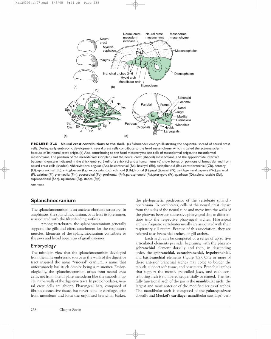
EmbryologyThe mistaken view that the splanchnocranium developedfrom the same embryonic source as the walls of the digestivetract inspired the name “visceral” cranium, a name thatunfortunately has stuck despite being a misnomer. Embry-ologically, the splanchnocranium arises from neural crestcells, not from lateral plate mesoderm like the smooth mus-cle in the walls of the digestive tract. In protochordates, neu-ral crest cells are absent. Pharyngeal bars, composed offibrous connective tissue, but never bone or cartilage, arisefrom mesoderm and form the unjointed branchial basket,
the phylogenetic predecessor of the vertebrate splanch-nocranium. In vertebrates, cells of the neural crest departfrom the sides of the neural tube and move into the walls ofthe pharynx between successive pharyngeal slits to differen-tiate into the respective pharyngeal arches. Pharyngealarches of aquatic vertebrates usually are associated with theirrespiratory gill system. Because of this association, they arereferred to as branchial arches, or gill arches.
Each arch can be composed of a series of up to fivearticulated elements per side, beginning with the pharyn-gobranchial element dorsally and then, in descendingorder, the epibranchial, ceratobranchial, hypobranchial,and basibranchial elements (figure 7.5). One or more ofthese anterior branchial arches may come to border themouth, support soft tissue, and bear teeth. Branchial archesthat support the mouth are called jaws, and each con-tributing arch is numbered sequentially or named. The firstfully functional arch of the jaw is the mandibular arch, thelargest and most anterior of the modified series of arches.The mandibular arch is composed of the palatoquadratedorsally and Meckel’s cartilage (mandibular cartilage) ven-
238 Chapter Seven
Lacrimal
NasalJugalMaxilla
Premaxilla
Mandible
LaryngealsHyoidsOccipitals
Petrosus
FrontalParietal
Po
PrfN
NcPm
DEg
BhBb
CbJ
EbOticStp
Eo
Sq
PSoc
F
BsPs
Pl
AnQ Pt
s c i
Pharynx
Neuralcrest
Branchial arches 3–6Hyoid arch
Mandibular archStomodeum
Diencephalon
Mesodermalmesenchyme
Neural crestmesenchyme
Neural crest-mesoderminterface
MesencephalonMyelen-cephalon
Oto
(b)(a)
(d)(c)
Sphenoid
Sq.
Eth
FIGURE 7.4 Neural crest contributions to the skull. (a) Salamander embryo illustrating the sequential spread of neural crestcells. During early embryonic development, neural crest cells contribute to the head mesenchyme, which is called the ectomesodermbecause of its neural crest origin. (b) Also contributing to the head mesenchyme are cells of mesodermal origin, the mesodermalmesenchyme.The position of the mesodermal (stippled) and the neural crest (shaded) mesenchyme, and the approximate interfacebetween them, are indicated in the chick embryo. Skull of a chick (c) and a human fetus (d) show bones or portions of bones derived fromneural crest cells (shaded).Abbreviations: angular (An), basibranchial (Bb), basihyal (Bh), basisphenoid (Bs), ceratobranchial (Cb), dentary(D), epibranchial (Eb), entoglossum (Eg), exoccipital (Eo), ethmoid (Eth), frontal (F), jugal (J), nasal (N), cartilage nasal capsule (Nc), parietal(P), palatine (Pl), premaxilla (Pm), postorbital (Po), prefrontal (Prf), parasphenoid (Ps), pterygoid (Pt), quadrate (Q), scleral ossicle (Sci),supraoccipital (Soc), squamosal (Sq), stapes (Stp).
After Noden.
kar28303_ch07.qxd 3/9/05 9:41 AM Page 238

trally. The hyoid arch, whose most prominent element isthe hyomandibula, follows the mandibular arch. A varyingnumber of branchial arches, often designated with romannumerals, follow the hyoid arch (figure 7.5).
Origin of JawsIn agnathans, the mouth is neither defined nor supportedby jaws. Instead, the splanchnocranium supports the roof ofthe pharynx and lateral pharyngeal slits. Lacking jaws,ostracoderms would have been restricted to a diet of small,particulate food. The ciliary-mucous feeding surfaces of pro-tochordates probably continued to play a large part in thefood-gathering technique of ostracoderms. In some groups,small teethlike structures, derived from surface scales, sur-rounded the mouth. Perhaps ostracoderms used these rough“teeth” to scrape rock surfaces and dislodge encrusted algaeor other organisms. As these food particles became sus-pended in water, ostracoderms drew them into their mouthwith the incurrent flow of water. The mucus-lined walls ofthe pharynx collected these dislodged food particles fromthe passing stream.
Jaws appear first in acanthodian and placoderm fishesthat used them as food traps to grab whole prey or take bitesfrom large prey. Within some groups, jaws also served ascrushing or chewing devices to process food in the mouth.With the advent of jaws, these fishes became more free-ranging predators of open waters.
Jaws arose from one of the anterior pair of gill arches.Evidence supporting this comes from several sources. First,the embryology of sharks suggests that jaws and branchial
arches develop similarly in series (figure 7.6) and both arisefrom neural crest. The spiracle appears to have once been afull-sized gill slit, but in modern sharks it is crowded andmuch reduced by the enlarged hyoid arch next in series. Fur-thermore, nerves and blood vessels are distributed in a pat-tern similar to branchial arches and jaws. Finally, themusculature of the jaws appears to be transformed and mod-ified from branchial arch musculature.
So it seems reasonable to conclude that branchialarches phylogenetically gave rise to jaws. But the specificsremain controversial. For example, we are not sure whetherjaws represent derivatives of the first, second, third, or evenfourth branchial arches of primitive ancestors. Derivation ofthe mandibular arch also excites some controversy. Theserial theory is the simplest view and holds that the first orperhaps second ancient branchial arch gave rise exclusivelyto the mandibular arch, the next branchial arch exclusivelyto the hyoid arch, and the rest of the arches to the branchialarches of gnathostomes (figure 7.7a).
Erik Jarvik, a Swedish paleontologist, proposed thecomposite theory, a more complex view based on his exam-ination of fossil fish skulls and embryology of living forms(figure 7.7b). He hypothesized that ten branchial arches were present in primitive species, the first and followingarches being named terminal, premandibular, mandibular,hyoid, and six branchial arches. Rather than the “one arch,one mandible” view, he envisioned a complex series of losses or fusions between selective parts of several arches thatcame together to produce the single composite mandible.
Skeletal System: The Skull 239
Pharyngobranchial
Epibranchial
Ceratobranchial
Hypobranchial
BasibranchialBranchial arches Hyoid
archMandibulararch
Pharyngeal slitsPharyngeal slits
HyomandibulaPalatoquadrate
Meckel'scartilage
FIGURE 7.5 Primitive splanchnocranium. Sevenarches are shown. Up to five elements compose an arch on eachside, beginning with the pharyngobranchial dorsally and insequence to the basibranchials most ventrally.The first twocomplete arches are named: mandibular arch for the first andhyoid arch for the second that supports it.The characteristic five-arch elements are reduced to just two in the mandibular arch: thepalatoquadrate and Meckel’s cartilage.The large hyomandibula,derived from an epibranchial element, is the most prominentcomponent of the next arch, the hyoid arch. Behind the hyoidarch are variable numbers of branchial arches I, II, and so on.Labial cartilages are not included.
Oa
Ne
HyPc
Tr
Oc
Pq
Mk
Ch
Branchial arches
FIGURE 7.6 Shark embryo, the dogfish Scyllium. Jawsappear to be in series with the branchial arches.The mandibulararch is first, followed by the hyoid and then several branchialarches. Such a position of the jaws, in series with the arches, istaken as evidence that the jaws derive from the most anteriorbranchial arch.Abbreviations: ceratohyal (Ch), hyomandibula (Hy),Meckel’s cartilage (Mk), neural arch (Ne), occipital arch (Oa),orbital cartilage (Oc), polar cartilage (Pc), palatoquadrate (Pq),trabecula (Tr). Labial cartilages are not included.
After deBeer.
kar28303_ch07.qxd 2/17/05 12:37 PM Page 239

According to his theory, the mandibular arch of gnathos-tomes is formed by fusion of parts of the premandibular archand parts of the mandibular arch of jawless ancestors. Thepalatoquadrate forms from the fusion of the epibranchial ofthe premandibular arch with the epibranchial and onepharyngobranchial of the mandibular arch. Meckel’s carti-lage arises from the expanded ceratobranchial element.Next, the hyoid arch arises phylogenetically from the epi-branchial, ceratobranchial, and hypobranchial elements ofthe third primitive gill arch. The remaining branchial archespersist in serial order. The other elements of the primitivearches are lost or fused to the neurocranium.
Descriptive embryology provides much of the evi-dence put forth in these theories. However, descriptiveembryology alone cannot trace arch components fromembryo to adult structures with complete confidence. Wecan look forward to the use of more modern techniques tohelp settle this. For example, populations of cells can bemarked with chemical or cellular markers early in embry-onic development and followed to eventual sites of resi-dence in the adult. These markers would permit us to detectthe contributions of gill arches to jaws or chondrocranium.
Nevertheless, even though some argue over details, we knowin general that vertebrate jaws are derivatives of ancient gillarches (table 7.2).
Types of Jaw AttachmentsBecause of the mandible’s prominence, evolution of the jawsis often traced through how the mandible is attached (i.e.,its suspensorium) to the skull (figure 7.8). Agnathans rep-resent the earliest paleostylic stage in which none of thearches attach themselves directly to the skull. The earliestjawed condition is euautostylic, found in placoderms and
240 Chapter Seven
Branchial archGill slit
Agnathan
(a) Serial theory (b) Composite theory
Contributions to
Otic shelf Neurocranium
Mandibulararch
Hyoidarch
Branchialarches
Mandibulararch
Hyoidarch
Branchialarches
FIGURE 7.7 Serial and composite theories of jawdevelopment. (a) The serial theory holds that jaws arisecompletely from one of the anterior branchial arches. Elementsmay be lost within it, but other elements from other arches donot contribute. (b) In the composite theory, the mandibular archis formed from elements of several adjacent arches that alsocontribute to the neurocranium.
Paleostyly(agnathans)
Meckel's cartilage
Palatoquadrate
Euautostyly(placoderms,acanthodians)
Amphistyly(primitive fish)
Dentary
Stapes
Quadrate
Hyostyly(some fish)
Metautostyly(most amphibians,reptiles, and birds)
TemporalDentary
Symplectic
Modified hyostyly(teleosts)
Craniostyly(mammals)
Hyomandibula
FIGURE 7.8 Jaw suspension. The points at which thejaws attach to the rest of the skull define the type of jawsuspension. Note the mandibular arches (yellow, crosshatchedareas) and hyoid arches (yellow areas).The dermal bone (whiteareas) of the lower jaw is the dentary.
kar28303_ch07.qxd 3/9/05 9:41 AM Page 240

acanthodians. The mandibular arch is suspended from theskull by itself (hence, “auto”), without help from the hyoidarch. In early sharks, some osteichthyans, and rhipistians,jaw suspension is amphistylic; that is, the jaws are attachedto the braincase through two primary articulations, anteri-orly by a ligament connecting the palatoquadrate to theskull and posteriorly by the hyomandibula. Many, perhapsmost, modern sharks exhibit a variation of amphistylic jawsuspension. In most modern bony fishes, jaw suspension ishyostylic because the mandibular arch is attached to thebraincase primarily through the hyomandibula. Often a newelement, the symplectic bone, aids in jaw suspension. Thevisceral cranium remains cartilaginous in elasmobranchs,but within bony fishes and later tetrapods, ossification cen-ters appear, forming distinctive bony contributions to theskull. In most amphibians, reptiles, and birds, jaw suspensionis metautostylic. Jaws are attached to the braincase directlythrough the quadrate, a bone formed in the posterior part ofthe palatoquadrate (figure 7.8). The hyomandibula plays nopart in supporting the jaws; instead, it gives rise to the slen-der columella or stapes, involved in hearing. Other ele-ments of the second arch and parts of the third contribute tothe hyoid or hyoid apparatus that supports the tongue andthe floor of the mouth. In mammals, jaw suspension is cran-iostylic. The entire upper jaw is incorporated into the brain-case, but the lower jaw is suspended from the dermalsquamosal bone of the braincase. The lower jaw of mammalsconsists entirely of the dentary bone, which is also of dermalorigin. The palatoquadrate and Meckel’s cartilages stilldevelop, but they remain cartilaginous except at their pos-terior ends, which give rise to the incus and malleus of the
middle ear, respectively (figure 7.9). Thus, in mammals, thesplanchnocranium does not contribute to the adult jaws orto their suspension. Instead, the splanchnocranium formsthe hyoid apparatus, styloid, and three middle ear bones:malleus, incus, and stapes. Through Meckel’s cartilage, thesplanchnocranium contributes the scaffolding around whichthe dentary bone forms.
Skeletal System: The Skull 241
TABLE 7.2 Derivatives of Branchial Arches in Sharks,Teleosts, and Tetrapods
Arch Sharks Teleosts Amphibians Reptiles/Birds Mammals
I Meckel’s cartilage Articulara Articular Articular Malleusb
Palatoquadrate Quadrate Quadrate Quadrate Incusb
Epipterygoid Epipterygoid Epipterygoid Alisphenoid
II Hyomandibula Hyomandibula Stapes Stapes Stapesb
Symplectic Extracolumella ExtracolumellaInterhyal
Ceratohyal Ceratohyal Ceratohyal Ceratohyal Anterior horn hyoidHypohyal Hypohyal
Basihyal Basihyal Body of hyoid Body of hyoid
III Pharyngobranchial PharyngobranchialEpibranchial EpibranchialCeratobranchial Ceratobranchial Body of hyoid Second horn of hyoid Second horn of hyoidHypobranchial Hypobranchial
IV Branchial arch Last horn and body of hyoid Last horn and body of hyoid Thyroid cartilages (?)Laryngeal cartilages (?) Laryngeal cartilages (?)
V Branchial arch Branchial arch Laryngeal cartilages (?) Laryngeal cartilages (?) Laryngeal cartilages
VI Branchial arch Branchial arch Not present Not present Not present
VII Branchial arch Branchial arch
aSometimes dermal bone contributes.bSee figure 7.53 and related text for discussion of middle ear evolution.
SquamosalPremaxilla
Supra-occipital
ParietalFrontal
LacrimalMaxilla
Nasal
Meckel'scartilage
JugalDentary
Trachea
IncusStapes
MalleusTympanic
FIGURE 7.9 Skull of armadillo embryo. Duringembryonic formation of the three middle ear ossicles (incus,stapes, malleus), the incus and stapes arise from the mandibulararch, testifying to the phylogenetic derivation of these bones fromthis arch.The dermal dentary is cut away to reveal Meckel’scartilage, which ossifies at its posterior end to form the malleus.(Blue, chondrocranium contribution; yellow, splanchnocraniumcontribution; red, dermatocranium.)
After Goodrich.
U
v
kar28303_ch07.qxd 2/17/05 12:37 PM Page 241

Dermatocranium
Dermal bones that contribute to the skull belong to thedermatocranium. Phylogenetically, these bones arise fromthe bony armor of the integument of early fishes and sinkinward to become applied to the chondrocranium andsplanchnocranium. Bony elements of the armor alsobecome associated with the endochondral elements of thepectoral girdle to give rise to the dermal components ofthis girdle.
Dermal girdle (p. 330)
Dermal bones first become associated with the skull inostracoderms. In later groups, additional dermal bones of theoverlying integument also contribute. The dermatocraniumforms the sides and roof of the skull to complete the protec-tive bony case around the brain; it forms most of the bonylining of the roof of the mouth, and encases much of thesplanchnocranium. Teeth that arise within the mouth usu-ally rest on dermal bones.
As the name suggests, bones of the dermatocraniumarise directly from mesenchymal and ectomesenchymal tis-sues of the dermis. Through the process of intramembranousossification, these tissues form dermatocranial bones.
Parts of the DermatocraniumDermal elements in modern fishes and living amphibianshave tended to be lost or fused so that the number ofbones present is reduced and the skull simplified. Inamniotes, bones of the dermatocranium predominate,forming most of the braincase and lower jaw. The dermalskull may contain a considerable series of bones joinedfirmly at sutures in order to box in the brain and otherskull elements. As a convenience, we can group theseseries and recognize the most common bones in each (fig-ure 7.10; table 7.3).
Dermal Bone SeriesFacial Series The facial series encircles the external narisand collectively forms the snout. The maxilla and premax-illa (incisive) define the margins of the snout and usuallybear teeth. The nasal lies medial to the naris. The sep-tomaxilla is a small dermal bone of the facial series that isoften absent. When present, it is usually sunken below thesurface bones and aids in forming the nasal cavity.
Orbital Series The dermal bones encircle the eye to definethe orbit superficially. The lacrimal takes its name from thenasolacrimal (tear) duct of tetrapods that passes through ornear this bone. The prefrontal, postfrontal, and postorbitalcontinue the ring of bones above and behind the orbit. Thejugal usually completes the lower rim of the orbit. Not to beconfused with these dermal bones are the scleral ossicles ofneural crest origin that, when present, reside within theorbit defined by the ring of dermal bones.
Temporal Series The temporal series lies behind the orbit,completing the posterior wall of the braincase. In many prim-itive tetrapods, this series is indented posteriorly by a tempo-ral notch. Once thought in life to suspend an eardrum, thisnotch was named accordingly an otic notch. This now seemsunlikely, and instead the notch perhaps accommodated aspiracle, a respiratory tube. Openings called fenestrae (sing.,fenestra) arise within this region of the outer braincase inmany tetrapods in association with the jaw musculature. Arow of bones, the intertemporal, supratemporal, and tabu-lar, make up the medial part of the temporal series. This rowis reduced in early tetrapods and usually lost in later species.Laterally, the squamosal and quadratojugal complete thetemporal series and form the “cheek.”
Vault Series The vault, or roofing bones, run across thetop of the skull and cover the brain beneath. These includethe frontal anteriorly and the postparietal (interparietal)posteriorly. Between them is the large parietal, occupyingthe center of the roof and defining the small parietal fora-men if it is present. The parietal foramen is a tiny skylight inthe skull roof that exposes the pineal gland, an endocrinegland, to direct sunlight.
242 Chapter Seven
Facial series
Orbital series
Vaultseries
Temporalseries
PalatalDorsal
V
It
St
T
J Ec
Pl
Qj
Po
L PrfF
MN
Pm
P
PpSq
Pt
Ps
Pf
SaD
SpAnSp
Pa
Lateral Medial
Coronoids
Palatalseries
FIGURE 7.10 Major bones of the dermatocranium.Sets of dermal bones form the facial series surrounding thenostril.The orbital series encircles the eye, and the temporalseries composes the lateral wall behind the eye.The vault series,the roofing bones, run across the top of the skull above the brain.Covering the top of the mouth is the palatal series of bones.Meckel’s cartilage (not shown) is encased in the mandibular seriesof the lower jaw.Abbreviations: angular (An), dentary (D),ectopterygoid (Ec), frontal (F), intertemporal (It), jugal (J), lacrimal(L), maxilla (M), nasal (N), parietal (P), prearticular (Pa), palatine(Pl), premaxilla (Pm), postorbital (Po), postparietal (Pp), prefrontal(Prf), parasphenoid (Ps), pterygoid (Pt), quadratojugal (Qj),surangular (Sa), splenial (Sp), squamosal (Sq), supratemporal (St),tabular (T), vomer (V).
kar28303_ch07.qxd 2/17/05 12:37 PM Page 242

Palatal Series The dermal bones of the primary palatecover much of the roof of the mouth. The largest and mostmedial is the pterygoid. Lateral to it are the vomer, palatine,and ectopterygoid. Teeth may be present on any or all fourof these palatal bones. In fishes and lower tetrapods, therealso is an unpaired medial dermal bone, the parasphenoid.
Mandibular Series Meckel’s cartilage is usually encased indermal bones of the mandibular series. Laterally, the wall ofthis series includes the tooth-bearing dentary and one ortwo splenials, the angular at the posterior corner of themandible and the surangular above. Many of these boneswrap around the medial side of the mandible and meet theprearticular and one or several coronoids to complete themedial mandibular wall. Left and right mandibles usuallymeet anteriorly at the midline in a mandibular symphysis.If firm, the mandibular symphysis unites them into anarched unit. Most notably in snakes, the mandibular symph-ysis is composed of soft tissues, permitting independentmovement of each mandible.
Overview of Skull Morphology
BraincaseIn chondrichthyan fishes, the braincase is an elaborate car-tilaginous case around the brain. The dermatocranium isabsent, reflecting the elimination of almost all bone fromthe skeleton. However, in most bony fishes and tetrapods,the braincase is extensively ossified with contributions fromseveral sources. For descriptive purposes, it is useful to thinkof the braincase as a box with a platform of endoskeletal ele-ments supporting the brain, all encased in exoskeletal bones(figure 7.11). The endoskeletal platform is assembled from aseries of sphenoid bones. The occipital bones, which appar-ently are derived from anterior vertebrae, form the end ofthis sphenoid platform. These occipital bones, up to four innumber (basioccipital, supraoccipital, and paired exoccipi-tals), close the posterior wall of the braincase except for alarge hole they define, the foramen magnum, throughwhich the spinal cord runs. Articulation of the skull with
Skeletal System: The Skull 243
TABLE 7.3 Major Dermal Bones of the SkullB R A I N C A S E M A N D I B L E
Facial Series Orbital Series Temporal Series Vault Series Palatal Series Mandibular Series
Premaxilla Lacrimal Intertemporal Frontal Vomer Lateral bones:
Maxilla Prefrontal Supratemporal Parietal Palatine Dentary (teeth)
Nasals Postfrontal Tabular Postparietal Ectopterygoid Splenials (2)(septomaxilla) Postorbital
Jugal Squamosal Pterygoid AngularQuadratojugal Parasphenoid Surangular
(unpaired)Medial bones:
PrearticularCoronoids
Columella (stapes)
Quadrate (incusof mammals)
Articular(malleus ofmammals)
Epipterygoid
Vault seriesOrbital series
Temporal series
Hyoidapparatus
PalatalseriesMandibularseries
Facialseries
Orbitosphenoid Presphenoid Mesethmoid
Sphenethmoid(lower tetrapods)
Basisphenoid
Basi-Ex-Supraoccipitals
Vertebrae
Opisthotic Prootic
Otic capsule
Hyomandibula
Meckel's cartilage PalatoquadrateMandibular arch
Hyoidarch
3d–5tharches
FIGURE 7.11 Contributions to the skull. Thechondrocranium (blue) establishes a supportive platform that is joinedby contributions from the splanchnocranium (yellow), in particular theepipterygoid.Other parts of the splanchnocranium give rise to thearticular,quadrate, and hyomandibula, as well as to the hyoid apparatus.The dermatocranium (red) encases most of the chondrocraniumtogether with contributions from the splanchnocranium.
kar28303_ch07.qxd 2/17/05 12:37 PM Page 243

While disposing of the vertebral theory,Huxley substituted a segmental theory,tracing the segmentation to somites, not tovertebrae (box figure 1c). He took the oticcapsule housing the ear as a “fixed” land-mark and envisioned four somites (preotic)in front and five somites (postotic) behindit as segmental sources for segmental adultderivatives of the head.
244 Chapter Seven
The idea that the skull is derived fromserial compacted vertebrae dates to theeighteenth century.The German naturalistand poet, W. Goethe (1749–1832), wasapparently the first to think of but not thefirst to publish this idea.Goethe gave us theword morphology, which meant to him thesearch for underlying meaning in organicdesign or form.Among his discoveries wasthe observation that plant flowers aremodified stem petals compacted together.His venture into vertebrates and verte-brate skulls in particular occurred in 1790whilst he was strolling in an old cemeteryin Venice. He spied a dried ram’s skull disin-tegrated at its bony sutures but held insequence by the soil.The separated bonesof the ram’s skull seemed to be the fore-shortened anterior vertebrae of the back-bone, but Goethe did not publish this ideauntil about 1817. Public credit for this ideaand for elaborating it goes to another Ger-man naturalist, L. Oken (1779–1851). In1806, Oken was strolling in a forest andcame upon a dried sheep skull. He was sim-ilarly struck by its serial homology with thevertebrae, and shortly thereafter publishedthe idea (box figure 1a).
Next, the vertebral theory of skull originfell into the hands of Richard Owen andbecame part of his much embellished theo-retical view on animal archetypes (box fig-ure 1b). Because of Owen’s prominence inearly nineteenth-century science, the ideaof skull from vertebrae became a centralissue within European scientific communi-ties. One of the most persuasive dissentersfrom this view of a vertebral source for theskull was T. H. Huxley, who based his cri-tique upon a detailed comparative study ofvertebrate skulls and their development.This came to a head (no pun intended) in aninvited lecture, the Croonian lecture of1858, in which Huxley argued that thedevelopment of the skull showed that it wasnot composed of vertebrae. He suggestedthat the “skull was no more derived fromvertebrae, than vertebrae are derived from
the skull.” The skull, Huxley argued, arose inmuch the same way in most vertebrates, byfusing into a unit, not as a jointed series.Skull ossification showed no similarity withossification of the following vertebrae.Although Huxley was probably right aboutthis for most of the skull, the occipitalregion does ossify in a manner similar tovertebrae.
BOX ESSAY 7 .1 Getting a Head
3
P
2
F
1
N
2Or
II
3
Pro
4
Pp
5
So
Ex
5
o p s
4
2345
(a)
BsBo
BOX FIGURE 1 Getting a head. Derivation of the head from anteriorvertebrae was proposed separately by Goethe and Oken. Owen expanded on their ideas.(a) Ram’s skull, showing how its presumed segmental pattern might be interpreted as beingderived from parts of anterior vertebrae that expanded. (b) Richard Owen’s elaboratedview of head segmentation from vertebrae. Owen proposed that anterior vertebrae withinthe body moved forward to contribute to skeletal elements to the head. Therefore, Owenbelieved, the bony elements of the head could be homologized to the parts of afundamental vertebral pattern. (c) Taking several vertebrates, he indicated how namedparts of the skull might represent respective parts of this underlying vertebral pattern fromwhich they derive. (d) T. H. Huxley proposed alternatively that, rather than being derivedfrom vertebrae moved forward into the head, the components of the head were derivedfrom a basic segmentation unrelated to the vertebral segmentation behind the skull. Thesebasic segments (roman numerals) are laid out across a generalized vertebrate skull to showthe respective contributions to specific parts. Abbreviations: basioccipital (Bo),basisphenoid (Bs), exoccipital (Ex), frontal (F), nasal (N), opisthotic (Ops),orbitosphenoid (Or), parietal (P), postparietal (Pp), prootic (Pro), supraoccipital (So).
(a) After Jollie.
kar28303_ch07.qxd 3/9/05 9:41 AM Page 244

Skeletal System: The Skull 245
Today, some would argue that the headis a unique developmental system withoutany tie to the segmental somites (somito-meres). The neural crest cells that alsocontribute to parts of the skull show no
segmental pattern in the head. However, atleast in fishes, the branchial arches are seg-mental, as is the head paraxial mesoderm(somitomeres), and segmentation appar-ently can be carried into the accompanying
neurocranium. Matched shading in verte-brate series (box figure 1c) shows deriva-tives from parts of theoretical ancestor(box figure 1b).
(b) Theoretical ancestor
(c) Fish (teleost)
(d)
Reptile (alligator) Canine (dog) Human
V
IVIII II
I
(b–c) After Reader; (d) after Jollie.
kar28303_ch07.qxd 2/17/05 12:37 PM Page 245

the vertebral column is established through the occipitalcondyle, a single or double surface produced primarilywithin the basioccipital but with contributions from theexoccipitals in some species.
The otic capsule rests on the posterior part of theendoskeletal platform and encloses the sensory organs of theear. The splanchnocranium contributes the epipterygoid(alisphenoid of mammals) to the endoskeletal platform andgives rise to one (columella/stapes) or more (malleus andincus of mammals) of the middle ear bones housed in theotic capsule.
In most vertebrates, these endoskeletal elements,along with the brain and sensory organs they support, areenclosed by the exoskeletal elements, derivatives of the der-mis, to complete the braincase.
JawsThe upper jaw consists of the endoskeletal palatoquadratein primitive vertebrates. The palatoquadrate is fully func-tional in the jaws of chondrichthyans and primitive fishes,but in bony fishes and tetrapods, the palatoquadrate usuallymakes limited contributions to the skull through its twoderivatives: the epipterygoid, which fuses to the neurocra-nium, and the quadrate, which suspends the lower jawexcept in mammals. The dermal maxilla and premaxillareplace the palatoquadrate as the upper jaw.
The lower jaw, or mandible, consists only of Meckel’scartilage in chondrichthyans. In most fishes and tetrapods,Meckel’s cartilage persists but is enclosed in exoskeletalbone of the dermatocranium, which also supports teeth.Meckel’s cartilage, encased in dermal bone, usually remainsunossified, except in some tetrapods where its anterior endossifies as the mental bone. In most fishes and tetrapods(except mammals), the posterior end of Meckel’s cartilagecan protrude from the exoskeletal case as an ossified articu-lar bone.
In mammals, the lower jaw consists of a single bone,the dermal dentary. The anterior tooth-bearing part of thedentary is its ramus. Jaw-closing muscles are inserted on thecoronoid process, an upward extension of the dentary. Pos-teriorly, the dentary forms the transversely expandedmandibular condyle, a rounded process that articulates withthe glenoid fossa, a depression within the temporal bone ofthe braincase. Thus, in mammals, the mandibular condyle ofthe dentary replaces the articular bone as the surface of thelower jaw through which is established mandibular articula-tion with the braincase.
Hyoid ApparatusThe hyoid or hyoid apparatus is a ventral derivative of thesplanchnocranium behind the jaws. In fishes, it supports thefloor of the mouth. Elements of the hyoid apparatus arederived from the ventral parts of the hyoid arch and fromparts of the first few branchial arches. In larval and paedo-morphic amphibians, the branchial bars persist but form a
reduced hyoid apparatus that supports the floor of the mouthand functional gills. In adults, the gills and the associatedpart of the hyoid apparatus are lost, although elements per-sist within the floor of the mouth usually to support thetongue. Typically, the hyoid apparatus includes a main body,the corpus, and extensions, the cornua (“horns”). In manymammals, including humans, the distal end of the hyoidhorn fuses with the otic region of the braincase to form thestyloid process.
Cranial Kinesis
Kinesis means movement. Cranial kinesis refers literallythen to movement within the skull. But if left this general,the definition becomes too broad to provide a useful contextin which to discuss skull function. Some authors restrict theterm to skulls with a transverse, hingelike joint across theskull roof and a transverse, sliding basal joint in the roof ofthe mouth. But this restricted definition precludes mostteleost fishes, despite their highly mobile skull elements.Here, we use cranial kinesis to mean movement between theupper jaw and the braincase about joints between them (fig-ure 7.12a). Such kinetic skulls characterize most verte-brates. They are found in ancient fishes (crossopterygiansand probably palaeoniscoids), bony fishes (especiallyteleosts), very early amphibians, most reptiles (includingmost Mesozoic forms), birds, and early therapsid ancestors tomammals. Kinetic skulls are not present in modern amphib-ians, turtles, crocodiles, and mammals (with the possibleexception of rabbits). The widespread presence of cranialkinesis among vertebrates, but its essential absence amongmammals, seems to create a problem for humans. Becausewe, like most other mammals, have akinetic skulls with nosuch movement between upper jaw and braincase, we tendto underestimate its importance (figure 7.12b).
Kinesis and akinesis each have advantages. Cranialkinesis provides a way to change the size and configurationof the mouth rapidly. In fishes and other vertebrates thatfeed in water, rapid kinesis creates a sudden reduction ofpressure in the buccal cavity so that the animal can suck ina surprised prey. This method of prey capture, which takesadvantage of a sudden vacuum to gulp in water carrying theintended food, is known as suction feeding. Cranial kinesisalso allows tooth-bearing bones to move quickly into strate-gic positions during rapid feeding. Some teleost fishes, forinstance, swing their anterior tooth-bearing bones forwardat the last moment to reach out quickly at the intended prey.In many venomous snakes, linked bones along the sides ofthe skull can rotate forward. The venomous viper erects themaxillary bone bearing the fang and swings it from a foldedposition along its upper lip to the front of the mouth, whereit can more easily deliver venom into prey. In many fishesand reptiles with kinetic skulls, teeth on the upper jaw canbe reoriented with respect to the prey in order to assume amore favorable position during prey capture or to align
246 Chapter Seven
kar28303_ch07.qxd 2/17/05 12:37 PM Page 246
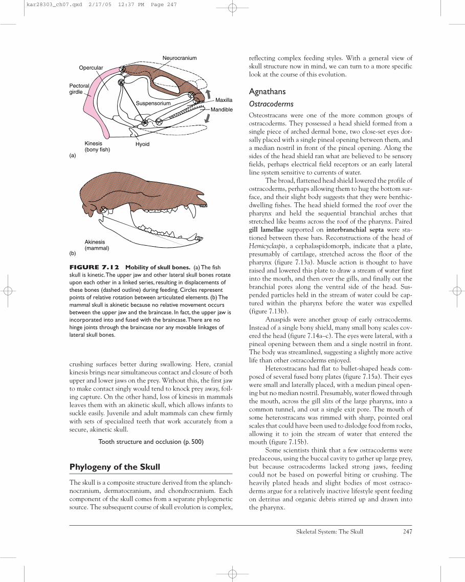
crushing surfaces better during swallowing. Here, cranialkinesis brings near simultaneous contact and closure of bothupper and lower jaws on the prey. Without this, the first jawto make contact singly would tend to knock prey away, foil-ing capture. On the other hand, loss of kinesis in mammalsleaves them with an akinetic skull, which allows infants tosuckle easily. Juvenile and adult mammals can chew firmlywith sets of specialized teeth that work accurately from asecure, akinetic skull.
Tooth structure and occlusion (p. 500)
Phylogeny of the Skull
The skull is a composite structure derived from the splanch-nocranium, dermatocranium, and chondrocranium. Eachcomponent of the skull comes from a separate phylogeneticsource. The subsequent course of skull evolution is complex,
reflecting complex feeding styles. With a general view ofskull structure now in mind, we can turn to a more specificlook at the course of this evolution.
AgnathansOstracodermsOsteostracans were one of the more common groups ofostracoderms. They possessed a head shield formed from asingle piece of arched dermal bone, two close-set eyes dor-sally placed with a single pineal opening between them, anda median nostril in front of the pineal opening. Along thesides of the head shield ran what are believed to be sensoryfields, perhaps electrical field receptors or an early lateralline system sensitive to currents of water.
The broad, flattened head shield lowered the profile ofostracoderms, perhaps allowing them to hug the bottom sur-face, and their slight body suggests that they were benthic-dwelling fishes. The head shield formed the roof over thepharynx and held the sequential branchial arches thatstretched like beams across the roof of the pharynx. Pairedgill lamellae supported on interbranchial septa were sta-tioned between these bars. Reconstructions of the head ofHemicyclaspis, a cephalaspidomorph, indicate that a plate,presumably of cartilage, stretched across the floor of thepharynx (figure 7.13a). Muscle action is thought to haveraised and lowered this plate to draw a stream of water firstinto the mouth, and then over the gills, and finally out thebranchial pores along the ventral side of the head. Sus-pended particles held in the stream of water could be cap-tured within the pharynx before the water was expelled(figure 7.13b).
Anaspids were another group of early ostracoderms.Instead of a single bony shield, many small bony scales cov-ered the head (figure 7.14a–c). The eyes were lateral, with apineal opening between them and a single nostril in front.The body was streamlined, suggesting a slightly more activelife than other ostracoderms enjoyed.
Heterostracans had flat to bullet-shaped heads com-posed of several fused bony plates (figure 7.15a). Their eyeswere small and laterally placed, with a median pineal open-ing but no median nostril. Presumably, water flowed throughthe mouth, across the gill slits of the large pharynx, into acommon tunnel, and out a single exit pore. The mouth ofsome heterostracans was rimmed with sharp, pointed oralscales that could have been used to dislodge food from rocks,allowing it to join the stream of water that entered themouth (figure 7.15b).
Some scientists think that a few ostracoderms werepredaceous, using the buccal cavity to gather up large prey,but because ostracoderms lacked strong jaws, feedingcould not be based on powerful biting or crushing. Theheavily plated heads and slight bodies of most ostraco-derms argue for a relatively inactive lifestyle spent feedingon detritus and organic debris stirred up and drawn intothe pharynx.
Skeletal System: The Skull 247
Pectoralgirdle
Opercular
Neurocranium
Maxilla
Mandible
HyoidKinesis(bony fish)
Suspensorium
Akinesis(mammal)
(b)
(a)
FIGURE 7.12 Mobility of skull bones. (a) The fishskull is kinetic.The upper jaw and other lateral skull bones rotateupon each other in a linked series, resulting in displacements ofthese bones (dashed outline) during feeding. Circles representpoints of relative rotation between articulated elements. (b) Themammal skull is akinetic because no relative movement occursbetween the upper jaw and the braincase. In fact, the upper jaw isincorporated into and fused with the braincase.There are nohinge joints through the braincase nor any movable linkages oflateral skull bones.
kar28303_ch07.qxd 2/17/05 12:37 PM Page 247

CyclostomesLampreys and hagfishes are the only surviving agnathansand heirs of the ostracoderms. However, subsequent special-izations have left cyclostomes with anatomies quite unlikethose of the early ostracoderms. Cyclostomes lack boneentirely and are specialized for parasitic or scavenging lives
that depend on a rasping tongue to scrape up tissue for ameal. Lampreys have a single medial nostril and a pinealopening. Branchial pouches are present. The braincase iscartilaginous. Branchial arches, although present, form anunjointed branchial basket. Hagfishes have a median nostrilbut no external pineal opening.
248 Chapter Seven
In hares or “jackrabbits” (but not in dis-tantly related pikas or in their fossilancestors), a suture between regions ofthe fetal braincase remains open in theadult, forming an intracranial joint (boxfigure 1).This intracranial joint runs alongthe sides and base of the adult braincaseand hinges across the top via the post-parietal.The joint permits relative motionbetween anterior and posterior parts ofthe braincase. It has been hypothesizedthat this joint helps absorb the impactforces sustained as the forelimbs strikethe ground when a rabbit runs. Uponimpact, mechanical deformation of thejoint would absorb some kinetic energyas the hinge is strained.This deformationand absorption would reduce the shocksustained by the anterior part of thebraincase. Additionally, the impact forceswould tend to drive blood from intracra-nial sinuses into a complex association ofvenous channels and spaces within theskull. This would help dissipate thesekinetic forces further as they actedagainst resistance offered by the walls ofthe blood vascular system.
The external ears (pinnae) of haresradiate heat generated during strenuousactivity, but apparently only after locomo-tor exercise ceases.During locomotion, theears are usually held erect by strong mus-cles at their bases. It has been hypothesizedthat these erect ears help reopen theintracranial joint as the hare pushes off onanother leap to accelerate again, thus in asense “resetting” this cranial mechanismand preparing it to act as a shock-absorbingdevice when the forelimbs again strike theground (box figure 1c).
The functional significance of theintracranial joint is still debated. However, ifsuch hypotheses are confirmed, this spe-cialized joint in hares, together with theirprojecting ears, might also serve to reduce
jarring of the eyes carried in the anteriorbraincase. Among mammals, rabbit kinesisrepresents an independent and apparentlyunique condition that did not evolve from
therapsid kinesis. Further, it evolved not forits advantages during feeding but rather forits advantages during rapid locomotion.(Based on the research of D. Bramble.)
BOX ESSAY 7 .2 Cranial Kinesis in Hares?
Impact
Fro
nt
supp
ort
Gat
here
dsu
spen
sion
Hin
dsu
ppor
t Extended suspension Front support
50 m sec
Intracranialjoint
Pp
PSo
B
J
Pat
Fa
Fd
(a)
(b) (c)
PetSq
BOX FIGURE 1 Possible cranial kinesis in hares. (a) Phases during arunning stride are illustrated. Note that the forelimbs receive the initial impact uponlanding. (b) Posterior regions of the skull of the jackrabbit Lepus. The intracranial jointextends along the sides of the skull between squamosal (Sq) and otic regions and thenalong the base of the skull. The interparietal bone forms the hinge across the top of theskull. (c) External ears held erect and attached to the posterior part of the skull may helpto reposition the posterior part of the skull relative to the anterior part during theextended suspension phase of running. The presumed motion (slightly exaggerated) of theanterior braincase relative to the posterior braincase is indicated. Fa is the force vector dueto acceleration resulting from thrust, and Fd is the force vector due to drag of the ears inthe oncoming wind. Abbreviations: bulla (B), postparietal (Pp), jugal (J), parietal (P),petrosal (Pet), supraoccipital (So), squamosal (Sq).
kar28303_ch07.qxd 2/17/05 12:37 PM Page 248

GnathostomesAll vertebrates, except agnathans, have jaws and form theembracing group gnathostomes (“jaw mouth”). Some biolo-gists mark the advent of vertebrate jaws as one of the mostimportant transitions in their evolution. Powerful closingmuscles, derivatives of the branchial arch musculature,make the jaws strong biting or grasping devices. It is not sur-prising, then, that with the advent of jaws, gnathostomesexperience a dietary shift away from suspension feeding ofthe ostracoderms to larger food items. With a change in dietalso comes a more active lifestyle.
FishesPlacoderms As much as a third to a half of the anteriorplacoderm body was composed of heavy plates of dermalbone that also enclosed the pharynx and braincase. The restof the body was covered with small bony scales. The dermalplates of the head were thick and tightly joined into a unittermed the cranial shield (figure 7.16a,b). Although thepattern of these dermal plates has been compared to scales ofbony fishes, their arrangement was sufficiently different thatit seems best to follow the convention of using differentnames until some agreement is reached on their homologies.
The braincase was heavily ossified, and the upper jawsattached to it. In most, a well-defined joint existed betweenthe braincase and the first vertebra. A spiracle was appar-ently absent. Water departing from the mouth exited poste-riorly at the open junction between cranial and trunkshields. Most placoderms were 1 m in length, although onespecies possessing strong jaws reached nearly 6 m overall.
Acanthodians The gnathostomes with the earliest surviv-ing fossil record are the acanthodians. Most were small, sev-eral centimeters in length, with streamlined bodies,suggesting an active swimming lifestyle. Their bodies werecovered with nonoverlapping, diamond-shaped, dermalbony scales. The bony scales of the head region wereenlarged into small plates. The pattern of cranial dermalscales resembled bony fishes, but as with placoderms, theseare usually given their own names. Some species had anoperculum, a bony flap that covered the exit gill slits. Eyeswere large, suggesting that visual information was especiallyimportant to these fishes. Acanthodes (early Permian) pos-sessed a lateral cranial fissure, a gap that partially dividedthe posterior braincase. This fissure is an important fixturein actinopterygian fishes, where it allows exit of the tenthcranial nerve. The mandibular arch that formed the jaws was
Skeletal System: The Skull 249
Ventralscales
Branchialpores
Mouth
Gilllamellae
Interbranchialseptum
Cranial cavityBony head shield
Branchial arch
Branchialpouch andpore
Buccal cavity Throat cartilage
Feeding on silt
Water
(a) (b)
FIGURE 7.13 Ostracoderm Hemicyclaspis, a cephalaspidomorph. (a) Ventral view showing branchial pores, the presumedsites of exit for water moving through the pharynx. (b) Cross section through the pharynx illustrating respiratory gill lamellae andsupporting branchial arch. Presumably, the floor of the pharynx could be raised and lowered to actively draw water into the mouth anddrive it out through the several branchial pores.The current crossed the respiratory gills before exiting. Suspended food may have beencollected in the pharynx and then passed to the esophagus.
After Jarvik.
kar28303_ch07.qxd 3/9/05 9:41 AM Page 249

much like that of sharks and bony fishes. Three centers ofossification appear within the palatoquadrate: Themetapterygoid and autopalatine both articulated with partsof the braincase, and the posterior quadrate articulated withthe ossified Meckel’s cartilage (figure 7.17a). A dermal bone,the mandibular, reinforced the ventral edge of the lowerjaw. A hyoid arch and five successive branchial arches werepresent in Acanthodes (figure 7.17b).
Chondrichthyans Cartilaginous fishes possess almost nobone. Denticles are present, vestiges of scales made up ofthe minerals enamel and dentin. A dermatocranium isabsent. Instead, the chondrocranium has been expandedupward and over the top of the head to form the braincase.As a consequence, the chondrocranium is a much moreprominent component of the skull than it is in most othervertebrates. The ethmoid and orbital anterior regions andposterior oticooccipital region are merged into an undi-vided braincase. The splanchnocranium is present. In prim-itive chondrichthyans, six gill arches trailed the mandibles(figure 7.18a,b). The upper jaw (palatoquadrate) of primi-tive sharks was supported by the braincase and probably bythe hyomandibula.
Modern sharks usually lack a strong, direct attach-ment between hyomandibula and palatoquadrate. Instead,the jaws are suspended at two other sites, by the ceratohyaland Meckel’s cartilage and by a strong, ligamentous connec-tion running from the base of the nasal capsule to the orbitalprocess of the palatoquadrate. As the ceratohyal, and tosome extent the hyomandibula, have moved in to aid in sup-porting the jaws, the gill slit in front has become crowded,leaving only a small opening, the spiracle. In some sharks(great whites, makos, hammerheads) and in most bony
250 Chapter Seven
(a)
(b)
(c)
Myotomes
Visceralmusculature
Branchialtube
External branchialpores
Dorsal plates
ExternalbranchialporesPectoral
scale
Pectoralspine
Surfacebodyscale
Neural andhemal arches
Premandibulararch
Oralcartilage
Throat cartilages
Velar cartilage
Branchialbasket
Notochord Dorsal spineBranchial pouches
Premandibular cartilageBranchial
pouchCommonbranchialduct
Common external branchial pore
Common external branchial pore(a) Pteraspis
(b) Poraspis
Branchial arches Gills
Hyoid archMandibular arch
Sensory barbels
Oral scale
DorsalspineNeural
arches
Notochord
Centra
FIGURE 7.14 Ostracoderm Pterolepis, an anaspid.(a) Exposed skull.The splanchnocranium included a few elementsaround the mouth, and the chondrocranium held the eye.Anotochord was present and vertebral elements rested on it. (b,c)Restoration of muscles and some of the surface scales.The throatcartilages supported the floor of the buccal cavity, which mighthave been part of a pump to draw water into the mouth and thenforce it across the gills and out through the external branchialpores.
After Stensiö.
FIGURE 7.15 Ostracoderm feeding. (a) Lateral viewof Pteraspis, a heterostracan.Water flowed through the mouth,over the gills, suspended in branchial pouches, and into a commonchamber before finally exiting via the branchial pore. Large, fusedbony plates formed the head shield.Throughout the tail, the bonyscales were small to accommodate the lateral bending of the tail.(b) Schematic reconstruction of the head of a heterostracan.Pointed, rough oral scales rimmed the mouth and might havebeen used to scrape or dislodge food from rock surfaces.Thisreconstruction of a heterostracan is based primarily on Poraspis.
After Stensiö.
kar28303_ch07.qxd 2/17/05 12:37 PM Page 250

fishes, the spiracle has vanished altogether. In chon-drichthyans, such as holocephalans, the jaws mechanicallycrush hard shells of prey, but in active chondrichthyans,such as predaceous sharks, the jaws capture prey.
Sharks may use suction to draw small prey toward orinto the mouth, but more commonly, they attack preydirectly, approaching it head-on. As sharks raise their head,the lower jaw descends (figure 7.19a). Upper and lower jawsarticulate with each other, and both in turn are suspendedlike a pendulum from the hyoid arch. The hyoid arch swingsabout its attachment to the braincase, which permits the
jaws to descend and shift downward and forward over theprey (figure 7.19b). Teeth along the upper (palatoquadrate)and lower (Meckel’s cartilage) jaws are often oriented withtheir points in an erect position to engage the surface of theprey. Occasionally a nictitating membrane, a movable flap ofopaque skin, is drawn protectively across each eye.
Jaw protrusion may also assist the synchronized meet-ing of upper and lower jaws on the prey. If the lower jawalone was responsible for closing the mouth, it might pre-maturely strike the prey before the upper jaw was suitablypositioned to assist. Protracting the mandibles away from
Skeletal System: The Skull 251
FIGURE 7.16 Placoderm skull. Bothriolepis was about 15 cm long and lived in the middle Devonian. (a) Lateral view ofsplanchnocranium and chondrocranium. (b) Skull with overlying dermatocranium in place. Note the dermal plates.
After Stensiö, 1969.
Branchialarch
Lateral line
Paranuchalplate
Nuchal plate
Postpineal
Sclerotic rim
Premedian
Lateral plate
Lateral plate
Prelateral plate
Lateral plate
Extra-lateral
Anteriorventrolateralplate
Articular process for pectoral appendage
Nasal capsule
Fenestraendonarina
Pineal opening
Orbital cavityIX
X
Hyomandibula
Palatoquadrate
Meckel's cartilage
(a) (b)
Hyoideangill rays
Meckel'scartilage
Mandibularbone Endoskeletal
shouldergirdle
CeratobranchialsCeratohyal
Braincase
Pharyngobranchial
Epibranchial
Autopalatine
Hyomandibula
Sub-terminal mouth
MetapterygoidQuadrate
Gillrakers
(a) (b)
FIGURE 7.17Acanthodian skull,Acanthodes. (a) Lateralview with mandibular archshown in its natural position.(b) Mandibular arch isremoved to better reveal thechondrocranium, hyoid arch,and five successive branchialarches. (Red, dermal bone;yellow, splanchnocranium; blue,chondrocranium)
After Jarvik.
kar28303_ch07.qxd 2/17/05 12:37 PM Page 251

252 Chapter Seven
Mandibulararch
Hyoid arch
ScapulocoracoidFin spine
Pectoral fin
Cladoselache
Pq
Mk
(a)
EthmoidOrbitalOtico-occipital
Ceratobranchial
Pharyngobranchial
Epibranchial
Bh
Labial cartilage
Squalus
(b)
Pq
ChMk
Hy
FIGURE 7.18 Shark skull. (a) Primitive shark Cladoselache, a late Devonian shark that reached perhaps 55 cm in length.Mandibles were followed by a complete hyoid arch and five branchial arches. Full gill slits were present between each arch. (b) Modernshark Squalus, the dogfish shark.The hyoid arch, second in series, is modified to support the back of the mandibular arch.As the hyoidmoves forward to help suspend the jaw, the gill slit in front is crowded and reduced to the small spiracle.Although fused into one unit, thethree basic regions of the chondrocranium are ethmoid, orbital, and oticooccipital.Abbreviations: basihyal (Bh), ceratohyal (Ch),hyomandibula (Hy), Meckel’s cartilage (Mk), palatoquadrate (Pq).
(a) After Zangerl.
FIGURE 7.19 Feeding in sharks. (a) Sketches of shark with jaws retracted (top) and manually protracted (bottom).(b) Interpreted positional changes in the mandibular arch as it rides forward on its suspension from the ceratohyal. Position depicted isnear the completion of jaw closure on the prey.Arrow indicates ventral and forward shift of the jaws.
Based on, and simplified from, the research of T. H. Frazzetta.
Hyomandibula
Ceratohyal
(b)(a)
kar28303_ch07.qxd 3/9/05 9:41 AM Page 252

the head allows the jaws to assume a more favorable geo-metric configuration so that they meet the prey simultane-ously and avoid deflecting it when they close. As the jawsclamp on the prey, the mandibular arch often is protractednear the end of closure. If the prey is large, the shark mayviolently shake its head to cut free a section of the prey andswallow it.
When protracted, the jaws disrupt the streamlinedbody silhouette characteristic of an active, open-water fish.Retraction of the jaws following feeding restores the hydro-dynamic, streamlined shape of the fish and tucks the jawsback up against the chondrocranium.
Actinopterygians Early actinopterygians had relativelylarge eyes and small nasal capsules. The jaws were long,extending to the front of the head. The jaws carried numer-ous teeth, and an operculum covered the gill arches. Thehyoid arch increased its support of the mandibles. Homolo-gies of dermal bones in some groups have been difficult toassign, partly because of the proliferation of extra bones,especially facial bones. Around the external naris, there maybe many tiny bones variously ascribed by position to nasals,rostral, antorbitals, and others. One common scheme isshown in figure 7.20a,b, but several varieties occur as well.Notice in particular the set of opercular bones covering thegills and the set of extrascapulars at the dorsal, posterior rimof the skull. These are major dermal bones in actinoptery-gians that are lost in tetrapods (figure 7.21a,b).
Within actinopterygians, an extraordinary radiationoccurred that continues to the present. It is difficult to gen-eralize about trends within the skull because so many var-ied specializations of modern bony fishes are part of thisradiation. If a common trend exists, it is for increased lib-eration of bony elements to serve diversified functions infood procurement.
Most actinopterygians employ rapid suction feeding,with prey capture completed within 1/40 of a second. Thealmost explosive expansion of the buccal cavity creates avacuum to accomplish swift capture. Negative pressure, relative to ambient pressure, sucks a pulse of water carryingthe prey into the mouth. Once captured, teeth hold the prey.Compression of the buccal cavity expels excess water posteriorly out the gill slits. Fishes that feed by suction takein larger chunks of food than suspension feeders. Larger foodparticles have more inertia and require a stronger feedingdevice. Suction feeders consequently possess a well-muscularized buccal cavity and powerful, kinetic jaws.
In primitive actinopterygians, such as the fossilCheirolepis and living Amia (figures 7.21a,b and 7.22a,b), thefeeding apparatus includes several units. One is the neuro-cranium, to which the premaxilla and maxilla are usuallyfused. The posterior part of the neurocranium articulateswith and is free to rotate on the anterior vertebra. The oper-cular bones form a unit along the side of the head. The sus-pensorium is formed from the fusion of various bones indifferent species but usually includes the hyomandibula, var-
ious pterygoids, and quadrate. The suspensorium is shapedlike an inverted triangle, its two upper corners articulatingwith the snout and braincase, its third lower corner articu-lating with the mandible. During jaw opening, epaxial mus-cles of the trunk raise the neurocranium and the attachedupper jaw. Sternohyoideus muscles in the throat move thehyoid apparatus to lower the mandible (figure 7.23a,b).Strong adductor muscles of the jaws run from the suspenso-rium directly to the mandible to close the lower jaw.
In advanced actinopterygians, the teleosts, there isusually even greater freedom of skull bone movement (fig-ure 7.24a–e). The premaxilla and maxilla are now usuallyfreely articulated with each other and with the neurocra-nium (figure 7.25). During jaw opening, the neurocraniumis raised, and the mandible is lowered. In addition, the geo-metric arrangement of the jaws allows it to move forward.The hyoid apparatus forms struts within the floor of thebuccal cavity. When pulled backward by the throat muscu-lature, these hyoid struts help push the lateral walls of thebuccal cavity apart and so contribute to its sudden enlarge-ment and creation of suction within.
Sarcopterygians In early lungfishes, the upper jaw (pala-toquadrate) was fused to the ossified braincase, which was asingle unit with teeth flattened into plates. This suggeststhat the earliest lungfishes fed on hard foods, like their liv-ing counterparts that have similar tooth plates and jaws forfeeding on shellfishes, snails, and crustaceans. The othergroup of sarcopterygians, the rhipidistians, had strong jawswith small, pointed teeth. However, in contrast to the teeth
Skeletal System: The Skull 253
J
M Pal
Ec Ps
Q
V
Pm
PmR
NM
L
P
It
Po
J
St Pp
T
Es
Dorsal Palatal(a) (b)
Pt
FIGURE 7.20 Major skull bones of anactinopterygian fish. (a) Dorsal view. (b) Palatal (ventral)views. Opercular bones are represented by dashed lines.Abbreviations: ectopterygoid (Ec), extrascapulars (Es),intertemporal (It), jugal (J), lacrimal (L), maxilla (M), nasal (N),parietal (P), palatine (Pal), premaxilla (Pm), postorbital (Po),postparietal (Pp), parasphenoid (Ps), pterygoid (Pt), quadrate (Q),rostral (R), supratemporal (St), tabular (T), vomer (V).
kar28303_ch07.qxd 2/17/05 12:37 PM Page 253

of other fishes, the walls of rhipidistian teeth were exten-sively infolded, producing distinct labyrinthodont teeth.Large teeth were carried on the dentary of the lower jaw andalong the lateral bones of the palate—vomer, palatine,ectopterygoid. Bones of the dermatocranium resembledthose of actinopterygians, and like actinopterygians, thepalatoquadrate articulated anteriorly with the nasal capsuleand laterally with the maxilla. Unlike actinopterygians andextant lungfishes, the braincase of rhipidistians typicallyossified into two articulated units: an anterior ethmoid unit(ethmosphenoid unit) and a posterior oticooccipital unit,with a flexible joint between them. In the dermal roofingbones above this joint, a hinge formed between the parietal
and postparietal. Consequently, the snout could rotateupward about the rest of the skull, a displacement thoughtto be important during feeding (figure 7.26). The functionalnotochord also extended well forward into the head, passingthrough a tunnel in the oticooccipital segment, eventuallyabutting the back of the ethmoid unit and perhaps bringingadded support into this region of the skull.
Labyrinthodont teeth (p. 505)
Nasal Capsules From fishes to tetrapods, the nasal cap-sules have had a complex history. The nasal capsules holdthe olfactory epithelium in the form of a paired nasal sac
254 Chapter Seven
FIGURE 7.21 Skull of theprimitive palaeoniscoid fishCheirolepis, from the late Devonian.Overall length of the fish was about 24cm. (a,b) Dorsal and lateral views of theskull, respectively. Bones of the pectoralgirdle (red) are tightly connected to theposterior wall of the skull.Abbreviations:angular (Ang), branchiostegals (Br),clavicle (Cl), cleithrum (Ct), dentary (D),dermohyal (Dhy), external naris (En),lateral extrascapular (Esl), intertemporal(It), jugal (J), lacrimal (L), maxilla (M), nasal(N), opercular (O), accessory opercular(Opa), parietal (P), premaxilla (Pm),postorbital (Po), preopercular (Pop),posttemporal (Pot), postparietal (Pp),preorbital (Pro), quadratojugal (Qj),rostral (R), sclerotic ring (Sc),supracleithrum (Sct), subopercular (Sop),supraorbital (Spo), supratemporal (St).
After Carroll.
Preopercularcanal
Sct
Esl
Posteriorpit line
Pineal foramenNPro
Spo
LPoIt
J
SpiraclePop
St
Dhy
Ang
Qj
OpaSct
PotSt
Esl DhyPpSpiracle
It P
NSpo
REnProPm
Sc
L
BrCl(a) (b)
D
M
PopPoO
Sop
Ct
Pot
P
R
Pp
Premaxilla
Supracleithrum
Postcleithrum
JugalMaxilla
Supramaxilla
Dentary
GularSurangular
QuadrateAngularInteropercularBranchiostegals
Subopercular Preopercular
Cleithrum
Opercular
PosttemporalExtrascapular
PostparietalHyomandibula
Supratemporal
Infraorbito-suborbitals("postorbitals")
IntertemporalLacrimal
Premaxilla
PosttemporalOpercularExtrascapular
Supratemporal
Postparietal
IntertemporalParietalJugalLacrimalPosterior nostril
AntorbitalNasal
Anterior nostril
Rostral
(b)(a)
FIGURE 7.22 Skull of the bowfin, Amia, a chondrostean. Lateral (a) and dorsal (b) views.
kar28303_ch07.qxd 2/17/05 12:37 PM Page 254

Skeletal System: The Skull 255
FIGURE 7.23 Jaw opening in a primitive actinopterygian fish. (a) Jaws are closed. (b) Jaws are open.The mandible rotateson its articulation with the suspensorium, which in turn is articulated with the opercular bones.The pectoral girdle remains relatively fixedin position, but the neurocranium rotates on it to lift the head. Lines of action of major muscles are shown by arrows.
After Lauder.
Pectoralgirdle
Opercularbones
(a) Closed jaws
Epaxial muscleNeurocranium
Suspensorium
Mandible
Sternohoideusmuscle
Hypaxial muscle(b) Open jaws
(a) Small mouth bass
(b) Sheepshead fish
Premaxilla
(c) Butterfly fish
(d) Cutlass fish
(e) Needlefish
Opercular
Maxilla
FIGURE 7.24 Teleost skulls.Despite the great diversification ofteleosts in many habits, the basic patternof skull bones is preserved. (a) Smallmouth bass (Micropterus dolomieu).(b) Sheepshead fish (Archosargusprobatocephalus). (c) Butterfly fish(Chaetodon ocellatus). (d) Cutlass fish(Trichiurus lepturus). (e) Needlefish(Tylosurus marinus).
After Radinsky.
kar28303_ch07.qxd 2/17/05 12:37 PM Page 255

(figure 7.27a). In actinopterygians, the nasal sac typicallydoes not open directly into the mouth. Instead, its ante-rior (incurrent) and posterior (excurrent) narial openingsestablish a route for one-way water flow across the olfac-tory epithelium, delivering to it fresh chemical odors. Bycontrast, each nasal sac of tetrapods opens directly intothe mouth via an internal naris, or choana (figure 7.27b).Each nasal sac also opens to the exterior by way of anexternal naris (nostril), thus establishing a respiratoryroute for airflow in and out of the lungs. In addition tointernal and external nares, a third opening within thenasal sac begins as a tube, the nasolacrimal duct, that runstoward the orbit in order to drain away excess secretionsof the adjoining lacrimal gland after helping to moistenthe surface of the eye.
Olfactory organs (p. 666)
Among sarcopterygians, the nasal capsules of rhipidis-tians are similar to those of tetrapods. In rhipidistians, thenasolacrimal duct is an adaptation that benefits surfacefishes that poke their eyes and nostrils out of the water. Thelacrimal gland moistens exposed sensory organs that are sub-jected to drying. The nasolacrimal duct is probably homolo-gous to the posterior (excurrent) naris of actinopterygianfishes. Rhipidistians (but not coelacanths) also possess inter-nal nares, apparently representing a new derivative of thenasal sac connecting it with the mouth. However, lungfishesprobably lack internal nares, although this is still debated. Inlungfishes, the posterior (excurrent) naris opens near the
margin of the mouth but does not pierce the palatal series ofdermal bones as does the true internal naris of rhipidistiansand tetrapods.
Early TetrapodsThe earliest tetrapods arose from rhipidistian ancestors andretained many of their skull features, including most of thebones of the dermatocranium. Numerous bones in the snoutwere reduced, leaving a distinct nasal bone occupying aposition medial to the external naris (figure 7.28a,b).Beginning in tetrapods, the hyomandibula ceases to beinvolved in jaw suspension and instead becomes dedicatedto hearing as the stapes (or columella) within the middleear. The opercular series of bones covering the gills are typ-ically lost. Extrascapulars across the back of the fish skullalso disappear in primitive tetrapods. Along with this, thepectoral girdle loses its attachment to the back of the skull.Roofing bones and chondrocranium become more tightlyassociated, reducing the neurocranial mobility of the snoutin comparison with rhipidistians.
The lateral line system, an aquatic sensory system,is evident in skulls of the earliest tetrapods, at leastamong the juveniles that were presumably aquatic stages(figure 7.29a,c). The skull is flattened, and in some a tem-poral notch at the back of the skull is present. The stapesconveys sound vibrations to the inner ear. But the stapesin early tetrapods is still a robust bone that also seems tobe a buttress between the braincase and the palato-quadrate. Teeth were conical in labyrinthodonts, with the
256 Chapter Seven
FIGURE 7.25 Suction feedingof a teleost fish. Top series are tracesfrom a high-speed film of jaw opening(food not shown). Note changes inposition of the jaws. Lateral and ventralviews, respectively, of the major kineticbones of the skull are shown when jawsare closed (left) and when they are open(right). Note the forward movement ofthe jaws (stippled areas) and outwardexpansion of the buccal cavity. Lines ofmuscle action are shown by arrows.
After Liem.
MaxillaPremaxilla
Epaxialmuscles Neurocranium
MaxillaPremaxilla
Mandible
Suspensorium
Hyoidapparatus
Hyobranchialmuscles
Pectoral girdle Lateral view
Ventral view
Opercularbones
kar28303_ch07.qxd 3/9/05 9:41 AM Page 256

enamel folded into complex patterns. Teeth of lep-ospondyls lacked the highly folded enamel, and the oticnotch was absent.
The skulls of modern amphibians are greatly simpli-fied compared with those of their fossil ancestors, with manyof the dermal bones being lost or fused into composite bones.Caecilian skulls are compact and firmly ossified, althoughthe pattern of dermal bones can be quite varied. In sala-manders, the chondrocranium consists primarily oforbitosphenoid and prootic bones, with exoccipitals closingthe posterior wall of the braincase (figure 7.30). Nasal bonesare usually present. Up to four pairs of roofing bones con-tribute to the skull: Frontals and parietals are present in all,but prefrontals and lacrimals vary among groups. In anurans(figure 7.31), ossification of the chondrocranium is highly
variable, usually with just five bones present, a singlesphenethmoid and paired prootics and exoccipitals. A nasalbone is present, but only a paired, composite frontoparietalremains of the roofing bones. In both frogs and salamanders,the single parasphenoid has expanded to form a large platethat has crowded other palatal bones.
The splanchnocranium, a major component of thefish skull, is reduced in amphibians. In modern amphibians,the hyomandibula plays no role in jaw suspension. This taskis taken over exclusively by the articular and quadrate bonesthrough which the mandible articulates with the skull. Thebranchial arches composing the hyobranchial apparatus sup-port external respiratory gills in the larvae, but when the lar-vae metamorphose into the adult, these arches are reducedto the hyoid apparatus that supports the action of thetongue.
Salamanders commonly use suction feeding in water.The floor of the throat is rapidly expanded and the jawsparted enough so that the pulse of water carrying theintended prey enters (figure 7.32). Excess water gulped inwith the prey exits at the back of the mouth through the gillslits. In salamanders, as in fishes, there is a unidirectionalflow of food and water into the mouth and out the gill slits.In metamorphosed salamanders and in adult frogs, gill slitsare absent, so excess water entering the mouth during feed-ing must reverse its flow to exit via the mouth. Such flow issaid to be bidirectional. On land, amphibians commonly usea sticky, projectile tongue. At close range, muscles catapultthe tongue over the parted mandibles and into contact withthe prey. At longer range, muscle action works in coopera-tion with fluid-filled spaces within the tongue to accelerateit along the hyoid apparatus. Retraction of the tonguereturns the attached food to the mouth, and teeth close onit to control the struggling prey.
Skeletal System: The Skull 257
Hyomandibula
(a)
(b)
Pterygoid complex
Operculum
Symplectic Quadrate
Mandible
Pectoralgirdle
EthmosphenoidOticooccipital
FIGURE 7.26 Cranial kinesis of a coelacanth,Latimeria. (a) Lateral view of the skull. (b) Biomechanical modelof major functional elements showing displacement patternduring jaw opening (solid lines) compared with closed position(dashed lines). Pterygoid complex includes entopterygoid,ectopterygoid, and epipterygoid.
(a) After Millot, Anthony, and Robineau; (b) based on Lauder.
Externalnaris
Nasolacrimal duct
Nasal sac
Internal naris
Anteriornaris
Posterior naris
Nasal sac
(a) Fish (b) Tetrapod
FIGURE 7.27 Openings of the nasal sac. (a) In anactinopterygian fish, the nasal sac typically has an anterior naristhrough which water enters and a posterior naris through whichwater exits, but the nasal sac does not open into the mouth.(b) In a tetrapod, the nasal sac has an external naris (homologouswith the anterior naris of the fish) and a nasolacrimal duct to theorbit (presumed homologue of the posterior naris of the fish). Inaddition to these, a third extension of the nasal sac, the internalnaris, opens into the buccal cavity through the roof of the mouth.
kar28303_ch07.qxd 2/17/05 12:37 PM Page 257

Primitive AmniotesThe first amniotes were small and would probably remindus of lizards in general appearance. The skull roof, likethat of early amphibians, was formed from the derma-tocranium, with openings for eyes, pineal organ, and nos-trils (figure 7.33a–d). Robust attachment flanges andprocesses are evidence of strong jaw-closing muscles. Thepalatoquadrate of the mandibular arch was reduced to thesmall epipterygoid and separate quadrate. The hyoid archproduced a stapes, a stout bone that braced the back ofthe dermatocranium against the chondrocranium. Theseearly tetrapods lacked a temporal notch. Sound transmis-sion to the inner ear may have occurred along bones ofthe lower jaw.
Skull Fenestrae As mentioned earlier, the temporal regionof the dermatocranium contains features particularly reveal-ing of amniote lineages (figure 7.34). Fenestrae are openingsin the outer dermatocranium. The anapsid skull lacks tem-poral fenestrae. In recent turtles, emarginations oftenencroach upon the posterior margin of the skull roof. Theseemarginations are large notches that function like fenestrae,but they are independent phylogenetic derivatives. Thediapsid skull includes two temporal fenestrae, a conditioncarried forward in Sphenodon and in crocodiles and theirallies. However, the lower and upper temporal bars are oftenlost in other modern forms. This gives us several contempo-rary varieties of a modified diapsid skull in which the diap-sid condition is substantially altered, such as birds, lizards,and especially snakes.
258 Chapter Seven
FIGURE 7.28 Diagrammaticviews of skull modifications fromrhipidistian to early tetrapod tomodern amphibian (salamander).(a) Dorsal views. (b) Ventral (palatal) views.Skull bones lost in the derived group areshaded in the skull of the preceding group.Abbreviations: basioccipital (Bo),basisphenoid (Bs), ectopterygoid (Ec),exoccipital (Eo), frontal (F), intertemporal(It), jugal (J), lacrimal (L), maxilla (M), nasal(N), opercular (Op), opisthotic (Ops),parietal (P), palatine (Pal), postfrontal (Pf),premaxilla (Pm), postorbital (Po), prefrontal(Prf), prootic (Pro), postparietal (Pp),parasphenoid (Ps), pterygoid (Pt), quadrate(Q), quadratojugal (Qj), supratemporal (St),squamosal (Sq), tabular (T), vomer (V).
Pm
M
QjSq
T
Pp
PIt
PJ
Pf
PrfL
Pm
LNM
J
QjSq
PpT
St
It
P
FPfPo
Prf M F
P
EoSq
Ops
Pro
Q
Pm
Modern amphibian(salamander)
Early tetrapodRhipidistian
V
Pal
Pt
J
Qj
Q
PsEc
QOpsTPro
Qj
J
Ec
Ps
Bs
Pal
Pm
Eo
Pt
Bo
Pm
M
MV
Ps
PtPro
Ops
Sq Eo
Q
(b) Ventral
(a) Dorsal
Op Extrascapulars
Internalnaris V
St
M
Pm
kar28303_ch07.qxd 2/17/05 12:37 PM Page 258

Skeletal System: The Skull 259
FIGURE 7.29 Skull of Ichthyostega, a primitivetetrapod of the late Devonian. Dorsal (a), ventral (b), andlateral (c) views. Parallel tracks of dashed lines indicate course ofthe aquatic lateral line system on the skull bones.Abbreviations:angular (An), dentary (D), ectopterygoid (Ect), frontal (F),internasal (Itn), jugal (J), lacrimal (L), maxilla (M), nasal (N), parietal(P), palatine (Pal), postfrontal (Pf), premaxilla (Pm), postorbital(Po), preopercular (Pop), postparietal (Pp), prefrontal (Prf),parasphenoid (Ps), pterygoid (Pt), quadrate (Q), quadratojugal(Qj), surangular (Sa), septomaxilla (Sm), splenial (Sp),supratemporal (St), squamosal (Sq), tabular (T), vomer (V).
FIGURE 7.31 Frog skull. Lateral (a) and ventral (b)views.Abbreviations: articular (Ar), angulosphenoid (As), basihyal(Bh), dentary (D), exoccipital (Eo), frontoparietal (Fp), maxilla (M),mento-Meckelian (Mm), nasal (N), naris (Na), palatine (Pal),premaxilla (Pm), prootic (Pro), parasphenoid (Ps), pterygoid (Pt),quadrate (Q), quadratojugal (Qj), sphenethmoid (Se), squamosal(Sq), stapes (Stp), vomer (V). Roman numerals indicate foraminaserving specific cranial nerves.
After Marshall.
FIGURE 7.32 Suction feeding by an aquaticsalamander. Before, during, and after suction feeding tracedfrom a high-speed film feeding sequence (top series). Note theinterpreted positions of the skull elements when the jaws areclosed (bottom left) and open (bottom right).
After Lauder.
FIGURE 7.30 Skull of Necturus, a modernamphibian. Superficial skull bones are indicated on the left.These bones have been removed to reveal the chondrocraniumand derivatives of the splanchnocranium on the right.
ItnPm
Sm
LPf
Pop
PpT
StSq
Qj
PoJ P
FPrf
N
PmExternal naris
Internal narisPalM
Ps
Ect
Qj
Q
Pt
V
St P F
Sm
Sp
Pop
QjSa
An
JSq Po
Prf
M
D
Pm
(a) (b)
(c)
N
Parietal
Squamosal
Quadratecartilage
Quadrate bone
Prootic
Opisthotic
Synotic tectumExoccipital
Basal plateForamenmagnum
Pterygoid
Frontal
Prevomer
Premaxilla
Trabecular horn
Ethmoid plate
Fenestra
Antorbital cartilage
Trabecularcartilage
Pm
Hyoidapparatus
Stp
EoPro Sq
Fp II
IX + XSe
NNa
Mm
MPs
AsPt
BhQj
Ar
Q
Pm
V
Na
Sq
Q
ColEo
Ps
Qj
Pt
M
Pal
Se
Fp
Pro
(a)
(b)
D
VisceralskeletonPectoral
girdle
Mandible
BraincaseQuadrate
Closed Open
Food
kar28303_ch07.qxd 2/17/05 12:37 PM Page 259

The synapsid skull of pelycosaurs, therapsids, andmodern mammals contains a single temporal opening. Lossof the postorbital bone in modern mammals allows mergingof the temporal fenestra with the orbit.
Taxonomic implications of temporal fenestrae (p. 107)
Although used by taxonomists to delineate phyloge-netic lineages within tetrapods, the functional significanceof fenestrae is not clear. With few exceptions, most notablylepospondyls, fenestrae are absent in early tetrapods andprimitive reptiles. Because fenestrae are associated withstrong jaw adductor muscles, it has been suggested that theyopen space in the skull for these muscles to bulge during con-traction (figure 7.35a–c). But it is difficult to see how such afunction could have afforded some initial advantage favor-ing their evolution. Initially fenestrae would have been toosmall to provide space for bulging muscles that presumablyfavored their appearance. Alternatively, some have sug-gested that unstressed bone of the dermatocranium mighthave little selective value if it did not contribute to muscleattachment. Its loss would have been expected, leading to
the initial appearance of fenestrae in these areas. More pos-itively, it has been proposed that the rims of open fenestraeoffer a more secure attachment site for muscles than does aflat surface. Muscle tendons merge with the periosteum,spread the tensile forces around the rim, and distribute themacross the extended surface of the bone. This might renderthe attachment site less susceptible to being torn loose fromthe bone.
Whatever the function of fenestrae, their presencewould be possible only if holes would not unduly weaken theability of the skull to withstand stresses. Their absence inlabyrinthodonts and primitive amniotes, their presence inlater amniotes, and the appearance of emarginations by adifferent route in turtles imply a complex and not fullyunderstood interaction between function and design in earlytetrapods.
Cranial Kinesis in Reptiles Skull elements of reptilesexhibit varying degrees of mobility. The most extensivemotions are found in the skulls of lizards and especiallysnakes. In these two groups, a transverse hinge extendsacross the skull roof, a transcranial joint. Depending on theposition of this hinge, three names apply. Where a hingepasses across the back of the skull, permitting rotationbetween the neurocranium and outer dermatocranium, theskull is said to exhibit metakinesis (figure 7.36a). If a jointpasses through the dermatocranium behind the eye, theskull exhibits mesokinesis. If a joint in the dermatocraniumpasses in front of the orbits, the skull exhibits prokinesis.Depending on the number of hinges, the skull may bemonokinetic, having one joint, or dikinetic (amphiki-netic), having two joints. Although rare, mesokinesis is pos-sibly present in amphisbaenians and some burrowing lizards.Prokinesis is typical in snakes and birds. Most modern lizardsare dikinetic, with both meta- and mesokinetic joints acrosstheir skull roofs.
The term streptostyly applies not to the skull roof butto the quadrate and describes the condition in which thequadrate is free to undergo some degree of independentrotation about its dorsal connection with the braincase (fig-ure 7.36b). Most lizards, snakes, and birds are streptostylic.
Modern ReptilesModern turtles have anapsid skulls, but emarginations thatdevelop from the posterior region forward often result in theopening of large regions within outer bones of the derma-tocranium (figure 7.37a–e). Large jaw-closing musclesoccupy this space. Although turtles lack teeth, the opposingsurfaces of upper and lower jaws are usually covered withkeratinized “tooth” plates that deliver powerful biting forcesto food.
Several modern reptiles are surviving members of thediapsid radiation. In Sphenodon, complete upper and lowertemporal bars firmly join the front and the back of the lat-eral skull wall (figure 7.38a–d). A mobile transcranial joint
260 Chapter Seven
(a)
Pm
Prf
L
J
J
Qj Qj
QSt
St
T(b)
(c) (d)
Pp
F PrfN
L
DSa
An
Pm
Qj
Sq
St
P
M
Pm
V
Stp
Ps
Pt
Ec
Pal
J
PpT
So
OpsEo Bo Stp
Sq
Sq
P
P
F
Po
Po
Pf
N
M
Qj
FIGURE 7.33 Skull of Paleothyris, an early amniotefrom the Carboniferous. Dorsal (a), ventral (b), lateral (c), andposterior (d) views.Abbreviations: angular (An), basioccipital (Bo),dentary (D), ectopterygoid (Ec), exoccipital (Eo), frontal (F), jugal(J), lacrimal (L), maxilla (M), nasal (N), opisthootic (Ops), parietal(P), palatine (Pal), postfrontal (Pf), premaxilla (Pm), postorbital(Po), postparietal (Pp), prefrontal (Prf), parasphenoid (Ps),pterygoid (Pt), quadrate (Q), quadratojugal (Qj), surangular (Sa),supraoccipital (So), supratemporal (St), squamosal (Sq), stapes(Stp), tabular (T), vomer (V).
After Carroll.
kar28303_ch07.qxd 2/17/05 12:37 PM Page 260

and movable palate are absent. Consequently, no significantmobility is permitted within the dermatocranium. However,the mandible slides back and forth on the fixed quadratefrom which it is suspended. The single row of teeth of themandible moves between a double row of teeth on the upperjaw, an action that seems to be important in slicing throughsome types of prey.
Loss of the lower temporal bar produces the modifieddiapsid skull of lizards (figure 7.39). Loss of this lower bonystrut laterally liberates the posterior part of the skull fromthe snout, thereby permitting streptostyly, and hence pro-moting the mesokinetic part of lizard dikinesis. Lizard ances-tors, the younginiformes, apparently possessed a singlemetakinetic joint across the back of the skull. A secondkinetic joint, the mesokinetic joint, has been added to thisin most modern lizards, making the skulls of most lizardsdikinetic. Although skulls of some specialized lizards, such asburrowers, anteaters, and some herbivores, seem monoki-netic, this is likely a secondary condition. This kineticmachinery of lizard jaws has been modeled as a four-bar link-age system (figure 7.40a,b). One unit is the triangular-shaped snout. Its posterior wall forms one of the four
Skeletal System: The Skull 261
Modified anapsid testudines
Sq PPPo Po P
Modified diapsid snake
Modified diapsid lizard
Modified diapsid bird
Modified synapsid mammal
EmarginationSq
Sq Sq
Po
Qj J J
P P
AnapsidJQj
SqP Po Orbit
Diapsid
PoPSq
Sq
P Po
PoP
SqSynapsid
Euryapsid
FIGURE 7.34 Major lineagesof dermatocranium evolutionwithin amniotes. The anapsid skulloccurs in cotylosaurs and their moderndescendants, turtles and tortoises.Twomajor groups, the diapsids and synapsids,independently evolved from the anapsids.Sphenodon and crocodilians retain theprimitive diapsid skull, but it has beenmodified in diapsid derivatives such assnakes, lizards, and birds. Shadingindicates positions of temporal fenestraeand orbit.Abbreviations: jugal (J), parietal(P), postorbital (Po), quadratojugal (Qj),squamosal (Sq).
Dematocranium
NeurocraniumTemporalmuscle
Temporalfenestra
ZygomaticarchLower
jaw
(a) (b) (c)
Primitive amniote Therapsid Mammal
Palate
FIGURE 7.35 Temporal fenestrae. The shift in jaw muscle attachment to the skull is shown. (a) Anapsid skull. In earlyamniotes, temporal muscles run from the neurocranium to the lower jaw. Such a skull is retained in modern turtles. (b) Perforation in thedermatocranium opens fenestrae, and attachment of jaw muscles expands to the edges of these openings. (c) Extensive attachment of jawmuscles to the surface of the dermatocranium. Such development of fenestrae characterizes the diapsid and synapsid radiations.
After Smith.
(a)
Neuro-cranium
Dermatocranium
Metakinesis Mesokinesis Prokinesis
(b)
Quadrate
Streptostyly
FIGURE 7.36 Cranial kinesis in squamates.(a) There are three types of cranial kinesis based largely on theposition at which the hinge (X) lies across the top of the skull.The hinge may run across the back of the skull roof (metakinesis),behind the orbit (mesokinesis), or in front of the orbit where thesnout articulates (prokinesis). (b) The ability of the quadrate torotate about its dorsal end is called streptostyly.
kar28303_ch07.qxd 2/17/05 12:37 PM Page 261
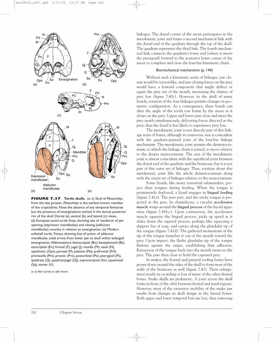
linkages. The dorsal corner of the snout participates in themesokinetic joint and forms a second mechanical link withthe dorsal end of the quadrate through the top of the skull.The quadrate represents the third link. The fourth mechan-ical link connects the quadrate’s lower end (where it meetsthe pterygoid) forward to the posterior lower corner of thesnout to complete and close the four-bar kinematic chain.
Biomechanical mechanisms (p. 144)
Without such a kinematic series of linkages, jaw clo-sure would be scissorslike, and jaw-closing forces on the preywould have a forward component that might deflect orsquirt the prey out of the mouth, increasing the chance ofprey loss (figure 7.40c). However, in the skull of manylizards, rotation of the four linkages permits changes in geo-metric configuration. As a consequence, these lizards canalter the angle of the tooth row borne by the snout as itcloses on the prey. Upper and lower jaws close and meet theprey nearly simultaneously, delivering forces directed at theprey; thus the lizard is less likely to experience prey loss.
The metakinetic joint is not directly part of this link-age train of bones, although its transverse axis is coincidentwith the quadrate-parietal joint of the four-bar linkagemechanism. The metakinetic joint permits the dermatocra-nium, to which the linkage chain is joined, to move relativeto the deeper neurocranium. The axis of the metakineticjoint is almost coincident with the superficial joint betweenthe dorsal end of the quadrate and the braincase, but it is notpart of this outer set of linkages. Thus, rotation about thismetakinetic joint lifts the whole dermatocranium alongwith the entire set of linkages relative to the neurocranium.
Some lizards, like many terrestrial salamanders, pro-ject their tongues during feeding. When the tongue isprominently deployed, a lizard engages in lingual feeding(figure 7.41a). The jaws part, and the sticky tongue is pro-jected at the prey. In chameleons, a circular acceleratormuscle wraps around the lingual process of the hyoid appa-ratus (figure 7.41b,c). Upon contraction, the acceleratormuscle squeezes the lingual process, picks up speed as itslides down the tapered process, perhaps like squeezing aslippery bar of soap, and carries along the glandular tip ofthe tongue (figure 7.41d). The gathered momentum of thetip of the tongue launches it out of the mouth toward theprey. Upon impact, the fleshy glandular tip of the tongueflattens against the target, establishing firm adhesion.Retraction of the tongue back into the mouth retrieves theprey. The jaws then close to hold the captured prey.
In snakes, the frontal and parietal roofing bones havegrown down around the sides of the skull to form most of thewalls of the braincase as well (figure 7.42). Their enlarge-ment results in crowding or loss of many of the other dermalbones. Snake skulls are prokinetic. A joint across the skullforms in front of the orbit between frontal and nasal regions.However, most of the extensive mobility of the snake jawresults from changes in skull design in the lateral bones.Both upper and lower temporal bars are lost, thus removing
262 Chapter Seven
Eo Ops
Bs
Bo
Pal
Po
Qj
Q
Sq
J
VM
Pm
Pt
NPrfM F
Prf
Po P
SoEo
EmarginationOps
Sq
Qj
(a) (b)
(c)
Sq
Qj
Po
M
FPrf
N
Pm
PtQ
M
Qj
Q
Ops
Pro
So
(e)
M
J
Qj
Mandible
Q
SqOps
Eo
Adductormandibulae
Depressormandibulae
P
F
Prf
P
So
Pro
(d)
Sq
P
PPo
F
Prf
FIGURE 7.37 Turtle skulls. (a–c) Skull of Pleisochelys,from the late Jurassic. Pleisochelys is the earliest known memberof the cryptodires. Note the absence of any temporal fenestraebut the presence of emarginations etched in the dorsal, posteriorrim of the skull. Dorsal (a), ventral (b), and lateral (c) views.(d) European pond turtle Emys, showing site of residence of jawopening (depressor mandibulae) and closing (adductormandibulae) muscles in relation to emargination. (e) Modernsoftshell turtle Trionyx, showing line of action of adductormandibulae, solid arrow, from lower jaw to skull within enlargedemargination.Abbreviations: basioccipital (Bo), basisphenoid (Bs),exoccipital (Eo), frontal (F), jugal (J), maxilla (M), nasal (N),opisthotic (Ops), parietal (P), palatine (Pal), prefrontal (Prf),premaxilla (Pm), prootic (Pro), postorbital (Po), pterygoid (Pt),quadrate (Q), quadratojugal (Qj), supraoccipital (So), squamosal(Sq), vomer (V).
(a–d) After Carroll; (e) after Romer.
kar28303_ch07.qxd 2/17/05 12:37 PM Page 262

Pt P PoQj
QPro
(a) (b)
(c) (d)
OpsSo
PfPrf
M
Pm Pro
Bs
Bo
EoSocSqOps
PmN
Qj
Stp Bo
StpOps Q
F
N Pt
Pal
Qj J EcPs
MPm
PrfFPrf
M
J
PoP
Sq
SaAr
Ar
Ept Ps Pt
Pa D
C
An
SaD
Pr
VFIGURE 7.38 Livingrhynchocephalian. The two temporalfenestrae are still bounded by bone inSphenodon, a living diapsid. Dorsal (a),ventral (b), lateral (c), and posterior (d)views.Abbreviations: angular (An),articular (Ar), basioccipital (Bo),basisphenoid (Bs), coronoid (C), dentary(D), ectopterygoid (Ec), exoccipital (Eo),epipterygoid (Ept), frontal (F), jugal (J),maxilla (M), nasal (N), opisthotic (Ops),parietal (P), prearticular (Pa), palatine (Pal),postfrontal (Pf), premaxilla (Pm),postorbital (Po), prefrontal (Prf), prootic(Pro), parasphenoid (Ps), pterygoid (Pt),quadrate (Q), quadratojugal (Qj),surangular (Sa), supraoccipital (Soc),squamosal (Sq), stapes (Stp), vomer (V).
After Carroll.
FIGURE 7.39 Lizard skull. Lizards are modified diapsids.Two fenestrae are present, but the ventral bony border of the lowerfenestra is absent, a result of changes serving increased cranial kinesis.Abbreviations: angular (An), articular (Ar), basioccipital (Bo),basisphenoid (Bs), coronoid (C), dentary (D), ectopterygoid (Ec), exoccipital (Eo), epipterygoid (Ept), frontal (F), jugal (J), lacrimal (L),maxilla (M), nasal (N), opisthotic (Ops), orbitosphenoid (Osp), parietal (P), prearticular (Pa), postfrontal (Pf), premaxilla (Pm), postorbital(Po), prefrontal (Prf), parasphenoid (Ps), prootic (Pro), pterygoid (Pt), quadrate (Q), surangular (Sa), septomaxilla (Sm), splenial (Sp),supraoccipital (So), squamosal (Sq), supratemporal (St), stapes (Stp), vomer (V).
After Jollie.
Foramen forJacobson's organ
Occipitalcondyle
St
StQ
So
Bo
Eo
Sq
Sq
EptPo Pf
PfJ
F
FP
OspPrf
PrfM
LN
N
Sm
Pm
Mandibularfossa
Ar
AnSa
SplD
Medial
Lateral
AnPa
C
D
J
Sa
EcPsPt
BsPro
Temporalfenestra
Pro
Po
VIIPt
Ops
BEoBo
Bs
X, XIPro
Basipterygoidprocess
Foramenmagnum
Eo
QJ
SqPo
EptPSo
Pro
St
Osp
PsV
Pm
Ept Ec MSm
Stp
Pt
Jugularforamen
Hypoglossalforamen
Bo
L
C
kar28303_ch07.qxd 2/17/05 12:37 PM Page 263

struts that in other diapsid skulls form restrictive bracesacross the temporal region. The kinematic machinery of thesnake skull includes more elements than the linkage systemof lizards (figure 7.43a–c). The quadrate, as in lizards, isstreptostylic but more loosely articulated with the pterygoid.Muscle forces imparted directly to the pterygoid are trans-mitted to the tooth-bearing maxilla via the linkingectopterygoid. The maxilla rotates upon the prefrontal, fromwhich it is suspended from the braincase. In many snakes,especially in advanced venomous snakes such as vipers, theprefrontal and supratemporal also enjoy some degree of rota-tion upon the braincase. Thus, the kinetic system can bemodeled on a linkage chain with up to six links (supratem-poral, quadrate, pterygoid, ectopterygoid, maxilla, pre-frontal) suspended at either end from a seventh link, thebraincase (figure 7.43d).
The mandible of snakes, suspended from thequadrate, includes a tooth-bearing dentary that articu-lates with a posterior compound bone derived from thefused surangular and angular. A tiny splenial is usuallypresent on the medial side. Both halves of the lower jaware joined at the mandibular symphysis, not by bonyfusion but by flexible soft tissues that unite the tips ofthe mandible. Mandibular tips thus enjoy independentmovement. Because there are no bony cross connectionsbetween chains of movable bones on left and right sides,each kinematic set of linkages can spread and moveindependently of the other on the opposite side. This isparticularly important during swallowing when alternat-ing left and right sets of bones are walked over the prey(see figure 7.63). It is a mistaken view that snakes“unhinge” their jaws when they swallow. Instead, the
264 Chapter Seven
FIGURE 7.40 Kinesis of a lizard skull. (a) Joints within the skull allow the snout to lift upward or bend downward about itsmesokinetic articulation with the rest of the braincase.This results in a change in the angle of closure of the teeth when the animal grabsits prey. (b) These movable units of the lizard skull can be represented as a kinematic mechanism by linkages (heavy lines) and points ofrotation (circles). Compared with the rest position of these linkages (a), geometric changes are shown during opening (middle) and closing(bottom) on the prey. (c) The functional significance of cranial kinesis in lizards is related to the resulting change in angle of tooth rows.Kinesis bends the snout so that both rows close directly on the prey (bottom).Were this not the case (middle and top), jaw closure wouldbe more of a scissors action, tending to squirt the prey back out of the mouth.
Based on the research of T. H. Frazzetta.
Snoutunit
(a)
(b)
Mandible
Opening
Basalunit
Quadrate unit
Parietalunit
Closing
Prey
(c)
kar28303_ch07.qxd 2/17/05 12:37 PM Page 264

great freedom of rotation between elements of the kine-matic chains, the independent movement of each, andthe ability of flaring the flexible jaws outward to accom-modate bulky prey all account for the suppleness ofsnake jaws. These processes, not disarticulation, permitsnakes to swallow (although slowly) relatively large,whole prey.
Crocodilians, together with Sphenodon and squamates,represent the surviving reptiles with a diapsid skull. Thecrocodilian skull is a composite of chondro-, dermato-, andsplanchnocranial elements, although the dermatocraniumtends to predominate (figure 7.44). Both temporal bars arepresent, and the skull is firm, without any evidence of cra-nial kinesis. However, crocodile ancestors possessed kineticskulls, suggesting that modern forms have lost this feature.Further, crocodiles possess a secondary palate, a furtherdeparture from diapsid ancestors. Within the roof of themouth, marginal bones (premaxilla, maxilla, palatine) growinward to meet at the midline beneath the sphenoid region.
Together with the pterygoid, these marginal bones producethe bony secondary palate that separates the nasal passage-way from the mouth.
BirdsBirds also arise from a diapsid ancestry, but like squamates,they show considerable modification of this skull pattern(figure 7.45). The braincase is much inflated and ossified inbirds, accommodating a relatively expanded brain within.Sutures between bones are usually overgrown in the adult sothat boundaries are not easily delineated. The palatal bonesare quite varied, but generally all show some degree of reduc-tion and lightening. Vomers and ectopterygoids are small,pterygoids are short struts articulating with the quadrate,and epipterygoids are usually lost (figure 7.46a–d).
Like turtles and some dinosaurs, birds are toothless,and their jaws are covered by keratinized sheaths. Birds thatfeed on slippery prey, such as fish-eating shorebirds, have
Skeletal System: The Skull 265
FIGURE 7.41 Lingual feeding in lizards. (a) Jackson’s chameleon uses its projectile tongue to “shoot” long distances at prey.(b) Hyoid apparatus of the chameleon includes an elongated lingual process (Lp) along which the tongue slides during launch. (c) Hyoidapparatus of a lizard without a projectile tongue. (d) Mechanical basis of tongue projection.The accelerator muscle, a circular band aroundthe lingual process, contracts to squeeze the lingual process.The squeeze of accelerator muscles causes the muscle to slide rapidly towardthe tip of the lingual process, carrying with it the glandular surface of the tongue.With gathered momentum, the tongue is launched fromthe lingual process toward the prey.The folded glossohyal muscle attached to the tongue’s tip is carried out as well and eventually isresponsible for retrieving the tongue and the adhering prey.Abbreviations: anterior process (Ap), basihyal (Bh), ceratobranchials I and II(CbI and CbII), ceratohyal (Ch), lingual process (Lp).
(b,c) After Bramble and Wake.
Lp
Lp
(a)
Bh
Bh
ApAp
Ch
Ch
CbICbI CbII(b) (c)
Glossohyal(retractormuscle)
(d)
Tendon
Lingualprocess Glandular
surface“Dimple”
Accelerator (“ring”) muscle
kar28303_ch07.qxd 3/9/05 9:42 AM Page 265

(a) (b)
(c)
(d)
Vg
Braincase
Lower jaw
St
St
Q
Q
Ec
Ec
Pt
Pt
Vg
M
M
PrfPrf
Prf
266 Chapter Seven
FIGURE 7.43 Kinematic model of movable skullbones in a venomous snake, the water moccasin. Wholehead (a) with successive removal of skin and muscles (b) revealsbones of the skull (c). Linkage bones movable relative to thebraincase are in color; lower jaw is crosshatched.(d) Biomechanical model of movable bones rotatable about pinconnections. Movable bones include the ectopterygoid (Ec),maxilla (M), pterygoid (Pt), prefrontal (Prf), quadrate (Q),supratemporal (St). Location of the main venom gland (Vg) isshown as well.
After Kardong.
Pp
P
P
Sq
F
F
N
N
Pm
TSt
Qj
Pf
Prf
Po
Po
Pt
Q
J
Prf
L
M M
Pm
St Eo So
Primitive amnioteDerived reptile(viperid snake)
FIGURE 7.42Diagrammatic comparison of aderived modern snake skull withan amniote skull. Bones lost inthe modern snake are indicated byshading in the primitivecaptorhinomorph.Abbreviations:exoccipital (Eo), frontal (F), jugal (J),lacrimal (L), maxilla (M), nasal (N),parietal (P), postfrontal (Pf),premaxilla (Pm), postorbital (Po),postparietal (Pp), prefrontal (Prf),pterygoid (Pt), quadrate (Q),quadratojugal (Qj), supraoccipital(So), squamosal (Sq), supratemporal(St), tabular (T).
kar28303_ch07.qxd 2/17/05 12:37 PM Page 266

beaks whose keratinized margins are serrated to improvefriction grip. The jaws are drawn out into a beak. The uppertemporal bar is absent, and the lower temporal bar is a slen-der rod called the jugal bar (quadratojugal-jugal bar), whichextends from the beak posteriorly to the side of the movable(streptostylic) quadrate. The skull is prokinetic. A strongpostorbital ligament extends from behind the eye to thelower jaw. In neognathous birds, the palate is functionallydivided at the pterygo-palatine joint (figure 7.47a,b). Theconverging paired pterygoids meet or nearly meet at themidline to couple there with the palatines (figure 7.47b).At this coupling, these bones form a joint that slides alongthe ventral edge of the orbital septum. The nasofrontal andpalato-maxilla joints are thin, flexible, bony joints, not syn-ovial, but they can be represented as hinges about whichrotation occurs. The mechanically important bones aremodeled as a linkage system on each side (figure 7.47c).When inserting muscles pull the quadrate and palate for-ward, the divided palate slides along the septum pushingthe palatines forward, which in turn push against the baseof the beak, rotating it about the nasofrontal joint, and rais-ing the beak. The coupled pair of gliding linkages is a slider-crank mechanism. (Muscles acting directly on the lowerjaw activate its opening.) Jaw-closing muscles act in theopposite fashion to move the beak downward to grasp food,and return the kinetic machinery to the rest position. Thethin jugal bar usually bows outward during jaw opening, butdoes not contribute significantly to the mechanism produc-ing upper jaw elevation.
Many birds use their beak like a probe to reach buriedgrubs or insects embedded in tree bark or soft soil. Such birdsoften use a form of rhynchokinesis, the lifting of the beaktips about points of rotation within the jaws (figure 7.47e).The jaws need not be parted far to seize the food. Other birdshave beaks that open tough seeds and short, stout jaws thatconcentrate closing forces at the base of their beaks.
In paleognathous birds, the pterygoids do not meet atthe midline, but slide on projecting struts, the basipterygoidprocesses (figure 7.47d). So structurally distinct is the palate,
that it has been used to argue that all paleognaths (ratitesand tinamous) represent a primitive condition and a mono-phyletic group.
SynapsidsPrimitive Synapsids Dimetrodon represents a primitivesynapsid. Therapsids continue the synapsid line and exhibitconsiderable diversity (figure 7.48). For a time in the Per-mian and early Triassic, they were fairly abundant. Somewere herbivores; most were carnivores. Most skull bones ofearly amniotes persist, but characteristic of synapsids, thetemporal region develops a single opening bound horizon-tally along its lower border by a bony connection betweenjugal and squamosal bones. In advanced therapsids andprimitive mammals, the vertical bar dividing the orbit fromthe single temporal fenestra is lost, leaving the narrow bonyconnection between jugal and squamosal bowing outward inthe cheek region. This bony squamosal-jugal bar is nowcommonly called the zygomatic arch. Within therapsids,there is a tendency for the temporal opening to move dor-sally and for the zygomatic arch to become delineated.
Mammals The skull of mammals represents a highly modi-fied synapsid pattern. Various dermal elements are lost intherian mammals, including the prefrontal, postorbital, post-frontal, quadratojugal, and supratemporal (figure 7.49). Thepostparietals, typically paired in reptiles, fuse into a single,medial interparietal in therapsids, which in mammals mayincorporate the tabular and fuse with the occipital bones.Monotremes retain several early synapsid skull features,including prefrontal, postfrontal, and pleurosphenoid bonestogether with unfused occipitals. Monotremes are somewhatspecialized as well. The lacrimal of therians is absent in them,and the jugal bones are small (figure 7.50a–d). A tympanicring encircles the middle ear bones of monotremes, but inmost therians, this ring has expanded into a large swollencapsule, the auditory bulla, that houses the middle ear ossi-cle (figure 7.51a–c).
Skeletal System: The Skull 267
The jaws of snakes are highly kinetic, withgreat freedom of motion. Skull bones thatin other reptiles are fixed to the braincaseor have restricted movement are joined insnakes into linked chains with extensivemotion relative to the braincase. Further,the series of linked bones on left and rightsides are not joined directly; so they expe-rience independent displacement, a featureallowing alternating left and right recipro-cating motion of jaw bones over the prey
being swallowed.This independent motionand outward spreading of the jaws (not“unhinging” of the jaws) allows most snakesto swallow large prey. Little by little, thedistended jaws are walked in alternatingsteps over the prey until it is completelyengulfed.
During the rattlesnake strike, the for-ward swing of these linked bones erectsthe maxilla and the fang it carries into posi-tion to inject venom into the prey. Snake
fangs are modified teeth with hollow coresso that venom flows from their base intothe prey. The fangs of most venomoussnakes are longer than other teeth in themouth, and the fangs of vipers and pitvipers are especially long. Extensive rota-tion of the fangbearing maxilla in suchsnakes allows this long fang to be folded upand out of the way along the upper lipwhen it is not in use.
BOX ESSAY 7 .3 Striking Features of Snakes
kar28303_ch07.qxd 2/17/05 12:37 PM Page 267

Placental Mammals Fusions between separate centers ofossification produce composite bones in the skull of placen-tal mammals. The single occipital bone represents the fusionof basioccipital, paired exoccipitals, supraoccipital, andinterparietal (and perhaps tabular) (figure 7.52a). The occip-ital bone defines the foramen magnum and closes the poste-rior wall of the braincase. As in monotremes and marsupials,there is a ventrally located, bilobed occipital condyle thatarticulates with the atlas, the first vertebra of the cervicalregion. Dorsally, a raised nuchal crest may form across theback of the occipital region, offering a secure attachment sitefor neck muscles and ligaments that support the head.
Several embryonic centers contribute to the sphenoidbone, representing the orbitosphenoid, presphenoid,basisphenoid, and a large alisphenoid (the epipterygoid oflower vertebrates) (figure 7.52b).
On the side of the braincase behind the orbit, a largetemporal bone is formed by the fusion of contributions fromall three parts of the skull (figures 7.52c and 7.53). The der-matocranium contributes the squamosal and the tympanicbulla (a derivative of the angular) in many mammals. Thechondrocranium contributes the petrosal, itself a deriva-tive of prootic and opisthotic bones (figure 7.52c). The pet-rosal often bears a ventrally directed projection, themastoid process. The splanchnocranium contributes threetiny middle ear bones (malleus, incus, stapes) and the sty-loid (figure 7.54).
In most tetrapods, the nasal capsule remains unossi-fied. However, in mammals, the ethmoid portion ossifies toform scroll-like turbinates (turbinals, conchae). There areusually three sets of turbinates attached to respective neigh-boring bones: the nasoturbinate, maxilloturbinate, and
268 Chapter Seven
Retroarticularprocess
Quadratojugal
PostorbitalFrontal
Supraciliary
PrefrontalNasolacrimal canal
Lacrimal
OrbitosphenoidProoticOpisthotic
Preoccipitalprocess
Carotid canal
BasioccipitalBasisphenoid
Basisphenoid
Quadrate
Stapes
Articular Surangular
Dentary
PrearticularAngular
NasalPremaxilla
MaxillaJugal
Dentary
Splanchnocranium Chondrocranium
Dermatocranium
FIGURE 7.44 Alligator skull. Acomposite skull design characteristic ofvertebrates.The skull is a combination ofelements receiving contributions from thechondrocranium (blue), the splanchnocranium(yellow), and the dermatocranium (red).
kar28303_ch07.qxd 2/17/05 12:37 PM Page 268

Skeletal System: The Skull 269
So
BirdPrimitive amniote
QjP
FJ
L
NM
Pm
PpSt
T
PSq
F
PfPoJ
Qj
L Prf
N
M
Pm
Sq
FIGURE 7.45 Diagrammaticcomparison of a derived bird skullwith a primitive amniote. Bones lostin birds are shaded in the primitivereptile.Abbreviations: frontal (F), jugal (J),lacrimal (L), maxilla (M), nasal (N), parietal(P), postfrontal (Pf), premaxilla (Pm),postorbital (Po), postparietal (Pp),prefrontal (Prf), quadratojugal (Qj),supraoccipital (So), squamosal (Sq),supratemporal (St), tabular (T).
FIGURE 7.46 Bird skull. In the adult bird, sutures between skull bones fuse to obliterate identifiable borders. Dorsal (a), ventral(b), lateral (c), and posterior (d) views of the skull of a young gosling (Anser) before bones fuse.Abbreviations: angular (An), articular (Ar),basioccipital (Bo), basisphenoid (Bs), dentary (D), exoccipital (Eo), frontal (F), jugal (J), lacrimal (L), laterosphenoid (Ls), maxilla (M), nasal(N), opisthotic (Ops), parietal (P), palatine (Pal), premaxilla (Pm), postorbital (Po), parasphenoid (Ps), pterygoid (Pt), quadrate (Q),quadratojugal (Qj), surangular (Sa), supraoccipital (So), squamosal (Sq), vomer (V).
P
So
Sq
P
Postorbitalprocess
L
NFrontal
PmM
JPoQjQ
F
So
Ops
Ar
An
D
V M
PalPt
JQj
N
LPo
Q
LsSqP
PSo
Eo
F
Sq
Bo
(a)
(c)
(b)
Bs
(d)
QjJ
M
So
EoSq
Jugal bar
FPt
L
M
PalPs
QV
Pm
SaBs
Ops
kar28303_ch07.qxd 2/17/05 12:37 PM Page 269

ethmoturbinate. The coiled walls of the turbinates supportthe mucous membrane within the nasal passage. Air enter-ing these passages is warmed and moistened before reachingthe lungs, functions that are especially important inendotherms. Absent in ungulates but present in most otherorders, such as rodents, carnivores, and primates, is anotherregion of the nasal capsule, the mammal mesethmoid. Thiselement forms the septum between the nasal capsules andusually remains cartilaginous. Between the nasal area andcranial cavity stands the transverse and finely perforatedcribriform plate (figure 7.54). Olfactory nerves originatingin the olfactory epithelium of the nasal capsule pass throughthis plate to reach the olfactory bulb of the brain.
Middle Ear Bones Two profound changes in the lower jawmark the transition from therapsid to mammal (figure 7.55).Both changes go hand in glove. They result in such an alter-ation in skull design that some anatomists doubted themuntil the surprisingly good fossil record made the evolution-
ary transition undeniable. One of these changes is the loss ofthe postdentary bones of the lower jaw. The other is thepresence of three middle ear bones. In vertebrates, the innerear is embedded deep within the otic capsule and holds thesensory apparatus responsive to sounds. The hyomandibulaor its derivatives deliver sound vibrations to the sensitiveinner ear. In all tetrapods, the hyomandibula tends tobecome reduced to a slender, light bone called the stapes (� columella). Sometimes there is a second hyomandibula-derived bone, the extracolumella. The stapes is usually sus-pended in the middle ear cavity where damping byrestrictive attachments is minimized. As sounds set the tym-panum into motion, these vibrations are imparted to thesmall, responsive stapes. Its opposite end often expands toreach the sensitive inner ear apparatus that responds to thevibrations the stapes delivers.
In mammals, two tiny, additional bones join the stapesin the middle ear. Together these bones transmit sound tothe inner ear. Specifically, these three middle ear bones are
270 Chapter Seven
Nasofrontal hinge
Premaxilla
Maxilla
Quadrate Pterygoid Palatine
Orbital septum(parasphenoid)
Jugalbar
Upperjaw
(a)
(d)
(e)
(b)
(c)
Quadrate
Palatine
Pterygoid
Basipterygoidprocess
Jugal bar
FIGURE 7.47 Cranial kinesis in the crow skull (Corvus). (a) Lateral view. (b) Ventral view. (c) Linkage model of cranialkinesis, slider-crank mechanism. From the rest position (solid lines), the point of coupling between pterygoids and palatines slides forwardalong the orbital septum to a new position (dashed lines), which lifts the upper jaw about the prokinetic joint (nasofrontal hinge).(d) Paleognath. Ostrich palate. (e) Rhynchokinesis. Flexions within the beak permit the tips of the upper and lower jaws to part withoutopening of the entire mouth.
kar28303_ch07.qxd 2/17/05 12:37 PM Page 270
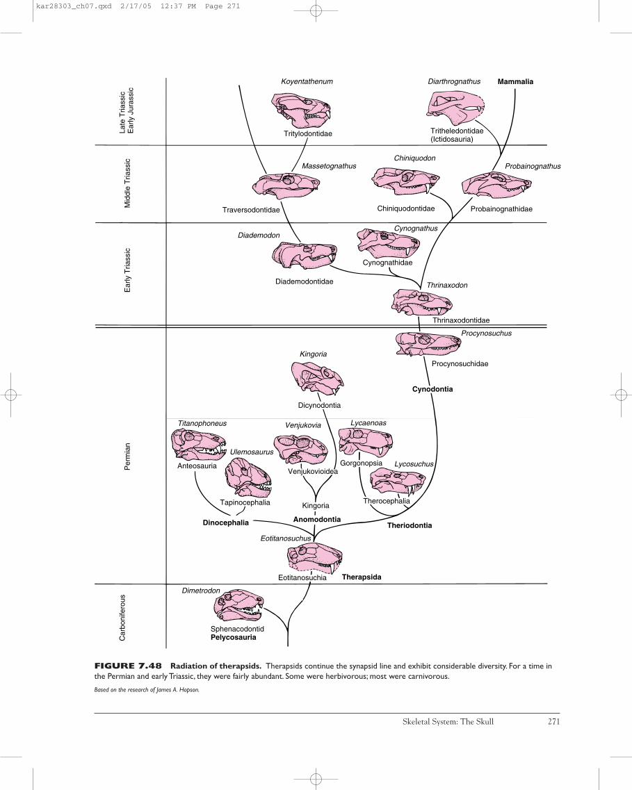
Skeletal System: The Skull 271
FIGURE 7.48 Radiation of therapsids. Therapsids continue the synapsid line and exhibit considerable diversity. For a time inthe Permian and early Triassic, they were fairly abundant. Some were herbivorous; most were carnivorous.
Based on the research of James A. Hopson.
Koyentathenum
Tritylodontidae
Diarthrognathus
Tritheledontidae(Ictidosauria)
Mammalia
ChiniquodonMassetognathus Probainognathus
Traversodontidae Chiniquodontidae Probainognathidae
CynognathusDiademodon
Diademodontidae Thrinaxodon
Thrinaxodontidae
Procynosuchus
Procynosuchidae
Cynodontia
Kingoria
Dicynodontia
Titanophoneus Venjukovia Lycaenoas
Ulemosaurus
Anteosauria Lycosuchus
Dinocephalia Theriodontia
Eotitanosuchus
Dimetrodon
SphenacodontidPelycosauriaC
arbo
nife
rous
Per
mia
nE
arly
Tria
ssic
Mid
dle
Tria
ssic
Late
Tria
ssic
Ear
ly J
uras
sic
Gorgonopsia
Eotitanosuchia
TherocephaliaKingoria
Venjukovioidea
Therapsida
Cynognathidae
Tapinocephalia
Anomodontia
kar28303_ch07.qxd 2/17/05 12:37 PM Page 271

272 Chapter Seven
Pm
Pm
Pp
M
M
N
F
L
L
JJ
Sq PSq P
Pp(Ip)
Mammal(opossum)
Primitive amniote
Sq P
TSt
Qj
Po Pf
Prf
F
N
FIGURE 7.49 Diagrammaticcomparison of a derived mammalskull with a primitive amniote skull.Bones lost in the derived mammal areshaded in the primitive amniote. Inmammals, orbital and temporal openingsmerge.Abbreviations: frontal (F), jugal (J),interparietal (Ip), lacrimal (L), maxilla (M),nasal (N), parietal (P), postfrontal (Pf),premaxilla (Pm), postorbital (Po),postparietal (Pp), prefrontal (Prf),quadratojugal (Qj), squamosal (Sq),supratemporal (St), tabular (T).
FIGURE 7.50 Monotreme, skull of the duckbill platypus Ornithorhynchus. Dorsal (a), ventral (b), lateral (c), andposterior (d) views.Abbreviations: dentary (D), ectopterygoid (Ec), exoccipital (Eo), frontal (F), jugal (J), maxilla (M), nasal (N),orbitosphenoid (Osp), parietal (P), palatine (Pal), premaxilla (Pm), supraoccipital (So), squamosal (Sq), vomer (V).
Osp
Sq(a)
(c)
(b)
(d)
Sq OspF N M
Pm
Foramenovale Foramen
rotundum Anterior palatineforamen
Palatineprocesses ofpremaxilla
Nuchalcrest
Temporalcrest
Occipitalcondyle
EcV
Pal M
Infraorbitalcanal
DJ
Eo
So
P
P
FN
kar28303_ch07.qxd 2/17/05 12:37 PM Page 272
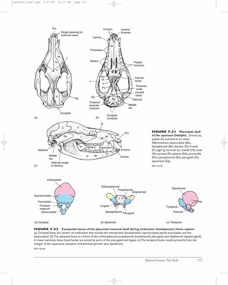
Skeletal System: The Skull 273
Middleear
Glenoid
Carotid canal
Foramen ovale
Internalnares
Palatelforamina
Incisiveforamen
Incisors
Canine
Premolars
Molars
J
Sq
Posteriorlacerateforamen
Occipitalcondyles
PalPt
V
Ps
Single opening forexternal nares
Pm
L
Sq
Occipital
P
J
F
N
M
(a)
(c)
(b)
Mastoid
Middleear
Inflected angleof dentary
N
Pm
Incisors
Canine
PremolarsMolarsPt
M
L
F
J
P
Sq
D
Bs
Bo
M
FIGURE 7.51 Marsupial, skullof the opossum Didelphis. Dorsal (a),palatal (b), and lateral (c) views.Abbreviations: basioccipital (Bo),basisphenoid (Bs), dentary (D), frontal(F), jugal (J), lacrimal (L), maxilla (M), nasal(N), parietal (P), palatine (Pal), premaxilla(Pm), parasphenoid (Ps), pterygoid (Pt),squamosal (Sq).
After Carroll.
FIGURE 7.52 Composite bones of the placental mammal skull during embryonic development, Homo sapiens.(a) Occipital bone has centers of ossification that include the interparietal (postparietal), supraoccipital, paired exoccipital, and thebasioccipital. (b) The sphenoid bone is a fusion of the orbitosphenoid, presphenoid, basisphenoid, pterygoid, and alisphenoid (epipterygoid).In many mammals, these fused bones are joined by parts of the pterygoid and lingula. (c) The temporal bone results primarily from themerger of the squamosal, tympanic, and petrosal (prootic plus opisthotic).
After Hyman.
Interparietal
Supraoccipital
Exoccipital
Foramenmagnum
Basioccipital
(a) Occipital
PterygoidBasisphenoid
Lingula
OrbitosphenoidPresphenoid
Alisphenoid
(b) Sphenoid
Petrosal
Tympanic
Squamosal
(c) Temporal
kar28303_ch07.qxd 2/17/05 12:37 PM Page 273

malleus (derived from the articular), incus (derived from thequadrate), and stapes (derived from the hyomandibula). Sodistinctive is the presence of three middle ear bones, thatmany anatomists mark the fossil transition to mammals atthe point of their acquisition.
Anatomy and function of the ear (p. 696)
Coupled with the derivation of the three middle earbones are changes in the posterior bones of the mandible. Inearly synapsids (pelycosaurs), the lower jaw includes thetooth-bearing dentary in addition to several postdentarybones (angular, articular, coronoid, prearticular, splenial,surangular) (figure 7.56). In derived synapsids (mammals),this set of postdentary bones has been entirely lost from thelower jaw, and the dentary has enlarged to assume the exclu-sive role of lower jaw function. From pelycosaur to therapsidto mammal, the anatomical details of these changes are welldocumented in an ordered time sequence by the fossilrecord. In pelycosaurs, the articular (future malleus) residesat the back of the mandible and establishes lower jaw artic-ulation with the quadrate (future incus). In early to latertherapsids, these two bones become reduced, along with thepostdentary bones, eventually moving out of the lower jawand taking up a position in the middle ear. The functionalreason for these changes is thought to be related to improvedhearing, especially to a wider range of sounds. The phyloge-netic reduction in size of these bones would reduce theirmass and thus increase their oscillatory responsiveness toairborne vibrations. Their removal from the jaw joint per-mits their more specialized role in transmitting sound to theinner ear. Alternatively, or along with such changes relatedto hearing, some morphologists have proposed that changesin feeding style led to changes in the preferred site of inser-tion of the jaw-closing muscles, specifically a shift forwardon the dentary and closer to the teeth. Larger jaw musclesacting close to the tooth row lessen the stresses at the backof the jaw where it articulates with the skull. Loss of post-dentary bones then might reflect this shift in forces forwardto the tooth row and away from the joint these bonesformed.
These changes in the lower jaw were accompanied bychanges in the method of food preparation prior to swallow-ing. Most reptiles bolt their food, swallowing it whole or inlarge chunks. Mammals typically chew their food beforeswallowing it, a process termed mastication. Masticationalso occurs in a few groups of fishes and lizards. But, it iswithin mammals that feeding strategy is based on mastica-tion of food. If mastication became a more characteristicpart of food preparation, then changes in jaw-closing mus-cles might be expected, with greater emphasis shifting to thedentary.
Secondary Palate and Akinesis In addition to changes inthe mammalian lower jaw, the presence of a secondarypalate is also related to mastication. The secondary palateincludes a hard palate of bone and a posterior continuation
274 Chapter Seven
Oc
P F
JSq
B
Sq
D
Canis dirus
L
Fio
Pm
M
Tympanic
annulusAngular*
Mandibular arch
ArticularQuadrate Stapes
Hyoidarch
StyloidMalleus Incus Stapes
Mastoid
portion
Bu
lla
Sq
uamosal bone
Petrosal bone
Prootic
Opisthotic
Sty-
l oidear ossiclesMiddle
portion
Sq
uamous Petrous
portion
Mas-
toid
Tym
panic
po
rtion
Temporalbone
Squamosal*
FIGURE 7.53 Mammalian temporal bone. Thetemporal bone forms phylogenetically from the dermatocranium(angular, squamosal), and chondrocranium (prootic, opisthotic),into which is set contributions from the splanchnocranium(articular, quadrate, stapes, styloid).The separate bony elements inearly amniotes (outer circle) contribute to the compositetemporal bone of mammals (middle and inner circle). Some ofthese contributions are dermal bones (*).The otic capsule isburied beneath the surface of the skull, leaving the exposed andoften elongated mastoid process.The bulla or auditory bullaforms, at least in part, from the tympanic annulus, itself aphylogenetic derivative of the angular bone.The exposedsquamous portion of the temporal bone is illustrated in color onthe skull of the Pleistocene wolf Canis dirus. Abbreviations:auditory bulla (B), dentary (D), frontal (F), infraorbital foramen(Fio), jugal (J), lacrimal (L), maxilla (M), occipital (Oc), parietal (P),premaxilla (Pm), squamosal (Sq).
kar28303_ch07.qxd 3/9/05 9:42 AM Page 274

of fleshy tissue, the soft palate (figure 7.57a,b). The hardpalate is formed from the inward growth of bony processes ofthe premaxilla, maxilla, and palatine that meet at the mid-line as a bony platform (figure 7.58a–c). This hard palateand its fleshy continuation effectively separate the foodchamber below from the respiratory passage above. Someturtles and also crocodilians have a secondary palate, andthey benefit from the advantages of separation of routes forfood and air. But chewing requires that food stays in themouth for an extended period of time in mammals; there-fore, separation of respiratory and oral passages is especiallyimportant. Mastication can proceed without impeding regu-lar breathing. Similarly, the secondary palate completes thefirm roof of the food chamber, so that the pumping action ofthe throat of a suckling infant creates effective negativepressure within the mouth without interfering with the res-piratory passage.
Mastication (p. 529)
Mastication in mammals has been accompanied byvery precise tooth occlusion to serve the mechanical break-down of food. Precise, strong occlusion requires a firm skull,so mammals have lost cranial kinesis, leaving them an aki-netic skull. The mammalian mandibular condyle fits into avery precise articulation with the squamosal bone. Whenjaws close about this joint, upper and lower rows of teeth areplaced in very precise alignment. This allows specializedteeth to function properly. As a further consequence of pre-cise occlusion, the pattern of tooth eruption in mammals dif-fers from that of most other vertebrates. In lower vertebrates,teeth wear and are replaced continuously (polyphyodonty);therefore, the tooth row is always changing. If teeth functionprimarily to snag prey, this causes little difficulty. However,continuous replacement means that at some location in thejaws, worn teeth are missing or new ones are moving intoposition. To avoid disruption of occlusion, teeth in most
mammals are not continuously replaced. Mammals exhibitdiphyodonty. Only two sets of teeth erupt during the life-time of a mammal, the “milk teeth” of the young and the“permanent” teeth of the adults.
Tooth types and their development (p. 500)
The chain of events leading from mastication to aki-nesis and to diphyodonty should not be viewed as inevitable.Some fishes chew their food but retain kinetic skulls andpolyphyodonty. But evolutionary events that produced themammalian skull underscore the importance of examininganatomical changes in partnership with functional changesthat must accompany phylogenetic modification of verte-brate design. Form and function necessarily go together, anissue to which we next turn.
Overview of Skull Function and Design
The skull performs a variety of functions. It protects andsupports the brain and its sensory receptors. It may func-tion to cool the brain during sustained activity or during arise in environmental temperature. In many active terres-trial mammals, the nasal epithelium lining the nasal pas-sages dissipates excess heat by evaporation as air movesacross this moist lining. A similar function has been pro-posed for the elaborate air passageway in some groups ofhadrosaurs, the duck-billed dinosaurs (figure 7.59). Airentering their nostrils would have coursed through intri-cate passageways formed within premaxilla and nasalbones to provide evaporative cooling. The skull of manyanimals also supports the voice box and occasionallyserves as a sound resonator to deepen or amplify an ani-mal’s call. The Weddell seal takes advantage of its jaws toopen and maintain its breathing holes in surface ice (figure 7.60).
Skeletal System: The Skull 275
FIGURE 7.54 Diagram of adog’s skull. Sources of the variousbones are outlined: dermatocranium(red), chondrocranium (blue), andsplanchnocranium (yellow).
After Evans.
Cribriform plateLacrimal
Nasal
Premaxilla
Meckel's cartilage
DentaryJugal
Pala-tine
Ptery-goid
Mesethmoid
FrontalOrbitosphenoid
IIV. man V. max
III, IV, opV, VI
Alisphe-noid
Parietal
Postparietal
Supraoccipital
Petrosal
Exoccipital
XII
IX, X, XITympanic ring
StapesIncus
Malleus
Basioccipital
BasisphenoidSquamosal
Tympanohyoid
StylohyoidEpiglottis
Thyrohyoid
Epihyoid
Ceratohyoid
BasihyoidArytenoid
Maxilla
VII
Presphenoid
Vomer
kar28303_ch07.qxd 2/17/05 12:37 PM Page 275
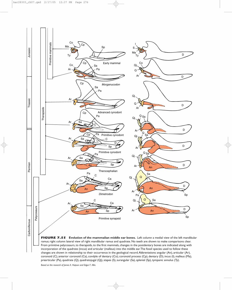
CoMa
Prim
itive
mam
mal
s
Jura
ssic
Tria
ssic
Per
mia
n
The
raps
ids
Car
boni
fero
us Pel
ycos
aurs
Ty
Sp
Early mammal
Advanced cynodont
Co
Ar
Co
IS
MaTy
Ar
CpSa
Pa
Pa
MorganucodonCp
Cp
Sa
SaArC
Primitive cynodont
Primitive synapsid
Primitive cynodont
Dimetrodon
Therocephalian
Pa
PaCp
SaAr
SpD
D
D
D
D
D
D
D
D
D
C
PaSaAr
An Sp
An An
An
An
Sa
Sa
Sa
Sa
Sa
Sa
Ar
Ar
Pa
Pa
C
C
Ca
Ca
Sp
Sp
Sp
Qj
Qj
Qj
Qj
Qj
Qj
Qj
Ar
Ar
Ar
Ar
Ar
Ar
Q
Q
Q
Ar
Q
Q
Q
Q
An
Sa
Cp
Sp
FIGURE 7.55 Evolution of the mammalian middle ear bones. Left column a medial view of the left mandibularramus; right column lateral view of right mandibular ramus and quadrate. No teeth are shown to make comparisons clear.From primitive pelycosaurs, to therapsids, to the first mammals, changes in the postdentary bones are indicated along withincorporation of the quadrate (incus) and articular (malleus) into the middle ear.The fossil species used to follow thesechanges are shown in relationship to their occurrence in the geological record.Abbreviations: angular (An), articular (Ar),coronoid (C), anterior coronoid (Ca), condyle of dentary (Co), coronoid process (Cp), dentary (D), incus (I), malleus (Ma),prearticular (Pa), quadrate (Q), quadratojugal (Qj), stapes (S), surangular (Sa), splenial (Sp), tympanic annulus (Ty).
Based on the research of James A. Hopson and Edgar F. Allin.
kar28303_ch07.qxd 2/17/05 12:37 PM Page 276
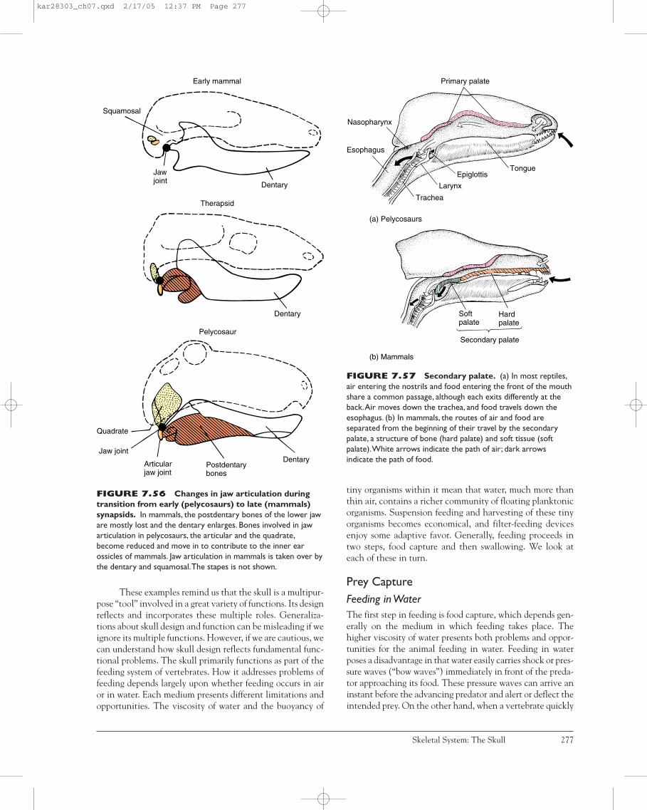
These examples remind us that the skull is a multipur-pose “tool” involved in a great variety of functions. Its designreflects and incorporates these multiple roles. Generaliza-tions about skull design and function can be misleading if weignore its multiple functions. However, if we are cautious, wecan understand how skull design reflects fundamental func-tional problems. The skull primarily functions as part of thefeeding system of vertebrates. How it addresses problems offeeding depends largely upon whether feeding occurs in airor in water. Each medium presents different limitations andopportunities. The viscosity of water and the buoyancy of
tiny organisms within it mean that water, much more thanthin air, contains a richer community of floating planktonicorganisms. Suspension feeding and harvesting of these tinyorganisms becomes economical, and filter-feeding devicesenjoy some adaptive favor. Generally, feeding proceeds intwo steps, food capture and then swallowing. We look ateach of these in turn.
Prey CaptureFeeding in WaterThe first step in feeding is food capture, which depends gen-erally on the medium in which feeding takes place. Thehigher viscosity of water presents both problems and oppor-tunities for the animal feeding in water. Feeding in waterposes a disadvantage in that water easily carries shock or pres-sure waves (“bow waves”) immediately in front of the preda-tor approaching its food. These pressure waves can arrive aninstant before the advancing predator and alert or deflect theintended prey. On the other hand, when a vertebrate quickly
Skeletal System: The Skull 277
DentaryPostdentarybones
Articularjaw joint
Jaw joint
Quadrate
Pelycosaur
Dentary
Therapsid
Early mammal
Squamosal
Dentary
Jawjoint
FIGURE 7.56 Changes in jaw articulation duringtransition from early (pelycosaurs) to late (mammals)synapsids. In mammals, the postdentary bones of the lower jaware mostly lost and the dentary enlarges. Bones involved in jawarticulation in pelycosaurs, the articular and the quadrate,become reduced and move in to contribute to the inner earossicles of mammals. Jaw articulation in mammals is taken over bythe dentary and squamosal.The stapes is not shown.
Nasopharynx
Esophagus
Trachea
Larynx
EpiglottisTongue
Primary palate
Soft palate
Hardpalate
Secondary palate
(a) Pelycosaurs
(b) Mammals
FIGURE 7.57 Secondary palate. (a) In most reptiles,air entering the nostrils and food entering the front of the mouthshare a common passage, although each exits differently at theback.Air moves down the trachea, and food travels down theesophagus. (b) In mammals, the routes of air and food areseparated from the beginning of their travel by the secondarypalate, a structure of bone (hard palate) and soft tissue (softpalate).White arrows indicate the path of air; dark arrowsindicate the path of food.
kar28303_ch07.qxd 2/17/05 12:37 PM Page 277

gulps water into its mouth, the viscosity of the water dragsalong the prey as well. This viscosity makes suction feeding,used with relatively large prey, possible.
To capture small foods, aquatic animals use suspen-sion feeding. The density of water gives it the viscosity toretard the fall of particulate material out of suspension.Compared to air, water holds a floating bounty of tinyorganic particles and microorganisms, a rich potential nutri-tional resource for an organism with the equipment to har-vest it. Cilia move and control currents of water (andtransport captured food), and sticky mucus snatches sus-pended food from the current of water as it glides by.
Suspension Feeding Suspension feeding is a feeding strat-egy confined largely, perhaps exclusively, to animals livingin water. Some, making an esoteric point, argue that bats“filter” insects “suspended” in air, but this misses the point.Air is too thin to hold suspended food for long. Bats catch orgrasp prey, but they do not really use a filtering apparatus norface the same mechanical problems as aquatic organisms ina viscous medium; so they are not suspension feeders. Mostsuspension feeders are benthic (bottom-dwelling) organismsor are associated with a herbivorous/detritus feeding style.Respiration and feeding are tightly coupled. Often the samewater currents support both activities.
Suspension feeders use several methods to intercept andgather nutrients traveling within streams of water. Capturedparticles are usually smaller than the pores of the filter. Theymay collide directly with the filter (figure 7.61a), or becauseof their inertia, they deviate from the streamlines to collidewith the mucus-covered surface of the filter (figure 7.61b).Upon impact, the particles cling to the sticky mucus and arerolled up in mucous cords, and then are passed by cilia into thedigestive tract.
Less commonly, a sieve can be used to strain sus-pended particles larger than the pores of the sieve. As thestream of water passes through the sieve, the particles areheld back and then collected from the face of the selectivefilter (figure 7.61c). This method is rare among animals,perhaps because the relatively large particles filtered tend to
278 Chapter Seven
(a) Early tetrapod
(b) Therapsid
(c) Mammal
Primary palate
Vomer
Pterygoid
Premaxilla Internal naris
MaxillaPalatine
Ectoptery-goid
Palatalprocess
Primary palate
Primary palate
Secondarypalate
Nasopharynx
Premaxilla
Maxilla
Palatine
Internalnaris
FIGURE 7.58 Evolution of the secondary palate.(a) Early tetrapod with a primary palate in cross section (left) andventral (right) views. (b) Therapsid with a partial secondary palateformed by the medial extension of the premaxilla and maxilla.(c) Mammal with a secondary palate that, in addition toextensions of the premaxilla and maxilla, includes part of thepalatine bone.
After Smith.
Air
Premaxilla
Nasal
Nasal cavity
Brain
Nasopharynx
FIGURE 7.59 Air passage of duck-billed dinosaurs.The air passageway is formed by the premaxillae and nasal bonesof the hadrosaur.Air that flowed through the nasal cavity on itsway to the lungs cooled the nasal epithelial lining and hence theblood flow through it.Although the vascular system of thehadrosaur is not known, if it were similar to some mammals, thenthis cooled blood might have circulated in such a fashion as toprecool the blood flowing to the brain. In this way, the brain wasprotected from elevated temperatures.Alternatively oradditionally, such an expanded air passage might have been aresonating chamber to amplify vocalizations.
After Wheeler.
kar28303_ch07.qxd 3/9/05 9:42 AM Page 278
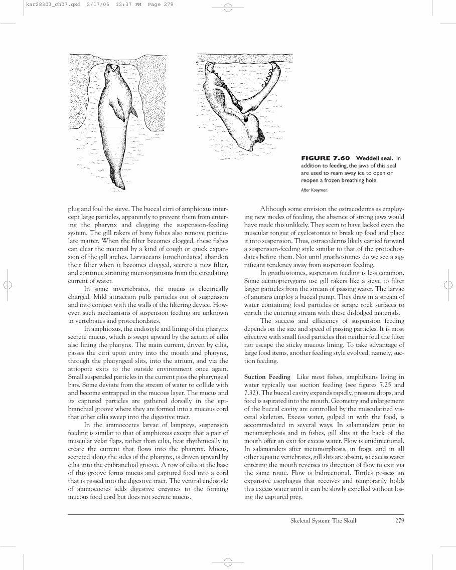
plug and foul the sieve. The buccal cirri of amphioxus inter-cept large particles, apparently to prevent them from enter-ing the pharynx and clogging the suspension-feedingsystem. The gill rakers of bony fishes also remove particu-late matter. When the filter becomes clogged, these fishescan clear the material by a kind of cough or quick expan-sion of the gill arches. Larvaceans (urochordates) abandontheir filter when it becomes clogged, secrete a new filter,and continue straining microorganisms from the circulatingcurrent of water.
In some invertebrates, the mucus is electricallycharged. Mild attraction pulls particles out of suspensionand into contact with the walls of the filtering device. How-ever, such mechanisms of suspension feeding are unknownin vertebrates and protochordates.
In amphioxus, the endostyle and lining of the pharynxsecrete mucus, which is swept upward by the action of ciliaalso lining the pharynx. The main current, driven by cilia,passes the cirri upon entry into the mouth and pharynx,through the pharyngeal slits, into the atrium, and via theatriopore exits to the outside environment once again.Small suspended particles in the current pass the pharyngealbars. Some deviate from the stream of water to collide withand become entrapped in the mucous layer. The mucus andits captured particles are gathered dorsally in the epi-branchial groove where they are formed into a mucous cordthat other cilia sweep into the digestive tract.
In the ammocoetes larvae of lampreys, suspensionfeeding is similar to that of amphioxus except that a pair ofmuscular velar flaps, rather than cilia, beat rhythmically tocreate the current that flows into the pharynx. Mucus,secreted along the sides of the pharynx, is driven upward bycilia into the epibranchial groove. A row of cilia at the baseof this groove forms mucus and captured food into a cordthat is passed into the digestive tract. The ventral endostyleof ammocoetes adds digestive enzymes to the formingmucous food cord but does not secrete mucus.
Although some envision the ostracoderms as employ-ing new modes of feeding, the absence of strong jaws wouldhave made this unlikely. They seem to have lacked even themuscular tongue of cyclostomes to break up food and placeit into suspension. Thus, ostracoderms likely carried forwarda suspension-feeding style similar to that of the protochor-dates before them. Not until gnathostomes do we see a sig-nificant tendency away from suspension feeding.
In gnathostomes, suspension feeding is less common.Some actinopterygians use gill rakers like a sieve to filterlarger particles from the stream of passing water. The larvaeof anurans employ a buccal pump. They draw in a stream ofwater containing food particles or scrape rock surfaces toenrich the entering stream with these dislodged materials.
The success and efficiency of suspension feedingdepends on the size and speed of passing particles. It is mosteffective with small food particles that neither foul the filternor escape the sticky mucous lining. To take advantage oflarge food items, another feeding style evolved, namely, suc-tion feeding.
Suction Feeding Like most fishes, amphibians living inwater typically use suction feeding (see figures 7.25 and7.32). The buccal cavity expands rapidly, pressure drops, andfood is aspirated into the mouth. Geometry and enlargementof the buccal cavity are controlled by the muscularized vis-ceral skeleton. Excess water, gulped in with the food, isaccommodated in several ways. In salamanders prior tometamorphosis and in fishes, gill slits at the back of themouth offer an exit for excess water. Flow is unidirectional.In salamanders after metamorphosis, in frogs, and in allother aquatic vertebrates, gill slits are absent, so excess waterentering the mouth reverses its direction of flow to exit viathe same route. Flow is bidirectional. Turtles possess anexpansive esophagus that receives and temporarily holdsthis excess water until it can be slowly expelled without los-ing the captured prey.
Skeletal System: The Skull 279
FIGURE 7.60 Weddell seal. Inaddition to feeding, the jaws of this sealare used to ream away ice to open orreopen a frozen breathing hole.
After Kooyman.
kar28303_ch07.qxd 2/17/05 12:37 PM Page 279

The early stages in vertebrate evolution took place inwater, mostly in marine waters, occasionally in fresh water.Adaptations for feeding and respiration took advantage ofthese conditions. Adaptations for suspension and suctionfeeding are present in early vertebrates. With the transitionof vertebrates to land and air, neither suspension nor suctionfeeding provided efficient ways to procure or process food.The jaws became specialized for grasping.
Feeding in AirTerrestrial feeding in most amphibians and many lizardsrequires a projectile tongue. The term lingual feeding recog-nizes the use of a rapid, projected, and sticky tongue to cap-ture prey (figures 7.62 and 7.41a–d). However, in many otheranimals, prey is captured by prehension, a method by whichthe animal rapidly grasps the prey with its jaws. In such ani-mals, the jaws are prey traps, designed to snare the unwary.
As a strategy to capture prey, prehension does notalways involve the jaws. Birds of prey snatch quarry with theirtalons, and mammalian predators often use claws to catch andthen control intended prey. Jaws are used secondarily to helphold the struggling victim or to deliver a killing bite.
SwallowingOnce an animal has captured and dispatched its prey, it mustswallow the prey in order to digest it. In suspension feeders,the food-laden cords of mucus are swept by synchronized cil-iary action into the esophagus. Other animals usually swal-low prey whole or in large pieces. Suction feeders rapidlyexpand the buccal cavity repeatedly to work the capturedprey backward into the esophagus. Terrestrial vertebrates usethe tongue to reposition the food bolus and work it towardthe back of the mouth. The highly kinetic skull of snakesallows great freedom of jaw movement. A snake swallows arelatively large animal by stepping the tooth-bearing bonesover the dispatched prey (figure 7.63a,b).
Swallowing mechanisms of terrestrial vertebrates (p. 510)
As we have seen in many vertebrates, swallowinginvolves mastication (the chewing of food). Masticationoccurs in a few groups of fishes and lizards. Within mammals,mastication has had a profound influence upon skull design,producing an akinetic skull with precise tooth occlusion andonly two replacement sets of teeth, a secondary palate, largejaw-closing musculature, and changes in lower jaw structure.
Overview
Cranial Neural CrestThe cranial neural crest cells leave their initial sites next to theneural tube and form streams of cells contributing to the mes-enchyme, which differentiates into bone, cartilage, cranialnerve cells, and various other structures in the head. In partic-ular, note that the hindbrain is segmented into compartmentscalled rhombomeres. Neural crest cells derived from particularrhombomeres migrate into and come to populate particularpharyngeal arches, which in turn give rise to particular cranialstructures (figure 7.64). Generally, in tetrapods, neural crestcells from rhombomeres 1 and 2 (and in some taxa, neural crestfrom forebrain and midbrain) migrate into the first pharyngeal(mandibular) arch, producing some of the facial bones,Meckel’s cartilage of the lower jaw, and, in mammals, the incusand malleus. Cells of rhombomere 4 enter the second pharyn-
280 Chapter Seven
Filter
(a)
(b)
(c)
Streamline
FIGURE 7.61 Suspension feeding: interception ofsuspended food particles. (a) Direct interception of foodparticles occurs when particles strike the filtering device. Food iscarried in streamlines flowing around bars through openings inthe feeding filter. (b) Small, dense particles flow along thestreamlines until the fluid is sharply diverted. Particle inertiacauses food particles to deviate from the streamlines, collide withthe filtering device, and adhere to the mucous coat of the filter.(c) The filtering apparatus can function as a sieve by holding backlarge particles that fail to pass through the small pores. Cilia drivethe food-laden mucus to the digestive tract.
kar28303_ch07.qxd 2/17/05 12:37 PM Page 280

geal (hyoid) arch, producing the stapes, styloid process, and acontribution to the hyoid. Rhombomere 6 releases neural crestcells into pharyngeal arches 3 and 4, which contribute to thehyoid and to the thyroid cartilages. Cells from rhombomeres 3and 5 do not migrate.
Somewhat surprisingly, in at least tetrapods both ele-ments of the mandibular arch, the palatoquadrate and Meckel’scartilages, derive solely from a single condensation of neuralcrest cells populating the first pharyngeal arch. However, themaxillary arch is not derived from the first pharyngeal arch butinstead from several other neural crest sources of the head.
Hox genes expressed in various combinations andregions of the neural crest (figure 7.64) instruct the cells quiteearly as to which tissues they will form. Although details arestill forthcoming, many major evolutionary changes in verte-brate jaws, teeth, and facial bones seem based on changes inthe placement or instructed fate of neural crest cells. Forexample, a Hox gene is expressed in the mandibular arch of a
species of lamprey, but not in gnathostomes. This apparentloss of Hox gene expression may have been the genetic basisfor facilitating the evolution of jaws in gnathostomes.
Neural crest (p. 238)
Emergence of MammalsMammals brought many innovations to the vertebratedesign; several involved the skull. One change alreadynoted is in the lower jaw. In early amniotes, as in gnathos-tomes generally, the jaws articulate with the braincase viathe articular-quadrate joint. In mammals, this is quite dif-ferent. The jaws articulate via the dentary-temporaljoint. Several postdentary bones become lost during thistransition to mammals, and a few move to the middle ear.The dentary expands posteriorly to form a new articula-tion with the skull, namely, via the dentary-temporaljoint. Although the factors favoring these changes aredisputed, the reality of these changes is not. Boneslocated at the back of the primitive amniote lower jawwere either lost or altered in function from jaw articula-tion to hearing. But this fact raises a new problem. Howcould bones involved in jaw suspension change functionwithout disrupting the intermediate species? If postden-tary bones moved to the middle ear, how could they aban-don jaw suspension without producing an individual withno method of supporting the jaw against the skull? G. Cuvier, nineteenth-century French anatomist, wouldhave understood the dilemma. He argued that evolutioncould not occur for just such a reason, because a changein structure would disrupt function and stop evolution inits tracks before it had begun.
Diarthrognathus, a late cynodont close to primitivemammals, suggests an answer. Its name means two (di-) sitesof articulation (arthro-) of the jaw (gnathus). In addition tothe articular-quadrate joint inherited from reptiles, a den-tary-squamosal joint was apparently present. We do notknow the feeding style of Diarthrognathus, so we cannot besure of the biological role played by this second articulation.
What do living vertebrates suggest? Some birds, suchas the skimmer, for example, feed by holding their lowerjaw just below the water’s surface and flying swiftly alonguntil they strike a fish. Then the jaws snap shut to snatchthe fish. A secondary articulation seems to strengthen thelower jaw and help prevent its dislocation as it collideswith the fish. Diarthrognathus did not feed on fish, but itmay have wrestled with struggling prey or fought with com-petitors. A second jaw articulation would make the jawstronger. Whatever its advantages, a dentary-squamosaljoint was established before the postdentary bonesdeparted from the lower jaw; therefore, when the quadrateand articular bones departed, an alternative method oflower jaw-skull articulation was already in place. This issignificant because loss or movement of these bones to sup-port hearing did not disrupt the function they abandoned,
Skeletal System: The Skull 281
FIGURE 7.62 Terrestrial feeding by a salamander.In this filmed sequence, the salamander’s jaws open (top). Itstongue starts to project (middle), approaches (bottom), and thenmakes contact with the prey.
Based on the research of J. H. Larsen.
kar28303_ch07.qxd 3/9/05 9:42 AM Page 281

282 Chapter Seven
The largest animal alive today is the bluewhale. It makes its living as a filter feeder.The filtering device is the baleen, a brush-like specialization of the oral epitheliumoccupying the site where teeth might oth-erwise be expected in the upper jaw.“Whalebone” is a misnomer for baleen.The term is inaccurate because no boneoccurs within the baleen. Because baleen isfrayed, it acts like a strainer to hold backfood from the stream of water passedthrough it. Food preference depends a littleon species, but most baleen whales strainsmall fishes or shrimplike crustaceanscalled “krill,” which school or gather indense swarms. Food collected in the baleenis licked free by the tongue and swallowed.
Blue and humpback whales representone subgroup of baleen whales called finwhales, or rorquals. Right whales are theother subgroup. In both groups, the teethare absent, the baleen is present, and theskull is long and arched to hold the filter-feeding equipment.
To feed, right whales part their jawsslightly and swim through swarms of krill.The stream of water enters the front of themouth and passes out the lateral suspendedwall of baleen. Here the krill become entan-gled in the frilly baleen and are licked up andswallowed (box figure 1a). The blue whalefeeds differently. As it approaches a schoolof fishes or krill, it opens its mouth wide toswim over and engulf the concentrated preyand accompanying water. Pleated furrowsalong its neck and belly allow the throat toinflate like a pouch and fill with this hugemass of water (box figure 1b).Up to 70 tonsof water are temporarily held in the
BOX ESSAY 7 .4
Tongue
Baleen
Dentary
Pleatedthroat
Baleen
(a)
(b)
Tongue
BOX FIGURE 1 Whales feeding. (a) The right whale has long plates of baleensuspended from its upper jaw, and it feeds by swimming through plankton with its mouthparted. Water enters, passes along the sides of the tongue, and then departs through thecurtain of baleen, leaving the plankton entangled in the baleen. (b) As a fin whaleapproaches a concentration of planktonic organisms, usually krill, it opens its mouth andengulfs these organisms together with the water in which they reside. Its pleated throatallows for considerable expansion of the mouth in order to accommodate the plankton-filled water. The whale lifts its throat to force water out through its baleen, which holdsback the food but allows excess water to filter out. Its tongue licks this food from the baleen
jaw suspension. The existing dentary-squamosal articula-tion was in a sense “ready to serve,” preadapted for a newor expanded function.
Preadaptation (p. 18)
Probainognathus, another late cynodont, like Diarthrog-nathus exhibits a posterior extension of the dentary to estab-lish a secondary point of jaw articulation with the skull(figure 7.65). Probainognathus, Diarthrognathus, and severalother late cynodonts with similar transitional double jawarticulations suggest how a harmonious transition in formand function might have occurred. They remind us againthat a series of anatomical changes alone are an incomplete
statement about evolutionary events. They must be coupledwith hypotheses about the accompanying functional series ofchanges. Form and function go together, and both mustreceive attention if we are to bring some understanding tothe process of evolutionary change.
Evolutionary Modifications of ImmatureForms: Akinesis in MammalsWe sometimes forget that an evolutionary modification candebut in an embryonic or infant stage and later becomeincorporated or expanded in the adult. Such may have beenthe case with akinesis in mammals. In all mammals, infantssuckle milk from their mothers. Suckling requires a pump
kar28303_ch07.qxd 2/17/05 12:37 PM Page 282

Skeletal System: The Skull 283
expanded throat.The whale then contractsthe swollen pouch, forcing water throughthe baleen where the food is strained, col-lected up by the tongue, and swallowed.
Humpback whales have been observedto release air bubbles while circling aschool of prey swimming above them. Asthe air bubbles rise, they form a “bubblecloud” that may corral or drive the schoolup to the surface ahead of the whale.The
bubble cloud may also immobilize or con-fuse schools of prey, causing them to clumptogether, or it may disguise the whale as itsurges upward with its mouth openthrough the center of the bubble cloud.Some humpbacks begin feeding at the sur-face by lopping or slapping their tail againstthe water as they dive. Just as the tail flukesare about to reenter the water, a whaleflexes its tail so that the flukes hit the
water, leaving a bubbling effervescence atthe surface.This is thought to startle preyand stimulate them to clump tightlytogether in a school. The whale thenreleases a bubble cloud as it dives, which itfollows by a feeding lunge back up throughthe bubble cloud to collect the prey in itsmouth.
The oldest fossil whales come from theOligocene and bear unmistakable resem-blances to primitive terrestrial mammals.Distinct incisor, canine, premolar, and molarteeth were present. From these earliestwhales, two major modern lines soonarose. One is the baleen whales, formallycalled the mysticetes.The other major lineof whales is the toothed whales, or theodontocetes, including sperm, killer, andother whales with teeth.
In both baleen and toothed whales, theskull is telescoped. Some bones are pushedtogether and even overlap, yet a long snoutpersists (box figure 1c). In the odontocetes,the backward lengthening of the facialbones creates the snout. In the mysticetes,the occipital bones are pushed forward.Although achieved differently, the result isthe same—to reposition the nostrils to amore central and dorsal position. When awhale surfaces to breathe, this position ofthe nostrils allows easy venting of the lungsand drawing in of fresh air without thewhale having to tip its entire head out ofwater.
(c) Eocenecarnivore
(Andresarchus)
Eocenecetacean
(Prozeuglodon)
Oligocenewhale
(Actiocetus)
Baleenwhale
P
F
Pm
N
M
J
MPm
N
F
P
and swallows it. (c) Skulls of whales have been highly modified during their evolution,especially the design of the face and the position of the nostrils. Andresarchus, a terrestrialcarnivore from the Eocene, may have belonged to a group from which early cetaceans arose.For comparison, Eocene (Prozeuglodon), Oligocene (Actiocetus), and a modern baleen whaleare illustrated. Although not on a direct evolutionary line with each other, these comparisonsshow the changes in cetacean skull design, especially in the facial region. Abbreviations:frontal (F), jugal (J), maxilla (M), nasal (N), parietal (P), premaxilla (Pm).
(a,b) After Pivorunas; (c) after Olsen; Romer.
and a seal. Fleshy lips provide the seal around the teat of themammary gland, the mouth is the chamber that receives themilk, and the up-and-down action of the tongue pumps themilk from the mother to the infant’s mouth and esophagus.If respiration and feeding shared a common chamber, as inmost reptiles, the infant would have to interrupt nursing andrelease its attachment to the nipple in order to breathe. Asecondary palate makes this inefficient interruption in feed-ing unnecessary. It separates feeding from breathing by sep-arating the mouth from the nasal chambers. But a secondarypalate that separates the mouth from the nasal passages alsofuses left and right halves of the skull, thus preventing anymovement within or across the braincase. The result is anakinetic skull.
Further changes in the adult evolved later. With lossof kinesis, the skull is firm and ready to serve strong jaw-clos-ing muscles. Mastication, development of specialized teethto serve chewing (accurately occlusal tooth rows), and amuscular tongue (to move food into position between toothrows) might then find adaptive favor. Certainly there areother ways to chew food. Some fishes with kinetic skulls andteeth that are continuously replaced chew their food. Inmammals, the conditions seem especially favorable for mas-tication, and we find this adaptation in almost all mam-malian species, including its appearance in some latetherapsids. Analysis of evolutionary events often centers onadult stages, yet understanding of these events must comefrom a knowledge of the entire life history of species.
kar28303_ch07.qxd 2/17/05 12:37 PM Page 283

284 Chapter Seven
FIGURE 7.63 Swallowing by a rat snake, Elaphe, as seen in dorsal view. (a) Outline of snake’s head during successiveswallowing motions, left to right. Previous head position is indicated by the dotted outline.With alternating left and right advances, thejaws walk over the prey along a line of progress, the axis of swallowing, until the jaws pass over the entire prey.These jaw-walkingdisplacements place the prey at the back of the throat where contractions of neck muscles move the prey along to the stomach.(b) Movable bones of the skull (shaded) on one side swing outward from the prey and advance farther forward where they come to restmomentarily on the surface of the prey at a new position. Movable bones of the opposite side now take their turn. By such reciprocatingmotion, jaws walk along the prey. In addition to jaw displacement, the skull itself swings outward from the swallowing axis (arrow) throughan angle (�) in the direction of the advancing bones to place them farther along the prey.Abbreviations: ectopterygoid (Ec), maxilla (M),mandible (Md), palatine (Pal), pterygoid (Pt), quadrate (Q), supratemporal (St).
After Kardong.
FIGURE 7.64 Cranial neural crest migration and Hox genes, generalized tetrapod. (a) Various cranial structuresderived from particular pharyngeal arches. (b) In turn, these pharyngeal arches are themselves populated by neural crest migrating(arrows) out of the rhombomeres of the hindbrain. (c) Embryo showing location of pharyngeal arches and hindbrain. Hox expressionpatterns in the neural crest show the boundaries of these gene domains. Key to abbreviations: r2–r8, rhombomeres 2–8.
After McGinnis and Krumlauf; Carlson.
(a) (b)
Pal
M
Ec
St
Md
Pt
Q
β
Styloidprocess
Stapes
Meckel’scartilage
Facial bones
IncusMalleus
Hyoid
Thyroid cartilage
Cricoid cartilage
Tracheal rings
Pharyngealarches
Pharyngealarch 1
Pharyngealarch 2
Pharyngealarch 3
Pharyngealarch 4
Nervoussystem
Mesenchyme Cranialganglia
Neuraltube
Rhombomere 1
r2
r3
r4
r5
r6
r7
r8
Spinalcord
Hoxb-2
Hoxb-2
Hoxb-3
Hoxb-4
Hoxb-3
Hoxb-4
Hoxb-1
(a) (b) (c)
Hindbrain
kar28303_ch07.qxd 2/17/05 12:37 PM Page 284

Skeletal System: The Skull 285
Composite SkullDermatocranium, chondrocranium, and visceral craniumcontribute to the skull. Although their phylogenetic back-grounds are different, parts of each combine into a functionalunit, the skull. If species were stamped out one at a time, eachbeing a unique creation, then there would be little reason toexpect a composite skull. Yet, clearly the skull is a combina-tion from different phylogenetic sources. In Darwin’s day asnow, we can point to this as evidence that supports evolution,not special creation. Evolution is behind the origin of newstructures and the species that display them.
The skull also illustrates a point made earlier. Evolu-tion proceeds by remodeling, seldom by new construction.Consider the splanchnocranium. From fishes to mammals, it
is variously modified to serve the adaptive demands of theorganism of the moment, first as support for gill slits, andthen as the source of jaws, as support for the tongue, and inmammals as part of the ear (figure 7.66). In this series oftransformations seen in retrospect, we have no reason tobelieve that the splanchnocranium of the first fish antici-pates its eventual contribution to the hearing devices ofmammals. Evolution does not look ahead. The sequences ortrends we see are apparent only after the fact, after evolutionhas taken place. There is no evolutionary arrow anticipatingthe future. We have seen that changes in the splanchnocra-nium, chondrocranium, and dermatocranium serve biologi-cal roles of the moment. They are not predestined changespreparing for the future.
Lateral Ventral
SmPm
M
M
MN
PrfJ
L
Pt
Ept
PSq
Q
ArSa An
D
Ar
Pro Fenestraovalis(b)(a)
Jugularforamen
Posttemporalfossa
Stapes
Q
Ept
Pal
Pm
Pt
V
Bs
Bo
Po
FIGURE 7.65 Double jaw articulation. Skull of Probainognathus, a late cynodont (therapsid). Shown in lateral (a) and ventral (b)views, double jaw articulation occurs between the quadrate and articular (solid arrow), the primitive condition, and another articulationoccurs between dentary and temporal (squamosal) (open arrow) that came to predominate in later mammals. (c) Posterior view, withwindow cut to show the stapes and its contact with the reduced quadrate.The new articulation, dentary-temporal (via squamosal), openarrow, as well as the primitive jaw articulation, quadrate-articular, are present.Abbreviations: angular (An), articular (Ar), basioccipital (Bo),basisphenoid (Bs), dentary (D), epipterygoid (Ept), jugal (J), lacrimal (L), maxilla (M), nasal (N), parietal (P), prefrontal (Prf), premaxilla (Pm),prootic (Pro), postorbital (Po), pterygoid (Pt), quadrate (Q), surangular (Sa), septomaxilla (Sm), squamosal (Sq), vomer (V).
After Carroll; after Romer.
Squamosal
Dentary QuadrateStapes
Articular
(c)
kar28303_ch07.qxd 2/17/05 12:37 PM Page 285

FIGURE 7.66 Phylogeny of the splanchnocranium. Notice how the branchial arches are remodeled to serve variousfunctions within each succeeding group. Parts of the branchial basket become the jaws, tracheal cartilages, tongue supports, components ofthe neurocranium, and ear ossicles. Roman numerals indicate branchial arch number.
286 Chapter Seven
Palatoquadratecartilage
Meckel'scartilage
Palatoquadratecartilage
Meckel'scartilage
Palatoquadratecartilage
Palatoquadratecartilage
Palatoquadratecartilage
Meckel's cartilage
Meckel's cartilage
Meckel's cartilage
Labial cartilages
Labial cartilages
Primitivereptile
Primitivetetrapod
Derivedgnathostome
Primitivegnathostome
Agnathus vertebrate Branchial basket
AlisphenoidMalleus IIncus IStapes II
Epiglottis VI
Cricoid VIITracheal rings VII
Thyroid IV-V
Arytenoid VII
Thyrohyoid III Basihyal
Anterior cornuof hyoid
QuadrateStapes
Arytenoidcartilage
Cricoid cartilage
Tracheal rings
Cricoid
Glossal skeletonArticular
Most mammals
Mandibular arch I
Larynx and tracheal rings
Hyoid arch II
QuadrateStapes
Arytenoid cartilage
Cricoid cartilage
Tracheal rings
Glossal skeletonGill slits Hyomandibula
Spiracle
Articular
HyobranchiumGill slits
HyobranchiumBranchial archesPharyngeal slits
kar28303_ch07.qxd 3/9/05 9:42 AM Page 286

Skeletal System: The Skull 287
SELECTED REFERENCES
Allin, E. F. 1986. The auditory apparatus of advancedmammal-like reptiles and early mammals. In The ecologyand biology of mammal-like reptiles, edited by N. Hotton,III, P. D. MacLean, J. J. Roth, and E. C. Roth.Washington, D.C.: Smithsonian Institution Press, pp. 283–94.
Allin, E. F., and J. A. Hopson. 1992. Evolution of the auditorysystem in Synapsida (“mammal-like reptiles”) andprimitive mammals as seen in the fossil record. In Theevolutionary biology of hearing, edited by D. B. Webster,R. R. Fay, and A. N. Popper. New York: Springer-Verlag, pp. 587–614.
Bramble, D. M. 1989. Cranial specialization and locomotorhabit in the lagomorpha. Amer. Zool. 29:303–17.
Clack, J. A. 1998. The neurocranium of Acanthostega gunnariJarvik and the evolution of the otic region in tetrapods.Zool. J. Linn. Soc. 122:61–97.
Cerny, R., P. Lwigale, R. Ericsson, D. Meulemans, H.-H.Epperlein, and M. Bronner-Fraser. 2004. Developmentalorigins and evolution of jaws: new interpretation of“maxillary” and “mandibular”. Dev. Biol. 276:225–236.
Erdman, S., and D. Cundall. 1984. The feeding apparatus ofthe salamander Amphiuma tridactylum: Morphology andbehavior. J. Morph. 181:175–204.
Frazzetta, T. H. 1968. Adaptive problems and possibilities inthe temporal fenestration of tetrapod skulls. J. Morph.125:145–57.
———. 1986. The origin of amphikinesis in lizards. Evol.Biol. 20:419–61.
German, R. Z., and A. W. Crompton. 1996. Ontogeny ofsuckling mechanisms in opossums (Didelphis virginiana).Brain Behav. Evol. 48:157–64.
Gorniak, G. C., H. I. Rosenberg, and C. Gans. 1982.Mastication in the tuatara, Sphenodon punctatus(Reptilia: Rhynchocephalia): Structure and activity ofthe motor system. J. Morph. 171:321–54.
Grogan, E. D., R. Lund, and D. Didier. 1999. Description ofthe chimaerid jaw and its phylogenetic origins. J. Morph. 239:45–59.
Homberger, D. G., and R. A. Meyers. 1989. Morphology ofthe lingual apparatus of the domestic chicken, Gallusgallus, with special attention to the structure of thefasciae. Amer. J. Anat. 186:217–57.
Hopson, J. A., and H. R. Barghusen. 1986. An analysis oftherapsid relationships. In The ecology and biology ofmammal-like reptiles, edited by N. Hotton, III, P. D.MacLean, J. J. Roth, and E. C. Roth. Washington, D.C.:Smithsonian Institution Press, pp. 83–106.
Jarvik, E. 1980. Basic structure and evolution of vertebrates. 2vols. New York and London: Academic Press. Primarilya detailed study of certain extinct or primitive fishes.
Jollie, M. 1977. Segmentation of the vertebrate head. Amer.Zool. 17:323–33.
Kardong, K. V. 1977. Kinesis of the jaw apparatus duringswallowing in the cottonmouth snake, Agkistrodonpiscivorus. Copeia 1977:338–48.
———. 1986. Kinematics of swallowing in the yellow ratsnake, Elaphe obsoleta quadrivittata: A reappraisal. Jpn. J.Herpet. 11:96–109.
Kardong, K. V., P. Dullemeijer, and J. A. M. Fransen. 1986.Feeding mechanism in the rattlesnake Crotalus durissus.Amph.Rept. 7:271–302.
Larsen, J. H., Jr., J. T. Beneski, Jr., and D. B. Wake. 1989.Hyolingual feeding systems of the Plethodontidae:Comparative kinematics of prey capture bysalamanders with free and attached tongues. J. Exp.Zool. 252:25–33.
Larsen, J. H., Jr., and D. J. Guthrie. 1975. The feeding systemof terrestrial tiger salamanders (Ambystoma tigrinummelanostictum Baird). J. Morph. 147:137–54.
Lauder, G. V. 1979. Feeding mechanics in primitive teleostsand in the halecomorph fish Amia calva. J. Zool.(London) 187:543–78.
———. 1980. Evolution of the feeding mechanism inprimitive actinopterygian fishes: A functionalanatomical analysis of Polypterus, Lepisosteus, and Amia.J. Morph. 163:283–317.
———. 1982. Patterns of evolution in the feeding mechanismof actinopterygian fishes. Amer. Zool. 22:275–85.
———. 1985. Aquatic feeding in lower vertebrates. InFunctional vertebrate morphology, edited by M.Hildebrand, D. M. Bramble, K. F. Liem, and D. B.Wake. Cambridge: Harvard University Press, pp. 210–29, 397–99.
Liem, K. F. 1980. Acquisition of energy by teleosts: Adaptivemechanisms and evolutionary patterns. InEnvironmental physiology of fishes, edited by M. A. Ali.New York: Plenum Press, pp. 299–334.
———. 1990. Aquatic versus terrestrial feeding modes:Possible impacts on the trophic ecology of vertebrates.Amer. Zool. 30:209–21.
Lombard, R. E., and D. B. Wake. 1977. Tongue evolution inthe lungless salamanders, family Plethodontidae. J. Morph. 153:39–80.
Mallatt, J. 1996. Ventilation and the origin of jawedvertebrates: A new mouth. Zool. J. Linn. Soc.117:329–404.
———. 1998. Crossing a major morphological boundary: Theorigin of jaws in vertebrates. Zool., Anal. Comp. Sys.100:128–40.
McGinnis, W., and R. Krumlauf. 1992. Homeobox genes andaxial patterning. Cell 68:283–302.
Miller, B. T., and J. H. Larsen, Jr. 1990. Comparativekinematics of terrestrial prey capture in salamanders andnewts (Amphibia: Urodela: Salamandridae). J. Exp.Zool. 256:135–53.
kar28303_ch07.qxd 3/9/05 9:42 AM Page 287

Nishikawa, K. C., and D. C. Cannatella. 1991. Kinematics ofprey capture in the tailed frog Ascaphus truei (Anura:Ascaphidae). Zool. J. Linn. Soc. 103:289–307.
Noden, D. M. 1984. Craniofacial development: New views onold problems. Anat. Rec. 208:1–13.
Rafferty, K. L., and S. W. Herring. 1999. Craniofacial sutures:Morphology, growth, and in vivo masticatory strains. J. Morph. 242:167–79.
Rieppel, O. 1980. The evolution of the ophidian feedingsystem. Zool. J. Anat. 103:551–64.
Sanderson, S. L., J. J. Cech, Jr., and M. R. Patterson. 1991.Fluid dynamics in suspension-feeding blackfish. Science251:1346–48.
Sanderson, S. L., and R. Wassersug. 1990. Suspension-feedingvertebrates. Sci. Amer. (March):96–101.
Schwenk, K. 2000. An introduction to tetrapod feeding. InFeeding, form, function and evolution in tetrapodvertebrates, edited by K. Schwenk. San Diego:Academic Press, pp. 21–61.
So, K-K. J., P. C. Wainwright, and A. F. Bennett. 1992.Kinematics of prey processing in Chamaeleo jacksonii:Conservation of function with morphologicalspecialization. J. Zool. (London) 226:47–64.
Vorobya, E., and H.-P. Schultze. 1991. Description andsystematics of panderichthid fishes with comments ontheir relationship to groups of tetrapods. In Origins ofhigher groups of tetrapods, edited by H.-P. Schultze and L.Trueb. Ithaca, N.Y.: Comstock Pub. Assoc., pp. 68–109.
Wainwright, P. C., D. M. Kraklau, and A. F. Bennett. 1991.Kinematics of tongue projection in Chamaeleo oustaleti.J. Exp. Biol. 159:109–33.
Wainwright, P. C., and A. F. Bennett. 1992. The mechanismof tongue projection in chameleons. I. Electromyographic tests of functional hypotheses. II. Role of shape change in a muscular hydrostat. J. Exp.Biol. 168:1–21; 23–40.
Weinrich, M. T., M. R. Schilling, and C. R. Belt. 1992.Evidence for acquisition of a novel feeding behaviour:Lobtail feeding in humpback whales, Megapteranovaeangliae. Anim. Behav. 44:1059–72.
288 Chapter Seven
WEB LINKS
Visit this book’s website at www.mhhe.com/zoology (click onthis book’s title) to find Web links for further reading on thesetopics:
Skeletal SystemsVertebrates: Macroscopic Anatomy of the Skeleton
Vertebrates: Microscopic Anatomy of the SkeletonVertebrate Laboratory Exercises.
kar28303_ch07.qxd 3/9/05 9:42 AM Page 288