sall4-gli3 system in early limb progenitors is … system in early limb progenitors is essential for...
TRANSCRIPT

Sall4-Gli3 system in early limb progenitors is essentialfor the development of limb skeletal elementsRyutaro Akiyamaa,b, Hiroko Kawakamia,b, Julia Wonga, Isao Oishic, Ryuichi Nishinakamurad,and Yasuhiko Kawakamia,b,e,f,1
aDepartment of Genetics, Cell Biology and Development, bStem Cell Institute, eDevelopmental Biology Center, and fLillehei Heart Institute, University ofMinnesota, Minneapolis, MN 55455; cHealth Research Institute, National Institute of Advanced Industrial Science and Technology, Ikeda, Osaka 563-8577,Japan; and dDepartment of Kidney Development, Institute of Molecular Embryology and Genetics, Kumamoto University, Kumamoto 860-0811, Japan
Edited by Denis Duboule, University of Geneva, Geneva, Switzerland, and approved February 26, 2015 (received for review November 16, 2014)
Limb skeletal elements originate from the limb progenitor cells, whichundergo expansion and patterning to develop each skeletal element.Posterior-distal skeletal elements, such as the ulna/fibula and poste-rior digits develop in a Sonic hedgehog (Shh)-dependent manner.However, it is poorly understood how anterior-proximal elements,such as the humerus/femur, the radius/tibia and the anterior digits,are developed. Here we show that the zinc finger factors Sall4 andGli3 cooperate for proper development of the anterior-proximal skel-etal elements and also function upstream of Shh-dependent poste-rior skeletal element development. Conditional inactivation of Sall4in the mesoderm before limb outgrowth caused severe defects in theanterior-proximal skeletal elements in the hindlimb. We found thatGli3 expression is reduced in Sall4 mutant hindlimbs, but not in fore-limbs. This reduction caused posteriorization of nascent hindlimbbuds, which is correlated with a loss of anterior digits. In proximaldevelopment, Sall4 integrates Gli3 and the Plzf-Hox system, in addi-tion to proliferative expansion of cells in the mesenchymal core ofnascent hindlimb buds. Whereas forelimbs developed normally inSall4 mutants, further genetic analysis identified that the Sall4-Gli3system is a common regulator of the early limb progenitor cells inboth forelimbs and hindlimbs. The Sall4-Gli3 system also functionsupstream of the Shh-expressing ZPA and the Fgf8-expressing AER infore- and hindlimbs. Therefore, our study identified a critical role ofthe Sall4-Gli3 system at the early steps of limb development forproper development of the appendicular skeletal elements.
Sall4 | Gli3 | limb progenitors | appendicular skeletal elements | Plzf-Hox
How progenitor cells are spatially and temporarily organizedto construct an organ is a central question in developmental
biology. Limb skeletal elements develop from limb progenitors,which arise from the lateral plate mesoderm (LPM) that is origi-nated from epithelial somatopleure (1). Limb progenitors initiallyform two paired protrusions, fore- and hindlimb buds, whose ini-tiation occurs around embryonic day (E) 9.0 and E9.5, respectively,in mouse embryos. In the following steps, limb signaling centers,known as the zone of polarizing activity (ZPA) and apical ecto-dermal ridge (AER), are established. SHH (Sonic hedgehog) fromthe ZPA and FGF8 from the AER are major signal molecules thatregulate proliferative expansion and patterning of early limb pro-genitor cells (reviewed in ref. 2). These processes lead to de-velopment of functional limbs with each skeletal element adoptinga unique shape at a distinct location.Several studies suggest that limb progenitors consist of two dis-
tinct pools, an anterior progenitor pool and a posterior progenitorpool. The posterior progenitor pool consists of cells that onceexpressed Shh and cells that received paracrine effects of SHH,which contribute to digit 2 (d2), d3, d4, and d5 and the posteriorzeugopod (ulna, fibula) (3–6). Contrary to this, the anterior pro-genitors are not well characterized. However, a recent report sug-gested that the putative anterior progenitor pool contributes to d1and anterior-proximal skeletal elements (tibia, femur) in hindlimbs.Inactivating Irx3 and Irx5 (Irx3/5), two homeodomain encodinggenes expressed in the LPM and the anterior-proximal domain of
developing fore- and hindlimb buds, resulted in failure to developthese anterior-proximal skeletons in hindlimbs, whereas forelimbsdeveloped normally (7). The lack of the anterior-proximal hindlimbskeleton was observed only when Irx3/5 were inactivated beforehindlimb initiation, suggesting that the putative anterior-progenitorpool is specified before hindlimb outgrowth.In addition, limb anterior genes appear to have a critical role in
establishing limb signaling centers. One of factors involved in thisprocess is a zinc finger factor GLI3 (8). Gli3 functions for speci-fying anterior-posterior polarity in the nascent limb buds (9), andGli3 null embryos develop polydactyly without polarity (10).Furthermore, simultaneous inactivation of Gli3 and Irx3/5 causedfailure to express Shh and significantly reduced levels of Fgf8,which resulted in severe limb truncation (11). This result suggeststhat cooperation of anterior genes is upstream of establishing limbsignaling centers, and therefore expansion of SHH-dependentposterior progenitors. This phenotype was observed only in thehindlimbs, although Irx3/5 and Gli3 are similarly expressed in bothfore- and hindlimb buds. Despite these recent reports, factors thatregulate the putative anterior progenitors are not well understood.Moreover, it is unknown whether similar or distinct mechanismsinvolving anterior genes regulate limb signaling center establish-ment in fore- and hindlimbs.Sall genes encode zinc finger transcription factors that are
vertebrate homologs of the Drosophila homeotic gene, spalt. Theyplay diverse roles in embryonic development, including the de-velopment of limbs (12, 13). Among four Sall genes in mammals,Sall4 is a key regulator of stemness in stem cells and progenitorcells, such as embryonic stem cells, induced pluripotent stem cells,spermatogenial progenitor cells and cancer cells (14–19). Despiteits robust expression during limb development, functions of Sall4
Significance
The limb skeletal elements that have unique morphology anddistinct locations are developed from limb progenitors, derivedfrom the lateral plate mesoderm. These skeletal elements ariseduring limb development. In this study, we show genetic evidencethat function of Sall4 is essential prior to limb outgrowth for de-velopment of the anterior-proximal skeletal elements. Further-more, genetic interaction between Sall4 and Gli3 is upstream ofestablishing Shh (Sonic hedgehog) expression, and therefore, Shh-dependent posterior skeletal elements. Our study identified earlyrequirements of the Sall4-Gli3 system for two putative progenitorpools that develop into distinct sets of limb skeletal elements.
Author contributions: R.A., I.O., and Y.K. designed research; R.A., H.K., J.W., I.O., and Y.K.performed research; R.N. contributed new reagents/analytic tools; R.A., H.K., I.O., andY.K. analyzed data; and R.A. and Y.K. wrote the paper.
The authors declare no conflict of interest.
This article is a PNAS Direct Submission.1To whom correspondence should be addressed. Email: [email protected].
This article contains supporting information online at www.pnas.org/lookup/suppl/doi:10.1073/pnas.1421949112/-/DCSupplemental.
www.pnas.org/cgi/doi/10.1073/pnas.1421949112 PNAS | April 21, 2015 | vol. 112 | no. 16 | 5075–5080
DEV
ELOPM
ENTA
LBIOLO
GY

in limb development are not understood well. A previous reportsuggested a role of Sall4-Tbx5 interaction in most-anterior digit(d1) development in forelimbs (20). However, this study usedembryos heterozygous for the Sall4 gene trap allele (Sall4GT),which exhibited a different phenotype from Sall4+/− mice (15),indicating that Sall4GT allele wound not be a simple loss-of-function model. Moreover, Sall4 null embryos die at peri-implantation stages (15), excluding the possibility of identifyingSall4 functions in postimplantation stages. We show that in-activation of Sall4 in the mesoendoderm before limb outgrowthcauses defects of proximal-anterior skeletal elements specificallyin hindlimbs. We provide genetic evidence that Sall4 and Gli3interact and regulate establishment of Shh-expression, and thisregulation is common in fore- and hindlimb buds. Therefore, theSall4-Gli3 system is an early and critical regulator acting on limbprogenitor cells.
ResultsSall4 Is Required Before Outgrowth for Development of the Anterior-Proximal Skeletal Elements in Hindlimbs. Sall4 is broadly expressedbefore limb outgrowth and is expressed in the developing limbbud (Fig. S1 A–C). To investigate the role of Sall4 in limb de-velopment, we conditionally inactivated Sall4 using three Credeleters, with which recombination occurs at different timings(Fig. S2). Recombination by Tcre occurs in mesoendoderm line-ages at E7.5, much earlier than limb outgrowth (21), and Sall4transcripts were undetectable by E8.5 (Fig. S1D). Neonatal mu-tants (Tcre; Sall4flox/flox) exhibited severe skeletal defects specifi-cally in hindlimbs (Fig. 1 A–F). In the stylopod, the femur was notformed, instead, a small cartilage aggregate was present (Fig. 1 Dand F). In the zeugopod the tibia was missing, whereas the fibuladeveloped with variable length. In the autopod, two to three digitswere present in most of mutants (Table S1, 60.5% and 28.9%,respectively). Based on the morphology, the missing digits includethe anterior d1, which is consistent with the expression of Hoxd12and Hoxd13 in the entire autopod region of Tcre; Sall4 CKOhindlimbs (Fig. S3), whereas the remaining digits include d5 andthe identity of other digits was difficult to determine. Theseskeletal defects were observed in both left and right hindlimbs.A second Cre line, Hoxb6Cre, recombines in the LPM around
E9.0 (22). Approximately half of the mutants (neonatal and>E16.5) exhibited milder hindlimb skeletal defects (Fig. 1 G andH and Table S1). The Hoxb6Cre; Sall4flox/flox mutants exhibitedfour (50%) or three (7.7%) digits. All mutants with reduced digitnumbers developed the tibia, except for two hindlimbs (Fig. S4A–C and Table S2). The expression pattern of Hoxd12 andHoxd13 is consistent with the loss of d1 (Fig. S3). The loss of d1has been reported in Hoxa13 mutants (23); however, Hoxa13 wasdetected in both Tcre; Sall4 and Hoxb6Cre; Sall4 CKO hindlimbs(Fig. S3), indicating that the loss of d1 is not caused by loss ofHoxa13 expression. A third deleter, Prx1Cre recombines at thetime of limb initiation in the forelimbs and slightly after the onsetof outgrowth in hindlimbs (24). All neonatal mutants exhibitednormal skeletons in fore- and hindlimbs (Fig. 1 I and J).These results indicate that Sall4 is required specifically for the
development of the proximal and anterior hindlimb skeletons.Although forelimbs develop largely normal in the absence ofSall4, some severely affected mutants with tail truncationexhibited reduced forelimb size (Fig. 1E and Fig. S4 D and E).Tcre-dependent recombination occurs much earlier than that ofHoxb6Cre in the LPM (21, 22). Thus, the difference between theskeletal phenotype by inactivating Sall4 using Tcre and Hoxb6Cresuggests a temporal difference in Sall4 requirement before andafter hindlimb initiation. Sall4 seems to act before hindlimboutgrowth for subsequent development of the femur and tibia,and at the early hindlimb outgrowth stage Sall4 seems to be re-quired for anterior digit development. Moreover, developmentof normal hindlimbs in Prx1Cre; Sall4 mutant suggests that Sall4
is dispensable around E9.75 and later on. A recent study sug-gested that the anterior-proximal skeletons in hindlimbs arederived from the putative anterior progenitors that express Irx3/5(7). Therefore, our data suggest that Sall4 functions in the pu-tative anterior progenitors in hindlimbs.
Sall4 Regulates Anterior Genetic Programs for Proper Anterior–Posterior Patterning. We focused our analyses on Tcre; Sall4flox/flox
conditional knockout mutants (hereafter referred to as Sall4 CKO),given that all mutants exhibited strong phenotypes. The Sall4 CKOneonates were obtained at a significantly lower than expected ratio(Table S3), suggesting that a large portion of mutants died in utero.Due to the early requirement of Sall4, we first examined expressionof genes that mark hindlimb progenitors at the onset of hindlimboutgrowth (E9.5–9.75). We detected weak expression of Tbx4 andFgf10 (25, 26), and, in particular, their expression in the anteriorportion exhibited significant down-regulation (Fig. 2A). Expressionof Irx3/5, whose functions are required for development of theanterior-proximal skeletal elements (7), seemed not to be signifi-cantly affected in Sall4 CKO embryos (Fig. 2B). Although quan-titative analysis indicated moderate reduction of Irx5 levels, we didnot detect alteration of Irx3 levels (Fig. 2D). IRX3 immunoreac-tivity was also detected similarly in control and Sall4 CKO hindlimbprogenitor cells. Costaining with an anti-SALL4 antibody showedthat immunoreactive IRX3 signals were similar in wild-type cells,relative to cells that lost SALL4 or cells that escaped from Sall4inactivation in Sall4 CKO embryos. (Fig. 2C). These results suggestthat Sall4 and Irx3/5 might function independently to regulate de-velopment of anterior-proximal skeletal elements.We detected down-regulation of Gli3, a critical anterior factor,
in nascent hindlimb buds at E10. Consistent with mutual antago-nism between Gli3 and Hand2, ectopic expression of Hand2, aposterior factor, was detected in the anterior portion of nascenthindlimb buds (Fig. 2E). HAND2 acts in concert with GLI3 topolarize the nascent limb mesenchyme and control transcriptional
Fig. 1. Sall4 is required for development of the anterior-proximal skeletalelements in a temporally restricted manner. (A–J) Appendicular skeletalpreparations of control (A and B) and Tcre; Sall4 CKO (C–F), Hoxb6Cre; Sall4CKO (G and H) and Prx1Cre; Sall4 CKO (I and J) neonatal mice. (A, C, E, G, andI) Forelimbs develop without significant defects in mutants with size re-duction in severely affected Tcre; Sall4 mutants. (D and F) In Tcre; Sall4hindlimbs a small cartilage aggregate was present instead of the femur (fe).The fibula (fi) was present, but the tibia (ti) was missing. The pelvic girdle(pg) was small, but patterned normally. (H) In Hoxb6Cre; Sall4 CKO hind-limbs, d1 was missing in the autopod. (J) Hindlimbs of Prx1Cre; Sall4 CKOdeveloped normally. fe, femur; fi, fibula; h, humerus; pg, pelvic girdle;r, radius; ti, tibia; u, ulna. Digits are numbered as 1–5. Asterisks mark digitswhose identity is not completely determined.
5076 | www.pnas.org/cgi/doi/10.1073/pnas.1421949112 Akiyama et al.
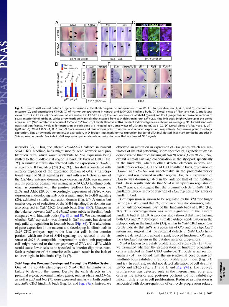
networks (27). Thus, the altered Hand2-Gli3 balance in nascentSall4 CKO hindlimb buds might modify gene network and pro-liferation rates, which would contribute to Shh expression beingshifted to the middle-distal region in hindlimb buds at E10.5 (Fig.2F). A similar shift was also detected with the expression ofHoxd13,a target of SHH signaling (28) (Fig. 2F). This shift is correlated withanterior expansion of the expression domain of Gli1, a transcrip-tional target of SHH signaling (8), and with a reduction in size ofthe Gli1-free anterior domain. Fgf8 expressing AER was narrowerand its posterior domain was missing in Sall4 CKO hindlimb buds,which is consistent with the positive feedback loop between theZPA and AER (29, 30). Accordingly, expression of Fgf10, whoseexpression in developing limb buds is maintained by FGF8 signaling(26), exhibited a smaller expression domain (Fig. 2F). A similar butsmaller degree of reduction of the SHH signaling-free domain wasalso observed in Sall4 CKO forelimb buds (Fig. S5C). Changes inthe balance between Gli3 and Hand2 were subtle in forelimb budscompared with hindlimb buds (Fig. S5 A and B). We also examinedwhether Sall4 expression was altered in Gli3 mutants, but detectedonly mild up-regulation in forelimb buds (Fig. S6). The alterationsof gene expression in the nascent and developing hindlimb buds inSall4 CKO embryos support the idea that cells in the anteriorportion, which are free of SHH signaling, are reduced in the ab-sence of Sall4. A compatible interpretation is that limb progenitorcells might respond to the new geometry of ZPA and AER, whichwould cause fewer cells to be specified as anterior digit precursors.Such a reduction of the anterior cells would result in the lack ofanterior digits in hindlimbs (Fig. 1) (7).
Sall4 Regulates Proximal Development Through the Plzf-Hox System.One of the notable phenotypes of Sall4 CKO hindlimbs is thefailure to develop the femur. Despite the early defects in theproximal region, proximal marker genes, such asMeis2 and Efnb2,as well as Irx3 and Irx5 (7), were expressed similarly in both controland Sall4 CKO hindlimb buds (Fig. 3A and Fig. S7B). Instead, we
observed an alteration in expression of Hox genes, which are reg-ulators of skeletal patterning. More specifically, a genetic study hasdemonstrated that mice lacking all Hox10 genes (Hoxa10, c10, d10)exhibit a small cartilage condensation in the stylopod, specificallyin the hindlimbs, whereas other skeletal elements in fore- andhindlimbs develop (31). In Sall4 CKO hindlimb buds, expression ofHoxa10 and Hoxd10 was undetectable in the proximal-anteriorregion, and was reduced in other regions (Fig. 3B). Expression ofHoxc10 was down-regulated in the anterior half of the hindlimbbud. These results indicate that Sall4 is an upstream regulator ofHox10 genes, and suggest that the proximal defects in Sall4 CKOhindlimbs involve reduced function of Hox10 genes in the anteriorhindlimb bud.Hox expression is known to be regulated by the Plzf zinc finger
factor (32). We found that Plzf expression was also down-regulatedin the anterior-proximal part of the hindlimb buds at E10.5 (Fig.3C). This down-regulation was more significant in the nascenthindlimb bud at E10.0. A previous study showed that mice lackingboth Gli3 and Plzf developed a small cartilage condensation in thestylopod only in the hindlimbs (33), similar to Sall4 CKO mice. Ourresults indicate that Sall4 acts upstream of Gli3 and the Plzf-Hox10system and suggest that the proximal defects in Sall4 CKO hind-limbs are derived from, at least in part, reduced function ofGli3 andthe Plzf-Hox10 system in the putative anterior progenitors.Sall4 is known to regulate proliferation of stem cells (15), thus,
we examined whether the proliferation of hindlimb progenitorcells is affected in Sall4 CKO embryos. Through serial sectionanalysis (34), we found that the mesenchymal core of nascenthindlimb buds exhibited a reduced proliferation index (Fig. 3 Dand E). In contrast, we did not detect alteration of cell death atE9.75 and E10.5 (Fig. 3 D and E and Fig. S7A). The reducedproliferation was detected only in the mesenchymal core, andcells in the anterior and posterior portions did not exhibit sig-nificant difference in cell proliferation. Reduced proliferation isassociated with down-regulation of cell cycle progression related
Fig. 2. Loss of Sall4 caused defects of gene expression in hindlimb progenitors independent of Irx3/5. In situ hybridization (A, B, E, and F), immunofluo-rescence (C), and quantitative RT-PCR (D) of marker genes/proteins in control and Sall4 CKO hindlimb buds. (A) Dorsal views of Tbx4 and Fgf10, and lateralviews of Tbx4 at E9.75. (B) Dorsal views of Irx3 and Irx5 at E9.5-E9.75. (C) Immunofluorescence of SALL4 (green) and IRX3 (magenta) on transverse sections ofE9.75 anterior hindlimb buds. White arrowheads point to cells that escaped from Sall4 deletion in Tcre; Sall4 CKO hindlimb buds. (Right) Close up of the boxedareas in Left. (D) Quantitative analysis of Irx3 and Irx5 transcript levels. Relative mRNA levels of indicated genes are shown as average ± SD. Asterisks indicatestatistical significance. P values for expression of each gene are included. (E) Dorsal views of Gli3 and Hand2 at E10.0. (F) Dorsal views of Shh, Hoxd13, Gli1,Fgf8 and Fgf10 at E10.5. (A, B, E, and F) Black arrows and blue arrows point to normal and reduced expression, respectively. Red arrows point to ectopicexpression. Blue arrowheads denote loss of expression. In D, broken lines mark normal expression border of Gli3. In E, dotted lines mark somite boundaries inShh expression panels. Brackets in Gli1 expression panels denote anterior domains that are free of Gli1 signals.
Akiyama et al. PNAS | April 21, 2015 | vol. 112 | no. 16 | 5077
DEV
ELOPM
ENTA
LBIOLO
GY

genes, such as Ccnd1, Ccne1, and Mycn (Fig. S8 A–L). Theseresults suggest that loss of Sall4 caused failure to expand cells ina discrete region in the nascent hindlimb buds.
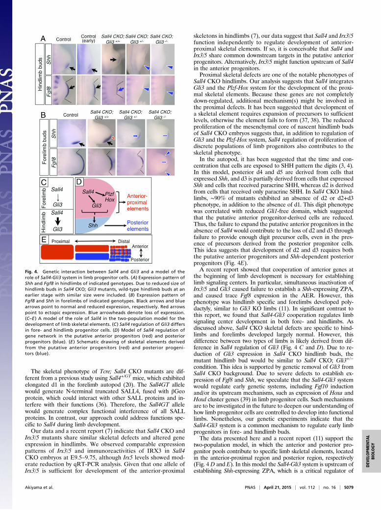
Interaction Between Sall4 and Gli3 Regulates Development of theLimb Signaling Centers in both Fore- and Hindlimbs. A major dif-ference between fore- and hindlimbs of Sall4 CKOmutants is Sall4regulation of Gli3, which was evident in hindlimb buds but wassubtle in forelimb buds (Fig. 2E and Fig. S5). To test whetherhindlimb-specific skeletal defects in Sall4 CKO limbs involve re-duced Gli3 rather than loss of Sall4 alone, we genetically removedGli3 from the Sall4 CKO background. Because Sall4 CKO; Gli3+/−
embryos did not survive beyond E10.5, we focused our analysis onShh, whose expression is significantly affected in Sall4 CKO hind-limb buds (Fig. 2F). We also monitored Fgf8 expression, given arecent genetic study, involving Irx3/5 and Gli3, indicated that co-operation of anterior genes is necessary for establishing limb sig-naling centers, such as Shh-expressing ZPA and Fgf8-expressingAER (11).In hindlimbs, removing one allele of Gli3 from the Sall4 CKO
background caused a failure to express both Shh and Fgf8 (Fig.4A), which was also observed in Sall4 CKO; Gli3−/− double KOhindlimbs. Although Sall4 CKO; Gli3 double mutants exhibitedsmaller hindlimb buds, the lack of Shh and Fgf8 is unlikely simplydue to small size of hindlimb buds, because wild-type hindlimbswith similar size (earlier stages) showed evident expression ofboth genes. Given that Gli3 is down-regulated in Sall4 CKOhindlimb buds, this result suggests that the Sall4 CKO; Gli3+/−
genotype caused a condition in the hindlimb similar to the Sall4CKO; Gli3−/− genotype.In forelimb buds of Sall4 CKO; Gli3+/− mutants, Shh expression
became weak, and its expression domain shifted to the middle-distal region (Fig. 4B). In addition, the Fgf8 expression domain wasshort. These expression patterns in forelimb buds resembled thoseof Sall4 CKO hindlimb buds. In Sall4 CKO; Gli3−/− double KOforelimbs, expression of Shh and Fgf8 was further down-regulatedand only trace levels of signals were detected. The residual Shh and
Fgf8 in Sall4 CKO; Gli3−/− forelimb buds implies that additionalanterior factor(s) might participate in signaling center establish-ment in forelimb buds. Comparison of the expression pattern ofShh and Fgf8 between forelimb and hindlimb buds supports theidea that the hindlimb-specific phenotype in Sall4 CKO mutantsinvolves reduced Gli3 levels. These results demonstrate that ge-netic interaction between two anterior factors, Sall4 and Gli3, isnecessary to establish limb signaling centers in both fore- andhindlimbs. The results also suggest that the difference of Sall4CKO skeletal phenotypes in fore- and hindlimbs would be de-rived from differences in Gli3 regulation by Sall4 (Fig. 2E andFig. S5).Because SALL4 and GLI3 are both nuclear proteins, they might
physically interact. Indeed, we observed both SALL4a (long form)and SALL4b (short form) could interact with GLI3, when trans-fected in cultured cells (Fig. S8M and N). This result suggests thatSALL4 and GLI3 might directly interact, in addition to geneti-cally, to regulate downstream developmental programs.
DiscussionProper development of skeletal elements depends on specifica-tion, expansion and patterning of limb progenitor cells (35).Genetic experiments have shown that posterior skeletal elementsare developed in a Shh-dependent manner (3, 4). However, howanterior skeletal elements are developed has been poorly un-derstood. A recent report demonstrated that anterior progenitorcells, marked by Irx3/5, give rise to the femur, tibia, and d1 in thehindlimb (7). These skeletal elements are missing (d1, tibia) orare present as a small cartilage condensation (femur) in Sall4CKO hindlimbs, suggesting that Sall4 function is required for theputative anterior progenitors. This phenotype was observed onlywhen Tcre was used to inactivate Sall4, and Hoxb6Cre-dependentSall4 inactivation only resulted in the lack of d1 in approximatelyhalf of the mutants. These different skeletal phenotypes fromusing Tcre and Hoxb6Cre suggest that the progenitors for thefemur and tibia are specified before digits, and this specificationoccurs before hindlimb outgrowth.
Fig. 3. Sall4 integrates Plzf-Hox10 system and proliferative expansion of limb progenitors. (A) Expression pattern of Meis2, Efnb2, Irx3, and Irx5 in controland Sall4 CKO hindlimbs at E10.5. (B) Expression pattern of Hoxa10, Hoxc10, and Hoxd10 in control and Sall4 CKO hindlimbs at E10.5. (C) Expression pattern ofPlzf in control and Sall4 CKO hindlimbs at E9.75 and E10.5. Black arrows and blue arrows point to normal and reduced expression, respectively. In A, Meis2expression is indicated by white arrows. Blue arrowheads denote loss of expression. In B, a broken line indicates the anterior boundary of Hoxc10 expressionin Sall4 CKO hindlimb buds. (D) Representative images of pHis3 and TUNEL double staining of transverse sections of nascent hindlimb bud at E9.75 in controland Sall4 CKO embryos. Dotted lines indicate boundary between the nascent hindlimb bud and trunk. (E) Quantitative evaluation of cell proliferation and celldeath in transverse sections of nascent hindlimb buds at E9.75 in control and Sall4 CKO embryos. P values are included.
5078 | www.pnas.org/cgi/doi/10.1073/pnas.1421949112 Akiyama et al.
The skeletal phenotype of Tcre; Sall4 CKO mutants are dif-ferent from a previous study using Sall4+/GT mice, which exhibitedelongated d1 in the forelimb autopod (20). The Sall4GT allelewould generate N-terminal truncated SALL4, fused with βGeoprotein, which could interact with other SALL proteins and in-terfere with their functions (36). Therefore, the Sall4GT allelewould generate complex functional interference of all SALLproteins. In contrast, our approach could address functions spe-cific to Sall4 during limb development.Our data and a recent report (7) indicate that Sall4 CKO and
Irx3/5 mutants share similar skeletal defects and altered geneexpression in hindlimbs. We observed comparable expressionpatterns of Irx3/5 and immunoreactivities of IRX3 in Sall4CKO embryos at E9.5–9.75, although Irx5 levels showed mod-erate reduction by qRT-PCR analysis. Given that one allele ofIrx3/5 is sufficient for development of the anterior-proximal
skeletons in hindlimbs (7), our data suggest that Sall4 and Irx3/5function independently to regulate development of anterior-proximal skeletal elements. If so, it is conceivable that Sall4 andIrx3/5 share common downstream targets in the putative anteriorprogenitors. Alternatively, Irx3/5 might function upstream of Sall4in the anterior progenitors.Proximal skeletal defects are one of the notable phenotypes of
Sall4 CKO hindlimbs. Our analysis suggests that Sall4 integratesGli3 and the Plzf-Hox system for the development of the proxi-mal skeletal elements. Because these genes are not completelydown-regulated, additional mechanism(s) might be involved inthe proximal defects. It has been suggested that development ofa skeletal element requires expansion of precursors to sufficientlevels, otherwise the element fails to form (37, 38). The reducedproliferation of the mesenchymal core of nascent hindlimb budsof Sall4 CKO embryos suggests that, in addition to regulation ofGli3 and the Plzf-Hox system, Sall4 regulation of proliferation ofdiscrete populations of limb progenitors also contributes to theskeletal phenotype.In the autopod, it has been suggested that the time and con-
centration that cells are exposed to SHH pattern the digits (3, 4).In this model, posterior d4 and d5 are derived from cells thatexpressed Shh, and d3 is partially derived from cells that expressedShh and cells that received paracrine SHH, whereas d2 is derivedfrom cells that received only paracrine SHH. In Sall4 CKO hind-limbs, ∼90% of mutants exhibited an absence of d2 or d2+d3phenotype, in addition to the absence of d1. This digit phenotypewas correlated with reduced Gli1-free domain, which suggestedthat the putative anterior progenitor-derived cells are reduced.Thus, the failure to expand the putative anterior progenitors in theabsence of Sall4 would contribute to the loss of d2 and d3 throughfailure to provide enough digit precursor cells, even in the pres-ence of precursors derived from the posterior progenitor cells.This idea suggests that development of d2 and d3 requires boththe putative anterior progenitors and Shh-dependent posteriorprogenitors (Fig. 4E).A recent report showed that cooperation of anterior genes at
the beginning of limb development is necessary for establishinglimb signaling centers. In particular, simultaneous inactivation ofIrx3/5 and Gli3 caused failure to establish a Shh-expressing ZPA,and caused trace Fgf8 expression in the AER. However, thisphenotype was hindlimb specific and forelimbs developed poly-dactyly, similar to Gli3 KO limbs (11). In significant contrast tothis report, we found that Sall4-Gli3 cooperation regulates limbsignaling center development in both fore- and hindlimbs. Asdiscussed above, Sall4 CKO skeletal defects are specific to hind-limbs and forelimbs developed largely normal. However, thisdifference between two types of limbs is likely derived from dif-ference in Sall4 regulation of Gli3 (Fig. 4 C and D). Due to re-duction of Gli3 expression in Sall4 CKO hindlimb buds, themutant hindlimb bud would be similar to Sall4 CKO; Gli3+/−
condition. This idea is supported by genetic removal of Gli3 fromSall4 CKO background. Due to severe defects to establish ex-pression of Fgf8 and Shh, we speculate that the Sall4-Gli3 systemwould regulate early genetic systems, including Fgf10 inductionand/or its upstream mechanisms, such as expression of Hoxa andHoxd cluster genes (39) in limb progenitor cells. Such mechanismsare to be investigated in the future to deepen our understanding ofhow limb progenitor cells are controlled to develop into functionallimbs. Nonetheless, our genetic experiments indicate that theSall4-Gli3 system is a common mechanism to regulate early limbprogenitors in fore- and hindlimb buds.The data presented here and a recent report (11) support the
two-population model, in which the anterior and posterior pro-genitor pools contribute to specific limb skeletal elements, locatedin the anterior-proximal region and posterior region, respectively(Fig. 4 D and E). In this model the Sall4-Gli3 system is upstream ofestablishing Shh-expressing ZPA, which is a critical regulator of
Fig. 4. Genetic interaction between Sall4 and Gli3 and a model of therole of Sall4-Gli3 system in limb progenitor cells. (A) Expression pattern ofShh and Fgf8 in hindlimbs of indicated genotypes. Due to reduced size ofhindlimb buds in Sall4 CKO; Gli3 mutants, wild-type hindlimb buds at anearlier stage with similar size were included. (B) Expression pattern ofFgf8 and Shh in forelimbs of indicated genotypes. Black arrows and bluearrows point to normal and reduced expression, respectively. Red arrowspoint to ectopic expression. Blue arrowheads denote loss of expression.(C–E ) A model of the role of Sall4 in the two-population model for thedevelopment of limb skeletal elements. (C ) Sall4 regulation of Gli3 differsin fore- and hindlimb progenitor cells. (D) Model of Sall4 regulation ofgene network in the putative anterior progenitors (red) and posteriorprogenitors (blue). (E ) Schematic drawing of skeletal elements derivedfrom the putative anterior progenitors (red) and posterior progeni-tors (blue).
Akiyama et al. PNAS | April 21, 2015 | vol. 112 | no. 16 | 5079
DEV
ELOPM
ENTA
LBIOLO
GY
proliferative expansion of the posterior progenitor pool. Moreover,the Sall4, together with Gli3 and the Plzf-Hox system, regulatesthe putative anterior progenitor pool. Therefore, our data sug-gests that the Sall4-Gli3 system is a critical upstream regulatorof both progenitor pools, and thus, development of the limbskeletal elements.
Materials and MethodsMouse Mutants. The mouse lines for Sall4 (15, 40),Gli3 (41), Tcre (21), Hoxb6Cre(22), and Prx1Cre (24) were maintained on a mixed genetic background.Skeletal preparation was done as published (42). Animal breeding and pro-cedures were performed according to the approval by the Institutional AnimalCare and Use Committee of the University of Minnesota.
Immunofluorescence and TUNEL Assay. Immunofluorescence of IRX3 (43) andSALL4 (Santa Cruz, sc-101147, 1/300 dilution) was performed on cryo-sec-tions according to standard procedures (42). Simultaneous cell proliferationand cell death analyses were performed by using anti-phospho histone H3(Upstate, catalog no. 06–570) and the In Situ Cell Death Detection kit(Roche) as published (34). Cell death analysis at E10.5 was performed oncoronal sections by using the In Situ Cell Death Detection kit. DAPI was usedfor nuclear staining. Fluorescent confocal images were obtained by usingZeiss LSM 710 laser scanning microscope system (Carl Zeiss Microscopy), andanalyzed using ZEN2009 software (Carl Zeiss Microscopy).
Skeletal Preparation and in Situ Hybridization. Preparation of skeletal stainingand in situ mRNA detection were performed according to a standard pro-cedure (42).
Quantitative RT-PCR for Irx3/5. Relative expression of Irx3/5 in wild-type andSall4 CKO was determined by isolating RNA from the hindlimb forming re-gion/nascent hindlimb buds at E9.5–9.75 and performing quantitative PCRafter reverse transcription. Details are provided in SI Materials and Methods.
Coimmunoprecipitation Assay. HEK293T cells were transfected with expres-sion constructs by using Polyethylenimine MAX (Polysciences). Cell lysateswere prepared after two days and coimmunoprecipitation assay were per-formed by using protein G-Sepharose (GE Healthcare) and anti-Flag antibody(M2, Sigma). Proteins were resolved by SDS/PAGE, transferred to PVDFmembranes (Millipore) and detected by anti-HA antibodies (4B2, Wako), HRPgoat anti-mouse IgG, and a chemiluminescence system.
ACKNOWLEDGMENTS. We thank Drs. Maria Barna, Juan Carlos IzpisuaBelmonte, C-c Hui, Michael Kuehn, Mark Lewandoski, Jonathan Licht, SridharRao, Sestuko Sahara, Toshihiko Shiroishi, and Stephanie Ware for sharingplasmids and/or mouse lines. We also thank Susan Morton for IRX3 antibody,Dr. Michael O’Connor for the use of LSM 710, Dr. David Zarkower for criticalreading of the manuscript, and Drs. C-c Hui and Sevan Hopyan for sharingunpublished data. We thank Jessica Burlingame, Asha Elogna, Jenna Matson,Thu Quach, and Elizabeth West for their excellent technical support and StevePehoski for editorial assistance. This work is supported by the National Insti-tute of Arthritis and Musculoskeletal and Skin Diseases of NIH (R01AR064195).
1. Gros J, Tabin CJ (2014) Vertebrate limb bud formation is initiated by localized epi-thelial-to-mesenchymal transition. Science 343(6176):1253–1256.
2. Zeller R, López-Ríos J, Zuniga A (2009) Vertebrate limb bud development: Movingtowards integrative analysis of organogenesis. Nat Rev Genet 10(12):845–858.
3. Harfe BD, et al. (2004) Evidence for an expansion-based temporal Shh gradient inspecifying vertebrate digit identities. Cell 118(4):517–528.
4. Ahn S, Joyner AL (2004) Dynamic changes in the response of cells to positivehedgehog signaling during mouse limb patterning. Cell 118(4):505–516.
5. Kraus P, Fraidenraich D, Loomis CA (2001) Some distal limb structures develop in micelacking Sonic hedgehog signaling. Mech Dev 100(1):45–58.
6. Chiang C, et al. (2001) Manifestation of the limb prepattern: Limb development in theabsence of sonic hedgehog function. Dev Biol 236(2):421–435.
7. Li D, et al. (2014) Formation of proximal and anterior limb skeleton requires earlyfunction of Irx3 and Irx5 and is negatively regulated by Shh signaling. Dev Cell 29(2):233–240.
8. Hui CC, Angers S (2011) Gli proteins in development and disease. Annu Rev Cell DevBiol 27:513–537.
9. te Welscher P, Fernandez-Teran M, Ros MA, Zeller R (2002) Mutual genetic antago-nism involving GLI3 and dHAND prepatterns the vertebrate limb bud mesenchymeprior to SHH signaling. Genes Dev 16(4):421–426.
10. Hui CC, Joyner AL (1993) A mouse model of greig cephalopolysyndactyly syndrome:The extra-toesJ mutation contains an intragenic deletion of the Gli3 gene. Nat Genet3(3):241–246.
11. Zhulyn O, et al. (2014) A switch from low to high Shh activity regulates establishmentof limb progenitors and signaling centers. Dev Cell 29(2):241–249.
12. de Celis JF, Barrio R (2009) Regulation and function of Spalt proteins during animaldevelopment. Int J Dev Biol 53(8-10):1385–1398.
13. Kawakami Y, et al. (2009) Sall genes regulate region-specific morphogenesis in themouse limb by modulating Hox activities. Development 136(4):585–594.
14. Rao S, et al. (2010) Differential roles of Sall4 isoforms in embryonic stem cell pluri-potency. Mol Cell Biol 30(22):5364–5380.
15. Sakaki-Yumoto M, et al. (2006) The murine homolog of SALL4, a causative gene inOkihiro syndrome, is essential for embryonic stem cell proliferation, and cooperateswith Sall1 in anorectal, heart, brain and kidney development. Development 133(15):3005–3013.
16. Hobbs RM, et al. (2012) Functional antagonism between Sall4 and Plzf definesgermline progenitors. Cell Stem Cell 10(3):284–298.
17. Yong KJ, et al. (2013) Oncofetal gene SALL4 in aggressive hepatocellular carcinoma.N Engl J Med 368(24):2266–2276.
18. Buganim Y, et al. (2014) The developmental potential of iPSCs is greatly influenced byreprogramming factor selection. Cell Stem Cell 15(3):295–309.
19. Lim CY, et al. (2008) Sall4 regulates distinct transcription circuitries in differentblastocyst-derived stem cell lineages. Cell Stem Cell 3(5):543–554.
20. Koshiba-Takeuchi K, et al. (2006) Cooperative and antagonistic interactions betweenSall4 and Tbx5 pattern the mouse limb and heart. Nat Genet 38(2):175–183.
21. Perantoni AO, et al. (2005) Inactivation of FGF8 in early mesoderm reveals an essentialrole in kidney development. Development 132(17):3859–3871.
22. Lowe LA, Yamada S, Kuehn MR (2000) HoxB6-Cre transgenic mice express Cre re-combinase in extra-embryonic mesoderm, in lateral plate and limb mesoderm and atthe midbrain/hindbrain junction. Genesis 26(2):118–120.
23. Fromental-Ramain C, et al. (1996) Hoxa-13 and Hoxd-13 play a crucial role in thepatterning of the limb autopod. Development 122(10):2997–3011.
24. Logan M, et al. (2002) Expression of Cre Recombinase in the developing mouse limbbud driven by a Prxl enhancer. Genesis 33(2):77–80.
25. Chapman DL, et al. (1996) Expression of the T-box family genes, Tbx1-Tbx5, duringearly mouse development. Dev Dyn 206(4):379–390.
26. Ohuchi H, et al. (1997) The mesenchymal factor, FGF10, initiates and maintains theoutgrowth of the chick limb bud through interaction with FGF8, an apical ectodermalfactor. Development 124(11):2235–2244.
27. Osterwalder M, et al. (2014) HAND2 targets define a network of transcriptionalregulators that compartmentalize the early limb bud mesenchyme. Dev Cell 31(3):345–357.
28. Riddle RD, Johnson RL, Laufer E, Tabin C (1993) Sonic hedgehog mediates the po-larizing activity of the ZPA. Cell 75(7):1401–1416.
29. Niswander L (2002) Interplay between the molecular signals that control vertebratelimb development. Int J Dev Biol 46(7):877–881.
30. Zúñiga A, Haramis AP, McMahon AP, Zeller R (1999) Signal relay by BMP antagonismcontrols the SHH/FGF4 feedback loop in vertebrate limb buds. Nature 401(6753):598–602.
31. Wellik DM, Capecchi MR (2003) Hox10 and Hox11 genes are required to globallypattern the mammalian skeleton. Science 301(5631):363–367.
32. Barna M, Hawe N, Niswander L, Pandolfi PP (2000) Plzf regulates limb and axialskeletal patterning. Nat Genet 25(2):166–172.
33. Barna M, Pandolfi PP, Niswander L (2005) Gli3 and Plzf cooperate in proximal limbpatterning at early stages of limb development. Nature 436(7048):277–281.
34. Akiyama R, et al. (2014) Distinct populations within Isl1 lineages contribute to ap-pendicular and facial skeletogenesis through the β-catenin pathway. Dev Biol 387(1):37–48.
35. Mariani FV, Martin GR (2003) Deciphering skeletal patterning: Clues from the limb.Nature 423(6937):319–325.
36. Kiefer SM, et al. (2003) Expression of a truncated Sall1 transcriptional repressor isresponsible for Townes-Brocks syndrome birth defects. Hum Mol Genet 12(17):2221–2227.
37. Wolpert L, Tickle C, Sampford M (1979) The effect of cell killing by x-irradiation onpattern formation in the chick limb. J Embryol Exp Morphol 50:175–193.
38. Galloway JL, Delgado I, Ros MA, Tabin CJ (2009) A reevaluation of X-irradiation-induced phocomelia and proximodistal limb patterning. Nature 460(7253):400–404.
39. Sheth R, et al. (2013) Decoupling the function of Hox and Shh in developing limbreveals multiple inputs of Hox genes on limb growth. Development 140(10):2130–2138.
40. Yuri S, et al. (2009) Sall4 is essential for stabilization, but not for pluripotency, ofembryonic stem cells by repressing aberrant trophectoderm gene expression. StemCells 27(4):796–805.
41. Büscher D, Grotewold L, Rüther U (1998) The XtJ allele generates a Gli3 fusiontranscript. Mamm Genome 9(8):676–678.
42. Itou J, et al. (2012) Islet1 regulates establishment of the posterior hindlimb fieldupstream of the Hand2-Shh morphoregulatory gene network in mouse embryos.Development 139(9):1620–1629.
43. Chen JA, et al. (2011) Mir-17-3p controls spinal neural progenitor patterning byregulating Olig2/Irx3 cross-repressive loop. Neuron 69(4):721–735.
5080 | www.pnas.org/cgi/doi/10.1073/pnas.1421949112 Akiyama et al.