salivary mucins protect surfaces from colonization …aem.asm.org/content/81/1/332.full.pdf ·...
TRANSCRIPT
Salivary Mucins Protect Surfaces from Colonization by CariogenicBacteria
Erica Shapiro Frenkel,a,b Katharina Ribbeckb
Biological Sciences in Dental Medicine, Harvard University, Cambridge, Massachusetts, USAa; Department of Biological Engineering, Massachusetts Institute ofTechnology, Cambridge, Massachusetts, USAb
Understanding how the body’s natural defenses function to protect the oral cavity from the myriad of bacteria that colonize itssurfaces is an ongoing topic of research that can lead to breakthroughs in treatment and prevention. One key defense mechanismon all moist epithelial linings, such as the mouth, gastrointestinal tract, and lungs, is a layer of thick, well-hydrated mucus. Themain gel-forming components of mucus are mucins, large glycoproteins that play a key role in host defense. This study focuseson elucidating the connection between MUC5B salivary mucins and dental caries, one of the most common oral diseases. Dentalcaries is predominantly caused by Streptococcus mutans attachment and biofilm formation on the tooth surface. Once S. mutansattaches to the tooth, it produces organic acids as metabolic by-products that dissolve tooth enamel, leading to cavity formation.We utilize CFU counts and fluorescence microscopy to quantitatively show that S. mutans attachment and biofilm formation aremost robust in the presence of sucrose and that aqueous solutions of purified human MUC5B protect surfaces by acting as anantibiofouling agent in the presence of sucrose. In addition, we find that MUC5B does not alter S. mutans growth and decreasessurface attachment and biofilm formation by maintaining S. mutans in the planktonic form. These insights point to the impor-tance of salivary mucins in oral health and lead to a better understanding of how MUC5B could play a role in cavity preventionor diagnosis.
One of the body’s key defense mechanisms on wet epitheliallinings, such as the mouth, gastrointestinal tract, and lungs, is
a layer of thick, well-hydrated mucus. The viscoelastic propertiesof mucus are attributed to mucins, large glycoproteins that play akey role in host defense and maintaining a healthy microbial en-vironment (1–3). Defects in mucin production can lead to dis-eases such as ulcerative colitis when mucins are underproduced orcystic fibrosis and asthma when mucins are overproduced (4–6).In addition, studies have shown that mucins can interact withmicrobes, such as Helicobacter pylori, Haemophilus parainfluen-zae, and human immunodeficiency virus (7–10). These diseasesand microbial interactions highlight the necessity of mucins asone of the body’s key natural defenses; however, few studies havefocused specifically on the connection between MUC5B salivarymucins and oral diseases. This study fills this gap in understandingby exploring the connection between purified human MUC5Band the virulence of Streptococcus mutans, one of the main cavity-causing bacteria naturally found in the oral cavity (11). MUC7 isanother salivary mucin, but MUC5B is the primary mucincomponent of the dental pellicle coating the soft and hard tis-sues in the oral cavity (12, 13). Importantly, the effects ofMUC5B are characterized in a clinically relevant three-dimen-sional model that mimics the natural environment in the oralcavity; mucins are secreted into an aqueous phase as opposed toa two-dimensional surface coating, which can create artificiallyconcentrated amounts of surface mucins (14–16). SuspendingMUC5B in media allows polymer domains to interact, prevent-ing collapse of the hydrogel structure (8, 17, 18).
Understanding the structure of MUC5B illustrates how thisspecific glycoprotein can play such a dominant role in maintain-ing oral health. There are several serotypes of mucins throughoutthe body, but MUC5B is the predominant polymeric mucin foundin the oral cavity and female genital tract (19, 20). In the oralcavity, MUC5B is produced by goblet cells in the submandibular
and sublingual glands (2). The peptide backbone is composed of avariable number of tandem repeats (VNTR) section that has re-peating sequences rich in serine, threonine, and proline, whichparticipate in O-glycosylation (1, 2, 21). Because of the extendedVNTR region, MUC5B is composed of approximately 80% carbo-hydrate in the form of O-linked glycan chains and 20% protein,consisting of the peptide backbone (22, 23). MUC5B’s complexstructure allows it to interact with an array of different salivaryproteins and microbes to maintain a healthy oral cavity (24, 25).The exact mechanisms through which MUC5B provides defenseare not well understood, but it has been proposed that it acts as aphysical protective barrier, provides lubrication, and has antimi-crobial properties (13, 25, 26).
S. mutans is a biofilm-forming facultative anaerobic bacteriumthat produces three glucosyltransferase enzymes to synthesize glu-cans from dietary sugar (27–29). Glucans are sticky polymers thatallow the cells to attach to the tooth surface and form an extracel-lular matrix that protects it from host defenses and mechanicalremoval (30, 31). Once S. mutans attaches to the tooth surface,organic acids, which are produced as metabolic by-products, be-come concentrated within the extracellular matrix and cause adrop in pH from neutral to 5 or below. This acidic environment
Received 6 August 2014 Accepted 19 October 2014
Accepted manuscript posted online 24 October 2014
Citation Frenkel ES, Ribbeck K. 2015. Salivary mucins protect surfaces fromcolonization by cariogenic bacteria. Appl Environ Microbiol 81:332–338.doi:10.1128/AEM.02573-14.
Editor: J. L. Schottel
Address correspondence to Katharina Ribbeck, [email protected].
Copyright © 2015, American Society for Microbiology. All Rights Reserved.
doi:10.1128/AEM.02573-14
332 aem.asm.org January 2015 Volume 81 Number 1Applied and Environmental Microbiology
on August 9, 2018 by guest
http://aem.asm
.org/D
ownloaded from
begins dissolving tooth enamel, leading to cavity formation, andthe high tolerance of S. mutans for acidic environments gives it anecological advantage. Without proper hygiene and nutritionalawareness, S. mutans can proliferate quickly, causing serious dam-age to the tooth structure. S. mutans biofilm formation is partic-ularly problematic in the interproximal spaces between teeth,where mechanical removal is difficult.
Because S. mutans attachment and biofilm formation are crit-ical steps in cavity formation, we use CFU counts and fluorescencemicroscopy to quantify the effects of supplemental sugar and pu-rified human salivary MUC5B on these key stages of disease pro-gression. We first validate our mucin studies by showing that S.mutans attachment and biofilm formation are most robust in thepresence of sucrose as opposed to glucose. When supplementalMUC5B is added in the presence of sucrose, however, S. mutansattachment and biofilm formation are significantly decreased. Al-though the number of surface-attached bacteria decreases in thepresence of MUC5B, we show that bacterial growth is unchangedin the presence of MUC5B and the observed effects are due toincreased numbers of S. mutans cells in the planktonic form.These findings that link MUC5B with S. mutans virulence couldsignificantly impact our understanding of the pathogenesis of cav-ity formation and aid in the development of novel oral diagnosticmethods or strategies for disease prevention.
MATERIALS AND METHODSBacterial strains and growth conditions. The bacterial strain Streptococ-cus mutans UA159 was kindly given as a gift by Dan Smith (Forsyth Insti-tute). For sucrose and glucose experiments, S. mutans was grown over-night in brain heart infusion (BHI) medium (Becton, Dickinson andCompany) containing 1% (wt/vol) sucrose and BHI with 1% (wt/vol)glucose (Sigma). For experiments determining the effects of MUC5B, S.mutans was grown overnight in BHI with 1% sucrose. BHI with 1% su-crose and either 0.3% MUC5B or 0.3% (wt/vol) methylcellulose (Sigma)was used to resuspend S. mutans cells before inoculating them into theexperiment. Hydroxyapatite disc (Clarkson Chromatography, Inc.) orchambered glass slide (LabTek) surfaces were used to test S. mutans at-tachment and biofilm formation. S. mutans was grown and incubated at37°C with 5% CO2.
Saliva collection. Submandibular saliva was collected from 10 volun-teers using a custom vacuum pump setup. Specifically, two holes werecut into the cap of a 50-ml conical tube (Falcon); the vacuum line wasinserted into one hole and a small-diameter Tygon collection tube wasinserted into the other hole (Saint Gobain Performance Plastics). Cottonswabs were used to absorb the volunteers’ parotid gland secretions. The col-lection tube was used to suck up pooled unstimulated submandibular glandsecretions from under the tongue. The collection vessel was kept on ice at alltimes. Saliva from volunteers was pooled before MUC5B purification. Proto-cols involving the use of human subjects were approved by MassachusettsInstitute of Technology’s Committee on the Use of Humans as ExperimentalSubjects.
MUC5B purification. Immediately after collection, saliva was dilutedusing 5.5 M sodium chloride containing 0.04% sodium azide so that thefinal concentration of sodium chloride was 0.16 M. The following anti-bacterial agents and protease inhibitors were then added at the indicatedfinal concentrations: benzamidine HCl (5 mM), dibromoacetophenone(1 mM), phenylmethylsulfonyl fluoride (1 mM), and EDTA (5 mM, pH 7)(Sigma). The mucins in the saliva were solubilized overnight by gentlestirring at 4°C. Saliva was then centrifuged at 3,800 � g for 10 min in aswinging-bucket centrifuge to remove cellular debris. MUC5B was puri-fied using a Bio-Rad NGC fast protein liquid chromatography (FPLC)system equipped with an XK 50 column packed with Sepharose CL-2Bresin (GE Healthcare Bio-Sciences). Mucin-containing fractions were
identified using a periodic acid-Schiff’s reagent assay and analysis of UVabsorbance at 280 nm from FPLC. Fractions were then combined, dia-lyzed, and concentrated using an ultrafiltration device and were then ly-ophilized for storage at �80°C.
Assay of CFU counts to evaluate S. mutans attachment and biofilmformation. To test the effects of sucrose or glucose on S. mutans physiol-ogy, S. mutans was grown to mid-exponential phase in BHI with 1%sucrose and BHI with 1% glucose, and then equal numbers of bacteria(107) from each culture were seeded in triplicate into wells containingglass or hydroxyapatite surfaces. For experiments testing the effect ofMUC5B, S. mutans was grown to mid-exponential phase in BHI with 1%sucrose and then seeded in triplicate into wells containing BHI with 1%sucrose and 0.3% MUC5B or control medium. For all experiments, at-tachment was evaluated at 20, 40, and 60 min and biofilm formation wasevaluated at 6, 18, and 24 h. Attachment was defined to occur at timepoints up to 1 h because the doubling time of S. mutans is approximately1.5 h. Biofilm formation was defined to occur at all time points after 1 h. Atthe time point being evaluated, the surface was washed with phosphate-buffered saline (PBS) to remove nonadherent cells, fresh PBS was added,and then adherent cells were lifted using a sterile pipette tip. The sus-pended bacteria were vigorously pipetted to individualize the cells. Thesuspension was diluted (10�1 to 10�7) and plated on BHI agar. The num-bers of CFU were counted after 24 to 36 h of incubation. Statisticallysignificant differences were determined using Student’s t test, with P val-ues of �0.02 considered significant.
Fluorescent staining and microscopy. To visually assess the effects ofsucrose, glucose, or MUC5B on S. mutans attachment and biofilm forma-tion, fluorescent SYTO9 staining with light microscopy was used (LifeTechnologies). S. mutans was grown to mid-exponential phase, thenseeded into a chambered glass slide with BHI containing 1% sucrose and0.3% MUC5B or with control medium. At the time point being evaluated,the surface was washed with PBS to remove nonadherent cells, and then200 �l SYTO9 (0.6 �l SYTO9/200 �l Milli-Q water) was added. Thebiofilm was incubated with SYTO9 in the dark for 30 min. After incuba-tion, the biofilm was washed with Milli-Q water to remove excess dye andfresh Milli-Q water was added. A Zeiss Axio Observer Z1 fluorescenceinverted microscope was used for imaging. All experiments were repeatedin triplicate.
Assay to evaluate S. mutans growth. Overnight cultures of S. mutansat an optical density at 600 nm (OD600) of 0.05 were seeded in triplicateinto a 96-well polystyrene plate containing BHI medium supplementedwith 1% sucrose and 0.3% MUC5B or control medium. Bacteria wereincubated at 37°C with 5% CO2. At 1-h intervals, the cultures were mixedand the OD600 was recorded using a microplate reader. The averages foreach time point were plotted, and a comparison of S. mutans’ growth ratesin the various media was evaluated within the estimated error.
Time-kill assay. CFU counts were used to evaluate the effect ofMUC5B on S. mutans viability at time points up to 24 h. S. mutans wasgrown to mid-exponential phase in BHI with 1% sucrose, and then equalnumbers of bacteria (107) were seeded in triplicate into glass-bottom wellscontaining BHI with 1% sucrose and 0.3% MUC5B or control medium.The cultures were incubated for 6, 18, and 24 h. At the time point beingevaluated, the contents of the wells were gently mixed and then the super-natant was removed and diluted (10�1 to 10�8). The remaining biofilmwas then washed with PBS, scraped off with a sterile pipette tip, anddiluted (10�1 to 10�6). Dilutions were plated on BHI agar. The numbersof CFU were counted after 24 to 36 h of incubation to quantify the numberof viable bacteria.
RESULTS AND DISCUSSIONSucrose enhances S. mutans attachment and biofilm formation.To determine the growth conditions where S. mutans attachmentand biofilm formation are most robust, we investigated the effectof the addition of sucrose or glucose to BHI medium. S. mutanswas inoculated into BHI containing 1% sucrose or 1% glucose.
Mucins Protect Surfaces from S. mutans Colonization
January 2015 Volume 81 Number 1 aem.asm.org 333Applied and Environmental Microbiology
on August 9, 2018 by guest
http://aem.asm
.org/D
ownloaded from

CFU counts were used to evaluate attachment at 20, 40, and 60min and biofilm formation at 6, 18, and 24 h. Attachment wasdefined to occur at time points up to 60 min because the doublingtime of S. mutans in exponential phase is approximately 1.5 h.Experiments were carried out on glass and hydroxyapatite discsbecause there are surface-specific effects on S. mutans attachmentand biofilm formation (32). S. mutans attachment on glass andhydroxyapatite was increased by 15 and 6 times, respectively,when sucrose was present compared to when glucose was present(Fig. 1A and C). S. mutans biofilm formation in the presence ofsucrose was increased by 45% on glass and 8% on hydroxyapatitecompared to that in the presence of glucose (Fig. 1B and D). Thenumber of S. mutans cells in the biofilm for each condition isrepresented as a fraction of the total number of bacteria in the well,because S. mutans’ growth rate changes in the presence of sucrosecompared with that in the presence of glucose (Fig. 2). The in-crease in S. mutans attachment and biofilm formation in the pres-
20 40 60 100
101
102
103
104
105
Time (min)
Atta
ched
Bac
teria
(L
og10
CFU
/µL)
* * *
20 40 60 100
101
102
103
Time (min)
* *
6 18 240
20
40
60
80
100
Biof
ilm B
acte
ria /
Tota
l Bac
teria
(%)
Time (h)
**
Time (h)6 18 24
0
5
10
15
20
25*
*
A B
C D
E
Atta
ched
Bac
teria
(L
og10
CFU
/µL)
Biof
ilm B
acte
ria /
Tota
l Bac
teria
(%)
F
20 m
in40
min
60 m
in
6 h
18 h
24 h
Medium+Glucose Medium+Sucrose
Medium+Glucose Medium+Sucrose Medium+SucroseMedium+Glucose
*
FIG 1 Sucrose enhances S. mutans attachment and biofilm formation. The levels of S. mutans attachment (A) and biofilm formation (B) on glass are significantlyenhanced at all time points when the bacteria are grown in BHI containing 1% sucrose (Medium�Sucrose) compared to the levels achieved in BHI containing1% glucose (Medium�Glucose). S. mutans attachment (C) and biofilm formation (D) on hydroxyapatite are similarly increased in the presence of sucrose,illustrating that the effect of sucrose on S. mutans physiology is not surface specific. Fluorescence microscopy images verify the findings of the CFU countexperiments by showing an increase in S. mutans attachment (E) and biofilm formation (F) on glass in the presence of sucrose. *, statistically significant differencedetermined by Student’s t test (P � 0.02). Error bars represent SDs. Scale bars, 20 �m.
Time (h)
OD
600
0 5 100.0
0.2
0.4
0.6
0.8 Medium+Glucose Medium+Sucrose
FIG 2 Supplemental sugar alters S. mutans growth. A growth curve of S.mutans in BHI with added 1% glucose (Medium�Glucose) or 1% sucrose(Medium�Sucrose) shows that S. mutans’ growth rate changes based on thespecific sugar present in the growth medium. Error bars represent SDs.
Frenkel and Ribbeck
334 aem.asm.org January 2015 Volume 81 Number 1Applied and Environmental Microbiology
on August 9, 2018 by guest
http://aem.asm
.org/D
ownloaded from

ence of sucrose is supported by a fluorescence microscopy timeseries using SYTO9 nucleic acid stain to visualize S. mutans attach-ment and biofilm formation in BHI containing 1% sucrose or 1%glucose on a glass surface (Fig. 1E and F). The role of sucrose inenhancing S. mutans biofilm formation has been well establishedusing genetic analysis and biochemical assays studying biofilmarchitecture (33–36). Here we characterized this phenomenon us-ing a quantitative method that directly evaluates the number oflive S. mutans cells attached and producing biofilm on varioussurfaces. Our results support those of previous studies by quanti-tatively showing that the addition of 1% sucrose enhanced S. mu-tans attachment and biofilm formation at all time points com-pared to attachment and biofilm formation in the presence of 1%glucose. There was little or no growth in BHI without a sugarsource, illustrating that protein alone cannot support S. mutansattachment or biofilm formation. Furthermore, our results show-ing that sucrose enhances S. mutans attachment and biofilm for-mation were consistent on hydroxyapatite and glass surfaces, in-dicating that the effect is not surface specific. These findings set thegroundwork for our investigation of the role of MUC5B in S.
mutans attachment and biofilm formation. When testing the ef-fect of MUC5B on S. mutans physiology, 1% sucrose was added toBHI medium to challenge the effect of MUC5B by ensuring that S.mutans attachment and biofilm formation are most robust.
MUC5B decreases S. mutans attachment and biofilm forma-tion. The effect of MUC5B on S. mutans attachment and biofilmformation was evaluated using CFU counts on various surfacesand fluorescence microscopy. S. mutans (107 bacteria) was grownin a chambered glass slide or on a hydroxyapatite disc in the pres-ence of BHI with 1% sucrose and 0.3% MUC5B. BHI with 1%sucrose and 0.3% methylcellulose and BHI with no added poly-mer served as controls. Methylcellulose is a gel-forming com-pound that, like mucins, imparts viscosity but does not containthe complex, glycosylated structure that is characteristic ofMUC5B. On glass, the addition of MUC5B to growth mediumdecreased S. mutans attachment by 88% and biofilm formation by74% compared to the levels of attachment and biofilm formationin BHI with 1% sucrose (Fig. 3A and B). In comparison, the ad-dition of methylcellulose reduced S. mutans attachment and bio-film formation on glass by 50% and 16%, respectively (Fig. 3A and
*
20 40 60 0
50
100
150
200
Time (min)
Atta
chm
ent
(%of
cont
rol)
* *
*
Time (min)
Atta
chm
ent
(%of
cont
rol)
20 40 60 0
50
100
150
200
* *
* *
6 18 240
50
100
150
200
Time (h)
Biof
ilm(%
ofco
ntro
l)
**
Time (h)
Biof
ilm(%
ofco
ntro
l)
6 18 240
50
100
150
200
* ** *
*
A B
C D
FE SMedium+Mucins
SMedium+Methyl. SMedium
6 h
24 h
20 m
in40
min
60 m
in
SMedium+MucinsSMedium+MethylcelluloseSMedium
SMedium+Mucins
SMedium+Methyl. SMedium
18 h
FIG 3 Salivary mucins reduce S. mutans attachment and biofilm formation. The addition of 0.3% mucins to the control medium, BHI containing 1% sucrose(SMedium), significantly reduces the levels of S. mutans attachment and biofilm formation on glass (A, B) and hydroxyapatite (C, D) compared to the levelsobtained with the control consisting of BHI with 1% sucrose. Similarly, the addition of 0.3% methylcellulose to BHI with 1% sucrose reduces S. mutansattachment and biofilm formation; however, the effect is not significant for the majority of time points studied. Fluorescence microscopy was used to visuallyassess S. mutans attachment (E) and biofilm formation (F) on glass when the bacteria are grown in BHI with 1% sucrose and 0.3% mucins, BHI with 1% sucroseand 0.3% methylcellulose (Methyl.), and BHI with 1% sucrose. *, statistically significant difference from BHI with 1% sucrose determined by Student’s t test (P �0.02). Error bars represent SDs. Scale bars, 20 �m.
Mucins Protect Surfaces from S. mutans Colonization
January 2015 Volume 81 Number 1 aem.asm.org 335Applied and Environmental Microbiology
on August 9, 2018 by guest
http://aem.asm
.org/D
ownloaded from
B). When S. mutans was grown on hydroxyapatite discs in thepresence of MUC5B, attachment was decreased by 77% and bio-film formation was decreased by 95% compared to the levels inBHI with 1% sucrose (Fig. 3C and D). In comparison, the pres-ence of methylcellulose reduced S. mutans attachment by 27% andbiofilm formation by 76% on hydroxyapatite discs (Fig. 3C andD). There was an overall decrease in biofilm formation at 18and 24 h due to the dissolution of the hydroxyapatite discs. Byevaluating the attachment and biofilm formation of S. mutans inmedium containing methylcellulose, which simulates an environ-ment that has physical properties similar to those of mucins, wecan better understand if MUC5B is acting as a physical barrier toattachment most likely through increased viscosity or if the ob-served effect is due to specific MUC5B properties. We can con-clude that the latter is most likely because S. mutans attachmentand biofilm formation in the presence of MUC5B are significantlydecreased at most time points compared to the levels in the pres-ence of methylcellulose. There are at least three potential mecha-nisms by which MUC5B could protect the surface from bacterialcolonization: (i) MUC5B could bind or agglutinate bacteria,which would allow planktonic bacteria to be swept out of the oralcavity with salivary flow but enhance bacterial attachment to sur-faces coated with MUC5B (14, 25, 37–44), (ii) MUC5B could havethe opposite effect, where its heterogeneous glycan chains repelbacteria, thereby preventing surface attachment (9, 11, 45–47), or(iii) MUC5B could directly downregulate S. mutans genes in-volved in attachment and biofilm formation. In our case, itappears that MUC5B is repelling S. mutans and/or directly influ-encing genetic modifications that protect the glass and hydro-xyapatite surfaces from bacterial attachment and biofilm forma-tion. The decrease in attachment caused by methylcelluloseindicates that increased viscosity may also be playing some role inreducing attachment at early time points. Fluorescence micros-copy experiments using SYTO9 staining confirm the findings ob-tained by CFU counts by showing a visually detectable decrease inthe amount of S. mutans on glass and that the characteristic mi-crocolony morphology of S. mutans biofilms is unchanged (Fig.3E and F) (48, 49).
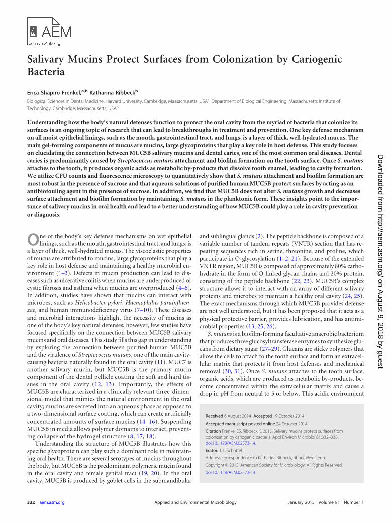
MUC5B does not alter S. mutans growth. To evaluate theeffect of MUC5B on bacterial growth, S. mutans was grown in BHImedium containing 1% sucrose and 0.3% MUC5B. BHI with 1%sucrose and 0.3% methylcellulose and BHI with 1% sucrose wereused as controls. These are the same media used in experimentsthat determine the effects of MUC5B on S. mutans attachment and
biofilm formation. Optical density readings over the course of 12h show that the addition of MUC5B or methylcellulose does notalter S. mutans growth compared to its growth in BHI containing1% sucrose (Fig. 4). Because the growth of S. mutans is unchangedby MUC5B, we can conclude that the observed decrease in S.mutans attachment and biofilm formation in the presence ofMUC5B is not due to slower growth but, rather, is due to theintrinsic properties of the MUC5B glycoprotein.
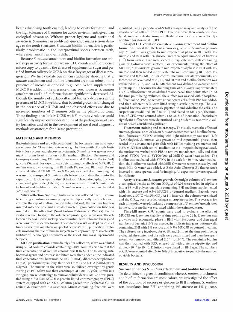
MUC5B keeps S. mutans in planktonic form. By quantifyingthe biofilm and supernatant bacteria in S. mutans cultures grownfrom 6 to 24 h, we determined the effect of MUC5B on S. mutansover time after stationary phase is reached. When the numbers ofplanktonic and biofilm S. mutans CFU are combined to determinethe total number of viable S. mutans cells in a given experiment,results show that there is no significant difference between thetotal numbers of live bacteria in cultures containing 0.3% MUC5Band in control media (Fig. 5). Based on these findings, we showthat the reduction in S. mutans attachment and biofilm formationon glass and hydroxyapatite in the presence of MUC5B (Fig. 3) isnot due to bactericidal properties of MUC5B. The presence ofMUC5B reduces S. mutans attachment and biofilm formation bymaintaining bacteria in the planktonic phase. These findingspoint to the importance of MUC5B in establishing a healthy oralmicrobiome that allows species diversity but, at the same time,protects teeth from bacterial damage.
Conclusion. In summary, we used CFU counts and fluores-cence light microscopy to quantitatively show that S. mutans at-tachment and biofilm formation are most robust when the organ-ism is grown in the presence of sucrose and that the addition ofpurified human salivary MUC5B significantly decreases S. mutansattachment and biofilm formation even in the presence of sucrose.We determined that MUC5B does not alter S. mutans growth orlead to bacterial killing over 24 h but limits biofilm formation bymaintaining S. mutans primarily in the planktonic form (Fig. 6).We speculate that the observed decrease in bacterial attachmentand biofilm formation is due to a combination of genetic changesthat decrease bacterial virulence and repulsion by MUC5B’s het-erogeneous glycans. S. mutans attachment and biofilm formationare key steps in the development of cavities; therefore, these find-ings have particularly important clinical implications. The pres-ence or absence of MUC5B in the oral cavity could alter individ-uals’ susceptibility to dental cavity formation, which could then bean easily accessible, highly predictable clinical diagnostic marker
0 5 100.0
0.1
0.2
0.3
SMedium+MucinsSMedium+MethylcelluloseSMedium
FIG 4 S. mutans growth is unaffected by the presence of salivary mucins. Agrowth curve of S. mutans in BHI with 1% sucrose (SMedium), BHI with 1%sucrose and 0.3% mucins, or BHI with 1% sucrose and 0.3% methylcelluloseindicates that the presence of mucins and methylcellulose does not alter thegrowth of S. mutans. Error bars represent SDs.
Time (h)
Tota
lBac
teria
(Log
10CF
U/µ
l)
6 18 24100
102
104
106
108SMedium+MucinsSMedium+MethylcelluloseSMedium
FIG 5 S. mutans survival is unaffected by salivary mucins. The graph repre-sents the total number of viable S. mutans cells per well in the supernatant andbiofilm in BHI with 1% sucrose (SMedium), BHI with 1% sucrose and 0.3%mucins, or BHI with 1% sucrose and 0.3% methylcellulose. Salivary mucinsand methylcellulose show no bactericidal effects at time points up to 24 h.Error bars represent SDs.
Frenkel and Ribbeck
336 aem.asm.org January 2015 Volume 81 Number 1Applied and Environmental Microbiology
on August 9, 2018 by guest
http://aem.asm
.org/D
ownloaded from

of disease. From a therapeutic standpoint, exogenous MUC5Bcould potentially be utilized as a treatment or preventative mea-sure for cavities. These findings illustrate that MUC5B may helpprotect teeth from cavity formation, but further studies, such asthose that use RNA sequencing or other genetic profiling tech-niques, are needed to fully characterize the mechanism underlyingthe observed decrease in S. mutans attachment and biofilm forma-tion.
ACKNOWLEDGMENTS
We thank Nicole Kavanaugh for her critical review of the figures.This work was made possible by generous funding support from Col-
gate-Palmolive (to K.R.) and Biological Sciences in Dental Medicinegrants for tuition and a stipend (to E.S.F.).
REFERENCES1. Brockhausen I, Schachter H, Stanley P. 2009. O-GalNAc glycans. In
Varki A, Cummings R, Esko J, Freeze H, Stanley P, Bertozzi C, Hart G,Etzler M (ed), Essentials of glycobiology, 2nd ed. Cold Spring HarborLaboratory Press, Cold Spring Harbor, NY.
2. Perez-Vilar J, Mabolo R. 2007. Gel-forming mucins. Notions from invitro studies. Histol Histopathol 22:455– 464.
3. Tabak LA. 1995. In defense of the oral cavity: structure, biosynthesis, andfunction of salivary mucins. Annu Rev Physiol 57:547–564. http://dx.doi.org/10.1146/annurev.ph.57.030195.002555.
4. Van der Sluis M, De Koning B, De Bruijn A, Velcich A, Meijerink J, VanGoudoever J, Büller H, Dekker J, Van Seuningen I, Renes I, EinerhandA. 2006. Muc2-deficient mice spontaneously develop colitis, indicatingthat MUC2 is critical for colonic protection. Gastroenterology 131:117–129. http://dx.doi.org/10.1053/j.gastro.2006.04.020.
5. Rose MC, Voynow JA. 2006. Respiratory tract mucin genes and mucinglycoproteins in health and disease. Physiol Rev 86:245–278. http://dx.doi.org/10.1152/physrev.00010.2005.
6. Derrien M, van Passel MW, van de Bovenkamp JH, Schipper RG, deVos WM, Dekker J. 2010. Mucin-bacterial interactions in the human oralcavity and digestive tract. Gut Microbes 1:254 –268. http://dx.doi.org/10.4161/gmic.1.4.12778.
7. Habte HH, Mall AS, de Beer C, Lotz ZE, Kahn D. 2006. The role of crudehuman saliva and purified salivary MUC5B and MUC7 mucins in theinhibition of human immunodeficiency virus type 1 in an inhibition assay.Virol J 3:99. http://dx.doi.org/10.1186/1743-422X-3-99.
8. Bansil R, Turner BS. 2006. Mucin structure, aggregation, physiologicalfunctions and biomedical applications. Curr Opin Colloid Interface Sci11:164 –170. http://dx.doi.org/10.1016/j.cocis.2005.11.001.
9. Veerman EC, Ligtenberg AJ, Schenkels LC, Walgreen-Weterings E,Nieuw Amerongen AV. 1995. Binding of human high-molecular-weightsalivary mucins (MG1) to Hemophilus parainfluenzae. J Dent Res 74:351–357. http://dx.doi.org/10.1177/00220345950740011101.
10. Bansil R, Celli JP, Hardcastle JM, Turner BS. 2013. The influence ofmucus microstructure and rheology in Helicobacter pylori infection.Front Immunol 4:310. http://dx.doi.org/10.3389/fimmu.2013.00310.
11. Loesche WJ. 1986. Role of Streptococcus mutans in human dental decay.Microbiol Rev 50:353–380.
12. Al-Hashimi I, Levine MJ. 1989. Characterization of in vivo salivary-derived enamel pellicle. Arch Oral Biol 34:289 –295. http://dx.doi.org/10.1016/0003-9969(89)90070-8.
13. Cárdenas M, Elofsson U, Lindh L. 2007. Salivary mucin MUC5B couldbe an important component of in vitro pellicles of human saliva: an in situellipsometry and atomic force microscopy study. Biomacromolecules8:1149 –1156. http://dx.doi.org/10.1021/bm061055h.
14. Gibbons RJ, Cohen L, Hay DI. 1986. Strains of Streptococcus mutansand Streptococcus sobrinus attach to different pellicle receptors. InfectImmun 52:555–561.
15. Bushnak IA, Labeed FH, Sear RP, Keddie JL. 2010. Adhesion of micro-organisms to bovine submaxillary mucin coatings: effect of coating depo-sition conditions. Biofouling 26:387–397. http://dx.doi.org/10.1080/08927011003646809.
16. Shi L, Ardehali R, Caldwell KD, Valint P. 2000. Mucin coating onpolymeric material surfaces to suppress bacterial adhesion. Colloids SurfB Biointerfaces 17:229 –239. http://dx.doi.org/10.1016/S0927-7765(99)00121-6.
17. Kesimer M, Makhov AM, Griffith JD, Verdugo P, Sheehan JK. 2010.Unpacking a gel-forming mucin: a view of MUC5B organization aftergranular release. Am J Physiol Lung Cell Mol Physiol 298:L15–L22. http://dx.doi.org/10.1152/ajplung.00194.2009.
18. Yakubov GE, Papagiannopoulos A, Rat E, Easton RL, Waigh TA. 2007.Molecular structure and rheological properties of short-side-chain heavilyglycosylated porcine stomach mucin. Biomacromolecules 8:3467–3477.http://dx.doi.org/10.1021/bm700607w.
19. Thornton DJ, Khan N, Mehrotra R, Howard M, Sheehan JK, VeermanE, Packer NH. 1999. Salivary mucin MG1 is comprised almost entirely ofdifferent glycosylated forms of the MUC5B gene product. Glycobiology9:293–302. http://dx.doi.org/10.1093/glycob/9.3.293.
20. Andersch-Björkman Y, Thomsson KA, Larsson JMH, Ekerhovd E,Hansson GC. 2007. Large scale identification of proteins, mucins, andtheir O-glycosylation in the endocervical mucus during the menstrualcycle. Mol Cell Proteomics 6:708 –716. http://dx.doi.org/10.1074/mcp.M600439-MCP200.
21. Gerken TA. 1993. Biophysical approaches to salivary mucin structure,conformation and dynamics. Crit Rev Oral Biol Med 4:261–270.
22. Slomiany BL, Murty VLN, Piotrowski J, Slomiany A. 1996. Salivarymucins in oral mucosal defense. Gen Pharmacol Vasc Syst 27:761–771.http://dx.doi.org/10.1016/0306-3623(95)02050-0.
23. Van Klinken BJ, Dekker J, Büller HA, Einerhand AW. 1995. Mucin genestructure and expression: protection vs. adhesion. Am J Physiol 269:G613–G627.
24. Iontcheva I, Oppenheim FG, Troxler RF. 1997. Human salivary mucinMG1 selectively forms heterotypic complexes with amylase, proline-richproteins, statherin, and histatins. J Dent Res 76:734 –743. http://dx.doi.org/10.1177/00220345970760030501.
25. Tabak LA, Levine MJ, Mandel ID, Ellison SA. 1982. Role of salivarymucins in the protection of the oral cavity. J Oral Pathol 11:1–17. http://dx.doi.org/10.1111/j.1600-0714.1982.tb00138.x.
26. Linden SK, Sutton P, Karlsson NG, Korolik V, McGuckin MA. 2008.Mucins in the mucosal barrier to infection. Mucosal Immunol 1:183–197.http://dx.doi.org/10.1038/mi.2008.5.
27. Bowen WH, Koo H. 2011. Biology of Streptococcus mutans-derivedglucosyltransferases: role in extracellular matrix formation of cariogenicbiofilms. Caries Res 45:69 – 86. http://dx.doi.org/10.1159/000324598.
28. Nakano YJ, Kuramitsu HK. 1992. Mechanism of Streptococcus mutansglucosyltransferases: hybrid-enzyme analysis. J Bacteriol 174:5639 –5646.
29. Krzyœciak W, Jurczak A, Koœcielniak D, Bystrowska B, Skalniak A.2014. The virulence of Streptococcus mutans and the ability to form bio-films. Eur J Clin Microbiol Infect Dis 33:499 –515. http://dx.doi.org/10.1007/s10096-013-1993-7.
30. Cross SE, Kreth J, Zhu L, Sullivan R, Shi W, Qi F, Gimzewski JK. 2007.Nanomechanical properties of glucans and associated cell-surface adhe-sion of Streptococcus mutans probed by atomic force microscopy underin situ conditions. Microbiology 153:3124 –3132. http://dx.doi.org/10.1099/mic.0.2007/007625-0.
31. Socransky SS, Haffajee AD. 2002. Dental biofilms: difficult therapeu-
mucinsS. mutans
A B
FIG 6 Summary of conclusions. S. mutans utilizes sucrose to form stickyextracellular polysaccharides that facilitate attachment to the tooth surface andsubsequent biofilm formation. (A) In the biofilm, bacterial metabolism ofsucrose causes a decrease in the local pH, leading to demineralization of thetooth structure. (B) The presence of mucins in sucrose-supplemented growthmedium decreases S. mutans attachment and biofilm formation on the toothsurface by maintaining S. mutans in the planktonic state.
Mucins Protect Surfaces from S. mutans Colonization
January 2015 Volume 81 Number 1 aem.asm.org 337Applied and Environmental Microbiology
on August 9, 2018 by guest
http://aem.asm
.org/D
ownloaded from

tic targets. Periodontol 2000 28:12–55. http://dx.doi.org/10.1034/j.1600-0757.2002.280102.x.
32. Shemesh M, Tam A, Aharoni R, Steinberg D. 2010. Genetic adapta-tion of Streptococcus mutans during biofilm formation on differenttypes of surfaces. BMC Microbiol 10:51. http://dx.doi.org/10.1186/1471-2180-10-51.
33. Shemesh M, Tam A, Steinberg D. 2007. Expression of biofilm-associatedgenes of Streptococcus mutans in response to glucose and sucrose. J MedMicrobiol 56:1528 –1535. http://dx.doi.org/10.1099/jmm.0.47146-0.
34. Koo H, Xiao J, Klein MI, Jeon JG. 2010. Exopolysaccharides produced byStreptococcus mutans glucosyltransferases modulate the establishment ofmicrocolonies within multispecies biofilms. J Bacteriol 192:3024 –3032.http://dx.doi.org/10.1128/JB.01649-09.
35. Duarte S, Klein MI, Aires CP, Cury JA, Bowen WH, Koo H. 2008.Influences of starch and sucrose on Streptococcus mutans biofilms. OralMicrobiol Immunol 23:206 –212. http://dx.doi.org/10.1111/j.1399-302X.2007.00412.x.
36. Kreth J, Zhu L, Merritt J, Shi W, Qi F. 2008. Role of sucrose in the fitnessof Streptococcus mutans. Oral Microbiol Immunol 23:213–219. http://dx.doi.org/10.1111/j.1399-302X.2007.00413.x.
37. McBride BC, Gisslow MT. 1977. Role of sialic acid in saliva-inducedaggregation of Streptococcus sanguis. Infect Immun 18:35– 40.
38. van Houte PJ. 1982. Bacterial adherence and dental plaque formation.Infection 10:252–260. http://dx.doi.org/10.1007/BF01666923.
39. Levine MJ, Herzberg MC, Levine MS, Ellison SA, Stinson MW, Li HC,van Dyke T. 1978. Specificity of salivary-bacterial interactions: role ofterminal sialic acid residues in the interaction of salivary glycoproteinswith Streptococcus sanguis and Streptococcus mutans. Infect Immun 19:107–115.
40. Klein A, Carnoy C, Wieruszeski JM, Strecker G, Strang AM, VanHalbeek H, Roussel P, Lamblin G. 1992. The broad diversity of neutraland sialylated oligosaccharides derived from human salivary mucins. Bio-chemistry (Mosc) 31:6152– 6165. http://dx.doi.org/10.1021/bi00141a028.
41. Williams RC, Gibbons RJ. 1975. Inhibition of streptococcal attachmentto receptors on human buccal epithelial cells by antigenically similar sali-vary glycoproteins. Infect Immun 11:711–718.
42. Thomsson KA, Prakobphol A, Leffler H, Reddy MS, Levine MJ, FisherSJ, Hansson GC. 2002. The salivary mucin MG1 (MUC5B) carries arepertoire of unique oligosaccharides that is large and diverse. Glycobiol-ogy 12:1–14. http://dx.doi.org/10.1093/glycob/12.1.1.
43. Gibbons RJ, Dankers I. 1981. Lectin-like constituents of foods whichreact with components of serum, saliva, and Streptococcus mutans. ApplEnviron Microbiol 41:880 – 888.
44. Gibbons RJ, Qureshi JV. 1978. Selective binding of blood group-reactivesalivary mucins by Streptococcus mutans and other oral organisms. InfectImmun 22:665– 671.
45. Murray PA, Prakobphol A, Lee T, Hoover CI, Fisher SJ. 1992.Adherence of oral streptococci to salivary glycoproteins. Infect Immun60:31–38.
46. Amerongen AVN, Bolscher JGM, Veerman ECI. 1995. Salivary mucins:protective functions in relation to their diversity. Glycobiology 5:733–740.http://dx.doi.org/10.1093/glycob/5.8.733.
47. Schenkels LCPM, Ligtenberg AJM, Veerman ECI, Van Nieum Amer-ongen A. 1993. Interaction of the salivary glycoprotein EP-GP with thebacterium Streptococcus salivarius HB. J Dent Res 72:1559 –1565. http://dx.doi.org/10.1177/00220345930720120501.
48. Xiao J, Koo H. 2010. Structural organization and dynamics of exopoly-saccharide matrix and microcolonies formation by Streptococcus mutansin biofilms. J Appl Microbiol 108:2103–2113. http://dx.doi.org/10.1111/j.1365-2672.2009.04616.x.
49. Xiao J, Klein MI, Falsetta ML, Lu B, Delahunty CM, Yates JR, III,Heydorn A, Koo H. 2012. The exopolysaccharide matrix modulates theinteraction between 3D architecture and virulence of a mixed-species oralbiofilm. PLoS Pathog 8:e1002623. http://dx.doi.org/10.1371/journal.ppat.1002623.
Frenkel and Ribbeck
338 aem.asm.org January 2015 Volume 81 Number 1Applied and Environmental Microbiology
on August 9, 2018 by guest
http://aem.asm
.org/D
ownloaded from