salivary gland basal cell adenomas - rjme.ro
TRANSCRIPT
Romanian Journal of Morphology and Embryology 2005, 46(1):29–40
Salivary gland basal cell adenomas – immunohistochemical evaluation of four cases and review of the literature
CL. MĂRGĂRITESCU1), VERONICA MERCUŢ2), L. MOGOANTĂ3), MARIA FLORESCU 4), CRISTIANA SIMIONESCU1), LUCIA CIONCA5), MĂDĂLINA MANEA6)
1)Department of Pathology, Faculty of Dentistry, University of Medicine and Pharmacy of Craiova 2)Department of Prosthetic Dentistry, Faculty of Dentistry, University of Medicine and Pharmacy of Craiova
3)Department of Histology, Faculty of Medicine, University of Medicine and Pharmacy of Craiova 4)Department of Pathology, Faculty of Medicine, University of Medicine and Pharmacy of Craiova
5)University Hospital of Dentistry “Prof. Dr. Dan Theodorescu”, Bucharest 6)Nursing College, University of Medicine and Pharmacy of Craiova
Abstract Objective — evaluate the antigen profile of cellular population from basal cell adenomas. Material and methods — histopathological and immunohistochemical evaluation of four-salivary gland basal cell adenomas; the pathological samples were provided by the Oral Maxilla Facial Surgery Department of the Clinical County Hospital from Craiova. Results — all basal cell adenoma exhibit differentiation toward 3 cell phenotypes: ductal luminal, basal and myoepithelial. Conclusions — the antigen profile of these tumors closely regard with other variants of salivary gland adenomas, such as pleomorphic adenoma and myoepithelioma. So, we can conclude that these tumors had a common origin, most probably from intercalated ducts and the proportion of those 3 cellular types, their cytoarhitectural arrangements and the quantity of extracellular matrix production can do the differentiation between them. Keywords: salivary gland adenomas, basal cell adenomas, immunohistochemistry.
Introduction
Confusion and disagreement about the terminology of this tumor has existed since her designation by Kleinsasser and Klein in 1967 [1]. Other terms that have been associated with this group of tumors include tubular adenoma, trabecular adenoma, dermal analogue tumor, canalicular adenoma, basaloid adenoma, clear cell adenoma, and monomorphic adenoma [2–6].
In 1983, Gardner and Daley were the first authors who made the distinction between basal cell adenoma and canalicular adenomas [7]. In the second WHO classification of salivary gland tumors, from 1991, was establish that in the category of basal cell adenomas are included the benign epithelial neoplasm with a uniform, monomorphous histologic appearance that is dominated by basaloid cells and is without the myxochondroid tissue characteristic of mixed tumor [8].
The literature data state an incidence of this tumor between 2-4% from all salivary neoplasms [9–14].
Basal cell adenomas are tumors of adults with a mean age of 58 years, with a small peak incidence in the seventh decade of life [8, 15]. There is about a 2 to 1 predominance of women over men [15] with the exception of membranous type of basal cell adenoma that prevail in men [16–18].
The parotid gland is the dominant site of occurrence of the basal cell adenoma (75%), followed by minor salivary gland from upper lip with 6% and submandibular gland with 5% [9, 17].
For several years many reviewers erroneously interpreted the "monomorphous" appearance to indicate isomorphism of the epithelial cell type and absence of myoepithelial differentiation [19–25].
More recently evaluations, including electron microscopic and immunohistochemical studies, have shown that basal, ductal, and myoepithelial cell differentiation occurs to variable degrees in basal cell adenomas [26–35].
Different investigators reported varying results of immunohistochemical staining in basal cell adenomas [30, 33, 34, 36, 37].
Cytokeratin is demonstrable in nearly all tumors, but the number of reactive cells varies from few to many. Similarly, immunoreactivity to S-100 protein, smooth muscle actin (SMA), and vimentin can be demonstrated in most basal cell adenomas but is typically localized to the peripheral tumor cells adjacent to the connective tissue stroma [34, 36–38].
Morinaga et al. [40] have reported focal reactivity for myosin. Williams et al. [34] have even found rare, faint staining for glial fibrillary acidic protein (GFAP), a tumor marker most often associated with mixed tumors; others have not seen this [35, 37, 39].
Carcinoembryonic antigen and epithelial membrane antigen reactivity is mostly confined to luminal cells [30, 33, 34].
Dardick et al. described some tubulo-trabecular basal cell adenomas with S-100 protein immunoreactivity of spindled "stromal" cells that they interpreted as myoepithelial cells with electron microscopy [38].
Material and methods
There were selected eight cases of basal cell adenomas from 334 salivary gland tumors retrieved during 1994–2003 from the surgical pathology files of

Cl. Mărgăritescu et al.
30
Oral Maxilla Facial Surgery Department of the Clinical County Hospital from Craiova.
The surgical pieces were routinely fixed with 10% buffered formalin and sent to the Laboratory of Cytology and Pathology of the same hospital, where were processed by the classical histopathological technique with paraffin embedding and stained with Haematoxylin & Eosin (HE) stain, Trichromic Goldner Szeckelly stain and Periodic Acid Schiff–Mayer Haematoxylin stain.
The immunohistochemical processing was making in the Laboratory of histological, histopathological and immunohistochemical techniques from University of Medicine and Pharmacy of Craiova.
The paraffin blocks acquired by histopathological processing were section at microtome resulting sections with 5µm in thickness mounted on microscopically
slides cover with polylysin. Subsequently, the sections were deparaffinized
(three successive baths of benzene, 1 hour at thermostat, at 58°C, for first bath and 10 minutes, at room temperature, for the remainder baths) and rehydrated (three successive alcohol baths with decreased concentration: 96%, 80% and 70%, 10 minutes per each bath, followed by a bath with distillated water, where the sections were hold for 10 minutes).
We used streptavidin-biotin immuno-peroxidase staining method with 30 minutes primary antibodies incubation.
The used antibodies, their characteristics and their peculiar immunostaining procedures are summarized in Table 1.
Table 1 – The list of antibodies used in immunohistochemical investigation of basal cell adenomas
Antibodies Clone Class/subclass Code Antigen unmasking methods: microwave retrieval Dilution
Monoclonal Mouse Anti-Human Cytokeratin
AE1/AE3
AE1 and AE3
AE1: IgG1, kappa; AE3: IgG1, kappa M3515
15 minutes heating – induced epitope retrieval (750W) in
10mmol/L citrate buffer, pH 6
1:50 in 0.02M PBS, pH 7.2–7.6
Monoclonal Mouse Anti-Human Cytokeratin 19
(CK 19) RCK 108 IgG1, kappa M0888 Ascites
15 minutes heating – induced epitope retrieval (750W) in
10mmol/L citrate buffer, pH 6
1:50 in 0.02M PBS, pH 7.2–7.6
Polyclonal Rabbit Anti-Human Carcinoembryonic
Antigen (CEA) – – A0115 Ig
fraction
10 minutes heating – induced epitope retrieval (750W) in
10mmol/L citrate buffer, pH 6
1:100 in 0.02M PBS, pH 7.2–7.6
Monoclonal Mouse Anti-Human Vimentin (VIM) V9 IgG1, kappa M0725 Culture
supernatant
10 minutes heating – induced epitope retrieval (750W) in
10mmol/L citrate buffer, pH 6
1:30 in 0.02M PBS, pH 7.2–7.6
Polyclonal rabbit Anti-S 100 – – Z0311 Ig fraction
20 minutes heating – induced epitope retrieval (750W) in
10mmol/L citrate buffer, pH 6
1: 300 in 0.02M PBS, pH 7.2–7.6
Monoclonal Mouse Anti-Human Smooth Muscle Actin (SMA)
1A4 IG2a, kappa M0851 15 minutes heating – induced
epitope retrieval (750W) in 10mmol/L citrate buffer, pH 6
1:75 in 0.02M PBS, pH 7.2–7.6
Monoclonal Mouse Anti- Proliferating Cell Nuclear
Antigen (PCNA) PC-10 IG2a, kappa M0879
15 minutes heating – induced epitope retrieval (750W) in
10mmol/L citrate buffer, pH 6
1:75 in 0.02M PBS, pH 7.2–7.6
The tumors have been diagnosed according to WHO classification [8].
As internal control we use the residual normal salivary gland parenchyma (the fragments that were placed at a suitable distance away from the tumoral tissue).
The external tissue control for the performed immunohistochemical reactions was attain by using fragments of sublingual salivary parenchyma, removed during necropsy of three patients older than 50 years.
The number of immunoreactive cells from each investigated cases were semi quantitative evaluate as follow: (+++) >75% positive cells, (++) 75–25% positive cells, (+) <25% positive cells, (−) negative cells.
For the appreciation of the intensity of immunostaining reaction we use the interpretation criteria from literature data (Jasani B and Schmid KW, 1993), consigned like below:
▪ (+++), when immunostaining reaction is intense positive or with peculiar „all over”, obvious at small magnification.
▪ (++), when immunostaining reaction is focal or with moderate intensity, evident on average magnification.
▪ (+), when immunostaining reaction is weak or very focal, visible only at strong magnification.
▪ (±), when immunostaining reaction is very reduced, at limit.
▪ (−), when immunostaining reaction is negative. Were excluding from interpretation the microscopic
fields with folds, necrosis and hemorrhages. The cell kinetics in the investigated basal cell
adenoma was estimate by PCNA labeling indices determination:
100×=EN
PN
NNPI
PI – PCNA labeling indices; NPN – number of positive nuclei in the reference epithelium; NEN –total number of nuclei in the reference epithelium.
Results and discussions
Epidemiological aspects
According to the data presented in the following table (Table 2) the investigated cases of basal cell adenoma develops mostly in the 6th decade of life (5 cases), in female (5 cases) and in the parotid location (5 cases).

Salivary gland basal cell adenomas – immunohistochemical evaluation of four cases and review of the literature
31
Table 2 – Epidemiological, macroscopic and microscopic data of the investigated basal cell adenomas
Cases Age Sex (F/M) Location Size Cytological
differentiations The dominant
pattern The dominant
stroma 1 46 years F Left parotid 2/3 cm Basaloid cell Solid-insular Fibrous 2 52 years M Right parotid 2/2 cm Basaloid cell Solid-insular Fibrous 3 54 years F Left parotid 2/4 cm Basaloid cell Membranous Fibrous 4 55 years M Upper lip 2/2 cm Basaloid cell Solid-insular Fibrous
5 56 years F Right submandibular gland 2.5/2 cm Basaloid + luminal
ductal cell Tubular + solid Fibrous + loose stroma
6 57 years F Upper lip 1.5/2 cm Basaloid cell Solid-insular Fibrous
7 62 years F Left parotid 3/2 cm Basal cell Trabecular + solid Fibrous + loose stroma
8 66 years M Right parotid 3/4 cm Basaloid cell Solid-insular Fibrous
Gross aspects
The investigated basal cell adenomas presents as solitary tumoral masses of small dimensions (2–4 cm), sharply circumscribed, firm, with homogeneous, gray-white aspect on the cut surface (Table 2).
Microscopic aspects
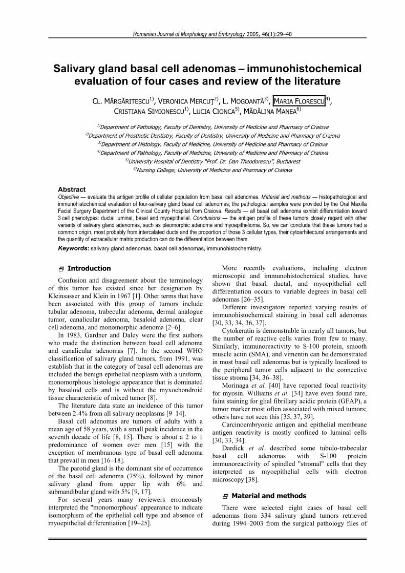
Histopathologically, these tumors were made up by a monomorphous epithelial proliferation; dominate by basal cell in a fibrous stroma. Thus, the main cellular population of these tumors was the basaloid one with “dark” and “light” aspects (Figure 1a) and compact disposition (Table 2). Only in two investigated cases we notice the presence of luminal duct cells with cuboidal morphology that surround small lumens.
The solid pattern was the most encountered (Figure 1b). Other tumoral patterns observed by as were tubular, trabecular and membranous (Figure 1c–e). Usually, they were associated with solid one, and they are not the dominant growth pattern; exception for membranous type (Table 2).
The dominant stroma was of fibrous type, which varies from moderately dense to very dense. In the tumors with tubular or trabecular growth patterns the stroma is often less densely collagen than in the solid type and is sometimes very loose with only a few thin strands of collagen (Table 2). Peculiar for membranous case is the production of a conspicuous amount of basal lamina that is recognized as an eosinophilic, PAS-positive hyaline material; material that forms thick bands at the periphery of the basaloid cell islands.
Immunohistochemical aspects ▪ of the residual fragments of the normal salivary
gland parenchyma
On the remainder major salivary gland parenchyma we notice the presence of all excretory system components: intralobular (intercalated ducts of Boll and striated ducts of Pfluger) and extralobular ducts.
The intercalated ducts lumina were outline by a single layer of flatten epithelium, positive to cytokeratins (AE1/AE3 and CK 19), CEA and negative to Vimentin, S-100 and α-Smooth Muscle Actin.
The epithelial lining of striated ducts was simple columnar and was positive to cytokeratins (AE1/AE3 and CK), CEA, versatile to S-100 and negative to Vimentin and α-Smooth Muscle Actin. The extralobular ducts were lined by a bilayered cuboidal epithelium, positive to (AE1/AE3 and CK), CEA and negative to Vimentin, S-100 and α-Smooth Muscle Actin.
The myoepithelial cells bounded the acini, the intercalated ducts and occasionally the striated ducts of the major salivary glands. These cells were constantly positive to AE1/AE3, α-Smooth Muscle Actin and S-100, versatile positive for Vimentin, and negative to CK19 and CEA.
The acinar cells were positive to AE1/AE3 and CEA, weak positive for CK 19, negative to vimentin, α- Smooth Muscle Actin and S-100. Moreover, we notice the positivity for α-Smooth Muscle Actin of the pericytes and smooth muscle cells from wall of blood vessels (internal positive control).
Also, vimentin was positive in stromal fibroblasts (internal positive control) and vascular endothelial cells. As well, we constantly observe S-100 positivity of the adipocytes present in interlobular stroma of the major salivary glands and for the nervous fibers.
▪ of the pathological specimens
The semi-quantitative and qualitative investigation of immunostaining for AE1/AE3 of the four basal cell adenomas is reproducing on Table 3 from below.
The reactivity to AE1/AE3 was confine only to tumoral epithelial component. The quantitative evaluation shows different reactivity from case to case and even in the same tumor base on dominate pattern. The immunostaining pattern was a homogenous cytoplasmic one.
Table 3 – The semi-quantitative and qualitative evaluation of AE1/AE3 immunostaining Semi-quantitative evaluation of AE1/AE3
immunostaining Qualitative evaluation of
AE1/AE3 immunostaining Types of epithelial cells
Negative <25% 25–75% >75% 100% – + ++ +++ Luminal ductal cells 0 3/4 1/4 0 0 0 0 3/4 1/4
„Light” basaloid cells 0 1/4 2/4 1/4 0 0 2/4 2/4 0
„Dark” basaloid cells 2/4 2/4 0 0 0 2/4 2/4 0 0
No. of positive cases / No. of
all cases

Cl. Mărgăritescu et al.
32
The mostly intense reaction occurs in luminal ductal cells, especially in those cases with dominant tubular growth pattern; the % age of positive cells was nearly 75% (Figure 1f).
In the remainder cases these % age was below 25% and the intensity of the reaction was moderate (++). The great majority of „dark” basaloid cells were positive, but their proportion varies from 25% to 75%
(the latest was found especially in tumors with predominance of solid growth pattern).
The intensity varies from weak (+) to moderate (++). We notice a positive reaction of „dark” basaloid cells to AE1/AE3 only in two cases and the intensity was weak.
The semi-quantitative and qualitative results of cytokeratin 19 (CK 19) investigation of basal cell adenoma are illustrated in the following table (Table 4).
Table 4 – The semi-quantitative and qualitative evaluation of CK 19 immunostaining Semi-quantitative evaluation of CK19
immunostaining Qualitative evaluation of CK19
immunostaining Types of epithelial cells
Negative <25% 25–75% >75% 100% – + ++ +++ Luminal ductal cells 2/4 2/4 0 0 0 0 1/4 1/4 0
„Light” basaloid cells 3/4 1/4 0 0 0 3/4 1/4 0 0
„Dark” basaloid cells 4/4 0 0 0 0 4/4 0 0 0
No. of positive cases/ No. of
all cases
The positive reaction was present only in epithelial component and was influence by the type of dominant growth pattern. The most intense reaction was notice in luminal ductal cells, especially in those cases with tubular pattern as dominant growth pattern; the % age of positive cells was small, below 25% (Figure 2a).
In two cases the reaction was negative. The great majority of „light” basaloid cells were negative, only in
one case of basal cell adenoma with solid growth pattern we could notice a positive reaction confine to a small group of cells from the center of the compact epithelial proliferations. The „dark” basaloid cells were negative to CK 19 reaction in all investigated cases.
The semi-quantitative and qualitative investigation of immunostaining for CEA four basal cell adenomas is reproducing on Table 5 from below:
Table 5 – The semi-quantitative and qualitative evaluation of CEA immunostaining Semi-quantitative evaluation of CEA
immunostaining Qualitative evaluation of CEA
immunostaining Types of epithelial cells
Negative <25% 25–75% >75% 100% – + ++ +++ Luminal ductal
cells 0 3/4 1/4 0 0 0 1/4 2/4 1/4
„Light” basaloid cells 4/4 0 0 0 0 4/4 0 0 0
„Dark” basaloid cells 4/4 0 0 0 0 4/4 0 0 0
No. of positive cases/ No. of
all cases
We notice a positive reaction to CEA only in luminal ductal cells. The maximum intensity was notice in basal cell adenoma with tubular pattern as dominant growth pattern; in this case were positive nearly 75% of luminal ductal cells (Figure 2b). In the remainder cases only 25% of epithelial tumoral cells were positive; the
intensity of immunostaining to CEA varies from weak (one case) to moderate (two cases). Both „dark” and „light” basaloid cells were negative to this reaction.
The semi-quantitative and qualitative investigation of immunostaining for vimentin of the four basal cell adenomas is reproducing on Table 6 from below:
Table 6 – The semi-quantitative and qualitative evaluation of vimentin immunostaining Semi-quantitative evaluation of vimentin
immunostaining Qualitative evaluation of
vimentin immunostaining Types of epithelial cells
Negative <25% 25–75% >75% 100% – + ++ +++ Luminal ductal
cells 4/4 0 0 0 0 4/4 0 0 0
„Light” basaloid cells 4/4 0 0 0 0 4/4 0 0 0
„Dark” basaloid cells 0 4/4 0 0 0 0 3/4 1/4 0
Stromal cells 0 4/4 0 0 0 0 1/4 1/4 2/4
No. of positive cases/ No. of all
cases
Both the epithelial and stromal components were positive to such immunostaining.
The reactivity of epithelial component was limited only to some of the basaloid cells from the periphery of epithelial neoplastic proliferations; they correspond to the „dark” basaloid cells observed in light microscopy (Figure 2c–d).
The reaction intensity to this level varies from weak to moderate and the proportion of positive cells was below 20%. In the stromal areas the reaction was
positive for interstitial fibroblasts and cells with stellate morphology from myxoid areas.
The positive reaction to vimentin was present in all investigated cases and it varies in intensity from weak (+) to intense (+++). The immunostaining pattern was homogenous cytoplasmic.
The results of semi-quantitative and qualitative investigation of immunostaining for S-100 of the four basal cell adenomas are reproducing on Table 7 from below.

Salivary gland basal cell adenomas – immunohistochemical evaluation of four cases and review of the literature 33
a b
c d
5 6
e f
Figure 1a)
b) c)d) e)
f)
Basal cell adenomaepithelial component, proliferation of „light” and „dark” basal cells (HE, ×200);
with the predominance of solid pattern (HE, ×100); with solid-tubular pattern (HE, ×100);with the predominance of trabecular pattern (HE, ×100); with membranous patter (HE, ×100);
with the predominance of tubular pattern, AE1–AE3 immunostaining ( ) of„light” basal cells and ( ) of the peripheral „dark” basal cells, ×200.
++
+

34 Cl. Mărgăritescu et al..
a b
c d
5
fe
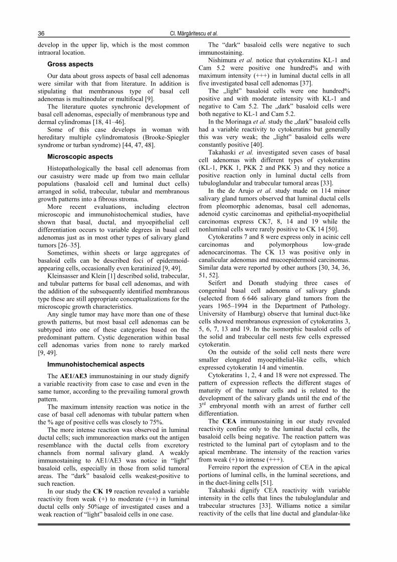
Figure 2a)
b)c)
d)
e)f)
Basal cell adenomawith the predominance of tubular pattern, CK 19 immunostaining ( ) of the luminal cells and (–)
of basal cells, ×200; with the predominance of tubular pattern, CEA immunostaining ( ) of theluminal cells and (–) of the basal cells, ×200; with the predominance of tubular pattern, vimentineimmunostaining ( ) of the luminal cells and ( ) of the basal cells, ×200; with the predominance of
trabecular pattern, vimentine immunostaining ( ) of „dark” basal cells and (+) of the stromal cells, ×200;with the predominance of solid pattern, S-100 immunostaining ( ) of „light” basal cells and ( )
of the „dark” basal cells, ×200; with the predominance of tubular pattern, SMA immunostaining (–)of „light” basal cells and ( ) of the peripheral „dark” basal cells, ×200
++
+++
- +
+
++ +
+

Salivary gland basal cell adenomas – immunohistochemical evaluation of four cases and review of the literature
35
Table 7 – The semi-quantitative and qualitative evaluation of S–100 immunostaining
Semi-quantitative evaluation of S-100 immunostaining
Qualitative evaluation of S-100 immunostaining Types of
epithelial cells Negative <25% 25–75% >75% 100% – + ++ +++
Luminal ductal cells 4/4 0 0 0 0 4/4 0 0 0
„Light” basaloid cells 1/4 3/4 0 0 0 1/4 2/4 1/4 0
„Dark” basaloid cells 2/4 2/4 0 0 0 2/4 2/4 1/4 0
Stromal cells 0 4/4 0 0 0 0 1/4 2/4 1/4
No. of positive cases/ No. of all
cases
We notice a positive reaction both in epithelial and stromal components of the investigated cases. In the epithelial component the reaction was positive only to the basaloid cells. The most intense positive reaction was present in „light” basaloid cells, especially in the cases with solid growth pattern (Figure 2e).
However, the % age of positive cells was small, below 25% and the intensity varies from weak (+) to moderate (++).
The „dark” basaloid cells from the periphery of neoplastic proliferations were also positive, but in a small % age and with weak (+) intensity.
The luminal ductal cells from all investigated cases were negative to such immunostaining. In stromal areas the positive reaction was present especially in myxoid zones at cells with stellate morphology.
At this level the immunostaining had maximum intensity. The pattern of this reaction was preponderant cytoplasmic, but in some „light” basaloid cells and peculiar in stellate stromal cells the pattern was predominant of nuclear type.
The results of semi-quantitative and qualitative investigation of SMA immunostaining of the four basal cell adenomas are reproducing on Table 8 from below:
Table 8 – The semi-quantitative and qualitative evaluation of SMA immunostaining Semi-quantitative evaluation of SMA
immunostaining Qualitative evaluation of SMA
immunostaining Types of epithelial cells
Negative <25% 25–75% >75% 100% – + ++ +++ Luminal ductal
cells 4/4 0 0 0 0 4/4 0 0 0
„Light” basaloid cells 4/4 0 0 0 0 4/4 0 0 0
„Dark” basaloid cells 0 4/4 0 0 0 0 2/4 2/4 0
Stromal cells 2/4 2/4 0 0 2/4 0 2/4 0 0
No. of positive cases/ No. of all
cases
We notice a positive reaction both in stromal and epithelial components. The epithelial reaction was confine to „dark” basaloid cells from the periphery of neoplastic proliferations (Figure 2f). The reaction intensity at this level was not purchase by the prevailing growth pattern; it varies from weak (+) to moderate (++). The „light” basaloid cells and luminal ductal cells were negative to such immunostaining. In stromal areas with myxoid differentiation we notice the presence of fusiform cells with myoepithelial-like morphology; the intensity was weak. The reaction’s pattern to SMA was a homogenous cytoplasmic one.
The results of PCNA investigation of those four basal cell adenomas are illustrated in the following table (Table 9).
Table 9 – PCNA labeling indices (PI) estimation of the investigated basal cell adenomas
No. of cases PCNA labelling indices (PI)
The average value of PI
1 9.2 2 8.3 3 7.5 4 12.5
9.37
The nuclei of both „dark” and „light” basaloid cells were positive to PCNA immunostaining. This reaction was more obvious to the nuclei of „dark” basaloid cells, especially in those tumors with predominant solid growth pattern.
Epidemiological data
In our study, during 1994–2003, the basal cell adenoma investigated cases represents 3.38% of all benign salivary tumor and 2.40% of all salivary gland tumors. In literature, different authors quoted an incidence of 2–4% from all primary salivary gland tumors [6, 9–13].
Basal cell adenoma from our casuistry develops during 6th and 7th decade of life, with a pick incidence in 6th decade, especially in women (63%, with a ratio F/M about 1:1.66) and in parotid salivary gland locating (62.5%).
In the AFIP files basal cell adenomas are tumors of adults [9] and Seifert et al. [15] also reported a virtual absence of monomorphic adenomas in children in the Salivary Gland Register at the University of Hamburg, Germany.
The mean age of patients is 58 years, with a small peak incidence in the seventh decade of life, which is an older population than patients with mixed tumors.
There is about a 2 to 1 predominance of women over men at the AFIP [9], while Batsakis and Brannon [16] and other authors [2, 18] observed a marked predominance of men among patients with membranous type of basal cell adenoma.
In the AFIP's data [9], over 75% occur in the parotid gland, 5% arise in the submandibular gland and 6%
Cl. Mărgăritescu et al.
36
develop in the upper lip, which is the most common intraoral location.
Gross aspects
Our data about gross aspects of basal cell adenomas were similar with that from literature. In addition is stipulating that membranous type of basal cell adenomas is multinodular or multifocal [9].
The literature quotes synchronic development of basal cell adenomas, especially of membranous type and dermal cylindromas [18, 41–46].
Some of this case develops in woman with hereditary multiple cylindromatosis (Brooke-Spiegler syndrome or turban syndrome) [44, 47, 48].
Microscopic aspects
Histopathologically the basal cell adenomas from our casuistry were made up from two main cellular populations (basaloid cell and luminal duct cells) arranged in solid, trabecular, tubular and membranous growth patterns into a fibrous stroma.
More recent evaluations, including electron microscopic and immunohistochemical studies, have shown that basal, ductal, and myoepithelial cell differentiation occurs to variable degrees in basal cell adenomas just as in most other types of salivary gland tumors [26–35].
Sometimes, within sheets or large aggregates of basaloid cells can be described foci of epidermoid- appearing cells, occasionally even keratinized [9, 49].
Kleinsasser and Klein [1] described solid, trabecular, and tubular patterns for basal cell adenomas, and with the addition of the subsequently identified membranous type these are still appropriate conceptualizations for the microscopic growth characteristics.
Any single tumor may have more than one of these growth patterns, but most basal cell adenomas can be subtyped into one of these categories based on the predominant pattern. Cystic degeneration within basal cell adenomas varies from none to rarely marked [9, 49].
Immunohistochemical aspects
The AE1/AE3 immunostaining in our study dignify a variable reactivity from case to case and even in the same tumor, according to the prevailing tumoral growth pattern.
The maximum intensity reaction was notice in the case of basal cell adenomas with tubular pattern when the % age of positive cells was closely to 75%.
The more intense reaction was observed in luminal ductal cells; such immunoreaction marks out the antigen resemblance with the ductal cells from excretory channels from normal salivary gland. A weakly immunostaining to AE1/AE3 was notice in “light” basaloid cells, especially in those from solid tumoral areas. The “dark” basaloid cells weakest-positive to such reaction.
In our study the CK 19 reaction revealed a variable reactivity from weak (+) to moderate (++) in luminal ductal cells only 50%age of investigated cases and a weak reaction of “light” basaloid cells in one case.
The “dark“ basaloid cells were negative to such immunostaining.
Nishimura et al. notice that cytokeratins KL-1 and Cam 5.2 were positive one hundred% and with maximum intensity (+++) in luminal ductal cells in all five investigated basal cell adenomas [37].
The „light” basaloid cells were one hundred% positive and with moderate intensity with KL-1 and negative to Cam 5.2. The „dark” basaloid cells were both negative to KL-1 and Cam 5.2.
In the Morinaga et al. study the „dark” basaloid cells had a variable reactivity to cytokeratins but generally this was very weak; the „light” basaloid cells were constantly positive [40].
Takahaski et al. investigated seven cases of basal cell adenomas with different types of cytokeratins (KL-1, PKK 1, PKK 2 and PKK 3) and they notice a positive reaction only in luminal ductal cells from tubuloglandular and trabecular tumoral areas [33].
In the de Arujo et al. study made on 114 minor salivary gland tumors observed that luminal ductal cells from pleomorphic adenomas, basal cell adenomas, adenoid cystic carcinomas and epithelial-myoepithelial carcinomas express CK7, 8, 14 and 19 while the nonluminal cells were rarely positive to CK 14 [50].
Cytokeratins 7 and 8 were express only in acinic cell carcinomas and polymorphous low-grade adenocarcinomas. The CK 13 was positive only in canalicular adenomas and mucoepidermoid carcinomas. Similar data were reported by other authors [30, 34, 36, 51, 52].
Seifert and Donath studying three cases of congenital basal cell adenoma of salivary glands (selected from 6 646 salivary gland tumors from the years 1965–1994 in the Department of Pathology. University of Hamburg) observe that luminal duct-like cells showed membranous expression of cytokeratins 3, 5, 6, 7, 13 and 19. In the isomorphic basaloid cells of the solid and trabecular cell nests few cells expressed cytokeratin.
On the outside of the solid cell nests there were smaller elongated myoepithelial-like cells, which expressed cytokeratin 14 and vimentin.
Cytokeratins 1, 2, 4 and 18 were not expressed. The pattern of expression reflects the different stages of maturity of the tumour cells and is related to the development of the salivary glands until the end of the 3rd embryonal month with an arrest of further cell differentiation.
The CEA immunostaining in our study revealed reactivity confine only to the luminal ductal cells, the basaloid cells being negative. The reaction pattern was restricted to the luminal part of cytoplasm and to the apical membrane. The intensity of the reaction varies from weak (+) to intense (+++).
Ferreiro report the expression of CEA in the apical portions of luminal cells, in the luminal secretions, and in the duct-lining cells [51].
Takahaski dignify CEA reactivity with variable intensity in the cells that lines the tubuloglandular and trabecular structures [33]. Williams notice a similar reactivity of the cells that line ductal and glandular-like

Salivary gland basal cell adenomas – immunohistochemical evaluation of four cases and review of the literature
37
neoplastic structures from basal cell adenomas and basal cell adenocarcinomas [34].
Other authors point out that the pericytoplasmic rim pattern of CEA immunostaining from ductal structures of basal cell adenomas is similar to that express by luminal columnar cells from striated ducts of normal salivary glands [53, 54].
In normal salivary glands the CEA immunostaining is positive in serous acinar cells (pericytoplasmatic and on luminal membrane slope), in luminal columnar cells of striated ducts (on luminal membrane slope) and in the luminal secretions [33, 55].
We notice a positive reaction to vimentin both epithelial and stromal components of investigated basal cell adenomas.
The maximum intensity was present in stromal fibroblasts and cells with stellate morphology from myxoid stromal areas.
In the epithelial component the positive reaction was confine to some „dark” basaloid cells and with a weak intensity.
Similar results were acquired by Ferreiro, Ogawa and Williams [30, 34, 51]. In their study vimentin was positive in stromal cells and to the periphery of epithelial proliferations.
Some authors showed that vimentin can by express not only by mesenchymal cells but also carcinomatous cells [56].
Ben-Ze`ev suggest that vimentin synthesis ratio depend on the changing morphology and cellular migration, while the keratin synthesis ratio intermediate by the degree of intercellular cohesion [57].
Vimentin positive epithelial cells can be less differentiate or can have a wastage or diminution of their cohesion. This fact suggests that vimentin maybe a common protein of noncohesive cells, including carcinomatous cells.
In the salivary gland tumors the ultrastructural studies revealed presence of intercellular spaces between myoepithelial neoplastic cells with basal membrane-like material. For this reason it was considerate that neoplastic myoepithelial cells are more noncohesive than normal myoepithelial cells.
Concept confirm by vimentin expression in myoepithelial neoplastic cells. The same co-expression of vimentin and keratin was notice in „mesenchymal” cells or hyaline cells from pleomorphic adenomas [40, 58–60] and even in the lining cells of cystic proliferations, the other cells from ductal-like structures and „unconcerned” cells from cystic adenoid carcinomas and basal cell adenomas [40, 61].
The S-100 reaction in our study was positive both in epithelial and stromal components. The maximum intensity was observed in the rarely cells with fusiform morphology.
In the epithelial component the „light” basaloid cells were more positive than „dark” basaloid cells; the luminal cells were negative.
Such immunoreactivity shows a possible myoepithelial differentiation of some basaloid cells. Other authors acquired similar results. Thus, Nishimura notice a moderate reaction in almost all transitional cells
(the equivalent of „light” basaloid cells) from all seven investigated basal cell adenomas [37].
The proportion of positive basaloid cells diminishes to the periphery of neoplastic proliferations.
In a study of nine basal cell adenomas Cho and Kim observed a more positive reaction to S-100 in tumoral stroma and a variable reaction in the solid neoplastic epithelial proliferations [62].
The same results were acquired by Ferreiro, who notice an intense reaction in stromal cells and a focal expression in the epithelial component [51].
These results suggest the myoepithelial cell participation to basal cell adenoma enlightening and also the tight histogenetic link of these tumors with pleomorphic adenomas [51].
Takahashi notice a focal reaction to S-100 α of some cells that lines ductal and tubuloglandular structures [33]. As well, in four of seven investigated cases of basal cell adenomas a co-expression of polyclonal S-100 and S-100 β subunit with actin and neuronal specific enolase (NSE) in some basaloid cells was notice; such antigen profile indicate a myoepithelial differentiation.
In one case the author ascertain a co-expression of vimentin and S-100 in some basaloid cells. This fact suggests a moderate participation of myoepithelial cells to basal cell adenoma enlightening [33].
Other authors like Hamano and Williams demonstrated S-100 reactivity typically located at the periphery of epithelial neoplastic proliferations adjacent to tumoral stroma [34, 36].
Dardick describe a subtype of basal cell adenoma in which the tubulotrabecular epithelial proliferations were separated by a stroma rich in fusiform cells, intense positive to S-100 [38].
Ultrastructural these cells were proved to be of myoepithelial type [38].
The smooth muscle actin reaction (SMA) in our study shows a positive reaction especially in “dark” basaloid cells from the periphery of neoplastic epithelial cells. The % age of positive cells was small, below 25% and the intensity varies from weak to moderate.
The stromal cells with fusiform morphology of myoepithelial type were constantly positive to SMA actin.
Such reactivity suggests a possible differentiation of some “dark” basaloid cells from epithelial-stroma interface toward myoepithelial cells. Other authors acquired similar results.
Thus, in studies of Dardick, Hamano, Nishimura and Williams a positive reaction to SMA was typically observed in cells from the periphery of neoplastic epithelial proliferations adjacent to tumoral stroma [34, 36–38].
Co-expression of SMA and vimentin in such cells designate a myoepithelial differentiation at this level. Unlike the myoepithelial cells in other neoplasms, studies at the AFIP have found that these spindled stromal cells were unreactive for cytokeratin, SMA, and GFAP [9].
In a study of 14 cases of basal cell adenomas Zarbo establish that staining with antibodies to α-SMA and

Cl. Mărgăritescu et al.
38
calponin were of equivalent quantity, whereas anti- Smooth Muscle Myosin Heavy Chain (SMMH) was focally positive in all basal cell adenoma variants [63].
Of the tubular variants, positively stained cells were noted in tumors composed microscopically of single-layered tubules and bilayered tubules. In the trabecular and trabecular-tubular adenomas, the peripheral stromal interface tumor cells that formed trabeculae showed myoepithelial differentiation in contrast to the central basaloid cells within the trabeculae.
In addition, the stromal-like spindled myoepithelial cells in between trabeculae also stained positive in the trabecular-tubular type.
In the solid type, the peripheral, palisading dark cuboidal cells stained positively, whereas in the membranous type, in addition to this staining pattern, the cells surrounding the hyaline cylinders within solid tumor islands also stained positively for all three immunohistochemical stains.
This antigen profile proves the existence in the basal cell adenomas beside the luminal and basaloid cell populations of some myoepithelial cells located to the epithelium-stroma interface.
Rather than being isomorphic, as described in the World Health Organization classification all basal cell adenoma variants are verified to be truly pleomorphic in cell content when stained with antibodies that detect the smooth muscle differentiation antigens of myoepithelium [63].
There is an overlapping histomorphologic and common cellular composition of the basal cell adenoma variants with other recognized adenomas, such as pleomorphic adenoma and myoepithelioma.
Relative differentiation toward three cell phenotypes (ductal luminal, basal, and myoepithelial) and the character of extracellular matrix production in varying proportions by the neoplastic myoepithelial cells distinguishes the spectrum of salivary gland adenomas identified in current classification schemes.
Consequently, it appears that pleomorphic adenoma and basal cell adenoma essentially differ in the type and amount of extracellular matrix produced by their constituent myoepithelial cells.
In pleomorphic adenoma, the extracellular matrix is predominantly chondroitin sulfate proteoglycans (myxochondroid), whereas basement membrane elements (hyalinized matrix) characterize some basal cell adenomas [63]. The variant with the most prominent myoepithelial component is the trabecular-tubular adenoma with its mesenchymal-like cellular stroma.
Zarbo et al. using S-100 protein staining identified this in 1986. These authors suggested that the entity might better be classified as a variant of pleomorphic adenoma [64].
Dardick and van Nostrand added ultrastructural evidence of myoepithelial differentiation of the stroma and postulated that the entity was either a hybrid basal cell adenoma–myoepithelioma or a cellular pleomorphic adenoma [28].
Zarbo believe that the spindled stromal-like myoepithelial cell participation in this basal cell
adenoma variant reflects kinship to ‘‘mixed’’ tumors, whereas more abundant myoepithelial cells give rise to other mesenchymal patterns that differ only in a matrix that is chondroid, mucoid, or myxoid in appearance [63]. Whatever it’s nosologic designation, increased awareness of this rare, benign entity should avoid confusion with malignancy despite its solid, cellular nature.
The PCNA investigation in our study shows a Proliferating Index that varies from 7.5 to 12.5 with an average of 9.37. The maximum index was observed in tumors with solid dominant pattern and the mostly intense marked nuclei were those of „dark” basaloid cells these results suggest that the index of proliferation is in relation of inverted proportion with the neoplastic differentiation degree.
Nagao T. et al. (1998) investigated eleven cases of basal cell adenocarcinoma comparative to basal cell adenoma through assessment of cell proliferative activity, apoptosis, and expression of p53, bcl-2, and epidermal growth factor receptor (EGFR) because these two tumors show close similarity in some cytological and architectural characteristics [12].
Cell proliferative activity, including mitotic count, Ki-67 labeling index (LI), and apoptotic index were significantly higher in basal cell adenocarcinoma than basal cell adenoma.
More than four mitotic figures per ten high-power fields or a Ki-67 LI >5% appeared to be limited to cases of basal cell adenocarcinoma.
Considering those cases expressing p53 or EGFR in >10% of tumor cells as positive, 6 of the 11 basal cell adenocarcinoma cases were positive for p53 and 3 were positive for EGFR.
In contrast, all basal cell adenoma cases were negative for p53 and EGFR. Although all cases of basal cell adenoma were strongly positive for bcl-2 (>50% of tumor cells), 3 of 11 cases of basal cell adenocarcinoma were completely negative.
Other data about immunohistochemical investigation of basal cell adenoma
Nishimura investigate immunohistochemicaly five cases of basal cell adenomas with desmin, glial fibrillary acidic protein (GFAP) and secretory component [37].
He notice a positive reaction to secretory component only in columnar cells from tubular and tubulotrabecular areas, and a negative reaction to desmin and GFAP in all investigated cases.
Ferreiro observe that epithelial membrane antigen (EMA) showed a similar pattern of expression as CEA, but was less frequently present in duct-lining cells [51].
Hamano notice a positive reaction of tumoral cells from basal cell adenoma to polyclonal keratin, prekeratin, monoclonal PKK-1, polyclonal S-100 protein, monoclonal S-100 protein (alpha), secretory component, actin and laminin [36].
However, no cells, which stained positively with monoclonal KL-1, amylase, carcinoembryonic antigen, or epithelial membrane antigen, were recognized. From these immunohistochemical results and other

Salivary gland basal cell adenomas – immunohistochemical evaluation of four cases and review of the literature
39
ultrastructural observations reported previously, the author conclude that the cells constituting the basal cell adenoma are ductal, myoepithelial, and squamous cells but not secretory ones.
It is also suggested that the origins of basal cell adenoma as well as those of pleomorphic and clear cell adenoma are undifferentiated cells of intercalated duct [36].
In addition, Takahashi et al. notice that basaloid cells in the trabecular and solid patterns expressed two immunophenotypes: one had actin, neuron-specific enolase (NSE), S-100 protein and S-100 beta subunit patterns typical of myoepithelial cells in normal glands [33].
The authors' results indicated that the duct lining cells of tubulo-glandular and trabecular patterns have distinct epithelial features with cytokeratins (KL 1, PKK 1, PKK 2 and PKK 3), alpha-one-antichymo-trypsin (alpha 1-ACT), carcinoembryonic antigen (CEA) and S-100 alpha subunit positivity.
Moreover they observe a positive reaction to alpha-one-antitrypsin (alpha 1-AT) at basement membrane and stromal connective tissue around the neoplastic cells.
Morinaga et al. [40] have reported focal reactivity for myosin. Williams et al. [34] have even found rare, faint staining for glial fibrillary acidic protein (GFAP), a tumor marker most often associated with mixed tumors; others have not seen this [9, 35, 37, 39].
Weber et al. in a study of p53, p63, and p73 expression in benign salivary gland tumors demonstrate a positive reaction to p53 and a expression of DeltaNp63 isoforms in basal cell adenoma, fact that reflect their possible malignant potential [65].
Conclusions
According to our investigation basal cell adenomas are not a „pure” monomorphic adenoma; in their composition we identify basaloid cells, luminal duct cells and myoepithelial cells.
Our results may suggest that the proportion and arrangement of heterogeneous tumor cells are responsible for different histologic patterns of the salivary basal cell adenoma.
However, the monomorphous character of these tumors is a result of more or less uniform, monotonous growth patterns with an absence of the myxochondroid type tissue of mixed tumors.
Their immunohistochemical profiles suggested that the origins of basal cell adenoma are undifferentiated cells of intercalated duct.
References [1] KLEINSASSER O., KLEIN H.J., Basalzelladenome der
speidheldrüsen, Arch Klin Exp Ohren Nasen Kehlkopfheilkd, 1967, 189:302–316.
[2] BATSAKIS J.G., LUNA M.A., EL-NAGGAR A.K., Basaloid monomorphic adenomas, Ann Otol Rhinol Laryngol, 1991, 100:687–690.
[3] FANTASIA J.E., NEVILLE B.W., Basal cell adenomas of the minor salivary glands. A clinicopathologic study of seventeen new cases and a review of the literature, Oral Surg Oral Med Oral Pathol, 1980, 50:433–440.
[4] LEVINE J., KRUTCHKOFF D.J., EISENBERG E., Monomorphic adenoma of minor salivary glands: a reappraisal and report of nine new cases, J Oral Surg, 1981, 39:101–107.
[5] MINTZ G.A., ABRAMS A.M., MELROSE R.J., Monomorphic adenomas of the major and minor salivary glands. Report of twenty-one cases and review of the literature, Oral Surg Oral Med Oral Pathol, 1982, 53:375–386.
[6] WALDRON C.A., EL-MOFTY S.K., GNEPP D.R., Tumors of the intraoral minor salivary glands: a demographic and histologic study of 426 cases, Oral Surg Oral Med Oral Pathol, 1988, 66:323-333.
[7] GARDNER D.G., DALEY T.D., The use of the terms monomorphic adenoma, basal cell adenoma, and canalicular adenoma as applied to salivary gland tumors, Oral Surg Oral Med Oral Pathol, 1983, 56:608–615.
[8] SEIFERT G., SOBIN L.H., Histological typing of salivary gland tumours. World Health Organization international histological classification of tumours, 2nd edition, Springer Verlag, New York, 1991.
[9] ELLIS G.L., AUCLAIR P.L., Mixed tumors. In: Tumors of the salivary glands. Atlas of tumor pathology. Chapter IV: Benign epithelial neoplasm, 3rd series, Fascicle 17, Armed Forces Institute of Pathology, Washington, 1996, 39–63.
[10] EVANS R.W., CRUICKSHANK A.H., Epithelial tumours of the salivary glands, W.B. Saunders, Philadelphia, 1970, 58–76.
[11] MAURIZI M., SALVINELLI F., CAPELLI A., CARBONE A., Monomorphic adenomas of the major salivary glands: clinicopathological study of 44 cases, J Laryngol Otol, 1990, 104:790–796.
[12] NAGAO K., MATSUZAKI O., SAIGA H. et al., Histopathologic studies of basal cell adenoma of the parotid gland, Cancer, 1982, 50:736–745.
[13] TAKAHASHI H., FUJITA S., TSUDA N. et al., Intraoral minor salivary gland tumors: a demographic and histologic study of 200 cases, Tohoku J Exp Med, 1990, 161:111–128.
[14] WALDRON C.A., EL-MOFTY S.K., GNEPP D.R., Tumors of the intraoral minor salivary glands: a demographic and histologic study of 426 cases, Oral Surg Oral Med Oral Pathol, 1988, 66:323–333.
[15] SEIFERT G., MIEHLKE A., HAUBRICH J., CHILLA R., Diseases of the salivary glands: diagnosis, pathology, treatment, facial nerve surgery, Georg Thieme Verlag, Stuttgart, 1986, 171–179.
[16] BATSAKIS J.G., BRANNON R.B., Dermal analogue tumours of major salivary glands, J Laryngol Otol, 1981, 95:155–164.
[17] BATSAKIS J.G., LUNA M.A., EL-NAGGAR A.K., Basaloid monomorphic adenomas, Ann Otol Rhinol Laryngol, 1991, 100:687–690.
[18] LUNA M.A., TORTOLEDO M.E., ALLEN M., Salivary dermal analogue tumors arising in lymph nodes, Cancer, 1987, 59:1165–1169.
[19] BATSAKIS J.G., LITTLER E.R., LEAHY M.S., Sebaceous cell lesions of the head and neck, Arch Otolaryngol Head Neck Surg, 1972, 95:151–157.
[20] BERNACKI E.G., BATSAKIS J.G., JOHNS M.E., Basal cell adenoma. Distinctive tumor of salivary glands, Arch Otolaryngol, 1974, 99:84–87.
[21] CHILLA R., CASJENS R., EYSHOLDT U., DROESE M., Maligne speicheldrusentumoren. Der einfluss von histologie und lokalisation auf die prognose, HNO, 1983, 31:286–290.
[22] CHRIST T.F., CROCKER D., Basal cell adenoma of minor salivary gland origin, Cancer, 1972, 30:214–219.
[23] EVANS R.W., CRUICKSHANK A.H., Epithelial tumours of the salivary glands, W.B. Saunders, Philadelphia, 1970, 58–76.
[24] THAWLEY S.E., WARD S.P., OGURA J.H., Basal cell adenoma of the salivary glands, Laryngoscope, 1974, 84:1756–1765.
[25] YOUNGBERG G., RAO M.S., Ultrastructural features of monomorphic adenoma of the parotid gland, Oral Surg Oral Med Oral Pathol, 1979, 47:458–461.
[26] ABIKO Y., SHIMONO M., HASHIMOTO S. et al., Ultrastructure of basal cell adenoma in the parotid gland, Bull Tokyo Dent Coll, 1989, 30:145–153.
[27] DARDICK I., KAHN H.J., VAN NOSTRAND A.W., BAUMAL R., Salivary gland monomorphic adenoma. Ultrastructural, immunoperoxidase, and histogenetic aspects, Am J Pathol, 1984, 115:334–348.

Cl. Mărgăritescu et al.
40
[28] DARDICK I., VAN NOSTRAND A.W., Myoepithelial cells in salivary gland tumors – revisited, Head Neck Surg, 1985, 7:395–408.
[29] JAO W., KEH P.C., SWERDLOW M.A., Ultrastructure of the basal cell adenoma of parotid gland, Cancer, 1976, 37:1322–1333.
[30] OGAWA I., NIKAI H., TAKATA T. et al., The cellular composition of basal cell adenoma of the parotid gland: an immunohistochemical analysis, Oral Surg Oral Med Oral Pathol, 1990, 70:619–626.
[31] SUZUKI K., Basal cell adenoma with acinic differentiation, Acta Pathol Jpn, 1982, 32:1085–1092.
[32] SUZUKI K., MORI I., MASAWA N., OONEDA G., A case report of basal cell adenoma showing elastic fiber (elastin- basement membrane complex) formation of the submandibular gland, Acta Pathol Jpn, 1980, 30:275–283.
[33] TAKAHASHI H., FUJITA S., OKABE H. et al., Immunohisto-chemical characterization of basal cell adenomas of the salivary gland, Pathol Res Pract, 1991, 187:145–156.
[34] WILLIAMS S.B., ELLIS G.L., AUCLAIR P.L., Immunohisto-chemical analysis of basal cell adenocarcinoma, Oral Surg Oral Med Oral Pathol, 1993, 75:64–69.
[35] ZARBO R.J., RICCI A. JR, KOWALCZYK P.D. et al., Intranasal dermal analogue tumor (membranous basal cell adenoma). Ultrastructure and immunohistochemistry, Arch Otolaryngol, 1985, 111:333–337.
[36] HAMANO H., ABIKO Y., HASHIMOTO S. et al., Immunohistochemical study of basal cell adenoma in the parotid gland, Bull Tokyo Dent Coll, 1990, 31:23–31.
[37] NISHIMURA T., FURUKAWA M., KAWAHARA E., MIWA A., Differential diagnosis of pleomorphic adenoma by immuno-histochemical means, J Laryngol Otol, 1991, 105:1057–1060.
[38] DARDICK I., DALEY T.D., VAN NOSTRAND A.W., Basal cell adenoma with myoepithelial cell- derived "stroma": a new major salivary gland tumor entity, Head Neck Surg, 1986, 8:257–267.
[39] GUPTA R.K., NARAN S., DOWLE C., SIMPSON J.S., Coexpression of vimentin, cytokeratin and S-100 in monomorphic adenoma of salivary gland; value of marker studies in the differential diagnosis of salivary gland tumours, Cytopathol, 1992, 3:303–309.
[40] MORINAGA S., NAKAJIMA T., SHIMOSATO Y., Normal and neoplastic myoepithelial cells in salivary glands: an immunohistochemical study, Hum Pathol, 1987, 18:1218–1226.
[41] BATSAKIS J.G., BRANNON R.B., Dermal analogue tumours of major salivary glands, J Laryngol Otol, 1981, 95:155–164.
[42] FERRANDIZ C., CAMPO E., BAUMANN E., Dermal cylindromas (turban tumour) and eccrine spiradenomas in a patient with membranous basal cell adenoma of the parotid gland, J Cutan Pathol, 1985, 12:72–79.
[43] GERBER J.E., DESCALZI M.E., Eccrine spiradenoma and dermal cylindroma, J Cutan Pathol, 1983, 10:73–78.
[44] HEADINGTON J.T., BATSAKIS J.G., BEALS T.F. et al., Membranous basal cell adenoma of parotid gland, dermal cylindromas, and trichoepitheliomas. Comparative histochemistry and ultrastructure, Cancer, 1977, 39:2460–2469.
[45] HERBST E.W., UTZ W., Multifocal dermal-type basal cell adenomas of parotid glands with co-existing dermal cylindromas, Virchows Arch [A], 1984, 403:95–102.
[46] SCHMIDT K.T., MA A., GOLDBERG R., MEDENICA M., Multiple adnexal tumors and a parotid basal cell adenoma, J Am Acad Dermatol, 1991, 25:960–964.
[47] REINGOLD I.M., KEASBEY L.E., GRAHAM J.H., Muticentric dermal-type cylindromas of the parotid glands in a patient with florid tumor, Cancer, 1977, 40:1702–1710.
[48] JUNGEHULSING M., WAGNER M., DAMM M., Turban tumour with involvement of the parotid gland, J Laryngol Otol, 1999, 113:779–783.
[49] CHEUK W., CHAN J.K.C., Salivary gland tumors. In: FLETCHER C.D.M. (ed), Diagnostic histopathology of tumors, volume I, chapter 7, 2000, 231–312.
[50] DE ARAUJO V.C., DE SOUSA S.O., CARVALHO Y.R., DE ARAUJO N.S., Application of immunohistochemistry to the diagnosis of salivary gland tumors, Appl Immunohistochem Mol Morphol, 2000, 8(3):195–202.
[51] FERREIRO J.A., Immunohistochemistry of basal cell adenoma of the major salivary glands, Histopathol, 1994, 24(6):539–542.
[52] HEMACHANDRAN M., LAL A., VAIPHEI K., Basal cell adenoma – an unusual presentation, Ann Diagn Pathol, 2003, 7(5):292–295.
[53] KORSRUD F.R., BRANDTZAEG P., Immunohistochemical characterization of cellular immunoglobulins and epithelial marker antigens in Warthin's tumor, Hum Pathol, 1984, 15(4):361–367.
[54] SEGAMI N., FUKUDA M., MANABE T., Immunohistological study of the epithelial components of Warthin's tumor, Int J Oral Maxillofac Surg, 1989, 18(3):133–137.
[55] SUMITOMO S., KUMASA S., MITANI H., MORI M., Comparison of CEA distribution in lesions and tumors of salivary glands as determined with monoclonal and polyclonal antibodies, Virchows Arch [B] Cell Pathol Incl Mol Pathol, 1987, 53(3):133–139.
[56] UPTON M.P., HIROHASHI S., TOME Y. et al., Expression of vimentin in surgically resected adenocarcinoma and large cell carcinomas of lung, Am J Surg Pathol, 1986, 10:560.
[57] BEN-ZE`EV A., Differential control of cytokeratins and vimentin synthesis by cell-cell contact and cell spreading in cultured epithelial cells, J. Cell Biol, 1984, 99:1424.
[58] CASTELITY J., OSBORN M., SEIFERT G. et al., Intermediate-sized filament proteins (prekeratin, vimentin, desmin) in the normal parotid gland and parotid gland tumours: immunoflorescence study, Virchows Arch [Pathol Anat], 1981, 393:273.
[59] ERLANDSON R.A., CARDON-CARDO C., HIGGINS P.J., Histogenesis of benign pleomorphic adenoma (mixed tumor) of the major salivary glands. An ultrastructural and immunohistochemical study, Am J Surg Pathol, 1984, 8:803–820.
[60] KREPLER R., DENK H., ARTLIEB U. et al., Immunocyto-chemistry of intermediate filament protein in pleomorphic adenomas of the parotid gland: characterization of different cell types in the same tumor, Differentiation, 1982, 21:191.
[61] CASTELITY J., BECKER J., SEIFERT G. et al., Coexpression of keratin and vimentin filaments in adenoid cystic carcinomas of salivary glands, Vichows Arch [Pathol Anat], 1984, 403:337.
[62] CHO K.J., KIM Y.I., Monomorphic adenomas of the salivary gland. A clinico-pathological study of 12 cases with immunohistochemical observation, Pathol Res Pract, 1989, 184(6): 614–620.
[63] ZARBO R.J., PRASAD A.R., REGEZI J.A. et al., Salivary gland basal cell and canalicular adenomas: immunohistochemical demonstration of myoepithelial cell participation and morphogenetic considerations, Arch Pathol Lab Med, 2000, 124:401–405.
[64] ZARBO R.J., REGEZI J.A., BATSAKIS J.G., S-100 protein in salivary gland tumors: an immunohistochemical study of 129 cases, Head Neck Surg, 1986, 8:268–275.
[65] WEBER A., LANGHANKI L., SCHUTZ A. et al., Expression profiles of p53, p63, and p73 in benign salivary gland tumors, Virchows Arch, 2002, 441(5):428–436.
Mailing address Claudiu Mărgăritescu, Assistant Professor, M.D., Ph.D., University of Medicine and Pharmacy of Craiova, Faculty of Dentistry, Department of Pathology, Street Petru Rareş no. 2–4, 200349 Craiova, Romania; Phone +40251–523 654, E-mail: [email protected], [email protected] Received: 10 January, 2005
Accepted: 25 March, 2005