saellariou et al, trauma treat 21, s2 journal of trauma ... · 1department of orthopaedic surgery,...
TRANSCRIPT
Open Access
Sakellariou et al., J Trauma Treat 2014, S2 DOI: 10.4172/2167-1222.S2-005
Open Access
Review Article
J Trauma Treat ISSN: 2167-1222, an open access journalTrauma Injury and Orthopaedic Surgery
Fractures of the Proximal Part of the 5th MetatarsalVasileios I Sakellariou1*, Stamatios Kyriakopoulos2, Ioannis P Sofianos2 and Panayiotis J Papagelopoulos1
1Department of Orthopaedic Surgery, Athens Univeristy Medical School, ATTIKON University General Hospital, Chaidari, Greece2Orthopaedic Department, General Hospital of Levadia, Greece
*Corresponding author: Vasileios I Sakellariou, Consultant Orthopaedic Surgeon, Department of Orthopaedic Surgery Athens Univeristy Medical School, AttikonUniversity General Hospital, Chaidari, Greece, Rimini Street, 12462, ChaidariGreece, Tel: +30 2130299117; E-mail: [email protected]
Received March 07, 2014; Accepted March 26, 2014; Published March 28, 2014
Citation: Sakellariou VI, Kyriakopoulos S, Sofianos IP, Papagelopoulos PJ (2014) Fractures of the Proximal Part of the 5th Metatarsal. J Trauma Treat S2: 005. doi:10.4172/2167-1222.S2-005
Copyright: © 2014 Sakellariou VI, et al. This is an open-access article distributed under the terms of the Creative Commons Attribution License, which permits unrestricted use, distribution, and reproduction in any medium, provided the original author and source are credited.
Keywords: Orthopaedic surgery; 5th metatarsal; Fractures
AnatomyUnderstanding of the anatomy of the proximal part of the fifth
metatarsal helps to differentiate the types of fractures and recognize variables affecting healing. The proximal part of the fifth metatarsal includes the head, the metaphyseal region, the tuberosity and the proximal part of diaphysis, with the tuberosity being the most proximal and plantar structure. The proximal part of the 5th metatarsal is articulated with the base of the 4th metatarsal and the cuboid bone. Dorsal, plantar, and interosseus ligaments are attaching the base of the fourth and fifth metatarsals. The most prominent part of the tuberosity is connected with the lateral process of the tuberosity of the calcaneus with a strong band of the plantar aponeurosis.
Soft tissue attachments to the base of the 5th metatarsal include [1] the peroneal brevis tendon, which attaches over the dorsolateraltuberosity, [2] the peroneus tertius tendon, which inserts on thedorsal region of the metaphysis, and the lateral band of the plantaraponeurosis.
ClassificationDameron [2] classified the fractures of the proximal part of the
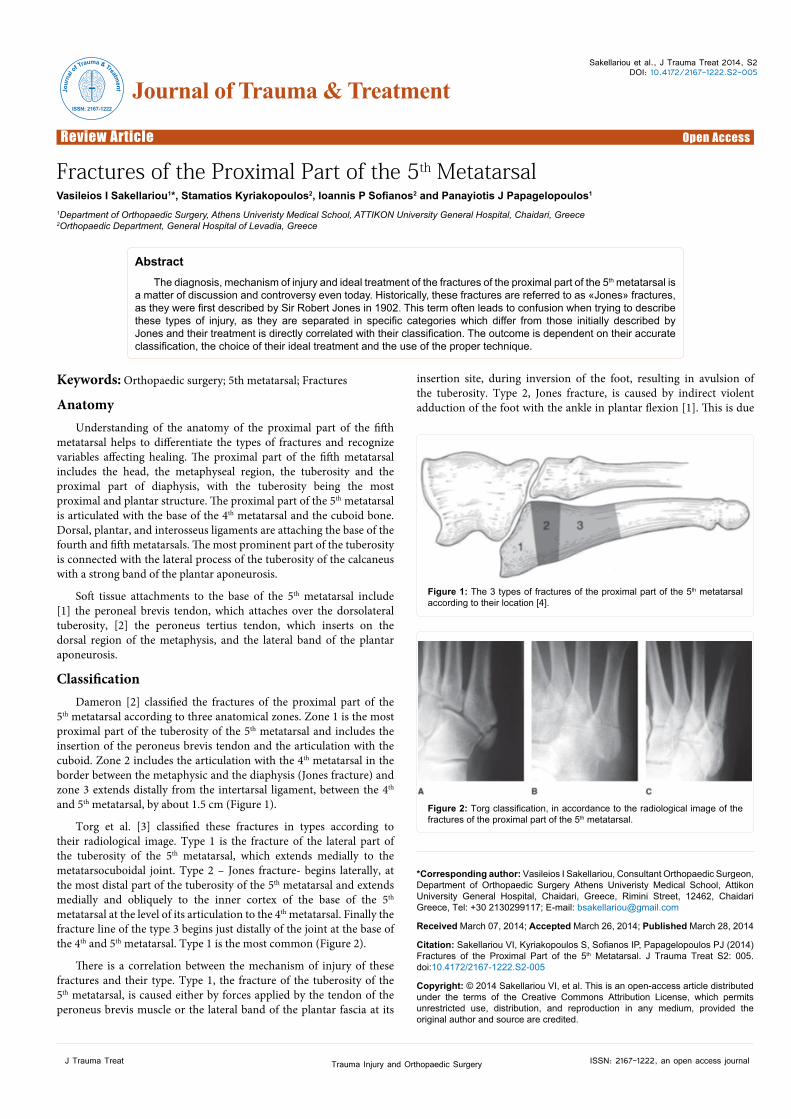
5th metatarsal according to three anatomical zones. Zone 1 is the most proximal part of the tuberosity of the 5th metatarsal and includes the insertion of the peroneus brevis tendon and the articulation with the cuboid. Zone 2 includes the articulation with the 4th metatarsal in the border between the metaphysic and the diaphysis (Jones fracture) and zone 3 extends distally from the intertarsal ligament, between the 4th and 5th metatarsal, by about 1.5 cm (Figure 1).
Torg et al. [3] classified these fractures in types according to their radiological image. Type 1 is the fracture of the lateral part of the tuberosity of the 5th metatarsal, which extends medially to the metatarsocuboidal joint. Type 2 – Jones fracture- begins laterally, at the most distal part of the tuberosity of the 5th metatarsal and extends medially and obliquely to the inner cortex of the base of the 5th metatarsal at the level of its articulation to the 4th metatarsal. Finally the fracture line of the type 3 begins just distally of the joint at the base of the 4th and 5th metatarsal. Type 1 is the most common (Figure 2).
There is a correlation between the mechanism of injury of these fractures and their type. Type 1, the fracture of the tuberosity of the 5th metatarsal, is caused either by forces applied by the tendon of the peroneus brevis muscle or the lateral band of the plantar fascia at its
insertion site, during inversion of the foot, resulting in avulsion of the tuberosity. Type 2, Jones fracture, is caused by indirect violent adduction of the foot with the ankle in plantar flexion [1]. This is due
AbstractThe diagnosis, mechanism of injury and ideal treatment of the fractures of the proximal part of the 5th metatarsal is
a matter of discussion and controversy even today. Historically, these fractures are referred to as «Jones» fractures, as they were first described by Sir Robert Jones in 1902. This term often leads to confusion when trying to describe these types of injury, as they are separated in specific categories which differ from those initially described by Jones and their treatment is directly correlated with their classification. The outcome is dependent on their accurate classification, the choice of their ideal treatment and the use of the proper technique.
Figure 1: The 3 types of fractures of the proximal part of the 5th metatarsal according to their location [4].
Figure 2: Torg classification, in accordance to the radiological image of the fractures of the proximal part of the 5th metatarsal.
Journal of Trauma & TreatmentJour
nal o
f Trau am & Treatment
ISSN: 2167-1222

Citation: Sakellariou VI, Kyriakopoulos S, Sofianos IP, Papagelopoulos PJ (2014) Fractures of the Proximal Part of the 5th Metatarsal. J Trauma Treat S2: 005. doi:10.4172/2167-1222.S2-005
Page 2 of 4
J Trauma Treat ISSN: 2167-1222, an open access journalTrauma Injury and Orthopaedic Surgery
to the stability of the intertarsal ligaments, which withstand ruptures or possible luxations. Type 3 is caused either by excessive bearing of the region or is part of the fatigue fractures group. This type of injury can be acute or chronic.
Even though many support that the location of a stress fracture differs from that of an acute one [4,5], it has never been proven.
Physical HistoryIn a series of 21 fractures of the base of the 5th metatarsal, Carp [6]
was one of the first to note a tendency of delayed union of these fractures and was the first who attributed this to the poor blood supply of the region. The vascular anatomy of the 5th metatarsal has meticulously been described by Smith et al. [7] and Shereff et al. [8]. A widespread arteriolar network, which enters though its base, is responsible for the blood supply of the metaphysis. The main nutrient artery of the 5th metatarsal enters though the nutrient foramen approximately in the middle of the diaphysis and branches proximally and distally. It has been observed that the proximal part is slightly shorter. As a result a critical area exists (watershed area), at the border of metaphysic and diaphysis, where the blood supply is poor, almost avascular, which makes this area prone to delayed union or even pseudoarthrosis (Figure 3).
The patient with a fracture at the base of the 5th metatarsal reports sudden onset of pain in the area after torsional injury of the foot. Local edema and hematoma may be observed. Exceptions are the fatigue fractures of zone 3, where a dull pain may be present for days or even weeks before the appearance of the fracture. They are usually observed in athletes and are prone to delayed union [2,4,9,10]. Plain radiological imaging is essential and usually an anteroposterior and a lateral view are enough to set the diagnosis. They are usually transverse fractures, vertical to the diaphysis of the 5th metatarsal.
We need to differentiate between an avulsion fracture of the tuberosity of the 5th metatarsal and a secondary calcification center at the proximal end of the metatarsal (apophysis). The apophysis becomes visible at radiographs in girls at ages 9 to 11 and boys 11 to 14 as a cortex of calcification at the base of the 5th metatarsal, perpendicular to the diaphysis of the bone (Figure 4).
The osteochondritis of the base of the 5th metatarsal (Iselin`s disease), is a self-restrained disorder observed in young ages which regresses spontaneously when the patient reaches skeletal maturity. The child complains of local pain after vigorous physical exercise, which subsides with resting. Radiologically an unusual density and shape of the apophysis is observed.
An adjuvant ossicle can sometimes be mistaken as an avulsion fracture. Os vesalianum is very rare and is found next to the insertion of the peroneus brevis muscle. Os perineum is more commonly seen and is embedded in the tendon of the peroneus brevis muscle. These ossicles are characterized by smooth surfaces, whereas fractures have uneven ones (Figure 5).
TreatmentThe undisplaced fractures of zone 1 are being easily managed
with walking casts and controlled bearing. Union is usually achieved after 6 to 8 weeks. Displaced fractures of this zone that include 30% or more of the metatarsocuboidal joint, or those that have intraarticular longitudinal displacement greater than 2 mm are usually treated with open reduction and internal fixation. Usually a Kirschner wire or a compression screw is enough to stabilize the displaced bony piece (Figure 6). According to Dameron [10] fractures with up to 3 mm displacement should be treated conservatively, unless they have torsional displacement. In case of a symptomatic pseudoarthrosis the fractured piece, if small, can be removed at a later time.
Zone 2 fractures can be more difficult to treat. Even though the literature states that conservative treatment with cast and avoidance of weight bearing is enough, this is a controversial matter [11-13]. In non-athletes conservative treatment is usually adequate in treating a Jones fracture as well as an acute meta-diaphyseal fracture of the 5th metatarsal. Radiologically the union of these fractures is seen to progress from the inside out. The formation of the callus at the fracture site, without intramedullary sclerotic signs, should be evident within 6 to 8 weeks. In case of delayed union or pseudoarthrosis applying an electromagnetic stimulator appears to be a good alternative to surgical treatment [14]. Kavanaugh et al. [15] and Delee et al. [16] reported a pretty high percentage of delayed union and pseudoarthrosis in athletes with type 2 and 3 fractures which did not undergo surgical treatment. As a result it has been proposed to treat these fractures as soon as possible in athletes with internal fixation using compression screws, whereas in non-athletes to use internal fixation only in case of delayed union [2,3,10,15-17]. Torg classified the fractures of the diaphysis of the 5th metatarsal in 3 subtypes according to the time of fracture (Table 1). He observed that patients with radiological signs of delayed union were less likely to be cured without surgical intervention (Figure 7). Quill [5] also reported, based on a literature review, that in approximately 1/3 of those fractures a new fracture occurred if the patient`s follow up was long enough and proposed that their treatment should be more aggressive.
Clapper et al. [13] reported a 100% union of Jones fractures that
Metaphysealarteries
Nutrient artery
Periosteal blood supplyAvascular
(watershed) zone
Figure 3: Survey of the vascular supply of the proximal part of the 5th metatarsal [2].
Figure 4: Apophysis of the 5th metatarsal (arrow).
Citation: Sakellariou VI, Kyriakopoulos S, Sofianos IP, Papagelopoulos PJ (2014) Fractures of the Proximal Part of the 5th Metatarsal. J Trauma Treat S2: 005. doi:10.4172/2167-1222.S2-005
Page 3 of 4
J Trauma Treat ISSN: 2167-1222, an open access journalTrauma Injury and Orthopaedic Surgery
were surgically treated. In the same study the mean time of union was 12.1 weeks for those patients who underwent surgical fixation, compared to 21.2 weeks for those who were treated conservatively. Clapper et al. concluded that surgical management of these fractures was highly successful, with minor risks and resulted in a higher patient satisfaction when compared to those with conservative management. However, the literature supports that surgical reconstruction has a role in treating these types of fractures in high demand athletes [5,13,15,16], informed patients that prefer to eliminate the risk of pseudoarthrosis due to conservative management [4,5,13,17] as well as patients with fatigue fractures of the diaphysis of the 5th metatarsal showing radiological signs of delayed union or pseudoarthrosis [3,16].
Surgical treatment is contraindicated in patients with vascular problems, local infection as well as in those who are unable to undergo spinal or general anesthesia due to systemic conditions. Diabetes mellitus is not an absolute contraindication for surgical treatment. Yue and Marcus showed positive results after internal fixation and use of bone grafts for the treatment of Jones fractures in diabetic patients [18].
The surgical management of fractures of the proximal part of the 5th metatarsal consists of inserting an intramedullary, cannulated or not, compression screw, reaming beforehand the medullary canal, using bone grafts if needed. Delee et al. [16] was the first to describe the technique of percutaneous screw osteosynthesis for type 2 and 3 Torg fractures as well as for Jones fractures. Nunley [19] evolved and improved this technique.
This procedure can be done either as part of a day care case through nervous block at the level of the ankle, or using spinal or general anesthesia. The width of the 5th metatarsal`s medullary canal varies from person to person. As a result it is important to be sure that the screw fits tightly with the endosteum and the thread is in good contact with the inner cortex in order to accomplish sufficient compression of the fracture. This is usually achieved with a cannulated screw larger than 5.5 mm or more usually 7 mm. it is important not to use excessive force during positioning of the screw as a larger screw will result in a diaphyseal fracture (Figure 8).
It is important to ensure that the whole length of the thread passes through the fracture line in order to achieve compression. The length of the screw should not exceed 50-60% of the total length of the metatarsal bone, as longer screws have a tendency to increase the gap between
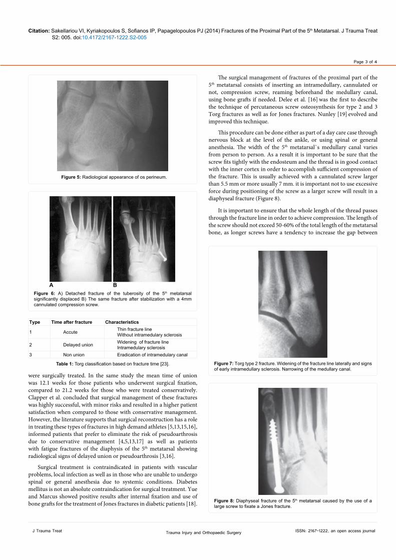
Figure 5: Radiological appearance of os perineum.
A BFigure 6: A) Detached fracture of the tuberosity of the 5th metatarsal significantly displaced B) The same fracture after stabilization with a 4mm cannulated compression screw.
Type Time after fracture Characteristics
1 Accute Thin fracture lineWithout intramedulary sclerosis
2 Delayed union Widening of fracture lineIntramedulary sclerosis
3 Non union Eradication of intramedulary canal
Table 1: Torg classification based on fracture time [23]. Figure 7: Torg type 2 fracture. Widening of the fracture line laterally and signs of early intramedullary sclerosis. Narrowing of the medullary canal.
Figure 8: Diaphyseal fracture of the 5th metatarsal caused by the use of a large screw to fixate a Jones fracture.
Citation: Sakellariou VI, Kyriakopoulos S, Sofianos IP, Papagelopoulos PJ (2014) Fractures of the Proximal Part of the 5th Metatarsal. J Trauma Treat S2: 005. doi:10.4172/2167-1222.S2-005
Page 4 of 4
J Trauma Treat ISSN: 2167-1222, an open access journalTrauma Injury and Orthopaedic Surgery
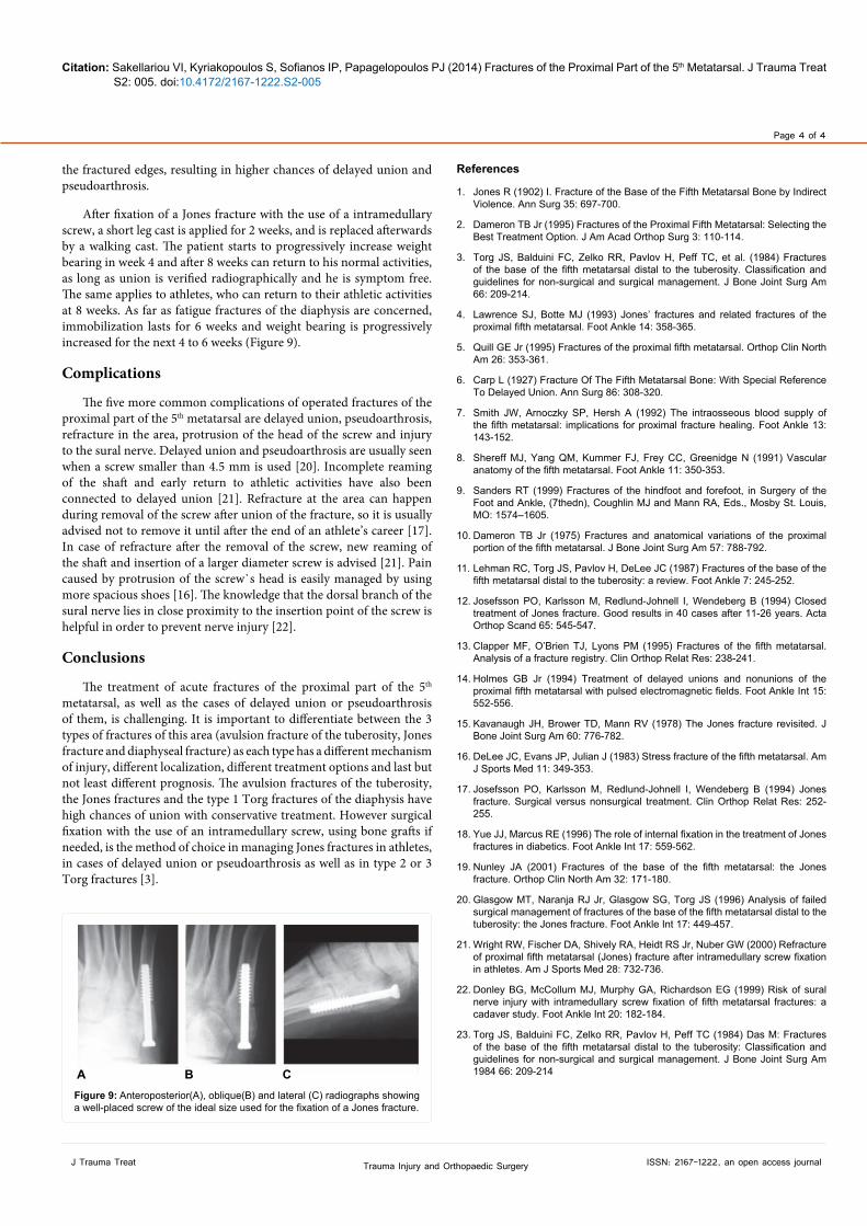
A B CFigure 9: Anteroposterior(A), oblique(B) and lateral (C) radiographs showing a well-placed screw of the ideal size used for the fixation of a Jones fracture.
the fractured edges, resulting in higher chances of delayed union and pseudoarthrosis.
After fixation of a Jones fracture with the use of a intramedullary screw, a short leg cast is applied for 2 weeks, and is replaced afterwards by a walking cast. The patient starts to progressively increase weight bearing in week 4 and after 8 weeks can return to his normal activities, as long as union is verified radiographically and he is symptom free. The same applies to athletes, who can return to their athletic activities at 8 weeks. As far as fatigue fractures of the diaphysis are concerned, immobilization lasts for 6 weeks and weight bearing is progressively increased for the next 4 to 6 weeks (Figure 9).
Complications
The five more common complications of operated fractures of the proximal part of the 5th metatarsal are delayed union, pseudoarthrosis, refracture in the area, protrusion of the head of the screw and injury to the sural nerve. Delayed union and pseudoarthrosis are usually seen when a screw smaller than 4.5 mm is used [20]. Incomplete reaming of the shaft and early return to athletic activities have also been connected to delayed union [21]. Refracture at the area can happen during removal of the screw after union of the fracture, so it is usually advised not to remove it until after the end of an athlete’s career [17]. In case of refracture after the removal of the screw, new reaming of the shaft and insertion of a larger diameter screw is advised [21]. Pain caused by protrusion of the screw`s head is easily managed by using more spacious shoes [16]. The knowledge that the dorsal branch of the sural nerve lies in close proximity to the insertion point of the screw is helpful in order to prevent nerve injury [22].
Conclusions
The treatment of acute fractures of the proximal part of the 5th metatarsal, as well as the cases of delayed union or pseudoarthrosis of them, is challenging. It is important to differentiate between the 3 types of fractures of this area (avulsion fracture of the tuberosity, Jones fracture and diaphyseal fracture) as each type has a different mechanism of injury, different localization, different treatment options and last but not least different prognosis. The avulsion fractures of the tuberosity, the Jones fractures and the type 1 Torg fractures of the diaphysis have high chances of union with conservative treatment. However surgical fixation with the use of an intramedullary screw, using bone grafts if needed, is the method of choice in managing Jones fractures in athletes, in cases of delayed union or pseudoarthrosis as well as in type 2 or 3 Torg fractures [3].
References
1. Jones R (1902) I. Fracture of the Base of the Fifth Metatarsal Bone by Indirect Violence. Ann Surg 35: 697-700.
2. Dameron TB Jr (1995) Fractures of the Proximal Fifth Metatarsal: Selecting the Best Treatment Option. J Am Acad Orthop Surg 3: 110-114.
3. Torg JS, Balduini FC, Zelko RR, Pavlov H, Peff TC, et al. (1984) Fractures of the base of the fifth metatarsal distal to the tuberosity. Classification and guidelines for non-surgical and surgical management. J Bone Joint Surg Am 66: 209-214.
4. Lawrence SJ, Botte MJ (1993) Jones’ fractures and related fractures of the proximal fifth metatarsal. Foot Ankle 14: 358-365.
5. Quill GE Jr (1995) Fractures of the proximal fifth metatarsal. Orthop Clin North Am 26: 353-361.
6. Carp L (1927) Fracture Of The Fifth Metatarsal Bone: With Special Reference To Delayed Union. Ann Surg 86: 308-320.
7. Smith JW, Arnoczky SP, Hersh A (1992) The intraosseous blood supply of the fifth metatarsal: implications for proximal fracture healing. Foot Ankle 13: 143-152.
8. Shereff MJ, Yang QM, Kummer FJ, Frey CC, Greenidge N (1991) Vascular anatomy of the fifth metatarsal. Foot Ankle 11: 350-353.
9. Sanders RT (1999) Fractures of the hindfoot and forefoot, in Surgery of the Foot and Ankle, (7thedn), Coughlin MJ and Mann RA, Eds., Mosby St. Louis, MO: 1574–1605.
10. Dameron TB Jr (1975) Fractures and anatomical variations of the proximal portion of the fifth metatarsal. J Bone Joint Surg Am 57: 788-792.
11. Lehman RC, Torg JS, Pavlov H, DeLee JC (1987) Fractures of the base of the fifth metatarsal distal to the tuberosity: a review. Foot Ankle 7: 245-252.
12. Josefsson PO, Karlsson M, Redlund-Johnell I, Wendeberg B (1994) Closed treatment of Jones fracture. Good results in 40 cases after 11-26 years. Acta Orthop Scand 65: 545-547.
13. Clapper MF, O’Brien TJ, Lyons PM (1995) Fractures of the fifth metatarsal. Analysis of a fracture registry. Clin Orthop Relat Res: 238-241.
14. Holmes GB Jr (1994) Treatment of delayed unions and nonunions of the proximal fifth metatarsal with pulsed electromagnetic fields. Foot Ankle Int 15: 552-556.
15. Kavanaugh JH, Brower TD, Mann RV (1978) The Jones fracture revisited. J Bone Joint Surg Am 60: 776-782.
16. DeLee JC, Evans JP, Julian J (1983) Stress fracture of the fifth metatarsal. Am J Sports Med 11: 349-353.
17. Josefsson PO, Karlsson M, Redlund-Johnell I, Wendeberg B (1994) Jones fracture. Surgical versus nonsurgical treatment. Clin Orthop Relat Res: 252-255.
18. Yue JJ, Marcus RE (1996) The role of internal fixation in the treatment of Jones fractures in diabetics. Foot Ankle Int 17: 559-562.
19. Nunley JA (2001) Fractures of the base of the fifth metatarsal: the Jones fracture. Orthop Clin North Am 32: 171-180.
20. Glasgow MT, Naranja RJ Jr, Glasgow SG, Torg JS (1996) Analysis of failed surgical management of fractures of the base of the fifth metatarsal distal to the tuberosity: the Jones fracture. Foot Ankle Int 17: 449-457.
21. Wright RW, Fischer DA, Shively RA, Heidt RS Jr, Nuber GW (2000) Refracture of proximal fifth metatarsal (Jones) fracture after intramedullary screw fixation in athletes. Am J Sports Med 28: 732-736.
22. Donley BG, McCollum MJ, Murphy GA, Richardson EG (1999) Risk of sural nerve injury with intramedullary screw fixation of fifth metatarsal fractures: a cadaver study. Foot Ankle Int 20: 182-184.
23. Torg JS, Balduini FC, Zelko RR, Pavlov H, Peff TC (1984) Das M: Fractures of the base of the fifth metatarsal distal to the tuberosity: Classification and guidelines for non-surgical and surgical management. J Bone Joint Surg Am 1984 66: 209-214