saccharomyces cerevisiae paf1 transcription elongation...
TRANSCRIPT

YYou\
The Saccharomyces cerevisiae Paf1 transcription elongation complex is connected to chromatin modification through the multifunctional Rtf1 subunit and the inositol
polyphosphate signaling pathway
by
Marcie Helene Warner
B.S., Duquesne University, 2000
Submitted to the Graduate Faculty of
Arts and Sciences in partial fulfillment
of the requirements for the degree of
Doctor of Philosophy
University of Pittsburgh
2008

ii
UNIVERSITY OF PITTSBURGH
SCHOOL OF ARTS AND SCIENCES
This dissertation was presented
by
Marcie Helene Warner
It was defended on
October 10, 2008
and approved by
Deborah Chapman, PhD, Associate Professor, Dept. of Biological Sciences
Sanford Leuba, PhD, Assistant Professor, Dept. of Cell Biology and Physiology
William Saunders, PhD, Associate Professor, Dept. of Biological Sciences
Anthony Schwacha, PhD, Assistant Professor, Dept. of Biological Sciences
Dissertation Advisor: Karen Arndt, PhD,
Associate Professor, Dept. of Biological Sciences

iii
Copyright © by Marcie Helene Warner
2008

iv
Transcription in eukaryotes takes place in the context of a repressive chromatin template.
Access to the DNA is facilitated by histone modifying enzymes and ATP-dependent chromatin
remodeling complexes, which modify chromatin structure. The activities of chromatin
modifying proteins are often coordinated by nonenzymatic accessory factors that interact with
actively transcribing RNA Polymerase II (Pol II). One such factor is the Saccharomyces
cerevisiae Paf1 transcription elongation complex. This complex, which is minimally composed
of Paf1, Ctr9, Rtf1, Cdc73, and Leo1, physically interacts with Pol II and localizes to the coding
regions of active genes.
The Rtf1 subunit of the Paf1 complex performs several cotranscriptional functions: it
facilitates recruitment of the chromatin remodeling enzyme Chd1, promotes covalent
modification of specific lysine residues in histones H2B and H3, and mediates association of
other Paf1 complex subunits. Using a collection of internal deletion mutations that remove 20 to
50 amino acid segments across the length of Rtf1, I demonstrated that Rtf1's known functions are
mediated by nonoverlapping regions, implying that the multiple functions of this protein are not
completely interrelated. Deletion of the regions of Rtf1 that are required for promoting histone
modification or its association with active genes resulted in the strongest transcription-related
phenotypes, which suggested that promoting cotranscriptional histone modification is a critical
means by which Rtf1 exerts its effects on transcription. Detailed analysis of the region of Rtf1
The Saccharomyces cerevisiae Paf1 transcription elongation complex is connected to chromatin modification through the multifunctional Rtf1 subunit and the inositol
polyphosphate signaling pathway
Marcie Helene Warner, PhD
University of Pittsburgh, 2008

v
required for histone modification determined that it is sufficient to promote Rtf1-dependent
histone modifications and that this function is dependent on several highly conserved residues.
Additionally, a screen for factors that become essential in the absence of Rtf1 uncovered
mutations in the first two enzymes of the inositol polyphosphate (IP) signaling pathway: Plc1
and Arg82. The IP signaling pathway has been linked to the function of several chromatin
remodeling complexes. I uncovered strong genetic interactions between Arg82, Paf1, and
mutations in the SWI/SNF and INO80 chromatin remodeling complexes and demonstrated that
the expression of several target genes was strongly impaired by mutations in these factors.
Together, these data suggest that transcription elongation, IP signaling, and chromatin
remodeling cooperate to coordinate proper gene expression.

vi
TABLE OF CONTENTS
PREFACE ................................................................................................................................. XVI
1.0 INTRODUCTION ........................................................................................................ 1
1.1 THE EUKARYOTIC GENOME IS PACKAGED INTO CHROMATIN .... 2
1.2 CHROMATIN STRUCTURE IS DYNAMICALLY REGULATED BY
SEVERAL MECHANISMS ................................................................................................ 5
1.2.1 Nucleosome disassembly and the incorporation of histone variants .......... 5
1.2.2 Histone modification........................................................................................ 7
1.2.2.1 Histone Acetylation ............................................................................. 11
1.2.2.2 Histone Ubiquitylation ........................................................................ 14
1.2.2.3 Histone Methylation ............................................................................ 18
1.2.3 ATP-Dependent Chromatin Remodeling .................................................... 25
1.2.3.1 SWI/SNF .............................................................................................. 29
1.2.3.2 RSC ....................................................................................................... 30
1.2.3.3 ISW1 ..................................................................................................... 31
1.2.3.4 ISW2 ..................................................................................................... 32
1.2.3.5 CHD1 .................................................................................................... 33
1.2.3.6 INO80 ................................................................................................... 33
1.2.3.7 SWR1 .................................................................................................... 34

vii
1.3 TELOMERIC SILENCING IS CONTROLLED BY GLOBAL PATTERNS
OF HISTONE MODIFICATION ..................................................................................... 35
1.4 TRANSCRIPTION BY RNA POLYMERASE II IS HIGHLY
REGULATED BY ACCESSORY PROTEINS ............................................................... 37
1.4.1 Promoter Binding and Transcription Initiation ......................................... 44
1.4.1.1 Assembly of the Preinitiation Complex ............................................. 45
1.4.1.2 Transcriptional activators and coactivators ..................................... 49
1.4.2 Transcription Elongation .............................................................................. 51
1.4.2.1 Paf1 complex ........................................................................................ 51
1.4.2.2 Spt4/Spt5 complex ............................................................................... 61
1.4.2.3 TFIIS .................................................................................................... 62
1.4.2.4 yFACT .................................................................................................. 63
1.4.3 3’ End Formation and Transcription Termination at Protein Coding
Genes ………………………………………………………………………………. 64
1.4.4 mRNA export ................................................................................................. 68
1.5 THE INOSITOL POLYPHOSPHATE SIGNALING PATHWAY
AFFECTS TRANSCRIPTION ......................................................................................... 70
1.5.1 Arg82 is required for normal expression of arginine metabolic genes ..... 73
1.5.2 IP6 is necessary for mRNA export................................................................ 74
1.5.3 Chromatin remodeling is affected by inositol polyphosphates .................. 74
1.6 THESIS AIMS ................................................................................................... 76
2.0 RTF1 IS COMPOSED OF DISCRETE FUNCTIONAL REGIONS ................... 77
2.1 INTRODUCTION ............................................................................................. 77
viii
2.2 MATERIALS AND METHODS ...................................................................... 78
2.2.1 Media and yeast strains ................................................................................. 78
2.2.2 Plasmid construction ..................................................................................... 79
2.2.3 Yeast growth assays ....................................................................................... 80
2.2.4 Sequence Alignment ...................................................................................... 80
2.2.5 Immunoblotting analyses .............................................................................. 81
2.2.6 Analysis of histone H2B K123 monoubiquitylation ................................... 82
2.2.7 Chromatin immunoprecipitation assays ..................................................... 82
2.3 RESULTS ........................................................................................................... 86
2.3.1 Conserved regions of Rtf1 direct normal transcription ............................. 86
2.3.2 A region near the amino-terminus of Rtf1 mediates physical interaction
with Chd1 .................................................................................................................... 92
2.3.3 Conserved regions of Rtf1 are required for Rtf1-dependent histone
modifications and telomeric silencing ...................................................................... 96
2.3.4 Association of Rtf1 with active ORFs requires a conserved central region
………………………………………………………………………………. 99
2.3.5 Rtf1 interacts with other Paf1 complex components through its carboxy-
terminus .................................................................................................................... 102
2.4 CONCLUSIONS .............................................................................................. 107
3.0 CONSERVED RESIDUES IN THE RTF1 HISTONE MODIFICATION
DOMAIN DIRECT HISTONE H2B UBIQUITYLATION AND H3 METHYLATION .. 111
3.1 INTRODUCTION ........................................................................................... 111
3.2 MATERIALS AND METHODS .................................................................... 112

ix
3.2.1 Media and yeast strains ............................................................................... 112
3.2.2 Plasmid construction ................................................................................... 113
3.2.3 Error prone mutagenesis of the RTF1 HMD coding sequence ............... 114
3.2.4 Yeast growth assays ..................................................................................... 115
3.2.5 Immunoblotting analyses ............................................................................ 115
3.2.6 Analysis of histone H2B K123 monoubiquitylation ................................. 116
3.2.7 Chromatin immunoprecipitation assays ................................................... 117
3.3 RESULTS ......................................................................................................... 121
3.3.1 Point mutations were generated in the Rtf1 HMD by biased and unbiased
methods ..................................................................................................................... 121
3.3.2 Conserved residues in the Rtf1 HMD differentially affect H2B
ubiquitylation and H3 methylation ........................................................................ 125
3.3.3 rtf1 108-110A and rtf1 F80V F123S may alter histone-DNA contacts .... 129
3.3.4 Point mutations in the Rtf1 HMD cause defects in transcription ........... 130
3.3.5 Mutations in the Rtf1 HMD allow aberrant transcription initiation from
cryptic promoters internal to coding regions and the telomeres ......................... 133
3.3.6 The Rtf1 HMD is sufficient to promote Rtf1-dependent histone
modifications ............................................................................................................. 136
3.4 CONCLUSIONS .............................................................................................. 148
4.0 THE PAF1 COMPLEX IS CONNECTED TO CHROMATIN REMODELING
THROUGH GENETIC INTERACTIONS WITH THE INOSITOL POLYPHOSPHATE
SIGNALING CASCADE ......................................................................................................... 151
4.1 INTRODUCTION ........................................................................................... 151

x
4.2 MATERIALS AND METHODS .................................................................... 153
4.2.1 Genetic methods ........................................................................................... 153
4.2.2 Analysis of mRNA export ........................................................................... 154
4.2.3 Northern analysis ......................................................................................... 154
4.3 RESULTS ......................................................................................................... 157
4.3.1 arg82∆ genetically interacts with mutations in several known elongation
factors ……………………………………………………………………………... 157
4.3.2 Enzymes that function downstream of Arg82 are not essential in the
absence of Paf1 or Rtf1 ............................................................................................ 163
4.3.3 The synthetic lethality caused by the combined loss of ARG82 and PAF1 is
not the result of an enhanced defect in mRNA export. ......................................... 163
4.3.4 paf1∆ and arg82∆ genetically interact with mutations in the INO80,
SWI/SNF, and SWR1 chromatin remodeling complexes ..................................... 166
4.3.5 Expression of several genes is affected by loss of Paf1, Arg82, or
mutations in the INO80 and SWI/SNF chromatin remodeling complexes ......... 172
4.4 CONCLUSIONS .............................................................................................. 175
5.0 DISCUSSION AND FUTURE DIRECTIONS ...................................................... 179
5.1 RTF1 IS COMPOSED OF FUNCTIONALLY DISTINCT REGIONS ..... 179
5.2 THE HISTONE MODIFICATION DOMAIN OF RTF1 IS NECESSARY
AND SUFFICIENT TO PROMOTE RTF1-DEPENDENT HISTONE
MODIFICATIONS ........................................................................................................... 184

xi
5.3 GENETIC INTERACTIONS IMPLY CONNECTIONS BETWEEN THE
INOSITOL POLYPHOSPHATE SIGNALING PATHWAY, TRANCRIPTION
ELONGATION, AND CHROMATIN REMODELING .............................................. 191
BIBLIOGRAPHY ..................................................................................................................... 196

xii
LIST OF TABLES
Table 1. Saccharomyces cerevisiae strains used in Chapter 2 ..................................................... 84
Table 2. Saccharomyces cerevisiae strains used in Chapter 3 ................................................... 118
Table 3. Saccharomyces cerevisiae strains used in Chapter 4 ................................................... 155
Table 4. Genetic interactions between arg82∆ and mutations in known elongation factors ..... 160
Table 5. paf1∆ or arg82∆ genetically interact with disruptions of chromatin remodeling
complexes ................................................................................................................................... 169

xiii
LIST OF FIGURES
Figure 1. The crystal structure of the nucleosome. ........................................................................ 4
Figure 2. Posttranslational histone modifications in yeast. .......................................................... 10
Figure 3. Distribution of H2B K123 ubiquitylation and H3 K4, K36, and K79 methylation at a
typical active yeast gene. .............................................................................................................. 16
Figure 4. The states of methylation on histone lysine and arginine residues. ............................. 20
Figure 5. Potential mechanism of intranucleosomal looping and nucleosome repositioning
induced by chromatin remodeling enzymes. ................................................................................ 27
Figure 6. The crystal structure of RNA Polymerase II. ............................................................... 39
Figure 7. Phosphorylation of the CTD is dynamically regulated throughout the transcription
cycle. ............................................................................................................................................. 43
Figure 8. Assembly of the preinitiation complex. ........................................................................ 48
Figure 9. Physical interactions and functions of the Paf1 complex. ............................................ 56
Figure 10. Factors involved in RNA 3’ end formation. ............................................................... 66
Figure 11. The mRNA export pathway. ....................................................................................... 69
Figure 12. The inositol polyphosphate signaling pathway. ......................................................... 72
Figure 13. Rtf1 internal deletion mutants. ................................................................................... 87

xiv
Figure 14. Deletions of discrete regions of Rtf1 cause phenotypes associated with transcriptional
defects. .......................................................................................................................................... 89
Figure 15. Rtf1 homologs contain two clusters of highly conserved residues. ........................... 91
Figure 16. The amino-terminus of Rtf1 is required for recruitment of Chd1 to active genes. .... 93
Figure 17. Mutations that disrupt the Rtf1-Chd1 interaction suppress a TBP mutant. ................ 95
Figure 18. Conserved regions of Rtf1 are essential for Rtf1-dependent histone modifications and
telomeric silencing. ....................................................................................................................... 97
Figure 19. A central conserved region mediates association of Rtf1 with active ORFs and
influences protein stability. ......................................................................................................... 101
Figure 20. The carboxy-terminus of Rtf1 mediates physical interaction with other Paf1 complex
subunits. ...................................................................................................................................... 105
Figure 21. Rtf1 is composed of functionally distinct regions. ................................................... 108
Figure 22. Substitution of conserved residues in the Rtf1 HMD affects H3 K4 trimethylation. 123
Figure 23. Conserved residues in the Rtf1 HMD differentially affect Rtf1-dependent histone
modifications. .............................................................................................................................. 128
Figure 24. Rtf1 HMD substitution mutations cause phenotypes associated with transcription
defects. ........................................................................................................................................ 132
Figure 25. Substitution mutations in the Rtf1 HMD cause phenotypes associated with defects in
chromatin structure. .................................................................................................................... 135
Figure 26. Expression of GBD-HMD fusion proteins. .............................................................. 138
Figure 27. The Rtf1 HMD is sufficient to induce H3 K4 trimethylation at the GAL7 UAS. .... 141
Figure 28. The Rtf1 HMD does not require targeting to DNA to induce Rtf1-dependent histone
modifications. .............................................................................................................................. 145

xv
Figure 29. The Rtf1 HMD can partially restore global Rtf1-dependent histone modifications. 147
Figure 30. arg82∆ genetically interacts with deletions of the genes encoding Paf1 complex
subunits. ...................................................................................................................................... 159
Figure 31. arg82∆ genetically interacts with mutations in the Spt4/5 elongation complex, but not
TFIIS. .......................................................................................................................................... 162
Figure 32. The inviability of an arg82∆ paf1∆ strain is not due to an enhanced defect in mRNA
export. ......................................................................................................................................... 165
Figure 33. paf1∆ genetically interacts with disruptions in chromatin remodeling complexes. . 168
Figure 34. arg82∆ genetically interacts with disruptions in chromatin remodeling complexes.
..................................................................................................................................................... 171
Figure 35. The Paf1 complex, Arg82, and the INO80 and SWI/SNF chromatin remodeling
complexes are necessary for normal expression of SPL2 and VTC3. ......................................... 174
Figure 36. Genetic interactions between Paf1, Arg82, and chromatin remodeling complexes. 177

xvi
PREFACE
My experience as a graduate student in the Department of Biological Sciences at the University
of Pittsburgh has been a fantastic experience that would not have been possible without the
support and assistance of so many wonderful people. Most importantly, I must acknowledge
Karen Arndt; thank you, Karen, for welcoming me into your lab and sharing your knowledge
while teaching me to think critically and independently. I also need to thank past and present
members of the Arndt lab. It has been a privilege to work with so many talented and intelligent
people.
I also must thank the members of my thesis committee: Debbie Chapman, Sanford
Leuba, Bill Saunders, and Tony Schwacha. I appreciate the helpful suggestions and advice as I
progressed through my graduate career. Thanks also to the Martens, VanDemark, Winston,
Prelich, Tansey, and Wente labs for their assistance and collaboration. The main and fiscal
offices also deserve acknowledgement for their assistance in helping me meet my requirements
for graduation. I also need to thank the community of graduate students in this department. I’ve
been fortunate to meet so many wonderful people and develop many lasting friendships.
Finally, I must acknowledge my family who have always supported me and encouraged
my pursuit of whatever made me happy. I’m thankful to have always been surrounded by
unconditional love and know that I never would’ve made it this far without you.

1
1.0 INTRODUCTION
The genomes of eukaryotes are intricately packaged into a nucleoprotein material known as
chromatin by association with the histone proteins; this arrangement facilitates their efficient
storage and organization within the comparatively small cell nucleus. However, this storage
system imposes a significant impediment to processes that require access to the genetic material,
such as DNA repair and replication, recombination, and transcription. Eukaryotic cells have,
therefore, developed several methods to counter the repressive nature of chromatin structure,
including covalent modification of the histone proteins and disassembly or remodeling of
nucleosomes, which represent the basic unit of chromatin.
The studies described in this dissertation were performed using the yeast Saccharomyces
cerevisiae as a model system to explore the mechanisms underlying chromatin modification
during the transcription of messenger RNA. Therefore, the information provided in this chapter
is specifically tailored toward detailing the current understanding of these processes in S.
cerevisiae.
Chromatin modification during transcription appears to be an ongoing process that is
tightly regulated throughout the transcription cycle. This careful control is necessary to ensure
proper levels of gene expression and to prevent aberrant initiation from cryptic promoter
sequences present in active open reading frames (ORFs) that become exposed when chromatin
structure has been perturbed by the passage of RNA polymerase II (Pol II). The regulation of

2
chromatin modification during transcription depends, in part, upon accessory factors that interact
with actively transcribing Pol II and coordinate the recruitment of chromatin-modifying factors
to active genes and, in some cases, stimulate their enzymatic activity.
1.1 THE EUKARYOTIC GENOME IS PACKAGED INTO CHROMATIN
Eukaryotic cells condense and organize large genomes within the relatively small confines of
their nuclei. The genome of S. cerevisiae, which represents one of the simplest eukaryotic
genomes, totals nearly 13,000,000 base pairs (bp) in length and contains nearly 6,000 genes
(GOFFEAU et al. 1996). The S. cerevisiae haploid genome consists of 16 linear chromosomes of
varying size which must be packaged into a nuclear space that measures approximately 3µm3
(JORGENSEN et al. 2007).
Organization and compaction of eukaryotic DNA molecules is achieved through their
association with the histone proteins. The histones are a class of small, highly conserved
proteins that interact with each other to form an octamer containing two copies each of histones
H2A, H2B, H3, and H4 (LUGER et al. 1997). The histones are positively charged, leading to a
tight interaction between the histone octamer and the negatively-charged phosphodiester
backbone of a DNA molecule.
Approximately every 200 bp along the length of a eukaryotic chromosome, a 146 bp
length of DNA coils nearly two revolutions around a histone octamer to form a structure known
as a nucleosome (Figure 1) (LUGER et al. 1997). The resulting nucleoprotein material, known as
chromatin, resembles “beads on a string” and represents the first order of chromosome
organization (KORNBERG and THOMAS 1974). The genome can be further compacted through

3
the formation of higher-order chromatin structures which require, in part, a fifth histone protein
(histone H1) which contacts both the proteins in the histone octamer and DNA present in the
linker region between adjacent nucleosomes (ISHIMI et al. 1981). Highly compact chromatin
domains, known as heterochromatin, often contain important structural elements such as
telomeres or centromeres. The remainder of the genome is referred to as euchromatin, which is
largely composed of coding regions for both active and inactive genes.

4
Figure 1. The crystal structure of the nucleosome.
The crystal structure of the nucleosome was solved at 2.8A resolution. Models for the structure of 146 bp of DNA
wrapped around a histone octamer are shown (Reprinted by permission from Macmillan Publishers Ltd: [Nature]
LUGER et al. 1997, copyright 1997). Two copies each of H2A (orange), H2B (pink), H3 (blue), and H4 (green) can
be seen in the model on the left. In addition to the alpha-helical histone core domains, several histone “tails” can be
seen protruding from the nucleosome core in both models. Two coils of DNA around the histone octamer can be
seen clearly in the model on the right.

5
1.2 CHROMATIN STRUCTURE IS DYNAMICALLY REGULATED BY SEVERAL
MECHANISMS
The arrangement of eukaryotic chromosomes into chromatin is an efficient means of storing the
genetic material in a highly organized fashion. However, organization of the genome into
chromatin impacts DNA-based processes such as DNA repair, DNA replication, recombination,
and transcription, by introducing structural constraints and occluding the recognition sites of
DNA-binding proteins. Specifically, nucleosomes are known to impede Pol II processivity along
ORFs of active genes, often resulting in stalling or arrest of the polymerase (KIREEVA et al.
2005). Eukaryotic cells have, therefore, developed a number of mechanisms to modify
chromatin structure in order to facilitate the proper execution of DNA-based processes. The
most common mechanisms of chromatin modification in yeast, including chromatin disassembly,
incorporation of histone variants, covalent histone modification, and ATP-dependent chromatin
remodeling, are described in more detail in the following sections. Higher eukaryotes also
regulate chromatin modification through heritable methylation of the DNA, although this
mechanism appears to be largely absent from the S. cerevisiae genome.
1.2.1 Nucleosome disassembly and the incorporation of histone variants
Chromatin assembly is closely linked to genome replication. Expression of the histone genes
takes place specifically during the S phase of the cell cycle and histones are incorporated into
newly synthesized DNA immediately following the passage of DNA polymerase (TABANCAY

6
and FORSBURG 2006). Histone octamers are assembled from free H2A/H2B and H3/H4 dimers.
Incorporation of new nucleosomes into DNA takes place in a stepwise fashion in which two
H3/H4 dimers are incorporated first, followed by the addition of two H2A/H2B dimers
(SCHULTZ et al. 1997).
Nucleosome positioning is not entirely random; nearly 70% of the S. cerevisiae genome
is incorporated into well-positioned nucleosomes that consistently occupy a 140 bp length of
DNA (YUAN et al. 2005). Additionally, chromatin structure assumes a characteristic pattern
over the bodies of yeast genes. Many yeast gene promoters contain an approximately 200 bp
length of DNA that is devoid of nucleosomes. This nucleosome free region (NFR) is succeeded
into the ORF by several well-positioned nucleosomes (MAVRICH et al. 2008; YUAN et al. 2005).
In many cases, the precise location of nucleosomes becomes less well-defined as distance from
the promoter increases, suggesting the NFR may function as a nucleosome positioning signal.
The NFR is known to be flanked by two well-positioned nucleosomes that contain the
histone variant H2A.Z in place of canonical H2A. Histone variants are differentiated from
canonical histones largely by their continued expression outside of S phase and their primarily
replication-independent incorporation into chromatin (LI et al. 2007a). H2A.Z, encoded by the
HTZ1 gene, shares only about 60% sequence similarity with canonical H2A, which implies an
early evolutionary divergence between these two proteins (ZLATANOVA and THAKAR 2008).
H2A.Z containing nucleosomes appear to be destabilized more easily than their canonical H2A
containing counterparts (ZHANG et al. 2005). The presence of H2A.Z in nucleosomes flanking
promoters may, therefore, facilitate transcription initiation by promoting efficient eviction of
nucleosomes that occlude transcription factor binding sites or inhibit the ability of the
transcription machinery to assemble on gene promoters.

7
The presence of nucleosomes in the bodies of genes impedes the processivity of Pol II
(KIREEVA et al. 2005). Therefore, chromatin must be disassembled during active transcription to
ensure efficient gene expression. Removal of a single H2A/H2B dimer from each nucleosome
appears to be sufficient to allow the passage of Pol II in vitro (KIREEVA et al. 2002).
Translocating Pol II is believed to induce positive supercoiling of the DNA template ahead of its
position, which may be adequate to dislodge an H2A/H2B dimer (LEVCHENKO et al. 2005).
However, chromatin is also believed to be actively disassembled by the activity of numerous
histone chaperones including the CAF-I complex, Nap1, Spt6, and the yeast FACT complex;
these histone chaperone complexes also play important roles in reassembling chromatin
following the passage of Pol II (ARMSTRONG 2007). Chromatin reassembly is critical to prevent
aberrant initiation from cryptic promoters present within ORFs (KAPLAN et al. 2003).
1.2.2 Histone modification
Changes in chromatin structure can also be elicited through modification of the histone proteins.
The crystal structure of the nucleosome demonstrates that each histone protein contains an alpha-
helical core domain, which interacts with other histones and the DNA backbone; however,
unstructured amino- and carboxy-terminal tails are also apparent (Figure 1) (LUGER et al. 1997).
Histone modifications primarily occur on these histone tails, although modifications in the core
domains have also been identified.
Histones can be posttranslationally modified by the phosphorylation of serines, the
acetylation, ubiquitylation, or sumoylation of lysines, and the methylation of lysines and
arginines (Figure 2); additional modifications, such as isomerization, ADP-ribosylation and
deimination, have also been identified (KOUZARIDES 2007; KREBS 2007). Many histone

8
modifications are conserved throughout eukaryotes, which emphasizes the importance of
modifying chromatin structure for normal cellular function.

9

10
Figure 2. Posttranslational histone modifications in yeast.
The four core histones are represented; hexagons symbolize core domains and the sequences of amino- and/or
carboxy-terminal tails are provided. By convention, histone H2A is numbered from the methionine at position 1,
which is cleaved posttranslationally; all other histones are numbered according to the first amino acid present in the
mature protein. Modifications on the C-terminal tails of histones H3 and H4 have not been identified; the C-
terminal tails have, therefore, been omitted for simplicity. Known covalent histone modifications in yeast are
indicated; acetylation (Ac) is shown in green; phosphorylation (P) is shown in purple; monoubiquitylation (Ub) is
shown in blue; and methylation (Me) is shown in red. In several instances, the same residue is known to be
modified by both acetylation and methylation; these modifications are mutually exclusive. For simplicity, only the
most common modification or the modification most relevant to the data contained in this document is shown.
Select histone modifying enzymes that are of particular relevance to this thesis are illustrated in proximity to their
sites of action. Rad6 and Bre1 cooperate to monoubiquitylate histone H2B at K123; Ubp8 and Ubp10 are ubiquitin
proteases that remove this modification. Set1, Set2, and Dot1 are histone lysine methyltransferases that catalyze the
methylation of histone H3 at lysines 4, 36, and 79, respectively. The same enzyme catalyzes mono-, di-, and
trimethylation of the particular lysine that it modifies. The four core histones are also known to be sumoylated, but
specific sites of sumoylation have not been identified. This illustration is an adaptation of Figure 1 from KREBS
2007.

11
Specific enzymes that mediate each type of histone modification have been identified. In
many cases, enzymes have also been identified that reverse or remove these modifications. The
existence of these classes of enzymes underscores the extreme flexibility of this mode of
chromatin modification. Furthermore, the reversibility and complexity of histone modification
suggests that chromatin, which is generally believed to repress DNA-based processes, is also
utilized by eukaryotic cells in a regulatory capacity.
The placement and removal of histone modifications is tightly regulated, both spatially
and temporally. This results in the establishment of distinct patterns and combinations of histone
modifications at specific chromosomal landmarks, such as genes, telomeres, or areas of DNA
damage. These carefully crafted domains of histone modification patterns have, therefore, been
postulated to represent a “histone code” in which specific histone modification patterns elicit
distinct effects on different DNA-dependent processes (STRAHL and ALLIS 2000).
Histone modifications are believed to impact chromatin structure in at least three ways:
the attachment of functional groups to the histones inherently alters their structure, acetylation of
lysines is believed to weaken histone-DNA interactions by neutralizing the positive charge of
this residue, and the presence of histone modifications can introduce binding sites for additional
factors that further modify chromatin structure. The establishment and consequences of three of
the most well-studied classes of histone modifications, (acetylation, ubiquitylation, and
methylation) are described in more detail in the following sections.
1.2.2.1 Histone Acetylation
Histone acetylation was the first covalent histone modification to be identified and it
remains the most common. The four core histone proteins, H2A, H2B, H3, and H4, are
acetylated at multiple residues in vivo (Figure 2) and the histone variant H2A.Z is also known to

12
be acetylated. Histones are acetylated almost exclusively on their amino-terminal tails; although,
modifications within the histone core domain have also been identified.
Acetylation of histones is carried out by a group of enzymes known as histone
acetyltransferases (HATs); individual HATs often possess the ability to modify multiple histone
lysine residues. In vitro experiments have demonstrated that HATs often require association
with additional factors to facilitate acetylation of histones that are incorporated into a
nucleosomal template. At least four major HAT-containing complexes have been identified in
yeast: SAGA, ADA, NuA3, and NuA4. The activities of these complexes are carried out by the
Gcn5 (SAGA and ADA), Esa1, and Sas3 HATs, respectively (ALLARD et al. 1999; GRANT et al.
1997; JOHN et al. 2000). Gcn5 specifically acetylates histone H2B at lysines 11 and 16 and
histone H4 at lysines 9, 14, 18, and 23; Esa1 primarily acetylates histone H4 at lysines 5, 8, 12,
and 16, but also displays some activity toward lysine 7 of histone H2A; and Sas3 exclusively
acetylates histone H3 at lysines 14 and 23 (MILLAR and GRUNSTEIN 2006; STERNER and BERGER
2000). Several additional HATs, including Hpa3, Hat1, Elp3, Sas2, and Rtt109, have also been
identified in yeast (KLEFF et al. 1995; OSADA et al. 2001; TSUBOTA et al. 2007; WITTSCHIEBEN
et al. 1999; YOW et al. 2004).
HAT-containing complexes are frequently transcriptional coactivators, which are
specifically recruited to gene promoters to facilitate gene expression. It is, therefore, not
surprising that histone acetylation, primarily of histones H3 and H4, is predominantly associated
with promoter regions and is almost exclusively linked to transcription activation (KOUZARIDES
2007; LI et al. 2007a; STERNER and BERGER 2000). Acetylation has been proposed to weaken
histone-DNA contacts due to its ability to neutralize the positive charge of the lysine residue to
which it is attached (HONG et al. 1993). This idea is supported by the observation that acetylated

13
histones can be evicted from chromatin more easily than their unmodified counterparts (CHANDY
et al. 2006; ERKINA and ERKINE 2006; GOVIND et al. 2007; WILLIAMS et al. 2008). Because
histones are acetylated at multiple residues, it is likely that charge neutralization has a
cumulative effect whereby histone-DNA contacts are affected more significantly as multiple
modifications accumulate on a single histone tail. Histone acetylation has also been postulated
to promote transcription activation by hindering the compaction of chromatin into higher-order
structures (KOUZARIDES 2007; SHOGREN-KNAAK et al. 2006). Many HATs have also been
implicated in telomeric silencing, chromatin assembly, and DNA repair (COUTURE and TRIEVEL
2006; MILLAR and GRUNSTEIN 2006).
Another function that has been identified for histone acetylation is the recruitment of
additional chromatin modifying factors. Acetylated histones are the binding sites for proteins
that contain bromodomains, which are typically found in ATP-dependent chromatin remodeling
factors (described in more detail in Section 1.2.3) and other proteins that impact chromatin
structure (ZENG and ZHOU 2002). Histone acetylation, therefore, represents a means to target
additional enzymatic activities to specific locations in the genome.
Histone acetylation is actively reversed by enzymes known as histone deacetylases
(HDACs). These proteins generally function as gene repressors (KOUZARIDES 2007).
Additionally, low levels of histone acetylation have been identified within gene coding regions.
Accumulation of histone acetylation within ORFs can allow aberrant initiation from cryptic
promoters that become exposed when chromatin structure has been modified to allow the
passage of Pol II (CARROZZA et al. 2005; JOSHI and STRUHL 2005; LI et al. 2007b). Histone
acetylation within coding regions is removed by the Rpd3 HDAC by a mechanism described in
detail in Section 1.2.2.3.

14
1.2.2.2 Histone Ubiquitylation
Histones in S. cerevisiae are known to be ubiquitylated on lysine 123 in the carboxy-
terminal tail of histone H2B (H2B K123 Ub) (Figure 2) (ROBZYK et al. 2000). This modification
is conserved in higher eukaryotes, which also possess the ability to ubiquitylate histone H2A at
K119 (NICKEL and DAVIE 1989). These modifications are primarily limited to
monoubiquitylation, which is reversible and is believed to signal for involvement in non-
degradation associated functions. A recent study indicates that histone H2B may also be
polyubiquitylated, which likely signals for its degradation (GENG and TANSEY 2008).
Ubiquitylation of H2B K123 in S. cerevisiae is performed by the Rad6 ubiquitin
conjugating-enzyme and the Bre1 ubiquitin ligase (Figure 2) (HWANG et al. 2003; ROBZYK et al.
2000; WOOD et al. 2003a). An additional factor, Lge1, is found in a complex with Rad6 and
Bre1 and is also required for H2B K123 Ub, but its role is not clearly defined (HWANG et al.
2003). H2B K123 Ub is found along the promoters and coding regions of active genes (Figure
3), where the enzymes involved in this modification are known to localize; Bre1 is recruited to
gene promoters by interaction with transcriptional activator proteins, which leads to the
subsequent recruitment of Rad6 (HWANG et al. 2003; WOOD et al. 2003a; WOOD et al. 2003b;
XIAO et al. 2005). However, recruitment alone is not sufficient for Bre1 and Rad6 activation.
Rad6 associates with Pol II when it transitions to an elongation-competent form and remains
associated along the entirety of the ORF; this interaction and the presence of a Pol II-associated
transcription accessory complex, known as the Paf1 complex (described in greater detail in
Section 1.4.2.1), are required for Rad6 to become catalytically active (XIAO et al. 2005).
Additionally, in vitro experiments demonstrated that, in addition to Pol II and the Paf1 complex,

15
Rad6 requires the presence of NTPs to transition to an active form, suggesting that transcription
itself is also required for H2B K123 Ub (PAVRI et al. 2006).

16
Figure 3. Distribution of H2B K123 ubiquitylation and H3 K4, K36, and K79 methylation at a
typical active yeast gene.
The open reading frame (ORF) and promoter of a model yeast gene are illustrated at the top. A typical yeast gene
contains higher total histone levels in the ORF than at the promoter or immediately downstream of the ORF. The
relative abundance of total histones or each of the indicated histone modifications are indicated by the height of the
curved lines. Me3 = trimethylation; Me2 = dimethylation; Me1 = monomethylation. H3 K79 Me refers to all methyl-
lysine states for this residue. This figure is adapted from Figure 1 in LI et al. 2007a.

17
H2B K123 Ub is rapidly reversed by deubiquitylating enzymes. In yeast, this activity is
mediated by the ubiquitin specific proteases Ubp8 and Ubp10 (DANIEL et al. 2004; EMRE et al.
2005; GARDNER et al. 2005; HENRY et al. 2003). While the activities of Ubp8 and Ubp10 are
partially redundant (GARDNER et al. 2005), unique functions have been identified for each of
these enzymes. Ubp8 is a component of the SAGA transcriptional coactivator complex, which is
recruited to gene promoters and also contains HAT activity; integrity of the SAGA complex is
required for Ubp8 activity (DANIEL et al. 2004; HENRY et al. 2003). Ubp10 functions
independently of SAGA and has instead been shown to associate with Sir4, a protein involved in
establishing transcriptional silencing at the telomeres and rDNA loci (described in more detail in
Section 1.3). Accordingly, Ubp10 is required for normal telomeric and rDNA silencing
(KAHANA and GOTTSCHLING 1999; OSLEY 2006).
The exact function of H2B K123 Ub remains unknown, although several models have
been proposed. Ubiquitin, a 76 amino acid protein, likely represents a bulky attachment to
histone H2B, which itself is only 132 amino acids in length. The presence of a monoubiquitin
moiety on H2B has, therefore, been postulated to function as a “wedge” that might loosen the
interaction of the histone octamer with DNA or prevent tight packing of adjacent chromatin
fibers, thereby making the underlying DNA more accessible to the transcription machinery
(MURATANI and TANSEY 2003). It has also been reported that deubiquitylation of H2B K123 Ub
is required for association of the Ctk1 kinase with transcribing Pol II (WYCE et al. 2007). Ctk1
phosphorylates Pol II during transcription elongation at specific sites on an unstructured domain
at the C-terminus of the largest subunit (JONES et al. 2004). Ctk1-mediated phosphorylation of
Pol II is known to stimulate the association of numerous factors involved in chromatin
modification and mRNA maturation with active Pol II (AHN et al. 2004; LI et al. 2003;

18
LICATALOSI et al. 2002). Because the association of Ctk1 with Pol II is inhibited until H2B
K123 Ub is removed, it has been suggested that this modification may function to impose a
checkpoint during transcription elongation (WEAKE and WORKMAN 2008). Additionally, H2B
K123 Ub is known to be a prerequisite for the subsequent methylation of histone H3 at lysines 4
and 79 (BRIGGS et al. 2002; SUN and ALLIS 2002). These histone methylation events (described
in more detail in the following section) are believed to prevent aberrant silencing of
transcriptionally active regions (NG et al. 2003c); H2B K123 Ub is, therefore, also implicated in
this process.
1.2.2.3 Histone Methylation
Histones can also be modified by the enzymatic addition of methyl groups to arginine and
lysine residues on histones H3 and H4. Histone arginine residues can be mono- or dimethylated;
dimethylation may occur in symmetric or asymmetric configurations (Figure 4; bottom). The
arginine residue at position 2 of histone H3 is known to be methylated in S. cerevisiae; this
modification is typically associated with inactive genes (KIRMIZIS et al. 2007). To date, enzymes
that remove histone arginine methylation have not been identified.
Histone lysine methylation occurs in mono-, di-, or trimethylated forms (Figure 4; top);
all three methyl-lysine states are mediated by the same enzyme. Nucleosomes present in the
bodies of active genes in S. cerevisiae are frequently methylated on histone H3 at lysines 4, 36,
and 79 (K4, K36, and K79); these modifications are mediated by the Set1, Set2, and Dot1
methyltransferases, respectively (Figure 2) (BERNSTEIN et al. 2002; LI et al. 2007a; SANTOS-
ROSA et al. 2002; XIAO et al. 2005). The activity of Set1 and Set2 is carried out by a conserved
catalytic motif known as a SET domain. This domain is named for the Drosophila melanogaster
gene regulators Su(var) 3-9, Enhancer of zeste, and Trithorax, which represent the first proteins

19
in which this domain was identified (JONES and GELBART 1993; TSCHIERSCH et al. 1994). SET
domains, which have since been documented in numerous histone lysine methyltransferases
throughout eukaryotes, were originally characterized by their involvement in heterochromatin
formation, a process that has subsequently been demonstrated to be highly dependently on global
patterns of histone modifications (RUSCHE et al. 2003). Dot1 represents the only known histone
methyltransferase that lacks a SET domain (FENG et al. 2002). K4 and K36 are located in the H3
amino-terminal tail; while K79 is present on the outer surface of the H3 core domain. The
different localization of their substrates may account for the unique catalytic motifs present in the
SET-domain containing histone methyltransferases and Dot1.

20
Figure 4. The states of methylation on histone lysine and arginine residues.
Top; Histones can be mono-, di-, or trimethylated on lysine residues. Bottom; Histones can be mono- or
dimethylated on arginine residues. Arginine dimethylation can occur in symmetric or asymmetric configurations.
This figure is an adaptation of Figure 1 from SHILATIFARD 2006.

21
While Set2 and Dot1 appear to function independently of binding partners, Set1 acts in
the context of COMPASS (complex of proteins as
Histone H3 lysine methylation occurs in distinct patterns across the bodies of active
genes. H3 K4 methylation displays a graded pattern across ORFs in which a peak of
trimethylation is present at the 5’ end of a gene, dimethylation is enriched in the center of a gene,
and the 3’ end is marked by monomethylation (Figure 3) (LI et al. 2007a; POKHOLOK et al.
2005). H3 K36 di- and tri-methylation are distributed across the coding regions of active genes
with a slight bias toward the 3’ end (Figure 3); no distinct pattern has been identified for
monomethylation of H3 K36. H3 K79 methylation marks nearly all histone H3 in euchromatin,
which accounts for 90% of the yeast genome (VAN LEEUWEN et al. 2002). This modification is
found in the coding regions of active genes, although no particular distribution of individual
methyl-lysine states is apparent (LI et al. 2007a; POKHOLOK et al. 2005).
sociated with Set1), which is conserved
throughout eukaryotes. In addition to Set1, COMPASS contains Cps60, Cps50, Cps40, Cps35,
Cps30, Cps25, and Cps15 (MILLER et al. 2001; NAGY et al. 2002; ROGUEV et al. 2001).
Association of Set1 with other COMPASS subunits is required for its enzymatic activity and
unique subunits have distinct effects on the fate of COMPASS and the methyl-lysine states that
can be achieved at H3 K4 (DEHE and GELI 2006; MUELLER et al. 2006; ROGUEV et al. 2001;
SCHNEIDER et al. 2005): Cps50 and Cps30 are required for Set1 stability and COMPASS
integrity and are, therefore, required for all H3 K4 methyl states; loss of Cps25 and/or Cps60
decreases H3 K4 dimethylation and eliminates H3 K4 trimethylation; and Cps40 specifically
affects H3 K4 trimethylation, potentially by influencing Set1 stability. Cps35, which has also
been found to associate with the RNA 3’ end formation machinery, is essential for cell viability
(DEHE and GELI 2006; ROGUEV et al. 2001).

22
These distinct patterns of individual histone H3 lysine methylation marks are established
through several mechanisms. Set1 and Set2 are specifically recruited to the coding regions of
active genes through a physical interaction with Pol II itself. Rpb1, the largest subunit of Pol II,
features an unstructured “tail” at its carboxy-terminus, which is composed of 26 repeats of a
tyrosine-serine-proline-threonine-serine-proline-serine (YSPTSPS) consensus heptapeptide
repeat (ALLISON et al. 1985). Pol II is phosphorylated on this unstructured domain, known as the
carboxy-terminal domain (CTD), in a distinct pattern as it transcribes an mRNA. The serine at
position 5 (Ser5) of the heptapeptide repeat is phosphorylated during early transcription
elongation. Ser5 phosphorylation is erased as Pol II approaches the 3’ end of a gene, while
phosphorylation of the serine at position 2 (Ser2) concomitantly accumulates (PALANCADE and
BENSAUDE 2003). Set1 is known to interact with the Ser5 phosphorylated form of Pol II
(KROGAN et al. 2003) and Set2 contains a motif that binds directly to the CTD when it is
phosphorylated at Ser2 (KIZER et al. 2005). Dot1 is not known to physically interact with Pol II,
but instead is likely recruited to chromatin through an interaction with a basic patch on the
surface of histone H4 (FINGERMAN et al. 2007).
In addition to targeted recruitment of their corresponding histone methyltransferases, H3
K4 and K79 di- and tri-methylation are also confined to discrete spatial domains through a trans-
histone modification pathway; H2B K123 Ub is a prerequisite for the subsequent di- and
trimethylation of H3 at these sites (BRIGGS et al. 2002; NG et al. 2002; SUN and ALLIS 2002;
WOOD et al. 2003b). Monomethylation of H3 at K4 and K79 does not require H2B K123 Ub
(DEHE et al. 2005; SHAHBAZIAN et al. 2005). In vitro studies have demonstrated that
monoubiquitylation of K120 of human H2B, which corresponds to H2B K123 of S. cerevisiae,

23
leads to methylation of H3 K79 by the human homolog of Dot1 within the same nucleosome
(MCGINTY et al. 2008).
Several models have been proposed to explain the mechanism by which H2B K123 Ub
controls di- and trimethylation of histone H3 at K4 and 79. In the simplest model, the addition of
ubiquitin to H2B at K123 decondenses chromatin structure to allow better access of the Set1 and
Dot1 methyltransferases to their corresponding substrates. Additionally, a recent study
demonstrated that H2B K123 Ub is necessary for the recruitment of the COMPASS subunit
Cps35 to active genes and that this interaction is required for normal di- and trimethylation of H3
K4 (LEE et al. 2007). Interestingly, the same study identified a physical interaction between
Cps35 and Dot1, suggesting that a similar mechanism may also control Dot1’s ability to catalyze
di- and trimethylation. The proteasomal ATPases Rpt4 and Rpt6 and the Ccr4-Not mRNA
production and processing complex are also involved in the steps linking H2B K123 Ub to H3
K4 and K79 di- and trimethylation, but the mechanism by which these factors participate in this
process is unclear (EZHKOVA and TANSEY 2004; LARIBEE et al. 2007).
Histone lysine methylation is a relatively stable mark; it endures for several hours at
recently transcribed yeast genes despite dissociation of the histone lysine methyltransferases
from coding regions coincident with cessation of transcription (NG et al. 2003c). It was,
therefore, believed that histone lysine methylation was not actively removed and was instead
turned over slowly as a passive result of the incorporation of unmodified histones during
subsequent rounds of genome replication. However, multiple enzymes have recently been
discovered that possess the ability to enzymatically remove specific methyl marks from histones.
Two classes of histone lysine demethylases, which utilize unique catalytic mechanisms, have
been identified: the first class, represented by LSD1 (lysine specific demethylase 1; originally

24
identified in humans) can remove mono- and dimethylation of H3 K4 (SHI et al. 2004); the
second class is composed of multiple proteins containing a Jumonji domain. Histone lysine
demethylases containing Jumonji domains have been shown to catalyze the removal of di- and
trimethylation from specific histone lysine residues (KIM and BURATOWSKI 2007; TSUKADA et
al. 2006). In spite of the discovery of histone demethylases, histone methylation remains a
persistent mark. This suggests that histone lysine demethylases function to fine tune histone
lysine methylation domains and do not catalyze their complete erasure. While the effects of
histone demethylases on global histone methylation levels and transcription are relatively subtle,
they have been demonstrated to have roles in development in higher eukaryotes (AGGER et al.
2008).
Histone lysine methylation is conserved throughout evolution, suggesting that it serves an
important purpose and experimental evidence implies that these modifications are linked to
several processes. H3 K4 and K79 methylation have been proposed to function as a
transcriptional memory mechanism (NG et al. 2003c). Because these modifications are present
on the bodies of active genes and persist for an extended period of time in the absence of
transcription, histone lysine methylation may represent a means of maintaining recently
transcribed genes in a state that is poised for reactivation. The presence of histone lysine
methylation in recently transcribed coding genes is also known to prevent the spread of
transcriptional silencing into areas of active gene expression (described in detail in Section 1.3.)
Additionally, histone lysine methylation is known to introduce binding sites for several
classes of enzymes that contain chromo-, Tudor, or PHD domains (KREBS 2007). These histone
methyl-lysine binding proteins can sometimes discriminate among different methylation states,
implying that unique stages of methylation can lead to distinct downstream effects. Proteins

25
containing histone methyl-lysine binding motifs are often found in multisubunit complexes that
contain enzymatic activity, thus functioning as a mechanism to tether catalytic activity to specific
sites in the genome. For example, H3 K36 di- and trimethylation are known to function as
binding sites for the chromodomain containing Eaf3 protein, a subunit of the Rpd3(S) complex
(CARROZZA et al. 2005; JOSHI and STRUHL 2005; LI et al. 2007b). Rpd3 is an HDAC and its
targeted recruitment to domains where H3 K36 di- and trimethylation are present leads to the
removal of acetylation from histones present in the bodies of active genes. Deacetylation of
histones in coding regions by this mechanism prevents aberrant initiation from cryptic promoters
present within open reading frames.
1.2.3 ATP-Dependent Chromatin Remodeling
An additional method of modifying chromatin is to reposition or restructure nucleosomes to
facilitate or restrict access to the underlying DNA. This is achieved through the activity of
several families of multisubunit complexes, called chromatin remodeling complexes, that contain
a catalytic subunit with homology to the super family 2 (SF2) of DEAD/H-box nucleic acid-
stimulated ATPases (EISEN et al. 1995). Although the SF2 family of enzymes includes DNA
helicases, chromatin remodeling enzymes do not possess helicase activity.
Chromatin remodeling enzymes are likely recruited to chromatin through interactions that
are mediated by regulatory subunits present within their respective complexes; the ATPase
subunits themselves do not appear to possess any sequence specific DNA-binding activity and
are instead believed to interact with DNA nonspecifically through the phosphodiester backbone.
Chromatin remodeling enzymes utilize the energy derived from ATP hydrolysis to disrupt the 14
points of contact between a histone octamer and the DNA backbone (LUGER et al. 1997). In

26
some cases, disrupting the interactions between histones and DNA is sufficient to permit access
of DNA-binding proteins to the underlying DNA or to allow the passage of RNA or DNA
polymerase.
However, chromatin remodeling complexes do not typically cause only transient
disruptions in histone-DNA contacts, but rather result in the repositioning, eviction, or
restructuring of entire nucleosomes. The current model for how chromatin remodeling enzymes
facilitate these effects on nucleosome positioning is believed to involve a mechanism of
intranucleosomal looping (GANGARAJU and BARTHOLOMEW 2007; VAN VUGT et al. 2007).
Chromatin remodeling enzymes are thought to contact DNA at two points and spool the DNA
from one contact point through its active site, while holding the other point of contact stationary,
resulting in the formation of an intranucleosomal loop (Figure 5). The force of translocating the
DNA is likely sufficient to disrupt histone-DNA contacts.

27
Figure 5. Potential mechanism of intranucleosomal looping and nucleosome repositioning induced
by chromatin remodeling enzymes.
A chromatin remodeling enzyme (pink) may contact nucleosomal DNA nonspecifically through the phosphodiester
backbone at two points. The energy derived from ATP hydrolysis might lead to a shifting of one DNA contact point
while the second point of contact is held stationary. This would result in an intranucleosomal loop, as depicted in
the center illustration. A nucleosome could be repositioned if the histone octamer forms stable contact with the
DNA that was translocated into proximity with the histones during formation of the intranucleosomal loop (shown
in orange). Propagation of the intranucleosomal loop could then result in shifting of the DNA at the other end of the
nucleosome (shown in purple). For simplicity, only one revolution of DNA around the histone octamer is shown.
This figure is adapted from Figure 5 in GANGARAJU and BARTHOLOMEW 2007.
28
Individual complexes create different lengths of intranucleosomal loops. It is likely that
the size of intranucleosomal loop created by a particular complex is responsible for the
characteristic style of activity by which that complex functions, such as the incorporation of
histone variants or the formation of regularly spaced nucleosome arrays. The mechanism by
which a particular chromatin remodeling complex functions is generally thought to determine
whether it facilitates or restricts access to the underlying DNA.
S. cerevisiae contains 8 known chromatin remodeling complexes, which range in
complexity from 2 to 17 subunits. Homologs of S. cerevisiae chromatin remodeling complexes
have been identified in all eukaryotes. These complexes are split into 4 families, which are
classified by similarities in their ATPase subunits (GANGARAJU and BARTHOLOMEW 2007).
SWI2/SNF2-type complexes are characterized by bromodomains, which bind acetylated lysines,
in their catalytic subunit; ISWI-type complexes contain catalytic subunits that contain SANT and
SLIDE domains, which facilitate histone tail and DNA binding, respectively, CHD-type
chromatin remodeling proteins harbor chromodomains that are believed to interact with
methylated lysines on histones, and INO80-type complexes contain DNA-binding DBINO
domains in their catalytic subunits.
In yeast, the SWI2/SNF2 family is represented by the SWI/SNF and RSC complexes; the
ISWI family is represented by the ISW1a, ISW1b, and ISW2 complexes, the INO80 family is
represented by the INO80 and SWR1 complexes, and Chd1 is the sole CHD-type chromatin
remodeling enzyme. With the exception of the essential RSC complex, different chromatin
remodeling complexes appear to be somewhat redundant, although each complex also appears to
fulfill its own unique roles. Each of these complexes is described in more detail in the following
subsections.

29
1.2.3.1 SWI/SNF
SWI/SNF was the first chromatin remodeling complex discovered in yeast. Mutations in
genes encoding the members of this complex were identified in independent genetic screens for
factors that could not activate mating type switching (SWI = mating type swi
The product of the SNF2 gene was found to encode a protein with similarity to the SF2
family of DEAD/H-box nucleic acid-stimulated ATPases (CAIRNS et al. 1994; VAN VUGT et al.
2007). Snf2 was purified as a member of a 12 subunit complex that also contains the products of
many other SWI and SNF genes (CAIRNS et al. 1994; PETERSON et al. 1994). This complex was,
therefore, named SWI/SNF and was found to be present at about 100-200 copies per cell
(GHAEMMAGHAMI et al. 2003). In addition to its ATPase domain, Snf2 also contains
bromodomains and other subunits of SWI/SNF contain ARID, SWIRM, and SANT domains
which participate in interactions with DNA and histones (GANGARAJU and BARTHOLOMEW
2007). SWI/SNF has subsequently been shown to disrupt nucleosome structure and increase
DNA accessibility to activators in an ATP-dependent manner (COTE et al. 1994); additional
tching defect) or
use sucrose as a carbon source (SNF = sucrose non-fermenting) (WINSTON and CARLSON 1992).
SWI and SNF mutations were discovered to affect overlapping sets of factors. The likely cause
of the defects in SWI and SNF mutants was the inability to induce expression of the gene
encoding the mating type switching HO endonuclease or SUC2, which encodes a sucrose
hydrolyzing enzyme; these observations suggested SWI and SNF mutants resulted in a general
defect in transcription activation. Suppressors of SWI and SNF mutations were generally found
to relieve the repressive nature of chromatin (LAURENT et al. 1991; PETERSON and HERSKOWITZ
1992), suggesting a role for factors encoded by SWI and SNF genes in gene activation through
effects on chromatin structure.

30
biochemical analysis indicates that SWI/SNF functions at individual nucleosomes to create large
intranucleosomal loops that are approximately 50 bp in length (ZOFALL et al. 2006). SWI/SNF
is strongly required for expression of only about 5% of genes in rich medium (SUDARSANAM et
al. 2000). However, it is likely that there is a requirement for this complex during activation of
many genes; this idea is supported by the initial identification of SWI/SNF subunits as
participating in HO and SUC2 activation. In addition to its known role in transcription
activation, less well-defined roles for SWI/SNF have also been identified in rDNA and telomeric
silencing, nucleotide excision repair, and double strand break repair (VAN VUGT et al. 2007).
1.2.3.2 RSC
RSC, which stands for Remodels the Structure of Chromatin, is an additional member of
the SWI/SNF family of chromatin remodeling complexes in S. cerevisiae. This complex is
present in the cell at a 10-fold greater abundance than SWI/SNF (GHAEMMAGHAMI et al. 2003).
The catalytic component of RSC, Sth1, was identified based on sequence homology to Snf2
(LAURENT et al. 1992). Sth1 contains a similar domain composition as Snf2 and creates similar-
sized intranucleosomal loops (VAN VUGT et al. 2007). RSC is a 17-member complex that exists
in two isoforms that are defined by the presence or absence of the Rsc3 and Rsc30 subunits; 10
of the 17 subunits of RSC, including Sth1, are essential for cell viability (CAIRNS et al. 1999).
This is likely due to a general requirement for RSC in global transcription; 40% of all yeast
genes are strongly affected by depletion of Sth1 (VAN VUGT et al. 2007).
RSC likely functions in concert with histone acetylation to maintain global transcriptional
activity. Multiple bromodomains are found in the Rsc1, Rsc2, and Rsc4 subunits of RSC, in
addition to those in the Sth1 ATPase. Together, the bromodomains present in RSC account for 7
of the 15 known bromodomains in yeast (VAN VUGT et al. 2007). The abundance of

31
bromodomains contained in the RSC complex may suggest that RSC is recruited to genes
through an interaction with acetylated histones. This idea is supported by the observation that
RSC stimulates the passage of Pol II through nucleosomes most efficiently in the presence of the
SAGA and NuA4 histone acetyltransferase complexes (CAREY et al. 2006). In addition to its
role in transcription regulation, RSC has also been implicated in sister chromatin cohesion,
chromosome stability, and DNA repair (VAN VUGT et al. 2007).
1.2.3.3 ISW1
The Imitation SWItch, or ISWI, family of chromatin remodeling complexes in yeast is
represented by ISW1a, ISW1b, and ISW2; these ATPases are present in the cell at approximately
1500 copies each (GHAEMMAGHAMI et al. 2003). The ISWI ATPases, Isw1 and Isw2, are very
similar in structure to SWI/SNF-type ATPases except that they lack bromodomains. The SANT
domains of ISWI-type ATPases, known as SLIDE domains (SANT-like ISWI domain) mediate
the DNA-binding activity of ISWI-family members and are necessary for their full catalytic
activity (GANGARAJU and BARTHOLOMEW 2007). The full activity of these enzymes also
requires interaction with the same basic patch on histone H4 that interacts with the histone lysine
methyltransferase Dot1 (CLAPIER et al. 2002).
ISWI-containing complexes are the smallest chromatin remodeling complexes identified;
the 4 known ISWI-containing complexes in yeast contain only 2 to 4 subunits. ISW1a is
composed of Isw1 and Ioc3 and functions primarily at promoters to repress gene expression;
ISW1b contains Isw1, Ioc2, and Ioc4 and is believed to restore nucleosomes within ORFs, and
is, therefore, also typically associated with inhibition of transcription (VAN VUGT et al. 2007).
However, the repositioning of nucleosomes into ORFs by ISW1b may also function to assist in

32
promoter clearance of Pol II during transcription initiation, which suggests that this complex may
also have a positive impact on transcription (MORILLON et al. 2003).
ISW1a and ISW1b move nucleosomes in opposite directions in vitro, suggesting that
regulatory subunits impart directionality to the catalytic subunit (GANGARAJU and
BARTHOLOMEW 2007; VAN VUGT et al. 2007). ISW1a creates regularly spaced nucleosome
arrays (SIF 2004), which are believed to inhibit transcription by restricting access to the
underlying DNA. ISWI-type ATPases result in the formation of considerably smaller
intranucleosomal DNA loops than SWI/SNF family members; the loops created by ISWI-type
ATPases appear to measure only about 10 bp in length (ZOFALL et al. 2006).
1.2.3.4 ISW2
The Isw2 ATPase forms a complex with Itc1, Dbp4, and Dls1, which is believed to have
a role in maintenance of telomeric silencing and in preventing inappropriate antisense
transcription (WHITEHOUSE et al. 2007) (IIDA and ARAKI 2004). A complex consisting of only
Isw2 and Itc1 has also been identified; this two subunit complex appears to be recruited to gene
promoters through interaction with the transcriptional repressor Ume6 and functions in parallel
with the Rpd3(L) histone deacetylase complex to repress gene expression (VAN VUGT et al.
2007).
Isw2-containing complexes are thought to function in a primarily repressive capacity,
similar to their Isw1-containing counterparts. Isw2 also functions to create regularly spaced
nucleosome arrays, although the activity of this enzyme does not appear to be as tightly regulated
as that of Isw1. Isw2 results in the spacing of nucleosomes approximately every 200 bp, while
Isw1-positioned nucleosomes are about 175 bp apart (GANGARAJU and BARTHOLOMEW 2007).

33
1.2.3.5 CHD1
Chd1 is the sole representative of the CHD (chromodomain helicase DNA-binding)-type
ATPase in yeast cells. It shares significant domain similarity to Snf2, but also contains two
chromodomains, which recognize methyl-lysine residues on histones (WOODAGE et al. 1997).
The presence of chromodomains suggests that Chd1 might be targeted to active chromatin
through a physical interaction with methylated H3 K4, but conflicting data regarding this
interaction have been reported in yeast (OKUDA et al. 2007; PRAY-GRANT et al. 2005). The
normal expression of about 2-4% of all yeast genes requires Chd1, which is present at about
1600 copies per cell (GHAEMMAGHAMI et al. 2003; TRAN et al. 2000). This chromatin
remodeling enzyme demonstrates the ability to reposition nucleosomes, but does not create large
stretches of free DNA like those created by SWI/SNF (TRAN et al. 2000).
Chd1 interacts with chromatin throughout the yeast genome (SIMIC et al. 2003; TRAN et
al. 2000). It is known to associate with the SAGA transcriptional coactivator/histone
acetyltransferase complex, implying a potential role in transcription activation (PRAY-GRANT et
al. 2005). However, yeast Chd1 also physically interacts with the Paf1 and Spt4/Spt5
transcription elongation complexes and human CHD1 co-immunoprecipitates the FACT histone
chaperone/transcription elongation complex (SIMIC et al. 2003; SIMS et al. 2007); these
interactions strongly suggest a role for Chd1 in transcription elongation.
1.2.3.6 INO80
The INO80 family of chromatin remodeling proteins is the most recently identified class
of these factors in yeast. The catalytic components (Ino80 and Swr1) of these complexes
(INO80 and SWR1) are characterized by a split ATPase domain that contains a large spacer
region (GANGARAJU and BARTHOLOMEW 2007). The INO80 complex, which is composed of 15

34
subunits, is present at about 7000 copies per cell and is necessary for normal transcription of
about 150 genes (GHAEMMAGHAMI et al. 2003; JONSSON et al. 2001).
Ino80 contains Rvb1 and Rvb2; these proteins share homology with the bacterial
Holliday junction helicase RuvB and impart 3’-5’ helicase activity to the INO80 complex (SHEN
et al. 2000). Additionally, the INO80 complex contains actin and three actin-related proteins
(ARPs), Arp4, Arp5, and Arp8. While the exact mechanism by which ARPs participate in
chromatin remodeling is not understood, they have been hypothesized to interact with histones or
components of the nuclear matrix (SIF 2004; VAN VUGT et al. 2007).
In addition to its known role in regulating a subset of yeast genes, the INO80 complex
has also been implicated in DNA repair. Mutations in INO80 complex members result in an
increased sensitivity to DNA-damaging agents (MORRISON et al. 2004; VAN ATTIKUM et al.
2004). INO80 is targeted to DNA double stranded breaks through an interaction of the Arp4
subunit with H2A that is phosphorylated on serine 129 in response to DNA damage (DOWNS et
al. 2004). However, the exact mechanism by which INO80 participates in the repair of DNA
double strand breaks is not well understood.
1.2.3.7 SWR1
The SWR1 (SWI-related) complex, which is composed of 15 subunits, is present at about
700 copies per cell (GHAEMMAGHAMI et al. 2003; KOBOR et al. 2004; VAN VUGT et al. 2007).
The catalytic component of this complex, Swr1, participates in the only known example of ATP-
dependent chromatin remodeling that results in the incorporation of a histone variant. Swr1
physically interacts with H2A.Z and catalyzes the replacement of canonical H2A/H2B dimers
with H2A.Z/H2B dimers (KOBOR et al. 2004; MIZUGUCHI et al. 2004). This exchange happens
primarily at gene promoters (ZHANG et al. 2005). H2A.Z/H2B dimers are more easily displaced

35
from chromatin than their canonical counterparts and are, therefore, believed to poise genes for
activation (ZHANG et al. 2005). Although the exact mechanism by which the SWR1 complex is
targeted to promoters is not entirely understood, its recruitment may be facilitated by interaction
of two bromodomains within the Bdf1 subunit with acetylated histones (VAN VUGT et al. 2007).
In addition to its effects on transcription through H2A.Z incorporation, the SWR1 complex is
also known to have roles in DNA repair and preventing the spread of telomeric silencing
(GANGARAJU and BARTHOLOMEW 2007).
1.3 TELOMERIC SILENCING IS CONTROLLED BY GLOBAL PATTERNS OF
HISTONE MODIFICATION
Structural segments of linear chromosomes, such as telomeres and centromeres, are assembled
into compact, transcriptionally silent heterochromatin domains. Compaction of these areas of the
genome is believed to protect their structural integrity, in part, by excluding the transcription
machinery and other DNA-based enzymes from accessing the genomic material in these regions.
In S. cerevisiae, rDNA repeats and the mating type loci are also actively silenced to properly
control their expression. Because transcriptional silencing of the telomeres is the category of
silencing that is most relevant to the data presented in this document, the remainder of this
section focuses specifically on establishment and maintenance of this type of silencing.
However, silencing of rDNA repeats and the mating type loci are carried out by similar, though
not identical, mechanisms (RUSCHE et al. 2003).
Telomeric DNA is composed of repetitive sequences that contain binding sites for the
sequence specific DNA binding protein Rap1 (CONRAD et al. 1990). The silent information

36
regulator (Sir) proteins Sir4 and Sir2 form a complex that is recruited to the telomeres by a
physical interaction between Rap1 and Sir4 (MORETTI et al. 1994). Sir2 is an HDAC that
functions to deacetylate the amino-terminal tails of histones H3 and H4 in nearby telomeric
nucleosomes (LANDRY et al. 2000; SMITH et al. 2000). Hypoacetylated histone H3 and H4 tails
are bound tightly by Sir4 and also recruit a third Sir family member, Sir3 (HECHT et al. 1995).
Recruitment of Sir4 to hypoacetylated H3 and H4 tails results in the arrival of additional
molecules of Sir2 as part of the Sir4-Sir2 complex. Multiple cycles of Sir2-dependent histone
deacetylation followed by recruitment of Sir3 and the Sir4-Sir2 complex leads to the spreading
of these factors over several kilobases of DNA.
Telomere-associated Sir proteins bind tightly to each other, leading to compaction of the
underlying DNA. This creates a dense and highly ordered arrangement of nucleosomes in
telomeres. The tightly packed Sir proteins and closely spaced nucleosomes create a barrier that
results in inaccessibility of the underlying genomic DNA to DNA-binding proteins.
Transcriptional silencing of the telomeres is highly dependent on global histone
modification patterns. In addition to the absence of histone H3 and H4 acetylation, telomeric
nucleosomes also lack H2B K123 Ub and H3 K4 and K79 methylation (BERNSTEIN et al. 2002;
NG et al. 2003a; SANTOS-ROSA et al. 2004; VAN LEEUWEN et al. 2002). H2B K123 Ub is likely
excluded from histones in the telomeres due to the known physical interaction between the
Ubp10 deubiquitylating enzyme and Sir4 (GARDNER et al. 2005; KAHANA and GOTTSCHLING
1999). Because H2B K123 Ub is a prerequisite for H3 K4 and K79 methylation, the active
exclusion of H2B K123 Ub from telomere-associated histones likely prevents H3 K4 and K79
methylation in these nucleosomes. H3 K4 and K79 methylation that was present when silencing
was established is likely eliminated by histone replacement during subsequent rounds of genome

37
replication. However, it is also possible that histone demethylases are involved in the removal of
H3 K4 and K79 methylation from telomeric nucleosomes.
The enzymes required for H2B K123 Ub and H3 K4 and K79 methylation are necessary
for normal telomeric silencing (HUANG et al. 1997; KROGAN et al. 2002a; VAN LEEUWEN et al.
2002). The absence of these enzymes leads to an abundance of hypomodified histone tails
throughout the genome, which results in titration of the Sir proteins away from the telomere (NG
et al. 2003b). If the Sir proteins are not present at a high enough concentration in telomeric
regions, DNA compaction is not achieved thereby allowing the transcription machinery and
other DNA-binding proteins to access the underlying DNA. Because H2B K123 Ub and H3 K4
and K79 methylation inhibit the formation of silenced chromatin, it is likely that a major function
of the specific targeting of these modifications to the bodies of actively transcribed genes is to
protect areas of ongoing or recent gene activity from being silenced incorrectly.
1.4 TRANSCRIPTION BY RNA POLYMERASE II IS HIGHLY REGULATED BY
ACCESSORY PROTEINS
The genetic information contained in DNA is copied into RNA by the activity of RNA
polymerases. Three major RNA polymerases exist in eukaryotic cells: Pol I, Pol II, and Pol III.
Pol I transcribes ribosomal RNA (rRNA); Pol III primarily transcribes transfer RNA, but also
transcribes 5S rRNA. Transcription of protein coding genes into messenger RNA (mRNA) is
carried out by Pol II; this enzyme also catalyzes the expression of several species of noncoding
RNAs, including small nuclear RNAs and small nucleolar RNAs.

38
Pol II is a highly conserved 550 kDa complex that is composed of 12 subunits (CRAMER
2004). Structural studies of Pol II indicate that it adopts a “crab claw”-like configuration. The
upper and lower “jaws” are composed primarily of the two largest subunits of Pol II: Rpb1 and
Rpb2 (Figure 6) (CRAMER et al. 2000; KETTENBERGER et al. 2004). Template DNA is funneled
through this cleft, where the Watson and Crick strands are separated and brought into contact
with the active site of the polymerase. Individual nucleotides enter the active site through a
channel (CRAMER et al. 2000) and base pair with the template strand where Pol II catalyzes the
formation of a phosphodiester bond between the arriving nucleotides. The “bubble” introduced
between the Watson and Crick strands of the DNA can accommodate an RNA-DNA hybrid that
measures approximately 8 nucleotides in length (KETTENBERGER et al. 2004).

39
Figure 6. The crystal structure of RNA Polymerase II.
The crystal structure of Pol II was solved in complex with template DNA and a short RNA product (Reprinted from
KETTENBERGER et al. 2004, Copyright 2004, with permission from Elsevier). Two views (left = side view; right =
top view) are shown. Pol II subunits 1-12 (Rpb1-Rpb12) are colored according to the key shown between the two
structural representations. Template DNA, nontemplate DNA, and product RNA are shown in blue, cyan, and red,
respectively; downstream DNA is shown in light pink.

40
As the length of the RNA chain increases, it exits the polymerase through a channel that
emerges near the carboxy-terminus of Rpb1 (KETTENBERGER et al. 2004). This places the
growing RNA in close proximity to the Rpb1 carboxy-terminal domain (CTD) (KETTENBERGER
et al. 2004). In yeast, the CTD is composed of 26 repeats of the heptapeptide consensus
sequence YSPTSPS, which are unstructured in crystallographic studies (ALLISON et al. 1985).
The CTD is highly conserved throughout eukaryotes, although the number of repeats increases
with the complexity of the organism. The CTD is a unique feature of Pol II that is not present on
the largest subunits of Pols I and III.
Removal or substantial truncation of the Rpb1 CTD results in lethality in yeast (WEST
and CORDEN 1995), which likely reflects its critical role in coordinating the cotranscriptional
processing of nascent RNAs. Transcription of an mRNA consists of distinct initiation,
elongation, and termination stages. The CTD of Rpb1 is phosphorylated in a characteristic
pattern as Pol II progresses through each transcriptional stage (Figure 7), which influences the
association of unique classes of RNA processing factors with the CTD; at a minimum, these
factors participate in adding a 5’ cap and 3’ polyadenylated (poly(A)) tail to an emerging pre-
mRNA to produce a mature mRNA. The importance of the CTD in coordinating
cotranscriptional pre-mRNA processing is further supported by the identification of over 100
yeast proteins that interact with the CTD (PHATNANI et al. 2004). The emergence of the nascent
RNA from a channel near the CTD places it in close proximity to processing factors that bind to
the polymerase through this domain; this likely facilitates the coupling of transcription to pre-
mRNA processing.
The association of pre-mRNA processing factors is influenced by the phosphorylation
state of the CTD, which changes dynamically during the transcription cycle (Figure 7) (BENTLEY

41
2005; KOMARNITSKY et al. 2000; PALANCADE and BENSAUDE 2003; PRELICH 2002). During
transcription initiation, when Pol II is recruited to a gene promoter, the CTD is un- or
hypophosphorylated. Coincident with the release of Pol II into the ORF at the onset of
transcription elongation, CTD repeats become phosphorylated specifically on the serine at
position 5 (Ser5) by the protein kinase Kin 28. Ser5-phosphorylated CTD repeats facilitate the
binding of enzymes that participate in processing of the pre-mRNA 5’ end, including the 7-
methyltransferase Abf1, the guanylyltransferase Ceg1, and the RNA triphosphatase Cet1 which
catalyze the addition of a methylguanosine cap to the pre-mRNA at the 5’ end. Capping of the
pre-mRNA appears to positively influence transcription elongation, suggesting that checkpoints
exist throughout the transcription cycle to ensure an emerging RNA is properly maturing.

42

43
Figure 7. Phosphorylation of the CTD is dynamically regulated throughout the transcription cycle.
The predominant form of Pol II recruited to the promoter during PIC formation is un- or hypomodified on the Rpb1
CTD. Coincident with promoter escape and the transition to early elongation, CTD repeats become phosphorylated
on Ser5 by Kin28; this facilitates the recruitment of the pre-mRNA capping enzymes, which catalyze the addition of
a methylguanosine cap to the 5’ end of the emerging RNA (shown in orange). As transcription continues, Ser5
phosphorylation is removed from the CTD repeats by Ssu72, and the CTD is concomitantly phosphorylated on Ser2
by the Ctk1 kinase. Ser2 phosphorylated CTD repeats are essential for the recruitment of many cleavage,
polyadenylation, and termination factors; the association of these factors peak during late elongation and as Pol II
traverses the poly(A) site. Downstream of the poly(A) site, CTD phosphorylation at Ser2 is removed by the Fcp1
phosphatase and the polymerase dissociates from the template. Dephosphorylation of Pol II renders it competent to
initiate another round of transcription. This figure is an adaptation of Figure 1 in PRELICH 2002.

44
As transcription progresses, Ser5 phosphorylation is removed by the Ssu72 phosphatase,
resulting in a graded pattern of Ser5 phosphorylation across the ORF in which it is highest at the
5’ end. Conversely, CTD repeats are phosphorylated on the serine at position 2 (Ser2) by the
Ctk1 kinase beginning in early elongation and intensifying as the polymerase reaches the 3’ end
of the ORF. The activity of Ctk1, therefore, results in a Ser2 phosphorylation pattern that is
inverse to that of Ser5 phosphorylation. Phosphorylation of CTD repeats at Ser2 facilitates the
recruitment of factors involved in late transcriptional events including 3’ end processing,
transcript termination, and mRNA export (described in more detail in Sections 1.4.3 and 1.4.4).
As Pol II encounters the polyadenylation site, Ser2 phosphorylation is removed by the Fcp1
phosphatase. This restores the CTD to its unmodified form, thus rendering the polymerase
competent to reinitiate transcription of additional RNAs once it dissociates from the template.
In addition to the CTD-interacting factors described here, a plethora of additional
proteins participate in transcription. These factors facilitate recruitment of Pol II to gene
promoters, modification of the chromatin template during transcription, reactivation of stalled
polymerases, 3’ end formation of the RNA, transcript termination, and export of mRNA to the
cytoplasm for translation. Factors that play prominent roles in these processes are detailed in the
following sections.
1.4.1 Promoter Binding and Transcription Initiation
Genes are divided into coding regions and control regions. Coding regions contain the
DNA sequence information necessary to instruct production of the gene’s RNA or protein
product. Control regions, or promoters, are composed of various DNA sequence elements that
direct the binding and positioning of transcriptional activators, coactivators, and the basal

45
transcription machinery. Approximately 20% of eukaryotic promoters contain an element
known as a TATA box, which generally conforms to the consensus sequence
TATA(A/T)A(A/T)N and is found approximately 40 to 120 bases upstream of the transcription
start site in S. cerevisiae (BASEHOAR et al. 2004; BUCHER 1990; STRUHL 1989). The TATA box
is involved in assembly and positioning of the preinitiation complex (PIC), which is composed of
Pol II and several general transcription factors (GTFs).
Eukaryotic promoters contain several additional conserved sequence elements that
contribute to transcription initiation. The initiator (Inr) element defines the transcription start site
and generally contains an adenosine at the transcription start site, a cytosine at the -1 position,
and several neighboring pyrimidines (SMALE and KADONAGA 2003). Many TATA-less
promoters in higher eukaryotes also contain a downstream promoter element (DPE). This
element contains sequences that interact with the GTFs TFIID and TFIIB to define the site of
PIC assembly in the absence of a TATA box (KUTACH and KADONAGA 2000; SMALE and
KADONAGA 2003). Many gene promoters also contain upstream activating sequences (UASs) or
upstream repressing sequences (URSs); these sequence elements frequently contain binding sites
for sequence-specific transcriptional activators or repressors.
1.4.1.1 Assembly of the Preinitiation Complex
Pol II is insufficient to initiate transcription on its own and requires interaction with the
GTFs to stably associate with gene promoters (HA et al. 1993). In vitro experiments
demonstrated that the GTFs assemble sequentially into the PIC during transcription activation
(Figure 8) (BURATOWSKI et al. 1989; FANG and BURTON 1996; HA et al. 1993). The first step in
PIC assembly is binding of the TATA binding protein (TBP) to the TATA box. TBP, which is
encoded by the SPT15 gene in S. cerevisiae, is a component of the GTF TFIID. In addition to

46
TBP, TFIID also contains at least 12 TBP-associated factors, or TAFs, which interact with
promoter sequences other than the TATA box, as well as activators and coactivators, to enhance
or restrict interaction of TBP with the TATA box (WOYCHIK and HAMPSEY 2002). At TATA-
less promoters, the TAFs and the GTF TFIIB are critical for positioning the PIC through
association with other promoter elements. Binding of TFIID to the TATA box is enhanced by
the subsequent association of TFIIA (ORPHANIDES et al. 1996). PIC formation continues with
the recruitment of TFIIB, which interacts asymmetrically with the DNA upstream and
downstream of TATA-associated TFIID. Transcription generally initiates from a PIC in only
one direction, and the asymmetrical binding of TFIIB is thought to impart this directionality
(NOGALES 2000). TFIIB physically interacts with a complex of TFIIF and Pol II, which is
unmodified on its CTD, and recruits it to the promoter (FLORES et al. 1989). The association of
Pol II-TFIIF stabilizes the interaction of TFIIB and TFIID with promoter DNA and facilitates the
recruitment of TFIIE (BURATOWSKI et al. 1989; FLORES et al. 1989). TFIIE appears to assist
closure of Pol II around the DNA template and to stimulate TFIIH’s catalytic activities
(NOGALES 2000; WOYCHIK and HAMPSEY 2002). TFIIH, the final GTF recruited to the PIC,
contains at least two known catalytic functions. It contains two helicases of opposite polarity,
Rad3 and Rad25, which are believed to unwind the DNA template to facilitate Pol II binding
(GUZMAN and LIS 1999; WOYCHIK and HAMPSEY 2002).

47

48
Figure 8. Assembly of the preinitiation complex.
The preinitiation complex (PIC) is composed of Pol II and several general transcription factors (GTFs). In vitro, the
GTFs and Pol II are recruited in a stepwise fashion, as described in the text; the order of GTF and Pol II recruitment
is illustrated. Following promoter escape, a subset of GTFs remains at the promoter to facilitate additional rounds of
transcription. This figure is an adaptation of Figure 1 in MARTINEZ 2002.

49
Although PIC assembly is complete upon the association of TFIIH, in vitro experiments
have demonstrated that Pol II does not immediately transition to an elongation-competent form.
Initially, Pol II remains associated with the promoter as it generates short 8-10 bp transcripts; a
phenomenon known as abortive initiation. The exact trigger that releases Pol II from the
promoter is not entirely understood, but it appears to involve a stable association with the
template DNA and the availability of necessary cofactors and nucleotides (DVIR 2002).
Additionally, Pol II becomes phosphorylated at Ser5 coincident with promoter escape; this
activity is mediated by the Kin28 kinase, which is a subunit of TFIIH (MARTINEZ 2002; PRELICH
2002). Whether Ser5 phosphorylation precedes or immediately follows promoter escape is
unknown. Upon promoter clearance, a subset of the GTFs remain assembled at the promoter,
often in contact with activator and coactivator proteins, which facilitates reinitiation during
subsequent rounds of transcription of the same gene (MARTINEZ 2002; YUDKOVSKY et al. 2000).
The stepwise model of PIC assembly was determined through in vitro experiments. It
has also been proposed that the PIC may be recruited to gene promoters as a “holoenzyme” in
which Pol II is preassembled with the GTFs and coactivator proteins (BURATOWSKI 2000). In
vivo PIC formation likely proceeds by a mechanism that is intermediate to the stepwise and
holoenzyme models and that the precise pathway of PIC formation is likely to differ at individual
promoters.
1.4.1.2 Transcriptional activators and coactivators
The PIC is sufficient to support basal transcription from a promoter, but the level of gene
expression must often be enhanced or reduced to meet the physiological needs of the cell. The
intensity and timing of gene expression is generally controlled through the regulated binding of
sequence-specific DNA binding proteins, known as transcriptional activators or repressors, to

50
sites within the gene promoter. Transcriptional activators, such as Gal4, Gcn4, and Swi5 in S.
cerevisiae, are typically composed of a DNA binding domain and an activation domain. The
DNA binding domain of a transcriptional activator interacts with a binding site in the promoters
of its target genes. Activation domains enhance transcription by physically interacting with the
basal transcription machinery and facilitating its association with the promoter or by recruiting
coactivators (discussed in more detail in the following paragraph). Transcriptional repressors
generally function to antagonize assembly of the PIC, block the access of transcriptional
activators to their binding sites, or interfere with the downstream functions of activators by
physically interacting with activation domains.
Coactivators, which are generally thought to lack sequence-specific DNA-binding
activity, are typically recruited to promoters through physical interactions with transcriptional
activators. Coactivators usually function either to establish a physical link between the
transcriptional activator and Pol II or to modify chromatin structure at the promoter; both of
these modes of functionality are believed to assist PIC formation. Coactivator proteins that are
known to influence the structure of promoter chromatin include the SWI/SNF chromatin
remodeling complex and the SAGA HAT complex. These proteins appear to stimulate
transcription by creating a more accessible chromatin structure at the promoter that facilitates
binding of the GTFs and Pol II. Mediator, which is composed of 20 subunits, is the most
recognizable representative of coactivators that function to facilitate and stabilize the binding of
Pol II at gene promoters. This complex, which is believed to function at the majority of yeast
promoters, physically interacts with many transcriptional activators and with Pol II (KORNBERG
2005). Mediator has also been demonstrated to interact with several transcriptional repressors to
inhibit transcription initiation. Several of the TAFs associated with TFIID also function as co-

51
activators that appear to stabilize TBP binding through interaction with transcriptional activators
and promoter elements (FEATHERSTONE 2002; WOYCHIK and HAMPSEY 2002).
1.4.2 Transcription Elongation
Once Pol II clears the promoter, it enters the ORF and begins incorporating free nucleotides into
a growing RNA chain during the elongation phase of the transcription cycle. Transcription
elongation is a highly regulated process which requires numerous accessory factors.
Crystallographic studies have demonstrated that the structure of Pol II is modified in several
ways following the initiation to elongation transition (GNATT 2002); these structural
modifications may generate new protein-protein interaction surfaces which function to recruit
transcription elongation factors to the transcribing polymerase. Transcription elongation factors
have been identified that physically interact with Pol II to coordinate cotranscriptional
modification of the nascent mRNA or chromatin and to reactivate Pol II that has stalled or
arrested within the ORF. Additional factors are known to associate directly with the chromatin
template to modulate its structure during transcription. Four yeast transcription elongation
factors with particular relevance to the data presented in this document, the Paf1 complex, the
Spt4/Spt5 complex, TFIIS, and yFACT, are described in more detail in the following sections.
1.4.2.1 Paf1 complex
The S. cerevisiae Paf1 complex, minimally composed of Paf1, Ctr9, Rtf1, Cdc73, and
Leo1, colocalizes with Pol II from the transcription start site to the poly(A) site of actively
transcribed genes (KAPLAN et al. 2005; KROGAN et al. 2002b; MUELLER and JAEHNING 2002;
POKHOLOK et al. 2002; SIMIC et al. 2003; SQUAZZO et al. 2002). In the absence of Rtf1 or

52
Cdc73, the remaining Paf1 complex subunits remain physically associated with each other, but
are not recruited to genes and no longer interact with Pol II (MUELLER et al. 2004). Paf1, Cdc73,
and Rtf1 are present in the cell at about 25,000 copies each, which is roughly equivalent to the
expression level of Rpb3 (the 3rd largest subunit of Pol II) (MUELLER et al. 2004; PORTER et al.
2005); this suggests that there is a Paf1 complex available for interaction with every Pol II in an
individual cell. Consistent with this idea, the Paf1 complex has been found to associate with
every active ORF that has been tested.
Despite their ubiquitous association with active genes, the subunits of the Paf1 complex
are not essential for cell viability. However, deletion of genes encoding the Paf1 complex results
in numerous phenotypes. Deletion of CTR9 and PAF1 result in a severe growth defect, deletion
of CDC73 causes an intermediate growth phenotype, and deletion of RTF1 or LEO1 cause only
minor or undetectable effects on cell growth (BETZ et al. 2002). This pattern of mutational
severity (paf1∆ = ctr9∆ > cdc73∆ > rtf1∆ = leo1∆) is reflected in several other phenotypes
caused by deletion of the genes encoding the Paf1 complex. These phenotypes include genetic
interaction with CTD truncations or deletion of the gene encoding the H3 K36 methyltransferase
Set2 and sensitivity to the base analog 6-azauracil (6-AU) (CHU et al. 2007; SQUAZZO et al.
2002). These observations suggest that individual subunits of the Paf1 complex are necessary
for distinct functions.
Microarray and other gene expression analyses have determined that the Paf1 complex
affects the expression of a subset of genes. Most genes whose expression is affected by the Paf1
complex are involved in cell wall biosynthesis, rRNA maturation, and cell cycle progression
(KOCH et al. 1999; PENHEITER et al. 2005; PORTER et al. 2005). Accordingly, deletion of PAF1
causes phenotypes associated with cell wall defects and a decreased tolerance to cell stress (BETZ

53
et al. 2002). These phenotypes include sensitivity to high temperature, caffeine, and SDS.
Interestingly, 68% of genes who expression is downregulated in a paf1∆ strain are essential, and
many of these genes are also downregulated in the absence of Ctr9 (PENHEITER et al. 2005).
These observations likely account for the severe growth defects observed in paf1∆ and ctr9∆
cells.
The severity of phenotypes caused by loss of Paf1 or Ctr9 may also be accounted for by
the observation that these subunits are required for normal levels of Rtf1, Cdc73, and Leo1,
while the reverse effect is not observed (MUELLER et al. 2004; PORTER et al. 2005). Intriguingly,
Paf1 and Ctr9 appear to retain functionality even when they are not associated with active genes
or Pol II (MUELLER et al. 2004); this observation is supported by the less severe growth defect
observed in rtf1∆ or cdc73∆ strains, in which the remaining Paf1 complex subunits are no longer
associated with chromatin, as compared to the extreme slow growth of paf1∆ and ctr9∆ strains.
The Paf1 complex has been linked to the initiation, elongation, and termination stages of
transcription. Database analyses indicate that the subunits of the Paf1 complex contain no
recognizable functional motifs or domains suggestive of catalytic activity in their primary amino
acid sequences. Instead, the Paf1 complex may represent a recruitment platform for other factors
that positively stimulate transcription.
Paf1 and Cdc73 were initially purified in association with a component of the GTF TFIIH
and Rpb1 that is unphosphorylated on its CTD (the predominant form of Pol II found in the PIC)
(SHI et al. 1997). However, subsequent studies have demonstrated that a significantly greater
amount of Paf1 complex interacts with Rpb1 that is phosphorylated at Ser5 or Ser2 of the CTD
(MUELLER et al. 2004; QIU et al. 2006). This observation suggests that the Paf1 complex
predominantly associates with elongating polymerase. Additionally, expression of luciferase

54
was unaffected by the deletion of Paf1 in strains where the gene encoding luciferase was placed
under the control of promoters from genes that depend on the Paf1 complex for normal
expression (PENHEITER et al. 2005). These observations suggest that the Paf1 complex does not
play a major role in transcription initiation.
The presence of the Paf1 complex across the entirety of active ORFs implies that this
complex functions during transcription elongation. An initial link between the Paf1 complex and
transcription elongation was suggested by the results of a screen for factors that become essential
in the absence of the Paf1 complex subunit Rtf1. This screen identified mutations in the CTD
Ser2 kinase Ctk1, the CTD Ser2 phosphatase Fcp1, and the Pob3 component of the yFACT
histone chaperone/transcription elongation complex (COSTA and ARNDT 2000); these factors all
have established roles in transcription elongation. The Rtf1 subunit of the Paf1 complex is also
known to physically interact with the Chd1 chromatin remodeling enzyme, which has been
linked to transcription elongation, and this interaction is necessary for normal recruitment of
Chd1 to active genes (SIMIC et al. 2003).
Additional subunits of the Paf1 complex have subsequently been implicated in
transcription elongation. Deletion of the genes encoding subunits of the Paf1 complex results in
genetic interaction with mutations in known elongation factors (Spt4, Spt5, and TFIIS) and
sensitivity to the base analog 6-AU (SQUAZZO et al. 2002). Exposure to 6-AU results in
reduction of intracellular nucleotide pools (EXINGER and LACROUTE 1992); this is believed to
increase polymerase stalling and arrest and result in a greater dependence on elongation factors
for normal transcription. Therefore, sensitivity to 6-AU is a hallmark of defects in transcription
elongation. Physical interactions have been identified between the Paf1 complex and other
protein complexes which are known to affect transcription elongation, including the Spt4/Spt5

55
and yFACT complexes (Figure 9) (SQUAZZO et al. 2002). Additionally, the Spt4/Spt5 and
Bur1/Bur2 transcription elongation complexes are necessary for normal recruitment of the Paf1
complex to active genes (LARIBEE et al. 2005; QIU et al. 2006).

56
Figure 9. Physical interactions and functions of the Paf1 complex.
The Paf1 complex physically interacts with the Spt4/Spt5 and yFACT transcription elongation complexes. The
Spt4/Spt5 and Bur1/Bur2 complexes are required for association of the Paf1 complex with active genes; a physical
interaction between the Paf1 and Bur1/Bur2 complexes has not been demonstrated. The Paf1 complex is required
for normal levels of Ser2 CTD phosphorylation, which is necessary for normal recruitment of the H3 K36
methyltransferase Set2 and several factors involved in RNA 3’ end formation. Rtf1 is known to physically interact
with Chd1 and mediate its full recruitment to active ORFs. Subunits of the Paf1 complex are also required for
normal association of the Rad6 ubiquitin conjugating enzyme and the COMPASS complex (which contains the Set1
H3 K4 methyltransferase) with active genes. In addition to its role in the recruitment of histone modifying enzymes,
the Paf1 complex affects multiple covalent histone modifications: H2B K123 Ub and methylation of H3 at K4,
K36, and K79. The Paf1 complex affects only trimethylation of H3 K36. S2P = Ser2 phosphorylation

57
A known Paf1 complex-dependent function associated with transcription elongation is
the covalent modification of specific lysine residues on histones H2B and H3 (Figure 9).
Monoubiquitylation of histone H2B at lysine 123 (K123) by the ubiquitin-conjugating enzyme
Rad6 and the ubiquitin-protein ligase Bre1 is eliminated in strains lacking Rtf1 or Paf1 (NG et al.
2003b; WOOD et al. 2003b). This modification is a prerequisite for the methylation of histone
H3 on K4 and K79 by the methyltransferases Set1 and Dot1, respectively (BRIGGS et al. 2002;
SUN and ALLIS 2002). Therefore, Rtf1 and Paf1 are also required for histone H3 K4 and K79
methylation (KROGAN et al. 2003; NG et al. 2003b; NG et al. 2003c). Interestingly, low levels of
monomethylation at H3 K4 are still detected in rtf1∆ and paf1∆ cells, suggesting that the Paf1
complex controls the processivity of Set1 (DEHE et al. 2005). Rtf1 is likely the primary
component of the Paf1 complex that regulates these modifications because Rtf1 levels are
significantly reduced in strains lacking Paf1, while Paf1 levels remain unchanged in the absence
of Rtf1 (MUELLER et al. 2004; PORTER et al. 2005).
H2B K123 ubiquitylation and H3 K4 methylation are enriched in the coding regions of
active genes (BERNSTEIN et al. 2002; SANTOS-ROSA et al. 2002; XIAO et al. 2005). Rad6 and
Set1 are recruited to ORFs coincident with gene activation and modify histones during
transcription. Rtf1 colocalizes with these histone modifying enzymes on active genes and is
required for their optimal recruitment and activation (KROGAN et al. 2003; NG et al. 2003c; XIAO
et al. 2005). Rtf1-dependent histone modifications are required for normal Sir-mediated
silencing (HUANG et al. 1997; KROGAN et al. 2002a; VAN LEEUWEN et al. 2002). Consistent with
this observation, mutations in the Paf1 complex lead to defects in telomeric and rDNA silencing
(KROGAN et al. 2003; MUELLER et al. 2006; NG et al. 2003b).

58
The Paf1 complex also has additional roles in histone modification. Histone H3 is also
methylated on lysine 36 (K36) by the Set2 methyltransferase (STRAHL et al. 2002). The
trimethyl state of this modification is severely reduced by the deletion of PAF1 or CTR9 and is
decreased in the absence of CDC73; mono- and dimethylation of this residue are unaffected
(CHU et al. 2007). Interestingly, deletion of RTF1, which has severe effects on H3 K4 and K79
methylation, has no effect on methylation of K36. Chromatin immunoprecipitation analysis
demonstrated that H3 K36 trimethylation was severely reduced in the coding regions of several
genes and that this reduction was greater at the 5’ end of the ORF than at the 3’ end of the ORF.
The decrease in H3 K36 trimethylation caused by loss of Paf1 correlates with an increase
in acetylation of histones H3 and H4 (CHU et al. 2007). Histone acetylation and H3 K36
methylation have previously been linked; Set2-mediated H3 K36 methylation results in
recruitment of the Rpd3(S) histone deacetylase complex, which removes histone acetylation
from coding regions to prevent aberrant initiation from cryptic promoters (CARROZZA et al.
2005; JOSHI and STRUHL 2005; LI et al. 2007b). Interestingly, deletion of SET2 does not result
in increased acetylation at the 5’ ends of coding regions, as is seen in paf1∆ strains (CHU et al.
2007). Additionally, a paf1∆ set2∆ strain results in a significant increase in cryptic initiation
from the FLO8 ORF than is observed in either single mutant. These observations suggest the
Paf1 complex functions to control cryptic initiation and histone acetylation in the bodies of
active genes by a mechanism that is redundant with the function of Set2.
The Paf1 complex is also connected to the termination stage of transcription through
several recognized roles in RNA 3’ end formation. Components of the Paf1 complex are
necessary for normal Ser2 phosphorylation of the Rpb1 CTD (MUELLER et al. 2004). This
modification is necessary for the cotranscriptional recruitment of numerous cleavage,

59
polyadenylation, and termination factors to transcribing Pol II. Specifically, the Paf1 complex is
known to be required for association of the cleavage factors Cft1 and Pcf11 with active genes
(MUELLER et al. 2004; NORDICK et al. 2008). A physical interaction has also been identified
between the Paf1 complex and Hpr1, a subunit of the TREX transcription elongation and mRNA
export complex (CHANG et al. 1999). Consistent with a role in pre-mRNA cleavage and
polyadenylation, mutations in the Paf1 complex are known to affect poly(A) site selection and
result in the shortening of poly(A) tails from the normal length of 75 nucleotides to 65
nucleotides (MUELLER et al. 2004; PENHEITER et al. 2005). These effects on RNA 3’ end
formation can lead to nonsense mediated decay of the resulting transcripts and it has been
demonstrated that the decreased mRNA levels observed for some Paf1 complex-dependent genes
are a result of mRNA instability and not reduced gene expression (PENHEITER et al. 2005).
The Paf1 complex also affects 3’ end formation of several nonpolyadenylated transcripts.
Deletion of genes encoding subunits of the Paf1 complex leads to transcriptional readthrough of
the SNR13 and SNR47 snoRNA genes, resulting in the production of hybrid transcripts that
extend into the neighboring gene (SHELDON et al. 2005). snoRNAs are not polyadenylated;
instead, their 3’ ends are generated through the function of the Nab3 and Nrd1 heterologous
nuclear ribonucleoproteins and the nuclear exosome (STEINMETZ et al. 2001). The role of the
Paf1 complex in 3’ end formation of nonpolyadenylated RNAs can, therefore, likely be
attributed to its involvement in the recruitment of Nab3 and Nrd1 to snoRNA genes (SHELDON et
al. 2005).
Despite the evidence linking it to transcription termination through its roles in 3’ end
formation, the Paf1 complex is primarily considered an elongation factor. While a direct role for
the Paf1 complex in transcription termination cannot be ruled out, it is known to dissociate from
60
Pol II at the poly(A) site and has not been found to associate with RNA (KAPLAN et al. 2005);
these observations make it unlikely that the Paf1 complex participates directly in 3’ end
formation or transcription termination. The known roles for the Paf1 complex in 3’ end
formation or transcription termination can largely be attributed to the role of this complex in
facilitating phosphorylation of Rpb1 CTD repeats on Ser2, which occurs during transcription
elongation.
Although there is considerable genetic and biochemical evidence linking the Paf1
complex to transcription elongation, the absence of Paf1 complex subunits appears to have
relatively minor effects on the distribution of Pol II throughout the genome. However, Pol II
levels were assessed at constitutively active genes in many of these studies. Recent reports have
demonstrated that Pol II levels are reduced at the 5’ end of the galactose-inducible GAL1 gene
shortly after activation in strains lacking Paf1 complex subunits (MARTON and DESIDERIO 2008;
XIAO et al. 2005). Additionally, the sensitivity of strains lacking Paf1 complex subunits to heat
and caffeine suggests that this complex may be involved in the cellular stress response, which is
largely mediated by rapid gene induction. These results may imply that the Paf1 complex is
necessary to efficiently promote optimal gene expression during the pioneering rounds of
transcription and that it may become less important during the maintenance of steady-state gene
expression. This hypothesis is consistent with the known roles for the Paf1 complex in creating
a more permissible chromatin structure in ORFs through it roles in histone modification.
Further supporting the idea that the Paf1 complex contributes to a more accessible
chromatin structure, several studies have demonstrated that histone H3 levels are elevated in
ORFs in the absence of Paf1 complex subunits (DEHE et al. 2005; MARTON and DESIDERIO
2008). Interestingly, deletion of genes encoding the subunits of the Paf1 complex often cause

61
more severe phenotypes than mutation of the histone modifying enzymes that mediate Paf1
complex-dependent histone modifications. This suggests that the function of the Paf1 complex is
not solely mediated through its effects on histone modification. The role of Rtf1 in recruiting
Chd1 to ORFs, or an as-yet-unidentified function for the Paf1 complex, may also contribute to
modifying chromatin structure upon gene activation.
The subunits of the Paf1 complex are conserved throughout eukaryotes and they have
been demonstrated to carry out many of the same functions as their S. cerevisiae counterparts.
Deletion or multiplication of the genes encoding the human homologs of Paf1 complex subunits
have been implicated in the etiology of numerous cancers, and the homologs of Drosophila
melanogaster (fly) Paf1 complex subunits are involved in Wnt and Notch signaling (AKANUMA
et al. 2007; CHAUDHARY et al. 2007; TENNEY et al. 2006). Interestingly, while Rtf1 is conserved
throughout eukaryotes, it is absent from the human and fly Paf1 complex equivalents
(CHAUDHARY et al. 2007; YART et al. 2005; ZHU et al. 2005). Additionally, the human Paf1
complex has incorporated the Ski8 component of the human SKI complex, which cooperates
with the exosome to mediate 3’–5’ mRNA decay (ZHU et al. 2005). This observation implies
that the human Paf1 complex has incorporated additional functions.
1.4.2.2 Spt4/Spt5 complex
Spt4 physically associates with the essential Spt5 protein in vivo to form the Spt4/Spt5
transcription elongation complex in S. cerevisiae (HARTZOG et al. 1998). This complex is
conserved in higher eukaryotes, where it is known as DSIF (DRB sensitivity-inducing factor).
Human DSIF was identified as a factor that induced Pol II pausing in conjunction with the
transcription inhibitor 5,6-dichloro-1-β-D-ribofuranosylbenzimidazole (DRB) (WADA et al.
1998). The Spt4/Spt5 complex is recruited to the ORFs of active genes and is known to

62
physically interact with Pol II and multiple elongation factors including TFIIS, yFACT, the Paf1
complex, and Spt6 (KROGAN et al. 2002b; LINDSTROM et al. 2003; MUELLER and JAEHNING
2002; SQUAZZO et al. 2002). Spt5 shares a region of homology with the bacterial elongation
factor NusG (WADA et al. 1998). These observations suggested a role for the Spt4/Spt5 complex
in transcription elongation and it has been demonstrated to affect this process both positively and
negatively (HARTZOG et al. 1998; WADA et al. 1998). Mutations in SPT4 and SPT5 cause
growth defects when combined with CTD truncations or mutations in CTD kinases, suggesting
that the Spt4/Spt5 complex affects transcription through a pathway that functions in parallel to
the CTD of Rpb1 (LINDSTROM and HARTZOG 2001). Despite the experimental evidence linking
the Spt4/Spt5 complex to transcription elongation, the mechanism by which it participates in this
process remains unknown and the subunits of this complex are not predicted to function
enzymatically.
The Spt4/Spt5 complex is also known to interact with transcription initiation factors,
RNA processing factors, and mRNA capping enzymes and to affect polyadenylation of mRNA
(CUI and DENIS 2003; LINDSTROM et al. 2003). Additionally, Spt4 and Spt5 associate with Pol I
and are necessary for normal expression of rRNA (SCHNEIDER et al. 2006). These observations
suggest that the function of the Spt4/Spt5 complex is not restricted specifically to transcription
elongation by Pol II.
1.4.2.3 TFIIS
Pol II is known to stall or arrest as it translocates across an ORF during transcription
(KIREEVA et al. 2005). Pausing or arrest of Pol II can be induced by chromatin structure or by
interaction with unfavorable nucleotide sequences. Arrested Pol II can be reactivated by the
conserved transcription elongation factor TFIIS, which is encoded by the DST1 gene

63
(KETTENBERGER et al. 2003; RUDD et al. 1994; WEILBAECHER et al. 2003). Deletion of DST1
results in growth defects when strains harboring this deletion are grown in the presence of 6-AU
or are combined with deletion in genes encoding other elongation factors, including components
of the Paf1, Spt4/Spt5, and the yFACT complexes (COSTA and ARNDT 2000; HARTZOG et al.
1998; ORPHANIDES et al. 1999). These observations implicate TFIIS in transcription elongation.
TFIIS is known to physically interact with Pol II and the structure of this interaction has
been solved (KETTENBERGER et al. 2004). TFIIS binds to the surface of Pol II near the pore
through which nucleotides enter the active site. A hairpin loop extending from TFIIS inserts into
the pore where it stimulates an intrinsic cleavage activity of Pol II. This activity repositions the
new RNA 3’ end properly in the Pol II active site and stimulates the resumption of transcription.
1.4.2.4 yFACT
The FACT (FA
yFACT is also known to physically interact with histones and has been demonstrated to
possess histone chaperone activity (ORPHANIDES et al. 1998). yFACT can stimulate the
cilitates Chromatin Transcription) complex was first identified in HeLa
cell nuclear extracts as a factor that stimulates transcription on a chromatin template in vitro
(ORPHANIDES et al. 1998). This complex was found to be conserved in yeast (yFACT), where it
is composed of the Spt16, Pob3, and Nhp6 subunits. ChIP analyses have demonstrated that
yFACT associates with the coding regions of active genes and mutations in subunits of yFACT
genetically interact with mutations in the transcription elongation factors TFIIS and Spt4
(MASON and STRUHL 2003; ORPHANIDES et al. 1998). Furthermore, yFACT is known to
physically interact with the Chd1 chromatin remodeling enzyme, which has been linked to
transcription elongation, and the Paf1 complex (KROGAN et al. 2002b; SQUAZZO et al. 2002).
Together, these observations implicated yFACT in transcription elongation.

64
displacement of an H2A/H2B dimer from a nucleosome, which is sufficient to permit the
passage of Pol II (BELOTSERKOVSKAYA et al. 2003). These activities of yFACT are strongly
enhanced in vitro by the addition of the Paf1 complex and H2B K123 Ub (PAVRI et al. 2006).
The histone chaperone activity of yFACT has also been linked to the reassembly of chromatin
following cessation of transcription (BELOTSERKOVSKAYA et al. 2003). These observations
suggest that yFACT facilitates transcription elongation by creating a chromatin environment that
allows Pol II to navigate the template more easily; yFACT then functions to reassemble
chromatin once transcription has ceased.
1.4.3 3’ End Formation and Transcription Termination at Protein Coding Genes
The final steps in production of a functional mRNA are cleavage and polyadenylation of the 3’
end of the transcript. These processes are tightly coupled to dissociation of Pol II from the DNA
template. The factors involved in these processes are frequently essential and many associate
with Pol II on active genes through an interaction with Ser2 phosphorylated CTD repeats.
Cleavage and polyadenylation take place in yeast at a site downstream of the coding region that
contains 3 characteristic DNA sequence elements (Figure 10) (ZHAO et al. 1999). The most 5’
component of the cleavage and polyadenylation signal is a UA-rich efficiency element, which
functions to activate a nearby A-rich positioning element. Cleavage of the pre-mRNA typically
takes place about 20 nucleotides downstream of the positioning element at a PyAn (Py =
pyrimidine) consensus site.

65

66
Figure 10. Factors involved in RNA 3’ end formation.
Cleavage of a pre-mRNA requires two multisubunit complexes, CFI and CFII, which bind to the RNA. Two
sequence elements, the efficiency element (EE) and the positioning element (PE), are involved in positioning of the
cleavage complex. Cleavage takes place at a PyAn (Py = pyrimidine) element that is usually found about 20
nucleotides downstream of the PE. Association of a third multisubunit complex (polyadenylation factor = PF),
results in the addition of approximately 75 adenines to the new 3’ OH on the pre-mRNA that was generated by the
cleavage reaction. The addition of the poly(A) tail is catalyzed by poly(A) polymerase (Pap1). The activity of Pap1
is opposed by poly(A) nuclease (PAN). poly(A) binding protein (Pab1) coats the poly(A) tail to help stabilize the
transcript. This figure is an adaptation of Figure 4 in ZHAO et al. 1999.

67
Transcript cleavage is carried out by cleavage factor I (CFI; composed of Rna14, Rna15,
Pcf11, Clp1, and Hrp1) and cleavage factor II (CFII; composed of Cft1, Cft2, Brr5, and Pta1)
(ZHAO et al. 1999). The exact sites at which CFI and CFII bind RNA are unknown, but these
complexes are known to crosslink to RNA (MINVIELLE-SEBASTIA et al. 1998). The efficiency
element and positioning element are believed to interact with CFI and CFII to direct cleavage
activity to the PyAn site. Although the identity of the endonuclease that carries out the cleavage
reaction remains unknown, recent evidence suggests the Cft2 subunit of CFII may perform this
activity (RYAN et al. 2004).
Following cleavage, polyadenylation factor (PF; composed of Fip1, Pap1, Yth1, Pfs1,
and Pfs2), is recruited to the RNA through physical interaction with CFII and directs the addition
of a poly(A) tail to the free 3’ OH of the RNA at the cleavage site through the catalytic activity
of Pap1 (poly(A) polymerase) (PREKER et al. 1997; ZHAO et al. 1999). The action of Pap1 is
opposed by poly(A) nuclease (PAN) (LOWELL et al. 1992); together, the antagonism between
these enzymes results in the formation of a poly(A) tail that typically measures 75 nucleotides in
length (BLOCH et al. 1978). The poly(A) tail is coated by poly(A) binding protein (Pab1), which
protects the mature mRNA transcript from degradation in both the nucleus and cytoplasm (ZHAO
et al. 1999).
Transcription termination at protein coding genes in yeast does not appear to take place at
a consensus sequence, but instead occurs stochastically downstream of the poly(A) site.
Eukaryotic transcription has been proposed to take place by two mechanisms: the “anti-
terminator model” and the “torpedo model”. Experimental evidence supports both hypotheses,
suggesting that distinct mechanisms may operate downstream of specific genes. In the anti-
terminator model, the addition of the poly(A) tail to the transcript may lead to the displacement

68
of a positive elongation factor or the association of a negative elongation factor (BURATOWSKI
2005; LOGAN et al. 1987). This may lead to increased stalling and pausing of the polymerase,
making it more prone to dissociate from the DNA template. Interestingly, the Paf1 complex is
known to dissociate from Pol II near the poly(A) site and deletion of genes encoding Paf1
complex subunits lead to shortened poly(A) tails (KAPLAN et al. 2005; MUELLER et al. 2004).
Alternatively, the torpedo model of transcription termination proposes that cleavage of
the transcript at the poly(A) site introduces a new 5’ RNA end that is not capped; this may create
a loading point for an exonuclease or helicase that tracks along the transcript until it reaches the
polymerase and facilitates its dissociation from the DNA template (BURATOWSKI 2005). This
model is supported by the physical interaction of the Rat1 5’ to 3’ exonuclease with Rtt103, a
phospho-Ser2 CTD binding protein that is thought to play a role in mRNA 3’ end formation and
transcription termination (KIM et al. 2004b).
1.4.4 mRNA export
Before a mature mRNA can be translated into protein, it must be exported to the cytoplasm.
mRNA is exported to the cytoplasm in association with proteins as a messenger
ribonucleoprotein particle (mRNP) (Figure 11). Formation of mRNPs takes place
cotranscriptionally. The mRNA export adaptor proteins Npl3, Yra1, and Sub2 physically
interact with Pol II along active ORFs and are transferred to the growing pre-mRNA chain
during synthesis (KOHLER and HURT 2007; ROUGEMAILLE et al. 2008). Yra1 and Sub2 mediate
association of the assembling mRNP with the Mex67-Mtr2 export receptor complex. Mex67-
Mtr2 physically interacts with the nuclear pore to facilitate movement of the mRNP through the
channel (SANTOS-ROSA et al. 1998; SEGREF et al. 1997).

69
Figure 11. The mRNA export pathway.
mRNA is exported to the cytoplasm for transcription in complex with several proteins as a messenger
ribonucleoprotein particle (mRNP). mRNPs are generated cotranscriptionally; the Yra3, Sub2, and Npl3 mRNA
export adaptor proteins associate with Pol II and the bodies of active genes. These adaptor proteins associate with
the pre-mRNA as it is being transcribed. Following transcription termination, the mRNP is released from the
polymerase. Passage of the mRNP into the cytoplasm is facilitated by the Mex67-Mtr2 export receptor complex,
which associates with the nuclear pore (NP). Backtracking of mRNPs into the nucleus is inhibited by the helicase
activity of Dbp5, which is believed to unravel mRNPs as they enter the cytoplasm. The activity of Dbp5 is
specifically stimulated in the cytoplasm by physical interaction with Gle1. This figure is an adaptation of Figure 1
in ROUGEMAILLE et al. 2008.

70
To prevent backtracking of the mRNP through the NPC, mRNPs are remodeled as they
emerge into the cytoplasm by the helicase activity of Dbp5 (LUND and GUTHRIE 2005).
Although Dbp5 can shuttle between the nucleus and cytoplasm, its helicase activity is only
activated in the cytoplasm. This is because activation of Dbp5 requires interaction with Gle1,
which associates with filaments on the cytoplasmic side of the nuclear pore (ALCAZAR-ROMAN et
al. 2006; WEIRICH et al. 2006). Association with Gle1 tethers Dbp5 activity in a position to
unravel mRNPs immediately upon entry into the cytoplasm to prevent backtracking of these
particles into the nucleus.
1.5 THE INOSITOL POLYPHOSPHATE SIGNALING PATHWAY AFFECTS
TRANSCRIPTION
Signaling pathways represent a primary means by which cells sense and respond to changing
environmental stimuli. A major signaling pathway utilized by eukaryotic organisms is the
inositol polyphosphate signaling pathway. Inositol, a 6-carbon ring that bears a hydroxyl group
on each carbon, is imported into cells or is synthesized from glucose-6-phosphate through the
enzymatic activity of inositol-1-phosphate synthase (Ino1 in S. cerevisiae) (ALCAZAR-ROMAN
and WENTE 2008). Inositol, which is essential for cell viability, is incorporated into membrane-
anchored lipids resulting in the formation of phosphatidylinositols (PIPs). PIPs, which have
recognized functions in vesicular trafficking and actin cytoskeleton rearrangements, can be
phosphorylated in various arrangements.
PIP2 (phosphatidylinositol 4,5 bisphosphate) is cleaved by phospholipase C (Plc1 in S.
cerevisiae) to generate membrane-bound diacylglycerol and soluble inositol trisphosphate (IP3)

71
(Figure 12) (FLICK and THORNER 1993). IP3 is phosphorylated by the Arg82 kinase to inositol
tetrakisphosphate (IP4) and inositol pentakisphosphate (IP5) (SAIARDI et al. 1999). IP5 is further
phosphorylated to inositol hexakisphosphate (IP6) by the Ipk1 kinase (YORK et al. 1999).
Although Arg82 and Ipk1 are localized to the nucleus, IPs are found throughout the cell
(ALCAZAR-ROMAN and WENTE 2008). Plc1 is necessary for a variety of cellular functions
including survival in nutrient-limited conditions, transition from budding to pseudohyphal
growth, sporulation, heat-tolerance, and response to osmotic shock; the IP signaling pathway has
also been implicated in DNA repair and telomere homeostasis (ALCAZAR-ROMAN and WENTE
2008; MICHELL et al. 2003). These observations suggest that the inositol polyphosphate
signaling pathway is involved in the cellular stress response.

72
Figure 12. The inositol polyphosphate signaling pathway.
Inositol is a 6-carbon ring (represented by the blue hexagons) that bears a hydroxyl group on each carbon (hydroxyl
groups are not shown. Plc1 cleaves the membrane-associated phospholipid PIP2 into IP3 and diacylglycerol (DAG;
not shown). Arg82 is a bifunctional kinase that phosphorylates IP3 to IP4 and IP5. IP5 is further phosphorylated to
IP6 by Ipk1. Phosphate groups are represented by red dots. This figure is an adaptation of Figure 2 in ALCAZAR-
ROMAN and WENTE 2008.

73
IPs are known to bind to various proteins, although no consensus sequence for an IP
binding pocket has been identified (ALCAZAR-ROMAN and WENTE 2008). The association of IPs
with proteins has been proposed to modulate protein function in several ways: IPs may function
as essential structural cofactors, induce allosteric changes, or inhibit the binding of other ligands.
Many signaling pathways are known to impact gene expression, and the IP signaling
pathway has several recognized connections to this activity. The products of Arg82 and Ipk1
catalytic activity have roles in chromatin remodeling and mRNA export and Arg82 is a subunit
of the ArgR/Mcm1 transcription regulatory complex. These roles for the IP signaling pathway
are described in more detail in the following sections.
1.5.1 Arg82 is required for normal expression of arginine metabolic genes
Arg82 is known to associate with and stabilize Mcm1, a member of the MADS box transcription
factor family (EL BAKKOURY et al. 2000). Arg82 is, therefore, necessary for normal expression
of Mcm1 targets, which include genes involved in the pheromone response. Additionally, Arg82
and Mcm1 associate with Arg80 and Arg81 to form the ArgR/Mcm1 transcription regulatory
complex (EL BAKKOURY et al. 2000; MESSENGUY et al. 1991) (SHEARS 2000). This complex
binds to the promoters of 8 genes involved in arginine metabolism and regulates their expression.
Five genes involved in arginine synthesis (ARG8, ARG3, ARG1, and the bifunctional ARG5,6)
are repressed by ArgR/Mcm1 binding, while two genes involved in arginine catabolism (CAR1
and CAR2) are induced in response to association of this complex. Interestingly, Arg82 kinase
activity does not appear to required for its role in expression of arginine metabolic genes
(DUBOIS et al. 2000; EL ALAMI et al. 2003). This observation suggests that Arg82 may possess
functionality that is not related to the IP signaling pathway.

74
1.5.2 IP6 is necessary for mRNA export
Mutations in PLC1, ARG82, and IPK1 were identified in a screen for factors that became
essential in combination with a gle1-1 mutation (YORK et al. 1999). As discussed in Section
1.4.4, Gle1 is a cytoplasmic factor that interacts with Dbp5 and stimulates its helicase activity to
facilitate the forward translocation of mRNP particles from the nucleus to the cytoplasm. IP6 is
required for Gle1-dependent stimulation of Dbp5 activity (ALCAZAR-ROMAN et al. 2006;
KOHLER and HURT 2007; WEIRICH et al. 2006). The role of IP6 in mRNA export suggests that
this process can be modulated in response to physiological stress.
1.5.3 Chromatin remodeling is affected by inositol polyphosphates
The IP signaling pathway has been implicated in the control of chromatin remodeling in several
ways. A genetic screen for mutations that impaired induction of the PHO5 gene, which is highly
regulated by chromatin structure, identified a mutation in ARG82 (STEGER et al. 2003). Further
analysis demonstrated that nucleosomes, which are displaced from the PHO5 promoter upon
activation, were not efficiently remodeled in the absence of Arg82 kinase activity. The
requirement for the kinase function of Arg82 suggests that effects on PHO5 chromatin
remodeling are mediated through Arg82’s role in IP signaling and are likely not due to its
function as a transcriptional regulator as a subunit of the ArgR/Mcm1 complex.
This study further demonstrated that remodeling of chromatin structure at several
phosphate responsive promoters is impaired by mutations in the SWI/SNF and INO80 chromatin
remodeling complexes and that mutations in these factors also decreases expression of PHO5
and PHO84. In the absence of Arg82, SWI/SNF and INO80 fail to localize to the PHO84

75
promoter upon gene induction and INO80 is not recruited to the PHO5 promoter. Interestingly,
the Pho4 transcriptional activator is still efficiently recruited to PHO5 and PHO84 in the absence
of Arg82, suggesting that regulation of SWI/SNF and INO80 recruitment by the IP signaling
pathway takes place independent of transcription factor binding.
An additional study demonstrated that IPs could modulate the activity of chromatin
remodeling complexes in vitro. IP4 and IP5, the products of Arg82 catalytic function, stimulated
nucleosome sliding by SWI/SNF while IP6, the product of Ipk1 kinase activity, inhibited
nucleosome repositioning by the INO80, ISW2, and the fly ISWI-containing NURF complexes
(SHEN et al. 2003). SWI/SNF, ISW2, and INO80 are required for normal expression of the INO1
gene in S. cerevisiae (GOLDMARK et al. 2000; PETERSON et al. 1991; SHEN et al. 2000). Deletion
of PLC1 or ARG82, or mutation of the Arg82 catalytic domain, impaired INO1 induction (SHEN
et al. 2003). This observation suggests that the effects of IP4, IP5, and IP6 on chromatin
remodeling complexes in vitro may have physiological relevance.
The different effects of individual IPs on chromatin remodeling complexes implies that
activity of IP signaling enzymes can be modulated to elicit distinct effects on gene expression.
Interestingly, INO1 and the PHO genes are induced under conditions of nutrient limitations;
therefore, the effects of mutations in IP signaling enzymes on expression of these genes further
implicates the IP signaling pathway in the cellular stress response. Genes involved in the cellular
stress response are often poised for rapid activation and in several cases, chromatin remodeling
complexes are associated with the promoters of these genes under repressing conditions (RANDO
et al. 2003). These genes may then be rapidly activated or inhibited in response to
environmental stimuli by modulating the activity of enzymes in the IP signaling pathway.

76
1.6 THESIS AIMS
The S. cerevisiae Paf1 transcription elongation complex contributes to normal transcription in
multiple ways. However, the mechanisms underlying Paf1 complex function remain largely
unknown. A major focus of my thesis research was to investigate the means by which the Rtf1
subunit of the Paf1 complex carries out its known transcription-related functions. Rtf1
physically interacts with the ATP-dependent chromatin remodeling protein Chd1 and recruits it
to active genes, promotes posttranslational modification of specific lysine residues in histones
H2B and H3 along active ORFs, and mediates association of other Paf1 complex subunits with
transcriptionally active regions. Using a collection of sequential Rtf1 internal deletion mutants, I
identified nonoverlapping regions of Rtf1 that were necessary for each of these processes. These
observations suggest that the known activities of Rtf1 are not completely interrelated. I
performed a more detailed analysis of the region of Rtf1 required for histone modification and
found that this region of the protein was sufficient to promote Rtf1-dependent histone
modifications when expressed in isolation and that the function of this region depended on
several highly conserved residues.
Furthermore, a screen for factors that become essential when RTF1 is deleted identified
mutations in the IP signaling enzymes Plc1 and Arg82. I sought to better understand the nature
of the interaction between these factors. I performed genetic analyses which demonstrated that
deletion of ARG82 impaired cell growth in combination with loss of four subunits of the Paf1
complex (Paf1, Ctr9, Rtf1, and Cdc73). Additional genetic analyses established that deletion of
ARG82 or PAF1 genetically interacted with disruptions of the same chromatin remodeling
complexes. These results established a connection between IP signaling, transcription
elongation, and chromatin remodeling.

77
2.0 RTF1 IS COMPOSED OF DISCRETE FUNCTIONAL REGIONS
2.1 INTRODUCTION
The Paf1 complex colocalizes with actively transcribing Pol II (KROGAN et al. 2002b;
POKHOLOK et al. 2002; PORTER et al. 2005; SIMIC et al. 2003) and orchestrates modifications to
the chromatin template during transcription elongation. In particular, the Rtf1 subunit of the
Paf1 complex contributes to chromatin modification in several ways. Rtf1 is necessary for the
cotranscriptional monoubiquitylation of histone H2B at lysine 123 (H2B K123 Ub) along active
ORFs by the Rad6 ubiquitin conjugating enzyme and the Bre1 ubiquitin ligase (NG et al. 2003b;
WOOD et al. 2003b; XIAO et al. 2005). H2B K123 Ub is a prerequisite for the subsequent
methylation of histone H3 at lysine 4 (H3 K4 Me) and lysine 79 (H3 K79 Me) within active
genes by the Set1 and Dot1 methyltransferases, respectively (BERNSTEIN et al. 2002; BRIGGS et
al. 2001; SANTOS-ROSA et al. 2002; SUN and ALLIS 2002). Therefore, these histone H3
methylation marks are also Rtf1-dependent (KROGAN et al. 2003; NG et al. 2003b; NG et al.
2003c). Additionally, Rtf1 impacts chromatin structure through its physical interaction with the
ATP-dependent chromatin remodeling protein Chd1. Our lab has demonstrated that this physical
interaction is necessary for the normal recruitment of Chd1 to active genes (SIMIC et al. 2003).
Although multiple transcription-related roles have been identified for Rtf1, the
mechanism by which it functions in these processes remains unknown. Rtf1’s primary amino

78
acid sequence contains no recognizable motifs or domains that provide insight into the
mechanism by which it functions. Therefore, a series of sequential deletions along the length of
the protein was created in the Arndt lab to identify the functional regions of the protein. I
performed genetic and biochemical assays on these mutants to identify residues required for the
various activities of Rtf1. My results establish that discrete non-overlapping segments of Rtf1
are necessary for recruitment of the ATP-dependent chromatin remodeling protein Chd1 to
genes, promoting covalent modification of histones H2B and H3, recruitment to active ORFs,
and association with other Paf1 complex subunits. Transcription-related defects were observed
when regions of Rtf1 that mediate histone modification or association with active genes were
deleted, but disruption of the physical association between Rtf1 and other Paf1 complex subunits
caused only subtle mutant phenotypes. Together, our results indicate that Rtf1 influences
transcription and chromatin structure through several independent functional domains and that
Rtf1 may function independently of its association with other members of the Paf1 complex.
2.2 MATERIALS AND METHODS
2.2.1 Media and yeast strains
Rich (YPD), synthetic complete (SC), synthetic minimal (SD), and 5-fluoroorotic acid (5-FOA)
media were prepared as described previously (ROSE et al. 1990). Where indicated, 6-azauracil
was added to SC-Ura media at a final concentration of 50 μg/ml.
S. cerevisiae strains used in these studies are listed in Table 1. All strains, with the
exceptions of OKA93 and PJ69-4A, are GAL2+ derivatives of S288C (WINSTON et al. 1995).

79
Transformation, mating, sporulation, and tetrad dissection were performed according to standard
methods (ROSE et al. 1990). Complete disruptions of RTF1 and CHD1 were created by a PCR-
based gene replacement method (AUSUBEL et al. 1988). Constructs expressing HA-Rtf1Δ3 or
HA-Rtf1Δ4 were integrated into the yeast chromosome to replace the endogenous RTF1 locus by
a two-step gene replacement method (ROTHSTEIN 1991). Tagging of Ctr9 at the carboxy-
terminus with six copies of the Myc epitope and Chd1 at the amino-terminus with three copies of
the HA epitope was previously described (SIMIC et al. 2003; SQUAZZO et al. 2002).
2.2.2 Plasmid construction
Standard cloning techniques were used to construct all plasmids (AUSUBEL et al. 1988). pLS20
and pLS21-5 are derivatives of pRS314 (SIKORSKI and HIETER 1989) that express Rtf1 or triple
HA-tagged Rtf1, respectively (STOLINSKI et al. 1997). Site-directed mutagenesis (KUNKEL et al.
1987) or PCR-based approaches were used to remove segments of the RTF1 coding region from
pLS21-5 to create a series of sequential RTF1 internal deletion mutations, which are detailed in
Figure 13A. Each rtf1 deletion mutation was confirmed by DNA sequencing. pKR37, which
expresses HA-Rtf1Δ1, was digested with Nde1 to remove the triple HA-tag and religated to
generate pPC61. High-copy vectors used to overexpress HA-Rtf1 (pMW6) and HA-Rtf1Δ7
(pMW7) were constructed by subcloning a SalI-SpeI fragment from pLS21-5 or pKR14 (the
pLS21-5 derivative carrying HA-rtf1Δ7) into SalI-SpeI digested pRS426 (SIKORSKI and HIETER
1989). To create glutathione S-transferase (GST)-Rtf1 fusions, plasmid pJS2 was generated by
introducing a BamHI site immediately upstream of RTF1 by site directed-mutagenesis of
pKA61, which carries RTF1 on a 3.6 kb insert (STOLINSKI et al. 1997). A plasmid expressing a
fusion of GST to full-length Rtf1 (pJS4) was constructed by subcloning a 2.4 kb RTF1-

80
containing BamHI/EcoRI fragment from pJS2 into BamHI/EcoRI digested pGEX-3X (SMITH
and JOHNSON 1988). pJS3, which expresses a GST fusion to the most amino-terminal 261 amino
acids of Rtf1, was created by digesting pJS2 at the introduced BamHI and at a natural SmaI site
in the RTF1 coding region and subcloning this fragment into BamHI/SmaI digested pGEX-3X.
A plasmid expressing a GST fusion to the most carboxy-terminal 297 amino acids of Rtf1 (pJS1)
was constructed by digesting pKA61 at the internal RTF1 SmaI site and at a downstream EcoRI
site and cloning this 1.6 kb fragment into SmaI/EcoRI digested pGEX-3X. pMW4, which
expresses a GST fusion to Rtf1 segments 11 through 13, was created by amplifying the 3’ end of
the RTF1 coding region from pJS1 to introduce a SmaI site adjacent to nucleotide 1306 of RTF1
(corresponding to the start of amino acid 436). PCR product was digested with SmaI and EcoRI
and the resulting 1.1 kb fragment was cloned into SmaI/EcoRI digested pGEX-3X.
2.2.3 Yeast growth assays
Strains were grown to saturation at 30oC in appropriate media. Cells were washed twice and
serially diluted in sterile water. Three microliters of each dilution were spotted on indicated
media and incubated at 30oC.
2.2.4 Sequence Alignment
A BLAST search was conducted using S. cerevisiae Rtf1 protein sequence (GenBank accession
#: NP_011270.1) as the query. Sequences corresponding to Rtf1 homologs in
Schizosaccharomyces pombe (NP_595507.1), Caenorhabditis elegans (NP_505473.1), and
Homo sapiens (NP_055953.1) were downloaded from the NCBI database. A complete sequence

81
alignment was performed using Clustal X (JEANMOUGIN et al. 1998) and the resulting alignment
was exported into JalView (http://www.jalview.org) to apply grayscale shading at a threshold
value of 20% identity. To search for proteins containing a similar sequence, amino acids 63-152
of S. cerevisiae Rtf1 were used to query the nonredundant NCBI protein database in four
iterations of a Psi-BLAST search.
2.2.5 Immunoblotting analyses
Transformants of KY404 containing pRS314, pRS424, pMW6, pMW7, pLS20, pLS21-5, or
derivatives of pLS21-5 expressing each Rtf1 internal deletion mutant were grown to
approximately 4 x 10⁷ cells/mL in SC-Trp medium. Whole cell extracts were made by glass bead
lysis essentially as described previously (SHIRRA et al. 2005) except that
radioimmunoprecipitation assay (RIPA) buffer (50 mM HEPES, pH 7.9, 2 mM EDTA, 0.1%
SDS, 0.1% sodium deoxycholate, 1% Triton X-100, and protease inhibitors) was used. To
examine expression of Rtf1 internal deletion mutants, 20 µg of extract was run on a 15% SDS-
PAGE gel and transferred to nitrocellulose membrane. Membranes were probed with anti-HA
(Boehringer Mannheim) or anti-L3 (VILARDELL and WARNER 1997) primary antibodies at a final
concentration of 1:3000. Sheep anti-mouse horseradish peroxidase (HRP)-conjugated secondary
antibodies (GE Healthcare) were used at a 1:5000 dilution and the presence of immunoreactive
proteins was visualized by enhanced chemiluminescence detection (Perkin Elmer). Bulk levels
of histone modifications were assayed similarly except that 30 µg of protein was analyzed using
primary antibodies specific for H3 K4 Me3 (Abcam; 1:2000 dilution), H3 K79 Me2 (Upstate;
1:2500 dilution), or total H3 (Upstate; 1:2000 dilution). Donkey anti-rabbit HRP-conjugated
secondary antibodies (GE Healthcare) were used at a 1:5000 dilution.

82
2.2.6 Analysis of histone H2B K123 monoubiquitylation
Histone H2B K123 monoubiquitylation was measured using a protocol provided by William
Tansey. FY406, KY982, KY1216, and KY1217 were transformed with CEN/ARS/HIS3
plasmids carrying HTA1/HTB1 or HTA1/FLAG-HTB1 (NG et al. 2002). URA3-marked
HTA1/HTB1 plasmids were eliminated by counterselection on 5-FOA medium. Resulting strains
were transformed with 2-micron, URA3-marked plasmids expressing untagged or HIS-tagged
ubiquitin under control of the CUP1 promoter (pUb175 and pUb221, respectively; gift of Daniel
Finley). Transformants were grown to approximately 1 x 10⁷ cells/ml in SC-His-Ura medium
and expression of ubiquitin was then induced with a final concentration of 0.5 mM CuSO4 for
4.5 h. Whole cell extracts were made by glass bead lysis in Buffer A, pH 8.0 (6M guanidine-
HCl, 0.1 M sodium phosphate). Extract (2 mg) was incubated with 250 µl 50% Ni-NTA agarose
(Qiagen) at room temperature for 2 h. Affinity precipitated proteins were washed, separated on a
15% SDS-PAGE gel, and analyzed by immunoblotting using anti-FLAG (Sigma) or anti-HIS tag
(Invitrogen) antibodies at a final concentration of 1:5000.
2.2.7 Chromatin immunoprecipitation assays
Transformants of KY452 or KY995 containing pRS314, pRS424, pLS20, pMW6, pMW7,
pLS21-5, or derivatives of pLS21-5 and transformants of KY623 containing pRS314, pLS20, or
pPC61, were grown in SC-Trp medium to approximately 1 x 10⁷ cells/mL. Chromatin
preparation and treatment were performed essentially as described previously (SHIRRA et al.
2005). Sonicated chromatin was incubated overnight with anti-HA or anti-Myc conjugated
agarose (Santa Cruz Biotechnology) to immunoprecipitate HA-Rtf1, HA-Chd1 or Ctr9-Myc.

83
PCR was performed using primers that amplify segments in the 5’ ORFs of PYK1 (+195/381;
ATG = +1), CLN2 (+126/373; ATG = +1), or TEF2 (+40/291; ATG = +1). Reactions were
multiplexed with control primers that amplify an intergenic region of chromosome VIII
(coordinates: 535129/535268). Two dilutions of input and immunoprecipitated DNA were
amplified in the presence of [α-32P]dATP (Perkin Elmer) and Platinum Taq DNA polymerase
(Invitrogen). PCR products were separated on 6% native polyacrylamide gels and signals were
visualized and quantitated with a Fujifilm FLA-5100 phosphorimager and MultiGauge software.
Signals from input and immunoprecipitated DNA were normalized to the chr VIII control signal
and relative association of Rtf1, Chd1, or Ctr9 at each locus was determined by dividing the
average of the two immunoprecipitated samples by the average of the two input samples.

84
Table 1. Saccharomyces cerevisiae strains used in Chapter 2
Straina Genotype
FY406 MATa (hta1-htb1)Δ::LEU2 (hta2-htb2)Δ::TRP1 his3Δ200 lys2-128δ
leu2Δ1 ura3-52 trp1Δ63 [pSAB6= HTA1-HTB1/CEN/ARS/URA3]
OKA93 MATα rtf1Δ::kanMX4 TEL-VR::URA3 ura3-52 trp1-
KY100 MATa spt15-122 his4-917δ lys2-173R2 ura3-52 ade8
KY284 MATα his4-917δ ura3-52 trp1Δ63
KY343 MATa rtf1Δ::URA3 his4-917δ lys2-173R2 leu2Δ1 ura3-52 trp1Δ63
KY386 MATα spt15-122 rtf1Δ::URA3 his4-917δ lys2-173R2 ura3-52 leu2Δ1 ade8
KY404 MATa rtf1Δ::LEU2 his4-912δ lys2-128δ leu2Δ1 ura3-52 trp1Δ63
KY432 MATa rtf1-1 his4-917δ lys2-173R2 leu2Δ1 ura3-52 trp1Δ63
KY440 MATα spt15-122 rtf1-1 his4-917δ lys2-173R2 leu2Δ1 ade8
KY452 MATa rtf1Δ::URA3 his3Δ200 lys2-173R2 ura3-52 trp1Δ63
KY457 MATa rtf1Δ::URA3 leu2Δ1 ura3-52 trp1Δ63
KY619 MATa rtf1Δ::ARG4 his4-912δ lys2-173R2 leu2Δ1 trp1Δ63 arg4-12
KY623 MATα rtf1Δ ::LEU2 3XHA-CHD1 his4-912δ lys2-128δ leu2Δ1 ura3-52
trp1Δ63
KY638 MATα spt15-122 his4-917δ lys2-173R2 ura3-52
KY639 MATa spt15-122 chd1Δ::URA3 his4-917δ leu2Δ1 ura3-52 trp1Δ63
KY640 MATa his4-917δ ura3-52
KY641 MATα chd1Δ::URA3 his4-917δ lys2-173R2 leu2Δ1 ura3-52 trp1Δ63
KY680 MATα 3XHA-rtf1Δ1 his4-917δ lys2-173R2 leu2Δ1 ura3-52
KY982 MATa rtf1Δ::kanMX4 (hta1-htb1)Δ::LEU2 (hta2-htb2)Δ::TRP1 his3Δ200

85
lys2-128δ leu2Δ1 ura3-52 trp1Δ63 [pSAB6= HTA1-
HTB1/CEN/ARS/URA3]
KY995 MATα rtf1Δ::URA3 CTR9-6XMYC::LEU2 his3Δ200 leu2Δ(0 or 1)
ura3(Δ0 or -52) trp1Δ63
KY1216 MATa rtf1Δ3 (hta1-htb1)Δ::LEU2 (hta2-htb2)Δ::TRP1 his3Δ200 lys2-
128δ leu2Δ1 ura3-52 trp1Δ63 arg4-12 [pSAB6= HTA1-
HTB1/CEN/ARS/URA3]
KY1217 MATa rtf1Δ4 (hta1-htb1)Δ::LEU2 (hta2-htb2)Δ::TRP1 his3Δ200 lys2-
128δ leu2Δ1 ura3-52 trp1Δ63 arg4-12 [pSAB6= HTA1-
HTB1/CEN/ARS/URA3]
KY1265 MATa spt15-122 3XHA-rtf1Δ1 his4-917δ lys2-173R2 ura3-52 ade8
a FY406 was generated in the laboratory of Fred Winston.

86
2.3 RESULTS
2.3.1 Conserved regions of Rtf1 direct normal transcription
Although Rtf1 homologs are found in many eukaryotes, the primary amino acid sequences of
these proteins contain no recognizable functional domains or motifs and the mechanism by
which this protein affects transcription remains unknown. To begin dissecting the functional
regions of Rtf1, a series of sequential internal deletions across the RTF1 coding region was
constructed in the Arndt lab. These internal deletion mutations express HA-epitope tagged
mutant versions of Rtf1 that lack between 23 and 52 amino acids (Figure 13A) from CEN/ARS
plasmids. I performed immunoblot analysis to demonstrate that all Rtf1 internal deletion
mutants were expressed (Figure 13B) and verified by serial dilution analysis that cell growth was
not impaired by any of the mutations on control medium (Figure 14, right panel).

87
Figure 13. Rtf1 internal deletion mutants.
(A) Schematic representation of amino acids removed by rtf1 internal deletion mutations. Each Rtf1 mutant protein
is amino-terminally tagged with a triple HA epitope. (B) Immunoblot analysis of wild-type and mutant Rtf1 protein
levels. Extracts were prepared from an rtf1Δ strain (KY404) transformed with CEN/ARS plasmids that express the
indicated HA-tagged Rtf1 derivatives. Immunoblots were probed with anti-HA antibody and, as a loading control,
anti-L3. The faster migrating band observed in most lanes in the anti-HA immunoblot is likely a product of
proteolysis, which we frequently observe in extracts prepared by glass bead lysis, but is less pronounced in extracts
prepared by a rapid boiling method (data not shown).

88
I first utilized the Rtf1 internal deletion mutants to explore which regions of the protein
are responsible for the transcription defects observed when Rtf1 is absent from the cell. Deletion
of RTF1 causes the suppressor of Ty (Spt-) phenotype (STOLINSKI et al. 1997); that is, it
suppresses the transcriptional effects of promoter insertion mutations caused by a yeast
retrotransposon Ty or its long terminal repeat (δ). In particular, I examined the Spt- phenotype of
rtf1 deletion strains that contain his4-912δ, a HIS4 allele where a Ty δ element is positioned 37
bp upstream of the native TATA box (ROEDER and FINK 1982). Transcriptional effects at this
locus were assayed by growth on medium lacking histidine; growth on this medium indicates an
Spt- phenotype. Deletion of regions 2, 3, 4, or 13 of Rtf1 caused a weak Spt- phenotype, while
deletion of region 5 caused a moderate Spt- phenotype (Figure 14, left panel). In contrast, a
significantly stronger Spt- phenotype was observed when regions 6, 7, 8, or 9 were removed. To
rule out the possibility that the severe phenotype caused by these deletions arose from
mislocalization of the mutant proteins, indirect immunofluorescence assays were performed
(K.L. Roinick and K.M. Arndt, data not shown). These experiments demonstrated that Rtf1
internal deletion mutants 6, 7, 8, and 9 localized to the nucleus, where wild-type Rtf1 is known
to reside (STOLINSKI et al. 1997).

89
Figure 14. Deletions of discrete regions of Rtf1 cause phenotypes associated with transcriptional
defects.
Ten-fold serial dilutions, ranging from 1 x 10⁸ cells/mL to 1 x 10⁴ cells/mL, of an rtf1Δ strain (KY619) expressing
the indicated Rtf1 derivatives were spotted on SD-His-Trp medium to examine the Spt- phenotype, SC-Ura-Trp
medium containing 50 μg/mL 6-AU to assess 6-AU sensitivity, or SC-Trp medium as a control for growth. Plates
were incubated at 30oC for 5 days.

90
Deletion of RTF1 also causes sensitivity to the base analog 6-azauracil (6-AU) (COSTA
and ARNDT 2000), a drug that lowers intracellular nucleotide pools by inhibiting enzymes in the
ribonucleotide synthesis pathway (EXINGER and LACROUTE 1992). Sensitivity to 6-AU is
considered an indicator of defects in transcription elongation because Pol II is believed to
become more dependent on accessory factors to overcome elongation impediments under low
nucleotide conditions. My analysis demonstrated that removal of regions 6, 7, or 8 from Rtf1
caused strong sensitivity to 6-AU, while deletion of regions 3, 4, and 9 caused moderate
sensitivity (Figure 14, center panel).
Interestingly, loss of any single region of RTF1 did not cause an effect equal to a
complete RTF1 disruption for either phenotype tested, suggesting that each internal deletion
mutant retains some functionality. This raises the possibility that Rtf1 may be composed of
distinct functional parts. I performed an alignment of Rtf1 homologs from four species, which
revealed a total of 26 invariant residues. All of these invariant amino acids reside in either
regions 3-4 (8 invariant residues) or 6-9 (18 invariant residues) (Figure 15). These conserved
sections of the protein overlap with those that cause the most severe Spt- and 6-AUS phenotypes
when deleted. Together, these observations suggest that Rtf1 requires two clusters of highly
conserved amino acids to direct normal transcription in vivo.

91
Figure 15. Rtf1 homologs contain two clusters of highly conserved residues.
An alignment of Rtf1 protein sequences from four different species is shown. Conserved residues are highlighted in
gray, with darker shades indicating a greater degree of conservation. Black and gray lines above the S. cerevisiae
sequence denote Rtf1 regions 3-4 and 6-9, respectively.

92
2.3.2 A region near the amino-terminus of Rtf1 mediates physical interaction with Chd1
The observation that removal of discrete amino acid clusters from Rtf1 caused transcription
defects led us to investigate whether these residues are responsible for any of Rtf1’s known
functions. The Arndt lab has previously demonstrated that Rtf1 physically associates with the
ATP-dependent chromatin remodeling protein Chd1 and determined that this interaction is
mediated by region 1 of Rtf1 (residues 3-30) (COSTA 2001). Rtf1 is required for normal
recruitment of Chd1 to active ORFs (SIMIC et al. 2003) and I examined whether Rtf1 region 1
contributed to this association. I performed chromatin immunoprecipitation (ChIP) assays on
wild type, rtf1Δ1, and rtf1Δ strains expressing HA-tagged Chd1. Association of Rtf1 was
analyzed at the 5’ ends of two ORFs: PYK1, a highly expressed gene where association of the
Paf1 complex has been detected previously (QIU et al. 2006), and TEF2, where we have
previously demonstrated a role for Rtf1 in the recruitment of Chd1 (SIMIC et al. 2003). My
results show that deletion of region 1 of Rtf1 significantly reduced association of Chd1 at the 5’
ORFs of PYK1 and TEF2, although the effect was not as severe as that observed when RTF1 was
completely absent (Figure 16) suggesting that additional regions of Rtf1 may contribute to
stabilizing the interaction of Chd1 with chromatin.

93
Figure 16. The amino-terminus of Rtf1 is required for recruitment of Chd1 to active genes.
ChIP analysis of Chd1 recruitment to active ORFs in rtf1 mutants. HA-Chd1 and associated DNA were
immunoprecipitated from extracts of a formaldehyde-treated rtf1Δ strain (KY623) that had been transformed with an
empty vector or plasmids that express untagged wild-type Rtf1 or Rtf1∆1. An untagged Chd1 strain (KY452)
expressing untagged wild-type Rtf1 (pLS20) was used as a control. Association of Chd1 at the 5’ ORFs of PYK1
(A) or TEF2 (B) was assessed by PCR. The mean of three independent experiments with standard error is shown.
Signal from strains expressing full-length Rtf1 is set to 1.

94
A previous study in the Arndt lab identified a function for Rtf1 region 1 (STOLINSKI et al.
1997). An Rtf1 mutant with a substitution of phenylalanine for leucine at position 11 (Rtf1-1)
was identified as a suppressor of the Spt- phenotype caused by spt15-122, a mutation in the gene
encoding the TATA-binding protein (TBP). The TBP mutant encoded by spt15-122, TBP-
L205F, exhibits altered DNA binding specificity in vitro and in vivo (ARNDT et al. 1992).
Previous analysis in the lab demonstrated that Rtf1-1 also impairs the physical interaction
between Rtf1 and Chd1 (COSTA 2001). The observation that the Rtf1-1 mutant protein interacted
poorly with Chd1 suggested that suppression of spt15-122 by certain RTF1 mutations was due to
a disruption of the Rtf1-Chd1 interaction. This possibility was tested by examining the effect of
mutations that interfere with the Rtf1-Chd1 interaction on the Spt- phenotype of spt15-122 in a
strain carrying his4-917δ, a HIS4 allele where a Ty δ element is inserted between the native
TATA box and the transcription start site (ROEDER and FINK 1982). Previous work in the lab
determined that chd1∆ and rtf1-1 strongly suppressed the Spt- phenotype of the spt15-122
mutation, similar to the effect of an rtf1 null allele (COSTA 2001; STOLINSKI et al. 1997). I
extended this analysis to demonstrate that rtf1Δ1 also suppresses this phenotype (Figure 17).
These observations suggest that while disrupting the Rtf1-Chd1 interaction does not cause
noticeable transcription-related phenotypes in a wild-type strain (Figure 17), it may have effects
on chromatin structure that become apparent in a TBP mutant strain.

95
Figure 17. Mutations that disrupt the Rtf1-Chd1 interaction suppress a TBP mutant.
Mutations in RTF1 or CHD1 suppress the Spt- phenotype of an spt15-122 strain. KY343, KY386, KY638, KY639,
KY640, and KY641 (top row) or KY100, KY284, KY432, KY440, KY680, and KY1265 (bottom row) were grown
on YPD medium and transferred by replica printing to SC complete as a control or SC-His medium to examine the
Spt- phenotype. Plates were incubated at 30oC for 3 days.

96
2.3.3 Conserved regions of Rtf1 are required for Rtf1-dependent histone modifications
and telomeric silencing
I next sought to determine which residues are essential for Rtf1 to direct covalent modification of
lysine residues in histones H2B and H3. I began by examining the effects of the Rtf1 internal
deletion mutants on histone H3 methylation. Immunoblot analysis demonstrated that Rtf1
regions 3 and 4 are essential for dimethylation and trimethylation of histone H3 K4 and
dimethylation of histone H3 K79 (Figure 18A and data not shown). I also observed a slight
reduction of these modifications when regions 6, 7, 8, or 9 were eliminated from Rtf1.
Interestingly, the regions of Rtf1 that are required for histone methylation correspond to the most
highly conserved portions of the protein (Figure 15).

97
Figure 18. Conserved regions of Rtf1 are essential for Rtf1-dependent histone modifications and
telomeric silencing.
(A) Immunoblot analysis of Rtf1-dependent histone methylation in strains expressing the Rtf1 internal deletion
mutants. Extracts from an rtf1Δ strain (KY404) expressing the indicated Rtf1 derivatives were probed with
antibodies specific for H3 K4 Me3, H3 K79 Me2, and total H3. An anti-L3 immunoblot was performed as a loading
control. Me3 = trimethylation; Me2 = dimethylation. (B) Analysis of H2B K123 ubiquitylation levels in Rtf1
mutants defective for histone methylation. FY406, KY982, KY1216, and KY1217 were transformed with plasmids
expressing untagged or HIS-tagged ubiquitin and a plasmid expressing wild type or FLAG-tagged histone H2B.
The HIS-tagged protein fraction from each strain was isolated and analyzed by immunoblot using an anti-FLAG
antibody to detect FLAG-H2B (top panel). An anti-HIS tag immunoblot was also performed (lower panel) to
demonstrate expression and enrichment of HIS-ubiquitin (HIS-Ub) in the expected strains. (C) Analysis of
telomeric silencing in strains expressing Rtf1 internal deletion mutants. Ten-fold serial dilutions, ranging from 1 x
108 cells/mL to 1 x 10⁴ cells/mL, of an rtf1Δ strain (OKA93) expressing the indicated Rtf1 derivatives and
containing an ectopic URA3 gene integrated proximal to the telomere on the right arm of chromosome V were
spotted on SC-Ura-Trp medium containing 5-FOA to assess telomeric silencing defects or SC-Trp medium as a
control for growth. Plates were incubated at 30oC for 4 days.

98
Rtf1 is also required for monoubiquitylation of histone H2B K123 (NG et al. 2003b;
WOOD et al. 2003b). Because removal of Rtf1 region 3 or 4 eliminates H3 K4 and K79
methylation, modifications that lie downstream of H2B K123 Ub, I examined the state of H2B
K123 Ub in strains expressing these Rtf1 mutants. This modification was analyzed in strains
expressing HIS-tagged ubiquitin (HIS-Ub) and FLAG-tagged histone H2B (FLAG-H2B).
Expression of FLAG-H2B and HIS-Ub was confirmed by immunoblot analysis (data not shown
and Figure 18B). Ubiquitylated proteins from yeast extracts were captured on Ni-NTA agarose,
separated by SDS-PAGE, and analyzed by immunoblotting for the presence of FLAG-H2B. I
detected ubiquitylated H2B in wild-type cells, but not in cells where RTF1 had been deleted or
replaced with rtf1Δ3 or rtf1Δ4 (Figure 18B). This observation demonstrates that Rtf1-dependent
histone ubiquitylation is mediated by the same 90 amino acids (residues 63-152; regions 3 and 4)
as Rtf1-dependent histone methylation.
Rtf1 and its downstream histone modifications are required for normal telomeric
silencing (KROGAN et al. 2003; NG et al. 2003b). To assess which regions of Rtf1 affect
telomeric silencing, I analyzed the effects of the Rtf1 internal deletion mutants on expression of
an ectopic copy of URA3 integrated proximal to the telomere on the right arm of chromosome V
(TEL-VR::URA3). URA3 expression causes toxicity on media containing 5-fluoroorotic acid (5-
FOA). When telomeric silencing is active, transcription from TEL-VR::URA3 is repressed and
cell growth is largely unaffected by 5-FOA. However, mutations that compromise telomeric
silencing derepress TEL-VR::URA3, resulting in growth inhibition or cell death on 5-FOA
containing media. I found that deletion of Rtf1 regions 3, 4, 6, 7, 8, or 9 eliminated cell growth
on media containing 5-FOA in a TEL-VR::URA3 strain (Figure 18C, left panel). Therefore, the

99
regions of Rtf1 that affect telomeric silencing correspond to the same regions that eliminate or
reduce Rtf1-dependent histone modifications.
2.3.4 Association of Rtf1 with active ORFs requires a conserved central region
Rtf1 occupies active ORFs coincident with Pol II. I performed ChIP assays on strains expressing
the Rtf1 internal deletion mutants to investigate which regions of Rtf1 are required for its
association with active ORFs. Association of Rtf1 was analyzed at the 5’ ends of PYK1 and
CLN2, a gene that requires the Paf1 complex for normal expression (KOCH et al. 1999). My
results show that deletion of segments 6, 7, or 8 reduced association of Rtf1 at the 5’ ORFs of
PYK1 and CLN2 to background levels observed in untagged or rtf1Δ strains (Figure 19A and B).
A strong reduction in association of Rtf1 at these loci was also observed when Rtf1 region 9 was
absent. Deletion of Rtf1 regions 3 and 4, which are required for Rtf1-dependent histone
modifications, caused approximately a two-fold reduction of Rtf1 association with PYK1, but
this effect was not observed for CLN2. These observations suggest that a large central region of
Rtf1, spanning amino acids 201-350, is essential for recruitment of Rtf1 to the two genes tested
and amino acids 351-395 contribute significantly to this interaction.

100

101
Figure 19. A central conserved region mediates association of Rtf1 with active ORFs and influences
protein stability.
(A) and (B) ChIP analysis of Rtf1 association with active ORFs. HA-tagged Rtf1 mutants and associated DNA were
immunoprecipitated from extracts of a formaldehyde-treated rtf1Δ strain (KY452) that expressed the indicated Rtf1
derivatives. Association of wild-type or mutant Rtf1 proteins with the 5’ ORFs of PYK1 (A) or CLN2 (B) was
assessed by PCR. The mean of three independent experiments with standard error is shown. Signal from strains
expressing HA-tagged full-length Rtf1 is set to 1. (C) Effects of Rtf1Δ7 overexpression on protein levels and histone
modifications. 20 μg of extract from an rtf1Δ strain (KY404) expressing the indicated Rtf1 derivatives from low-
copy (CEN/ARS = C/A) or high-copy (2μ) plasmids were subjected to immunoblot analysis with antibodies specific
for the HA epitope, H3 K4 Me3, H3 K79 Me2 and total H3. An anti-L3 immunoblot was performed as a loading
control. (D) ChIP analysis to assess effects of Rtf1Δ7 overexpression on association with active ORFs. Analysis
was performed as described in (A) and (B) on extracts from KY452 transformed with plasmids expressing the
indicated Rtf1 derivatives from C/A or 2μ plasmids. (E) Overexpression of Rtf1Δ7 does not suppress transcription-
related phenotypes. Ten-fold serial dilutions, ranging from 1 x 10⁸ cells/mL to 1 x 10⁴ cells/mL, of an rtf1Δ strain
(KY619) expressing the indicated Rtf1 derivatives from C/A or 2μ plasmids were spotted on SD-His-Trp medium to
examine the Spt- phenotype, SC-Ura-Trp medium containing 50 μg/mL 6-AU to assess 6-AU sensitivity, or SC-Trp
medium as a control for growth. Plates were incubated at 30oC for 5 days.

102
Immunoblot analysis of the Rtf1 internal deletion mutants indicates that expression of
Rtf1 deletions 6, 7, 8, and 9 is somewhat reduced in comparison to full-length Rtf1 (Figure
13B.), raising the possibility that the effects I observe when regions 6-9 are deleted may be a
consequence of reduced protein levels. To address this possibility, I expressed Rtf1Δ7 from a
high-copy yeast vector and asked whether increased levels of the mutant protein reduced the
severity of its effects. I found that overexpression of Rtf1∆7 increased its levels to those
observed for wild-type Rtf1 that was expressed from a CEN/ARS (low copy) vector (Figure
19C), but did not increase association of Rtf1Δ7 with the 5’ ORF of PYK1 (Figure 19D) or
suppress the transcription-related or histone modification defects observed in an rtf1Δ7 strain
(Figure 19C and E). These results support the idea that amino acids 201-395 are important for
recruitment of Rtf1 to active genes and indicate that this association contributes to protein
function and stability.
2.3.5 Rtf1 interacts with other Paf1 complex components through its carboxy-terminus
Rtf1 is a component of the five-member Paf1 transcription elongation complex. To identify the
regions of Rtf1 that are necessary for its association with other Paf1 complex components, I
immunoprecipitated the Rtf1 internal deletion mutants with an anti-HA antibody and co-
immunoprecipitation of Paf1 or Myc-tagged Ctr9 was measured by immunoblot analysis (Figure
20A). I found that Paf1 and Ctr9 co-immunoprecipitated with full-length Rtf1 and Rtf1 internal
deletion mutants 1 through 11. The slight decrease in the amounts of Paf1 and Ctr9 co-
immunoprecipitated from strains expressing Rtf1 internal deletion mutants 6, 7, 8, and 9 is likely
related to the reduced expression of these Rtf1 mutants (Figure 13B). However, when the most

103
carboxy-terminal segments of Rtf1, regions 12 or 13, were deleted, co-immunoprecipitation of
Paf1 and Ctr9 was greatly reduced.

104

105
Figure 20. The carboxy-terminus of Rtf1 mediates physical interaction with other Paf1 complex
subunits.
(A) Co-immunoprecipitation analyses of Rtf1 internal deletion mutants and other Paf1 complex subunits. Extracts
from an rtf1Δ strain (KY995) expressing Ctr9-Myc and the indicated Rtf1 derivatives were subjected to
immunoprecipitation with an anti-HA antibody. The precipitated fraction was analyzed by immunoblotting using
anti-Myc, anti-Paf1, or anti-HA antibodies. (B) Paf1 and Ctr9 bind to the carboxy-terminus of Rtf1. GST alone or
GST fusions to full-length Rtf1 (GST-Rtf1), the amino-terminal half of Rtf1 (GST N-Rtf1), the carboxy-terminal
half of Rtf1 (GST Rtf1-C), or Rtf1 segments 11 through 13 (GST Rtf1(11-13)) were purified from bacteria and
incubated with yeast extract from an rtf1∆ strain (KY457) for GST pull-down assays. Bound proteins were detected
by immunoblotting using anti-Myc and anti-Paf1 antibodies. (C) ChIP analysis of Ctr9-Myc in Rtf1 internal
deletion mutant strains. Myc-tagged Ctr9 and associated DNA were immunoprecipitated from extracts of a
formaldehyde-treated rtf1Δ strain (KY995) expressing Ctr9-Myc and the indicated Rtf1 derivatives. Association of
Ctr9-Myc with the 5’ ORF of PYK1 was assessed by PCR. The mean of three independent experiments with
standard error is shown. Signal from transformants expressing HA-tagged full-length Rtf1 is set to 1.

106
To further test the importance of the Rtf1 carboxy-terminus in Paf1 complex assembly, I
purified bacterially-expressed GST fusions to full-length Rtf1, the amino-terminal half of Rtf1,
the carboxy-terminal half of Rtf1, or the extreme carboxy-terminus of Rtf1 (regions 11 through
13) and examined the ability of these fusions to bind Paf1 or Myc-tagged Ctr9 in yeast lysates. I
found that Paf1 and Ctr9 bound GST fusions to full-length Rtf1, the carboxy-terminal half of
Rtf1, and the extreme carboxy-terminus of Rtf1 (Figure 20B). However, a GST fusion to the
amino-terminal half of Rtf1 showed essentially no interaction with Paf1 or Ctr9. Together, these
observations indicate that the extreme carboxy-terminus of Rtf1 is both necessary and sufficient
to mediate interaction with other components of the Paf1 complex.
A previous report demonstrated that Paf1 complex components fail to associate with
active ORFs in a strain completely lacking Rtf1 (MUELLER et al. 2004). I therefore decided to
use our specific mutations to test whether disruption of the physical interaction between Rtf1 and
the Paf1 complex is sufficient to eliminate recruitment of other Paf1 complex components to
active ORFs. ChIP analyses were performed on a strain expressing Myc-tagged Ctr9 and a
subset of the Rtf1 internal deletion mutants. I found that regions of Rtf1 required for the
interaction with Chd1 (region 1) or histone modification (region 3) had only a modest effect on
Ctr9 association at the PYK1 gene (Figure 20C). However, removal of region 6, which is
required for association of Rtf1 with active ORFs, or region 13, which is responsible for the
interaction of Rtf1 with other Paf1 complex components, reduced Ctr9 association at PYK1
essentially to the level observed in an rtf1 null strain. Surprisingly, Ctr9 associates with the 5’
ORF of PYK1 at near wild-type levels in a strain expressing Rtf1Δ12. This observation suggests
that a weak physical interaction between Rtf1Δ12 and Ctr9 exists in vivo and that this interaction
can be detected once it has been stabilized by formaldehyde crosslinking. These results support

107
the conclusion that Rtf1 is required to tether the Paf1 complex to active ORFs. Additionally, I
observed that deletion of region 13 from Rtf1 slightly reduces its occupancy on PYK1 and CLN2
(Figure 19), suggesting that other Paf1 components may play a reciprocal, though minor, role in
stabilizing association of Rtf1 with active ORFs.
2.4 CONCLUSIONS
Using a collection of sequential rtf1 deletion mutations, I demonstrated that discrete
nonoverlapping segments of Rtf1 are required for recruitment of Chd1 to active genes, histone
modification, association of Rtf1 with active ORFs, and interaction with other Paf1 complex
subunits. These regions of the protein have been given designations which reflect their
functionality: Chd1-interaction domain (CID), histone modification domain (HMD), ORF-
association region (OAR), and the Paf1 complex interaction domain (PID), respectively. I
observed that distinct transcriptional effects resulted from disruption of individual functions of
Rtf1 (Figure 21). Combined with the observation that a complete deletion of RTF1 causes more
severe phenotypes than mutations that eliminate only one of its activities (Figure 14), these data
suggest that the functions of Rtf1 are not entirely interdependent.

108
Figure 21. Rtf1 is composed of functionally distinct regions.
A schematic diagram of Rtf1 indicating the relative size of each Rtf1 internal deletion (numbered boxes) is shown.
Regions of Rtf1 affecting each of the indicated processes or phenotypes are specified by colored lines. The width of
the line represents the degree of phenotype or effect that was observed.

109
My analyses and other data from the Arndt lab indicate that amino acids 3-30 of Rtf1
(region 1) are required for physical interaction with Chd1 and for normal association of Chd1
with active genes. Although I observed no transcription-related defects when region 1 was
deleted in an otherwise wild-type strain, previous work in the lab has demonstrated that rtf1-1,
which results in substitution of phenylalanine for a leucine at position 11, and chd1 and rtf1 null
alleles suppress the Spt- phenotype of the spt15-122 mutation (COSTA 2001; STOLINSKI et al.
1997). I report here that this effect is also caused by rtf1Δ1. This result indicates that the
interaction between Rtf1 and Chd1 is important for proper chromatin function particularly when
the cell is sensitized to small changes in nucleosome positioning or stability, such as in a TBP
mutant strain.
I also demonstrated that amino acids 63 through 152 of Rtf1, which correspond to regions
3 and 4, are required for monoubiquitylation of H2B K123 and methylation of histone H3 K4
and K79. Regions 3 and 4 of Rtf1 contain several highly conserved residues, but a Psi-BLAST
search did not identify similar domains in other proteins. This suggests that regions 3 and 4 of
Rtf1 stimulate the activity of the enzymes that promote Rtf1-dependent histone mechanisms
(Rad6, Set1, and Dot1) by a unique conserved mechanism.
Rtf1-dependent histone modifications were also moderately reduced by deletion of
regions 6-9, which span amino acids 201-395. My analyses show that residues in these regions
are important for association of Rtf1 with active ORFs. In addition to its role in normal Rtf1-
dependent histone modification levels, the association of Rtf1 with chromatin, and presumably
transcribing Pol II, appears to be required for its full stability and for normal telomeric silencing.
All amino acids that are invariant across the four Rtf1 homologs examined in Figure 15
reside in either regions 3-4, which are required for histone modification, or in regions 6-9, which

110
regulate localization of Rtf1 to active ORFs. I also observe that the most severe transcription-
related phenotypes result from deletion of these regions of the protein, indicating that directing
cotranscriptional modification of histones is a critical means by which Rtf1 exerts its effects on
transcription.
Finally, I demonstrated that the carboxy-terminus of Rtf1, defined by region 13, is
necessary and sufficient for the interaction between Rtf1 and other Paf1 complex subunits.
Consistent with a prior study, I confirmed that interaction with Rtf1 mediates recruitment of Paf1
and Ctr9 to active ORFs (MUELLER et al. 2004). Remarkably, disruption of the physical
interaction between Rtf1 and the Paf1 complex causes only a mild Spt- phenotype (Figure 14)
and the known biochemical functions of Rtf1 remain largely intact. This observation suggests
that Rtf1 retains function, independent of stable association with the Paf1 complex.

111
3.0 CONSERVED RESIDUES IN THE RTF1 HISTONE MODIFICATION DOMAIN
DIRECT HISTONE H2B UBIQUITYLATION AND H3 METHYLATION
3.1 INTRODUCTION
In Section 2.3.3, I demonstrated that a 90 amino acid segment of Rtf1 mediates its roles in
histone H2B monoubiquitylation, histone H3 K4 di- and trimethylation, and histone H3 K79
dimethylation. The region of Rtf1 required to promote these histone modifications spans amino
acids 63 through 152 (regions 3 and 4) of the full-length protein. I have termed this protein
region the histone modification domain (HMD) to reflect its functionality.
The Rtf1 HMD contains eight residues in a 33 amino acid span that are invariant across
Saccharomyces cerevisiae, Schizosaccharomyces pombe, Homo sapiens, and Caenorhabditis
elegans (Figure 15). Although the HMD is conserved, its primary amino acid sequence does not
resemble any recognized domains or motifs that imply the mechanism by which it functions; I,
therefore, sought to explore this mechanism in several ways.
First, I isolated substitution mutations in conserved residues of the HMD and investigated
their effects on the known functions for this region of the protein. Surprisingly, I found that
different HMD substitution mutations had unique effects on Rtf1-dependent histone
modifications. I also tested the effects of these mutations on transcription and chromatin-related
phenotypes that result when RTF1 is completely deleted. Interestingly, the results of these

112
analyses indicate that HMD substitution mutations often have similar effects on these
phenotypes, despite their differential effects on Rtf1-dependent histone modifications. However,
phenotypic effects of the HMD substitution mutations sometimes differ from those caused by an
rtf1 null mutation or partial deletions of the HMD. These results suggest that modifying the
HMD and removing it are not functionally equivalent.
Additionally, I determined that the HMD alone was sufficient to promote Rtf1-dependent
histone methylation in a strain where RTF1 had been deleted. Intriguingly, the HMD did not
need to be specifically targeted to DNA or to active genes to elicit this activity. Together, these
results suggest that promoting histone modification is an intrinsic property of the HMD and that
its function does not require a stable interaction with active genes.
3.2 MATERIALS AND METHODS
3.2.1 Media and yeast strains
Media were prepared and standard genetic methods were performed as described in Section
2.2.1. S. cerevisiae strains used in these studies are listed in Table 2. All strains are GAL2+
derivatives of S288C (WINSTON et al. 1995). Constructs expressing HA-Rtf1 102-104A, HA-
Rtf1 108-110A, or HA-Rtf1 F80V F123S, which are tagged at the amino-terminus with three
copies of the HA epitope, were integrated into the yeast chromosome to replace endogenous
RTF1 by a two-step gene replacement method (ROTHSTEIN 1991).

113
3.2.2 Plasmid construction
Standard cloning techniques were used to construct all plasmids (AUSUBEL et al. 1988). pLS20
and pLS21-5 are derivatives of pRS314 (SIKORSKI and HIETER 1989) that express Rtf1 or triple
HA-tagged Rtf1, respectively (STOLINSKI et al. 1997). pKR27 and pKR28 express HA-Rtf1∆3
and HA-Rtf1∆4, respectively, and were created as part of the Rtf1 internal deletion series as
described in Section 2.2.2. Alanine substitution mutations in the Rtf1 HMD were created by
site-directed mutagenesis using the QuikChange system from Stratagene. Using pLS21-5 as the
template plasmid, three separate plasmids were created by this method: pJB1, which expresses
HA-Rtf1 102-104A; pJB2, which expresses HA-Rtf1 108-110A, and pJB3, which expresses HA-
Rtf1 120-121A. These mutations cause changes in the native amino acids at the corresponding
numerical positions, i.e. 102-104, to alanines. The presence of the specified mutations in each
plasmid was confirmed by DNA sequencing.
An amino-terminal Gal4 DNA binding domain fusion to full-length Rtf1 (GBD-Rtf1) is
expressed from pLS28; construction of this plasmid is described in SIMIC et al. 2003. pLS28 is
based on pGBT9 (Clontech), which expresses untagged GBD (corresponding to amino acids 1-
147 of full-length Gal4.) pMW8 expresses an amino-terminal fusion of GBD to amino acids 63
through 152 of full-length Rtf1 (GBD-HMD); a single Myc tag is present between the GBD and
HMD; this plasmid was created by amplifying the HMD coding sequence (nucleotides 187
through 456 of full-length RTF1) from pLS21-5 using primers that add an NdeI site to the 5’ end
and two stop codons and an EcoRI site to the 3’ end. The resulting PCR product was digested
with the appropriate enzymes and ligated into NdeI/EcoRI digested pGBKT7. pGBKT7
(Clontech) expresses the GBD tagged with a single Myc epitope at the carboxy-terminus (GBD-

114
Myc). pMW16 and pMW17 express GBD-myc fusions to amino acids 63 through 152 of Rtf1
F123S (GBD-F123S-HMD) and Rtf1-102-104A (GBD-102-104A-HMD), respectively. pMW16
was created by amplifying the HMD coding region from pDS1, which expresses full-length HA-
Rtf1 F123S, and pMW17 was created by amplifying the HMD coding sequence from pJB1. A
GBD-Myc fusion to the Rtf1 108-110A HMD (pMW15) was created, but no expression of this
fusion protein could be detected in whole cell extracts (data not shown).
3.2.3 Error prone mutagenesis of the RTF1 HMD coding sequence
To create random substitution mutations in the Rtf1 HMD, a PCR product encompassing
nucleotides -23 through +1008 of RTF1 was generated using Promega Taq DNA polymerase, a
low fidelity DNA polymerase, under low nucleotide conditions (50µM each dNTP). The
resulting PCR product was transformed into KY960 along with pKR27 that was digested with
BglII and SmaI to remove nucleotides 190 through 783 (regions 3 through 6). Because the PCR
product contains slightly greater than 200 bp overlaps upstream and downstream of the deleted
region, homologous recombination should result in integration of the PCR product into the
digested plasmid (MUHLRAD et al. 1992). Repaired plasmids are TRP1 marked and can be
selected on medium lacking tryptophan (SC-Trp). KY960 is deleted for the genomic copies of
RTF1 and RKR1, but is kept viable due to the presence of pKA69, which carries RTF1 on a
URA3-marked plasmid. Trp+ transformants were exposed to medium containing 5FOA to
counterselect for pKA69; transformants that were Trp+, but sensitive to 5FOA, were further
characterized. Null mutations were ruled out due to the degree of Spt- phenotype they caused; an
rtf1 null mutation causes a strong Spt- phenotype in a his4-912δ strain, while the effect of partial
deletions of the HMD on this phenotype is relatively mild (Figure 14). Candidate plasmids that

115
caused a mild Spt- phenotype were isolated and sequenced. Two plasmids that were isolated in
this screen, pKB1 (HA-rtf1 F80V F123S) and pKB2 (HA-rtf1 E119K F123S) were used in
studies detailed in this chapter.
3.2.4 Yeast growth assays
Strains were grown to saturation at 30oC in appropriate media. Cells were washed twice and
serially diluted in sterile water. Three microliters of each dilution were spotted on indicated
media and incubated at 30oC.
3.2.5 Immunoblotting analyses
With the exception of the extracts analyzed in Figure 23C, all extracts were prepared by glass
bead lysis in RIPA buffer and analyzed as described in Section 2.2.5. Extracts prepared from
KY404 transformed with pLS21-5, pRS314, pKR27, pKR28, pJB1, pJB2, pJB3, pKB1, pKB2,
and pLS20 that had been grown in SC-Trp medium were analyzed in Figure 22B; extracts
prepared from KY279, KY1418, KY404, KY1193, KY1194, and KY1195 that had been grown
in YPD were analyzed in Figure 23B; and extracts prepared from MHY119 transformed with
pGBT9, pGBKT7, pLS20, pLS28, pMW8, pMW16, or pMW17 that had been grown in SC-Trp
medium were analyzed in Figure 26 and Figure 29. 30 µg of extract from each strain was run on
a 15% SDS-polyacrylamide gel and proteins were transferred to nitrocellulose for immunoblot
analysis. An antibody specific for H3 K4 dimethylation (H3 K4 Me2; Upstate) was used at
1:2500 dilution and an anti-Rtf1 antibody (SQUAZZO et al. 2002) was used at a 1:3000 dilution.
Antibodies specific for glucose-6-phosphate dehydrogenase (G6PDH; 1:100,000; Sigma) or L3

116
(described in Section 2.2.5) were used to demonstrate that equivalent amounts of protein were
loaded in each lane. All other antibodies were described in Section 2.2.5.
Extracts from KY1418, KY1194, and KY1195 were also prepared by a rapid boiling
method to assess total H3 levels. Cells were grown in YPD to approximately 3 x 10⁷ cells/mL
and a volume equal to approximately 4.5 x 10⁷ cells was centrifuged to remove growth media.
The cell pellet was resuspended in 20 µL sample buffer (80mM Tris-Cl pH 6.8, 2% SDS, 10%
glycerol, 0.01% bromophenol blue, 1% β-mercaptoethanol, and 1mM phenylmethylsulfonyl-
fluoride) and boiled for 2 minutes. Cells were frozen in liquid nitrogen, and then thawed and
vortexed at maximum speed in the presence of 0.12g acid-washed glass beads. 80 µL of
additional sample buffer were added, extracts were boiled for an additional 3 minutes, and a
volume equivalent to 1 x 10⁷ cells was loaded on a 15% SDS-polyacrylamide gel. Proteins were
transferred to nitrocellulose and analyzed with the indicated antibodies.
3.2.6 Analysis of histone H2B K123 monoubiquitylation
Histone H2B K123 monoubiquitylation was measured using the protocol described in Section
2.2.6 and strains FY406 (RTF1), KY982 (rtf1∆), KY1458 (HA-RTF1), MHY213 (HA-rtf1 102-
104A), MHY215 (HA-rtf1 108-110A), and KY1462 (HA-rtf1 F80V F123S) expressing untagged
or HIS-tagged ubiquitin and wild type or FLAG-tagged histone H2B. An anti-Ub (gift of
Richard Gardner; 1:50 dilution) immunoblot was performed to demonstrate expression of HIS-
Ub in the appropriate strains following enrichment with Ni-NTA agarose.

117
3.2.7 Chromatin immunoprecipitation assays
Chromatin was isolated and immunoprecipitated as described in Section 2.2.7. MHY119
transformed with pGBT9, pGBKT7, pLS20, pLS28, pMW8, pMW16, or pMW17 was analyzed
in these experiments. Anti-Myc immunoprecipitation was performed as described in Section
2.2.7. Other immunoprecipitations were performed overnight at 4oC with primary antibodies
specific for H3 K4 trimethylation (H3 K4 Me3; Abcam), H3 K79 dimethylation (H3 K79 Me2;
Upstate), total H3 (Upstate), or the largest subunit of Pol II (8WG16; Covance). Immune
complexes were captured by incubating with Protein A-coupled sepharose for histone antibody
immunoprecipitations (Amersham Biosciences) and Protein G-coupled sepharose (Amersham
Biosciences) for 8WG16 immunoprecipitations. Association of these factors was analyzed at
two loci: the GAL7 UAS (-298/-92; ATG = +1) and the 5’ end of PYK1 (+195/381; ATG = +1).
All PCR reactions were multiplexed with control primers that amplify a nontranscribed region in
the right arm of telomere VI (TEL-VIR; coordinates: 269487/269624). IP/input was calculated as
described in Section 2.2.7. IP/input values from the GAL7 UAS or PYK1 ORF are normalized to
background signal observed with TEL-VIR primers; a ratio equal to “1” indicates no enrichment
of the immunoprecipitated factors at the GAL7 UAS or PYK1 ORF over that observed at the
TEL-VIR. Signals for H3 K4 Me3 and H3 K79 Me2 were not normalized to TEL-VIR control
primers because histones in this region are modified at low levels in strains containing a WT
HMD. TEL-VIR signal is, therefore, not equivalent across all strains used in this analysis and is
unsuitable for normalization; specific signals are instead normalized to the amount of total H3
present at the locus being examined.

118
Table 2. Saccharomyces cerevisiae strains used in Chapter 3
Straina Genotype
FY406 MATa (hta1-htb1)Δ::LEU2 (hta2-htb2)Δ::TRP1 his3Δ200 lys2-128δ
leu2Δ1 ura3-52 trp1Δ63 [pSAB6= HTA1-HTB1/CEN/ARS/URA3]
KY279 MATa leu2Δ1 ura3-52 trp1Δ63
KY404 MATa rtf1∆::LEU2 his4-912δ lys2-128δ leu2Δ1 ura3-52 trp1Δ63
KY619 MATa rtf1∆::ARG4 his4-912δ lys2-173R2 leu2Δ1 trp1Δ63 arg4-12
KY960 MATa rtf1∆::LEU2 rkr1∆::kanMX4 his4-912δ lys2-128δ leu2Δ1 ura3-52
trp1Δ63 [pKA69 = RTF1/CEN/ARS/URA3]
KY982 MATa rtf1∆::kanMX4 (hta1-htb1)Δ::LEU2 (hta2-htb2)Δ::TRP1 his3Δ200
lys2-128δ leu2Δ1 ura3-52 trp1Δ63 [pSAB6= HTA1
HTB1/CEN/ARS/URA3]
KY1010 MATα his4-912δ lys2-128δ ade8
KY1116 MATa GAL1pr-FLO8-HIS3::kanMX his3Δ200 lys2-128δ leu2Δ1 ura3-52
trp1Δ63
KY1193 HA-rtf1 102-104A his3Δ200 lys2-128δ leu2Δ1 ura3-52 trp1Δ63 ade8
KY1194 HA-rtf1 108-110A his3Δ200 lys2-128δ leu2Δ1 ura3-52 ade8
KY1195 HA-rtf1 F80V-F123S his3Δ200 lys2-128δ leu2Δ1 ura3-52 trp1Δ63 ade8
KY1207 MATa rtf1∆::ARG4 GAL1pr-FLO8-HIS3::kanMX his3Δ200 (LYS2 or lys2-
173R2) leu2Δ1 ura3-52 arg4-12
KY1208 MATa HA-rtf1∆3 GAL1pr-FLO8-HIS3::kanMX his3Δ200 lys2-128δ
leu2Δ1 ura3-52

119
KY1209 MATa HA-rtf1∆4 GAL1pr-FLO8-HIS3::kanMX his3Δ200 lys2-128δ
leu2Δ1 ura3-52 ade8
KY1210 MATa HA-rtf1102−104Α GAL1pr-FLO8-HIS3::kanMX his3Δ200 lys2-
128δ leu2Δ1 ura3-52 arg4-12
KY1211 MATα HA-rtf1108−110Α GAL1pr-FLO8-HIS3::kanMX his3Δ200 lys2-
128δ leu2Δ1 ura3-52
KY1212 MATa HA-rtf1 F80V F123S GAL1pr-FLO8-HIS3::kanMX his3Δ200 lys2-
128δ leu2Δ1 ura3-52 ade8
KY1226 MATα TEL-VR::URA3 ura3-52
KY1227 MATα rtf1∆::kanMX TEL-VR ::URA3 lys2-128δ leu2Δ1 ura3-52 trp1Δ63
KY1418 MATα HA-RTF1 his3Δ200 lys2-128δ leu2Δ1 ura3-52 trp1Δ63
KY1458 MATα HA-RTF1 (hta1-htb1)Δ::LEU2 (hta2-htb2)Δ::TRP1 his3Δ200 lys2-
128δ leu2Δ1 ura3-52 trp1Δ63 [pSAB6= HTA1 HTB1/CEN/ARS/URA3]
KY1462 MATα HA-rtf1 F80V F123S (hta1-htb1)Δ::LEU2 (hta2-htb2)Δ::TRP1
his3Δ200 lys2-173R2 leu2Δ1 ura3-52 trp1Δ63 ade8 [pSAB6= HTA1
HTB1/CEN/ARS/URA3]
KY1506 MATα HA-rtf1∆3 his4-912δ lys2-128δ ade8
KY1508 MATa HA-rtf1∆4 his4-912δ lys2-128δ leu2Δ1 trp1Δ63
KY1510 MATα HA-rtf1 102-104A his4-912δ lys2-128δ leu2Δ1 trp1Δ63
KY1511 MATα HA-rtf1 108-110A his4-912δ lys2-128δ trp1Δ63 ade8
KY1512 MATa HA-rtf1 F80V F123S his4-912δ lys2-128δ leu2Δ1 ade8
MHY119 MATa rtf1∆::kanMX4 gal4∆::LEU2 lys2-128δ leu2Δ1 ura3-52 trp1Δ63

120
MHY188 MATa HA-rtf1∆3 TEL-VR::URA3 his4-912δ lys2-128δ leu2Δ1 ura3-52
trp1Δ63
MHY190 MATα HA-rtf1∆4 TEL-VR::URA3 his4-912δ lys2-128δ leu2Δ1 ura3-52
MHY192 MATα HA-rtf1 102−104Α TEL-VR::URA3 his4-912δ lys2-128δ leu2Δ1
ura3-52
MHY194 MATα HA-rtf1 108−110Α TEL-VR::URA3 his4-912δ lys2-128δ leu2Δ1
ura3-52 trp1Δ63
MHY197 MATα HA-rtf1 F80V F123S TEL-VR::URA3 his4-912δ lys2-128δ leu2Δ1
ura3-52 trp1Δ63
MHY213 MATα HA-rtf1 102-104A (hta1-htb1)Δ::LEU2 (hta2-htb2)Δ::TRP1
his3Δ200 lys2-173R2 leu2Δ1 ura3-52 trp1Δ63 [pSAB6= HTA1
HTB1/CEN/ARS/URA3]
MHY215 MATα HA-rtf1 108-110A (hta1-htb1)Δ::LEU2 (hta2-htb2)Δ::TRP1
his3Δ200 lys2-173R2 leu2Δ1 ura3-52 trp1Δ63 [pSAB6= HTA1
HTB1/CEN/ARS/URA3]
a FY406 was generated in the laboratory of Fred Winston.

121
3.3 RESULTS
3.3.1 Point mutations were generated in the Rtf1 HMD by biased and unbiased methods
To explore the mechanism by which the Rtf1 HMD functions, I utilized two strategies to create
mutations in this region. In a biased approach, site-directed mutagenesis was used to replace
several highly conserved residues in the HMD with alanines. The HMD contains eight residues
that are invariant across S. cerevisiae, S. pombe, H. sapiens, and C. elegans (Figure 15). These
invariant residues reside in a 33 amino acid stretch spanning residues 88 through 120. I created
three separate substitution mutations in this region: rtf1 102-104A, rtf1 108-110A, and rtf1 120-
121A. These particular mutations were chosen between these residues flank the border between
regions 3 and 4 of Rtf1. Each mutation changes at least one invariant residue and a neighboring
residue to alanines, resulting in the neutralization of a charged patch (Figure 22A).

122

123
Figure 22. Substitution of conserved residues in the Rtf1 HMD affects H3 K4 trimethylation.
(A) Primary amino acid sequence of the Rtf1 HMD. The sequence of the S. cerevisiae Rtf1 HMD, corresponding to
residues 63 through 152 of full-length Rtf1, is shown. Residues that are conserved across S. cerevisiae, S. pombe,
H. sapiens, and C. elegans are shown in red. Black bars above the sequence denote clusters of charged residues that
were mutated to alanines by site-directed mutagenesis; each mutation is referred to by its amino acid position
followed by an “A”, which signifies that the native residues have been replaced with alanines. Substitutions of the
residues shown in blue were isolated in a genetic screen, as described in the text; the specific change that was
isolated is indicated below the altered residue. (B) Immunoblot analysis of H3 K4 trimethylation levels in strains
with substitution mutations in the Rtf1 HMD. Extracts from an rtf1∆ strain (KY404) expressing the indicated Rtf1
derivatives were probed with antibodies specific for H3 K4 Me3 and total H3. An anti-L3 immunoblot was
performed as a loading control and an anti-HA blot was performed to demonstrate approximately equal expression
of HA-tagged wild type Rtf1 and mutant Rtf1 proteins harboring mutations in the HMD. The faster migrating band
observed in most lanes in the anti-HA immunoblot is likely a product of proteolysis, which we frequently observe in
extracts prepared by glass bead lysis, but is less pronounced in extracts prepared by a rapid boiling method (data not
shown).

124
Furthermore, I performed an unbiased genetic screen to isolate additional mutations in
the Rtf1 HMD. A deletion of RTF1 is synthetically lethal in a cell that also lacks RKR1, which
encodes a nuclear ubiquitin ligase (BRAUN et al. 2007). Previous work in the Arndt lab has
demonstrated that this synthetic lethality results from the combined loss of Rtf1-dependent H2B
ubiquitylation and an unidentified compensatory function that is mediated by Rkr1. I amplified a
DNA fragment containing the HMD coding sequence using error-prone PCR to introduce
random mutations. The resulting PCR products, along with a linearized TRP1-marked plasmid
carrying triple HA-tagged RTF1 that had been digested to remove the HMD coding sequence,
were transformed into a strain where the genomic copies of RTF1 and RKR1 had been deleted.
This strain was kept viable with a URA3-marked plasmid carrying wild type RTF1, which could
be counterselected by growing these cells on media containing 5-FOA. Colonies that grew on
media lacking tryptophan, but became inviable following exposure to 5-FOA, likely contained
repaired plasmids harboring copies of rtf1 with mutations in the HMD, since loss of Rtf1-
dependent H2B ubiquitylation is lethal in the absence of Rkr1. rtf1 null mutations were ruled out
based on the strength of Spt- phenotype; a strong Spt- phenotype is associated with a null allele
while HMD mutations cause a weak Spt- phenotype (Figure 14). Two HMD mutations were
identified in this screen: rtf1 F80V F123S and rtf1 E119K F123S (Figure 22A). Although these
residues are not invariant across the four Rtf1 homologs analyzed in Figure 15, the phenylalanine
at position 123 in S. cerevisiae is conserved as a non-polar hydrophobic amino acid in the other
three species examined.

125
3.3.2 Conserved residues in the Rtf1 HMD differentially affect H2B ubiquitylation and
H3 methylation
I first tested the effects of the HMD point mutations on Rtf1-dependent histone modifications.
Initially, I performed immunoblot analysis with an antibody specific for H3 K4 trimethylation on
extracts prepared from an rtf1∆ strain that had been transformed with plasmids carrying each of
the five rtf1 HMD point mutations: rtf1 102-104A, rtf1 108-110A, rtf1 120-121A, rtf1 F80V
F123S, and rtf1 E119K F123S. The resulting HMD mutant proteins are HA-tagged and I
confirmed their expression is equivalent to wild type Rtf1 by anti-HA immunoblot analysis
(Figure 22B). I also demonstrated that several of the HMD mutations do not impair cell growth
on complete medium (Figure 24; right panel) and that the resulting mutant proteins were
recruited normally to the 5’ end of CLN2 (data not shown). These results indicate that mutation
of the HMD does not result in a gross misfolding of the resulting proteins. I found that four of
the five HMD point mutations, with the exception of rtf1 120-121A, eliminated or greatly
reduced H3 K4 trimethylation (Figure 22B).
To further characterize the consequences of these mutations, I integrated three of the
alleles that affected H3 K4 trimethylation, rtf1 102-104A, rtf1 108-110A, and rtf1 F80V F123S,
into the genome to replace wild type RTF1. Because I had observed that these mutations
affected H3 K4 trimethylation, I next determined their effect on the upstream ubiquitylation of
H2B at K123. I analyzed this modification using a strain that expressed HIS-Ub and FLAG-
H2B, as described in Section 2.3.3. Interestingly, I found that rtf1 108-110A and rtf1 F80V
F123S eliminated this modification, while it was essentially unaffected in an rtf1 102-104A strain
(Figure 23A). I confirmed that the effect of rtf1 108-110A and rtf1 F80V F123S on H2B K123
ubiquitylation was not a consequence of decreased H2B levels by performing an anti-FLAG

126
immunoblot on whole cell extracts prepared from these strains and observed that equal or greater
amounts of FLAG-H2B were extracted from HMD mutant strains when compared to a wild type
strain (data not shown.)

127

128
Figure 23. Conserved residues in the Rtf1 HMD differentially affect Rtf1-dependent histone
modifications.
(A) Analysis of H2B K123 ubiquitylation levels in Rtf1 HMD substitution mutant strains. FY406 (RTF1), KY982
(rtf1∆), KY1458 (HA-RTF1), MHY213 (HA-rtf1 102-104A), MHY215 (HA-rtf1 108-110A), and KY1462 (HA-rtf1
F80V F123S) were transformed with plasmids expressing untagged or HIS-tagged ubiquitin and plasmids expressing
wild type or FLAG-tagged histone H2B as their only source of histone H2B. The HIS-tagged protein fraction from
each strain was isolated and analyzed by immunoblot using an anti-FLAG antibody to detect FLAG-H2B (top
panel). An anti-ubiquitin (anti-Ub) immunoblot was also performed (lower panel) to demonstrate expression and
enrichment of HIS-ubiquitin (HIS-Ub) in the expected strains. (B) Immunoblot analysis of Rtf1-dependent histone
methylation in Rtf1HMD substitution mutant strains. Extracts were prepared from KY279 (RTF1), KY1418 (HA-
RTF1), KY404 (rtf1∆), KY1193 (HA-rtf1 102-104A), KY1194 (HA-rtf1 108-110A), and KY1195 (HA-rtf1 F80V
F123S) by glass bead lysis and probed with antibodies specific for H3 K4 Me3, H3 K4 Me2, H3 K79 Me2, and total
H3. An anti-G6PDH immunoblot was performed as a loading control and an anti-HA immunoblot was performed to
demonstrate equivalent expression of HA-Rtf1 and Rtf1 HMD substitution mutant proteins. (C) Analysis of total
H3 levels in extracts prepared from select Rtf1 HMD substitution mutant strains by a rapid boiling method. Rapid
boiling extracts were prepared from KY1418 (HA-RTF1), KY1195 (HA-rtf1 F80V F123), and KY1194 (HA-rtf1
108-110A) and probed with antibodies specific for H3 K4 Me3, H3 K79 Me2, and total H3.

129
Due to the unexpected observation that unique HMD mutations had distinct effects on
H2B ubiquitylation, I sought to perform a more thorough evaluation of the effects of these
mutations on Rtf1-dependent H3 methylation. I prepared extracts from strains where rtf1 102-
104A, rtf1 108-110A, or rtf1 F80V F123S had been integrated to replace the genomic copy of
RTF1 and performed immunoblot analysis using antibodies specific for di- or trimethylated H3
K4 or dimethylated H3 K79. As expected, the HMD mutations which eliminated H2B K123 Ub
(rtf1 108-110A and rtf1 F80V F123S) also eliminated all three H3 methylation marks that were
examined (Figure 23B). However, rtf1 102-104A, which did not affect H2B K123
ubiquitylation, specifically eliminates only H3 K4 trimethylation. Together, these results
indicate that distinct conserved residues in the HMD differentially affect H2B ubiquitylation and
H3 K4 trimethylation.
3.3.3 rtf1 108-110A and rtf1 F80V F123S may alter histone-DNA contacts
Another surprising observation made from the immunoblot analysis shown in Figure 23B is that
rtf1 108-110A and rtf1 F80V F123S decrease the amount of total H3 present in whole cell
extracts prepared by glass bead lysis (Figure 23B). However, I demonstrated that the effects of
these mutations on Rtf1-dependent histone modifications are not simply a consequence of
decreased H3 expression by repeating this immunoblot analysis on extracts prepared from HA-
RTF1, rtf1 108-110A, and rtf1 F80V F123S strains by a rapid boiling method; when extracts are
prepared in this manner, H3 K4 trimethylation and H3 K79 dimethylation are still dramatically
decreased in rtf1 108-110A and rtf1 F80V F123S strains even though the amount of total H3
extracted under these conditions is equal to that observed in the HA-RTF1 strain (Figure 23C).

130
A major difference between the two extract preparation methods is that the rapid boiling
method lacks a centrifugation step that is performed during glass bead lysis. During this
centrifugation step, intact genomic DNA is likely discarded along with any histones that are still
associated. This suggests that the histone population being analyzed in extracts prepared by
glass bead lysis represents free histones and histones which were dislodged from DNA during
extract preparation. Because it appears that total histone H3 protein levels are unaffected by rtf1
108-110A and rtf1 F80V F123S mutations, my observations may instead suggest that these HMD
mutations alter histone-DNA contacts or affect the balance of histones associated with DNA and
in free pools. This effect is not likely due exclusively to the loss of H2B K123 ubiquitylation,
because this modification is also absent from an rtf1∆ strain, where wild type levels of total H3
are extracted by glass bead lysis (Figure 23B). The different effects of rtf1 108-110A and rtf1
F80V F123S mutations on extraction of total H3 when compared to an rtf1 null mutation may
instead indicate that these HMD mutations represent gain-of-function alleles.
3.3.4 Point mutations in the Rtf1 HMD cause defects in transcription
I demonstrated that defects in transcription, as measured by the Spt- phenotype and sensitivity to
the base analog 6-azauracil (6-AU), are caused by partial deletions of the HMD coding sequence:
rtf1∆3 and rtf1∆4 (Figure 14). I repeated this analysis to investigate whether Rtf1 HMD
substitution mutations also cause transcription-related phenotypes. In contrast to an rtf1 null
mutation or partial HMD deletions, the HMD point mutations cause no discernible Spt-
phenotype in a strain containing the his4-912δ allele (Figure 24; center panel). However, the
HMD mutations caused the same degree of 6-AU sensitivity as partial HMD deletions (Figure

131
24; left panel). Because sensitivity to 6-AU is an indicator of a transcription elongation defect,
this result supports a role for the Rtf1 HMD in proper transcription elongation.

132
Figure 24. Rtf1 HMD substitution mutations cause phenotypes associated with transcription defects.
Ten-fold serial dilutions, ranging from 1 x 10⁸ cells/mL to 1 x 10⁴ cells/mL, of KY1010 (RTF1), KY619 (rtf1∆),
KY1506 (HA-rtf1∆3), KY1508 (HA-rtf1∆4), KY1510 (HA-rtf1 102-104A), KY1511 (HA-rtf1 108-110A), and
KY1512 (HA-rtf1 F80V F123S) were spotted on SC-Ura medium containing 50 μg/mL 6-AU to assess 6-AU
sensitivity, SD-His medium to examine the Spt- phenotype, or SC Complete medium as a control for growth. Plates
were incubated at 30oC for 4 days.

133
3.3.5 Mutations in the Rtf1 HMD allow aberrant transcription initiation from cryptic
promoters internal to coding regions and the telomeres
In addition to the transcription-related defects that occur in its absence, deletion of RTF1 is also
known to result in several phenotypes indicative of effects on normal chromatin function. As
described in Section 2.3.3, loss of Rtf1 results in ineffective transcriptional silencing of the
telomeres, a process which is highly sensitive to disruptions in global histone modification
patterns. I analyzed the effectiveness of telomeric silencing in strains carrying the TEL-
VR::URA3 telomeric silencing reporter and integrated versions of rtf1 102-104A, rtf1 108-110A,
or rtf1 F80V F123S. I demonstrated that these HMD mutations prevent growth on media
containing 5-FOA, comparable to the effect observed when RTF1 is completely deleted or when
the HMD is partially removed (Figure 25A). Interestingly, rtf1 120-121A, the only rtf1 HMD
mutation that did not affect H3 K4 trimethylation in my initial analysis (Figure 22B), also did not
affect telomeric silencing (data not shown.) This result very closely links Rtf1’s roles in histone
modification and telomeric silencing.

134

135
Figure 25. Substitution mutations in the Rtf1 HMD cause phenotypes associated with defects in
chromatin structure.
(A) Analysis of telomeric silencing in strains expressing Rtf1 HMD substitution mutants. Ten-fold serial dilutions,
ranging from 1 x 10⁸ cells/mL to 1 x 10⁴ cells/mL, of KY1226 (RTF1), KY1227 (rtf1∆), MHY188 (HA-rtf1∆3),
MHY190 (HA-rtf1∆4), MHY192 (HA-rtf1 102-104A), MHY194 (HA-rtf1 108-110A), and MHY197 (HA-rtf1 F80V
F123S) were spotted on SC-Ura medium containing 5-FOA to assess telomeric silencing defects or SC Complete
medium as a control for growth. Plates were incubated at 30oC for 3 days. (B) Substitution mutations in the Rtf1
HMD allow aberrant initiation from a cryptic promoter within the FLO8 open reading frame. Top; Schematic of the
GAL1pr-FLO8-HIS3 reporter (PRATHER 2005). GAL1pr = GAL1 promoter; FLO8cr = FLO8 cryptic internal
promoter. Bottom; Ten-fold serial dilutions (1 x 10⁸ - 1 x 10⁴ cells/mL) of KY1116 (RTF1), KY1207 (rtf1∆),
KY1208 (HA-rtf1∆3), KY1209 (HA-rtf1∆4), KY1210 (HA-rtf1 102-104A), KY1211 (HA-rtf1 108-110A), and
KY1212 (HA-rtf1 F80V F123S) were spotted on SC-His + galactose medium to induce and monitor expression of
the reporter and SC Complete medium as a control for growth. Plates were incubated at 30oC for 7 days. The
GAL1pr-FLO8-HIS3 reporter was kindly provided by Fred Winston.

136
Rtf1 is also required to suppress internal initiation from a cryptic promoter within the
FLO8 gene (WINSTON, F.; unpublished communication). This internal promoter is expressed
when chromatin structure across the FLO8 gene is disrupted, as is thought to occur in strains
with mutations in certain transcription and chromatin-related factors. Effects on internal
initiation from FLO8 can be monitored using a reporter strain where the HIS3 gene, which
encodes an enzyme in the histidine biosynthesis pathway, is integrated into the FLO8 reading
frame downstream of the internal promoter; because FLO8 is expressed at low levels, the native
FLO8 promoter was replaced with the highly inducible GAL1 promoter (GAL1 pr; Figure 25B)
(PRATHER 2005). Expression from the internal promoter can then be monitored by growing
GAL1pr-FLO8-HIS3 reporter strains on galactose-containing media that lacks histidine. I used
this reporter to demonstrate that rtf1 102-104A, rtf1 108-110A, and rtf1 F80V F123S result in
internal initiation at FLO8 at a level equivalent to that observed when RTF1 is completely
deleted (Figure 25B). Interestingly, this effect is more severe than that caused by partial HMD
deletions. This result indicates that substitution of conserved residues in the HMD is not
functionally equivalent to completely removing these residues from the protein.
3.3.6 The Rtf1 HMD is sufficient to promote Rtf1-dependent histone modifications
I have established that the Rtf1 HMD is necessary for Rtf1-dependent histone modifications; I
next sought to determine if it was also sufficient. To this end, I created a fusion of the Rtf1
HMD, encompassing residues 63 through 152 of the full-length protein, to the DNA binding
domain of the Gal4 transcriptional activator (GBD-HMD). This construct also contains a single
Myc epitope between the GBD and HMD. As controls, I also created a fusion of the Rtf1 102-
104A HMD to GBD (GBD-102-104A-HMD) and a GBD fusion to an HMD containing a single

137
F123S substitution (GBD-F123S-HMD). I focused on the F123S substitution because it was
isolated multiple times in the error-prone mutagenesis screen described in Section 3.2.3. I
performed immunoblot analysis on extracts prepared from an rtf1∆ gal4∆ strain that had been
transformed with plasmids expressing the GBD-HMD fusions. All three GBD-HMD fusions
were expressed and recognized by an anti-Rtf1 antibody (Figure 26). I also analyzed expression
of these fusions from the same extracts using an anti-Myc antibody; expression of GBD-Myc,
GBD-HMD and GBD-F123S-HMD was detected in these extracts at nearly equivalent levels.
However, GBD-102-104A-HMD could not be detected with an anti-Myc antibody in whole cell
extracts (Figure 26). This is likely the result of the lower expression level of this protein when
compared with expression of the other GBD fusions, as is observed in the anti-Rtf1 immunoblot
analysis. Interestingly, all three GBD-HMD fusions impair growth of wild type, rtf1∆, and rtf1∆
gal4∆ strains when compared to the same strains expressing GBD-Myc alone (data not shown).
This result may suggest that the GBD-HMD fusions function as dominant-negatives, potentially
by sequestering another factor involved in histone modification.

138
Figure 26. Expression of GBD-HMD fusion proteins.
Extracts were prepared from an rtf1Δ gal4∆ strain (MHY119) transformed with 2µ plasmids that express the
indicated GBD constructs or pLS20, a CEN/ARS plasmid that expresses untagged full-length Rtf1. Immunoblots
were performed on these extracts with anti-Rtf1 and anti-Myc antibodies to assess expression of the GBD-HMD
fusions and, as a loading control, anti-G6PDH.

139
The motivation for creating the GBD-HMD fusions was to determine whether they were
sufficient to induce Rtf1-dependent histone modifications when recruited to Gal4 binding sites
present at a nontranscribed region of the genome. I performed ChIP assays on chromatin
extracted from an rtf1∆ gal4∆ strain expressing GBD-Myc, untagged GBD, untagged Rtf1, full-
length GBD-Rtf1, or each of the three GBD-HMD fusions. I determined whether H3 K4
trimethylation was present in these strains at the upstream activating sequence (UAS) of GAL7,
which contains two Gal4 binding sites (Figure 27A) (LORCH and KORNBERG 1985). I
determined that GBD-Myc and the three GBD-HMD fusions were recruited to this site, although
wild type GBD-HMD is recruited at significantly higher levels than mutant GBD-HMD fusions
or the GBD-myc control protein (Figure 27B). Chromatin utilized in these experiments was
prepared from strains grown in glucose, a condition under which GAL7 is transcriptionally
inactive. I confirmed that Pol II was present at this locus at levels roughly equivalent to that
observed at the telomeres, which are known to be transcriptionally silent (Figure 27C)
(GOTTSCHLING et al. 1990). Expression of these fusion proteins also did not significantly affect
levels of total histone H3 present at the GAL7 UAS (Figure 27D).

140

141
Figure 27. The Rtf1 HMD is sufficient to induce H3 K4 trimethylation at the GAL7 UAS.
(A) Schematic of the GAL7 promoter region. The GAL7 promoter contains a UAS with two 17 bp Gal4 binding
sites, GAL4-1 and GAL4-2, contained in a region spanning approximately 150 bp through 260 bp upstream of the
translation start site. The region amplified in the GAL7 UAS ChIP studies spans coordinates -298/-92 upstream of
GAL7. (B) through (E) ChIP analyses of the effects of GBD-HMD fusions at the GAL7 UAS. Antibodies specific
for the Myc epitope (B), the largest subunit of Pol II (8WG16) (C), total H3 (D), and H3 K4 Me3 (E) were used to
immunoprecipitate these factors and associated DNA from a formaldehyde-treated rtf1∆ gal4∆ strain (MHY119)
that had been transformed with 2µ plasmids expressing the indicated GBD or Rtf1 derivatives. Association of the
indicated proteins or levels of histone modifications were assessed by PCR. Results in (B) and (C) are shown as a
ratio of the signal observed at the GAL7 promoter to the signal derived from a nontranscribed region in the right
telomere of chromosome VI (TEL-VIR). Values in (D) and (E) are not normalized to TEL-VIR. GAL7 UAS signal
derived from the H3 K4 Me3 samples (E) was normalized to total H3 levels (E) at the same position. The mean of
three independent experiments with standard error is shown.

142
My analyses indicated that H3 K4 trimethylation at the GAL7 UAS was increased three-
fold in strains expressing GBD-HMD when compared to strains expressing GBD alone or GBD-
Myc (Figure 27E). GBD-102-104A-HMD and GBD-F123S-HMD caused no detectable increase
in H3 K4 trimethylation at this locus, consistent with the effect of these mutations in whole cell
extracts. This result indicates that promoting Rtf1-dependent histone modifications is an
intrinsic property of the HMD.
However, I also observed that full-length Rtf1 induced substantial levels of H3 K4
trimethylation at this locus, regardless of whether it was expressed as a GBD fusion or untagged
(Figure 27E). The presence of this modification at the GAL7 UAS in a strain expressing
untagged Rtf1 is unexpected because GAL7 should be inactive under these conditions and Rtf1-
dependent histone modifications are typically found in transcriptionally active regions. This may
indicate that transcription has recently occurred at this locus, or is currently occurring at a very
low level, because H3 K4 trimethylation is known to be highly stable even several hours
following the cessation of transcription (NG et al. 2003c).
Another explanation for the presence of H3 K4 trimethylation at this locus is that Rtf1
may not need to be recruited to DNA to induce Rtf1-dependent histone modifications. The
GAL7 UAS may be particularly poised to become modified in this way as long as a functional
Rtf1 HMD is present in the cell. Precedence for the idea that Rtf1 does not require targeting to
active genes to promote histone modifications can be found in my previous observation that
when the region of Rtf1 that mediates its association with active genes (regions 6 through 9;
Figure 19) is mutated, global levels of Rtf1-dependent histone methylation are relatively
unaffected (Figure 18A).

143
To determine whether the HMD needed to be recruited to DNA to elicit Rtf1-dependent
histone modifications, I examined the presence of these modifications at the 5’ end of PYK1, a
location which does not harbor Gal4 binding sites, in the same strains analyzed in the GAL7
UAS studies. PYK1 is constitutively expressed at high levels and I observed ten- to twenty-five-
fold greater association of Pol II at the 5’ end of PYK1 than at the telomeres in all strains
examined (Figure 28A). Due to the high expression level of PYK1, Rtf1-dependent histone
modifications should be present within its 5’ coding region in strains expressing full-length Rtf1.
Accordingly, higher levels of H3 K4 trimethylation are observed at this locus in strains
expressing untagged Rtf1 or GBD-Rtf1 than are seen in strains expressing GBD alone (Figure
28B). Full-length Rtf1 is known to associate with the 5’ end of PYK1 (WARNER et al. 2007).
However, the GBD-HMD fusions and control proteins should not associate with this locus due to
the absence of the Rtf1 OAR and Gal4 binding sites; I verified that these proteins were not
enriched in the 5’ end of PYK1 (Figure 28C). Total H3 levels at the 5’ end of PYK1 are also not
dramatically impacted by expression of different Rtf1 constructs (Figure 28D); the most
significant effect is a slightly greater than two-fold increase in total H3 levels at this locus in a
strain expressing GBD-102-104A-HMD compared to a strain expressing wild type Rtf1.
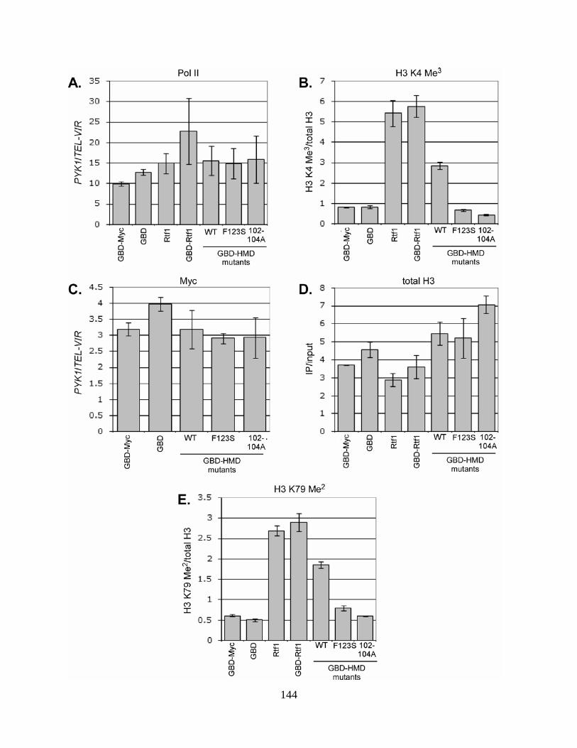
144

145
Figure 28. The Rtf1 HMD does not require targeting to DNA to induce Rtf1-dependent histone
modifications.
ChIP analyses of the effects of GBD-HMD fusions at the 5’ end of PYK1. Antibodies specific for the largest
subunit of Pol II (8WG16) (A), H3 K4 Me3 (B), the Myc epitope (C), total H3 (D), and H3 K79 Me2 (E) were used
to immunoprecipitate these factors and associated DNA from a formaldehyde-treated rtf1∆ gal4∆ strain (MHY119)
that had been transformed with 2µ plasmids expressing the indicated GBD or Rtf1 derivatives. Association of the
indicated proteins or levels of histone modifications are assessed by PCR. Results in (A) and (C) are shown as a
ratio of the signal observed at the 5’ end of PYK1 to the signal derived from a nontranscribed region in the right
telomere of chromosome VI (TEL-VIR). Values in (B), (D), and (E) are not normalized to TEL-VIR. PYK1 signal
derived from the H3 K4 Me3 and H3 K79 samples (B and E) was normalized to total H3 levels at the same position.
The mean of three independent experiments with standard error is shown.

146
Surprisingly, GBD-HMD induces H3 K4 trimethylation to the same extent at the 5’ end
of PYK1 as was observed at the GAL7 UAS (Figure 28B). I observed a similar induction of H3
K79 dimethylation at this locus in strains expressing untagged Rtf1, GBD-Rtf1, and GBD-HMD
(Figure 28E). The increase in H3 K4 trimethylation and H3 K79 dimethylation observed at the
5’ end of PYK1 in strains expressing GBD-HMD was not seen in strains expressing GBD fusions
to the 102-104A or F123S HMDs. rtf1 102-104A did not affect bulk H3 K79 dimethylation
levels in whole cell extracts and the effect on this modification observed in this analysis is likely
a consequence of the low expression level of the GBD-102-104A-HMD fusion protein.
To determine if the GBD-HMD fusions could affect Rtf1-dependent histone
modifications on a global scale, I performed immunoblot analysis with antibodies specific for H3
K4 trimethylation and H3 K79 dimethylation on whole cell extracts from an rtf1∆ gal4∆ strain
expressing GBD alone, GBD-Myc, full-length Rtf1, or the three GBD-HMD fusions.
Interestingly, I observed nearly wild type levels of H3 K79 dimethylation and a modest
restoration of H3 K4 trimethylation in the GBD-HMD strain, while these modifications were
absent from GBD-102-104A-HMD and GBD-F123S-HMD strains (Figure 29). Together, the
results of my PYK1 ChIP analysis and the immunoblot analysis described here support the idea
that the Rtf1 HMD possesses inherent activity and that it does not need to be specifically
recruited to DNA to promote Rtf1-dependent histone modifications.

147
Figure 29. The Rtf1 HMD can partially restore global Rtf1-dependent histone modifications.
Extracts were prepared from an rtf1Δ gal4∆ strain (MHY119) transformed with 2µ plasmids that express the
indicated GBD constructs or pLS20, a CEN/ARS plasmid that expresses untagged full-length Rtf1. Immunoblots
were probed with antibodies specific for H3 K4 Me3 and H3 K79 Me2 to assess the levels of these modifications in
the indicated extracts. A total H3 immunoblot was performed to demonstrate effects on these modifications are not
due to a significant change in histone H3 expression and an anti-G6PDH immunoblot was performed as a loading
control.

148
3.4 CONCLUSIONS
Rtf1 has been implicated in several transcription-related functions, but the mechanisms by which
it participates in these processes remain undefined. My previous analyses identified a region of
Rtf1, now termed the HMD, which is necessary for its role in the covalent modification of
histones that are present in the bodies of transcriptionally active genes. The experiments
described in this chapter were aimed at better characterizing the HMD to begin exploring the
mechanism by which Rtf1 promotes histone modification.
I created substitution mutations in several conserved residues in the HMD and assessed
the effects of these alterations on histone modification. Interestingly, I found that different
substitution mutations in the HMD resulted in unique effects on Rtf1-dependent histone
modifications. This result indicates that the Rtf1 HMD may promote histone H2B lysine 123
monoubiquitylation and histone H3 K4 trimethylation by unique mechanisms.
Because I had previously observed that partially deleting the HMD led to transcription-
related phenotypes (Figure 14), I explored the effects of the HMD substitution mutations on
these phenotypes. I found that all three mutations tested, rtf1 102-104A, rtf1 108-110A, and rtf1
F80V F123S, caused sensitivity to 6-AU. However, the HMD substitution mutations caused no
obvious Spt- phenotype.
A complete deletion of RTF1 also causes several phenotypes associated with defects in
chromatin structure. I demonstrated that these phenotypes, loss of telomeric silencing and
aberrant internal initiation from within the FLO8 open reading frame, are also caused by rtf1
102-104A, rtf1 108-110A, and rtf1 F80V F123S. Effects on telomeric silencing reflect a global

149
disruption of histone modifications patterns, while internal initiation from a cryptic promoter
within the FLO8 open reading frame is a read-out of an effect on chromatin structure at this
specific locus. These results therefore suggest that HMD substitution mutations lead to
chromatin defects on both global and local scales.
Interestingly, aberrant initiation from the FLO8 internal promoter and the Spt- phenotype
both reflect the ability of the transcription machinery to gain access to abnormal transcriptional
start sites. Rtf1-dependent histone modifications are found specifically within ORFs, where the
FLO8 internal promoter is located. The Spt- phenotype measures the ability to suppress
insertions at specific promoters, which are devoid of Rtf1-dependent histone modifications.
Because the Rtf1 HMD substitution mutations also display sensitivity to 6-AU, a phenotype
associated with transcription elongation defects, these results may suggest that mutations in the
Rtf1 HMD specifically affect chromatin structure within ORFs. Mutations in various elongation
factors, including an rtf1 null allele, are known to cause strong Spt- phenotypes. Therefore, it
remains likely that the Spt- phenotype is influenced by effects on transcription elongation.
However, the effect of Rtf1 on this phenotype appears to be mediated by a function other than its
role in histone modification.
Despite their differential effects on Rtf1-dependent histone modifications, HMD
substitution mutations always result in equivalent phenotypic effects. This may indicate that H3
K4 trimethylation is particularly important to prevent defects in transcription elongation and
chromatin structure, since this is the only modification absent in all HMD substitution mutants
tested in the phenotypic analysis. Interestingly, HMD substitution mutations sometimes result in
phenotypes that are different from those caused by an rtf1 null mutation or partial HMD
deletions, suggesting that mutating the HMD and removing it are not functionally equivalent.

150
Finally, I found that expressing only the residues encompassing the HMD as a fusion to
the Gal4 DNA binding domain (GBD-HMD) was sufficient to elicit a three-fold induction of H3
K4 trimethylation at the GAL7 UAS, where this fusion protein is recruited by Gal4 binding sites,
indicating that promoting histone modifications is an intrinsic property of the HMD.
Surprisingly, I found that expression of the GBD-HMD also induced H3 K4 trimethylation and
H3 K79 dimethylation at the 5’ end of PYK1, where GBD-HMD binding is not detected, and at
least partially restored both of these modifications in whole cell extracts. These observations
indicate that the HMD does not need to be specifically recruited to DNA to elicit histone
modifications. Overall, the analyses described in this chapter indicate that Rtf1’s role in histone
modification can be attributed to a number of highly conserved residues in the HMD and that
these residues differentially contribute to H2B ubiquitylation and H3 K4 trimethylation. In
addition to being necessary for Rtf1-dependent histone modifications, the HMD is also sufficient
for this function and may not require targeting to active genes to elicit this activity.

151
4.0 THE PAF1 COMPLEX IS CONNECTED TO CHROMATIN REMODELING
THROUGH GENETIC INTERACTIONS WITH THE INOSITOL POLYPHOSPHATE
SIGNALING CASCADE
4.1 INTRODUCTION
A screen for factors that became essential for viability when RTF1 was deleted identified
several transcription-related factors and suggested a role for Rtf1 in transcription (COSTA and
ARNDT 2000). The same screen also identified mutations in the genes that code for the first two
enzymes in the inositol polyphosphate (IP) signaling cascade: PLC1 and ARG82 (Figure 12)
(COSTA 2001). Plc1 catalyzes the conversion of phosphatidylinositol 4,5-bisphosphate to
diacylglycerol and inositol trisphosphate (IP3) (FLICK and THORNER 1993). Arg82 converts
nuclear IP3 to tetrakis- and pentakisphosphorylated derivatives (IP4 and IP5) (SAIARDI et al.
1999). Because several factors that were identified as essential in the absence of RTF1 were
ultimately linked to transcription elongation, the function of Plc1 and Arg82 may also be
connected to this process.
In addition to this possible connection to transcription elongation, disruption of the IP
signaling cascade has been implicated in several other nuclear processes including chromatin
remodeling and mRNA export. IP4 and IP5, the direct products of Arg82 activity, stimulate the
SWI/SNF chromatin remodeling complex to mobilize nucleosomes in vitro (SHEN et al. 2003).

152
A separate study reported that the chromatin structure of the PHO5 promoter was not efficiently
remodeled and the SWI/SNF and INO80 complexes were poorly recruited to the promoter of
PHO84 in the absence of Arg82 (STEGER et al. 2003). Additionally, mutations in PLC1 and
ARG82 were recovered in a screen for factors that become essential in combination with a
temperature-sensitive version of Gle1, an mRNA export factor that associates with the nuclear
pore complex (YORK et al. 1999). Further analysis demonstrated that mutations in PLC1 and
ARG82 result in a striking accumulation of polyadenylated transcripts in the nucleus, suggesting
that these mutations severely disrupt export of mRNA to the cytoplasm.
I investigated whether these known nuclear functions for the IP signaling cascade were
responsible for the synthetic lethality observed when PLC1 and ARG82 are mutated in an RTF1
deletion strain. Because a complete deletion of PLC1 causes a severe growth defect, I performed
these analyses using arg82∆ strains. I found that the absence of Arg82 strongly impaired the
growth of several strains lacking known elongation factors, including four of the five subunits of
the Paf1 complex. An arg82∆ paf1∆ strain was inviable; I determined that this lethality was not
the result of an enhanced defect in mRNA export, but instead was likely due to interactions
between Paf1, Arg82, and specific chromatin remodeling complexes. These results suggest that
IP signaling, transcription elongation factors, and ATP-dependent chromatin remodeling
cooperate to support normal cell growth.

153
4.2 MATERIALS AND METHODS
4.2.1 Genetic methods
S. cerevisiae strains used in these studies are listed in Table 3. All strains are GAL2+ derivatives
of S288C (WINSTON et al. 1995). Complete disruptions of RTF1, PAF1, CTR9, CDC73, LEO1,
SPT4, DST1, SET1, BRE1, ARG82, ISW1, ISW2, CHD1, INO80, ARP8, SNF2, and SWR1 were
created by a PCR-based gene replacement method (AUSUBEL et al. 1988). spt5-194 is a
temperature sensitive mutation in the essential SPT5 gene.
Mating, sporulation, and tetrad dissection were performed according to standard methods
(ROSE et al. 1990). Null mutations are marked by the incorporation of an auxotrophic marker in
place of the native gene (Table 3). Following sporulation and dissection, sorting of marked
deletions is assayed by replica printing to a series of media that lacks individual amino acids or
nucleic acids; the auxotrophic profile of each spore can be determined by this method. Synthetic
lethality of double mutants is inferred when spores containing marker genes for both knockouts
sorting in a genetic cross are consistently not recovered. If both knockouts in a cross are marked
by the same nutritional marker, the progeny of tetrads that sort 2:2 for growth on media lacking
the corresponding nutrient (nonparental ditypes) are analyzed; the two spores capable of growth
in the absence of this nutrient are surmised to contain both deletions. spt5-194 is not marked by
a nutritional marker; sorting of this mutation is followed by its known strong Spt- phenotype in a
strain containing his4-912δ. dst1∆ was followed by sensitivity to mycophenolic acid.

154
4.2.2 Analysis of mRNA export
mRNA export analyses were conducted as described in (MURPHY et al. 1996) except that oligo-
dT(30) prelabeled with digoxigenin (IDT) and Antidigoxigenin-rhodamine Fab fragments
(Boehringer Mannheim) were used and strains were grown at 30oC. KY660 (wild type), KY804
(paf1∆), MHY82 (ipk1∆ paf1∆), KY875 (ipk1∆), and KY743 (arg82∆) were used in these
analyses.
4.2.3 Northern analysis
KY292 (wild type), KY802 (paf1∆), KY930 (rtf1∆), MHY63 (arg82∆), KY508 (snf2∆), MHY88
(arp8∆), MHY57 (ino80∆), MHY75 (isw1∆), KY884 (isw2∆), KY632 (chd1∆), KY907 (set1∆),
and KY968 (bre1∆) were grown to approximately 1 x 10⁷ cells/ml in YPD. RNA isolation and
Northern analyses were performed as described previously (SWANSON et al. 1991). [α-
32P]dATP-labeled DNA hybridization probes were prepared by random prime labeling of VTC3,
SPL2, or SCR1 PCR products that had been amplified from yeast genomic DNA. SCR1 encodes
a cytoplasmic RNA that is transcribed by RNA Polymerase III (HANN and WALTER 1991) and is
used as a loading control.

155
Table 3. Saccharomyces cerevisiae strains used in Chapter 4
Strain Genotype
ECY57
KY292
MATα paf1∆::kanMX leu2∆0 ura3∆0
MATa his4-912δ lys2-128δ leu2Δ1 ura3-52 trp1Δ63
KY404 MATa rtf1∆::LEU2 his4-912δ lys2-128δ leu2Δ1 ura3-52 trp1Δ63
KY452 MATa rtf1∆::URA3 his3∆200 lys2-173R2 ura3-52 trp1Δ63
KY508 MATa snf2∆::HIS3 his3∆200 lys2-128δ leu2Δ1 ura3-52
KY528 MATα dst1∆::HISG his4-912δ lys2-128δ leu2Δ1 ura3-52
KY583 MATa chd1∆::HIS3 his3∆200 lys2-128δ leu2Δ1 ura3-52 trp1Δ63
KY632 MATα chd1∆::URA3 his3∆200 lys2-128δ leu2Δ1 ura3-52
KY678 MATα ctr9∆::kanMX4 his3∆200 lys2-128δ leu2Δ1 ura3-52
KY688 MATa cdc73∆::kanMX4 lys2∆0 ura3Δ0 trp1Δ63
KY715 MATa spt5-194 his3∆200 leu2Δ1 ura3-52
KY740 MATα arg82∆::URA3 leu2∆0 ura3Δ0 trp1Δ63 met15Δ0
KY741 MATa arg82∆::URA3 his3∆200 leu2∆0 ura3Δ0 trp1Δ63 met15Δ0
KY742 MATα arg82∆::URA3 leu2∆0 ura3Δ0 met15Δ0
KY743 MATa arg82∆::URA3 his3∆200 leu2∆0 ura3Δ0
KY744 MATα arg82∆::URA3 his3∆200 leu2∆0 ura3Δ0 met15Δ0
KY758 MATa spt4∆::HIS3 his3∆200 lys2-128δ leu2Δ1 ura3-52 trp1Δ63
KY802 MATa paf1∆::URA3 his3∆200 lys2-173R2 ura3(∆0 or -52)
KY804 MATα paf1∆::URA3 his3∆200 leu2Δ(0 or 1) ura3(∆0 or -52)
KY806 MATa leo1∆::URA3 his3∆200 lys2-173R2 ura3-52

156
KY875 MATa ipk1∆::HIS3 his3∆200 lys2-173R2 leu2∆1 ura3-52 trp1Δ63
KY879 MATα arp8∆::HIS3 his3∆200 lys2-173R2 leu2Δ1 ura3-52 trp1Δ63
KY884 MATa isw1∆::HIS3 his3∆200 lys2-173R2 leu2Δ1 ura3-52 trp1Δ63
KY885 MATα isw2∆::HIS3 his3∆200 lys2-128δ leu2Δ1 ura3-52 ade8
KY901 MATα isw1∆::HIS3 his3∆200 lys2-128δ leu2Δ1 ura3-52 trp1Δ63
KY907 MATa set1∆::HIS3 his3∆200 lys2-128δ leu2Δ1 ura3-52
KY930 MATα rad6∆::URA3 lys2-128δ ura3-52
KY968 MATa bre1∆::kanMX4 his3∆200 ura3-52
KY972 MATα swr1∆::kanMX4 his3∆200 lys2-128δ leu2Δ1 ura3-52
MHY8 MATa arp8∆::HIS3 his3∆200 lys2-128δ leu2Δ1 ura3-52 trp1Δ63 ade8
MHY57 MATa ino80∆::HIS3 his3∆200 his4-912δ lys2-173R2 leu2Δ1 ura3-52
MHY63 MATα arg82∆::URA3 his3∆200 leu2Δ(0 or 1) ura3(∆0 or -52)
MHY66 MATa paf1∆::URA3 his3∆200 leu2Δ( 0 or 1) ura3(∆0 or -52) trp1Δ63
MHY75 MATa isw1∆::HIS3 his3∆200 lys2-128δ leu2Δ1 ura3-52
MHY82 MATα ipk1∆::HIS3 paf1∆::URA3 his3∆200 ?lys2-173R2? leu2Δ( 0 or 1)
ura3(∆0 or -52) trp1Δ63
MHY86 MATa paf1∆::URA3 his3∆200 leu2Δ( 0 or 1) ura3(∆0 or -52) ade8
MHY88 MATa arp8∆::HIS3 his3∆200 lys2-128δ leu2Δ( 0 or 1) ura3(∆0 or -52)

157
4.3 RESULTS
4.3.1 arg82∆ genetically interacts with mutations in several known elongation factors
To determine whether the genetic interaction resulting from combination of mutations in
IP signaling enzymes and deletion of RTF1 was specific, I crossed an arg82 null strain by strains
harboring deletions in the genes encoding each subunit of the Paf1 complex: PAF1, CTR9,
RTF1, CDC73, and LEO1. Deletion of ARG82 results in synthetic lethality when combined with
paf1 or ctr9 null mutations (Figure 30A and B and Table 4) and combination of an ARG82
deletion with rtf1 or cdc73 null mutations resulted in considerable synthetic cell sickness (Figure
30C and D and Table 4). Although an arg82∆ leo1∆ strain displayed only a minor growth defect
(Figure 30E and Table 4), the strong genetic interactions I observe between an arg82 null
mutation and deletion of four of the five genes encoding Paf1 complex subunits suggest that the
connection between the IP signaling cascade and transcription elongation is not specifically
mediated through Rtf1.
The severity of the genetic interaction between rtf1 and arg82 null mutations differs from
the results of the original genetic screen where combination of an RTF1 deletion with a point
mutation in ARG82 (arg82-383) was lethal (COSTA 2001). This disparity may reflect differences
in the genetic background of the strains used in these analyses or may indicate that deletion of
ARG82 is not equivalent to arg82-383.

158

159
Figure 30. arg82∆ genetically interacts with deletions of the genes encoding Paf1 complex subunits.
Yeast strains of opposite mating type were mated, sporulated, and dissected on YPD. Strains used for each
individual cross are indicated in the legend to Table 4. Each vertical row contains the four spores derived from
meiosis of a single diploid yeast cell. Plates were incubated at 30oC for 4 days. Because the deletions of ARG82
and LEO1 are both marked by URA3, only nonparental ditypes tetrads are shown (E).

160
Table 4. Genetic interactions between arg82∆ and mutations in known elongation factors
Strain Relative growth rate
arg82∆ +++++
dst1∆ arg82∆ +++++
leo1∆ arg82∆ ++++
rtf1∆ arg82∆ ++
cdc73∆ arg82∆ ++
spt5-194 arg82∆ +
spt4∆ arg82∆ +
paf1∆ arg82∆ -
ctr9∆ arg82∆ -
The growth rate for an arg82∆ strain was arbitrarily set to “+++++”; an arg82∆ alone causes an intermediate growth
defect when compared to a wild type strain, although this effect is no longer noticeable on dissection plates after 2
days growth at 30oC. The growth rates of double mutants were determined by analyzing the progeny of the
following crosses: leo1∆ arg82∆ = KY806 X KY744; dst1∆ arg82∆ = KY741 X KY678; rtf1∆ arg82∆ = KY404 X
KY740; cdc73∆ arg82∆ = KY688 X KY740; spt5-194 arg82∆ = KY715 X KY742; spt4∆ arg82∆ = KY758 X
KY744; paf1∆ arg82∆ =KY742 X ECY57; ctr9∆ arg82∆ = KY741 X KY528.

161
I next investigated whether mutations in IP signaling enzymes interacted specifically with
mutations in the Paf1 complex or whether the growth effects caused by combination of mutations
in these factors might be indicative of a functional relationship between IP signaling and
transcription elongation. I crossed an arg82∆ strain by strains harboring mutations in SPT4 and
SPT5, which encode the subunits of the Spt4/Spt5 transcription elongation complex, or a deletion
of DST1, which encodes a transcription elongation factor (TFIIS) that reactivates stalled Pol II
by stimulating cleavage of nascent transcripts. I observed strong synthetic sickness in arg82∆
spt4∆ and arg82∆ spt5-194 strains, but observed no obvious genetic interaction between arg82∆
and dst1∆ (Figure 31 and Table 4). This result suggests that while the connection between IP
signaling and transcription elongation is not restricted specifically to mutation of the Paf1
complex, it is also not a general phenomenon that results from all disruptions of transcription
elongation factors.
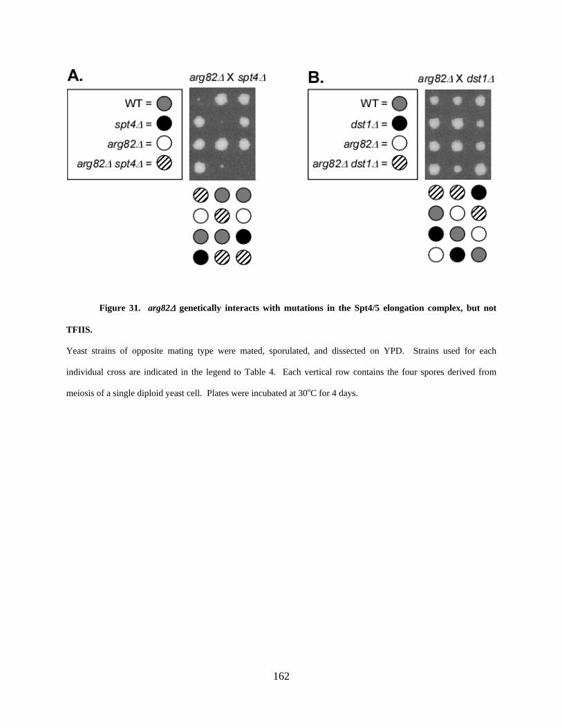
162
Figure 31. arg82∆ genetically interacts with mutations in the Spt4/5 elongation complex, but not
TFIIS.
Yeast strains of opposite mating type were mated, sporulated, and dissected on YPD. Strains used for each
individual cross are indicated in the legend to Table 4. Each vertical row contains the four spores derived from
meiosis of a single diploid yeast cell. Plates were incubated at 30oC for 4 days.

163
4.3.2 Enzymes that function downstream of Arg82 are not essential in the absence of Paf1
or Rtf1
The products of Arg82 kinase activity, IP4 and IP5, are further phosphorylated by two additional
kinases, Ipk1 and Kcs1. Ipk1 phosphorylates IP5 to IP6 (YORK et al. 1999) and Kcs1 converts
IP4 and IP6 to pyro-phosphorylated forms (SAIARDI et al. 1999). To determine if the Paf1
complex was required in all instances of disruptions in the IP signaling pathway, I created
deletions of IPK1 and KCS1 and crossed the null strains by paf1∆ and rtf1∆ strains. I observed
no major effects on growth of the resulting double mutant strains (data not shown). This result
indicates that the connection between transcription elongation and IP signaling is specifically
associated with Plc1 or Arg82 or the products of their enzymatic activity.
4.3.3 The synthetic lethality caused by the combined loss of ARG82 and PAF1 is not the
result of an enhanced defect in mRNA export.
Mutations in PLC1 and ARG82 are known to result in a failure to properly export polyadenylated
transcripts from the nucleus (YORK et al. 1999). To determine if the synthetic lethality observed
in an arg82∆ paf1∆ strain might be the result of a combined effect on mRNA export, I examined
the consequences of deleting PAF1 on this process. The efficiency of mRNA export was
assessed in fixed cells following in situ hybridization of a digoxigenin-labeled oligo-dT probe to
the polyadenylated tails of mRNA. Hybridized probe was then detected with a rhodamine-
labeled antidigoxigenin antibody and visualized on a fluorescence microscope. As expected, an
arg82∆ strain displayed a striking accumulation of polyadenylated transcripts in the nucleus

164
(Figure 32). However, paf1 null cells display diffuse cellular staining that is virtually
indistinguishable from that observed in wild type cells, indicating that mRNA export was largely
unaffected by this mutation (Figure 32). Deletion of RTF1 or CDC73 also does not result in a
noticeable effect on mRNA export (data not shown). These results suggest that the inviability of
an arg82∆ paf1∆ strain likely cannot be attributed to a combinatorial effect on mRNA export.

165
Figure 32. The inviability of an arg82∆ paf1∆ strain is not due to an enhanced defect in mRNA
export.
The efficiency of mRNA export was assessed in fixed cells by in situ hybridization of a digoxigenin-labeled oligo-
dT probe to polyadenylated (polyA) RNA followed by detection with a rhodamine-labeled antidigoxigenin antibody.
DAPI was used to visualize the location of nuclei.

166
However, deletion of Paf1 complex subunits is known to result in a decrease in the
average length of polyadenylated tails on RNA transcripts (MUELLER et al. 2004). This suggests
that the absence of a strong nuclear signal in the mRNA export analysis of a paf1∆ strain may
not reflect wild type levels of mRNA export, but instead be a simple consequence of the inability
of the oligo-dT probe to hybridize as efficiently to abnormally short polyadenylated tails. To
address this possibility, I performed mRNA export analysis on an ipk1∆ paf1∆ double mutant
strain. Mutation of IPK1 is known to cause a defect in mRNA export, similar to the effect
observed in arg82 mutant strains (YORK et al. 1999). A prominent signal is observed in the
nuclei of ipk1∆ paf1∆ cells (Figure 32), suggesting that the reduction in polyadenylated tail
length caused by deletion of PAF1 does not affect the efficiency of this assay.
4.3.4 paf1∆ and arg82∆ genetically interact with mutations in the INO80, SWI/SNF, and
SWR1 chromatin remodeling complexes
Inositol polyphosphates can modulate the activity of several ATP-dependent chromatin
remodeling complexes in S. cerevisiae, including the INO80 and SWI/SNF complexes (SHEN et
al. 2003). Additionally, the INO80 and SWI/SNF complexes are not efficiently recruited to the
PHO84 promoter and nucleosomes at the PHO5 promoter are not efficiently remodeled in
arg82Δ cells (STEGER et al. 2003). To investigate whether the inviability of an arg82∆ paf1∆
strain is the result of an enhanced impact on chromatin remodeling, paf1Δ cells were crossed by
disruptions of all known nonessential chromatin remodeling complexes in S. cerevisiae: INO80,
ISW1, ISW2, CHD1, SWI/SNF, and SWR1. Due to the requirement of the RSC complex for

167
viability, genetic interaction between deletion of PAF1 and mutations in this complex were not
analyzed.
The results of these crosses demonstrated that a paf1 null mutation was synthetically
lethal with deletions of SWR1, the catalytic subunit of the SWR1 complex, and ARP8, a
component of the INO80 complex that is not found in other known chromatin remodeling
complexes (Figure 33A and B and Table 5). An ino80 null mutation causes severe sickness on
its own (data not shown) and, therefore, was not used in my genetic analyses. Additionally, a
deletion of PAF1 resulted in severe sickness when combined with deletions of ISW1 or SNF2,
the catalytic subunits of the ISW1 and SWI/SNF chromatin remodeling complexes, respectively
(Table 5). No obvious genetic interaction was observed in paf1∆ chd1∆ strains or paf1∆ isw2∆
strains; CHD1 and ISW2 encode the catalytic subunits of chromatin remodeling complexes by
the same names (Table 5).

168
Figure 33. paf1∆ genetically interacts with disruptions in chromatin remodeling complexes.
Yeast strains of opposite mating type were mated, sporulated, and dissected on YPD. Strains used for each cross are
indicated in the legend to Table 5. Each vertical row contains the four spores derived from meiosis of a single
diploid yeast cell. Plates were incubated at 30oC for 4 (A) or 3 (B) days.

169
Table 5. paf1∆ or arg82∆ genetically interact with disruptions of chromatin remodeling
complexes
Chromatin remodeling
complex
Subunit deleted
Growth defect when crossed
by paf1∆
Growth defect when crossed
by arg82∆
CHD1 chd1∆ none none
ISW1 isw1∆ severe none
ISW2 isw2∆ none none
SWI/SNF snf2∆ severe lethal
INO80 arp8∆ lethal lethal
SWR1 swr1∆ lethal intermediate
paf1∆ and arg82∆ both cause growth defects on their own when compared to a wild type cell; the growth categories
shown are relative to the normal growth of a paf1∆ or arg82∆ strain. The growth rates of double mutants were
determined by analyzing the progeny of the following crosses: chd1∆ paf1∆ = KY583 X KY804; chd1∆ arg82∆ =
KY583 X KY744; isw1∆ paf1∆ = MHY86 X KY901; isw1∆ arg82∆ = KY741 X KY901; isw2∆ paf1∆ = KY884 X
KY804; isw2∆ arg82∆ = KY741 X KY885; snf2∆ paf1∆ = KY508 X KY804; arp8∆ paf1∆ = MHY8 X KY804;
arp8∆ arg82∆ = KY741 X KY879; swr1∆ paf1∆ = MHY66 X KY972; swr1∆ arg82∆ = KY452 X KY972. snf2∆
arg82∆ double mutants were previously reported to be inviable (SHEN et al. 2003).

170
Deletion of ARG82 is known to cause synthetic lethality in a cell lacking SNF2 (SHEN et
al. 2003). To more extensively analyze the connection between Arg82 and chromatin
remodeling complexes, I introduced an arg82 null mutation into strains harboring mutations in
integral subunits of the other nonessential chromatin remodeling complexes. I found that arg82∆
arp8∆ double mutants were inviable (Figure 34A and Table 5). Loss of ARG82 also resulted in
synthetic sickness when combined with a deletion of SWR1 (Figure 34B), while no discernible
genetic interaction was obvious in crosses between an arg82∆ strain and strains lacking CHD1,
ISW1, or ISW2 (Table 5). Additionally, I performed genetic analyses which revealed that
deletion of ARG82 caused no growth defect in strains lacking SET1, which encodes the H3 K4
methyltransferase, or BRE1, the gene for the ubiquitin ligase required for H2B K123
ubiquitylation (data not shown). This suggests that Arg82 is not required for normal cell growth
when chromatin structure is impacted by the loss of histone modifications, but instead appears to
be specifically connected to chromatin remodeling.

171
Figure 34. arg82∆ genetically interacts with disruptions in chromatin remodeling complexes.
Yeast strains of opposite mating type were mated, sporulated, and dissected on YPD. Strains used for each cross are
indicated in the legend to Table 5. Each vertical row contains the four spores derived from meiosis of a single
diploid yeast cell. Plates were incubated at 30oC for 3 days.

172
Together, the results detailed in this section demonstrate that, when combined with
disruptions of chromatin remodeling complexes, deletion of ARG82 caused a similar pattern to
that observed when PAF1 is absent. Three of the four chromatin remodeling complexes
(SWI/SNF, INO80, and SWR1) that were necessary for normal cell growth in the absence of
PAF1 were also required when ARG82 was deleted. These genetic results imply that IP
signaling and transcription elongation may work in parallel with a common set of chromatin
remodeling complexes.
4.3.5 Expression of several genes is affected by loss of Paf1, Arg82, or mutations in the
INO80 and SWI/SNF chromatin remodeling complexes
To further investigate the connection between IP signaling, transcription elongation, and ATP-
dependent chromatin remodeling, I investigated whether factors involved in these processes were
required for expression of common targets. Genes whose expression was known to be
compromised in strains containing disruptions of chromatin remodelers were chosen as
candidates. Expression of one such candidate gene, SPL2 (OHDATE et al. 2003), is virtually
eliminated in cells containing disruptions of the INO80 or SWI/SNF complexes or deletion of
PAF1, and its expression is reduced in arg82Δ and rtf1∆ strains (Figure 35A). Transcription of a
second candidate gene, VTC3, was essentially absent from snf2∆ and ino80∆ strains and was
severely compromised by deletion of PAF1, RTF1, ARG82, and ARP8 (Figure 35B). Chromatin
remodelers that caused no growth defects when disrupted in both paf1∆ strains and arg82∆
strains (ISW1, ISW2, CHD1), or deletion of genes encoding histone modifying enzymes (SET1
or BRE1), did not impact expression of these genes. These similar effects on expression of at

173
least two common genes further imply a specific relationship between the Paf1 complex, IP
signaling, and the INO80 and SWI/SNF chromatin remodeling complexes.

174
Figure 35. The Paf1 complex, Arg82, and the INO80 and SWI/SNF chromatin remodeling complexes
are necessary for normal expression of SPL2 and VTC3.
Expression of SPL2 (A) and VTC3 (B) was analyzed by Northern analysis. 10 µg of total RNA from strains
harboring the indicated deletions was analyzed with probes specific for SPL2 or VTC3 and, after stripping, SCR1
levels were assessed on the same filter as a loading control.

175
Surprisingly, deletion of SWR1, which was synthetically lethal in the absence of Paf1 or
Arg82, does not impact expression of VTC3 or SPL2. This may reflect the known function of the
SWR1 complex as primarily required to facilitate the incorporation of the Htz1 histone variant in
gene promoters whereas INO80 and SWI/SNF are primarily believed to reposition existing
nucleosomes (MIZUGUCHI et al. 2004; SHEN et al. 2003). The genes examined in this analysis,
VTC3 and SPL2, may require nucleosome repositioning for expression, but not Htz1
incorporation. If I extended this analysis to explore effects on genes that are known to require
Htz1 deposition for normal expression, I would likely uncover common target genes that are
affected by swr1∆, paf1∆, and arg82∆.
4.4 CONCLUSIONS
Previous work in the Arndt lab identified a synthetic lethal interaction between mutation of a
transcription elongation factor, Rtf1, and two enzymes that function in the inositol polyphosphate
signaling cascade, Plc1 and Arg82. While many connections between signaling pathways and
transcription have been previously described, the effect is often manifested at the level of
transcription initiation. The discovery of an interaction between the IP signaling cascade and the
Paf1 transcription elongation complex implies that signaling pathways may also exert control
during the elongation stage of transcription.
Because deletion of PLC1 results in a severe growth defect, my studies were focused
primarily on investigating the contribution of ARG82 to this synthetic lethality. Arg82 is known
to have at least three nuclear functions: transcriptional control of arginine-responsive genes as a
subunit of the ArgR/Mcm1 transcriptional regulatory complex; involvement in the proper export

176
of mRNA from the nucleus, and effects on the activity of several chromatin remodeling
complexes. I ruled out effects on Arg82’s role as a transcriptional regulator as the likely cause of
the synthetic lethality with rtf1∆ by demonstrating that deletion of ARG80, which encodes an
additional subunit of the ArgR/Mcm1 complex, caused no growth defect in an rtf1∆ strain (data
not shown). Furthermore, I demonstrated that deletion of multiple subunits of the Paf1 complex
(PAF1, CDC73, or RTF1) did not affect mRNA export. This result indicates that the synthetic
lethality caused by simultaneous mutation of RTF1 and ARG82 is not the result of a combined
effect on mRNA export.
My genetic results suggest that the IP signaling cascade is connected to transcription
elongation through its known interactions with several ATP-dependent chromatin remodeling
complexes. Synthetic lethality is thought to result when two or more parallel processes that elicit
a common essential function are simultaneously disrupted. Synthetic lethal interactions or strong
effects on cell growth resulted from combinatorial deletions of factors involved in IP signaling
(Arg82), transcription elongation (Paf1), and chromatin remodeling (Arp8, Snf2, and Swr1)
(Figure 36). This result may imply that all three of these processes function in parallel; however,
my genetic results demonstrate that a cell cannot tolerate the simultaneous loss of any two of
these functions.

177
Figure 36. Genetic interactions between Paf1, Arg82, and chromatin remodeling complexes.
A schematic is shown representing genetic interactions that were observed between the indicated factors; the
thickness of the line indicates the degree of interaction that was observed with the thickest lines signifying lethality;
a paf1 null allele showed a strong genetic interaction in a strain with an ISW1 deletion, but no interaction was
observed between arg82∆ and isw1∆.

178
Additionally, individual disruption of the Paf1 complex, Arg82, or the INO80 and
SWI/SNF chromatin remodeling complexes has a marked effect on at least two genes that are
known to require proper chromatin remodeling for normal expression. This observation, in
combination with my genetic data, may instead suggest that Arg82 and Paf1 both contribute to
the normal function of the INO80, SWI/SNF, and SWR1 chromatin remodeling complexes.
When both IP signaling and the Paf1 complex are concurrently disrupted, this may lead to
widespread effects on chromatin remodeling that are too severe to support transcription at a level
necessary to support cell viability. While the INO80, SWI/SNF, and SWR1 complexes are not
essential when disrupted individually, deletion of the catalytic subunits of the INO80 or
SWI/SNF complexes cause substantial growth defects. It is, therefore, likely that simultaneously
inactivating or dampening the activity of all three of these chromatin remodeling complexes
would have severe consequences for normal cell function.

179
5.0 DISCUSSION AND FUTURE DIRECTIONS
5.1 RTF1 IS COMPOSED OF FUNCTIONALLY DISTINCT REGIONS
A large number of accessory factors participate in mRNA synthesis by facilitating the
recruitment and modification of Pol II, the cotranscriptional processing of the nascent transcript,
the progression of Pol II along ORFs, and transcription termination. Accessory proteins that
carry out distinct cotranscriptional events, such as transcript cleavage, can be recruited to
discrete segments of a gene (BENTLEY 2005). In contrast, the Paf1 complex associates along the
entirety of active ORFs (KIM et al. 2004a; MUELLER et al. 2004; SIMIC et al. 2003), suggesting
that it functions throughout transcript elongation. My results indicate that the Paf1 complex
component Rtf1 utilizes several independent mechanisms to orchestrate alterations to the
chromatin template during gene expression. Using a collection of sequential rtf1 deletion
mutations, I demonstrated that discrete nonoverlapping segments of the protein are required for
recruitment of Chd1 to active genes, histone modification, association with active ORFs, and
physical interaction with the Paf1 complex; these regions of the protein have been given
designations that reflect their functionalities: CID (Chd1-interaction domain), HMD (histone
modification domain), OAR (ORF-association region), and PID (Paf1 complex interaction
domain).

180
I observed that distinct transcriptional effects resulted from disruption of individual
functions of Rtf1 (Figure 21). Combined with the observation that a complete deletion of RTF1
causes more severe phenotypes than mutations that eliminate only one of its activities, these data
suggest that the functions of Rtf1 are not entirely interdependent. Additionally, I observed that
the most severe transcription-related phenotypes were caused by deletions in the HMD or OAR,
which correspond to the most highly conserved regions of the protein; this observation suggests
that promoting cotranscriptional histone modification is a critical means by which Rtf1 directs
transcription.
Deletion of sequences required for either histone methylation or ORF association causes
similar 6-AU sensitivity, but disruption of the association of Rtf1 with ORFs causes a more
severe Spt- phenotype. This observation suggests that association of Rtf1 with active genes is
required for a function in addition to H2B K123 ubiquitylation or H3 K4 and K79 methylation.
Another indication that the 6-AU and Spt- phenotypes measure, at least partly, distinct functions
of Rtf1 comes from the observation that Rtf1Δ5 causes a moderate Spt- phenotype but no
sensitivity to 6-AU (Figure 14). Despite the Spt- phenotype resulting from deletion of this
region, none of Rtf1’s known functions are compromised in the absence of region 5; this may
indicate that Rtf1 participates in an additional transcription-related function that has not been
identified.
The OAR is the most highly conserved region of Rtf1; it contains 18 of the 26 residues
that are invariant across S. cerevisiae, S. pombe, H. sapiens, and C. elegans (Figure 15).
Recently, the structure of the corresponding region in human Rtf1 has been solved by NMR (DE
JONG et al. 2008). Interestingly, the structure of a subdomain within the human OAR shows
structural similarity to a number of nucleic acid binding proteins, including the PAZ domains of

181
Dicer and Argonaut (proteins involved in RNA interference), the Tudor domains of Sm and
SMN proteins (proteins involved in assembly and function of the spliceosome), and bacterial
transcription elongation factors including NusG. This observation led the authors to test whether
Rtf1 possessed DNA binding activity; they found that the human Rtf1 OAR could bind single
stranded DNA in vitro, but that it did not interact with double stranded DNA or RNA under
similar conditions. The authors, therefore, proposed that the OAR may have a role in stabilizing
the transcription “bubble” where the template and nontemplate strands of DNA have separated
during active transcription.
Due to the conservation observed between the human and S. cerevisiae OAR domains, it
will be beneficial to our understanding of Rtf1’s function to determine whether the S. cerevisiae
OAR also interacts with single stranded DNA. It is also possible that the OAR may be capable
of interacting with double stranded DNA or RNA in the context of the full length protein, or in
vivo. Rtf1 is known to physically interact with the Spt4/Spt5 transcription elongation complex,
which mediates interaction of Rtf1 with Pol II. In light of my observation that the OAR is
necessary for association of Rtf1 with active genes, it will be interesting to determine whether
this region of the protein is also necessary for its physical interaction with the Spt4/Spt5 complex
and/or Pol II and if these interactions are disturbed following treatment with DNase.
Alternatively, the structure of the human Rtf1 OAR also resembled the structures of several
proteins that contained Tudor domains, which have been proposed to possess histone methyl-
lysine binding properties. No interaction was observed between the human OAR domain and
un- or di-methylated H3 K4 (DE JONG et al. 2008), but it remains possible that the OAR of S.
cerevisiae may bind mono- or tri- methylated H3 K4 or that it may interact with methylated H3
K36 or K79.

182
Previous analyses in the Arndt lab demonstrated that physical interaction with Chd1 is
disrupted by deleting amino acids 3-30 of Rtf1 (region 1; CID) or by substituting phenylalanine
for leucine at position 11 (Rtf1-1) (COSTA 2001; WARNER et al. 2007). I demonstrated that the
rtf1∆1 mutation reduces association of Chd1 at the 5’ ORFs of PYK1 and TEF2 (Figure 16),
implying that the physical interaction between these two factors contributes to the normal
recruitment of Chd1 to active genes. However, removing the CID from Rtf1 does not decrease
Chd1 recruitment to the same extent as a complete RTF1 deletion. This observation suggests
that another function of Rtf1 may also contribute to Chd1 association with active genes. A likely
candidate for this additional function is the role of Rtf1 in promoting histone modifications. S.
cerevisiae Chd1 has been demonstrated to bind dimethylated H3 K4 through the second of the
two chromodomains present near its N-terminus (PRAY-GRANT et al. 2005). Because H3 K4
Me2 is an Rtf1-dependent histone modification that marks active ORFs, the HMD of Rtf1 may
contribute to association of Chd1 with active genes by promoting the placement of this
modification. However, conflicting reports exist regarding the validity of the physical
interaction between S. cerevisiae Chd1 and H3 K4 Me2. An additional study failed to detect
interaction of the S. cerevisiae Chd1 chromodomains with H3 K4 Me2 and demonstrated that S.
cerevisiae lacks several key functional residues that mediate the known interaction between
human CHD1 and an H3 K4 Me2 peptide (OKUDA et al. 2007). Therefore, it may be useful to
test the effect of all Rtf1 internal deletion mutants on the interaction of Chd1 with active genes.
Although I observed no transcription-related defects when the CID was deleted in an
otherwise wild-type strain, the Arndt lab has previously shown that rtf1-1 or deletion of RTF1 or
CHD1 suppresses the Spt- phenotype of the spt15-122 mutation (STOLINSKI et al. 1997). I report
here that this effect is also caused by rtf1Δ1. The similar effects of mutations in RTF1 and

183
CHD1 in an spt15-122 strain suggest that these factors elicit similar effects on chromatin
structure. The interaction between Rtf1 and Chd1 may, therefore, be important for proper
chromatin function particularly when the cell is sensitized to small changes in nucleosome
positioning or stability, such as in a TBP mutant strain.
Additionally, I observed that the carboxy-terminus of Rtf1, defined by region 13 (PID), is
necessary and sufficient for the interaction between Rtf1 and other Paf1 complex subunits. It is
surprising that Rtf1∆13 has apparently wild-type stability, because previous studies revealed a
ten- to twenty- fold reduction in Rtf1 levels in crude extracts from paf1∆ and ctr9∆ strains
(MUELLER et al. 2004; PORTER et al. 2005). My results indicate that a stable physical interaction
with the Paf1 complex is not required for Rtf1 stability or that residues within Rtf1 that confer
instability are deleted from the Rtf1∆13 mutant protein. Consistent with a prior study, I
demonstrated that interaction with Rtf1 mediates recruitment of Paf1 and Ctr9 to active ORFs
(MUELLER et al. 2004).
Remarkably, disruption of the physical interaction between Rtf1 and the Paf1 complex
causes only a mild Spt- phenotype and the known biochemical functions of Rtf1 remain largely
intact. This observation suggests that Rtf1 retains function, independent of stable association
with the Paf1 complex. In agreement with this idea, Rtf1 does not copurify with the human Paf1
complex (ROZENBLATT-ROSEN et al. 2005; YART et al. 2005; ZHU et al. 2005) and the carboxy-
terminus is poorly conserved (Figure 15). Human Rtf1 may only interact with other Paf1
complex subunits while associated with chromatin or the human Paf1 complex may execute
some functions independently of ORF association, as previously suggested from studies in yeast
(MUELLER et al. 2004). Alternatively, Cdc73, which also promotes association of Paf1 and Ctr9
with active genes in yeast (MUELLER et al. 2004), may be the sole Paf1 complex subunit required

184
for this function in humans. The observation that only mild transcription-related phenotypes
result when the CID or PID is removed from Rtf1 may also indicate that Rtf1 performs an
additional unidentified role in the cell that is not transcription-related; the interaction between
Rtf1 and Chd1 and/or other subunits of the Paf1 complex may ultimately be found to be
important in this unidentified process.
The goal of analyzing the Rtf1 internal deletion mutants was to gain insight into the
mechanism by which this protein functions. Through these analyses, I have demonstrated that
the known roles of Rtf1 are mediated by distinct functional regions. At least 3 of these
functional segments, the CID, OAR, and the PID, appear to be necessary to mediate protein-
protein (or potentially protein-DNA) interactions. The Paf1 complex is believed to function as a
platform for the recruitment of accessory factors during transcription elongation, and my data
provide additional support for this idea in regard to the Rtf1 subunit of this complex. The
exchange of interacting factors, allosteric changes in Rtf1’s structure, or posttranslational
modifications may control the association of Rtf1 with its binding partners. Alternatively, a
catalytic role for Rtf1 itself cannot be completely ruled out. Structural analyses on full-length
Rtf1 may reveal similarities to known enzymatic domains.
5.2 THE HISTONE MODIFICATION DOMAIN OF RTF1 IS NECESSARY AND
SUFFICIENT TO PROMOTE RTF1-DEPENDENT HISTONE MODIFICATIONS
My analyses of the Rtf1 internal deletion series also revealed that a 90 amino acid segment
spanning residues 63 through 152 (HMD) was necessary to promote Rtf1-dependent
monoubiquitylation of histone H2B at K123 and methylation of histone H3 at K4 and K79. The

185
dependence of both H2B K123 Ub and its downstream histone methylation marks on a relatively
small region of Rtf1 implied that the primary role of Rtf1 in histone modification may be to
promote H2B K123 Ub. However, I created substitution mutations in the Rtf1 HMD and
observed unique effects on Rtf1-dependent histone modifications; rtf1 108-110A and rtf1 F80V
F123S eliminated all Rtf1-dependent histone modifications tested (H2B K123 Ub, di- and
trimethylation of H3 K4 and dimethylation of H3 K79), while rtf1 102-104A affected only H3
K4 Me3. These results suggested that Rtf1 may promote distinct Rtf1-dependent histone
modifications by separate mechanisms.
However, it remains possible that the primary role of the HMD is to specifically promote
H2B K123 Ub. In contrast to eliminating H2B K123 Ub, the rtf1 102-104A mutation may
instead alter the genome-wide pattern of this modification. The progression of H3 K4 Me2 to H3
K4 Me3 may be particularly sensitive to proper patterns of H2B K123 Ub, and this effect may
account for the specific loss of this modification in an rtf1 102-104A strain. ChIP analyses to
explore the distribution of H2B K123 Ub within active genes will be useful in exploring this
possibility. It is also possible that Rtf1 controls the deubiquitylation of H2B K123 Ub;
deubiquitylation of H2B K123 has been demonstrated to be necessary for H3 K4 Me (WYCE et
al. 2004). Examining association of the H2B deubiquitylating enzymes Ubp8 and Ubp10 with
active genes in strains expressing Rtf1 HMD substitution mutations may reveal a role for Rtf1 in
the recruitment of these enzymes.
Alternatively, factors have previously been identified that are necessary for the
progression of H3 K4 Me2 to H3 K4 Me3; these factors include Not4, a subunit of the Ccr4-Not
mRNA production and processing complex, and the Cps35, Cps40 and Cps60 subunits of the
COMPASS complex (which contains the Set1 H3 K4 methyltransferase) (LEE et al. 2007;

186
MULDER et al. 2007; SCHNEIDER et al. 2005). Determining whether rtf1 102-104A impairs the
recruitment or activity of these factors may provide insight into the specific effect of this
mutation on H3 K4 Me3. It has been reported previously that deletion of RTF1 causes a more
severe effect on H3 K4 Me1 than is observed in the absence of Rad6 (the ubiquitin conjugating
enzyme necessary for H2B K123 Ub) (DEHE et al. 2005). This observation supports the idea that
Rtf1 may have effects on the activity of the Set1 methyltransferase that are independent of its
role in H2B K123 Ub.
The effectiveness of transcriptional silencing at the telomeres is tightly linked to global
patterns of histone modifications. Deletion of RTF1 is known to disrupt this process (KROGAN et
al. 2003; NG et al. 2003b) and I have demonstrated that partial deletion of the HMD or
substitution mutations in this region that affect Rtf1-dependent histone modifications also
compromise telomeric silencing. I also observed loss of telomeric silencing when regions of the
Rtf1 OAR were deleted, even though these mutations led to only a minor decrease in H3 K4 Me3
while other Rtf1-dependent H3 methylation marks remained at wild type levels. While these
results may indicate that telomeric silencing is dramatically affected by slight perturbations in
global histone modification patterns, it is more likely a reflection of the sensitivity of the TEL-
VR::URA3 reporter. To more accurately measure transcriptional activity at the telomeres,
Northern blots could be performed to assess levels of URA3 expression from the telomeric
silencing reporter in strains harboring Rtf1 HMD mutations.
Additionally, I tested the effects of Rtf1 HMD mutations on three other transcription- or
chromatin-related phenotypes: sensitivity to the base analog 6-AU, the Spt- phenotype, and
internal initiation from the FLO8 locus. All HMD mutations tested (rtf1∆3, rtf1∆4, rtf1 102-
104A, rtf1 108-110A, and rtf1 F80V F123S) caused similar sensitivity to 6-AU, indicating that

187
these mutations lead to a defect in transcription elongation. Partial deletions of the HMD (rtf1∆3
and rtf1∆4) resulted in a very weak Spt- phenotype, but no effect on this phenotype is observed
in strains expressing Rtf1 HMD substititution mutations. Conversely, the Rtf1 HMD substitution
mutations allowed internal initiation from a cryptic promoter in the FLO8 locus at a level
equivalent to that observed for a complete deletion of RTF1, while partial deletions of the HMD
led to a weaker effect on this phenotype.
The FLO8 internal initiation phenotype and the Spt- phenotype have both been proposed
to indicate that chromatin structure has been disrupted, allowing access to aberrant
transcriptional start sites (KAPLAN et al. 2003). The FLO8 internal promoter lies within the
ORF, where Rtf1-dependent histone modifications are known to localize. Conversely, the Spt-
phenotype typically results from utilizing alternate transcriptional start sites within the promoter,
which is devoid of most Rtf1-dependent histone modifications. Mutations in the Rtf1 HMD
allow internal initiation from FLO8, but cause a very mild or no Spt- phenotype; these
observations may suggest that the HMD contributes mainly to the function of chromatin within
active ORFs.
Another interesting observation made from these phenotypic studies is that partial
deletions and substitution mutations in the HMD are not always functionally equivalent. I also
observed that several HMD substitution mutations led to a decrease in the amount of total H3
that could be extracted from these strains; this effect is not caused by partial deletions of the
HMD. These observations may indicate that Rtf1 substitution mutations are gain of function
mutations that alter histone-DNA contacts. Consistent with the idea that the Paf1 complex may
affect nucleosome stability on active genes, absence of Paf1 complex subunits results in an
increase in levels of histone H3 present in the 5’ ORFs of several genes (DEHE et al. 2005;

188
MARTON and DESIDERIO 2008). This increase in total histone H3 levels correlates with a
decrease in levels of Pol II associated with the GAL1 locus following induction and leads to
decreased GAL1 mRNA levels at 90 minutes post-induction (MARTON and DESIDERIO 2008;
XIAO et al. 2005). ChIP analyses to determine if rtf1 108-110A and rtf1 F80V F123S, which
both decreased the amount of total H3 present in whole cell extracts prepared by glass bead lysis,
increase the level of total H3 present in the GAL1 ORF upon gene induction will be useful in
determining if Rtf1’s role in histone modification contributes to nucleosome dynamics during
transcription activation. It will also be interesting to determine if the Rtf1 CID contributes to
histone eviction during the activation of gene transcription.
Similar studies to explore the effects of Rtf1 substitution mutations on H2A and H2B
levels present in active ORFs following gene induction should also be performed. H2A/H2B
dimers are more readily evicted from nucleosomes during transcription and may, therefore, be
more strongly affected than histone H3 in the proposed ChIP analyses. Interestingly, the yFACT
histone chaperone complex that contributes to H2A/H2B dynamics during transcription
physically interacts with the Paf1 complex (SQUAZZO et al. 2002). It will be interesting to
determine if yFACT requires the Rtf1 HMD for recruitment to recently induced genes or whether
the HMD and yFACT physically interact.
I demonstrated that expressing the Rtf1 HMD as a fusion to the Gal4 DNA binding
domain (HMD-GBD) in an rtf1∆ strain could restore global levels of H3 K79 Me2 to those
present in a wild type strain; expression of this construct also restored a modest level of global
H3 K4 Me3 in an rtf1∆ background and induced a 3-fold increase in this modification at the
GAL7 UAS under repressing conditions. These observations demonstrated that promoting Rtf1-
dependent histone methylation is an intrinsic property of the HMD. Future experiments to

189
explore whether the HMD is also sufficient to promote H2B K123 Ub will further clarify the
function of this region of Rtf1.
The ultimate goal of this experiment was to determine if the HMD could induce histone
modifications in the absence of the transcription machinery. Although the GAL7 locus should be
silent under the conditions used in these experiments, it is possible that a low level of
transcription is occurring under these conditions. Integration of Gal4 binding sites into an
intergenic region and the inactive PRM1 gene is currently underway. ChIP analysis to determine
if Rtf1-dependent histone modifications are induced at these regions by expression of HMD-
GBD will provide more conclusive data.
I also observed that expression of the HMD-GBD induced H3 K4 Me3 and H3 K79 Me2
in the 5’ end of the PYK1 ORF, a locus at which Gal4 binding sites have not been detected. This
raises the possibility that Rtf1 may not need to be directly targeted to DNA to promote histone
modification. Rtf1 may perhaps possess the ability to modify histones that are not incorporated
into DNA.
To investigate this possibility, several experimental adjustments and additional lines of
experimentation must be pursued. The HMD-GBD constructs used in these analyses were
overexpressed from 2µ vectors. Because the HMD is fused to a DNA binding domain,
overexpression of this fusion protein may allow nonspecific interaction with DNA on a genome-
wide basis. This may lead to a diffuse, transient association of the HMD-GBD fusion throughout
the genome at levels below the limits of detection of the ChIP assay. To address this issue, ChIP
assays to determine if Rtf1-dependent histone modifications can be restored in an rtf1∆
background should be repeated utilizing constructs that express the HMD-GBD fusions from low
copy vectors or inducible promoters to achieve lower expression levels of the fusion protein.

190
Additionally, the HMD alone can be expressed to determine if the absence of a DNA-
binding domain affects the ability of this domain to induce Rtf1-dependent histone
modifications. Preliminary NMR structural analyses indicate that the HMD is largely structured
(collaboration between the laboratories of Dr. Andy VanDemark and Dr. Angela Gronenborn;
personal communication); therefore, the HMD alone should be stably expressed in vivo. In the
event that expression of the HMD alone cannot be achieved, it can be fused to a protein tag that
does not possess DNA-binding activity, such as GFP. If Rtf1-dependent histone modifications
are still induced in strains expressing HMD alone or HMD-GFP from low copy vectors, this
would provide more convincing evidence that Rtf1 does not need to be associated with DNA to
promote histone modification.
An alternative possibility is that the HMD itself possesses DNA binding activity; this
activity may no longer be regulated when the HMD is overexpressed and is not present in the
context of the full-length protein. This possibility could be tested by subjecting the HMD to the
DNA-binding analyses that were performed to demonstrate that the human Rtf1 OAR possessed
single stranded DNA binding activity (DE JONG et al. 2008). If DNA-binding activity is
identified for the HMD, this may indicate that Rtf1 participates in histone modification by
directly contacting DNA; this interaction may increase the accessibility of histone tails to histone
modifying enzymes.
Although the HMD may interact with DNA, the possibility that it mediates protein-
protein interactions should also be explored. Rtf1 is known to be necessary for the normal
recruitment of the Set1-containing COMPASS complex and Rad6 to active genes (KROGAN et al.
2003; NG et al. 2003c; XIAO et al. 2005). It is also possible that the HMD physically interacts
with the histones, histone deubiquitylating enzymes, or histone demethylases. Directed studies

191
to determine whether the HMD interacts directly with these factors, or with the yFACT histone
chaperone complex, should provide useful information. The HMD may also interact with an
intermediate factor that leads to the recruitment of these histone modifying enzymes. Several
unbiased protein purification studies are currently underway to determine if HMD-interacting
factors can be identified. The possibility that the HMD functions enzymatically also cannot be
conclusively ruled out. Completion of the HMD structural analyses may reveal similarity to a
known enzymatic fold, or other recognizable protein domain, and should significantly aid the
investigation into understanding the mechanism by which Rtf1 directs histone modification at
active genes.
5.3 GENETIC INTERACTIONS IMPLY CONNECTIONS BETWEEN THE
INOSITOL POLYPHOSPHATE SIGNALING PATHWAY, TRANCRIPTION
ELONGATION, AND CHROMATIN REMODELING
A screen for factors that become essential in the absence of Rtf1 uncovered mutations in several
proteins that function during transcription elongation (COSTA and ARNDT 2000); this screen
provided the first link between Rtf1 and transcription elongation. Interestingly, the same screen
also identified mutations in Plc1 and Arg82, the first two enzymes in the inositol polyphosphate
signaling pathway. The genetic interaction between mutations in these enzymes and Rtf1
suggested that IP signaling may also be connected to transcription elongation. To further
investigate this phenomenon, I performed a series of genetic crosses to assess whether the
genetic interaction between mutations in ARG82 and RTF1 was specific. I found that deletion of
ARG82 resulted in synthetic lethality or strong growth defects when combined with individual

192
deletions of four of the five members of the Paf1 complex; synthetic lethality resulted from
combining a deletion of ARG82 with a deletion of PAF1 or CTR9. I also observed genetic
interactions between deletion of ARG82 and mutations in the Spt4/Spt5 complex. Together,
these results further implied a connection between IP signaling and transcription elongation.
It is known that the Spt4/Spt5 complex is required for recruitment of the Paf1 complex to
active genes (QIU et al. 2006), which may indicate that the link between IP signaling and
transcription elongation may be mediated specifically through the Paf1 complex. It may be
useful to extend this genetic analysis to determine if deletion of ARG82 causes growth defects
when combined with mutations in other known elongation factors, such as CTK1, FCP1, and the
subunits of the yFACT complex, that have not been shown to affect the recruitment of the Paf1
complex to active genes. Additionally, the results of crosses between deletion of ARG82 and
mutations in the Bur1/Bur2 transcription elongation complex, which is known to affect
association of the Paf1 complex with active ORFs, will provide further information concerning
the connections between IP signaling and transcription elongation.
Arg82 is known to have at least three nuclear functions; it participates in the control of
arginine-responsive genes as a subunit of the ArgR/Mcm1 transcriptional regulatory complex
(BECHET et al. 1970), affects the activity and recruitment of several ATP-dependent chromatin
remodeling complexes (SHEN et al. 2003; STEGER et al. 2003), and is necessary for the export of
mRNA from the nucleus (YORK et al. 1999). My analyses demonstrated that the role of Arg82
as a transcriptional regulator or its function in mRNA export are likely not responsible for the
synthetic lethality observed when ARG82 and PAF1 are simultaneously deleted. Arg82’s role as
a transcriptional regulator is independent of its kinase function (DUBOIS et al. 2000). To more
conclusively demonstrate that the kinase function of Arg82 is specifically required in the absence

193
of PAF1, the effect of combining a deletion of PAF1 with a mutation in the Arg82 kinase domain
should be evaluated.
Additionally, it will be interesting to determine which function of the Paf1 complex is
necessary in the absence of Arg82. This could be assessed by combining a deletion of ARG82,
or the Arg82 kinase-dead mutant, with the Rtf1 internal deletion mutants. The results of this
analysis would determine which of Rtf1’s functions is necessary for normal cell growth when
ARG82 is disrupted. Paf1 and Ctr9 are known to have roles in trimethylation of histone H3 K36
and phosphorylation of elongating Pol II on Ser2 of CTD repeats (CHU et al. 2007; MUELLER et
al. 2004). Genetic analyses to determine if the loss of Set2 (the H3 K36 methyltransferase) or
Ctk1 (the CTD Ser2 kinase) cause growth defects in the absence of functional Arg82 kinase
activity will also be informative.
The IP signaling pathway was linked to the activity of several chromatin remodeling
complexes, including the INO80 and SWI/SNF complexes (SHEN et al. 2003; STEGER et al.
2003). I, therefore, expanded my genetic analyses to determine whether mutations in Arg82 or
the Paf1 complex genetically interacted with disruptions of nonessential chromatin remodeling
complexes. Strong synthetic genetic interactions are observed between deletion of PAF1 or
ARG82 and disruption of the INO80, SWI/SNF, or SWR1 chromatin remodeling complexes.
Disruption of these factors, with the exception of the SWR1 complex, was also found to impact
expression of SPL2 and VTC3. Interestingly, the IP signaling pathway, the Paf1 complex, and
the SWI/SNF complex have all been implicated in the cellular stress response (FLICK and
THORNER 1993; PORTER et al. 2002; SHIVASWAMY and IYER 2008). The connection between
these 3 pathways may indicate that the cellular stress response is tightly controlled during all
stages of transcription.

194
Several mechanisms could account for the synthetic lethality observed between factors
involved in IP signaling, transcription elongation, and chromatin remodeling. All three
processes may function in parallel for normal cell growth; this scenario is unlikely due to the
pairwise synthetic lethality observed between disruptions in any two of these processes.
Alternatively, the Paf1 complex and IP signaling may both contribute to the normal function of
chromatin remodeling enzymes; when both transcription elongation and IP signaling are
disrupted, chromatin remodeling is ineffective and results in cell inviability. A third possibility
is that the IP signaling pathway affects chromatin remodeling and these two processes work in
parallel to the effect of the Paf1 complex on transcription elongation; simultaneous disruption of
transcription elongation and chromatin remodeling could result in severe effects on gene
expression, which would have a dramatic effect on cell growth.
ChIP analyses will be useful in further investigating the connections between IP
signaling, chromatin remodeling, and transcription elongation. It will be interesting to determine
whether chromatin remodeling complexes are recruited to the ORFs of active genes; this may
indicate that chromatin remodeling complexes directly affect transcription elongation. Candidate
genes for these analyses will include VTC3 and SPL2; expression of these genes was affected by
deletion of ARG82, PAF1, or components of the INO80 and SWI/SNF chromatin remodeling
complex. Defects in expression of INO1 or recruitment of the INO80 and SWI/SNF to the
promoters of several phosphate-responsive genes were identified in arg82∆ strains (STEGER et
al. 2003); therefore, these genes would also represent ideal candidates for the proposed ChIP
analyses. Additionally, it may be interesting to analyze the association of chromatin remodeling
complexes with the bodies of heat-shock responsive genes. BRG1, the ATPase component of
the human SWI/SNF complex, has been demonstrated to associate with both the promoter and

195
ORF of the hsp70 gene following activation (COREY et al. 2003). If the INO80 and/or
SWI/SNF complexes are found to associate with the bodies of genes in these analyses, the
dependence of this association on the Paf1 complex should then be assessed; the results of these
experiments should indicate whether the Paf1 complex has a direct effect on chromatin
remodeling during transcription elongation.
The Paf1 complex has been demonstrated to be necessary for the eviction of histones
from the GAL1 locus upon gene activation (MARTON and DESIDERIO 2008). It would be
interesting to determine whether mutation of Arg82 or the INO80 and SWI/SNF complexes
result in a similar defect. The Paf1 complex has several known connections to histone
modification. A combined effect on histone modification and chromatin remodeling could result
in the inability to sufficiently modify chromatin structure to allow gene expression to take place
at levels necessary to support cell viability. In agreement with this idea, synthetic lethal
interactions have been reported between mutations in the SAGA histone acetyltransferase
complex and the SWI/SNF chromatin remodeling complex (ROBERTS and WINSTON 1997).

196
BIBLIOGRAPHY
AGGER, K., J. CHRISTENSEN, P. A. CLOOS and K. HELIN, 2008 The emerging functions of histone demethylases. Curr Opin Genet Dev 18: 159-168.
AHN, S. H., M. KIM and S. BURATOWSKI, 2004 Phosphorylation of serine 2 within the RNA
polymerase II C-terminal domain couples transcription and 3' end processing. Mol Cell 13: 67-76.
AKANUMA, T., S. KOSHIDA, A. KAWAMURA, Y. KISHIMOTO and S. TAKADA, 2007 Paf1 complex
homologues are required for Notch-regulated transcription during somite segmentation. EMBO Rep 8: 858-863.
ALCAZAR-ROMAN, A. R., E. J. TRAN, S. GUO and S. R. WENTE, 2006 Inositol hexakisphosphate
and Gle1 activate the DEAD-box protein Dbp5 for nuclear mRNA export. Nat Cell Biol 8: 711-716.
ALCAZAR-ROMAN, A. R., and S. R. WENTE, 2008 Inositol polyphosphates: a new frontier for
regulating gene expression. Chromosoma 117: 1-13. ALLARD, S., R. T. UTLEY, J. SAVARD, A. CLARKE, P. GRANT et al., 1999 NuA4, an essential
transcription adaptor/histone H4 acetyltransferase complex containing Esa1p and the ATM-related cofactor Tra1p. Embo J 18: 5108-5119.
ALLISON, L. A., M. MOYLE, M. SHALES and C. J. INGLES, 1985 Extensive homology among the
largest subunits of eukaryotic and prokaryotic RNA polymerases. Cell 42: 599-610. ARMSTRONG, J. A., 2007 Negotiating the nucleosome: factors that allow RNA polymerase II to
elongate through chromatin. Biochem Cell Biol 85: 426-434. ARNDT, K. M., S. L. RICUPERO, D. M. EISENMANN and F. WINSTON, 1992 Biochemical and
genetic characterization of a yeast TFIID mutant that alters transcription in vivo and DNA binding in vitro. Mol Cell Biol 12: 2372-2382.
AUSUBEL, F. M., R. BRENT, R. E. KINGSTON, D. D. MOORE, J. G. SEIDMAN et al., 1988 Current
Protocols in Molecular Biology. Greene Publishing Associates and Wiley-Interscience, New York.

197
BASEHOAR, A. D., S. J. ZANTON and B. F. PUGH, 2004 Identification and distinct regulation of yeast TATA box-containing genes. Cell 116: 699-709.
BECHET, J., M. GREENSON and J. M. WIAME, 1970 Mutations affecting the repressibility of
arginine biosynthetic enzymes in Saccharomyces cerevisiae. Eur J Biochem 12: 31-39. BELOTSERKOVSKAYA, R., S. OH, V. A. BONDARENKO, G. ORPHANIDES, V. M. STUDITSKY et al.,
2003 FACT facilitates transcription-dependent nucleosome alteration. Science 301: 1090-1093.
BENTLEY, D. L., 2005 Rules of engagement: co-transcriptional recruitment of pre-mRNA
processing factors. Curr Opin Cell Biol 17: 251-256. BERNSTEIN, B. E., E. L. HUMPHREY, R. L. ERLICH, R. SCHNEIDER, P. BOUMAN et al., 2002
Methylation of histone H3 Lys 4 in coding regions of active genes. Proc Natl Acad Sci U S A 99: 8695-8700.
BETZ, J. L., M. CHANG, T. M. WASHBURN, S. E. PORTER, C. L. MUELLER et al., 2002 Phenotypic
analysis of Paf1/RNA polymerase II complex mutations reveals connections to cell cycle regulation, protein synthesis, and lipid and nucleic acid metabolism. Mol Genet Genomics 268: 272-285.
BLOCH, J. C., F. PERRIN and F. LACROUTE, 1978 Yeast temperature-sensitive mutants specifically
impaired in processing of poly(A)-containing RNAs. Mol Gen Genet 165: 123-127. BRAUN, M. A., P. J. COSTA, E. M. CRISUCCI and K. M. ARNDT, 2007 Identification of Rkr1, a
nuclear RING domain protein with functional connections to chromatin modification in Saccharomyces cerevisiae. Mol Cell Biol 27: 2800-2811.
BRIGGS, S. D., M. BRYK, B. D. STRAHL, W. L. CHEUNG, J. K. DAVIE et al., 2001 Histone H3
lysine 4 methylation is mediated by Set1 and required for cell growth and rDNA silencing in Saccharomyces cerevisiae. Genes Dev 15: 3286-3295.
BRIGGS, S. D., T. XIAO, Z. W. SUN, J. A. CALDWELL, J. SHABANOWITZ et al., 2002 Gene
silencing: trans-histone regulatory pathway in chromatin. Nature 418: 498. BUCHER, P., 1990 Weight matrix descriptions of four eukaryotic RNA polymerase II promoter
elements derived from 502 unrelated promoter sequences. J Mol Biol 212: 563-578. BURATOWSKI, S., 2000 Snapshots of RNA polymerase II transcription initiation. Curr Opin Cell
Biol 12: 320-325. BURATOWSKI, S., 2005 Connections between mRNA 3' end processing and transcription
termination. Curr Opin Cell Biol 17: 257-261.

198
BURATOWSKI, S., S. HAHN, L. GUARENTE and P. A. SHARP, 1989 Five intermediate complexes in transcription initiation by RNA polymerase II. Cell 56: 549-561.
CAIRNS, B. R., Y. J. KIM, M. H. SAYRE, B. C. LAURENT and R. D. KORNBERG, 1994 A
multisubunit complex containing the SWI1/ADR6, SWI2/SNF2, SWI3, SNF5, and SNF6 gene products isolated from yeast. Proc Natl Acad Sci U S A 91: 1950-1954.
CAIRNS, B. R., A. SCHLICHTER, H. ERDJUMENT-BROMAGE, P. TEMPST, R. D. KORNBERG et al.,
1999 Two functionally distinct forms of the RSC nucleosome-remodeling complex, containing essential AT hook, BAH, and bromodomains. Mol Cell 4: 715-723.
CAREY, M., B. LI and J. L. WORKMAN, 2006 RSC exploits histone acetylation to abrogate the
nucleosomal block to RNA polymerase II elongation. Mol Cell 24: 481-487. CARROZZA, M. J., B. LI, L. FLORENS, T. SUGANUMA, S. K. SWANSON et al., 2005 Histone H3
methylation by Set2 directs deacetylation of coding regions by Rpd3S to suppress spurious intragenic transcription. Cell 123: 581-592.
CHANDY, M., J. L. GUTIERREZ, P. PROCHASSON and J. L. WORKMAN, 2006 SWI/SNF displaces
SAGA-acetylated nucleosomes. Eukaryot Cell 5: 1738-1747. CHANG, M., D. FRENCH-CORNAY, H. Y. FAN, H. KLEIN, C. L. DENIS et al., 1999 A complex
containing RNA polymerase II, Paf1p, Cdc73p, Hpr1p, and Ccr4p plays a role in protein kinase C signaling. Mol Cell Biol 19: 1056-1067.
CHAUDHARY, K., S. DEB, N. MONIAUX, M. P. PONNUSAMY and S. K. BATRA, 2007 Human RNA
polymerase II-associated factor complex: dysregulation in cancer. Oncogene 26: 7499-7507.
CHU, Y., R. SIMIC, M. H. WARNER, K. M. ARNDT and G. PRELICH, 2007 Regulation of histone
modification and cryptic transcription by the Bur1 and Paf1 complexes. EMBO J 26: 4646-4656.
CLAPIER, C. R., K. P. NIGHTINGALE and P. B. BECKER, 2002 A critical epitope for substrate
recognition by the nucleosome remodeling ATPase ISWI. Nucleic Acids Res 30: 649-655.
CONRAD, M. N., J. H. WRIGHT, A. J. WOLF and V. A. ZAKIAN, 1990 RAP1 protein interacts with
yeast telomeres in vivo: overproduction alters telomere structure and decreases chromosome stability. Cell 63: 739-750.
COREY, L. L., C. S. WEIRICH, I. J. BENJAMIN and R. E. KINGSTON, 2003 Localized recruitment of
a chromatin-remodeling activity by an activator in vivo drives transcriptional elongation. Genes Dev 17: 1392-1401.

199
COSTA, P. J., 2001 Characterization of the Saccharomyces cerevisiae Transcription Factor Rtf1. University of Pittsburgh Biological Sciences Ph.D. Thesis. University of Pittsburgh; Pittsburgh, PA.
COSTA, P. J., and K. M. ARNDT, 2000 Synthetic lethal interactions suggest a role for the
Saccharomyces cerevisiae Rtf1 protein in transcription elongation. Genetics 156: 535-547.
COTE, J., J. QUINN, J. L. WORKMAN and C. L. PETERSON, 1994 Stimulation of GAL4 derivative
binding to nucleosomal DNA by the yeast SWI/SNF complex. Science 265: 53-60. COUTURE, J. F., and R. C. TRIEVEL, 2006 Histone-modifying enzymes: encrypting an enigmatic
epigenetic code. Curr Opin Struct Biol 16: 753-760. CRAMER, P., 2004 RNA polymerase II structure: from core to functional complexes. Curr Opin
Genet Dev 14: 218-226. CRAMER, P., D. A. BUSHNELL, J. FU, A. L. GNATT, B. MAIER-DAVIS et al., 2000 Architecture of
RNA polymerase II and implications for the transcription mechanism. Science 288: 640-649.
CUI, Y., and C. L. DENIS, 2003 In vivo evidence that defects in the transcriptional elongation
factors RPB2, TFIIS, and SPT5 enhance upstream poly(A) site utilization. Mol Cell Biol 23: 7887-7901.
DANIEL, J. A., M. S. TOROK, Z. W. SUN, D. SCHIELTZ, C. D. ALLIS et al., 2004 Deubiquitination
of histone H2B by a yeast acetyltransferase complex regulates transcription. J Biol Chem 279: 1867-1871.
DE JONG, R. N., V. TRUFFAULT, T. DIERCKS, E. AB, M. A. DANIELS et al., 2008 Structure and
DNA binding of the human Rtf1 Plus3 domain. Structure 16: 149-159. DEHE, P. M., and V. GELI, 2006 The multiple faces of Set1. Biochem Cell Biol 84: 536-548. DEHE, P. M., M. PAMBLANCO, P. LUCIANO, R. LEBRUN, D. MOINIER et al., 2005 Histone H3
lysine 4 mono-methylation does not require ubiquitination of histone H2B. J Mol Biol 353: 477-484.
DOWNS, J. A., S. ALLARD, O. JOBIN-ROBITAILLE, A. JAVAHERI, A. AUGER et al., 2004 Binding of
chromatin-modifying activities to phosphorylated histone H2A at DNA damage sites. Mol Cell 16: 979-990.
DUBOIS, E., V. DEWASTE, C. ERNEUX and F. MESSENGUY, 2000 Inositol polyphosphate kinase
activity of Arg82/ArgRIII is not required for the regulation of the arginine metabolism in yeast. FEBS Lett 486: 300-304.

200
DVIR, A., 2002 Promoter escape by RNA polymerase II. Biochim Biophys Acta 1577: 208-223. EISEN, J. A., K. S. SWEDER and P. C. HANAWALT, 1995 Evolution of the SNF2 family of proteins:
subfamilies with distinct sequences and functions. Nucleic Acids Res 23: 2715-2723. EL ALAMI, M., F. MESSENGUY, B. SCHERENS and E. DUBOIS, 2003 Arg82p is a bifunctional
protein whose inositol polyphosphate kinase activity is essential for nitrogen and PHO gene expression but not for Mcm1p chaperoning in yeast. Mol Microbiol 49: 457-468.
EL BAKKOURY, M., E. DUBOIS and F. MESSENGUY, 2000 Recruitment of the yeast MADS-box
proteins, ArgRI and Mcm1 by the pleiotropic factor ArgRIII is required for their stability. Mol Microbiol 35: 15-31.
EMRE, N. C., K. INGVARSDOTTIR, A. WYCE, A. WOOD, N. J. KROGAN et al., 2005 Maintenance of
low histone ubiquitylation by Ubp10 correlates with telomere-proximal Sir2 association and gene silencing. Mol Cell 17: 585-594.
ERKINA, T. Y., and A. M. ERKINE, 2006 Displacement of histones at promoters of
Saccharomyces cerevisiae heat shock genes is differentially associated with histone H3 acetylation. Mol Cell Biol 26: 7587-7600.
EXINGER, F., and F. LACROUTE, 1992 6-Azauracil inhibition of GTP biosynthesis in
Saccharomyces cerevisiae. Curr Genet 22: 9-11. EZHKOVA, E., and W. P. TANSEY, 2004 Proteasomal ATPases link ubiquitylation of histone H2B
to methylation of histone H3. Mol Cell 13: 435-442. FANG, S. M., and Z. F. BURTON, 1996 RNA polymerase II-associated protein (RAP) 74 binds
transcription factor (TF) IIB and blocks TFIIB-RAP30 binding. J Biol Chem 271: 11703-11709.
FEATHERSTONE, M., 2002 Coactivators in transcription initiation: here are your orders. Curr Opin
Genet Dev 12: 149-155. FENG, Q., H. WANG, H. H. NG, H. ERDJUMENT-BROMAGE, P. TEMPST et al., 2002 Methylation of
H3-lysine 79 is mediated by a new family of HMTases without a SET domain. Curr Biol 12: 1052-1058.
FINGERMAN, I. M., H. C. LI and S. D. BRIGGS, 2007 A charge-based interaction between histone
H4 and Dot1 is required for H3K79 methylation and telomere silencing: identification of a new trans-histone pathway. Genes Dev 21: 2018-2029.
FLICK, J. S., and J. THORNER, 1993 Genetic and biochemical characterization of a
phosphatidylinositol-specific phospholipase C in Saccharomyces cerevisiae. Mol Cell Biol 13: 5861-5876.

201
FLORES, O., E. MALDONADO and D. REINBERG, 1989 Factors involved in specific transcription by mammalian RNA polymerase II. Factors IIE and IIF independently interact with RNA polymerase II. J Biol Chem 264: 8913-8921.
GANGARAJU, V. K., and B. BARTHOLOMEW, 2007 Mechanisms of ATP dependent chromatin
remodeling. Mutat Res 618: 3-17. GARDNER, R. G., Z. W. NELSON and D. E. GOTTSCHLING, 2005 Ubp10/Dot4p regulates the
persistence of ubiquitinated histone H2B: distinct roles in telomeric silencing and general chromatin. Mol Cell Biol 25: 6123-6139.
GENG, F., and W. P. TANSEY, 2008 Polyubiquitylation of histone H2B. Mol Biol Cell 19: 3616-
3624. GHAEMMAGHAMI, S., W. K. HUH, K. BOWER, R. W. HOWSON, A. BELLE et al., 2003 Global
analysis of protein expression in yeast. Nature 425: 737-741. GNATT, A., 2002 Elongation by RNA polymerase II: structure-function relationship. Biochim
Biophys Acta 1577: 175-190. GOFFEAU, A., B. G. BARRELL, H. BUSSEY, R. W. DAVIS, B. DUJON et al., 1996 Life with 6000
genes. Science 274: 546, 563-547. GOLDMARK, J. P., T. G. FAZZIO, P. W. ESTEP, G. M. CHURCH and T. TSUKIYAMA, 2000 The Isw2
chromatin remodeling complex represses early meiotic genes upon recruitment by Ume6p. Cell 103: 423-433.
GOTTSCHLING, D. E., O. M. APARICIO, B. L. BILLINGTON and V. A. ZAKIAN, 1990 Position effect
at S. cerevisiae telomeres: reversible repression of Pol II transcription. Cell 63: 751-762. GOVIND, C. K., F. ZHANG, H. QIU, K. HOFMEYER and A. G. HINNEBUSCH, 2007 Gcn5 promotes
acetylation, eviction, and methylation of nucleosomes in transcribed coding regions. Mol Cell 25: 31-42.
GRANT, P. A., L. DUGGAN, J. COTE, S. M. ROBERTS, J. E. BROWNELL et al., 1997 Yeast Gcn5
functions in two multisubunit complexes to acetylate nucleosomal histones: characterization of an Ada complex and the SAGA (Spt/Ada) complex. Genes Dev 11: 1640-1650.
GUZMAN, E., and J. T. LIS, 1999 Transcription factor TFIIH is required for promoter melting in
vivo. Mol Cell Biol 19: 5652-5658. HA, I., S. ROBERTS, E. MALDONADO, X. SUN, L. U. KIM et al., 1993 Multiple functional domains
of human transcription factor IIB: distinct interactions with two general transcription factors and RNA polymerase II. Genes Dev 7: 1021-1032.

202
HANN, B. C., and P. WALTER, 1991 The signal recognition particle in S. cerevisiae. Cell 67: 131-144.
HARTZOG, G. A., T. WADA, H. HANDA and F. WINSTON, 1998 Evidence that Spt4, Spt5, and Spt6
control transcription elongation by RNA polymerase II in Saccharomyces cerevisiae. Genes Dev 12: 357-369.
HECHT, A., T. LAROCHE, S. STRAHL-BOLSINGER, S. M. GASSER and M. GRUNSTEIN, 1995
Histone H3 and H4 N-termini interact with SIR3 and SIR4 proteins: a molecular model for the formation of heterochromatin in yeast. Cell 80: 583-592.
HENRY, K. W., A. WYCE, W. S. LO, L. J. DUGGAN, N. C. EMRE et al., 2003 Transcriptional
activation via sequential histone H2B ubiquitylation and deubiquitylation, mediated by SAGA-associated Ubp8. Genes Dev 17: 2648-2663.
HONG, L., G. P. SCHROTH, H. R. MATTHEWS, P. YAU and E. M. BRADBURY, 1993 Studies of the
DNA binding properties of histone H4 amino terminus. Thermal denaturation studies reveal that acetylation markedly reduces the binding constant of the H4 "tail" to DNA. J Biol Chem 268: 305-314.
HUANG, H., A. KAHANA, D. E. GOTTSCHLING, L. PRAKASH and S. W. LIEBMAN, 1997 The
ubiquitin-conjugating enzyme Rad6 (Ubc2) is required for silencing in Saccharomyces cerevisiae. Mol Cell Biol 17: 6693-6699.
HWANG, W. W., S. VENKATASUBRAHMANYAM, A. G. IANCULESCU, A. TONG, C. BOONE et al.,
2003 A conserved RING finger protein required for histone H2B monoubiquitination and cell size control. Mol Cell 11: 261-266.
IIDA, T., and H. ARAKI, 2004 Noncompetitive counteractions of DNA polymerase epsilon and
ISW2/yCHRAC for epigenetic inheritance of telomere position effect in Saccharomyces cerevisiae. Mol Cell Biol 24: 217-227.
ISHIMI, Y., Y. OHBA, H. YASUDA and M. YAMADA, 1981 The interaction of H1 histone with
nucleosome core. J Biochem 89: 1881-1888. JEANMOUGIN, F., J. D. THOMPSON, M. GOUY, D. G. HIGGINS and T. J. GIBSON, 1998 Multiple
sequence alignment with Clustal X. Trends Biochem Sci 23: 403-405. JOHN, S., L. HOWE, S. T. TAFROV, P. A. GRANT, R. STERNGLANZ et al., 2000 The something
about silencing protein, Sas3, is the catalytic subunit of NuA3, a yTAF(II)30-containing HAT complex that interacts with the Spt16 subunit of the yeast CP (Cdc68/Pob3)-FACT complex. Genes Dev 14: 1196-1208.
JONES, J. C., H. P. PHATNANI, T. A. HAYSTEAD, J. A. MACDONALD, S. M. ALAM et al., 2004 C-
terminal repeat domain kinase I phosphorylates Ser2 and Ser5 of RNA polymerase II C-terminal domain repeats. J Biol Chem 279: 24957-24964.

203
JONES, R. S., and W. M. GELBART, 1993 The Drosophila Polycomb-group gene Enhancer of zeste contains a region with sequence similarity to trithorax. Mol Cell Biol 13: 6357-6366.
JONSSON, Z. O., S. K. DHAR, G. J. NARLIKAR, R. AUTY, N. WAGLE et al., 2001 Rvb1p and Rvb2p
are essential components of a chromatin remodeling complex that regulates transcription of over 5% of yeast genes. J Biol Chem 276: 16279-16288.
JORGENSEN, P., N. P. EDGINGTON, B. L. SCHNEIDER, I. RUPES, M. TYERS et al., 2007 The size of
the nucleus increases as yeast cells grow. Mol Biol Cell 18: 3523-3532. JOSHI, A. A., and K. STRUHL, 2005 Eaf3 Chromodomain Interaction with Methylated H3-K36
Links Histone Deacetylation to Pol II Elongation. Mol Cell 20: 971-978. KAHANA, A., and D. E. GOTTSCHLING, 1999 DOT4 links silencing and cell growth in
Saccharomyces cerevisiae. Mol Cell Biol 19: 6608-6620. KAPLAN, C. D., M. J. HOLLAND and F. WINSTON, 2005 Interaction between transcription
elongation factors and mRNA 3'-end formation at the Saccharomyces cerevisiae GAL10-GAL7 locus. J Biol Chem 280: 913-922.
KAPLAN, C. D., L. LAPRADE and F. WINSTON, 2003 Transcription elongation factors repress
transcription initiation from cryptic sites. Science 301: 1096-1099. KETTENBERGER, H., K. J. ARMACHE and P. CRAMER, 2003 Architecture of the RNA polymerase
II-TFIIS complex and implications for mRNA cleavage. Cell 114: 347-357. KETTENBERGER, H., K. J. ARMACHE and P. CRAMER, 2004 Complete RNA polymerase II
elongation complex structure and its interactions with NTP and TFIIS. Mol Cell 16: 955-965.
KIM, M., S. H. AHN, N. J. KROGAN, J. F. GREENBLATT and S. BURATOWSKI, 2004a Transitions in
RNA polymerase II elongation complexes at the 3' ends of genes. Embo J 23: 354-364. KIM, M., N. J. KROGAN, L. VASILJEVA, O. J. RANDO, E. NEDEA et al., 2004b The yeast Rat1
exonuclease promotes transcription termination by RNA polymerase II. Nature 432: 517-522.
KIM, T., and S. BURATOWSKI, 2007 Two Saccharomyces cerevisiae JmjC domain proteins
demethylate histone H3 Lys36 in transcribed regions to promote elongation. J Biol Chem 282: 20827-20835.
KIREEVA, M. L., B. HANCOCK, G. H. CREMONA, W. WALTER, V. M. STUDITSKY et al., 2005
Nature of the nucleosomal barrier to RNA polymerase II. Mol Cell 18: 97-108.

204
KIREEVA, M. L., W. WALTER, V. TCHERNAJENKO, V. BONDARENKO, M. KASHLEV et al., 2002 Nucleosome remodeling induced by RNA polymerase II: loss of the H2A/H2B dimer during transcription. Mol Cell 9: 541-552.
KIRMIZIS, A., H. SANTOS-ROSA, C. J. PENKETT, M. A. SINGER, M. VERMEULEN et al., 2007
Arginine methylation at histone H3R2 controls deposition of H3K4 trimethylation. Nature 449: 928-932.
KIZER, K. O., H. P. PHATNANI, Y. SHIBATA, H. HALL, A. L. GREENLEAF et al., 2005 A novel
domain in Set2 mediates RNA polymerase II interaction and couples histone H3 K36 methylation with transcript elongation. Mol Cell Biol 25: 3305-3316.
KLEFF, S., E. D. ANDRULIS, C. W. ANDERSON and R. STERNGLANZ, 1995 Identification of a gene
encoding a yeast histone H4 acetyltransferase. J Biol Chem 270: 24674-24677. KOBOR, M. S., S. VENKATASUBRAHMANYAM, M. D. MENEGHINI, J. W. GIN, J. L. JENNINGS et al.,
2004 A protein complex containing the conserved Swi2/Snf2-related ATPase Swr1p deposits histone variant H2A.Z into euchromatin. PLoS Biol 2: E131.
KOCH, C., P. WOLLMANN, M. DAHL and F. LOTTSPEICH, 1999 A role for Ctr9p and Paf1p in the
regulation G1 cyclin expression in yeast. Nucleic Acids Res 27: 2126-2134. KOHLER, A., and E. HURT, 2007 Exporting RNA from the nucleus to the cytoplasm. Nat Rev
Mol Cell Biol 8: 761-773. KOMARNITSKY, P., E. J. CHO and S. BURATOWSKI, 2000 Different phosphorylated forms of RNA
polymerase II and associated mRNA processing factors during transcription. Genes Dev 14: 2452-2460.
KORNBERG, R. D., 2005 Mediator and the mechanism of transcriptional activation. Trends
Biochem Sci 30: 235-239. KORNBERG, R. D., and J. O. THOMAS, 1974 Chromatin structure; oligomers of the histones.
Science 184: 865-868. KOUZARIDES, T., 2007 Chromatin modifications and their function. Cell 128: 693-705. KREBS, J. E., 2007 Moving marks: dynamic histone modifications in yeast. Mol Biosyst 3: 590-
597. KROGAN, N. J., J. DOVER, S. KHORRAMI, J. F. GREENBLATT, J. SCHNEIDER et al., 2002a
COMPASS, a histone H3 (Lysine 4) methyltransferase required for telomeric silencing of gene expression. J Biol Chem 277: 10753-10755.

205
KROGAN, N. J., J. DOVER, A. WOOD, J. SCHNEIDER, J. HEIDT et al., 2003 The Paf1 complex is required for histone H3 methylation by COMPASS and Dot1p: linking transcriptional elongation to histone methylation. Mol Cell 11: 721-729.
KROGAN, N. J., M. KIM, S. H. AHN, G. ZHONG, M. S. KOBOR et al., 2002b RNA polymerase II
elongation factors of Saccharomyces cerevisiae: a targeted proteomics approach. Mol. Cell. Biol. 22: 6979-6992.
KUNKEL, T. A., J. D. ROBERTS and R. A. ZAKOUR, 1987 Rapid and efficient site-specific
mutagenesis without phenotypic selection. Methods Enzymol 154: 367-382. KUTACH, A. K., and J. T. KADONAGA, 2000 The downstream promoter element DPE appears to
be as widely used as the TATA box in Drosophila core promoters. Mol Cell Biol 20: 4754-4764.
LANDRY, J., A. SUTTON, S. T. TAFROV, R. C. HELLER, J. STEBBINS et al., 2000 The silencing
protein SIR2 and its homologs are NAD-dependent protein deacetylases. Proc Natl Acad Sci U S A 97: 5807-5811.
LARIBEE, R. N., N. J. KROGAN, T. XIAO, Y. SHIBATA, T. R. HUGHES et al., 2005 BUR kinase
selectively regulates H3 K4 trimethylation and H2B ubiquitylation through recruitment of the PAF elongation complex. Curr Biol 15: 1487-1493.
LARIBEE, R. N., Y. SHIBATA, D. P. MERSMAN, S. R. COLLINS, P. KEMMEREN et al., 2007
CCR4/NOT complex associates with the proteasome and regulates histone methylation. Proc Natl Acad Sci U S A 104: 5836-5841.
LAURENT, B. C., M. A. TREITEL and M. CARLSON, 1991 Functional interdependence of the yeast
SNF2, SNF5, and SNF6 proteins in transcriptional activation. Proc Natl Acad Sci U S A 88: 2687-2691.
LAURENT, B. C., X. YANG and M. CARLSON, 1992 An essential Saccharomyces cerevisiae gene
homologous to SNF2 encodes a helicase-related protein in a new family. Mol Cell Biol 12: 1893-1902.
LEE, J. S., A. SHUKLA, J. SCHNEIDER, S. K. SWANSON, M. P. WASHBURN et al., 2007 Histone
crosstalk between H2B monoubiquitination and H3 methylation mediated by COMPASS. Cell 131: 1084-1096.
LEVCHENKO, V., B. JACKSON and V. JACKSON, 2005 Histone release during transcription:
displacement of the two H2A-H2B dimers in the nucleosome is dependent on different levels of transcription-induced positive stress. Biochemistry 44: 5357-5372.
LI, B., M. CAREY and J. L. WORKMAN, 2007a The role of chromatin during transcription. Cell
128: 707-719.

206
LI, B., M. GOGOL, M. CAREY, D. LEE, C. SEIDEL et al., 2007b Combined action of PHD and chromo domains directs the Rpd3S HDAC to transcribed chromatin. Science 316: 1050-1054.
LI, B., L. HOWE, S. ANDERSON, J. R. YATES, 3RD and J. L. WORKMAN, 2003 The Set2 histone
methyltransferase functions through the phosphorylated carboxyl-terminal domain of RNA polymerase II. J Biol Chem 278: 8897-8903.
LICATALOSI, D. D., G. GEIGER, M. MINET, S. SCHROEDER, K. CILLI et al., 2002 Functional
interaction of yeast pre-mRNA 3' end processing factors with RNA polymerase II. Mol Cell 9: 1101-1111.
LINDSTROM, D. L., and G. A. HARTZOG, 2001 Genetic interactions of Spt4-Spt5 and TFIIS with
the RNA polymerase II CTD and CTD modifying enzymes in Saccharomyces cerevisiae. Genetics 159: 487-497.
LINDSTROM, D. L., S. L. SQUAZZO, N. MUSTER, T. A. BURCKIN, K. C. WACHTER et al., 2003 Dual
roles for Spt5 in pre-mRNA processing and transcription elongation revealed by identification of Spt5-associated proteins. Mol Cell Biol 23: 1368-1378.
LOGAN, J., E. FALCK-PEDERSEN, J. E. DARNELL, JR. and T. SHENK, 1987 A poly(A) addition site
and a downstream termination region are required for efficient cessation of transcription by RNA polymerase II in the mouse beta maj-globin gene. Proc Natl Acad Sci U S A 84: 8306-8310.
LORCH, Y., and R. D. KORNBERG, 1985 A region flanking the GAL7 gene and a binding site for
GAL4 protein as upstream activating sequences in yeast. J Mol Biol 186: 821-824. LOWELL, J. E., D. Z. RUDNER and A. B. SACHS, 1992 3'-UTR-dependent deadenylation by the
yeast poly(A) nuclease. Genes Dev 6: 2088-2099. LUGER, K., A. W. MADER, R. K. RICHMOND, D. F. SARGENT and T. J. RICHMOND, 1997 Crystal
structure of the nucleosome core particle at 2.8 A resolution. Nature 389: 251-260. LUND, M. K., and C. GUTHRIE, 2005 The DEAD-box protein Dbp5p is required to dissociate
Mex67p from exported mRNPs at the nuclear rim. Mol Cell 20: 645-651. MARTINEZ, E., 2002 Multi-protein complexes in eukaryotic gene transcription. Plant Mol Biol
50: 925-947. MARTON, H. A., and S. DESIDERIO, 2008 The Paf1 complex promotes displacement of histones
upon rapid induction of transcription by RNA polymerase II. BMC Mol Biol. 9. MASON, P. B., and K. STRUHL, 2003 The FACT complex travels with elongating RNA
polymerase II and is important for the fidelity of transcriptional initiation in vivo. Mol Cell Biol 23: 8323-8333.

207
MAVRICH, T. N., I. P. IOSHIKHES, B. J. VENTERS, C. JIANG, L. P. TOMSHO et al., 2008 A barrier nucleosome model for statistical positioning of nucleosomes throughout the yeast genome. Genome Res 18: 1073-1083.
MCGINTY, R. K., J. KIM, C. CHATTERJEE, R. G. ROEDER and T. W. MUIR, 2008 Chemically
ubiquitylated histone H2B stimulates hDot1L-mediated intranucleosomal methylation. Nature 453: 812-816.
MESSENGUY, F., E. DUBOIS and C. BOONCHIRD, 1991 Determination of the DNA-binding
sequences of ARGR proteins to arginine anabolic and catabolic promoters. Mol Cell Biol 11: 2852-2863.
MICHELL, R. H., N. M. PERERA and S. K. DOVE, 2003 Morton Medal Lecture. New insights into
the roles of phosphoinositides and inositol polyphosphates in yeast. Biochem Soc Trans 31: 11-15.
MILLAR, C. B., and M. GRUNSTEIN, 2006 Genome-wide patterns of histone modifications in
yeast. Nat Rev Mol Cell Biol 7: 657-666. MILLER, T., N. J. KROGAN, J. DOVER, H. ERDJUMENT-BROMAGE, P. TEMPST et al., 2001
COMPASS: a complex of proteins associated with a trithorax-related SET domain protein. Proc Natl Acad Sci U S A 98: 12902-12907.
MINVIELLE-SEBASTIA, L., K. BEYER, A. M. KRECIC, R. E. HECTOR, M. S. SWANSON et al., 1998
Control of cleavage site selection during mRNA 3' end formation by a yeast hnRNP. Embo J 17: 7454-7468.
MIZUGUCHI, G., X. SHEN, J. LANDRY, W. H. WU, S. SEN et al., 2004 ATP-driven exchange of
histone H2AZ variant catalyzed by SWR1 chromatin remodeling complex. Science 303: 343-348.
MORETTI, P., K. FREEMAN, L. COODLY and D. SHORE, 1994 Evidence that a complex of SIR
proteins interacts with the silencer and telomere-binding protein RAP1. Genes Dev 8: 2257-2269.
MORILLON, A., N. KARABETSOU, J. O'SULLIVAN, N. KENT, N. PROUDFOOT et al., 2003 Isw1
chromatin remodeling ATPase coordinates transcription elongation and termination by RNA polymerase II. Cell 115: 425-435.
MORRISON, A. J., J. HIGHLAND, N. J. KROGAN, A. ARBEL-EDEN, J. F. GREENBLATT et al., 2004
INO80 and gamma-H2AX interaction links ATP-dependent chromatin remodeling to DNA damage repair. Cell 119: 767-775.
MUELLER, C. L., and J. A. JAEHNING, 2002 Ctr9, Rtf1, and Leo1 are components of the
Paf1/RNA polymerase II complex. Mol Cell Biol 22: 1971-1980.

208
MUELLER, C. L., S. E. PORTER, M. G. HOFFMAN and J. A. JAEHNING, 2004 The Paf1 complex has functions independent of actively transcribing RNA polymerase II. Mol Cell 14: 447-456.
MUELLER, J. E., M. CANZE and M. BRYK, 2006 The requirements for COMPASS and Paf1 in
transcriptional silencing and methylation of histone H3 in Saccharomyces cerevisiae. Genetics 173: 557-567.
MUHLRAD, D., R. HUNTER and R. PARKER, 1992 A rapid method for localized mutagenesis of
yeast genes. Yeast 8: 79-82. MULDER, K. W., A. B. BRENKMAN, A. INAGAKI, N. J. VAN DEN BROEK and H. T. TIMMERS, 2007
Regulation of histone H3K4 tri-methylation and PAF complex recruitment by the Ccr4-Not complex. Nucleic Acids Res 35: 2428-2439.
MURATANI, M., and W. P. TANSEY, 2003 How the ubiquitin-proteasome system controls
transcription. Nat Rev Mol Cell Biol 4: 192-201. MURPHY, R., J. L. WATKINS and S. R. WENTE, 1996 GLE2, a Saccharomyces cerevisiae
homologue of the Schizosaccharomyces pombe export factor RAE1, is required for nuclear pore complex structure and function. Mol Biol Cell 7: 1921-1937.
NAGY, P. L., J. GRIESENBECK, R. D. KORNBERG and M. L. CLEARY, 2002 A trithorax-group
complex purified from Saccharomyces cerevisiae is required for methylation of histone H3. Proc Natl Acad Sci U S A 99: 90-94.
NG, H. H., D. N. CICCONE, K. B. MORSHEAD, M. A. OETTINGER and K. STRUHL, 2003a Lysine-79
of histone H3 is hypomethylated at silenced loci in yeast and mammalian cells: a potential mechanism for position-effect variegation. Proc Natl Acad Sci U S A 100: 1820-1825.
NG, H. H., S. DOLE and K. STRUHL, 2003b The Rtf1 component of the Paf1 transcriptional
elongation complex is required for ubiquitination of histone H2B. J Biol Chem 278: 33625-33628.
NG, H. H., F. ROBERT, R. A. YOUNG and K. STRUHL, 2003c Targeted recruitment of Set1 histone
methyltransferase by elongating Pol II provides a localized mark and memory of recent transcriptional activity. Mol Cell 11: 709-719.
NG, H. H., R. M. XU, Y. ZHANG and K. STRUHL, 2002 Ubiquitination of histone H2B by Rad6 is
required for efficient Dot1-mediated methylation of histone H3 lysine 79. J Biol Chem 277: 34655-34657.
NICKEL, B. E., and J. R. DAVIE, 1989 Structure of polyubiquitinated histone H2A. Biochemistry
28: 964-968.

209
NOGALES, E., 2000 Recent structural insights into transcription preinitiation complexes. J Cell Sci 113 Pt 24: 4391-4397.
NORDICK, K., M. G. HOFFMAN, J. L. BETZ and J. A. JAEHNING, 2008 Direct interactions between
the Paf1 complex and a cleavage and polyadenylation factor are revealed by dissociation of Paf1 from RNA polymerase II. Eukaryot Cell 7: 1158-1167.
OHDATE, H., C. R. LIM, T. KOKUBO, K. MATSUBARA, Y. KIMATA et al., 2003 Impairment of the
DNA binding activity of the TATA-binding protein renders the transcriptional function of Rvb2p/Tih2p, the yeast RuvB-like protein, essential for cell growth. J Biol Chem 278: 14647-14656.
OKUDA, M., M. HORIKOSHI and Y. NISHIMURA, 2007 Structural polymorphism of
chromodomains in Chd1. J Mol Biol 365: 1047-1062. ORPHANIDES, G., T. LAGRANGE and D. REINBERG, 1996 The general transcription factors of
RNA polymerase II. Genes Dev 10: 2657-2683. ORPHANIDES, G., G. LEROY, C. H. CHANG, D. S. LUSE and D. REINBERG, 1998 FACT, a factor
that facilitates transcript elongation through nucleosomes. Cell 92: 105-116. ORPHANIDES, G., W. H. WU, W. S. LANE, M. HAMPSEY and D. REINBERG, 1999 The chromatin-
specific transcription elongation factor FACT comprises human SPT16 and SSRP1 proteins. Nature 400: 284-288.
OSADA, S., A. SUTTON, N. MUSTER, C. E. BROWN, J. R. YATES, 3RD et al., 2001 The yeast SAS
(something about silencing) protein complex contains a MYST-type putative acetyltransferase and functions with chromatin assembly factor ASF1. Genes Dev 15: 3155-3168.
OSLEY, M. A., 2006 Regulation of histone H2A and H2B ubiquitylation. Brief Funct Genomic
Proteomic 5: 179-189. PALANCADE, B., and O. BENSAUDE, 2003 Investigating RNA polymerase II carboxyl-terminal
domain (CTD) phosphorylation. Eur J Biochem 270: 3859-3870. PAVRI, R., B. ZHU, G. LI, P. TROJER, S. MANDAL et al., 2006 Histone H2B monoubiquitination
functions cooperatively with FACT to regulate elongation by RNA polymerase II. Cell 125: 703-717.
PENHEITER, K. L., T. M. WASHBURN, S. E. PORTER, M. G. HOFFMAN and J. A. JAEHNING, 2005 A
posttranscriptional role for the yeast Paf1-RNA polymerase II complex is revealed by identification of primary targets. Mol Cell 20: 213-223.

210
PETERSON, C. L., A. DINGWALL and M. P. SCOTT, 1994 Five SWI/SNF gene products are components of a large multisubunit complex required for transcriptional enhancement. Proc Natl Acad Sci U S A 91: 2905-2908.
PETERSON, C. L., and I. HERSKOWITZ, 1992 Characterization of the yeast SWI1, SWI2, and SWI3
genes, which encode a global activator of transcription. Cell 68: 573-583. PETERSON, C. L., W. KRUGER and I. HERSKOWITZ, 1991 A functional interaction between the C-
terminal domain of RNA polymerase II and the negative regulator SIN1. Cell 64: 1135-1143.
PHATNANI, H. P., J. C. JONES and A. L. GREENLEAF, 2004 Expanding the functional repertoire of
CTD kinase I and RNA polymerase II: novel phosphoCTD-associating proteins in the yeast proteome. Biochemistry 43: 15702-15719.
POKHOLOK, D. K., N. M. HANNETT and R. A. YOUNG, 2002 Exchange of RNA polymerase II
initiation and elongation factors during gene expression in vivo. Mol Cell 9: 799-809. POKHOLOK, D. K., C. T. HARBISON, S. LEVINE, M. COLE, N. M. HANNETT et al., 2005 Genome-
wide map of nucleosome acetylation and methylation in yeast. Cell 122: 517-527. PORTER, S. E., K. L. PENHEITER and J. A. JAEHNING, 2005 Separation of the Saccharomyces
cerevisiae Paf1 complex from RNA polymerase II results in changes in its subnuclear localization. Eukaryot Cell 4: 209-220.
PORTER, S. E., T. M. WASHBURN, M. CHANG and J. A. JAEHNING, 2002 The yeast Paf1-RNA
polymerase II complex is required for full expression of a subset of cell cycle-regulated genes. Eukaryot Cell 1: 830-842.
PRATHER, D., KROGAN, N.J., EMILI, A., GREENBLATT, J.F., WINSTON, F., 2005 Identification and
characterization of Elf1, a conserved transcription elongation factor in Saccharomyces cerevisiae. Mol Cell Biol 25: 10122-10135.
PRAY-GRANT, M. G., J. A. DANIEL, D. SCHIELTZ, J. R. YATES, 3RD and P. A. GRANT, 2005 Chd1
chromodomain links histone H3 methylation with SAGA- and SLIK-dependent acetylation. Nature 433: 434-438.
PREKER, P. J., M. OHNACKER, L. MINVIELLE-SEBASTIA and W. KELLER, 1997 A multisubunit 3'
end processing factor from yeast containing poly(A) polymerase and homologues of the subunits of mammalian cleavage and polyadenylation specificity factor. Embo J 16: 4727-4737.
PRELICH, G., 2002 RNA polymerase II carboxy-terminal domain kinases: emerging clues to their
function. Eukaryot Cell 1: 153-162.

211
QIU, H., C. HU, C. M. WONG and A. G. HINNEBUSCH, 2006 The Spt4p subunit of yeast DSIF stimulates association of the Paf1 complex with elongating RNA polymerase II. Mol Cell Biol 26: 3135-3148.
RANDO, O. J., T. H. CHI and G. R. CRABTREE, 2003 Second messenger control of chromatin
remodeling. Nat Struct Biol 10: 81-83. ROBERTS, S. M., and F. WINSTON, 1997 Essential functional interactions of SAGA, a
Saccharomyces cerevisiae complex of Spt, Ada, and Gcn5 proteins, with the Snf/Swi and Srb/mediator complexes. Genetics 147: 451-465.
ROBZYK, K., J. RECHT and M. A. OSLEY, 2000 Rad6-dependent ubiquitination of histone H2B in
yeast. Science 287: 501-504. ROEDER, G. S., and G. R. FINK, 1982 Movement of yeast transposable elements by gene
conversion. Proc Natl Acad Sci U S A 79: 5621-5625. ROGUEV, A., D. SCHAFT, A. SHEVCHENKO, W. W. PIJNAPPEL, M. WILM et al., 2001 The
Saccharomyces cerevisiae Set1 complex includes an Ash2 homologue and methylates histone 3 lysine 4. Embo J 20: 7137-7148.
ROSE, M. D., F. WINSTON and P. HIETER, 1990 Methods in Yeast Genetics: A Laboratory Course
Manual. Cold Spring Harbor Laboratory Press, Cold Spring Harbor, New York. ROTHSTEIN, R., 1991 Targeting, disruption, replacement, and allele rescue: integrative DNA
transformation in yeast. Methods Enzymol 194: 281-301. ROUGEMAILLE, M., T. VILLA, R. K. GUDIPATI and D. LIBRI, 2008 mRNA journey to the
cytoplasm: attire required. Biol Cell 100: 327-342. ROZENBLATT-ROSEN, O., C. M. HUGHES, S. J. NANNEPAGA, K. S. SHANMUGAM, T. D. COPELAND
et al., 2005 The parafibromin tumor suppressor protein is part of a human Paf1 complex. Mol Cell Biol 25: 612-620.
RUDD, M. D., M. G. IZBAN and D. S. LUSE, 1994 The active site of RNA polymerase II
participates in transcript cleavage within arrested ternary complexes. Proc Natl Acad Sci U S A 91: 8057-8061.
RUSCHE, L. N., A. L. KIRCHMAIER and J. RINE, 2003 The establishment, inheritance, and function
of silenced chromatin in Saccharomyces cerevisiae. Annu Rev Biochem 72: 481-516. RYAN, K., O. CALVO and J. L. MANLEY, 2004 Evidence that polyadenylation factor CPSF-73 is
the mRNA 3' processing endonuclease. Rna 10: 565-573.

212
SAIARDI, A., H. ERDJUMENT-BROMAGE, A. M. SNOWMAN, P. TEMPST and S. H. SNYDER, 1999 Synthesis of diphosphoinositol pentakisphosphate by a newly identified family of higher inositol polyphosphate kinases. Curr Biol 9: 1323-1326.
SANTOS-ROSA, H., A. J. BANNISTER, P. M. DEHE, V. GELI and T. KOUZARIDES, 2004 Methylation
of H3 lysine 4 at euchromatin promotes Sir3p association with heterochromatin. J Biol Chem 279: 47506-47512.
SANTOS-ROSA, H., H. MORENO, G. SIMOS, A. SEGREF, B. FAHRENKROG et al., 1998 Nuclear
mRNA export requires complex formation between Mex67p and Mtr2p at the nuclear pores. Mol Cell Biol 18: 6826-6838.
SANTOS-ROSA, H., R. SCHNEIDER, A. J. BANNISTER, J. SHERRIFF, B. E. BERNSTEIN et al., 2002
Active genes are tri-methylated at K4 of histone H3. Nature 419: 407-411. SCHNEIDER, D. A., S. L. FRENCH, Y. N. OSHEIM, A. O. BAILEY, L. VU et al., 2006 RNA
polymerase II elongation factors Spt4p and Spt5p play roles in transcription elongation by RNA polymerase I and rRNA processing. Proc Natl Acad Sci U S A 103: 12707-12712.
SCHNEIDER, J., A. WOOD, J. S. LEE, R. SCHUSTER, J. DUEKER et al., 2005 Molecular regulation of
histone H3 trimethylation by COMPASS and the regulation of gene expression. Mol Cell 19: 849-856.
SCHULTZ, M. C., D. J. HOCKMAN, T. A. HARKNESS, W. I. GARINTHER and B. A. ALTHEIM, 1997
Chromatin assembly in a yeast whole-cell extract. Proc Natl Acad Sci U S A 94: 9034-9039.
SEGREF, A., K. SHARMA, V. DOYE, A. HELLWIG, J. HUBER et al., 1997 Mex67p, a novel factor
for nuclear mRNA export, binds to both poly(A)+ RNA and nuclear pores. Embo J 16: 3256-3271.
SHAHBAZIAN, M. D., K. ZHANG and M. GRUNSTEIN, 2005 Histone H2B ubiquitylation controls
processive methylation but not monomethylation by Dot1 and Set1. Mol Cell 19: 271-277.
SHEARS, S. B., 2000 Transcriptional regulation: a new dominion for inositol phosphate signaling?
Bioessays 22: 786-789. SHELDON, K. E., D. M. MAUGER and K. M. ARNDT, 2005 A Requirement for the Saccharomyces
cerevisiae Paf1 complex in snoRNA 3' end formation. Mol Cell 20: 225-236. SHEN, X., G. MIZUGUCHI, A. HAMICHE and C. WU, 2000 A chromatin remodelling complex
involved in transcription and DNA processing. Nature 406: 541-544.
213
SHEN, X., H. XIAO, R. RANALLO, W. H. WU and C. WU, 2003 Modulation of ATP-dependent chromatin-remodeling complexes by inositol polyphosphates. Science 299: 112-114.
SHI, X., M. CHANG, A. J. WOLF, C. H. CHANG, A. A. FRAZER-ABEL et al., 1997 Cdc73p and
Paf1p are found in a novel RNA polymerase II-containing complex distinct from the Srbp-containing holoenzyme. Mol Cell Biol 17: 1160-1169.
SHI, Y., F. LAN, C. MATSON, P. MULLIGAN, J. R. WHETSTINE et al., 2004 Histone demethylation
mediated by the nuclear amine oxidase homolog LSD1. Cell 119: 941-953. SHILATIFARD, A., 2006 Chromatin modifications by methylation and ubiquitination: implications
in the regulation of gene expression. Annu Rev Biochem 75: 243-269. SHIRRA, M. K., S. E. ROGERS, D. E. ALEXANDER and K. M. ARNDT, 2005 The Snf1 protein kinase
and Sit4 protein phosphatase have opposing functions in regulating TATA-binding protein association with the Saccharomyces cerevisiae INO1 promoter. Genetics 169: 1957-1972.
SHIVASWAMY, S., and V. R. IYER, 2008 Stress-dependent dynamics of global chromatin
remodeling in yeast: dual role for SWI/SNF in the heat shock stress response. Mol Cell Biol 28: 2221-2234.
SHOGREN-KNAAK, M., H. ISHII, J. M. SUN, M. J. PAZIN, J. R. DAVIE et al., 2006 Histone H4-K16
acetylation controls chromatin structure and protein interactions. Science 311: 844-847. SIF, S., 2004 ATP-dependent nucleosome remodeling complexes: enzymes tailored to deal with
chromatin. J Cell Biochem 91: 1087-1098. SIKORSKI, R. S., and P. HIETER, 1989 A system of shuttle vectors and yeast host strains designed
for efficient manipulation of DNA in Saccharomyces cerevisiae. Genetics 122: 19-27. SIMIC, R., D. L. LINDSTROM, H. G. TRAN, K. L. ROINICK, P. J. COSTA et al., 2003 Chromatin
remodeling protein Chd1 interacts with transcription elongation factors and localizes to transcribed genes. Embo J 22: 1846-1856.
SIMS, R. J., 3RD, S. MILLHOUSE, C. F. CHEN, B. A. LEWIS, H. ERDJUMENT-BROMAGE et al., 2007
Recognition of trimethylated histone H3 lysine 4 facilitates the recruitment of transcription postinitiation factors and pre-mRNA splicing. Mol Cell 28: 665-676.
SMALE, S. T., and J. T. KADONAGA, 2003 The RNA polymerase II core promoter. Annu Rev
Biochem 72: 449-479. SMITH, D. B., and K. S. JOHNSON, 1988 Single-step purification of polypeptides expressed in
Escherichia coli as fusions with glutathione S-transferase. Gene 67: 31-40.

214
SMITH, J. S., C. B. BRACHMANN, I. CELIC, M. A. KENNA, S. MUHAMMAD et al., 2000 A phylogenetically conserved NAD+-dependent protein deacetylase activity in the Sir2 protein family. Proc Natl Acad Sci U S A 97: 6658-6663.
SQUAZZO, S. L., P. J. COSTA, D. L. LINDSTROM, K. E. KUMER, R. SIMIC et al., 2002 The Paf1
complex physically and functionally associates with transcription elongation factors in vivo. Embo J 21: 1764-1774.
STEGER, D. J., E. S. HASWELL, A. L. MILLER, S. R. WENTE and E. K. O'SHEA, 2003 Regulation of
chromatin remodeling by inositol polyphosphates. Science 299: 114-116. STEINMETZ, E. J., N. K. CONRAD, D. A. BROW and J. L. CORDEN, 2001 RNA-binding protein
Nrd1 directs poly(A)-independent 3'-end formation of RNA polymerase II transcripts. Nature 413: 327-331.
STERNER, D. E., and S. L. BERGER, 2000 Acetylation of histones and transcription-related factors.
Microbiol Mol Biol Rev 64: 435-459. STOLINSKI, L. A., D. M. EISENMANN and K. M. ARNDT, 1997 Identification of RTF1, a novel
gene important for TATA site selection by TATA box-binding protein in Saccharomyces cerevisiae. Mol Cell Biol 17: 4490-4500.
STRAHL, B. D., and C. D. ALLIS, 2000 The language of covalent histone modifications. Nature
403: 41-45. STRAHL, B. D., P. A. GRANT, S. D. BRIGGS, Z. W. SUN, J. R. BONE et al., 2002 Set2 is a
nucleosomal histone H3-selective methyltransferase that mediates transcriptional repression. Mol Cell Biol 22: 1298-1306.
STRUHL, K., 1989 Molecular mechanisms of transcriptional regulation in yeast. Annu Rev
Biochem 58: 1051-1077. SUDARSANAM, P., V. R. IYER, P. O. BROWN and F. WINSTON, 2000 Whole-genome expression
analysis of snf/swi mutants of Saccharomyces cerevisiae. Proc Natl Acad Sci U S A 97: 3364-3369.
SUN, Z. W., and C. D. ALLIS, 2002 Ubiquitination of histone H2B regulates H3 methylation and
gene silencing in yeast. Nature 418: 104-108. SWANSON, M. S., E. A. MALONE and F. WINSTON, 1991 SPT5, an essential gene important for
normal transcription in Saccharomyces cerevisiae, encodes an acidic nuclear protein with a carboxy-terminal repeat. Mol Cell Biol 11: 4286.
TABANCAY, A. P., JR., and S. L. FORSBURG, 2006 Eukaryotic DNA replication in a chromatin
context. Curr Top Dev Biol 76: 129-184.

215
TENNEY, K., M. GERBER, A. ILVARSONN, J. SCHNEIDER, M. GAUSE et al., 2006 Drosophila Rtf1 functions in histone methylation, gene expression, and Notch signaling. Proc Natl Acad Sci U S A 103: 11970-11974.
TRAN, H. G., D. J. STEGER, V. R. IYER and A. D. JOHNSON, 2000 The chromo domain protein
chd1p from budding yeast is an ATP-dependent chromatin-modifying factor. Embo J 19: 2323-2331.
TSCHIERSCH, B., A. HOFMANN, V. KRAUSS, R. DORN, G. KORGE et al., 1994 The protein encoded
by the Drosophila position-effect variegation suppressor gene Su(var)3-9 combines domains of antagonistic regulators of homeotic gene complexes. Embo J 13: 3822-3831.
TSUBOTA, T., C. E. BERNDSEN, J. A. ERKMANN, C. L. SMITH, L. YANG et al., 2007 Histone H3-
K56 acetylation is catalyzed by histone chaperone-dependent complexes. Mol Cell 25: 703-712.
TSUKADA, Y., J. FANG, H. ERDJUMENT-BROMAGE, M. E. WARREN, C. H. BORCHERS et al., 2006
Histone demethylation by a family of JmjC domain-containing proteins. Nature 439: 811-816.
VAN ATTIKUM, H., O. FRITSCH, B. HOHN and S. M. GASSER, 2004 Recruitment of the INO80
complex by H2A phosphorylation links ATP-dependent chromatin remodeling with DNA double-strand break repair. Cell 119: 777-788.
VAN LEEUWEN, F., P. R. GAFKEN and D. E. GOTTSCHLING, 2002 Dot1p modulates silencing in
yeast by methylation of the nucleosome core. Cell 109: 745-756. VAN VUGT, J. J., M. RANES, C. CAMPSTEIJN and C. LOGIE, 2007 The ins and outs of ATP-
dependent chromatin remodeling in budding yeast: biophysical and proteomic perspectives. Biochim Biophys Acta 1769: 153-171.
VILARDELL, J., and J. R. WARNER, 1997 Ribosomal protein L32 of Saccharomyces cerevisiae
influences both the splicing of its own transcript and the processing of rRNA. Mol Cell Biol 17: 1959-1965.
WADA, T., T. TAKAGI, Y. YAMAGUCHI, A. FERDOUS, T. IMAI et al., 1998 DSIF, a novel
transcription elongation factor that regulates RNA polymerase II processivity, is composed of human Spt4 and Spt5 homologs. Genes Dev 12: 343-356.
WARNER, M. H., K. L. ROINICK and K. M. ARNDT, 2007 Rtf1 is a multifunctional component of
the Paf1 complex that regulates gene expression by directing cotranscriptional histone modification. Mol Cell Biol 27: 6103-6115.
WEAKE, V. M., and J. L. WORKMAN, 2008 Histone ubiquitination: triggering gene activity. Mol
Cell 29: 653-663.

216
WEILBAECHER, R. G., D. E. AWREY, A. M. EDWARDS and C. M. KANE, 2003 Intrinsic transcript cleavage in yeast RNA polymerase II elongation complexes. J Biol Chem 278: 24189-24199.
WEIRICH, C. S., J. P. ERZBERGER, J. S. FLICK, J. M. BERGER, J. THORNER et al., 2006 Activation
of the DExD/H-box protein Dbp5 by the nuclear-pore protein Gle1 and its coactivator InsP6 is required for mRNA export. Nat Cell Biol 8: 668-676.
WEST, M. L., and J. L. CORDEN, 1995 Construction and analysis of yeast RNA polymerase II
CTD deletion and substitution mutations. Genetics 140: 1223-1233. WHITEHOUSE, I., O. J. RANDO, J. DELROW and T. TSUKIYAMA, 2007 Chromatin remodelling at
promoters suppresses antisense transcription. Nature 450: 1031-1035. WILLIAMS, S. K., D. TRUONG and J. K. TYLER, 2008 Acetylation in the globular core of histone
H3 on lysine-56 promotes chromatin disassembly during transcriptional activation. Proc Natl Acad Sci U S A 105: 9000-9005.
WINSTON, F., and M. CARLSON, 1992 Yeast SNF/SWI transcriptional activators and the SPT/SIN
chromatin connection. Trends Genet 8: 387-391. WINSTON, F., C. DOLLARD and S. L. RICUPERO-HOVASSE, 1995 Construction of a set of
convenient Saccharomyces cerevisiae strains that are isogenic to S288C. Yeast 11: 53-55.
WITTSCHIEBEN, B. O., G. OTERO, T. DE BIZEMONT, J. FELLOWS, H. ERDJUMENT-BROMAGE et al.,
1999 A novel histone acetyltransferase is an integral subunit of elongating RNA polymerase II holoenzyme. Mol Cell 4: 123-128.
WOOD, A., N. J. KROGAN, J. DOVER, J. SCHNEIDER, J. HEIDT et al., 2003a Bre1, an E3 ubiquitin
ligase required for recruitment and substrate selection of Rad6 at a promoter. Mol Cell 11: 267-274.
WOOD, A., J. SCHNEIDER, J. DOVER, M. JOHNSTON and A. SHILATIFARD, 2003b The Paf1
complex is essential for histone monoubiquitination by the Rad6-Bre1 complex, which signals for histone methylation by COMPASS and Dot1p. J Biol Chem 278: 34739-34742.
WOODAGE, T., M. A. BASRAI, A. D. BAXEVANIS, P. HIETER and F. S. COLLINS, 1997
Characterization of the CHD family of proteins. Proc Natl Acad Sci U S A 94: 11472-11477.
WOYCHIK, N. A., and M. HAMPSEY, 2002 The RNA polymerase II machinery: structure
illuminates function. Cell 108: 453-463.

217
WYCE, A., K. W. HENRY and S. L. BERGER, 2004 H2B ubiquitylation and de-ubiquitylation in gene activation. Novartis Found Symp 259: 63-73; discussion 73-67, 163-169.
WYCE, A., T. XIAO, K. A. WHELAN, C. KOSMAN, W. WALTER et al., 2007 H2B ubiquitylation
acts as a barrier to Ctk1 nucleosomal recruitment prior to removal by Ubp8 within a SAGA-related complex. Mol Cell 27: 275-288.
XIAO, T., C. F. KAO, N. J. KROGAN, Z. W. SUN, J. F. GREENBLATT et al., 2005 Histone H2B
ubiquitylation is associated with elongating RNA polymerase II. Mol Cell Biol 25: 637-651.
YART, A., M. GSTAIGER, C. WIRBELAUER, M. PECNIK, D. ANASTASIOU et al., 2005 The HRPT2
tumor suppressor gene product parafibromin associates with human PAF1 and RNA polymerase II. Mol Cell Biol 25: 5052-5060.
YORK, J. D., A. R. ODOM, R. MURPHY, E. B. IVES and S. R. WENTE, 1999 A phospholipase C-
dependent inositol polyphosphate kinase pathway required for efficient messenger RNA export. Science 285: 96-100.
YOW, G. Y., T. UO, T. YOSHIMURA and N. ESAKI, 2004 D-amino acid N-acetyltransferase of
Saccharomyces cerevisiae: a close homologue of histone acetyltransferase Hpa2p acting exclusively on free D-amino acids. Arch Microbiol 182: 396-403.
YUAN, G. C., Y. J. LIU, M. F. DION, M. D. SLACK, L. F. WU et al., 2005 Genome-scale
identification of nucleosome positions in S. cerevisiae. Science 309: 626-630. YUDKOVSKY, N., J. A. RANISH and S. HAHN, 2000 A transcription reinitiation intermediate that is
stabilized by activator. Nature 408: 225-229. ZENG, L., and M. M. ZHOU, 2002 Bromodomain: an acetyl-lysine binding domain. FEBS Lett
513: 124-128. ZHANG, H., D. N. ROBERTS and B. R. CAIRNS, 2005 Genome-wide dynamics of Htz1, a histone
H2A variant that poises repressed/basal promoters for activation through histone loss. Cell 123: 219-231.
ZHAO, J., L. HYMAN and C. MOORE, 1999 Formation of mRNA 3' ends in eukaryotes:
mechanism, regulation, and interrelationships with other steps in mRNA synthesis. Microbiol Mol Biol Rev 63: 405-445.
ZHU, B., S. S. MANDAL, A. D. PHAM, Y. ZHENG, H. ERDJUMENT-BROMAGE et al., 2005 The
human PAF complex coordinates transcription with events downstream of RNA synthesis. Genes Dev 19: 1668-1673.
ZLATANOVA, J., and A. THAKAR, 2008 H2A.Z: view from the top. Structure 16: 166-179.

218
ZOFALL, M., J. PERSINGER, S. R. KASSABOV and B. BARTHOLOMEW, 2006 Chromatin remodeling by ISW2 and SWI/SNF requires DNA translocation inside the nucleosome. Nat Struct Mol Biol 13: 339-346.