saarc regional training manual on pediatric … · lp lumbar puncture . iv pediatric tuberculosis...
TRANSCRIPT

SAARC Regional Training Manual onPEDIATRIC TUBERCULOSIS
2017
SAARC Tuberculosis and HIV/AIDS CentreGPO Box NO 9517, Kathmandu, Nepal

2 Pediatric Tuberculosis

SAARC Regional Training Manual onPEDIATRIC TUBERCULOSIS
2017
SAARC Tuberculosis and HIV/AIDS CentreGPO Box No. 9517, Kathmandu, Nepal

4 Pediatric Tuberculosis

iiiPediatric Tuberculosis
ABBREVIATIONS
ADR Adverse Drug Reaction AFB Acid-Fast Bacilli ALT Alkaline Amino Transferase ART Antiretroviral Therapy BCG Bacille Calmette–Guérin CBC Complete Blood Count CHW Community Health Worker CSF Cerebrospinal Fluid CT Computed Tomography CXR Chest X-ray DGHS Director General of Health Services DOT Directly Observed Treatment DOTS The Internationally recommended strategy for TB control DPT Diptheria pertusis tetanus DST Drug Sensitivity Test E/ EMB Ethambutol EPTB Extra-Pulmonary Tuberculosis ELISA Enzyme Linked Immunosorbent Assay ESR Erythrocyte Sedimentation Rate FDC Fixed Dose Combination FNAC Fine Needle Aspiration Cytology GDF Global Drug Facility GFATM Global Fund to fi ght against AIDS, TB and Malaria H/INH Isoniazid HAART Highly Active Antiretroviral Therapy HCW Health Care Worker HIV Human Immunodefi ciency Virus ICDDRB International Center for Diarrhoeal Disease Research, Bangladesh IEC Information, Education Communication IGRAs Interferon-Gamma Release Assays IPT Isoniazid Preventive Therapy IRIS Immune Reconstitution Infl ammatory Syndrome IVIG Intravenous Immunoglobulin LIP Lymphocytic Interstitial Pneumonitis LP Lumbar Puncture
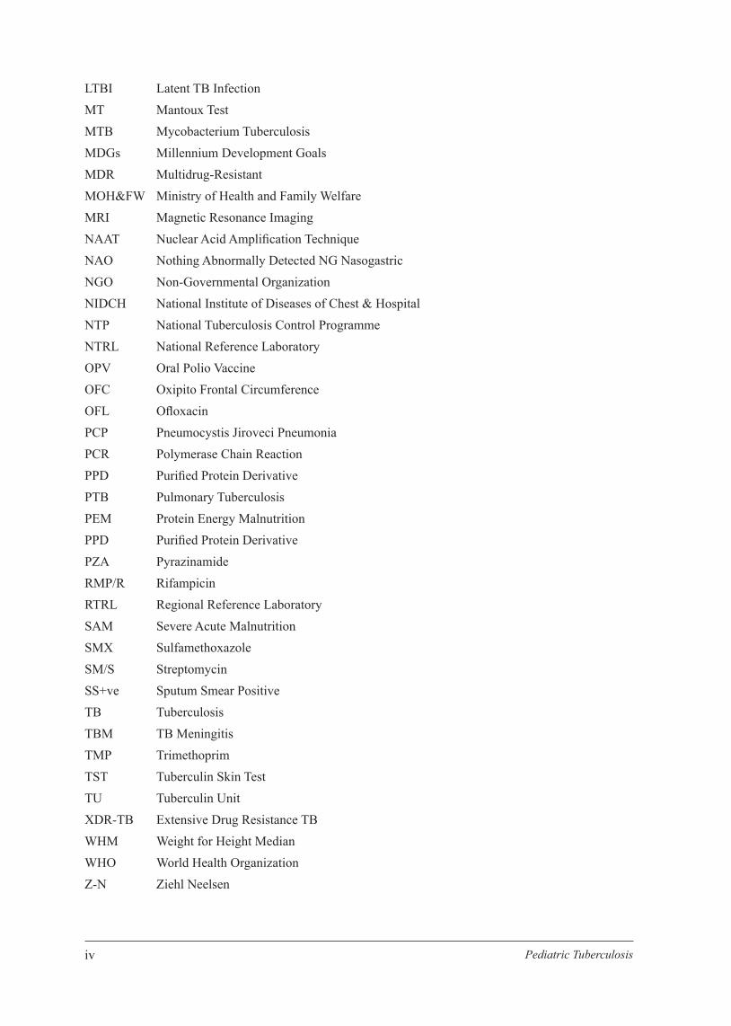
iv Pediatric Tuberculosis
LTBI Latent TB Infection MT Mantoux Test MTB Mycobacterium Tuberculosis MDGs Millennium Development Goals MDR Multidrug-Resistant MOH&FW Ministry of Health and Family Welfare MRI Magnetic Resonance Imaging NAAT Nuclear Acid Amplifi cation Technique NAO Nothing Abnormally Detected NG Nasogastric NGO Non-Governmental Organization NIDCH National Institute of Diseases of Chest & Hospital NTP National Tuberculosis Control Programme NTRL National Reference Laboratory OPV Oral Polio Vaccine OFC Oxipito Frontal Circumference OFL Ofl oxacin PCP Pneumocystis Jiroveci Pneumonia PCR Polymerase Chain Reaction PPD Purifi ed Protein Derivative PTB Pulmonary Tuberculosis PEM Protein Energy Malnutrition PPD Purifi ed Protein Derivative PZA Pyrazinamide RMP/R Rifampicin RTRL Regional Reference Laboratory SAM Severe Acute Malnutrition SMX Sulfamethoxazole SM/S Streptomycin SS+ve Sputum Smear Positive TB Tuberculosis TBM TB Meningitis TMP Trimethoprim TST Tuberculin Skin Test TU Tuberculin Unit XDR-TB Extensive Drug Resistance TB WHM Weight for Height Median WHO World Health Organization Z-N Ziehl Neelsen

vPediatric Tuberculosis

8 Pediatric Tuberculosis

viiPediatric Tuberculosis
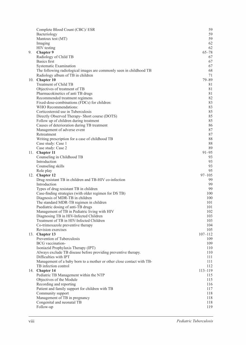
TABLE OF CONTENTS1. Chapter 1 1–5 Epidemiology of Child TB 3 Global 3 SAARC countries 3 How does childhood TB differ from adult TB? 4 Summary: 4 Revision exercises 52. Chapter 2 7–18 Pathology and pathogenesis 9 Spread from primary focus and lymph node enlargement 9 Risk of disease progression 13 Post-primary tuberculosis 14 Exposure, Infection, disease and close contact 16 Close contact 17 Household contact 17 Revision exercises 183. Chapter 3 19–24 Laboratory Diagnosis of Tuberculosis 21 Demonstration of Acid Fast Bacilli 21 Demonstration of Mycobacterium Tuberculosis 21 Collection of sputum samples 21 A. Spontaneous Sputum Collection 22 B. Gastric aspiration 22 C. Sputum induction 244. Chapter 4 25–30 Case taking in Pediatric TB 27 History 27 Regional and systemic examination 28 History and physical signs of importance 28 Practice of anthropometry 29 Calculation 305. Chapter 5 31–44 Approach to diagnosis of childhood TB 33 Detailed History 33 General symptoms suggestive of TB 33 Symptoms and signs highly suggestive of Childhood TB 34 Indications requiring hospitalization or referral- 34 Referral should also be considered if- 34 TB lymphadenitis (TBL) 38 Pleural and pericardial TB 39 TB meningitis 41 TB spine and TB arthritis 42 Miliary TB 43 Abdominal TB 43 Lesson Learnt 44 Danger signs requiring urgent hospital referral 44 Uncommon signs indicative of recent TB infection 446. Chapter 6 45–51 Child TB Album 47 Answer to cases 49 Case 1: 49 Case: 2: 50 Case 3: Answer 50 Case 4: Answer 50 Case 5: Answer 51 Case 6: Answer 51 Case 7: Answer 517. Chapter 7 53–56 Case screening 558. Chapter 8 57–63 Investigation 59
viii Pediatric Tuberculosis
Complete Blood Count (CBC)/ ESR 59 Bacteriology 59 Mantoux test (MT) 59 Imaging 62 HIV testing 629. Chapter 9 65–78 Radiology of Child TB 67 Basics fi rst 67 Systematic Examination 67 The following radiological images are commonly seen in childhood TB 68 Radiology album of TB in children 7110. Chapter 10 79–89 Treatment of Child TB 81 Objectives of treatment of TB 81 Pharmacokinetics of anti TB drugs 81 Recommended treatment regimens 82 Fixed-dose-combinations (FDCs) for children: 83 WHO Recommendations: 83 Corticosteroid use in Tuberculosis 85 Directly Observed Therapy- Short course (DOTS) 85 Follow up of children during treatment 85 Causes of deterioration during TB treatment 86 Management of adverse event 87 Retreatment 87 Writing prescription for a case of childhood TB 88 Case study: Case 1 88 Case study: Case 2 8911. Chapter 11 91–95 Counseling in Childhood TB 93 Introduction 93 Counseling skills 93 Role play 9512. Chapter 12 97–105 Drug resistant TB in children and TB-HIV co-infection 99 Introduction 99 Types of drug resistant TB in children 99 Case-fi nding strategies (with older regimen for DS TB) 100 Diagnosis of MDR-TB in children 100 The standard MDR-TB regimen in children 101 Paediatric dosing of anti-TB drugs 101 Management of TB in Pediatric living with HIV 102 Diagnosing TB in HIV-Infected Children 103 Treatment of TB in HIV-Infected Children 103 Co-trimoxazole preventive therapy 104 Revision exercises 10513. Chapter 13 107–112 Prevention of Tuberculosis 109 BCG vaccination- 109 Isoniazid Prophylaxis Therapy (IPT) 110 Always exclude TB disease before providing preventive therapy. 110 Diffi culties with IPT 111 Management of a baby born to a mother or other close contact with TB- 111 TB infection control 11214. Chapter 14 113–119 Pediatric TB Management within the NTP 115 Objectives of the Module 115 Recording and reporting 116 Patient and family support for children with TB 117 Community support 118 Management of TB in pregnancy 118 Congenital and neonatal TB 118 Follow-up 119

1Pediatric Tuberculosis
CHAPTER 1

2 Pediatric Tuberculosis

3Pediatric Tuberculosis
Epidemiology of Child TB
Global:
According to Global TB Report 2015, worldwide, 9.6 million people are estimated to have fallen ill with TB in 2014: 5.4 million men, 3.2 million women and 1.0 million children. Globally, 12% of the 9.6 million new TB cases in 2014 were HIV-positive. It is estimated that accurate diagnosis and good reporting system children are likely to contribute 10-20% of disease burden in areas where the TB is poorly controlled. The incidence of paediatric TB provides an accurate measure of ongoing transmission within communities, which is a key indicator of control. Despite decline in death due to TB globally is still very high. In 2014, TB killed 1.5 million people (1.1 million HIV-negative and 0.4 million HIV-positive), making it one of the deadliest disease in the planet. The toll comprised 890,000 men, 480,000 women and 140,000 children.
A common misconception is that children are not severely affected by the TB epidemic and rarely develop severe forms of disease. This is not the case in TB endemic areas, where children are often present with advanced and serious disease (TB meningitis, Miliary TB).
SAARC countries: SAARC countries lies in 2 (two) regions of WHO-South-East Asia and EMRO. In the eight SAARC member states total TB case new cases reported was 2,181,285, which is 36% of newly reported 6 million global TB cases. Among these 134,417 (6.16%) are children <15 years.
Underreporting and under-diagnosis of childhood TB cases are the main reason of low case detection in this region. Besides community awareness on child TB is also low.
Child TB and Total TB burden in SAARC countries, 2014
Country Total TB Child TB (<15 yrs)
Total Percentage Afghanistan 30,537 4,454 15 Bangladesh 187,005 6,262 3 Bhutan 1,066 56 5.25 India 1,609,547 95,709 6 Maldives 131 14 10.69 Nepal 35,277 354 1.00 Pakistan 308,417 27,245 9 Sri Lanka 9,305 323 3.47 Total 2,181,285 134,417 6.16
Contact with smear positive case (open case) when a child is in close contact with an open case of TB, the chance of getting infection is approximately 35%. This is higher during close contact than casual contact (11%). The chances of being infected, when in close contact with a smear negative TB case, are between 10-12%. The possibility of getting a TB disease following infection has been found to be between 3-9% among children in high burden countries. This is 4-6 times higher when the close contact is female (especially the mother) and even higher in a breastfeeding mother who is sputum smear positive case (think the distance between mother’s mouth and that of the baby). Most disease occurs within the 2 years
Chapter 1
4 Pediatric Tuberculosis
of contact (95% within 1 yr). TB disease in children is mostly pulmonary (60%) and is smear negative or smear not done. In children extra-pulmonary TB (EPTB) is found to be around 30%. Smear positive cases are found in older children and adolescents.
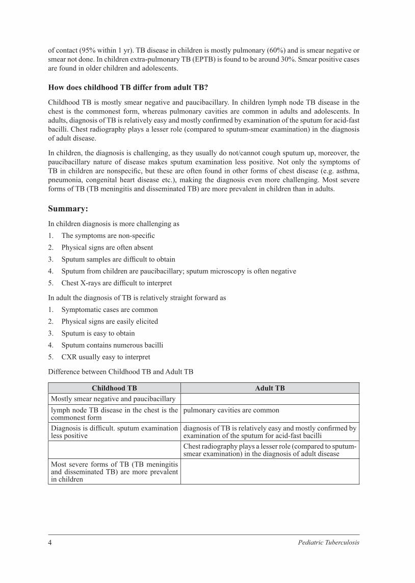
How does childhood TB differ from adult TB?
Childhood TB is mostly smear negative and paucibacillary. In children lymph node TB disease in the chest is the commonest form, whereas pulmonary cavities are common in adults and adolescents. In adults, diagnosis of TB is relatively easy and mostly confi rmed by examination of the sputum for acid-fast bacilli. Chest radiography plays a lesser role (compared to sputum-smear examination) in the diagnosis of adult disease.
In children, the diagnosis is challenging, as they usually do not/cannot cough sputum up, moreover, the paucibacillary nature of disease makes sputum examination less positive. Not only the symptoms of TB in children are nonspecifi c, but these are often found in other forms of chest disease (e.g. asthma, pneumonia, congenital heart disease etc.), making the diagnosis even more challenging. Most severe forms of TB (TB meningitis and disseminated TB) are more prevalent in children than in adults.
Summary:
In children diagnosis is more challenging as 1. The symptoms are non-specifi c 2. Physical signs are often absent 3. Sputum samples are diffi cult to obtain 4. Sputum from children are paucibacillary; sputum microscopy is often negative 5. Chest X-rays are diffi cult to interpret
In adult the diagnosis of TB is relatively straight forward as 1. Symptomatic cases are common 2. Physical signs are easily elicited 3. Sputum is easy to obtain 4. Sputum contains numerous bacilli 5. CXR usually easy to interpret
Difference between Childhood TB and Adult TB
Childhood TB Adult TBMostly smear negative and paucibacillarylymph node TB disease in the chest is the commonest form
pulmonary cavities are common
Diagnosis is diffi cult. sputum examination less positive
diagnosis of TB is relatively easy and mostly confi rmed by examination of the sputum for acid-fast bacilliChest radiography plays a lesser role (compared to sputum-smear examination) in the diagnosis of adult disease
Most severe forms of TB (TB meningitis and disseminated TB) are more prevalent in children
5Pediatric Tuberculosis
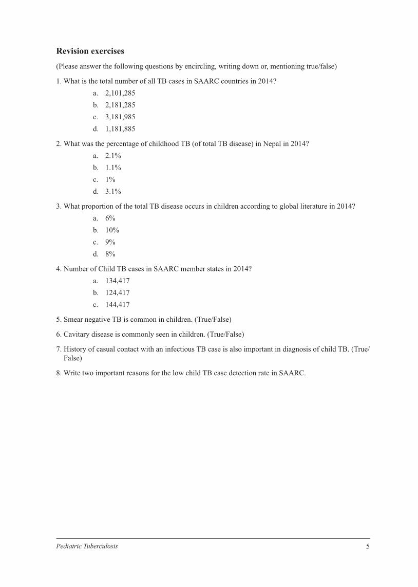
Revision exercises
(Please answer the following questions by encircling, writing down or, mentioning true/false)
1. What is the total number of all TB cases in SAARC countries in 2014? a. 2,101,285 b. 2,181,285 c. 3,181,985 d. 1,181,885
2. What was the percentage of childhood TB (of total TB disease) in Nepal in 2014? a. 2.1% b. 1.1% c. 1% d. 3.1%
3. What proportion of the total TB disease occurs in children according to global literature in 2014? a. 6% b. 10% c. 9% d. 8%
4. Number of Child TB cases in SAARC member states in 2014? a. 134,417 b. 124,417 c. 144,417
5. Smear negative TB is common in children. (True/False)
6. Cavitary disease is commonly seen in children. (True/False)
7. History of casual contact with an infectious TB case is also important in diagnosis of child TB. (True/ False)
8. Write two important reasons for the low child TB case detection rate in SAARC.

6 Pediatric Tuberculosis

7Pediatric Tuberculosis
CHAPTER 2

8 Pediatric Tuberculosis

9Pediatric Tuberculosis
Pathology and pathogenesis
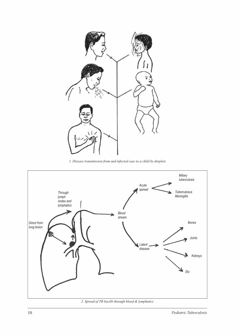
Mycobacterium tuberculosis (MTB) gains access to the lungs via droplets inhalation from an open case (smear positive) of tuberculosis. When an infectious adult coughs, sneezes, talks, shouts, sings or even laughs thousands of droplets spread into the air and remains suspended for hours; especially in a closed room (e.g. class room, hut) or other closed spaces where it causes cumulative effect. Droplets increase in numbers as the person continues to cough and people who share the room/environment have an increased chance of getting infected. Young children (less than 5years)have the greatest chance if their mother has TB. (Fig. 1)
In the lung bacilli settle in the alveoli, usually just beneath pleura, and multiplies slowly to form a small patch of pneumonia. From here the organism spreads via the lymphatic drainage to the regional lymph nodes (hilar, paratracheal and subcranial) causing lymph node enlargement (Fig. 2, 12, 14). This is known as the primary complex and sometimes detectable on CXR. It takes about 8-12 weeks to develop and most people after this period become sensitive to the protein of MTB; at this time tuberculin skin test (TST) or Mantoux skin test (MT) becomes positive. This process is called TB infection. The patient remains asymptomatic and TB infection is diagnosed by MT.
The subsequent course depends on the child’s immunity. Child with a good immunity will be able to contain the infection and prevent disease development. The primary complex will, in the majority of children, heal by fi brosis and calcifi cation; the bacilli can remain dormant for years. Very young children, child with severe malnutrition and HIV have impaired immunity making them vulnerable to disease. This is known as latent tuberculosis infection (LTBI). LTBI can reactivate after months to years when the immunity is suppressed and progress to TB disease. This is especially important for children younger than 5 years.
Spread from primary focus and lymph node enlargement
In most cases there is hematogenous and lymphatic dissemination from the primary focus to other parts of the body and the lungs. Acute massive seedling via the blood stream leads to miliary tuberculosis and tuberculous meningitis, usually within 3-6 months of initial infection. Certain organs favor survival of the bacilli and these organs may later be affected by disease, e.g. apical and sub-apical regions of the lungs (where there is a higher oxygen tension), renal parenchyma, epiphysis of bones, cerebral cortex and regional nodes. At these sites (bones, joints, kidney, genital tract etc.) bacilli remain dormant for months to years, another example of LTBI (Fig. 2). Fortunately, disease only occurs in a small percentage as a result of this dissemination.
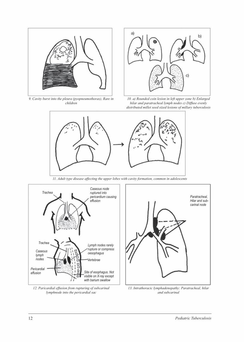
Rupture of the primary lesion into the pleural space can cause pleural effusion, rarely tuberculous empyema. As the lymph nodes lie near the trachea and large bronchi, enlarged lymph nodes can compress or erode into the air passages causing airway narrowing or tuberculous bronchopneumonia (Fig. 3). Airway involvement can result in collapse, obstructive emphysema, pneumonia, and even bronchiectasis in long standing cases (Fig. 4,5,6,7,15). At times the lymph node content may leak into pericardium, producing a pericardial effusion (Fig. 12). These clinical conditions will become more apparent during the radiology session.
Please go through all the diagrams with your pair to understand the natural progress of TB, which will also help you to understand and read X-ray. (a powerpoint presentation on these diagrams and pictures).
Chapter 2
10 Pediatric Tuberculosis
1. Disease transmission from and infected case to a child by droplets
2. Spread of TB bacilli through blood & lymphatics
Miliarytuberculosis
TuberculosusMeningitis
Bones
Joints
Kidneys
Etc
Latentdisease
Bloodstream
Acutespread
Throughlymphnodes andlymphatics
Direct fromlung lesion

11Pediatric Tuberculosis
Compensatoryexpansion ofmiddle andlower lobes
Bronchialobstruction.Absorptionof air.Collapse of lobe orsegment oflung beyondobstruction
Overinflationright, upperlobe
Compressedmiddle &lower zone
NodeerodingbronchusLumencloses onexpiration
Ball valveeffect
3. Tuberculous bronchopneumonia caused by erosion ofmediastinal lymphnode into bronchus
5. Collapse right middle lobe (lateral view)
7. Complications of tuberculous lymph nodes of primary complex
4. Complications intrathoracic lymphadenopatthy: collapse of upper lobe with compensatory expansion of middle and lower lobes
6. Bronchiectasis in right lower lobe: complication of lymph node disease
8. Large right Pleural effusion Heart and trachea pushed to left
12 Pediatric Tuberculosis
9. Cavity burst into the pleura (pyopneumothorax), Rare in children
11. Adult type disease affecting the upper lobes with cavity formation, common in adolescents
12. Paricardial effusion from rupturing of subcarinal lymphnode into the pericardial sac
10. a) Rounded coin lesion in left upper zone b) Enlarged hilar and paratracheal lymph nodes c) Diffuse evenly
distributed millet seed sized lesions of millary tuberculosis
13. Intrathoracic lymphadenopathy: Paratracheal, hilar and subcarinal
Paratracheal,Hilar and sub-carinal node
TracheaCaseous noderuptured intopericardium causingeffusion
Trachea
Caseouslymphnodes
Pericardialeffusion
Lymph nodes rarelyrupture or compressoesophagus
Vertebrae
Site of esophagus. Notvisible on X-ray exceptwith barium swallow
13Pediatric Tuberculosis
Risk of disease progression
In newly infected children,the risk of developing tuberculous disease is highest (95%) in the fi rst year following a primary infection and diminishes thereafter. Example: if a child is infected from an open case at 3 years of age, for him /her risk of developing TB disease is highest within 4 years of age. The risk is greatest in infants and children who are malnourished, taking steroids, HIV infected, or following measles and on cancer chemotherapy. The age specifi c risk is described in the table below.
Age-specifi c risk to progress to disease after primary infection with M. tuberculosis in immunocompetent children8
8 Marais BJ, Gie RP etal. Childhood Pulmonary Tuberculosis: Old wisdom and new challenges. Am J Res Crit Care Med. Vol-173. 1078-1090, 2006
Age at Primary Infection (yr) Risk to Progress to Disease
< 1 No disease- 50%
Pulmonary disease- 30-40%
Disseminated (millary) disease or TBM, 10-20%
1-2 No disease- 75-80%
Pulmonary disease- 10-20%
Disseminated (millary) disease or TBM- 2-5%
2-5 No disease- 95% Pulmonary disease- 5%
Disseminated (millary) disease or TBM- 0.5%
5-10 No disease- 98% Pulmonary disease- 2%
Disseminated (millary) disease or TBM- < 0.5%
> 10 No disease- 80-90%
Pulmonary disease- 10-20%
Disseminated (millary) disease or TBM, < 0.5%
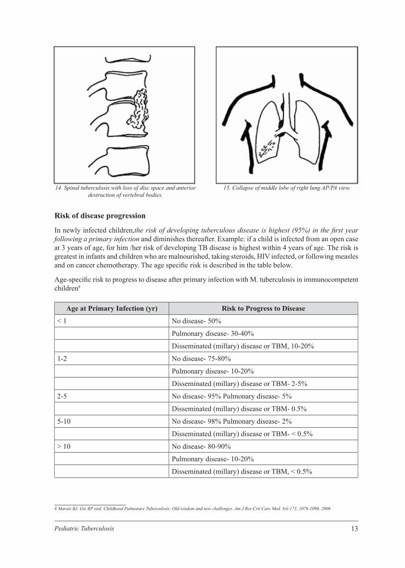
14. Spinal tuberculosis with loss of disc space and anteriordestruction of vertebral bodies.
15. Collapse of middle lobe of right lung AP/PA view.

14 Pediatric Tuberculosis
Post-primary tuberculosis
Post-primary tuberculosis can develop from reactivation of a dormant primary focus or from exogenous re-infection (new infection). This is common in adolescent (more in girls than boys) and is known as ‘adult-type’ pulmonary tuberculosis. This form of TB is associated with cavities and as a result sputum microscopy isusually smear positive for AFB in this type of disease. They are potentially infectious and can infect others.
In next couple of pages there are pictures of pathogenic specimen from TB disease. Go for a tour into these to make your understanding of pathology fi rmer, discuss with facilitator for a better guide of the tour.
Cavities are commonin the upper lobes
Granulomas
Hilar lymph node next toa bronchus
Sub-pleural granuloma
Closer look of a primary complex
Primarycomplex

15Pediatric Tuberculosis
Gross appearance of a lung with tuberculosis - scatteredgranulomas mostly in upper lobe
Miliary pattern evnly distributed throughout the lung Miliary pattern is seen throughout the lung
Granulomas with central caseation appear as imegularly sized nodules
Granulomotous disease of the upper lobe andapex of lower lobe

16 Pediatric Tuberculosis
Exposure, Infection, disease and close contact
Exposure
A child is exposed to M. tuberculosis when s/he comes in contact with an infectious TB patient (mostly smear positive). The risk of inhaling the organism and becoming infected is determined by:
1. The infectiousness of the source case (grade of smear positive)
2. The closeness of contact
3. Duration of contact
Multiplegranulomain histologicspecimen
Granuloma with Langhans giant cells
Multiple granuloma in spleen

17Pediatric Tuberculosis
Children are most likely to become infected if their mothers or other adolescent/adult household members have sputum smear-positive TB (Index Case) or having active pulmonary cavities in CXR. If a child has close contact with anindex case clinical screening should be done to decide whether s/he is symptomatic or not. It is estimated that one adult ss+ve patient can spread infection to 10 persons.
Infection
A child becomes infected when s/he inhales the TB organism. Only about 35% of children exposed to an infectious case of TB will be infected. Infection with M. tuberculosis does not mean that the child is sick or symptomatic. TB infection is indicated by a positive Mantoux test in an asymptomatic child. However, there are many limitations to the Mantoux test (learn more lately in Mantoux session on day 3). In HIV-infected and/or malnourished children, Mantoux test frequently gives a false negative result. After inhaling Mycobacterium tuberculosis, it takes up to 3 months for a positive MT test to develop. It should be noted that during this window period, infected children are usually asymptomatic and the MT test is negative (not reactive).
Disease
Only small percentages (5-10%) of children, who inhale the TB organism become infected and develop active disease. Certain groups are at greater risk than others (table: key risk factors below). TB disease may manifest in many different ways, but is usually indicated by the presence of well-defi ned symptoms and/or radiological changes.
Key risk factors developing TB disease in children-
Close contact with a known case of TB (parents, siblings, close relatives, caregivers, neighbors and teachers)
Age of the child (risk to develop TB disease is highest in very young, immune immature, children <5 yrs)
Severe malnutrition Other Immunosuppressive conditions:
Measles in the previous 3 months Whooping cough HIV infection Being on immune suppressive drugs like steroids
The time since exposure or infection; the vast majority (95%) of children who develops disease do so within the fi rst year of M. tuberculosis exposure or infection.
Contact: Any person who has been exposed to an index case.
Close contact
A person who is not in the household but who shared an enclosed space, such as a social gathering place, work place, or facility, with the index case for extended daytime periods during the 3 months before the start of the current treatment episode
Household contact
A person who shared the same enclosed living space as the index case for one or more nights or for frequent or extended daytime period during the 3 months before the start of current treatment episode.

18 Pediatric Tuberculosis
Slum dwellers have high risk of contact from unknown source cases as they live very close together. Infant of a mother with TB who breastfeeds them have the closest contact, hence at very high risk.
Revision exercises: Do this exercise, inform facilitator as soon you fi nish.
Encircle or write the answer in the space provided.
1. Arrange age group from highest to lowest risk for developing TB disease after infection:
2- 5 yrsa. >10 yrsb. <1 yrsc. 1-2 yrsd. 5-10 yrs
2. Primary complex can always be detected on CXR:a. Trueb. False
3. Intrathoracic lymph node enlargement can cause one or more of the following conditions:a. Lobar Collapseb. Empyemac. Compensatory Emphysemad. Bronchopneumoniae. Pleural effusion
4. How many weeks after infection with M. tuberculosis does the Mantoux test become positive?
…………. weeks
5. Which factors increase the likelihood of infection following close contact with an open case of TB?a.b.c.
6. Acute spread causes:a.b.
7. LTBI can ………………………. after months to years of initial infection.
8. Household contact is a person who
a. shared same enclosed space for………………………………….nights
b. extended daytime period for……………………………………….months
9. After exposure to an SS+ve case vast majority of children………………………………………….within…….yr

19Pediatric Tuberculosis
CHAPTER 3

20 Pediatric Tuberculosis

21Pediatric Tuberculosis
Laboratory Diagnosis of Tuberculosis
Tuberculosis is caused by Mycobacterium tuberculosis complex. It comprises of group of organisms;M. Tuberculosis, M. Bovis, M. Bovis BCG, M. Africanum, M. Macroftti, M. cannetti. The typical character of Mycobacterium is Acid Fastness. It can be demonstrated by using Acid Fast Bacillli Stain (AFB). This method was fi rst demonstratedby Robert Koch, a German scientist, on 24th March 1882. In 1896 it was named Mycobacterium (Greek, mykes- Fungus; bacterium- small rods) in reference to mould like growth in liquid medium. Defi nitive laboratory diagnosis of TB disease in children depends on identifying Mycobacterium tuberculosis in clinical specimens recovered from the patient. Bacteriological confi rmation though diffi cult, should be attempted whenever possible, especially in children with:
a. suspected drug-resistant TB b. complicated or severe cases of disease c. HIV infection d. Uncertain diagnosis
Demonstration of Acid Fast Bacilli
1. Microscopy:
There are two methods used for detecting Acid Fastness of bacilli. One is Ziehl Neelsen staining technique and other is Fuorochorme staining (Auramine- O stains ). Flurosneccne Miscrospy is more sensitive. Acid-fast bacilli (AFB) are seen by microscopy on slide stained by the Ziehl-Neelsen (ZN) method. ZN staining can be done on any clinical specimen e.g. sputum, gastric aspirate, biopsy specimen, CSF, pleural and peritoneal fl uid. The specimen should contain >10,000 bacilli/ ml to detect AFB under microscope. In <20% of children sputum/gastric aspirate microscopy will show AFB, compared to 75% in adults.
Demonstration of Mycobacterium Tuberculosis
Culture: Demonstration of Myobacteria by culture is considered “gold standard” for diagnosis of TB. Sensitivity ranges from 30-50%. Culture can detect 10-100 bacili/ml. Lowenstein-Jensen (L J) method takes 8-12 weeks to get a positive culture, with additional 4 weeks for drug sensitivity. Radiometric BACTEC method takes 7-14 days.
Molecular technology: Nuclear Acid Amplifi cation Technologies (NAAT) or Polymerase chain reaction (PCR) can be done on various specimens. It detects the presence of 10-100 bacilli/ml of specimen. Specifi city varies from 80-96%. These are costly tests and therefore not used routinely in SAARC countries. Gene-xpert can detect TB organism within 2 hours.
Collection of sputum samples
Collection can be done by the following techniques: 1. Sputum expectoration 2. Gastric aspirate 3. Sputum induction
Chapter 3

22 Pediatric Tuberculosis
A. Spontaneous Sputum Collection
Background
The sputum smear remains a valuable test. In any child who can produce sputum (usually >8 yrs), it is the test of choice. Sputum should always be obtained in older children who are TB suspects. Children able to produce sputum may be infectious, so, as with adults, they should be asked to produce a sputum outside the clinic or in specially equipped rooms and not in enclosed spaces (such as toilets). Two sputum specimens should be obtained: a spot specimen (at fi rst evaluation) and one early morning specimen.
Procedure 1. Counselling: Explain the purpose and procedure to the child/parent for sputum collection. 2. Instruct the child to rinse his/her mouth with water before producing the specimen. 3. Tell the child to take two deep breaths, holding the breath for a few seconds after each inhalation
and then exhale slowly. Ask him/her to breathe in for a third time and then forcefully blow out (cough). Ask the child to hold the sputum container close to the lips and to gently spit into it after the productive cough. Ask him/ her to breathe in again and then produce another specimen. This should produce good quality sputum specimen.
4. If the specimen is insuffi cient, encourage the patient to cough again until a satisfactory specimen is obtained. Give the child suffi cient time to produce a good specimen.
5. If there is no specimen collected, consider the container as contaminated and dispose of it in appropriate manner.
6. Carefully label the specimen and send to the laboratory. 7. The instructor must take measures for his/her personal protection by wearing a mask.
B. Gastric aspiration
Background
Since infants and young children do not expectorate rather swallow their own sputum, aspiration or lavage of gastric contents is a good procedure for obtaining a specimen. It can be also applied for older children who cannot produce a sputum specimen. As gastric aspiration causes discomfort in children; it is preferable to use this procedure if microscopy and culture facilities are available.
During sleep, the mucociliary system of lungs beats mucus up into the throat. The mucus is swallowed and remains in the stomach until the stomach empties. Therefore, the highest-yield specimens are obtained when collected in the morning before eating.
It is most useful for young hospitalized children. The diagnostic yield (positive culture) of a set of three gastric aspirates is about (25-30%) but the specifi city is very high (90-99%) with active TB. However, a negative smear or culture never excludes TB in a child.
Performing the test properly usually requires two people (one doing the test and an assistant). Fasting for a period of 3-4 hours prior to the procedure is necessary.
The following equipments are needed: 1. Gloves 2. Nasogastric tube (usually 10 French gauge or larger) 3. 5, 10, 20 or 30 ml syringe, with appropriate connector for the nasogastric tube 4. Specimen container 5. Pen (to label specimens)

23Pediatric Tuberculosis
6. Laboratory requisition forms 7. Sterile water or normal saline (0.9% NaCl) 8. Sodium bicarbonate solution (8.4%) 9. Alcohol/chlorhexidine.
Procedure
The procedure is best carried out as an inpatient, fi rst thing in the morning when the child wakes up. It can be done at the bedside or in a procedure room on the ward (if one is available). It can also be performed as an outpatient (provided that the facility is properly equipped). The child should have fasted for at least 4 hours (infants for 3 hours) before the procedure. 1. Counselling: Brief the purpose and procedure to the child/parent for sputum collection. 2. Find an assistant to help. 3. Prepare all equipment before starting the procedure.4. Position the child on his/her back or side. The assistant should hold the child.5. Measure the distance between the nose and stomach, to estimate distance that will be required to
insert the tube into the stomach. 6. Attach a syringe to the nasogastric tube 7. Gently insert the nasogastric tube through the nose and advance it into the stomach 8. Withdraw (aspirate) gastric contents (2–5 ml) using the syringe attached to the nasogastric tube. 9. Check that the position of the tube is correct (by pushing some air, 3–5 ml from the syringe rapidly
into the tube and listen with a stethoscope over the stomach). 10. If no fl uid is aspirated, insert 5–10 ml sterile water or normal saline and attempt to aspirate again.
If still unsuccessful, repeat the installation of fl uid (even if the nasogastric tube is in an incorrect position and water or normal saline is inserted into the airways, the risk of adverse events is still very small). Do not repeat more than three times.
11. Withdraw the gastric contents (ideally at least 5–10 ml) 12. Transfer gastric fl uid from the syringe into a sterile container (sputum collection cup). 13. Add an equal volume of 8.4% sodium bicarbonate solution to the specimen (in order to neutralize the
acidic gastric contents and so prevent destruction of tubercle bacilli).
After the procedure 1. Wipe the specimen container with alcohol/chlorhexidine to prevent cross- infection and label the
container. 2. Fill out the laboratory requisition forms. 3. Transport the specimen (in a cool box) to the laboratory for processing as soon as possible (within 4
hours). 4. If it is likely to take more than 4 hours for the specimens to be transported, place them in the refrigerator
(4–8 °C) and store until transported. 5. Give the child his or her usual meal.
Safety
Gastric aspiration is generally not an aerosol-generating procedure. As young children are also at low risk of transmitting infection, gastric aspiration can be considered a low risk procedure for TB transmission and can safely be performed at the child’s bedside or in a routine procedure room.

24 Pediatric Tuberculosis
C. Sputum induction
This procedure is safe and effective in children of all ages and the mycobacterial yields are good. Note that, unlike gastric aspiration, sputum induction is an aerosol-generating procedure. Infection control is important during sputum induction. Whenever possible, the procedure should be performed in an isolation room with an extractor fan. All the staff must wear face mask.
General approach
Examine children, as children that have fast breathing or are hypoxic should not undergo the procedure. Children with the following characteristics should not undergo sputum induction.
Contraindications: 1. Inadequate fasting: if a child has not been fasting for at least 3 hours, postpone the procedure until
the appropriate time. 2. Severe respiratory distress (including rapid breathing, wheezing, hypoxia). 3. Intubated children. 4. Bleeding: low platelet count (<50,000/cmm), bleeding tendency, severe nosebleeds. 5. Reduced level of consciousness. 6. History of signifi cant asthma (diagnosed and treated by a clinician).
Procedure 1. Counseling: Brief the purpose and procedure to the child/parent for sputum collection. 2. Administer nebulized bronchodilator (e.g. salbutamol) prior to the procedure to reduce the risk of
wheezing. 3. Administer nebulized hypertonic saline (3% NaCl) for 15 minutes or until 5 ml of solution have been
fully administered. 4. Give chest physiotherapy if necessary; to mobilize secretions. 5. For older children who are able to expectorate, follow procedures as described on how to collect
expectorated sputum. 6. For children unable to expectorate (e.g. young children): Collect a nasopharyngeal aspirate using a
Optional Session:
Facilitator may arrange a practice/demonstration session of specimen collection procedures: 1) Expectoration 2) Sputum induction: 3) Gastric aspiration: video

25Pediatric Tuberculosis
CHAPTER 4

26 Pediatric Tuberculosis

27Pediatric Tuberculosis
Case taking in Pediatric TB
History
History InquiriesParticulars of the patient
Name/age: (date of birth)/sex: male/female/address/occupation/sources of information: informant
Ask for Fever (3 weeks), cough (>3 weeks), weight loss, less playful, decreased activity breathing diffi culty, painless swelling of neck glands (with or without discharge), vomiting, convulsion, impaired consciousness, abdominal distension, vague pain in the back, angulation in spine, painless swelling of joints, limping.
Past illness Pneumonia/asthma/diarrhea/measles pertussis tuberculosis with treatmentFeeding history Breastfeeding-upto 6 month. Complementary feeding from 6 months.Immunization history
BCG and other vaccines
Family history H/O TB in the family within last 2 years, chronic cough, TB in the neighborhood or relatives, details of TB treatment with duration, type of tB drug response: cured/died
Socioeconomic history
Education and occupation of parents/housing (slum)
Points to examine Physical areas/featuresAppearance Ill-looking, distressed, wastedPallor Examined in lower eye lids/tongue/palms/soles/overall skinJaundice Examined in upper bulbar conjunctiva/under surface of tongue/palms/soles/skinDehydration Surken eyes/tongue/skin turgor of abdomenCyanosis Tip of tongue/fi nger nails/toe nailsLymph nodes Cervical, axillary, inguinal nodes (> 1 cm in cervical or axillary; >1.5 cm in
inguinal is signifi cant), Consistency, mobility, tenderness, matted, dischargeSkin BCG mark/features of PEM (dermatosis, skin changes, eye changes)Edema Above ankle over the shin/over the sacrum
Vital signs-Pulse/HR/RR Temperature Blood pressure
Pulse ratge in older child/heart rate in young child, Respiration rate per minute Blood pressure: Temperature, oral cavity/axilla
Spine Acute angulation (Gibbus)Anthropometry Weight and height will be measured and the nutritional status will be assessed.
The Weight-height median (WHM) will be calculated from the growth chart (weight-for-stature percentile).Child 6-59 months: the child will be severely malnourished if s/he has one or more of the following (a) bipedal edema/visible wasting (b) MUAC <110 cm or (c) WHM <70%
Chapter 4
28 Pediatric Tuberculosis
Regions Points to examineHead Bulged fontanel, hydrocephalusEyes Phlyctenular conjunctivitis, pailor, jaundice, movement of eyes, pupil size,
equally, response to light, fundoscopy (chorold tubercles)Neck Signs of meningeal irritation (neck stiffness, neck retraction), lymph nodes (vide
above)Chest and heart Look: asymmetry, chest movement, RR, subcostal recession
Feel: tracheal position, position of apex beat.
Percussion: stony dull in pleural effusion/hyperresonnant
Auscultate: breath sounds-vesicular or bronchlal, crepitations, rhonchi.
Heart: diminished/ absent/distant heart sounds in pericardial effusionAbdomen Look: distension
Feel: hepatomegaly, splenomegaly; ascites (fl uid thrill, shifting duuilness), Doughy feeling on palpation, lump bowel sound
Locomotor system Look: for non tender swelling of joints, walking diffi culty, muscle wasting
Feel: tenderness on the of spine and jointsNervous system Level of consciousness: exaggerated knee and ankle refl exex- may suggest
spinal TB even there is no gibbus, cranial nerve paisy - 6th, 7th (Fig-7, Page 37)
Child <6mo: the infant will be severely malnourished if s/he has one or more of the following (a) visible wasting/bipedal edema or (b) WHM <70%
Calculation of WHM: the weight for the child’s height in the 50th centlle is taken as 100% as found in the weight-for-stature chart of CDC growth chart. The observed weight of the child will be divided by the 50th cenrtile (100%) value.
Example: A male child of 4-years having weight 10.5 kg and height 100 cm. The 50th centile value for this height is 15.8 kg. So, the WHM of this child is 10.5/15.8 = 65%. The boy falls <70% WHM, so he is severely mainourished.
Regional and systemic examination
History and physical signs of importance 1. Documented weight loss 2. Household contact that is smear positive 3. Signifi cant respiratory symptoms, but normal chest fi ndings 4. Ascites 5. Matted cervical lymph gland enlargement 6. Sudden onset of gibbus 7. Signs of raised intracranial pressure or meningeal irritation 8. When associated with sign/symptoms in box of page 19.

29Pediatric Tuberculosis
Practice of anthropometry
Facilitator will show you ideal way to1. Measure the height & weight 2. Find weight for height median on growth chart 3. Calculate weight for height for the patient.
Assessment of severe acute malnutrition (SAM) on the basis of anthropometric and clinical basis is also to be practiced.
One example is given; do the other exercises. Fill the boxes from the case, fi nd WHM from the supplied growth chart, do calculations and then write/encircle your inference. A child aged 6-59 months, classifi ed as severely malnourished if s/he has one or more of the following:
(a) Weight for height median (WHM) <70%. (b) Bipedal edema
Exercises:
1. 4 year old male child presented with fever and cough for 2 months. He looked well with weight 14 kg and height 100 cm.
Question: What is the nutritional status of the child?
Calculation:
Age/Gender Height (cm) Weight (Kg) Wt for Ht median
% of median
Bipedal Edema Remark
4 Yr/M 100 cm 14 kg 15.6 kg 89% Absent Not SAM
Age/Gender Height (cm) Weight (Kg) Wt for Ht median
% of median
Bipedal Edema Remark
Calculation for % of median: 15.6 kg = 100% 14 kg = (14/15.6) X 100 = 89%
Not severely malnourished (SAM) as weight for height 89% (>70%) and does not have bipedal edema.
2. A 4-yr 6 mo old female child having weight 10.0 kg and height 100 cm. No bipedal edema.
Question: What is the nutritional status of the child?
Calculation:
Inference: SAM/ Not SAM
3. A 16 months old female child presented with swelling of the whole body for 8 days and itchy skin lesion for same duration. She has puffy appearance and bipedal edema. Weight for age 8.2 kg, length 73 cm.
Question: What is the nutritional status of the child?
30 Pediatric Tuberculosis
Age/Gender Height (cm) Weight (Kg) Wt for Ht median
% of median
Bipedal Edema Remark
Age/Gender Height (cm) Weight (Kg) Wt for Ht median
% of median
Bipedal Edema Remark
Calculation:
Inference: SAM/ Not SAM
4. A 3 yr 2 mo old male child presented with weight loss for 5 months; cough, fever and swelling of the body with skin changes for 1 month and pallor for 1 month. He was breast fed for 3 months and later given diluted cow’s milk along with breast feeding. His father is a truck helper.
He looked desperately sick, was moderately pale having bipedal edema. His weight was 6.8 kg and height 77 cm.
Question: Please fi nd the nutritional status of the child.
Calculation:
Inference: SAM/ Not SAM

31Pediatric Tuberculosis
CHAPTER 5

32 Pediatric Tuberculosis
33Pediatric Tuberculosis
Approach to diagnosis of childhood TB
1. Detailed history (including history of TB contact and symptoms suggestive of TB) 2. Clinical assessment 3. Diagnostic tests-
3.1 Mantoux test 3.2 Chest X-ray 3.3 AFB microscopy 3.4 Investigations relevant for suspected Extrapulmonary TB (EPTB) 3.5 HIV testing in high risk patient 3.6 Other tests
Detailed History
General symptoms suggestive of TB
TB in children commonly presents with poor appetite, weight loss, Persistent fever, reduced playfulness and unremitting cough, but these are non-specifi c symptoms. TB disease can be more severe and of rapid onset in infants and young children. Their symptoms will be of shorter duration and can be confused with acute bacterial pneumonia. Symptoms will depend upon the site of involvement: Pulmonary involvement will cause respiratory symptoms; and for extra pulmonary TB, symptoms depend on the organ(s) involved.
TB in children has different clinical presentations at different ages: Infants (<1 year): Primarily like pneumonia Children (1-9 years): Usually with a chronic cough Adolescents (10-19 years): Like adults, cough, expectoration, hemoptysis etc.
Key risk factors for TB in children Close contact with a known case of TB (parents, siblings, close relatives, caregivers, neighbors
and teachers) Age of the child (risk to develop TB disease is highest in very young, immune immature, children
<5 yrs) Severe malnutrition Other Immunosuppressive conditions:
Measles in the previous 3 months Whooping cough HIV infection Being on immune suppressive drugs like steroids
The time since exposure or infection; the vast majority (95%) of children who develops disease do so within the fi rst year of M. tuberculosis exposure or infection.
Chapter 5

34 Pediatric Tuberculosis
Symptoms and signs highly suggestive of Childhood TB
Persistent, non-remitting cough for >3 weeks not responding to conventional antibiotics (amosicillin, co-trimoxazole or cepnalosporins) and/or bronchodilators
and/or Persistent documented fever (>38oC/100oF) >2 weeks after common cases such as typhold
malaria or pneumonla have been excludedand/or
Documented weight loss or not gaining weight during the past 3 months (especially if not responding to de-worming and food and/or micronutrient supplementation) OR severe mainutrition
and/or Fatigue or reduced playfulness
Indications requiring hospitalization or referral-• Severe forms of PTB and EPTB for further investigation and initial management • Severe malnutrition for nutritional rehabilitation • Signs of severe pneumonia (i.e. chest in-drawing) or severe respiratory distress • Other co-morbidities e.g. severe anaemia, liver disease, nephrotic syndrome
Referral should also be considered if-• Diagnostic uncertainty requiring further investigation • Necessary for HIV-related care e.g. to commence ART
Case 1
A 5-month old female child presented with fever, persistent cough and poor growth for 1 month. Her mother complaints of short of breath for last 15 days. For this she was treated for pneumonia with various antibiotics with no improvement. She was born at term by vaginal delivery and was reasonably well during the fi rst month of age. The girl was breast-fed, supplemented with formula milk. She received all EPI vaccines including BCG; and developed neck control during the last 15 days. On enquiry, mother was found to have cough for last 4 months. Father is a rickshaw puller, lives in slum, and reported to be healthy.
The child looked unwell, fretful, mildly pale and febrile (100ºF). Weight 3.7Kg, length 64 cm, OFC 40 cm. RR 56/min, had chest in drawing, a central trachea and coarse crepitations in both lungs. She had mild hepatomegaly.
WBC -11,000/cmm, N -65%, L -28%, Peripheral Blood Film (PBF) microcytic hypochromic anemia. ESR 30 mm in 1st hour. MT 3 mm. CXR extensive diffuse opacities in both lung fi elds (R>L). Gastric aspirate showed AFB, in the second of three samples sent. CXR of mother: a cavitary lesion in the right apical region.
Clinical features that might suggest other causes of chronic lung disease1. Recurrent cough and/or wheeze responsive to bronchodilators suggests asthma2. Respiratory symptoms with fi nger clubbing suggests bronchiectasis
35Pediatric Tuberculosis
Questions
A. What are the key risk factors for development of TB in the baby? 1. 2. 3. 4.
B. What are the relevant investigations done in this patient?
Answer:
C. Write down the symptoms and signs highly suggestive to diagnose pulmonary TB in this girl?
Answer:
D. What are the radiological differences in the CXRs of the mother and girl?
Answer:
E. What is the most likely diagnosis in the child?
Answer:

36 Pediatric Tuberculosis
Infant’s CXR
Mother’s CXR

37Pediatric Tuberculosis
Case 2
A 2-year-old girl child came to your OPD with fever, cough, and recurrent respiratory infections for the past 3 months. The mother gave the history of poor feeding and weight loss for same duration.
Questions:
A. What history concerning the family would you want to know to suspect TB? 1. 2. 3. 4. Others
B. Name three investigations you want to do for Muna? 1. 2. 3.
C. What is your diagnosis?
Answer:
Case 3
A 12-years female child presented with fever and cough for 6 weeks. She was treated with various antibiotics that did not help. No history of contact with TB patient. BCG scar visible over the left deltoid.
She was not in respiratory distress; temp 100°F, RR 34/min, wt. 33 kg, no tracheal shifting, apex beat in left 5th space medial to mid-clavicular line. Bronchial breathing and crepitations heard over the posterior aspect of the left lower lung.
WBC 12,000/cmm, N 55%, L 40%, ESR 22 mm, MT 16 mm. CXR: consolidation of left lower zone on frontal projection; different segments of Left Lower Lobe on left lateral fi lm in the retro-cardiac space. She improved with anti-tubercular therapy.
12 years old girl with pulmonary TB CXR: consolidation in Left Lower Lobe CXR: Lateral projection

38 Pediatric Tuberculosis
Questions:
A. One investigation was done to confi rm the diagnosis. This is not mentioned in the case description above. What was that test?
Answer:
B. Mention the symptoms highly suggestive of tuberculosis in this case.
Answer: 1. 2. 3.
Symptoms and signs suggestive of extra pulmonary Childhood TB (EPTB)
Symptoms and Signs Extra pulmonary TBPainless enlarged mass of matted lymph nodes (>2x2 cm), usually in the neck, not fi xed to the underlying tissues, may present with sinus, does not respond to antibiotics
TB lymphadenitis (Commonly cervical)
Cough and shortness of breath Pleural TB, Pericardial TBReduced playfulness*, initabilty, weight loss, headache, vomiting without darrhee, drowsiness, lethargy, convulsions, unconsclousness; and meningitls of acute or sub-acute onset not responding to antibiotics.
TB meningitis
Abdominal pain, altered bowel habit, mass or ascites Abdominal TBGibbus (acute angulation) of spine TB spineChronic joint pain and swelling, mostly single joint and non-tendar
TB arthritis
* Reduced play fullness can be present in any form of childhood TB
TB lymphadenitis (TBL)
The most common extra-thoracic manifestation of TB is cervical lymphadenitis. This presents as a painless visible neck mass, usually composed of matted lymph nodes, not fi xed to the underlying tissues. Suppuration and spontaneous drainage of the lymph nodes may occur via a sinus. A size of >2X2 cm is usual in tuberculosis9. Fever, weight loss, fatigue, and malaise are usually absent or minimal. As lymphadenopathy is common in children other causes should be excluded. Check for infected impetigo, tinea capitis with secondary infection over scalp, face & neck, otitis externa and recurrent tonsilitis- these also can cause cervical lymphadenopathy. Besides tubercular lymphadenopathy non-visible causes are reactive nodes, nonspecifi c infl ammation and malignancy. Generalized lymphadenopathy is rare in tuberculosis, may be found in TB with HIV but usually indicates other pathology. History of contact may be found in 50% cases of TBL. AFB is positive in 20-70% of FNAC specimen10
Case 4
A 15 months old boy presented with fever and swelling on the right side of neck for 2 months. His mother also complains of poor feeding for 2 weeks and she noticed that the boy was not active and playful as compared to 3 weeks back. He was initially treated with amoxicillin. Subsequently, he was evaluated for persistent cervical lymph gland enlargement. The baby was immunized with BCG, Pentavelent, OPV & measles vaccines. His father had pulmonary tuberculosis 10 years ago and was treated with full course of anti-TB drugs.

39Pediatric Tuberculosis
The boy was irritable, mildly pale and febrile (101°F). Scalp, mouth and pharynx were normal. There was a swelling (3 X 3.5 cm) in the right upper neck below the ear. The swelling consisted of non-tender matted lymph nodes with a discharging sinus. Multiple small lymph nodes (1.0 X 1.0 cm or less) were palpable in the inguinal regions. Weight 10 kg, length 77 cm. Lungs were normal and there were no other physical signs on examination.
WBC 9000/cmm, P 57%, L 38, M 2%, E 3%, PBF non-specifi c, ESR 120 mm in fi rst hour. CXR non-specifi c. Mantoux test 12 mm.
Right cervical lymph node biopsy: multiple epitheloid granulomata with Langerhans type of giant cell. The appearance was compatible with tuberculous lymphadenitis.
Questions:
A. Describe examination fi ndings of the neck swelling.
Answer:
B. Which features favored the diagnosis of TB lymphadenitis?
Answer:
C. What is the signifi cance of father’s TB?
Answer:
Pleural and pericardial TB
The typical history of a pleural effusion is intermittent fever, chest pain that increases in intensity on deep inspiration, and shortness of breath. Chest pain is localized to one side of the chest which is stony dull on percussion. Pain in the chest may disappear once the fl uid separates the infl amed pleural surfaces; patient may complain of chest discomfort and diffi cult breathing. Pleural effusions due to TB usually occur in children older than fi ve years of age. Other signs include an increased respiratory rate and decreased breath sounds. Restricted movements of chest and intercostals fullness are highly suggestive of pleural effusion. Usually a child with tubercular pleural effusion does not look too sick.
Histology of lymphnode: Multiple Granulomas Child: TB cervical lymph adenitis

40 Pediatric Tuberculosis
The diagnosis is made of an effusion is made by CXR and TB should be suspected if the pleural tap reveals straw colored fl uid. USG of chest can also detect effusion.
Cardiovascular involvement in tuberculosis is relatively uncommon and mainly affects the pericardium. It occurs commonly due to rupture of medaistinal lymph node (sub-carinal) into pericardial space (page 7, fi g12). Clinical features are due to the presence of the pericardial fl uid and those due to pericardial constriction. Pericardial effusion is commonest presenting feature of the cardiac involvement of tuberculosis giving retrosternal chest pain, tightness of chest and respiratory distress on exertion (or even at rest).
Case 5
A 7-year old girl presented with recurrent fever for 3 months, chest pain and cough for 2 months. She suffered from enteric fever and was treated with ciprofl oxacin 9 months before the development of this fever. As she developed fever again; was treated with amoxicillin, cotrimoxazol, chloroquine and ceftriaxone, but temperature did not subside. On further enquiry, it was revealed that a neighbor was on anti-TB therapy for the last 3 months.
Weight 22 Kg, BCG scar mark present, Temp 100ºF, RR 24/min, restricted movement of chest on left side with intercostal fullness, trachea not shifted, percussion note was stony dull on left mid and lower zones. Breath sound diminished on the left mid and lower zones but bronchial in the upper zone.
ESR 90 mm in 1st hour, CXR showed left sided pleural effusion. Pleural fl uid obtained by pleural tap was straw colored with cells 2-3/cmm, sugar 36 mg/dl, protein 4.5 gm/dl. Mantoux 07 mm. Sputum negative for AFB.
Questions:
A. Write down clinical features of tuberculosis pleural effusion in this case.
Answer:
B. Frame a question which could have revealed the contact history in this patient.
Answer:
C. How does the girl look?
Answer:
7 years old girl with pleural effusion CXR: Left sided pleural effusion
41Pediatric Tuberculosis
TB meningitis
The most severe complication of TB is TB meningitis. Presenting clinical features in children with TB meningitis include hydrocephalus which manifests as increased intracranial pressure, vomiting, convulsions, signs of meningeal irritation, deterioration in level of consciousness, coma, and death. It is important to refer children with a history suggestive of TB meningitis as early as possible to prevent permanent neurological defects and death.
Case 6
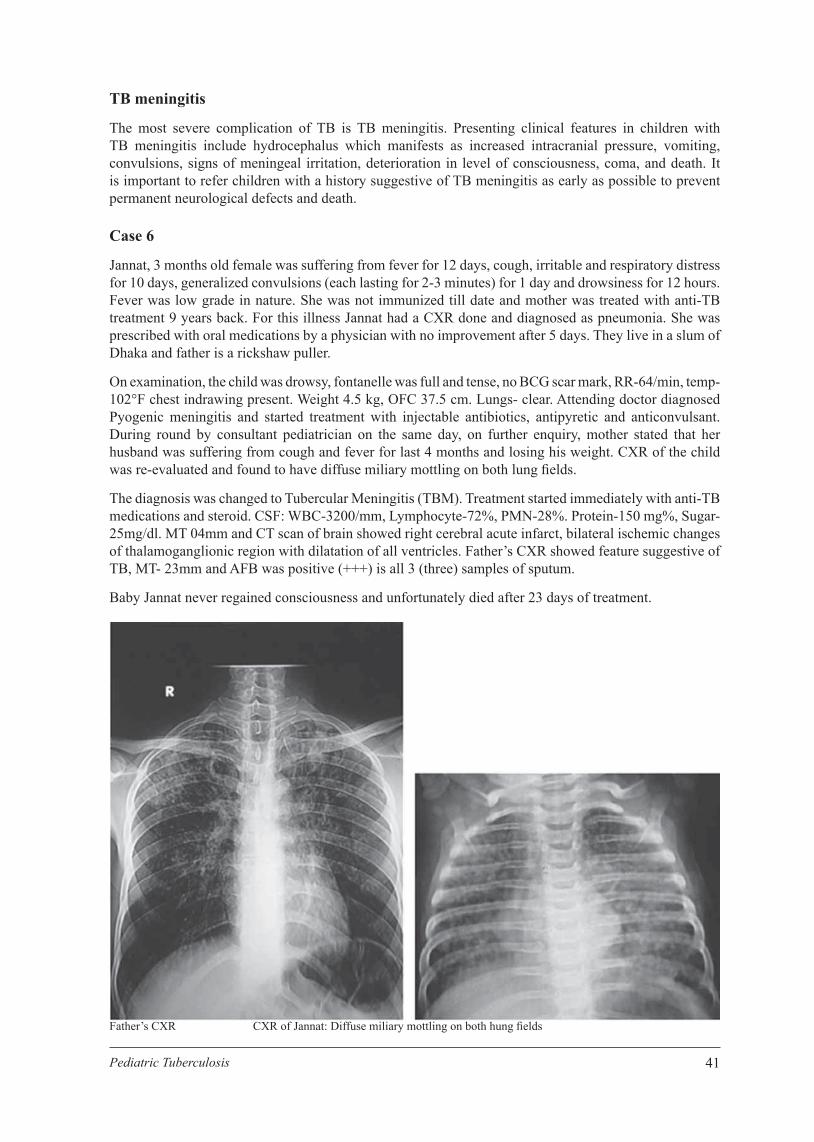
Jannat, 3 months old female was suffering from fever for 12 days, cough, irritable and respiratory distress for 10 days, generalized convulsions (each lasting for 2-3 minutes) for 1 day and drowsiness for 12 hours. Fever was low grade in nature. She was not immunized till date and mother was treated with anti-TB treatment 9 years back. For this illness Jannat had a CXR done and diagnosed as pneumonia. She was prescribed with oral medications by a physician with no improvement after 5 days. They live in a slum of Dhaka and father is a rickshaw puller.
On examination, the child was drowsy, fontanelle was full and tense, no BCG scar mark, RR-64/min, temp- 102°F chest indrawing present. Weight 4.5 kg, OFC 37.5 cm. Lungs- clear. Attending doctor diagnosed Pyogenic meningitis and started treatment with injectable antibiotics, antipyretic and anticonvulsant. During round by consultant pediatrician on the same day, on further enquiry, mother stated that her husband was suffering from cough and fever for last 4 months and losing his weight. CXR of the child was re-evaluated and found to have diffuse miliary mottling on both lung fi elds.
The diagnosis was changed to Tubercular Meningitis (TBM). Treatment started immediately with anti-TB medications and steroid. CSF: WBC-3200/mm, Lymphocyte-72%, PMN-28%. Protein-150 mg%, Sugar-25mg/dl. MT 04mm and CT scan of brain showed right cerebral acute infarct, bilateral ischemic changes of thalamoganglionic region with dilatation of all ventricles. Father’s CXR showed feature suggestive of TB, MT- 23mm and AFB was positive (+++) is all 3 (three) samples of sputum.
Baby Jannat never regained consciousness and unfortunately died after 23 days of treatment.
Father’s CXR CXR of Jannat: Diffuse miliary mottling on both hung fi elds

42 Pediatric Tuberculosis
Questions:
A. How an early diagnosis could have been made in this baby?
Answer:
B. What are the points favored the diagnosis of TBM?
Answer:
C. Describe father’s CXR fi ndings.
Answer:
TB spine and TB arthritis
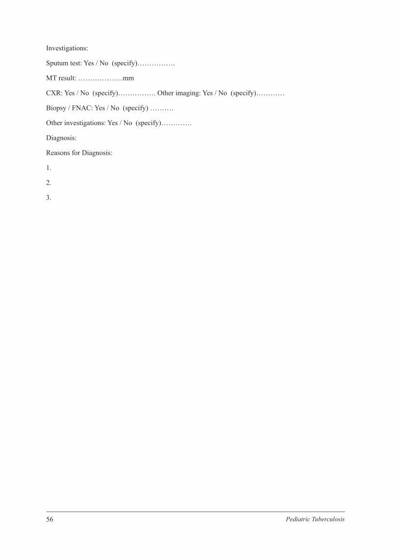
Tuberculosis of the bone and joint most commonly affects the spine. The lower thoracic or lumbar vertebrae are the commonest site. TB of the spine fi rst destroys the intervertebral discs followed by the body of vertebrae. This causes deformity of spine (gibbus) and neurological complications in growing children, if not treated properly.
TB bacteria does not directly affect bones and joints. The primary focus of infection is generally in the lungs or lymph nodes. Following the primary infection the bacilli spreads to the spine through blood dissemination. Common clinical features are local pain and tenderness in the affected spinal area, angulation of the spine called a gibbus deformity and/or Potts disease (severe kyphosis with destruction of the vertebral bodies). Destruction of vertebrae can cause spinal cord compression, leading to paraplegia or quadriplegia (diffi cultywalking) and diffi culty in passing urine.Any child with local pain and tenderness over the spine must be suspected of having spinal tuberculosis. A rapid onset of a gibbus (‘hump back’) is almost always due to tuberculosis.
Case 7
A 10 year old male child presented with pain in the back for 5 months and swelling over the spine for 3months. He was feeling unwell during the period and complained of diffi culty in walking for last 15 days.He was not febrile. He had no cough. He received BCG and other EPI vaccines. His father was a smokerand had chronic cough but was not a TB patient. His mother was healthy and other two younger sibs werewell. His father was a small trader and the family lived in a slum area.
He looked unwell, was mildly pale and had no lymph gland enlargement. Weight 23 kg, height 125 cm, BMI 14.7 (<10th centile). Examination of the spine a gibbus of the lower thorax was found. Deep tendon refl exes of lower limbs were exaggerated. Mantoux 13 mm. CXR: right paratracheal adenopathy. X-ray spine frontal projection: collapse of T 7-8.
Gibbs at T7 Level X-ray spine: collapse of T7 over T8

43Pediatric Tuberculosis
Questions:
A. Name the clinical features in this boy suggestive of TB spine.
Answer:
B. What are the laboratory fi ndings in favor of spinal TB in this boy?
Answer:
Miliary TB
Miliary TB is a disseminated form of TB, a serious complication of primary TB in young children. Children <3 years of age are at highest risk of developing miliary TB. Miliary TB may manifest with low-grade fever, malaise, weight loss, and fatigue. A rapid onset of fever and other manifestations of non-specifi c disease may be present. History of cough and respiratory distress may be present. The miliary TB may progress rapidly leading to death.
Physical examination fi ndings include enlarged lymph nodes; liver and spleen are often enlarged. Systemic signs include fever, increased respiratory rate and respiratory distress. Other signs are often subtle and should be carefully sought. CXR showsdiffuse/miliary mottling.
Less common signs include papular, necrotic, or purpuric lesions on the skin or choroid tubercles in the retina of the eye.
Abdominal TB
Abdominal TB is poorly understood and therefore often neglected by clinicians. Tuberculosis can involve any part of the gastrointestinal tract from mouth to anus. The most common site of involvement is the ileocaecal region. The spectrum of abdominal TB disease in children differs from adults. In children adhesive peritoneal and lymph nodal involvement is more common than gastrointestinal disease. Most children have constitutional symptoms of fever, abdominal pain, constipation, alternating constipation and diarrhoea, weight loss, anorexia and malaise. Other clinical features depend upon the site, nature and extent of involvement. Ascites is also a common physical sign of abdominal TB in children. An ascites tap will reveal straw-coloured ascitic fl uid (similar to that of pleural effusion). Palpable mass in ileocecal region is highly suggestive of abdominal TB.
Case 8
A 10-year old female child presented with fever and gradual weight loss for 6 months. Abdominal pain and distension were present for 3 months. She was fully immunized which included BCG. She had history of a TB contact.
She looked unwell. Her weight was 19 kg, height 122 cm with BMI 12.8 (<3rd centile). She had distended and soft abdomen. On examination, her fl anks were full with a palpable liver (3 cm) and spleen (2 cm). Shifting dullness and fl uid thrill were present. Bowel sounds were audible. She had clear lungs and normal heart.
Millary TB

44 Pediatric Tuberculosis
Urine examination revealed no albuminuria. HBsAg was negative, serum bilirubin 0.8 mg/dl, SGPT 45 U/L, serum total protein 7.8 mg/dl, serum albumin 3.5 mg/dl, cholesterol 170 mg/dl, partial prothrombin time 12.5 sec, control 12 sec. Ascitic fl uid: straw colored, cell 200/cmm, L 80%, N 20%, protein 6.2 g/dl, sugar 80 mg/dl. Microscopy of the ascites was AFB negative. MT 22 mm. Ultrasound of the abdomen: ascites with hepatosplenomegaly, normal liver echogenecity and no evidence of portal hypertension. CXR: right sided hilar adenopathy.
Lesson Learnt 1. TB of the abdomen causes ascites. 2. Peritoneal tap is straw coloured with abundant lymphocytes. 3. The biochemical reports of TB pleural and TB peritoneal fl uid are similar.
Danger signs requiring urgent hospital referral
10 years old girl: Abdominal TB with straw colored ascetic fl uid CXR: Right hilar adenopathy
1. Severe forms of PTB and EPTB for further investigation and initial management2. Severe respiratory distress (TB pneumonia with/without added bacterial infection, Pleural
effusion)3. Severe wheezing not responding to bronchodilators (sign of severe airway compression due to
enlarged lymph glands)4. Headache (especially if accompanied by vomiting), irritability, drowsiness, neck stiffness and
convulsions (signs of TB meningitis)5. Meningitis not responding to treatment6. Acutely ill with big liver and spleen and ascites (signs of disseminated TB)7. Breathlessness and peripheral oedema (signs of pericardial effusion)8. Acute angulation (bending) of the spine (sign of TB spine - gibbus)9. Other co-morbidities e.g. severe anaemia, severe malnutrition, very young infant (<3 months)
NB: Hospital referral should also be considered if there is any diagnostic uncertainty that requires further investigations.
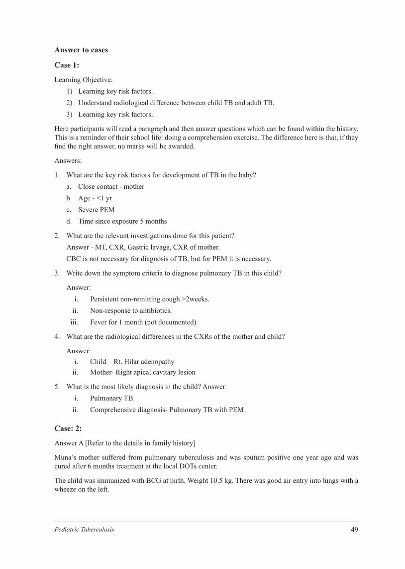
Uncommon signs indicative of recent TB infection 1) Phlyctenular conjunctivitis - Raised patch at the junction of the sclera and cornea surrounded by a red
area of conjunctivitis (has the shape of a comet (See child TB album). 2) Erythema nodosum - Raised, tender, purple patches on the shin.

45Pediatric Tuberculosis
CHAPTER 6

46 Pediatric Tuberculosis
47Pediatric Tuberculosis
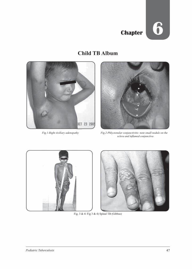
Chapter 6Child TB Album
Fig.1-Right Axillary adenopathy Fig.2-Phlyctenular conjunctivitis: note small nodule on the sclera and infl amed conjunctiva
Fig. 3 & 4: Fig 3 & 4) Spinal TB (Gibbus)

48 Pediatric Tuberculosis
Fig.9-TB Family : Father & Sons
Fig.5-TB right hip Fig.6-Lupus vulgaris
Fig.7-Mantoux test > 20 mm Fig.8-Complication of TBM: right sided hemiplegia with facialpalsy
49Pediatric Tuberculosis
Answer to cases
Case 1:
Learning Objective: 1) Learning key risk factors. 2) Understand radiological difference between child TB and adult TB. 3) Learning key risk factors.
Here participants will read a paragraph and then answer questions which can be found within the history. This is a reminder of their school life: doing a comprehension exercise. The difference here is that, if they fi nd the right answer, no marks will be awarded.
Answers:
1. What are the key risk factors for development of TB in the baby? a. Close contact - mother b. Age - <1 yr c. Severe PEM d. Time since exposure 5 months
2. What are the relevant investigations done for this patient? Answer - MT, CXR, Gastric lavage, CXR of mother. CBC is not necessary for diagnosis of TB, but for PEM it is necessary.
3. Write down the symptom criteria to diagnose pulmonary TB in this child?
Answer: i. Persistent non-remitting cough >2weeks.
ii. Non-response to antibiotics. iii. Fever for 1 month (not documented)
4. What are the radiological differences in the CXRs of the mother and child?
Answer: i. Child – Rt. Hilar adenopathy
ii. Mother- Right apical cavitary lesion
5. What is the most likely diagnosis in the child? Answer: i. Pulmonary TB.
ii. Comprehensive diagnosis- Pulmonary TB with PEM
Case: 2:
Answer A [Refer to the details in family history]
Muna’s mother suffered from pulmonary tuberculosis and was sputum positive one year ago and was cured after 6 months treatment at the local DOTs center.
The child was immunized with BCG at birth. Weight 10.5 kg. There was good air entry into lungs with a wheeze on the left.

50 Pediatric Tuberculosis
Answer B1. Mantoux test 22 mm induration 2. CXR: left hilar adenopathy with no lung lesion visible 3. Gastric lavage
Answer C. Intrathoracic TB lymphadenopathy
Case 3: Answer
A. Answer: Sputum AFB
B. Answer: 1. Cough for 6 weeks (>3 weeks) 2. Fever 3. Not responding to various causes of antibiotics.
Case 4: Answer
A. Answer: 1. Right sided anterior cervical lymph gland enlargement, size-3X3.5 cm
2. Matted glands
3. Discharging sinus
4. Non-tender
B. Answer:
1. All of above answers of A plus
2. Positive MT
3. Biopsy: Multiple epitheloid granuloma with Langerhans’s giant cell.
C. Answer:
No signifi cance, as the father had TB 10 years back, child is 15 months old.

51Pediatric Tuberculosis
Case 5: Answer
A. Answer:
1. Age > 5yrs
2. Recurrent fever
3. Chest pain
4. H/O contact
5. Non-response to antibiotics
6. Chest fi nding consistent with a pleural effusion (restricted movement, intercostal fullness, stony dullness to percussion)
B. Answer:
1. Is there any person suffering from cough for >3 weeks in the family OR, in the neighborhoods?
2. Is there any person taking anti-TB medication in the family OR, in the neighborhood?
3. Is there any person suffering from TB in the family OR, in the neighborhood?
4. Was there any family member or neighbor had TB in last 2 yrs?
C. Answer:
Healthy /Good looking /Not sick /Not distressed
Case 6: Answer 1. Detailed family history in a child living in urban slum could have revealed father’s TB.
Attentive reading of baby’s CXR by initial visit (miliary mottling on CXR) would defi nitely establish the diagnosis of TB and possible TBM.
2. C/F - Fever, respiratory distress, convulsion drowsiness, tense fontanelle. H/O close contact with father (suspected case). Non-immunized, Living in Slum
3. Cavitary lesion in left apical area, diffuse opacities in both lung. Sputum for AFB.
Case 7: Answer
A. Answer: 1. Back pain 2. Walking diffi culty 3. Feeling unwell 4. Gibbus 5. Exaggerated deep tendon refl exes of lower limb
B. Answer:
1. MT 13 mm
2. Collapse of T7 over T8
3. Right paratracheal shadow on CXR

52 Pediatric Tuberculosis

53Pediatric Tuberculosis
CHAPTER 7

54 Pediatric Tuberculosis

55Pediatric Tuberculosis
Chapter 7Case screening(Put Value or Encircle)
Case recording form for children with presumptive TB
Name: Sex: Male/ Female Age (Years / months): Address: (name and mobile no):
Temperature:……….°F/°C Weight:…………Kg Height: ………….Cm OFC: …………. Cm
Complaints:
Fever: Yes / No. Duration………day / month Persistent cough >2 weeks: Yes / No Weight loss: Yes / No Documented / Undocumented Less playfulness: Yes / No
BCG scar: Yes / No
H/O contact: Yes / No (if yes)
1. Close contact: Yes / No
2. Duration of contact: ……….year/month
3. Sputum positive: Yes / No / Not known
4. On treatment: Yes / No Duration of treatment: ……….month
5. Response to anti TB drugs: Good / Poor
6. Drug resistant: Yes / No / Not known
7. HIV positive: Yes / No / Not known (From document)
Physical fi ndings:
1. Weight/Height: ………% of median
2. Palpable lymph node: Yes (matted / sinus discharging) / No (specify)…………………
3. Gibbus: Yes / No
4. Chest examination: Normal Yes / No (specify)……………………. ………
5. Abdominal distension: Yes / No (specify)……………………. ………
6. Signs of meningeal irritation: Yes / No
7. Fontanelle: Bulged / Tensed / Normal / Closed
8. Others: (specify) …………………:
56 Pediatric Tuberculosis
Investigations:
Sputum test: Yes / No (specify)…………….
MT result: ……………….mm
CXR: Yes / No (specify)……………. Other imaging: Yes / No (specify)…………
Biopsy / FNAC: Yes / No (specify) ……….
Other investigations: Yes / No (specify)………….
Diagnosis:
Reasons for Diagnosis:
1.
2.
3.

57Pediatric Tuberculosis
CHAPTER 8

58 Pediatric Tuberculosis

59Pediatric Tuberculosis
Chapter 8Investigation
Confi rming diagnosis of TB in children is challenging; as it is diffi cult to detect mycobacteria in samples recovered from pediatric patients. The organism can be cultured in fewer than 50% of children with TB disease. Demonstrating or culturing Mycobacterium tuberculosis from clinical specimens establishes a defi nitive diagnosis of TB. Means of investigation include: direct microscopy, culture or DNA sequencing by molecular techniques etc. Supportive investigations are imaging (CXR, USG, CT, MRI) and Mantoux test (MT).
Complete Blood Count (CBC)/ ESR
CBC and ESR have no value in the diagnosis and follow-up of patient with childhood tuberculosis. ESR is a non-specifi c test and is infl uenced by several factors and is therefore not recommended.
Bacteriology
One must attempt at bacteriological diagnosis in every patient, as culture is the “gold standard” for diagnosis. Isolation of M. tuberculosis is possible in 30-40% of children with TB. Though younger children (<8 years) are unable to produce sputum, yet collection of sputum sample should always be attempted.Gastric aspirate is the alternative option used in younger children and already been described. The morespecimens you collect the greater the chance of culturing mycobacterium. Ideally two specimens (sputumor gastric aspirate) should be sent from each case and among these one sample should be early morningstrongly recommended. Bacteriological confi rmation is easy in cavitary (adult type) lesions. Mycobacterialculture and drug susceptibility is mandatory in case suspected of having multi-drug resistant TB.Collection of specimen has been detailed in earlier session. Besides sputum and gastric aspirate; pleural,peritoneal fl uid and CSF are also important specimens. These will not be discussed; anyone interested cango through text or talk to facilitator.
Each collected specimen should be properly labeled and sent to the laboratory as soon as possible. If forany reason transport is delayed, it can be stored at 4ºC to reduce overgrowth by bacterial contaminants.Sputum and gastric aspirate are potentially infective specimens. Proper personal protection must be takenas detailed in the infection control guideline. The specimen request form must have a brief descriptionof the patient’s clinical problem. The rate of positivity with other body fl uids and tissues are even lower.Microscopy can detect 1 X 10⁴ AFBs/ml of specimen. For this reason AFB is detected in less than 20% ofchildren, on the other hand it is 75% in adults.
Microscopy gives lower sensitivity because of two factors- paucity of mycobacteria in pediatric samplesand secondly the diffi culty in obtaining good quality samples from a child. Traditionally culture takes 4-6weeks, but positive yield can still be positive after 12 weeks.
Mantoux test (MT)
Also called Tuberculin Skin Test (TST) measures the delayed type hypersensitivity response to tuberculin Purifi edProtein Derivative (PPD). There are a number of TSTs available, but the most recommended test is the MT.A positive MT does not indicate active disease; it only indicates infection with M. tuberculosis. However, theMT is used in conjunction with other tests in diagnosing TB in children.

60 Pediatric Tuberculosis
MT is carried out by injecting 5 TU of tuberculin PPD-S or 2 TU of tuberculin PPD RT23 into the skin(intra-dermal) on the inner aspect of the left forearm. The MT is measured as the largest diameter ofi nduration and not the diameter of redness. Measure the diameter across the arm and not in its length.
The MT should be regarded as positive when the induration is:1. ≥10 mm 2. ≥5 mm in children with PEM, HIV infection and immunosuppression.
Interpretation of MT is done irrespective of previous BCG vaccination.
False negative MT may occur in-1. Severe malnutrition 2. Immune suppressive conditions:
Measles in last 3 months Whooping cough HIV infection On medications like steroids
3. Disseminated or (miliary) TB 4. TB meningitis 5. Very recent TB exposure (<3 months)
Reading best be taken within 48-72 hours of test. The result should not be recorded as positive/negative, rather as ……mm of induration.
Some tips on MT 1. Late reporters within 7 days with induration >10mm denotes tuberculous infection. 2. Repeat test-
a. No value in children with a previous positive test b. Useful after few weeks/months of an initial negative test. Any induration 6 mm or more than
previous test suggests recent infection. c. Best done on the opposite arm.
61Pediatric Tuberculosis
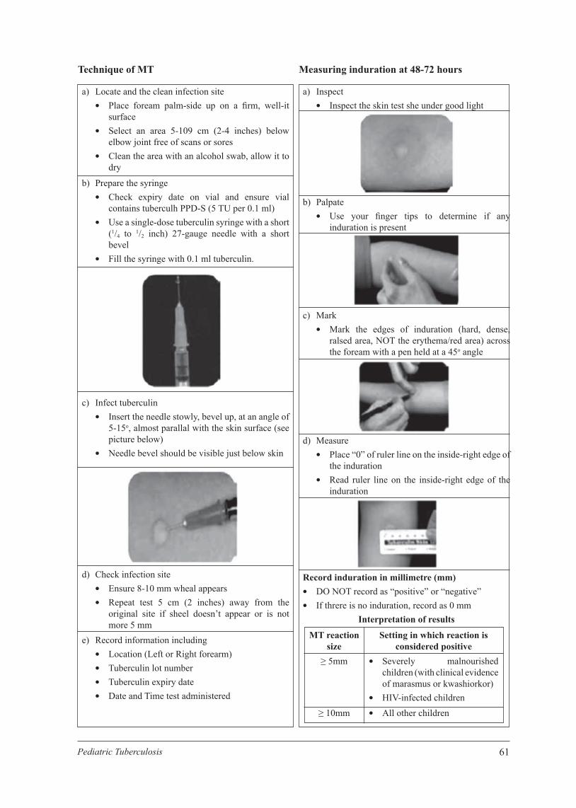
Technique of MT Measuring induration at 48-72 hours
a) Locate and the clean infection site Place foream palm-side up on a fi rm, well-it
surface Select an area 5-109 cm (2-4 inches) below
elbow joint free of scans or sores Clean the area with an alcohol swab, allow it to
dry
a) Inspect Inspect the skin test she under good light
b) Palpate Use your fi nger tips to determine if any
induration is present
c) Mark Mark the edges of induration (hard, dense,
ralsed area, NOT the erythema/red area) across the foream with a pen held at a 45o angle
d) Measure Place “0” of ruler line on the inside-right edge of
the induration Read ruler line on the inside-right edge of the
induration
Record induration in millimetre (mm) DO NOT record as “positive” or “negative” If threre is no induration, record as 0 mm
Interpretation of results
MT reaction size
≥ 5mm
≥ 10mm
Setting in which reaction is considered positive
Severely malnourished children (with clinical evidence of marasmus or kwashiorkor)
HIV-infected children All other children
b) Prepare the syringe Check expiry date on vial and ensure vial
contains tuberculh PPD-S (5 TU per 0.1 ml) Use a single-dose tuberculin syringe with a short
(1/4 to 1/2 inch) 27-gauge needle with a short bevel
Fill the syringe with 0.1 ml tuberculin.
c) Infect tuberculin Insert the needle stowly, bevel up, at an angle of
5-15o, almost parallal with the skin surface (see picture below)
Needle bevel should be visible just below skin
d) Check infection site Ensure 8-10 mm wheal appears Repeat test 5 cm (2 inches) away from the
original site if sheel doesn’t appear or is not more 5 mm
e) Record information including Location (Left or Right forearm) Tuberculin lot number Tuberculin expiry date Date and Time test administered
62 Pediatric Tuberculosis
Imaging
As pulmonary and lymph node disease in the chest predominates, CXR is the commonest imaging performed in diagnosis of TB in children. Ultrasonography is valuable in detecting ascites, pleural effusion and abdominal lymph gland enlargement. CT scan and MRI are done in CNS TB and TB spine.
In the majority of cases, children with pulmonary TB have CXR changes suggestive of TB. Good-quality CXRs are essential for proper evaluation. A lateral chest x-ray is helpful to evaluate hilar lymph node enlargement.
CXR will be detailed in next session. An X-Ray chest should be done in all form of TB.
Other tests for the diagnosis of Extrapulmonary TB
Site Practical approach to diagnosisPeripheral lymph nodes (especially cervical) Lymph node blopsyor fi ne needle aspiration
cytology(FNAC)Millary TB (disseminated) CXR and LP (to exclude meningitisTB meningitis LP (and CT scan of brain where available)Tuberculoma of Brain CT scan/MRI of brainPleural effusion CXR and pleural fl uid studyAbdominal TB Abdominal ultrasound and ascitic fl uid studyTB arthritis or Bone TB X-ray, joint fl uid study or synovial blopsyPericardial TB CXR, echocardiography and pericardial fl uid
study; pericardial blopsy and histopathology
In the diagnosis of EPTB all children should have a MT and CXR performed
HIV testing
HIV prevalence in SAARC countries varies (0.1% to 0.26%). HIV counseling and testing is indicated for TB patients with symptoms and/or signs of HIV-related conditions, and in TB patients having a history suggestive of high risk of HIV exposure (foreign expat, IV drug users, sex workers).
Tests for HIV: 1. ELISA for HIV antibody (screening), 2. Western blot (confi rmatory)
Other tests 1. Serological tests: WHO does not recommend test for antibodies against TB. These tests should not
be done to diagnose TB. 2. Interferon gamma assays (Quantiferon TB Gold, T-spot test): Costly and does not add more
information than MT. 3. PCR: Not a routine test for diagnosis for the diagnosis of childhood TB. 4. Gene Xpert: computer aided machine detects mycobacterium tuberculosis and rifampicin resistance
by detecting DNA of M. bacterium from sputum, CSF, lymphnode and gastric aspirate within 2 hours. WHO has recommended to use it as fi rst line investigation in suspected Tubercular meningitis.
5. BACTEC, a radiometric method, can detect mycobacterial growth within 7 days.
Revision exercises: 1. A child with >17 mm induration after 72 hrs should be diagnosed as mycobacterial disease. True/
False 2. MT measures induration not erythema. (True/ False) 3. High ESR is important in diagnosis of childhood TB. (True/ False)

63Pediatric Tuberculosis
4. CXR does not have pathognomonic features of childhood TB. (True/ False) 5. MT is false negative in severe PEM, recent measles and steroid therapy. (True/ False)
6. Bacteriological confi rmation is mandatory in: a. Suspected MDR TB (True/ False) b. TB/ HIV co-infection (True/ False) c. Cavitary lesion (True/ False)
7. Number of Gastric aspirate and sputum samples should be sent: a. 1 b. 2 c. 3 d. 4
8. HIV testing should be done in …a. 1 b. 2
9. Serological (immunoglobulin) tests are not recommended for childhood TB. (True/ False)
10. A 3 yrs child with cough for 3 weeks, fever and H/O contact with sputum positive case. On examination no chest fi ndings, weight 8 kg. With a provisional diagnosis of TB, write three investigations for the child: a. 1 b. 2 c. 3

64 Pediatric Tuberculosis

65Pediatric Tuberculosis
CHAPTER 9

66 Pediatric Tuberculosis

67Pediatric Tuberculosis
Chapter 9Radiology of Child TB
Imaging is a very important tool for evaluating children with suspected tuberculosis. As tuberculosis affects the chest in majority of cases, this session will highlight the CXR. Other X-rays and imaging will also be briefl y discussed. The knowledge you gain does not replace the expert opinion of a radiologist. The greatest danger is that the chest radiograph is seen in isolation, without taking into account the clinical history, examination and tuberculin skin test. An overall view is needed to ensure that the diagnosis is as accurate as possible.
Basics fi rst
The following basic conditions must be met 1. Full size CXR must be taken 2. Lateral view should be taken if possible 3. Previous CXR’s compared to present CXR (whenever possible) 4. A good viewing box. Do not view against the skylight. 5. The chest radiograph should be examined in a systematic manner.
Systematic Examination
A. Basic approach to the chest radiograph: rule of three (Figs. 1, 2, 3): a. Identifi cation: 3 aspects - name, date of CXR and supine or erect b. Now look at three aspects concerning the quality of the CXR
Quality aspects:
a. Rotation- (Fig-4, 5)
In older children check rotation by ensuring that the clavicle head ends are equidistant from the centre. In younger children ensure that the anterior rib ends are equidistant from the chest edge. The position of the patient is important, as lordotic views are diffi cult to evaluate.
b. Penetration- (Fig-6, 7, 8)
Correct penetration is ensured when the intervertebral spaces can just be distinguished through the lower part of the heart shadow. If these are too clear, this indicates over penetration and you may miss low density lesions e.g. cavity, pneumothorax, bullae. If you cannot see the intervertebral spaces then the fi lm is under penetrated and the lung fi eld will appear whiter.
c. Adequate, Inspiration (Fig-9, 10)
Adequate inspiration is when the 8-9th posterior rib, or the 6th anterior rib, is visible above the diaphragm. If more ribs are visible the lung is hyperinfl ated. A poor inspiration will make the heart look enlarged.
It is important to ensure that the chest radiograph is of acceptable quality, as a poor quality chest radiograph can lead to an incorrect diagnosis. Included is an example of a chest radiograph of unacceptable quality.
68 Pediatric Tuberculosis
The next step is to look at the three structures that are white:
a. Soft tissue
Examine the soft tissue of the chest for swelling, surgical emphysema or lumps.
b. Bony structures
Examine the bony tissue for fractures, signs of rickets or areas of infi ltration.
c. Heart shadow (Fig- 11, 12, 13, 14)
Examine the cardiac shadow for position, size and shape. The heart shadow should straddle the midline with one-third to the right and two-thirds to the left. Heart shadow will deviate to the side of collapse. Heart borders must be distinct; blurring indicates either collapse/ consolidation on that side. Blurred right border-right middle lobe collapse/consolidation. Blurred left border-lingula collapse/consolidation.
The next step is to look at the three structures that are black:
a. The trachea and the bronchi
Follow the trachea and bronchi carefully, looking for displacement or narrowing of the airways. Follow the width of major bronchi. Look for any area of narrowing.
b. The right and left lung. Right lung should be larger than the left- if not, suspect an area of right sided collapse. Right diaphragm is higher than the left.
c. Look at the three aspects of the lung i. Position ii. Size iii. Shape
Check three other aspects; diaphragm, pleura and mediastinum. a. The position of the left and right diaphragms. Right one is slight higher than the left (fi g.12).
Blurring of right dome indicates RLL consolidation/collapse (Fig.14). Blurring of left dome indicates consolidation/collapse of LLL (Fig. 15).
b. The two costophrenic angles- obliterated in pleural effusion c. The mediastinal structures
Thymic shadow (Fig-16, 17)
One of the normal structures that often cause considerable diffi culty in deciding if the mediastinum is wider than usual is the thymic shadow. The thymus is normally not visible in children older than four years. The classic sign of the thymic shadow is the sail sign. Look at the fi lm. If you are uncertain ask your facilitator.
The following radiological images are commonly seen in childhood TB
1. Uncomplicated lymph node disease
Uncomplicated primary tuberculosis is the most common form of TB seen in clinics. The radiological picture is that of a primary focus in the lung with accompanying mediastinal lymph gland enlargement.

69Pediatric Tuberculosis
Complete primary complex
The primary focus is often so small that it is most often not visible and only the accompanying mediastinal lymph gland enlargement is seen (Fig- 18). The primary focus can occur in any of the lobes and is not limited to the upper lobes, as in adults. Although the primary focus has no preference for any lobe, it tends to occur 1-2 cm from the pleura. It is normally poorly circumscribed and is less than 1 cm in diameter.
Primary complex with only mediastinal lymph gland enlargement visible
The mediastinal lymph gland enlargement is most commonly seen in the hilar regions of the lung (Fig. 19, 20). The lymph gland enlargement is usually unilateral, but bilateral lymph gland enlargement does occur in primary lymph node disease.
It is common to see the enlarged glands with infi ltration into the surrounding lung tissue. Visible hilar and paratracheal lymph gland (Fig-21) enlargement occurs, but is less common. A lateral chest radiograph is often useful in helping to visualize the hilar lymph gland enlargement (Fig-22, 23). It detects 11% more lymhadenopathy when coupled with a PA/AP fi lm. Care must be taken not to confuse the main pulmonary arteries with hilar lymph gland enlargement, but if the area designated by the arrow has well-circumscribed round lesions present, then lymph gland enlargement is certainly present.
Primary complex with mediastinal lymph gland enlargement not visible (Fig-24, 25, 26)
Often the hilar lymph gland enlargement is not clearly visible or distinguishable from the pulmonary vessels. In this case, careful evaluation of the airways is often helpful, as compression of the airways especially the right and left main bronchi is indirect evidence of hilar lymph gland enlargement. A more penetrated chest radiograph is often useful for visualizing the airways.
Airway compression due to lymph gland enlargement is more common in younger infants. Other causes of airway compression are bronchogenic cysts or vascular anomalies. But, in areas of high TB prevalence like most of the SSARC countries, the most common cause is tubercular lymphadenopathy. In a minority of cases, the diagnosis is enabled by observing a calcifi ed Ghon focus (Fig-27). Uncomplicated primary infection can pass unnoticed, with calcifi ed glands becoming visible on chest radiographs taken later for other reasons.
2. Complicated primary pulmonary disease
Understanding the pathogenesis of primary disease makes interpreting the radiographs of complicated primary disease easier. Complicated disease follows the involvement of the infected lymph nodes and the adjacent large airways, mainly the bronchi. As the airways become more involved, the airway lumen decreases. As the lumen narrows or the gland ulcerates into the airway, the clinical and radiological picture of the child changes.
Radiological pictures of airway obstruction
A. Large airway obstruction (Fig-24, 25, 26)
Occasionally the involved glands obstruct the bronchi, causing a clinical picture that is often confused with asthma. Clinically, the diagnosis is suspected as the airway obstruction responds poorly to bronchodilators. Obstruction of the bronchi is normally accompanied by visible glands in the mediastinum; but, in younger patients, the obstruction is only visible by following the trachea and bronchi. Narrowing of the major airways can then be seen. In most cases, the obstruction clears on medical treatment.
B. Unilateral hyperinfl ation (Fig-25)
This is not a common radiological picture. As the airways start to narrow, a point is reached where the narrowing acts as a “check valve”, allowing air to be trapped in the affected lobe or lung. The

70 Pediatric Tuberculosis
diagnosis is best made by combining the clinical examination with the radiological picture. On clinical examination, the affected lung is hyperinfl ated, with decreased air entry on auscultation. The radiological picture is that of a hyperinfl ated enlarged lung or lobe with decreased vascularity. In some cases, the glands are not directly visible, but compression of the airways can be seen. The most common cause of unilateral hyperinfl ation is foreign body aspiration.
C. Lobar or segmental collapse (Fig-28)
With complete obstruction of the airways by the infected lymph gland, collapse of the segment or lobe occurs. The lobes affected are usually the right middle lobe or the lower lobes (Fig-28). The most diffi cult collapse to observe is the left lower lobe, as the lobe remains hidden behind the cardiac shadow. The collapsed left lower lobe is visible as a double shadow seen through the cardiac shadow (Fig-29). Other common causes of collapse are mucus plugs and foreign-body aspiration.
D. Tubercular Consolidation - can result for primary focus (Fig-30-31)
E. Tuberculous bronchopneumonia (Fig-32)
With ulceration of the lymph nodes through the bronchus wall, aspiration of tuberculous material can pour throughout the lung, leading to bronchopneumonia. This clinical picture most often occurs in young children who are acutely ill, and they often require supplementary oxygen. Another mechanism that can lead to TB bronchopneumonia is aspiration of material from cavitating lesions.
F. Combination of the above complications
Some children develop a combination of the lesions above, or they may combine these with other radiological pictures like miliary TB or pleural effusions.
3. Pleural effusion (Fig-33, 34)
Children with tubercular pleural effusion usually presents with fever and an insidious onset of shortness of breath and are not toxic though have a high fever. The effusion can vary in size from complete opacifi cation of the whole hemithorax to a small effusion with only obliteration of the costophrenic angle (Fig. 33). Diagnosis of pleural effusion is made by the clinical and radiological pictures. In nearly all cases the TB effusion clears up rapidly on treatment. After three to four weeks of treatment, the pleural effusion will have cleared, with only slight pleural thickening still present (Fig. 34).
4. Miliary TB (Fig-35)
Dissemination of a large number of organisms into the blood circulation follows the involvement of blood vessels by the primary complex. These large numbers of bacilli are then spread throughout the body and lead to the development of granulomas in all the involved organs. These children are clinically very ill and often have accompanying TB meningitis. As the granulomas are all similar in size, they are seen on chest radiographs as evenly distributed, small, millet-sized (less than 2 mm), round opacities.
5. Post-primary TB (Fig-36)
Post-primary TB (adult-type) is seldom seen prior to adolescence, with the same clinical and radiological picture as TB in adult patients. The involvement is usually in the upper lobes or the apices of the lower lobes. As the disease progresses, the lesions become more dense and eventually develop cavities. The cavities, typically in the upper lobes, allow the spread of the TB to other parts of the lungs. The infection heals by fi brosis, leading to fi brotic upper lobes.
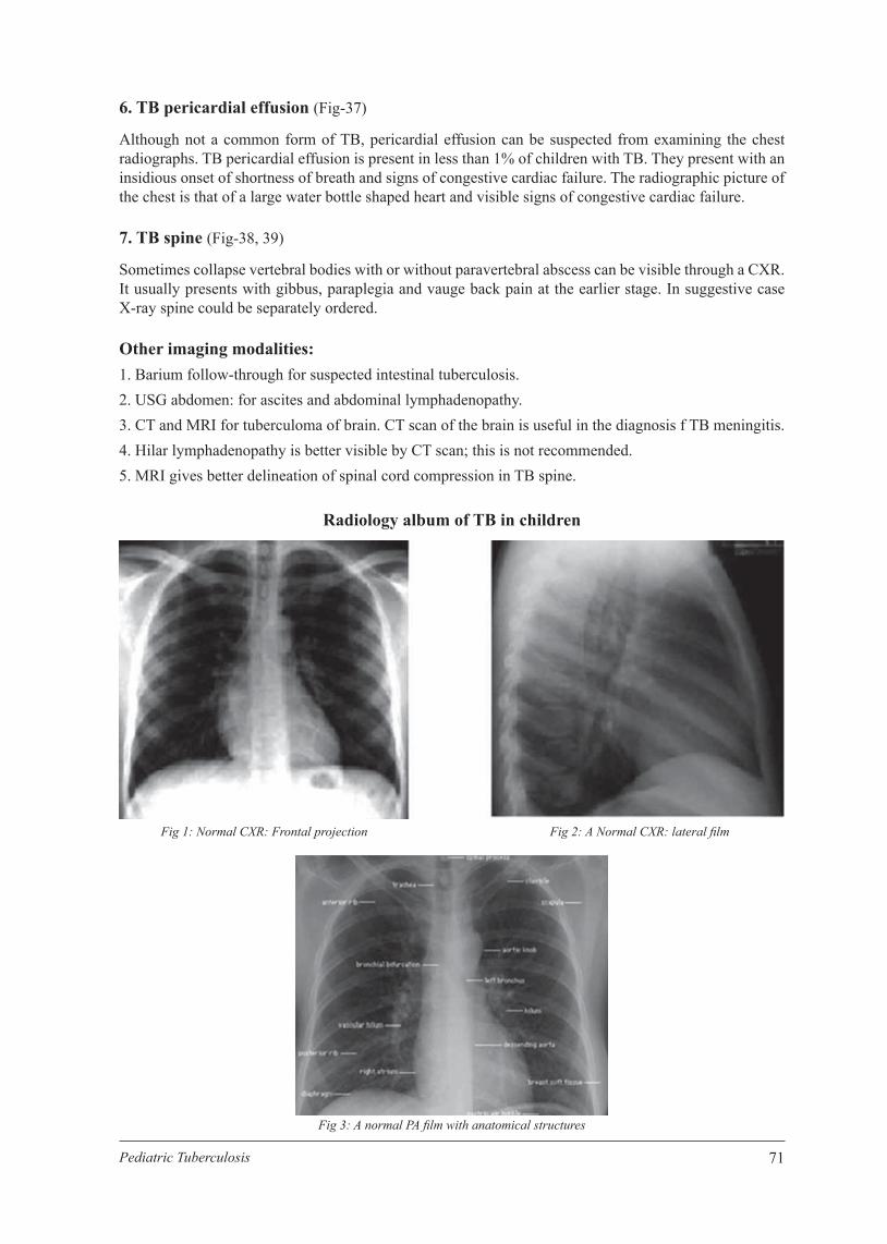
71Pediatric Tuberculosis
6. TB pericardial effusion (Fig-37)
Although not a common form of TB, pericardial effusion can be suspected from examining the chest radiographs. TB pericardial effusion is present in less than 1% of children with TB. They present with an insidious onset of shortness of breath and signs of congestive cardiac failure. The radiographic picture of the chest is that of a large water bottle shaped heart and visible signs of congestive cardiac failure.
7. TB spine (Fig-38, 39)
Sometimes collapse vertebral bodies with or without paravertebral abscess can be visible through a CXR. It usually presents with gibbus, paraplegia and vauge back pain at the earlier stage. In suggestive case X-ray spine could be separately ordered.
Other imaging modalities: 1. Barium follow-through for suspected intestinal tuberculosis. 2. USG abdomen: for ascites and abdominal lymphadenopathy. 3. CT and MRI for tuberculoma of brain. CT scan of the brain is useful in the diagnosis f TB meningitis. 4. Hilar lymphadenopathy is better visible by CT scan; this is not recommended. 5. MRI gives better delineation of spinal cord compression in TB spine.
Radiology album of TB in children
Fig 1: Normal CXR: Frontal projection Fig 2: A Normal CXR: lateral fi lm
Fig 3: A normal PA fi lm with anatomical structures

72 Pediatric Tuberculosis
Fig-4-CXR-Rotated fi lm
Fig-6-over penetration
Fig-8-adequqte penetration
Fig-5-CXR-central fi lm
Fig-7-under penetration
Fig-9-expiratory fi lm

73Pediatric Tuberculosis
Fig-10-adequqte inspiration
Fig-12-Silhouette sign-RML consolidation
Fig-14-Silhouette sign-RLL consolidation
Fig-11-silhouette sign-right border heart and right middle lobe
Fig-13-Silhouette sign-Lingular segment consolidation
Fig-15-Silhouette sign-LLL consolidation

74 Pediatric Tuberculosis
Fig-16-CXR-Enlarged thymus-1yr (Sail sign)
Fig18-Primary Pulmonary complex
Fig 20: Left hilar lymphadenopathy
Fig-17-CXR-enlarged thymus (Sail sign)
Fig 19: Right hilar adenopathy
Fig 21: Right paratracheal lymphadenopathy
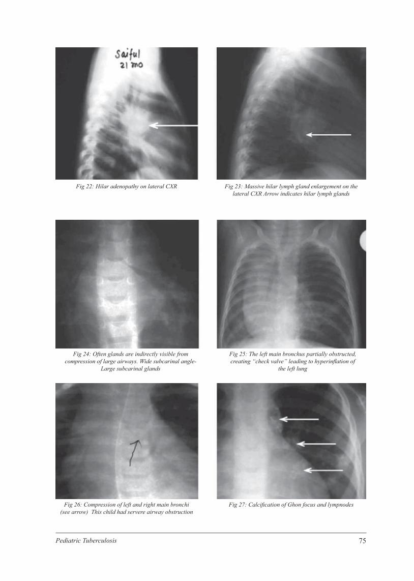
75Pediatric Tuberculosis
Fig 22: Hilar adenopathy on lateral CXR
Fig 24: Often glands are indirectly visible from compression of large airways. Wide subcarinal angle-
Large subcarinal glands
Fig 26: Compression of left and right main bronchi(see arrow) This child had servere airway obstruction
Fig 23: Massive hilar lymph gland enlargement on the lateral CXR Arrow indicates hilar lymph glands
Fig 25: The left main bronchus partially obstructed, creating “check valve” leading to hyperinfl ation of
the left lung
Fig 27: Calcifi cation of Ghon focus and lympnodes

76 Pediatric Tuberculosis
Fig-28-collapse-RML and RLL
Fig-30-consolidation-RUL (Frontal projection)
Fig 32: CXR: Bilateral Extensive opacities
Fig-29-Double left borders
Fig-31-consolidation-RUL (Lateral projection)
Fig 33: CXR: Right sided Pleural effusion
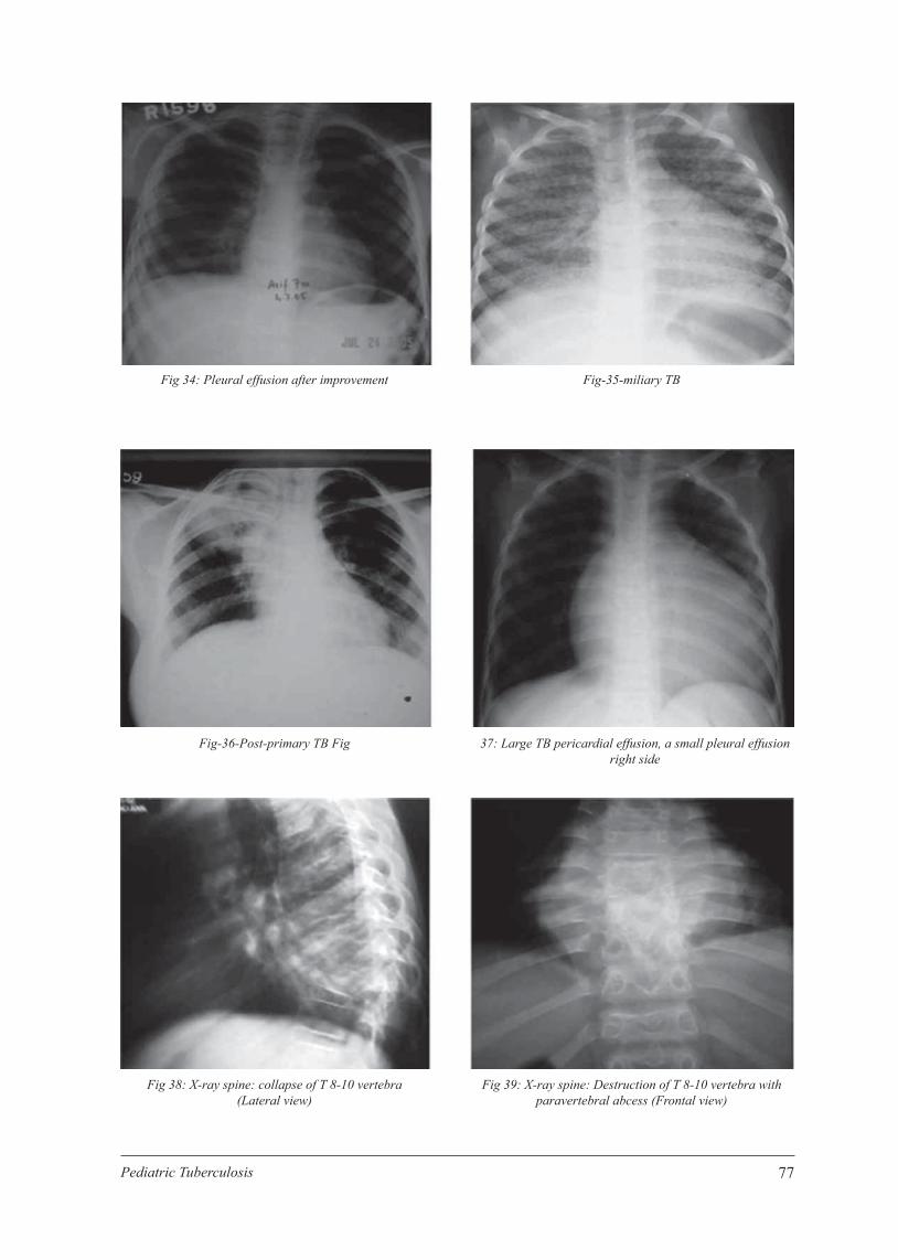
77Pediatric Tuberculosis
Fig 34: Pleural effusion after improvement
Fig-36-Post-primary TB Fig
Fig 38: X-ray spine: collapse of T 8-10 vertebra (Lateral view)
Fig-35-miliary TB
37: Large TB pericardial effusion, a small pleural effusion right side
Fig 39: X-ray spine: Destruction of T 8-10 vertebra with paravertebral abcess (Frontal view)

78 Pediatric Tuberculosis
Fig 40: A combination of features is sometimes sen. In the case above, there is expansile pneumonia with a cavity in the left upper lobe, and compression of the left main bronchus and
trachea with bronchopneumonia of the right upper lobe.
Fig 41: Left lower lobe collapse with the double shadow seen through the cardiac shadow. The left main bronchus can in some
cases be seen running down at a more acute angle, not seen in this radiograph.

79Pediatric Tuberculosis
CHAPTER 10

80 Pediatric Tuberculosis

81Pediatric Tuberculosis
Chapter 10Treatment of Child TB
Children who have been diagnosed with TB disease must receive directly observed TB treatment (DOT) with the appropriate TB regimen. All children being treated for TB must be recorded in the TB treatment register. Once TB treatment is started, it should be continued until completion, unless an alternative diagnosis has been confi rmed. Treatment outcomes in children are generally good, even in young and immune compromised children with severe and disseminated disease. Children with TB tolerate anti-TB drugs well and respond to treatment.
Objectives of treatment of TB 1. Cure individual patients 2. Prevent death from active TB or its late effects 3. Prevent relapse of TB (by eliminating the dormant bacilli) 4. Reduce transmission 5. Prevent the development of drug resistance
Pharmacokinetics of anti TB drugs
Rapid reduction in the organism load is important since it improves clinical symptoms, limits disease progression, terminates transmission and protects against drug resistance. A rapid reduction is achieved by bactericidal drugs that kill actively metabolizing organisms. However, there are multiple sub-populations of organisms, some extra- and others intra-cellular, with highly variable rates of metabolism. Permanent cure requires effective eradication of all organisms, including hypometabolic bacilli, which justifi es the use of multiple drugs and the prolonged duration of therapy.
Isoniazid (INH) has the most potent early bactericidal activity (EBA), killing the vast majority of rapidly metabolizing extra-cellular bacilli within the fi rst few days of treatment.
Rifampicin (RMP) is also bactericidal, but is more effective in eradicating intra-cellular organisms.
Pyrazinamide (PZA) contributes by killing extracellular bacilli that persist within the acidic centers of caseating granulomas.
Ethambutol (EMB) kills actively growing bacilli but has fairly limited potency; its role in RMP containing regimens is mainly to reduce the risk of acquired drug resistance in patients with high bacillary loads. Risk of ethambutol in causing optic neuritis has been assessed in a recent WHO analysis and is now recommended in the treatment of childhood TB. Meals, especially carbohydrates, reduce absorption antitubercular drugs especially INH, hence it is recommended that TB drugs be taken 30 minutes before breakfast/meal.
The main variables that infl uence the success of chemotherapy, apart from drug resistance, are the bacillary load and organs involved. Sputum smear-negative disease is usually paucibacillary (few bacilli) and therefore the risk of acquired (previously treated) drug resistance is low. Sputum smear-positive disease indicates a high bacillary load and an increased risk for developing drug resistance.

82 Pediatric Tuberculosis
Drug penetration into the anatomical sites involved is good and the success of 3 drugs (INH, RMP, PZA) during the 2-month intensive phase and 2-drugs (INH, RMP) during the 4-month continuation phase, is well established. In the presence of extensive disease (excluding TB meningitis), HIV co-infection and/or suspicion of INH resistance, the addition of EMB during the intensive phase is advised to improve outcome and reduce the risk of acquiring drug resistance. By the end of 2 months bacillary load is suffi ciently reduced so that daily therapy with INH and RMP during the 4-month continuation phase is suffi cient to ensure organism eradication without developing drug resistance.
It is essential to consider the cerebrospinal fl uid (CSF) penetration of drugs used in the treatment of TB meningitis. INH and PZA easily penetrate the CSF, while RMP and streptomycin (SM) only achieve therapeutic levels in the presence of meningeal infl ammation. EMB hardly penetrates the CSF, even in the presence of meningeal infl ammation, which explains why SM replaces EMB in the treatment of TBM.
Recommended treatment regimens
Anti-TB treatment is divided into two phases: an intensive phase and a continuation phase.
The purpose of the intensive phase is to rapidly eliminate the majority of organisms to prevent the emergence of drug resistance and render the patient non-infectious as quickly as possible. This phase uses more drugs than the continuation phase. The purpose of the continuation phase is to eradicate the dormant organisms.
Regular weight-based dose adjustment is important for children; as they respond to treatment, their weight increases. This is particularly evident in malnourished children. This adjustment is especially important during the intensive phase.
Treatment of TB in children younger than 8 years of age (with current FDCs)
Body weight Intensive Phase (2 months) Continuation phase (4 months)RHZ* 60,30,150 RH 60,30
2-2.9 kg 16 tab 16 tab
3-5.9 kg 1 tab 1 tab
6-8.9 kg 11/2 tabs 11/2 tabs9-11.9 kg 2 tabs 2 tabs
12-14.9 kg 21/2 tabs 21/2 tabs15-19.9 kg 3 tabs 3 tabs
20-24.9 kg 4 tabs 4 tabs
25-29.9 kg 5 tabs 5 tabs
30-35.9 kg 6 tabs 6 tabs
* R - Rlfamplcln, H - Isonlazld; Z -
Pyrazlnamlde
Recommended Doses of First-Line Anti-TB Drugs for Children (WHO 2014)
Drug Daily dose and range (mg/kg body weight) Isoniazid (H) 10 (7-15) [maximum 300mg] Rifampicin (R) Pyrazinamide (Z)
15 (10-20) [maximum 600mg] 35 (30-40) [Maximum 2000 mg]
Ethambutol (E) 20 (15-25) [maximum 1200mg]

83Pediatric Tuberculosis
NB: Higher dosage (mg/kg) are required for young children to achieve effective bactericidal activity, as these age group infl uences drug metabolism. Moreover, systematic review also shows an excellent safety profi le of revised dosages and are not associated with an increased risk of toxicity (no increased risk of drug-related hepatotoxicity due to INH or PZA, or of optic neuritis due to ethambutol.
Fixed-dose-combinations (FDCs) for children:
Child-friendly formulations are ideal for use to ensure adequate therapeutic blood levels and compliance to treatment regimen. These are dispersible tablets and currently available formulations contain 60 mg of Rifampicin, 30 mg of INH and 150 mg of Pyrazinamide per tablet. As there is a need to break these tablets for some weight bands (see Table 9), which pose uncertainty in reaching the desired levels and also not user friendly. The WHO, along with the Global Alliance for TB Drug Development, has taken the initiative for a new FDC. It contains 75 mg of rifampicin, 50 mg of INH and 150 mg of pyrazinamide per tablet1, which is child-friendly and in line with the higher WHO recommended dosage from the Rapid Advice 2010.
FDC tablet Current FDC Upcoming FDC*3 R60, H30, Z150 R75, H50, Z1502 R60, H30 R75, H50
Table: 10 Example of Weight band table for using “new”/upcoming FDCs
Weight Bands(Kg)
Number of TabletsIntensive Phase Continuation Phase
RHZ (mg) E (mg) RH (mg)75/50/150 per tablet 100 per tablet 75/50 per tablet
4-7 1 1 18-11 2 2 2
12-15 3 3 316-24 4 4 425+ Go to adult dosages and preparations
Children with extensive pulmonary disease (cavitary lesion, military TB) and severe EPTB (disseminated TB) living in settings of low HIV prevalence or low INH resistance should be treated with a four-drug regimen (HRZE) for 2 months followed by a two-drug regimen (HR) for 4 months. Ethambutol is considered to be safe in children at a dose of 20 mg/kg (range 15-25 mg/kg) daily.
Children 25 kg and over will also follow the recommended regimen for adults.
WHO Recommendations:
1. Children with suspected or confi rmed pulmonary TB or tuberculosis peripheral lymphadenitis who live in settings with low HIV prevalence or low prevalence of isoniazid resistance and children who are HIV-negative can be treated with a three-drug regimen (HRZ) for 2 months followed by a two-drug (HR) regimen for 4 months at the dosages specifi ed in table 2.
Note: The regimen for new patients should contain rifampicin for 6 months. Wherever feasible, the optimal dosing frequency for new patients is daily throughout the course of therapy.
2. Infants aged 0–3 months with suspected or confi rmed pulmonary TB or tuberculosis peripheral lymphadenitis should be promptly treated with the standard treatment regimens, as described in

84 Pediatric Tuberculosis
recommendation 1. Treatment may require dose adjustment to reconcile the effect of age and possible toxicity in young infants. The decision to adjust doses should be taken by a clinician experienced in managing pediatric TB.
Note: There are very limited data to inform drug dosages for neonates, who have certain characteristics - especially in the fi rst week of life - that are likely to affect drug metabolism. Treatment of neonates may require dose adjustment to reconcile the effect of age and possible toxicity and should therefore be undertaken by a clinician experienced in managing pediatric TB. If such expertise is not available, and TB has either been defi nitively diagnosed or is strongly suspected, treatment with the standard drug regimen may be considered.
3. During the continuation phase of treatment, thrice-weekly regimens can be considered for children known not to be HIV-infected and living in settings with well-established directly-observed therapy (DOT).
4. Streptomycin should not be used as part of fi rst-line treatment regimens for children with pulmonary TB or tuberculosis peripheral lymphadenitis.
5. Children with suspected or confi rmed tuberculosis meningitis and children with suspected or confi rmed osteoarticular TB should be treated with a four drug regimen (HRZ) plus streptomycin for 2 months, followed by a two-drug regimen (HR) for 10 months, the total duration of treatment being 12 months. The doses recommended for the treatment of tuberculosis meningitis are the same as those described for pulmonary TB. For easier understanding.
6. Children with extensive pulmonary disease (cavitary lesion, military TB) and severe EPTB (disseminated TB) living in settings of low HIV prevalence or low INH resistance should be treated with a four-drug regimen (HRZE) for 2 months followed by a two-drug regimen (HR) for 4 months. Ethambutol is considered to be safe in children at a dose of 20 mg/kg (range 15-25 mg/kg) daily.
7. Children 25 kg and over will also follow the recommended regimen for adults.
Table 11 lists all current recommended treatment regimens.
Table 11: Recommended treatment regimens for new cases of TB in children
TB diagnostic categoryAnti-TB drug regimens a
Intensive phase Continuation phaseLow HIV prevalence (and HIV-negative children) and low isoniazid resistance settings Smear-negative pulmonary TB
2HRZ 4HRIntrathoracic lymph node TBTuberculous peripheral lymphadenitisExtensive pulmonary disease
2HRZE 4HRSmear-positive pulmonary TBSevere forms of extra-pulmonary TB (other than tubercu-lous meningitis/osteoarticular TB)All SAARC countriesTuberculous meningitis and osteoarticular TB 2HRZ+SM 10HRMDR-TB Individualized regimens (MDR section)
NB: The standard code for anti-TB treatment regimens uses an abbreviation for each anti-TB drug: isoniazid (H), rifampicin (R), pyrazinamide (Z) and ethambutol (E). A regimen consists of two phases - the initial and continuation phases. The number at the front of each phase represents the duration of that phase in months. Example, 2HRZ: Duration of this phase is 2 months and drug treatment is daily (no subscript numbers after the abbreviations) with isoniazid, rifampicin and pyrazinamide.
85Pediatric Tuberculosis
For children with TB meningitis and osteo-articular tuberculosis, treatment may be extended up to 12 months based on clinical judgment.
Streptomycin should be avoided when possible in children because the injection is painful and irreversible auditory damage may occur. It may be given for the fi rst 2 months of treatment of TBM.
NB: Where treatment failure is in doubt, DR-TB should be considered and worked upon (DST needed).
Corticosteroid use in Tuberculosis
Corticosteroids may be used for the management of some complicated forms of TB, e.g. tuberculosis meningitis, complications of airway obstruction by TB lymph glands, endo-bronchial TB and pericardial TB. Corticosteroids have been shown to improve survival and reduce morbidity in advanced tuberculous meningitis and are thus recommended in all cases of tuberculous meningitis. According WHO, prednisone is used most frequently, in a dosage of 2 mg/kg daily, increased to 4 mg/kg daily in the case of the most seriously ill children, with a maximum dosage of 60 mg/day for 4 weeks. The dose should then be gradually reduced over 1–2 weeks before stopping.
Directly Observed Therapy- Short course (DOTS)
DOTS is a very effective way of internationally recommended policy package for TB control, called the DOTS strategy. DOTS means that an observer supports the patient during treatment (ensures that s/ he takes medication regularly), which is essential for completion of treatment and TB cure. This ensures patient takes right anti TB drugs, in the right doses, at the right interval and for the right period of time.
TB treatment should always be directly observed and drugs used should be in a fi xed drug combination (FDC). Ethambutol needs to be added with the FDC when indicated. Drug dosages, depending on the body weight of the child, are given daily (7 days a week). The dose should be adjusted as the weight changes during the course of treatment. Children should therefore be weighed at least after 1, 2, 3 (or at a lesser interval when necessary) and 6 months of therapy; their weight should be documented on the TB treatment card. If there is poor response to therapy (no weight gain, persistent symptoms after 2-3 months of treatment) children must be referred for urgent assessment.
[Referral-
The following children should be referred for expert opinion and management: All children with severe forms of TB (TB meningitis, miliary TB, TB peritonitis, TB pericarditis,
spinal or skeletal TB) Children suspected of having MDR TB, XDR TB (in contact with MDR TB, XDR TB case or not
responding to fi rst-line therapy) If there is poor response to therapy (no weight gain, persistent symptoms after 2-3 months of treatment)
Follow up of children during treatment
Children should be followed up monthly for the fi rst 3 months. In children responding to treatment the symptoms resolve, they gain weight and become playful within 1-2 months. It is important to accurately document the child’s weight at each of the follow-up visit and to adjust the drug dosages accordingly. Children with sputum smear-positive TB should be followed as adult clients with repeat sputum examinations done after 2, 5 and at 6 months of treatment.
The chest x-ray is a poor indicator of treatment response; and lymph nodes may initially enlarge as a result of an improvement in the child’s immune response. Routine follow-up chest x-rays are not required in children. Follow-up x-rays are only recommended in children with persistent symptoms or poor response to treatment, or if new symptoms develop while on treatment.
Key points for follow-up1. Praise for continuing treatment

86 Pediatric Tuberculosis
2. Symptomatic improvementsa. Fever- Subsided/ persisting b. Cough- Subsided/persistingc. Appetite – Regained/ not improvedd. Playfulness – Active child/less activee. Other symptoms …………………………
3. Weight: (readjustment of dose)4. Compliance: Good/Not compliant 5. Adverse effect: Jaundice/ peripheral neuropathy/Rash/Others6. Sputum test (in smear +ve cases) after 2 months of earlier if no improvement of deterioration 7. CXR- If no improvement/deterioration8. Ask of any person in the family developed/having chronic cough 9. Provide next follow-up date
Causes of deterioration during TB treatment
Children may sometimes deteriorate or experience a worsening of symptoms despite adequate therapy. The most important questions to answer are: Is the drug dosage correct? Is the child taking the drugs as prescribed (good adherence)? Is the child HIV-infected? Is the child severely malnourished? Is there a reason to suspect drug-resistant TB (the index case has drug resistant TB or is a re-
treatment case or is also not responding to therapy)? Is there another reason for the child’s illness other than TB? Is the diagnosis wrong? Does the child not have another disease?
In children recovering from severe malnutrition or being treated with antiretroviral treatment (ART) or those with intracranial tuberculomas the symptoms may worsen after starting TB treatment. This is due to the recovery of their immune responses. This is referred as immune reconstitution infl ammatory syndrome (IRIS). Any child with severe persistent symptoms should be referred for assessment.
Drug related adverse events
The toxicities are related to dose and regimens of TB drugs
TB Drugs Main toxicitiesIsoniazid Hepatitis
Peripheral neuropathyRifampicin Hepatitis
Orange discoloration of secretionsDrug interactions especially anticonvulsants and Antiretroviral treatment
Pyrazinamide HepatitisArthralgla
Ethambutol Visual disturbance (acuity, color vision)Streptomycin Ototoxic & nephrotoxic
hypersensitivity reac-tions and drug rashes may occur with any drug

87Pediatric Tuberculosis
Management of adverse event
Adverse events caused by TB drugs are much less common in children than in adults. Most serious adverse event is hepatotoxicity, which can be caused by PZA>INH>RMP.
Serum liver enzyme levels should not be routinely be monitored, as asymptomatic children started on TB children have commonly a mild elevation of serum liver enzymes (<5 times normal values). This is not an indication to stop treatment. However, the occurrence of liver tenderness with vomiting, hepatomegaly or jaundice during the course of treatment should lead to immediate stopping of all the drug and urgent referral for further investigation.
Management of drug induced hepatitis 1. Stop all anti-TB therapy 2. Perform serum liver enzyme levels: severe involvement indicated by - ALT >3 times (with
symptomssuggestive of liver disease) 3. Screen for viral hepatitis 4. F/up the Symptoms Biochemical ALT level 5. When the ALT<2 times upper limit and symptoms resolved 6. Reintroduce medicines-
a. RMP- gradual reintroduce in 48-72 hours (5mg/kg on Day-1, 10mg/kg on Day-2, 15mg/kg on Day-3)
b. Repeat ALT, if no rebound elevation gradually add in INH c. Repeat ALT, if no rebound elevation occurs RMP/INH
7. Avoid PZA 8. Start alternative non-hepatotoxic drugs- Ethambutol + Streptomycin + Fluroquinolones especially in
severe disease e.g. TBM, disseminated TB 9. A suggested regimen is 2SHE/10HE. An expert should be involved in the further management of
these cases.
INH-induced peripheral neuropathy
May occur in-1. Severely malnourished 2. HIV-infected children on HAART 3. Chronic liver disease 4. Renal failure
Supplemental pyridoxine (12.5 - 25mg = •-1 tablet/ day) is recommended for these patients. Pyridoxine is not routinely prescribed other than the group mentioned above.
Retreatment
Treatment failure in children is rare but should be managed in the same way that treatment failure is managed in adults. The most likely cause for treatment failure or relapse within 6 months of treatment completion is failure to adhere to treatment.
In children when anti-TB treatment fails or a relapse occurs, every effort should be made to fi nd the most likely cause for the failure or relapse. Non-adherence is the commonest cause. There are multiple (psychosocial, social taboo, false feeling of cure, economic and practical) reasons why people are non-adherent.

88 Pediatric Tuberculosis
Mycobacterial culture and drug susceptibility testing should be performed for all re-treatment cases before starting treatment either with a category II or category IV regimen. The decision on which regime to use is made by evaluating the possibility of MDR-TB. Children with a high risk of being infected with drug resistant bacilli are treated with category IV. The standard category II regimen is 2HRZES/1HRZE/5HRE.
Category IV regimens are specially designed and may be standardized or individualized. If an adult source case with drug-resistant TB is identifi ed, the child should be treated according to the drug susceptibility pattern of the source case’s strain, if an isolate from the child is not available. Two or more new drugs should be added to any re-treatment regimen in case of genuine failure of treatment and the duration of treatment should be not less than 9 months.
Writing prescription for a case of childhood TB
Writing prescription is not only writing medicines but also counseling the parents on duration of treatment, common side effects, low risk of transmission (except adolescent cases), danger of premature stopping the treatment (drug resistance development) and information on how and where to get medicines. Dietary counseling is important for all cases including children with PEM. The importance of planned follow up should be clearly emphasized.
Case scenario on regimens for the treatment of childhood TB-
(For countries still using older regimen)
1. A child with fever, weight loss and cervical lymphadenopathy. CXR-NAD, FNAC of cervical node-Caseating granuloma, MT-19mm.
Q. Which category of treatment you provide for this child?
Answer:
2. 12 year old girl diagnosed as tubercular pleural effusion.
Answer:
3. 5 months baby with fever and convulsions for 4 days and unconsciousness for 1 day. Mother on treatment for sputum positive. CSF- high protein, Sugar-low, lymphocyte-58/cmm. CXR-miliary TB.
Q. Which treatment regimen you will choose
Answer:
4. 13 yrs old boy was treated for pulmonary TB for 4 months. Then stopped treatment. After 5 months he developed extensive cavity on right upper lobe, sputum for AFB-+++.
Q. Which treatment regimen you will choose
Answer:
Case study: Case 1
Nazma a 2-year old female child presents with fever, cough, recurrent respiratory distress, poor feeding and weight loss for 3 months. Her mother suffered from pulmonary tuberculosis and was sputum smear positive one year ago. The child was immunized against BCG. Weight 10.5 kg. There was good air entry into lungs and wheeze on the left. MT = 22 mm, CXR: left hilar lymph gland enlargement visible, gastric lavage did not show any AFB.

89Pediatric Tuberculosis
Case study: Case 2
Roma, 5-month old female child presented with fever, cough and poor growth since 1 month of age presents at your health center. She suffered from respiratory distress for last 1 month. She was treated for pneumonia with various antibiotics and did not improve. She was born at term with LBW at home by vaginal delivery and was reasonably well during the fi rst month of life. She was breast fed and supplemented with diluted milk formula. She was immunized with all EPI vaccines including BCG. She developed neck control for last 15 days. The mother has been coughing for last 4 months. Father is a rickshaw puller, lives in a slum, and reported to be healthy. The child looked unwell and fretful. She was mildly pale and febrile (100ºF). Weight 3.7 Kg, length 64 cm, OFC 40 cm. RR 56/min; on examination, she had chest indrawing and coarse crepetations were audible over both lungs. She had mild hepatomegaly. MT 3 mm. CXR extensive diffuse opacities in both lung fi elds (R>L) were visible. Gastric lavage microscopy demonstrated AFB’s. CXR of mother: cavitary in the right apical region.
Prescription format for TB Patient
Prescription format for TB Patient

90 Pediatric Tuberculosis

91Pediatric Tuberculosis
CHAPTER 11

92 Pediatric Tuberculosis

93Pediatric Tuberculosis
Chapter 11Counseling in Childhood TB
Introduction
Counseling is a way of interpersonal communication where a Health Care Provider (HCP) can encourage others (parents or patient) in making their own decisions in favour of better health without hurting or humiliating others. The skill of counseling is very important to make the clients convinced about a health problem (tuberculosis), its course and further management.
Four skills required to make for good communication-1. First 2 skills are needed to be knowledgeable concerning the child’s problem. 2. Next 2 skills are to make the parent/patient confi dent to take decision.
The skills are
1. Listening skillsTo become knowledgeable abour the child’s problem
2. Learning skills3. Building confi dence
Required for making the client confi dent to takea decision4. Giving support
Major skills Minor skills with oxamplesIntroduction and Rapport building
Introducing the HCP to the parents: to start the process of bringing the client into the conversationRapport building: telling about the purpose of interaction/knowing the educational background of the parent/knowing baseline knowledge of parentHelpful non-verbal communication- Maintaining same level with parent: HCP and parent both sitting in chairs
- Taking your time: HCP should not be in a hurry
- Looking at father and mother while talking
- Appropriate touch: touching the child showing care
Principles of counseling o Parents are allowed to talk more o Concentrate on parent’s feelings o Parents should not be hurt or humiliated o Parents given one or two suggestions o Parents made confi dent of making decisions
Counseling skills
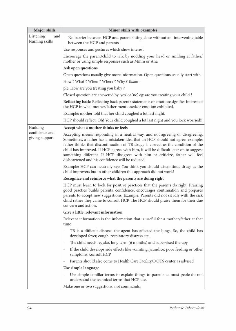
94 Pediatric Tuberculosis
Major skills Minor skills with oxamplesListening and learning skills
- No barrier between HCP and parent sitting close without an intervening table
between the HCP and parents
Use responses and gestures which show interest
Encourage the parent/child to talk by nodding your head or smilling at father/
mother or using simple responses such as Mmm or Aha
Ask open questionsOpen questions usually give more information. Open questions usually start with-
How ? What ? When ? Where ? Why ? Exam-
ple: How are you treating you baby ?
Closed question are answered by ‘yes’ or ‘no’, eg: are you treating your child ?
Refl ecting back: Refl ecting back parent’s statements or emotionssignifi es interest of
the HCP in what mother/father mentioned/or emotion exhibited.
Example: mother told that her child coughed a lot last night.
HCP should refl ect: Oh! Your child coughed a lot last night and you lock worried!!
Building confi dence and giving support
Accept what a mother thinks or feelsAccepting meens responding in a neutral way, and not agreeing or disagreeing.
Sometimes, a father has a mistaken idea that an HCP should not agree. example:
father thinks that discontinuation of TB drugs is correct as the condition of the
child has improved. If HCP agrees with him, it will be diffi cult later on to suggest
something diff erent. If HCP disagrees with him or criticize, father will feel
disheartened and his confi dence will be reduced.
Example: HCP can neutrally say: You think you should discontinue drugs as the
child improvers but in other children this approach did not work!
Recognize and reinforce what the parents are doing rightHCP must learn to look for positive practices that the parents do right. Praising
good practies builds parents’ confi dence, encourages continuation and prepares
parents to accept new suggestions. Example: Parents did not sit idly with the sick
child rather they came to consult HCP. Th e HCP should praise them for their due
concern and action.
Give a little, relevant informationRelevant information is the information that is useful for a mother/father at that
time
- TB is a diffi cult disease; the agent has aff ected the lungs. So, the child has
developed fever, cough, respiratory distress etc.
- Th e child needs regular, long term (6 months) and supervised therapy
- If the child develops side eff ects like vomiting, jaundice, poor feeding or other
symptoms, consult HCP
- Parents should also come to Health Care Facility/DOTS center as advised
Use simple language- Use simple famillar terms to explain things to parents as most peole do not
understand the technical terms that HCP use.
Make one or two suggestions, not commands.

95Pediatric Tuberculosis
Major skills Minor skills with oxamplesBe careful not to tell or command a mother to do something. Th is does not help her
to feel confi dent.
Example: You should continue the treatment of your child even though your child
will improve considerablysoon aft er treatment is started.
FeedbackAsk father what he should tell his wife aft er returning home about the treatment
provided for tuberculosis of the child. Final greetings- thank father/mother for
their time and consideration.
Role play
Case 1
A 12-years old female child presented with fever and cough for 3 months. She was treated with various antibiotics which did not help. No history of contact with a TB patient. Immunized with BCG scar mark over left deltoid. She had no respiratory distress, temp 100ºF, weight 33 kg. Breath sound bronchial and crepitations in left lower lung posteriorly.
WBC 12,000/cmm, N55%, L40%, ESR 22 mm in 1st hour, MT 16 mm. CXR: consolidation of left lower lobe on frontal projection. She is diagnosed as Pulmonary TB.
Counsel the parents on the diagnosis of TB.
Case 2
A 7-year old boy (Rana) presented with recurrent fever for 3 months, chest pain and cough for 2 months. He previously had suffered from enteric fever and was treated with ciprofl oxacin 9 months before the development of this fever. When he developed fever again, he was treated with cotrimoxazol, chloroquine, ceftriaxone and amoxycillin but temperature did not subside. Further enquiry could reveal contact with a neighbour, who was on anti-TB therapy for last 3 months.
After physical examination and investigation the boy was diagnosed as Tubercular Pleural effusion.
Counsel parent on treatment.
Case 3:
Father of Rana (case 2) came after 5 weeks. He was happy about the improvement of his boy- no fever, chest pain and cough. Wt gained- 19.6kg. He wanted to stop medication and she is ‘fully better’ now.
Counsel him to continue treatment.

96 Pediatric Tuberculosis

97Pediatric Tuberculosis
CHAPTER 12

98 Pediatric Tuberculosis

99Pediatric Tuberculosis
Chapter 12Drug resistant TB in children and TB-HIV co-infection
Introduction
MDR TB is on the rise among SAARC member states. Though the number of child MDR cases is not known, but estimates from the total MDR cases indicates large number of children are out of the NTP net. Among new cases this as high as 3.7% in Pakistan and 29% of re-treatment cases in Bangladesh.
MDR-TB in children is mainly transmitted drug resistance and acquired from an adult MDR case. Most of the MDR-TB in children develops within 12 months of infection. Contact tracing and follow-up of children exposed to MDR/ XDR-TB should receive priority.
Types of drug resistant TB in children
Mono drug resistance
1. Mono drug resistance means M. tuberculosis is resistant to only one fi rst-line anti-TB drug for example EMB or INH or SM resistance. Evidence suggests that CAT I regimen (INH, RMP, PZA, EMB) should be suffi cient for effective cure in most patients with INH mono-resistant TB. The risk of acquiring MDR-TB is increased in patients with high bacillary loads (mostly adult patients).
Poly drug resistance
2. When M. tuberculosis develops resistance to more than one fi rst line anti-TB drugs, organism is then called poly-drug resistant. This excludes MDR-TB. Examples of poly drug resistance are INH-EMB or EMB-SM or SM-RMP-EMB resistance or so on.
Multi drug resistance (MDR)
3. Multi drug resistant TB (MDR-TB): when TB is caused by organism that is resistant to isoniazid and rifampicin, the two most potent fi rst line anti-TB drugs. The resistance to isoniazid and rifampicin can occur with or without resistance to other anti-TB drugs.
Extensively drug resistance (XDR)
4. Extensively drug resistant TB or XDR-TB is defi ned as MDR-TB with additional resistance to any one of the fl uoroquinolones and to at least one of three injectable second line anti-TB drugs (amikacin, capreomycin or kanamycin).
Is drug resistant TB infectious?
Drug-resistant TB is as infectious as drug-susceptible TB. Children usually become infected after contact with adult or adolescent MDR-TB case.
How to recognize a drug resistant suspect?
Drug-resistant TB should be suspected if any of the following features are present:

100 Pediatric Tuberculosis
Features in the index case suggestive of drug resistant TBo Index case remaining smear-positive after 3 months of treatmento History of loss to follow-up or recurrence after completion of TB treatmento treatment failure of fi rst line treatment
Features in a child suggestive of having drug resistant TBo Contact with a known case of DR-TBo Child not responding to standard TB treatment when adherence is provedo Child with TB recurring after completing TB treatment in an adherent patient
Case-fi nding strategies (with older regimen for DS TB)
As MDR-TB is a laboratory diagnosis, the following groups will be targeted for culture and drug susceptibility testing (DST):
HIGH RISK: Failures of Category II; Failures of Category I; Close contact of a MDR-TB patient with symptoms
MEDIUM RISK: Delayed converters of Category I (remain positive at month 3); Delayed converters of Category II (remain positive at month 4); All relapses (Category I and II); All return after default (Category I and II); Others- Any smear negative or extrapulmonary TB patient clinically not improved in spite of
treatment as per NTP guidelines
LOW RISK:
All TB/HIV infected patients at the start of TB therapy;
Diagnosis of MDR-TB in children
Children, especially younger ones, may not be able to produce sputum specimens on demand. Children should not be excluded from treatment solely on the basis of non-availability of sputum specimens. Smear and culture negative children with active TB who are close contacts of patients with DR-TB can be started on DR-TB regimens .
Extra efforts can be used to get specimens for culture, induced sputum, tissue biopsy (including fi ne needle lymph node biopsy), gastric aspirate, urine and/or stool can be sent to NTRL/RTRL for diagnosis.
Principles of management of MDR-TB in children Manage in a specialized MDR-TB treatment facility (NIDCH/CDH) Children in Category II failure cases should go through the same algorithm as adults DOT with daily treatment is essential Be aware of cross-resistance between drug groups
NEVER add one drug to a failing regimen
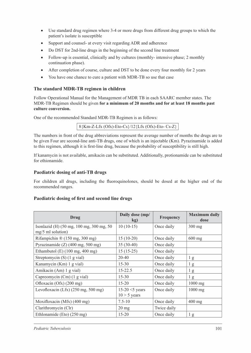
101Pediatric Tuberculosis
Use standard drug regimen where 3-4 or more drugs from different drug groups to which the patient’s isolate is susceptible
Support and counsel- at every visit regarding ADR and adherence Do DST for 2nd-line drugs in the beginning of the second line treatment Follow-up is essential, clinically and by cultures (monthly- intensive phase; 2 monthly
continuation phase). After completion of course, culture and DST to be done every four monthly for 2 years You have one chance to cure a patient with MDR-TB so use that case
The standard MDR-TB regimen in children
Follow Operational Manual for the Management of MDR TB in each SAARC member states. The MDR-TB Regimen should be given for a minimum of 20 months and for at least 18 months past culture conversion.
One of the recommended Standard MDR-TB Regimen is as follows:
8{Km-Z-Lfx (Ofx)-Eto-Cs}/12{Lfx (Ofx)-Eto- Cs-Z}
The numbers in front of the drug abbreviations represent the average number of months the drugs are to be given Four are second-line anti-TB drugs, one of which is an injectable (Km). Pyrazinamide is added to this regimen, although it is fi rst-line drug, because the probability of susceptibility is still high.
If kanamycin is not available, amikacin can be substituted. Additionally, protionamide can be substituted for ethionamide.
Paediatric dosing of anti-TB drugs
For children all drugs, including the fl uoroquinolones, should be dosed at the higher end of the recommended ranges.
Paediatric dosing of fi rst and second line drugs
Drug Daily dose (mp/kg) Frequency Maximum daily
doseIsonlazid (H) (50 mg, 100 mg, 300 mg, 50 mg/5 ml solution)
10 (10-15) Once daily 300 mg
Rifampichin ® (150 mg, 300 mg) 15 (10-20) Once daily 600 mgPyrazinamide (Z) (400 mg, 500 mg) 35 (30-40) Once dailyEthambutol (E) (100 mg, 400 mg) 15 (15-25) Once dailyStreptomycin (S) (1 g vial) 20-40 Once daily 1 gKanamycin (Km) 1 g vial) 15-30 Once daily 1 gAmikacin (Am) 1 g vial) 15-22.5 Once daily 1 gCapreomycin (Cm) (1 g vial) 15-30 Once daily 1 gOfl oxacin (Ofx) (200 mg) 15-20 Once daily 1000 mgLevofl oxacin (Lfx) (250 mg, 500 mg) 15-20 <5 years
10 > 5 yearsOnce daily 1000 mg
Moxifl oxacin (Mfx) (400 mg) 7.5-10 Once daily 400 mgClarithromycin (Clr) 20 mg Twice dailyEthlonamide (Eto) (250 mg) 15-20 Once daily 1 g

102 Pediatric Tuberculosis
Drug Daily dose (mp/kg) Frequency Maximum daily
doseProtionamide 15-20 Once daily 1 gCycloserine (Cs) (250 mg) 10-20 Once or twice
daily1 g
PAS (4 g sachets) 300 Twice or thrice daily
12 g
Clofazimine (Cfz) (50 and 100 mg) 2-3 mg Twice daily 200 mgAmoxycillin/clavulanate (Amx/Clv) (500/125) - dosing is base on Amoxacilling component
30 mg (<3 months) 45 mg (>3 months and less than 40 kg)
Twice daily 2000 mg Twice daily
Management of TB in Pediatric living with HIV
Background
Globally, tuberculosis (TB) is the most common opportunistic infection and leading cause of mortality in people living with the human immunodefi ciency virus (PLHIV), contributing to at least one in four of these deaths. HIV infection is a major threat in the global resurgence of tuberculosis (TB), especially for tuberculosis control in many developing countries. People living with HIV are at least 26 times more likely to develop TB disease as people without HIV. TB is the biggest killer of people with HIV/AIDS, shortening their lives by 6 to 24 months. Globally, 12% of the 9.6 million new TB cases in 2014 were HIV- positive. The World Health Organization estimates that the prevalence of HIV among children with TB in moderate to high prevalence countries ranges between 10% and 60%.
Tuberculosis, although curable, is one of the most common causes of HIV- related illness and death. 1.2 million People living with HIV/AIDS (PLHIV) are estimated to be co-infected with Mycobacterium tuberculosis, with 74% of those co-infected living in sub-Saharan Africa.
The annual risk of developing TB in a PLHIV who is co-infected with M tuberculosis ranges from 5 to 15 percent. HIV increases the rate of recurrent TB, which may be due to either endogenous reactivation (true relapse) or exogenous re-infection. Increasing tuberculosis cases in PLHIV poses an increased risk of TB transmission to the general community regardless whether they are infected with HIV or not.
The World Health Organization (WHO) has recommended the package of “collaborative TB/HIV activities” since 2004. To reduce the burden of TB in PLHIV, the WHO recommended the 3I’s approach in addition to early antiretroviral therapy. Treatment of latent TB infection with 6 months of isoniazid preventive therapy (IPT) which can reduce the progression to active TB in PLHIV by 32-62%.12 It is estimated that about 34% of children aged <15 years needing ART in low- and middle-income countries receive treatment compared with about 68% of adults.
Children living with HIV infection have increased risk of TB exposure, infection, progression to disease, and TB-related morbidity and mortality. This risk is infl uenced by the degree of immune suppression. All children living with HIV infection in a TB-endemic setting should therefore be regularly screened for TB by clinical assessment at each visit to a health facility or contact with a health worker. Evaluation should aim to identify those patients who are likely to have TB disease, requiring anti-TB treatment, and those who should start IPT. Suspicion of TB disease in children with HIV is initially based on the presence of clinical symptoms. Clinical evaluation may be followed up with further investigations as appropriate (e.g. chest radiography). As for any child with suspected TB, attempts should be made to confi rm diagnosis (e.g. culture, Xpert MTB/RIF assay) whenever possible.
Childhood HIV infection is particularly common in settings where antenatal HIV prevalence is high and PMTCT interventions are not widely implemented. The prevalence of HIV is therefore particularly

103Pediatric Tuberculosis
high among infants and young children, an age-group also at risk for TB. In regions endemic for TB/HIV, TB is common in children living with HIV, and HIV infection is common in children with TB. It is recommended that HIV testing be routinely offered to all children with suspected or diagnosed TB.
Diagnosing TB in HIV-Infected Children
In HIV-infected children the diagnosis of TB disease is more complex because:• Clinical features consistent with pulmonary TB are common in children living with HIV but may be
due to other diseases and therefore lack specifi city for a diagnosis of TB.• Most children living with HIV are infected by mother-to-child transmission. The peak age prevalence
for HIV is therefore in infants and young children (<5 years), who also make up the age group in which it is most diffi cult to confi rm the cause of acute or chronic lung disease, including TB.
• TST is less sensitive in children living with HIV than in HIV-negative children; induration of >5 mm is considered positive if the child is living with HIV.
• Children living with HIV have a very high incidence of acute and chronic lung diseases other than TB.
• Children living with HIV may have lung disease of more than one cause (co-infection), which can mask response to therapy.
• There is an overlap of radiographic fi ndings in TB and other HIV-related lung disease.
There is the dual risk that TB may either be over-diagnosed, resulting in unnecessary TB treatment, or under-diagnosed, resulting in increased morbidity and mortality. LIP is the most diffi cult condition to distinguish from TB, due to radiological similarities, although it is usually associated with typical clinical signs, such as clubbing and/or parotid enlargement. TB can occur in children with an underlying diagnosis of LIP, bronchiectasis, or any other lung infection. In spite of the diffi culties described, TB can usually be diagnosed with a fair degree of accuracy in the majority of HIV-infected children. The diagnostic approach in HIV-infected children is essentially the same as for HIV-uninfected children. Since the symptoms of TB can be confused with the symptoms of HIV disease and the CXR is more diffi cult to interpret, if possible every effort should be made to try and establish a bacteriological diagnosis.
Treatment of TB in HIV-Infected Children
Children living in settings where the prevalence of HIV is high should be treated with a four-drug regimen (HRZE) for 2 months followed by a two-drug regimen (HR) for 4 months at the dosages. Children with suspected or confi rmed pulmonary TB or tuberculosis peripheral lymphadenitis in settings with a high HIV prevalence (or with confi rmed HIV infection) should not be treated with intermittent regimens (that is, twice- or thrice-weekly doses).
Each child should be assessed 2 weeks after the start of TB treatment then reviewed monthly with clinical monitoring, which should include symptom assessment, weight measurement, assessment of adherence to treatment and enquiry about any adverse events. Dosages of anti-TB drugs should be adjusted to account for any weight gain. Most children living with HIV with drug-sensitive TB who are compliant with therapy have a good response to the 6-month regimen. Possible reasons for treatment failure are non-compliance with therapy, drug-resistant TB or alternative diagnoses (incorrect diagnosis of TB).
All children living with HIV who have successfully completed treatment for TB disease should receive isoniazid for an additional 6 months. When compared with HIV-negative children, responses to TB treatment and outcome are poorer for children living with HIV. Before the availability of ART, many deaths in children with TB/HIV occurred in the fi rst 2 months following the start of TB treatment. Medical risk factors for poor treatment response and mortality include severe malnutrition, co-infections, severe immune-suppression and high viral load. Additional therapy recommended for HIV-infected children with TB, which may help to improve TB treatment outcomes, includes co-trimoxazole preventive therapy, the early start of ART (see below) and pyridoxine supplementation along with nutritional support. (See details in Chapter 3).

104 Pediatric Tuberculosis
Co-trimoxazole preventive therapy
Co-trimoxazole is a broad-spectrum antimicrobial agent that prevents a range of secondary bacterial and parasitic infections in eligible adults and children living with HIV. Daily prophylaxis – co-trimoxazole preventive therapy (CPT) - prolongs survival in children living with HIV and reduces the incidence of co-morbidities. It also reduces the risk of co-infections such as pneumocystis pneumonia in HIV-exposed infants. CPT is therefore recommended for all HIV-exposed infants and children living with HIV, including those with TB, and should be implemented as an integral component of a package of HIV related services.
Table 7: Cotrimoxazole prophylaxis
Age Recommended daily cotrimoxazole prophylaxis
Under 6 months of age
20 mg trimethoprim (TMP) + 100 mg sulfamethoxazole (SMX)
Under 5 years 40 mg TMP + 200 mg SMX
5 years or older 80 mg TMP + 400mg SMX
5 Antiretroviral therapy
Antiretroviral therapy (ART) in children living with HIV aims to improve the length and quality of life, reduce HIV-related morbidity and mortality by reducing the incidence of opportunistic infections (including TB), reduce the viral load, restore and preserve immune function, and restore and preserve normal growth and development. ART improves TB treatment outcomes for children living with HIV.
Appropriate arrangements for access to ART should be made. All children with TB disease and HIV infection require ART. In HIV-infected children with confi rmed or presumptive TB disease, initiation of TB treatment is the priority. The decision on when to initiate ART after starting TB treatment should consider the child’s immune status and clinical severity of disease, the child’s age, pill burden, potential drug interactions, overlapping toxicities and possible IRIS.
This should be weighed up against the risk of further HIV disease progression and immunesuppression with associated increase in mortality and morbidity in the absence of ART. TB treatment should be started fi rst, followed by ART as soon as possible thereafter (and within 8 weeks of the start of TB treatment). For those with a CD4 count below 50 cells/mm3, ART should be provided within 2 weeks of the start of TB treatment.
Table : Use of fi rst-line ART
Age Treatment regimenChildren younger than 3 years
Protease inhibitor (PI)-based regimen in combination with ABC or zidovudine (AZT)
Adolescents and children older than 3 years
Regimens comprising a non-thymidine nucleoside reverse-transcriptase inhibitor (NRTI) backbone (tenofovir disoproxil fumarate (TDF) or abacavir (ABC) + la-mivudine (3TC)) and one non-nucleoside reverse-transcriptase inhibitor (NNRTI) efavirenz (EFV)
Note: For fi rst-line ART, use of simplifi ed and less toxic regimens – as fi xed-dose combinations whenever possible – is recommended as the most effective and convenient approach.
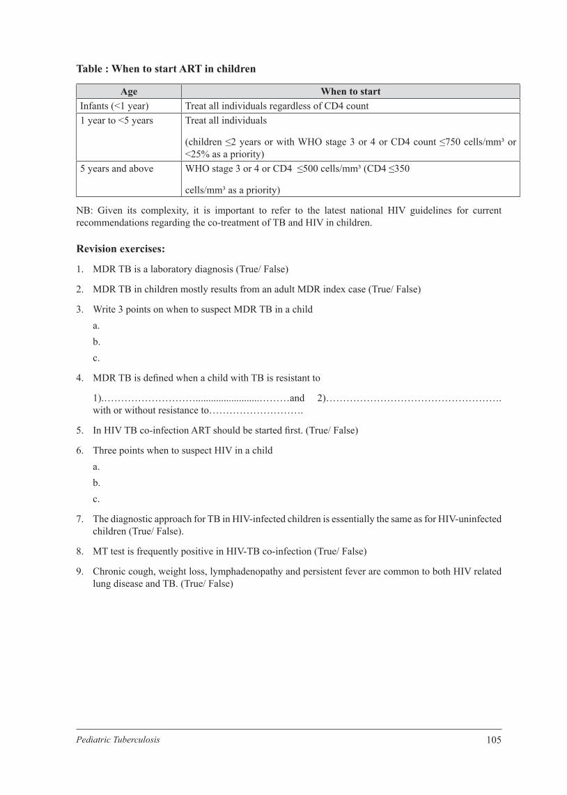
105Pediatric Tuberculosis
Table : When to start ART in children
Age When to startInfants (<1 year) Treat all individuals regardless of CD4 count1 year to <5 years Treat all individuals
(children ≤2 years or with WHO stage 3 or 4 or CD4 count ≤750 cells/mm³ or <25% as a priority)
5 years and above WHO stage 3 or 4 or CD4 ≤500 cells/mm³ (CD4 ≤350
cells/mm³ as a priority)
NB: Given its complexity, it is important to refer to the latest national HIV guidelines for current recommendations regarding the co-treatment of TB and HIV in children.
Revision exercises:
1. MDR TB is a laboratory diagnosis (True/ False)
2. MDR TB in children mostly results from an adult MDR index case (True/ False)
3. Write 3 points on when to suspect MDR TB in a child a. b. c.
4. MDR TB is defi ned when a child with TB is resistant to
1).……………………….........................………and 2)……………………………………………. with or without resistance to……………………….
5. In HIV TB co-infection ART should be started fi rst. (True/ False)
6. Three points when to suspect HIV in a child a. b. c.
7. The diagnostic approach for TB in HIV-infected children is essentially the same as for HIV-uninfected children (True/ False).
8. MT test is frequently positive in HIV-TB co-infection (True/ False)
9. Chronic cough, weight loss, lymphadenopathy and persistent fever are common to both HIV related lung disease and TB. (True/ False)

106 Pediatric Tuberculosis

107Pediatric Tuberculosis
CHAPTER 13

108 Pediatric Tuberculosis

109Pediatric Tuberculosis
Chapter 13Prevention of Tuberculosis
As with other infectious diseases, prevention is best achieved by interrupting the transmission of disease. The following methods have to be followed to prevent disease:
1. BCG vaccination 2. Identifi cation of infectious cases 3. Successfully complete treatment of TBcases 4. Contact tracing 5. Infection control 6. INH Prophylaxis Therapy (IPT) 7. Training of health care providers on TB 8. Community participation and ownership 9. Socio-economic change
In this section we shall be discussing on BCG vaccination, contact tracing & INH prophylaxis and infection control.
BCG vaccination-
BCG vaccine has been used for last 70 yrs and about 500 million vaccines have been given. Yet the TB epidemic continues. In the 22 high burden countries 200,000 deaths occurring each year from TB. Questions have been raised on the effi cacy of BCG. In a recent meta-analysis WHO has stated, effi cacy of BCG is 50% for PTB and 60-80% for TBM and disseminated TB. The protective effi cacy is best during the 15-20 yrs post-vaccination. Nevertheless, BCG vaccination is recommended to avoid life threatening TB diseases in children.
BCG is given as an intradermal injection (0.1ml) over the left deltoid at its insertion on the humerus and after a period of 8-14 weeks a small 5-8 mm rounded scar develops. BCG is generally a safe vaccine; but some complications may occur in HIV infected infants. BCG is best given during the neonatal period.
Complications of BCG vaccination: 1. Local BCG abscess 2. Prolonged large ulcer 3. Lymphadenitis (usually axillary): 1-10% 4. Others- Osteitis, disseminated infection (1/1,000,000 doses)
Mostly these complications do not need treatment. Needle aspiration of a local abscess is sometimes needed.
Contraindication: 1. Immunodefi ciency- symptomatic HIV, immunosupression from drugs 2. Defer 12 weeks after IVIG treatment and 4-6 weeks after viral infections like measles, chicken
pox and viral hepatitis

110 Pediatric Tuberculosis
What should be done when there is no scar?
Best course of action is to give BCG again on opposite arm.
Isoniazid Prophylaxis Therapy (IPT)
Tuberculosis is spread by droplets from an infected person especially those who are smear positive. Droplets are produced by coughing, laughing, talking, singing etc. One cough can produce 40-50,000 droplets, and when someone coughs inside a closed room the droplets accumulate. As person with active TB coughs in a closed room millions of droplets are suspend and persons sharing the same room can get infected. The smaller the child, the greater is the risk of getting disease, highest risk among children <5yrs (5-50%). The chance of getting disease is very high among PEM, baby born to infected mother (in last 2 trimester) and immunocompromised children after contact with a smear-positive TB case. Studies have proved that INH prophylaxis taken for 6 months can prevent TB disease especially in high risk groups. NTP has decided to provide IPT to all the children below 5 years with a history of close contact with a smear-positive patient.
Contact tracing: This strategy is to trace all individuals in contact with a case of suspected or confi rmed pulmonary TB and screened them for TB infection or disease. Those infected with a high risk of disease (children less than 5 years and all HIV infected people) are offered IPT while those with disease are treated.
Reverse contact tracing: This strategy is adopted when a child is the index case in whom active TB has been diagnosed. The potential source case (usually an adult or older child in the index case’s home) is sought by symptom screening and/or chest radiograph. Chest radiographs of parents, particularly the mother, may highlight the presence of previously undiagnosed TB disease in the parent.
Previous TB preventive therapy or treatment does not protect the child against subsequent TB exposure/ infection. Therefore high risk children (as defi ned above) should receive preventive therapy after each episode of documented TB exposure, unless the child is currently receiving TB prophylaxis or treatment.
Always exclude TB disease before providing preventive therapy.
Asymptomatic children (playful and thriving, no cough or wheeze, no fever, no unusual fatigue or lethargy, no visible neck mass) do not require additional tests to exclude TB disease, before providing preventive therapy.
Children <5 years of age or immunocompromised children of any age in close contact with an adult or adolescent with pulmonary TB should receive a course of INH prophylaxis (after TB disease is excluded). A child (<5 yrs) without history of contact but having a positive MT should also receive IPT.
Symptomatic children should be evaluated and TB disease excluded. When TB is excluded they should receive IPT.
How to give preventive therapy?
Preventive therapy comprises of isoniazid (INH) mono-therapy for 6 months (see dose recommendations below). This is usually not given as directly observed therapy (DOT). As poor adherence is a serious concern and parents/caregivers must be adequately counseled to explain why the medicine is given and to encourage good adherence. Parents/caregivers should also be counseled to recognize the symptoms of TB disease, such as a persistent non-remitting cough or fever, unusual fatigue or lethargy and/or weight loss, which should prompt them to immediately bring the children back to the clinic for further evaluation.
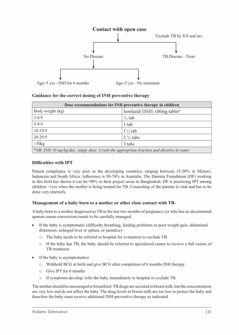
111Pediatric Tuberculosis
Contact with open caseExclude TB by S/S and inv.
TB Disease - TreatNo Disease
Age<5 yrs - INH for 6 months Age<5 yrs - No treatment
Guidance for the correct dosing of INH preventive therapy
Dose recommendations for INH preventive therapy in childrenBody weight (kg) Isonlazid (INH) 100mg tablet*
2-4.9 1/2 tab
5-9.9 1 tab
10-19.9 11/2 tab
20-29.9 2 1/2 tabs
>30kg 3 tabs
*NB. INH 10 mg/kg/day, single dose; Crush the appropriate fraction and dissolve in water
Diffi culties with IPT
Patient compliance is very poor in the developing countries, ranging between 15-20% in Malawi, Indonesia and South Africa. Adherence is 50-74% in Australia. The Damien Foundation (DF) working in this fi eld has shown it can be>90% in their project areas in Bangladesh. DF is practicing IPT among children <1yrs when the mother is being treated for TB. Counseling of the parents is vital and has to be done very intensely.
Management of a baby born to a mother or other close contact with TB-
A baby born to a mother diagnosed as TB in the last two months of pregnancy (or who has no documented sputum smear-conversion) needs to be carefully managed.
If the baby is symptomatic (diffi culty breathing, feeding problems or poor weight gain, abdominal distension, enlarged liver or spleen, or jaundice) -o The baby needs to be referred to hospital for evaluation to exclude TB.o If the baby has TB, the baby should be referred to specialized center to receive a full course of
TB treatment.
If the baby is asymptomaticoo Withhold BCG at birth and give BCG after completion of 6 months INH therapyo Give IPT for 6 monthso If symptoms develop, refer the baby immediately to hospital to exclude TB.
The mother should be encouraged to breastfeed. TB drugs are secreted in breast milk, but the concentrations are very low and do not affect the baby. The drug levels in breast milk are too low to protect the baby and therefore the baby must receive additional INH preventive therapy as indicated.

112 Pediatric Tuberculosis
TB infection control
Prevention of TB transmission and infection in the household and health facilities are important components of control and management of TB in children. The following simple procedures are effective in TB infection control at home and clinics:
1. Early diagnosis and treatment of adult TB cases
2. At the clinic promptly identify potential and known infectious cases of TB; separate them. Suspects must be fast tracked so that TB can be diagnosed as soon as possible and put on treatment. Place posters in all patient and staff areas containing TB IEC messages.
3. Provide health education about TB transmission without stigmatizing TB patients
4. Encourage proper cough hygiene both at home and at health facilities Cover nose and mouth with back of the hand(s), arm (sleeve), tissue, cloth or face mask when
coughing or sneezing; Turn head away from others when coughing or sneezing; Use in the nearest waste bin to dispose the tissue, cloth etc. after use; Spit in a cloth or container with lid; Perform hand hygiene (e.g. hand washing with soap and water, antiseptic hand wash) after
having contact with respiratory secretions;
5. Ensure Natural ventilation and sunlight: Keep doors and windows open on opposite sides of the TB clinic and other clinics (Effective
ventilation increases air exchanges, decreasing infection) Keep window open where children and adults live together. Apply the same principle in clinic
6. HCWs/ care givers should be screened out if symptomatic
Role play
Case scenario 1:
While reviewing Rahima Begum (32yrs) being treated for pulmonary TB during a ward round, you found she is breast feeding her 7 month old healthy looking playful boy (Jahur). On evaluation, Jahur was found to have no fever and cough, weighing 6.5 kg, BCG scar present, no lymphadenopathy. You have planned to treat the child with INH for 6 months.
Your colleague has a different opinion regarding IPT. Discuss with your colleague the value of IPT for Jahur.
Case scenario 2:
After convincing your colleague, now you talk with the parents on providing IPT.
Case scenario 3:
After 2 months at follow-up Rahima Begum revealed, they are not planning to continue INH for Jahur, as he is healthy and thriving well with no symptoms of TB. Convince mother on continuation of IPT.

113Pediatric Tuberculosis
CHAPTER 14

114 Pediatric Tuberculosis

115Pediatric Tuberculosis
Chapter 14Pediatric TB Management within the NTP
Objectives of the Module
Early case detection and effective management of TB cases in the community will reduce the burden of TB in children. It is important that NTP include child TB in funding and resource allocation, in policy guidelines/protocols and training opportunities in NTP. NTP should have a focal person for child TB and a child TB working group for monitoring and evaluation of child TB related issues. The responsibilities of the NTP are implementing childhood TB activities according to national guidelines.
Important objectives of training are to:- increase case-fi nding of child TB cases in the community;
- improve the overall management of TB in children
- increase implementation of child contact screening and preventive therapy;
- provide accurate data on childhood TB for NTPs for purposes of monitoring and evaluation.
The main focus of the training is for three likely common scenarios:- the child with suspected TB disease;
- the child treated for TB in the community;
- the child who is a close contact of a TB case.
To enable targeted service providers to implement childhood TB control activities:- health workers at secondary- and primary-level facilities that provide care for sick children;
- health workers involved in the management of adult TB cases in the community;
- health workers who are involved in the management of mothers and children with HIV;
- community health workers and volunteers and treatment support groups (who carry out contact tracing in the community).
NTP initiatives at the national level Formation of a child TB working group within the NTP, with representation from community and
national child TB experts as well as from the NTP.
Evaluation of surveillance data to target particular settings for implementation and assessment of specifi c activities.
Convening of stakeholders (including professional associations) to:
- analyse the capacity (public and private sector) at national and sub-national levels to implement the guidelines;
- identify and quantify factors that may constrain or facilitate successful implementation;
- develop national (or sub-national) policies and obtain endorsement.
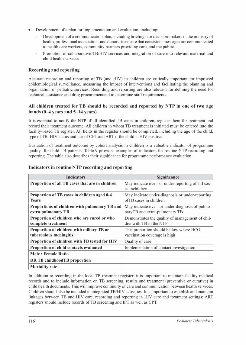
116 Pediatric Tuberculosis
Development of a plan for implementation and evaluation, including:- Development of a communication plan, including briefi ngs for decision-makers in the ministry of
health, professional associations and donors, to ensure that consistent messages are communicated to health care workers, community partners providing care, and the public.
- Promotion of collaborative TB/HIV services and integration of care into relevant maternal and child health services
Recording and reporting
Accurate recording and reporting of TB (and HIV) in children are critically important for improved epidemiological surveillance, measuring the impact of interventions and facilitating the planning and organization of pediatric services. Recording and reporting are also relevant for defi ning the need for technical assistance and drug procurementand to determine staff requirements.
All children treated for TB should be recorded and reported by NTP in one of two age bands (0–4 years and 5–14 years)
It is essential to notify the NTP of all identifi ed TB cases in children, register them for treatment and record their treatment outcome. All children in whom TB treatment is initiated must be entered into the facility-based TB register. All fi elds in the register should be completed, including the age of the child, type of TB, HIV status and use of CPT and ART if the child is HIV-positive.
Evaluation of treatment outcome by cohort analysis in children is a valuable indicator of programme quality for child TB patients. Table 9 provides examples of indicators for routine NTP recording and reporting. The table also describes their signifi cance for programme performance evaluation.
Indicators in routine NTP recording and reporting
Indicators Signifi cance Proportion of all TB cases that are in children May indicate over- or under-reporting of TB cas-
es inchildrenProportion of TB cases in children aged 0-4Years
May indicate under-diagnosis or under-reporting ofTB cases in children
Proportions of children with pulmonary TB and extra-pulmonary TB
May indicate over- or under-diagnosis of pulmo-naryTB and extra-pulmonary TB
Proportion of children who are cured or whocomplete treatment
Demonstrates the quality of management of chil-drenwith TB in the NTP
Proportion of children with miliary TB ortuberculous meningitis
This proportion should be low where BCGvaccination coverage is high
Proportion of children with TB tested for HIV Quality of careProportion of child contacts evaluated Implementation of contact investigationMale : Female RatioDR TB childhoodTB proportion Mortality rate
In addition to recording in the local TB treatment register, it is important to maintain facility medical records and to include information on TB screening, results and treatment (preventive or curative) in child health documents. This will improve continuity of care and communication between health services. Children should also be included in integrated TB/HIV activities. It is important to establish and maintain linkages between TB and HIV care, recording and reporting in HIV care and treatment settings; ART registers should include records of TB screening and IPT as well as CPT.

117Pediatric Tuberculosis
The use of a contact screening register should be encouraged. Important screening indicators include the number of children screened, symptoms suggestive of TB, the age and HIV status of child contacts, and the type of treatment each child has received (IPT or anti-TB treatment). The ratio of the number of sputum smear-positive pulmonary TB cases to the number of HIV-positive children or children ≤5 years of age screened could be used as an indicator of the effectiveness of contact tracing.
Supervision, monitoring, Training and Evaluation:
Supervision is the key element of TB control and a corner stone for the sustain ability of the program. Effective supervision, monitoring and evaluation ensure continued achievements and improved performance of health care professionals.
Training and supervision of public and private health workers will be needed to: increase case fi nding of child TB cases in the community; improve the management of children with TB; increase child contact screening and preventive therapy; and improve recording and reporting practices so that better data will become available for monitoring progress.
Integrated care
Children with TB often do not present, and are not managed, within the context ofspecifi c TB care services but rather in the context of services that provide care to the sickchild, including maternal and child health services and HIV care services. An importantstep towards improving the prevention and management of TB in children is the provisionof integrated care.
Roles and responsibilities
Health workers at all levels of care (including primary care staff, general clinicians and pediatricians in public and private health care facilities) have potential roles in, and responsibilities for, ensuring comprehensive and coordinated care for a child with TB or a child who is a TB contact. Health workers at all levels need to be aware of their responsibilities to ensure that all children treated for TB are registered with the NTP.
Patient and family support for children with TB
A patient may infect other people who may also develop tuberculosis. The patient should, therefore, encourage other people with whom he or she has been is in close contact with to undergo screening for TB. The TB patient should be encouraged to spend more time in open space so as to reduce indoor transmission Children, parents, and other family members should be educated about TB and the importance of completing treatment. Where possible, someone other than the child’s parent or immediate family member should observe or administer treatment.
When a child is diagnosed with TB, he or she should also be tested for HIV (if HIV status is unknown). In many settings, the diagnosis of TB and/or of HIV can result in stigma and discrimination; the impact of this on the family unit adds to the burden of caring for children with TB or TB/HIV during physical illness and death.
The model of family-centered care - an approach that focuses on the continuum of care for the whole family rather than the individual - requires a multidisciplinary approach to address all the needs of the family. Basic principles of this continuum of care include:
- integration of care with prevention for the provision of a comprehensive, holistic system of TB and TB/HIV management;
- provision of non-discriminatory/non-judgmental care and prevention;
- maintainance of confi dentiality and respect for basic rights;

118 Pediatric Tuberculosis
- provision of clinical and nursing care and home-based care to alleviate symptomsof TB and HIV and prevent opportunistic infections;
- provision of counseling and psychosocial support services;- community mobilization of resources for cost-effective comprehensive and holistic care;- Provision of education, training, supervision and support for staff and volunteers.
Community support
The usual approach to managing TB in children has been specialized care delivered at health care facilities. However, not all people with TB require referral for hospital-based investigations and care. Decentralization and delivery of care at the community level,when appropriate and available through an integration of family and child services, hasthe following advantages: All family members requiring care for TB can receive them at the same time and in the same place. Both time and money are saved when care is provided closer to home. There is continuity of care between the patient’s home, community and local health centre. Community support is increased, which may lead to better adherence to treatment and can be
instrumental in overcoming barriers to long-term care, including treatment adherence, transportation costs, and loss of wages care taker during sickness and clinic visits.
It is also important to involve local schools, assisting them through education of the teachers and other staff about the needs of children with TB/HIV, the necessity for frequent visits to clinics and the importance of taking drugs regularly. This may help toreduce stigma in schools. Additionally, because not all children have the opportunity to attend school, faith-based organizations, traditional healers and other strong community groups could be involved in supporting children with TB and their families.
Management of TB in pregnancy
The symptoms of TB disease in pregnancy are similar to those in non-pregnant women,with pulmonary TB being the commonest form of disease. Disseminated TB occurs in 5-10% of pregnant woman suffering from TB, and this is a particular risk for congenital TB. All pregnant women should therefore be screened for symptoms of TB as far as possible. It is equally important that a pregnant woman with suspected TB be tested for HIV.Maternal TB increases the risk of mother-to-child transmission (MTCT) of HIV. If TB is diagnosed, treatment must be started promptly to prevent transmission and improve outcome. The treatment of TB in pregnant women is similar to that for non-pregnant women (with the exception of streptomycin, which is not recommended in pregnancy). HIV-positive pregnant women with TB are treated with ART according to WHO guidelines.
Congenital and neonatal TB
Congenital TB is TB acquired in utero, through haematogenous spread via the umbilical vessels, or at the time of delivery through aspiration or ingestion of infected amniotic fl uid or cervico-vaginal secretions. Congenital TB usually presents in the fi rst 3 weeks of life and mortality is high.
Neonatal TB is TB acquired after birth through exposure to an infectious case of TB -usually the mother but sometimes another close contact.It is often diffi cult to distinguish between congenital and neonatal TB but managementis the same for both. Both forms will be referred to here as neonatal TB. The TB-exposed neonate may be asymptomatic or symptomatic.
Symptoms of neonatal TB are usually nonspecifi c and include lethargy, fever, poor feeding, low birth weight and poor weight gain. The clinical signs are also nonspecifi c and can include respiratory distress, non-resolving pneumonia, hepatosplenomegaly, lymphadenopathy, abdominal distension with ascites, or a clinical picture of “neonatal sepsis” with disseminated TB.

119Pediatric Tuberculosis
The diagnosis of TB should be included in the differential diagnosis of chronic neonatal infection with a poor response to antimicrobial therapy, congenital infections and atypical pneumonia. The most important clue to the diagnosis of neonatal TB is a maternal history of TB or HIV infection. Critical points in the maternal history includenon-resolving pneumonia, past treatment for TB, contact with an index case of TB and recent initiation of treatment for TB.
Key programmatic considerations to address the impact of TB on maternal, neonatal and child health- Integrated management of pregnancy and child health services- Prevention of mother-to-child HIV transmission services- Family planning and infertility services- Tuberculosis and HIV programme services
Follow-up
Each child should be clinically assessed a very 2 weeks during the intensive phase, and every month during the continuation phase until treatment completion. The assessment should include, at a minimum: Symptom assessment Assessment of adherence by reviewing the treatment card Inquiry about any adverse events Weight measurement Drug dosage adjustment to account for any weight gain
A follow-up sputum smear for microscopy at 2 months should be obtained for any child who was smear-positive at diagnosis. Follow-up chest radiographs are not routinely required in children, who are improving on treatment as radiological changes usually lag behind clinical response. A child who is not responding to TB treatment should be referred for further assessment and management.

120 Pediatric Tuberculosis

121Pediatric Tuberculosis
Birth to 36 months: Boys Weight-for-length percentiles
NAME ___________________________________________
RECORD# _________________
122 Pediatric Tuberculosis
Birth to 36 months: Girls Weight-for-length percentiles NAME
NAME ___________________________________________
RECORD # _________________
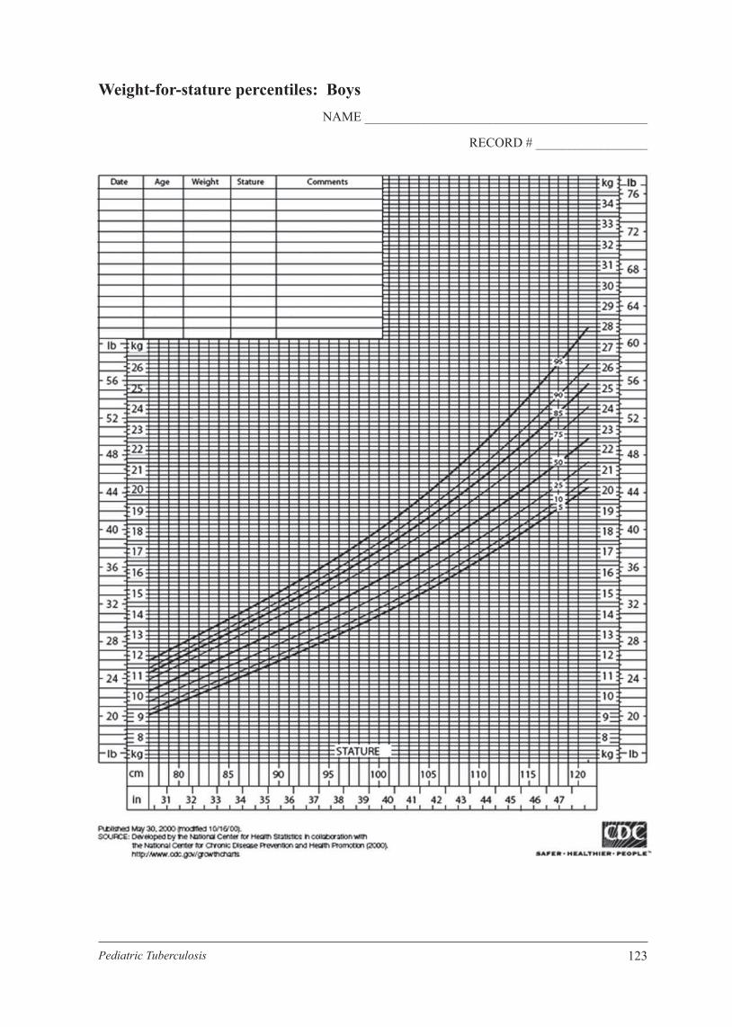
123Pediatric Tuberculosis
Weight-for-stature percentiles: BoysNAME ___________________________________________
RECORD # _________________
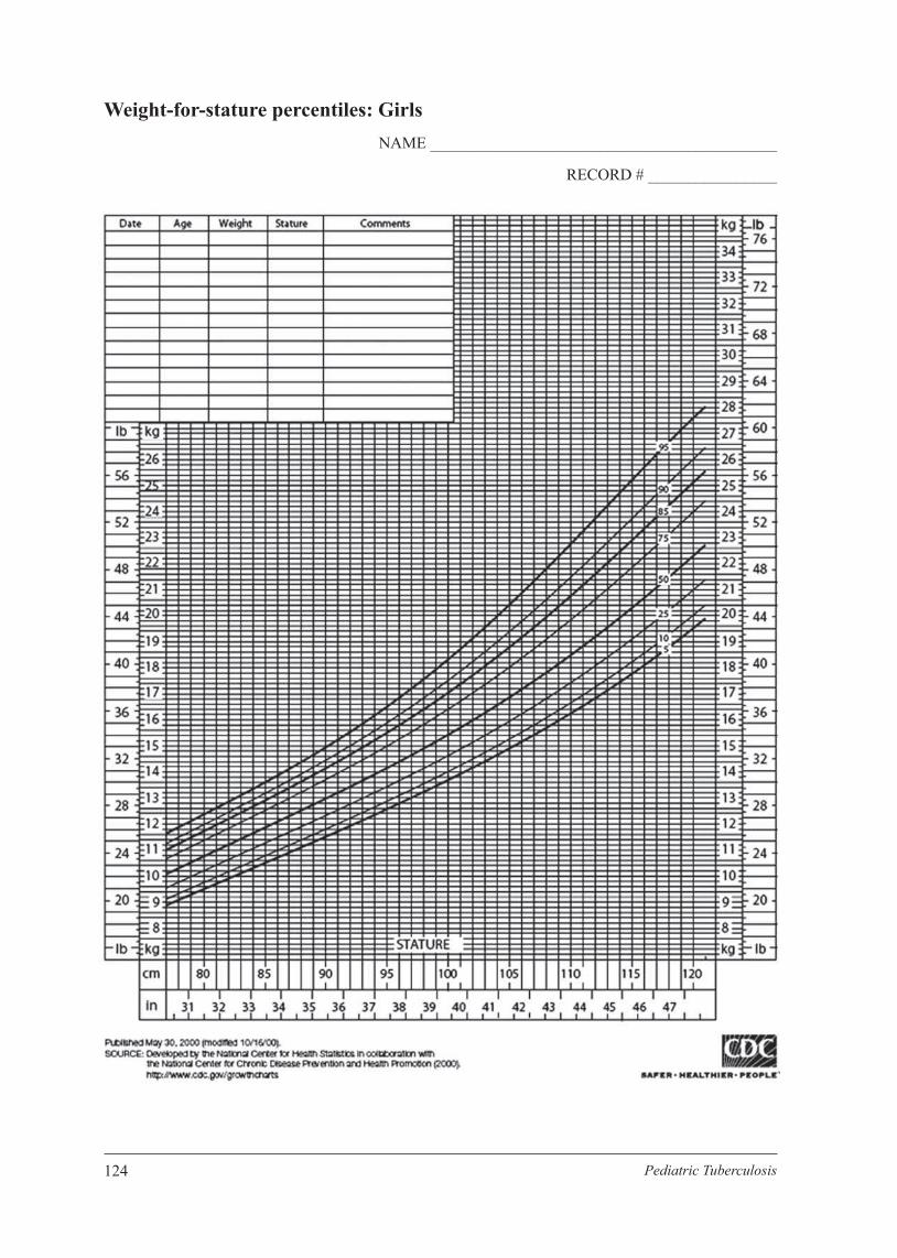
124 Pediatric Tuberculosis
Weight-for-stature percentiles: GirlsNAME ___________________________________________
RECORD # ________________

125Pediatric Tuberculosis
Training Schedule
SN Session Time RemarkDay-1
1. Reception and Pretest 8:30 - 9:00 am2. Epidemiology of TB in children 9:00 - 10:00 am3. Tea and socialization 10:00 - 10:30 am4. Pathology and pathogenesis 10:30 - 11:30 am5. Specimen sampling and microbiology 11:30 - 12:30 pm6. Lunch break 12:30 - 1:30 pm7. Case taking in CTB (History and 1:30 - 2:45 pm8. Approach to diagnosis of CTB 2:45 - 3:30 pm9. Tea and summary 4:00 pm
Day-21. Recap of Day-1 8:30 - 8:45 am2. Approach to diagnosis of CTB 8:45 - 10:15 am3. Tea break 10:15 - 10:30 am4. Live case Demonstration 10:30 - 11:30 am5. Investigation 11:30 - 12:00 am6. Lunch break 12:00 - 1:00 pm7. Imaging presentation (Multimedia) 1:00 - 1:45 pm8. Imaging practice 1:45 - 2:45 pm9. Summary of day-2 2:45 - 3:30 pm
Day-31. Recap of Day-2 8:30 - 8:45 am2. Imaging reading 8:45 - 9:15 am3. Counseling in Childhood TB 9:15 - 10:15 am4. Practical session on MT/DOTS
(Facility visit with tea)10:15 - 11:00 am
5. Photo show 11:00 - 11:15 am6. Treatment 11:15 - 1:00 pm7. Lunch and prayer 1:00 - 1:45 pm8. Prevention, Contact tracing and IPT 1:45 - 2:30 pm9. MDR and HIV TB 2:30 - 3:00 pm10. Closing 3:00 - 3:30 pm

126 Pediatric Tuberculosis