s2e guideline: positioning and early mobilisation in ... · 5 guidelines for use in positioning ......
TRANSCRIPT
Preface
The order to revise the S2 guideline ‘posi-tioning in prophylaxis or therapy of pul-monary disorders’, which was established in 2008, was issued by the German So-ciety for Anaesthesiology and Intensive Care Medicine (DGAI). Due to increasing clinical and scientific relevance, the guide-
line was expanded to include the topic ar-ea ‘early mobilisation’.
‘Guidelines are systematically devel-oped presentations and recommenda-tions with the purpose of assisting physi-cians and patients in deciding on appro-priate measures for medical care (preven-tion, diagnostics, therapy and after care) under specific medical conditions.’ (As-sociation of Scientific Medical Societies, AWMF).
The guideline is based on the following fundamental assumptions: 5 Guidelines for use in positioning therapy and early mobilisation in pro-phylaxis or therapy of pulmonary dis-orders aid in decision-making in spe-
cific situations. They are based on the current state of scientific knowledge and on procedures proven in practice. 5 Positioning and early mobilisation are supporting concepts in the treat-ment and prophylaxis of pulmonary disorders, wherein they are intend-ed to supplement basic medical mea-sures (e.g. mechanical ventilation, flu-id management, pharmacotherapy), but not to replace them. 5 There is no single ‘ideal’ position for all pulmonary disorders; rather the positioning plan must be customised individually to the circumstances sur-rounding a patient and condition.
Th. Bein1 · M. Bischoff1 · U. Brückner2 · K. Gebhardt1 · D. Henzler3 · C. Hermes4 · K. Lewandowski5 · M. Max6 · M. Nothacker7 · Th. Staudinger8 · M. Tryba9 · S. Weber-Carstens10 · H. Wrigge11
1 Clinic for Anaesthesiology, University Hospital Regensburg, Regensburg, Germany
2 Physiotherapy Department, Clinic Donaustauf, Centre for Pneumology, Donaustauf, Germany
3 Clinic for Anaesthesiology, Surgical Intensive Care Medicine, Emergency Care Medicine,
Pain Management, Klinikum Herford, Herford, Germany
4 HELIOS Clinic Siegburg, Siegburg, Germany
5 Clinic for Anaesthesiology, Intensive Care Medicine and Pain Management,
Elisabeth Hospital Essen, Essen, Germany
6 Centre Hospitalier, Soins Intensifs Polyvalents, Luxembourg, Luxemburg
7 Association of Scientific Medical Societies (AWMF), Marburg, Germany
8 University Hospital for Internal Medicine I, Medical University of Wien,
General Hospital of Vienna, Vienna, Austria
9 Clinic for Anaesthesiology, Intensive Care Medicine and Pain Management,
Klinikum Kassel, Kassel, Germany10 Clinic for Anaesthesiology and Surgical Intensive Care Medicine, Charité Universitätsmedizin Berlin,
Campus Virchow Klinikum, Berlin, Germany11 Clinic and Policlinic for Anaesthesiology and Intensive Care Medicine,
University Hospital Leipzig, Leipzig, Germany
S2e guideline: positioning and early mobilisation in prophylaxis or therapy of pulmonary disorders
Revision 2015: S2e guideline of the German Society of Anaesthesiology and Intensive Care Medicine (DGAI)
Anaesthesist 2015 · [Suppl 1] 64:S1–S26DOI 10.1007/s00101-015-0071-1Published online: 3 September 2015© The Author(s) 2015. This article is published with open access at Springerlink.com
S1Der Anaesthesist Suppl 1 · 2015 |
Guidelines and recommendations
First published in German language in: Bein T, Bischoff M, Brückner U et al. (2015) S2e-Leitli-nie: Lagerungstherapie und Frühmobilisation zur Prophylaxe oder Therapie von pulmona-len Funktionsstörungen. Revision 2015. Anästh Intensivmed 56:428–458
5 A sharp distinction of the indica-tion ‘prophylaxis’ versus ‘therapy’ is not possible for all eligible pulmo-nary disorders. As in other therapeu-tic fields, there is frequently a smooth transition between ‘prophylaxis’ , ‘ear-ly treatment’ and ‘therapy’. 5 On the basis of the present guideline, the majority of patients with pulmo-nary disorders should respond well to therapy in conjunction with a whole therapeutic plan. 5 Effective teamwork, the introduc-tion of practical algorithms and prop-er management of emergency situa-tions are the requirement for the safe implementation of positioning meth-ods and, in particular, for early mo-bilisation. In doing so, the integra-tion of these concepts into everyday work procedures will lead to a routine course of action and increased expe-rience. 5 The use of positioning and early mo-bilisation throughout the duration of therapy requires the continual critical review of the indication and customi-sation to the individual progression of the disease. 5 Objectives and methods of the treat-ment plan must be presented in a transparent manner for all involved (physicians, caregivers, physical ther-apists, relatives and, to the extent pos-sible, the patient).
Guideline topics
The guideline refers to the following top-ics of focus: 5 The use of positioning and early mo-bilisation in prophylaxis of pulmo-nary disorders. 5 The use of positioning and early mo-bilisation in treating pulmonary dis-orders. 5 Undesired effects and complications of positioning and early mobilisation. 5 Practical aspects when using posi-tioning and early mobilisation.
The statements made in the guideline with respect to acute respiratory distress syndrome (ARDS) refer to the ‘Berlin def-inition’ [90]. This includes the following criteria for the diagnosis of ARDS:
5 Begin: within a week after an acute incident or recently occurred or wors-ened symptoms 5 Imaging (X-ray or computed tomog-raphy (CT) scan of chest): bilateral in-filtrations that cannot be explained alone by effusion, pneumothorax or nodules 5 Cause of the oedema: respirato-ry distress cannot be explained alone through acute heart failure or volume overload (in the case of a lack of risk factors, the presence of hydrostatic oedema by means of echocardiogram must not be precluded) 5 Oxygenation: three degrees of severi-ty are differentiated 5 mild: partial arterial pressure of ox-ygen (PaO2)/fractional inspiratory concentration of oxygen (FIO2)= 200–299 mm Hg and positive end-expira-tory pressure (PEEP)/continuous pos-itive airway pressure (CPAP) ≥ 5 cm H2O 5 moderate: PaO2/FIO2 = 100–199 mm Hg and PEEP ≥ 5 cm H2O 5 severe: PaO2/FIO2 ≤ 100 mm Hg and PEEP ≥ 5 cm H2O.
All statements in the existing guideline were revised and the formulations were adapted pursuant to the Berlin definition.
Preparation process
This guideline is the result of systemat-ic literary research as well as the subse-quent critical evaluation of evidence us-ing scientific methods. The methodical approach of the guideline development process corresponds to the requirements for evidence-based medicine as they were defined by the AWMF as a standard. With respect to positioning, recently published papers were studied starting in 2005; the newly incorporated aspect of early mobil-isation comprises all previously published literature up to and including 06/2014.
The guideline was prepared in the fol-lowing steps:1. Definition of the search terms for all
topics of focus and determination of the relevant databases: z Pulmonary disorders: (adult; acute) respiratory distress syn-drome/ARDS, acute lung inju-
ry, severe lung injury, atelectasis, shock lung, acute respiratory fail-ure, postoperative respiratory fail-ure, lung failure, lung insufficien-cy, respiratory failure, respirat- ory insufficiency, ventilator-associ-ated/induced lung injury, ventila-tor-associated/induced pneumo-nia, prevention/prophylaxis pneu-monia. z Hospital infections: cross infec-tion, nosocomial infection, hospi-tal infection. z Ventilated patients, intensive care patients: critically ill, critical illness, catastrophic illness, critical care, intensive care, intensive care unit (ICU), respiratory care units, arti-ficial respiration, mechanical ven-tilation. z Positioning: prone position, su-pine position, lateral position, sit-ting/semi-seated position, horizon-tal position, semi-recumbent po-sition, positioning, rotation, body position, patient positioning, po-sitioning therapy, kinetic therapy, continuous lateral rotation, back-rest elevation, axial/body position change, facedown position, side po-sition, posture. z Early mobilisation: early ambula-tion, accelerated ambulation, oc-cupational therapy, physical thera-py, mobility therapy, exercise ther-apy, early mobilisation, early exer-cise, early activity, physical therapy modalities.
2. Systematic research of scientific litera-ture (University Library Regensburg), but also previously available guide-lines, recommendations and expert opinions.
3. The evaluation of these publications according to the evidence criteria of the Oxford Centre for Evidence-based Medicine (levels of evidence, www.cebm.net, as of 2001). Due to the fact that the guideline is a revision and not a new development, this schema was also applied.
4. Consensus process
The first author of the guideline was em-ployed as a speaker and commissioned by the DGAI committee to designate addi-
S2 | Der Anaesthesist Suppl 1 · 2015
Guidelines and recommendations
tional participants of the guideline group. In two consensus conferences as well as during two telephone conferences, the core statements and recommendations were coordinated with the entire guide-line group under the direction of a mod-erator from AWMF by means of a nom-inal group process. The individual steps were recorded in entirety and editorial-ly prepared by the speaker of the guide-line group together with Dr. M. Bischoff and Ms. K. Gebhardt. The guideline was adopted by the DGAI committee on 30 April 2015.
Members of the guideline group
The guideline was coordinated by the speaker of the group, Prof. Dr. Thomas Bein, Clinic for Anaesthesiology, Univer-sity Hospital Regensburg.
Dr. Monika Nothacker, Association of Scientific Medical Societies (AWMF), Marburg assumed the methodological guidance of guideline development.
The guideline group comprised the fol-lowing members:
Dr. Melanie Bischoff (DGAI), Uta Brückner (German Association for Phys-iotherapy), Kris Gebhardt (DGAI), Prof. Dr. Dietrich Henzler (DGAI), Carsten Hermes (German Association for Special-ised Nursing Care and Functional Servic-es), Prof. Dr. Klaus Lewandowski (DGAI), Prof. Dr. Martin Max (DGAI), Prof. Dr. Thomas Staudinger (Austrian Association for Internal and General Intensive Care Medicine and Emergency Medicine), Prof. Dr. Michael Tryba (DGAI), PD Dr. Steffen Weber-Carstens (DGAI) and Prof. Dr. Hermann Wrigge (DGAI).
Selection of literature
Extensive literary research was conduct-ed by the speaker of the guideline group at the University Library of Regensburg in collaboration with the director of the medical section (Dr. Helge Knüttel) based on preformulated keywords. The search was conducted via the German Institute for Medical Documentation and Informa-tion (DIMDI). This includes 40 extra da-tabases in addition to Medline, Embase, Cochrane and SciSearch.
All papers published in the databas- es as of 17 May 2005 (final date of last re-search) were inspected. Only German or
English-language publications were taken into account. The literary search primar-ily related to controlled studies, systemat-
Abstract · Zusammenfassung
Anaesthesist 2015 · [Suppl 1] 64:S1–S26 DOI 10.1007/s00101-015-0071-1© The Author(s) 2015. This article is published with open access at Springerlink.com
Th. Bein · M. Bischoff · U. Brückner · K. Gebhardt · D. Henzler · C. Hermes · K. Lewandowski · M. Max · M. Nothacker · Th. Staudinger · M. Tryba · S. Weber-Carstens · H. Wrigge
S2e guideline: positioning and early mobilisation in prophylaxis or therapy of pulmonary disorders. Revision 2015: S2e guideline of the German Society of Anaesthesiology and Intensive Care Medicine (DGAI)
AbstractThe German Society of Anesthesiology and Intensive Care Medicine (DGAI) commissione-da revision of the S2 guidelines on “position-ing therapy for prophylaxis or therapy of pul-monary function disorders” from 2008. Be-cause of the increasing clinical and scientifi-crelevance the guidelines were extended to include the issue of “early mobilization”and the following main topics are therefore in-cluded: use of positioning therapy and early-mobilization for prophylaxis and therapy of pulmonary function disorders, undesired ef-fects and complications of positioning ther-apy and early mobilization as well as practi-cal aspects of the use of positioning thera-py and early mobilization. These guidelines are the result of a systematic literature search and the subsequent critical evaluation of the
evidence with scientific methods. The meth-odological approach for the process of de-velopment of the guidelines followed the re-quirements of evidence-based medicine, as defined as the standard by the Association of the Scientific Medical Societies in Germany. Recently published articles after 2005 were examined with respect to positioning thera-py and the recently accepted aspect of early mobilization incorporates all literature pub-lished up to June 2014.
Keywordspositioning therapy · early mobilisation · prone position · pulmonary disorder · backrest elevation · continuous lateral rotation
S2e-Leitlinie: Lagerungstherapie und Frühmobilisation zur Prophylaxe oder Therapie von pulmonalen Funktionsstörungen. Revision 2015: S2e-Leitlinie der Deutschen Gesellschaft für Anästhesiologie und Intensivmedizin (DGAI)
ZusammenfassungDurch die Deutsche Gesellschaft für Anäs-thesiologie und Intensivmedizin (DGAI) wur-de der Auftrag erteilt, die seit 2008 bestehen-de S2-Leitlinie „Lagerungstherapie zur Pro-phylaxe oder Therapie von pulmonalen Funk-tionsstörungen“ zu revidieren. Aufgrund zu-nehmender klinischer und wissenschaft-licher Relevanz wurde die Leitlinie um den Themenkomplex „Frühmobilisation“ erwei-tert. Damit bezieht sie sich auf folgende the-matische Schwerpunkte: Einsatz von Lage-rungstherapie und Frühmobilisation zur Pro-phylaxe pulmonalerFunktionsstörungen, Ein-satz von Lagerungstherapie und Frühmobili-sation zur Therapie pulmonaler Funktionsstö-rungen, unerwünschte Wirkungen und Kom-plikationen von Lagerungstherapie und Früh-mobilisation sowie praktische Aspekte beim Einsatz von Lagerungstherapie und Frühmo-bilisation. Diese Leitlinie ist das Ergebnis ei-nersystematischen Literaturrecherche sowie der anschließenden kritischen Evidenzbe-
wertungmit wissenschaftlichen Methoden. Das methodische Vorgehen des Leitlinienent-wicklungsprozesses entspricht den Anforde-rungen an die evidenzbasierte Medizin, wie sie von der Arbeitsgemeinschaft der Wissen-schaftlichen Medizinischen Fachgesellschaf-ten als Standard definiert wurden. Bezüglich der Lagerungstherapie wurden neu publi-zierte Arbeiten ab 2005 untersucht; der neu aufgenommene Aspekt der Frühmobilisati-on umfasst die gesamte bisher publizierte Li-teratur bis einschließlich 06/2014. Der vorlie-gende Beitrag gibt die Kurzversion der Leitli-nie wieder.
SchlüsselwörterLagerungstherapie · Frühmobilisation · Bauchlagerung · Pulmonale Funktionsstörung · Oberkörper Hochlagerung · Kontinuierliche laterale Rotationstherapie
S3Der Anaesthesist Suppl 1 · 2015 |

ic reviews, meta-analyses, case series, case reports and comments/editorials. The fo-cus was on publications involving adult patients. Articles from the paediatric field were only included if statements were rec-ognised that enabled principle and age-in-dependent statements. Only studies con-ducted on humans were included. Papers relating to animal experimentation were only evaluated if significant pathophysio-logical conclusions could be made regard-ing the functional principle of position-ing therapy. Articles from textbooks were not used. Informational material from the medical device industry was only used for technical questions.
Initially, the literature of the already existing guideline was revised. Of the 287 publications included in the analysis at the time, only 170 articles were taken into con-sideration. A total of 117 articles (editori-als, case reports and smaller studies) were precluded after updating the data if new-er articles regarding the same subject mat-ter had been published.
Within the scope of research (May 2005–May 2014), 7051 publications were initially identified based on the search terms. After viewing the abstracts, exclud-ing duplicates and reviewing relevance, 952 publications were analysed at first. After reading the full texts, an addition-al 653 studies were precluded due to lack-ing relevance or inadequate study design (e.g. limited case numbers, probability for ‘bias’ statistical deficiencies) or lack of ref-erence (experimental animal studies, pae-diatric patients). In the analysis, 299 stud-
ies were ultimately included and evaluat-ed based on the aforementioned evidence schema. In the course of subsequently designating 29 relevant publications as well as a guideline (editorial deadline: 31 December 2014), ultimately 329 publica-tions were analysed. Of these, 149 articles were included in the final version of the revision, which results in a total of 319 in-cluding the 170 articles adopted from the first version (. Table 1).
Organisational and methodological process of the preparation of the guideline
The preparation of the guideline was methodologically supported by Dr. Mon-ika Nothacker, AWMF. In two conferenc-es in June and November 2014 as well as during two telephone conferences in Jan-uary and March 2015, the core statements of the existing guideline were revised by means of a nominal group process and recompiled with respect to early mobili-sation. Roll call votes were not necessary with regard to the preparation of the S2e; there were no potential influencing factors due to interests linked to industrial prod-ucts or other matters. Literary research and evaluation was prepared by the edito-rial team for the individual topics.
Financing
Travel expenses within the scope consen-sus conferences and literary research were financed through the German Anaesthe-siology Fund. Support was not provided from sponsors from the industry.
Evidence level and recommendation grading schema
The classification of the Oxford Centre for Evidence-based Medicine (May 2001) was the basis for the evidence level and recommendation grading schema. It was modified and adapted for use in Germa-ny [226] (see . Tables 2 and 3)
Explanation regarding recommendations
Recommendations are classified based on the best-available evidence and clini-
cal assessment in a formal consensus pro-cess (nominal group process). Thus, the essential findings extracted from litera-ture and assessed according to evidence are initially briefly outlined in the guide-lines. The recommendation statement in-cluding the evaluation is then made. The grading of the recommendation is thus deducible and comprehensible from the previously presented and evaluated clin-ically scientific statements. Recommen-dation classifications may deviate from the evidence level if the guideline group deems this necessary based on ethical or clinical aspects, the evaluation of side ef-fects or clinically practical application, for example in the case of cost/benefit con-siderations.
Furthermore, strong recommenda-tions for therapeutic forms or methods may be expressed, for which the available evidence is not sufficient, but which are indispensable for the clinical process. On the other hand, methods or therapeutic principles, for which a strong recommen-dation would have to be expressed based on the studies, may receive a low recom-mendation grade due to their limited clin-ical importance. The reasons of such a de-viating evaluation are mentioned in the text.
Prone position in patients with acute pulmonary disorders
Definition of prone position
The prone position implies the position-ing of a patient by 180° from the supine position. An incomplete prone position means a position between approximately 135° and < 180°.
Rational of the prone position
The primary goal of the prone position in patients with acute lung injury is to im-prove pulmonary gas exchange. Addi-tional goals are to prevent/reduce the lung damage and secretion mobilisation. This involves a significant therapeutic meth-od in addition to an optimised ventilation strategy [33, 62, 127, 163, 275] (evidence level 1a).
Table 1 Characterisation of the literature used for the revision of the guideline
Overviews/reviews 47
Systematic reviews 25
Meta-analyses 16
Randomised controlled studies 32
Cohort studies/controlled case series
135
Editorials 10
Case reports 13
Experimental/animal experimental publications
6
Expert opinions 23
General overview 8
Guidelines/recommendations 4
Total: 319
S4 | Der Anaesthesist Suppl 1 · 2015
Guidelines and recommendations

Physiological fundamentals: effects of the prone position
The significant physiological effects of the prone position are: (a) changes of the re-spiratory mechanics, (b) the reduction of the pleural pressure gradient [126, 127, 166, 183, 206, 227] and (c) the reduction of tidal hyperinflation [62] as well as the ven-tilation induced lung injury (‘stress and strain’) [193]. They may lead to the ho-mogenisation of pulmonary gas exchange [5, 102, 203], to a reduction of ventilation-perfusion mismatch [102, 215], to an in-crease of lung volume involved in gas ex-change in CT analyses due to a reduction of marginally or non-ventilated areas (at-electasis) [104, 107] and to a reduction of ventilation-associated lung injury [5, 45, 46, 194, 228, 278]. The assumption is made that an improvement of the drainage of bronchoalveolar secretion is affected.
Regarding (a): In ventilated patients with acute lung failure, the prone position leads to a reduction of thoracoabdominal compliance [227, 282]. Repositioning to the supine position leads to a general in-crease in compliance of the entire respira-tory system compared to the previous su-pine or prone position [227, 261]. This ef-fect becomes more distinctive the higher the elastance of the thorax and diaphragm (thoracoabdominal compliance) is at the beginning of the positioning method (ev-idence level 2a).
Regarding (b): The prone position leads to a homogenisation of pulmonary gas dispersion in healthy lungs [215] as well as in the case of acute respiratory in-sufficiency [102, 124, 204, 298] and pul-monary perfusion [147, 216, 252] and thus improves the overall ventilation/perfusion ratio [166, 203, 216, 225] (evidence level 2b). In some ventilated patients with an acute limitation of the pulmonary gas ex-change, the prone position may cause an increase of gas exchanging lung tissue (re-cruitment) through a reduction of atelec-tatic areas of the lungs. The significance of this effect overall is still unclear [6, 62, 104, 123] (evidence level 2b).
Regarding (c): Ventilation in the prone position leads to a delay and reduction of ventilation-induced lung injury in animal experimentation [45, 46, 293] as well as in patients with acute lung damage [62, 193] compared to ventilation in the supine po-sition (evidence level 2b). It is assumed that an increase of drainage of bronchoal-veolar secretion is caused by the prone po-sition, however there is no data to support this hypothesis (evidence level 4).
Effects of the prone position on the pulmonary gas exchange
In patients with acute respiratory insuf-ficiency and particularly in the stage of ARDS, ventilation in the prone position leads to an acute increase of arterial oxy-genation if the settings of the ventilation
device are not changed [1, 2, 8, 33, 36, 54, 91, 96, 100, 105, 123, 125, 146, 167, 171, 175, 177, 185, 204, 225, 245, 261, 266, 271, 273–275, 280, 288, 302, 307] (evidence level 1a). Not all patients experience an acute im-provement of oxygenation in the prone position; the rate of nonresponsiveness (absence of an increase in oxygenation by > 20 % of the initial value for sever-al hours after situation in the prone po-sition) is not systematically studied. The underlying disease, the time of onset and the type of application (length of time in prone position, positioning intervals) are of great significance for the effect (see be-low) [297]. Some patients experience in-creased CO2 elimination during ventila-tion in the prone position if the settings of the ventilation device remain unchanged, possibly as an expression of a recruitment [106, 124, 236] (evidence level 3).
Effect of the prone position on the duration of ventilation, incidence of pneumonia, length of hospitalisation and mortality
In two broad studies from multiple cen-tres, daily prone positioning (approxi-mately 8 h for 5–10 days) did not lead to a significantly shorter ventilation peri-od or to a survival advantage in patients with modest to moderate ARDS (PaO2/FIO2 < 300 mm Hg) despite an increase of oxygenation compared to patients who were not placed in the prone posi-tion [105, 126] (evidence level 2b). Like-wise, until then this did not reveal a short-er duration in intensive care or hospital treatment. In the most severe case of AR-DS (PaO2/FIO2 < 88 mm Hg), however, a post-hoc analysis [105] revealed a sur-vival advantage through daily prone po-sitioning compared to patients, who were not placed in the prone position (evidence level 2b). In one study, the occurrence of ventilator-associated pneumonia (VAP) was substantially lower in patients, who were repeatedly placed in the prone po-sition [124]. In one prospective observa-tional study [200], no reduction of VAP incidence could be demonstrated (evi-dence level 3).
In more recent studies conducted by multiple centres, patients with ARDS in an early stage of the disease spent approx-
Table 2 Evidence level schema
Source of evidence Level
Methodologically suitable meta-analysis/analyses from RCTs 1a
Suitable RCT(s) with a small confidence interval 1b
Well-designed controlled trial(s) without randomisation 2a
Controlled cohort trial(s), RCT(s) of an unlimited method 2b
Uncontrolled cohort trial(s), case control trial(s) 3
Expert opinion(s), editorial(s), case reports(s) 4
RCT randomised controlled trial.
Table 3 Schema for grading recommendations
Evidence level Recommendation classification Recommendation grade
1a, 1b Strong recommendationof ‘primary’ importance
A
2a, 2b Moderate recommendationof ‘secondary’ importance
B
3, 4 Low recommendation, minimal clinical impor-tance
0
S5Der Anaesthesist Suppl 1 · 2015 |
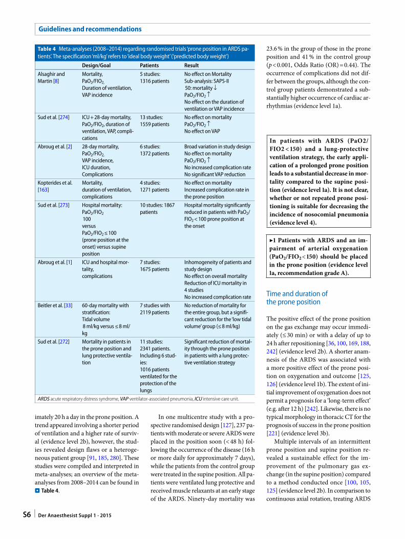
imately 20 h a day in the prone position. A trend appeared involving a shorter period of ventilation and a higher rate of surviv-al (evidence level 2b), however, the stud-ies revealed design flaws or a heteroge-neous patient group [91, 185, 280]. These studies were compiled and interpreted in meta-analyses; an overview of the meta-analyses from 2008–2014 can be found in . Table 4.
In one multicentre study with a pro-spective randomised design [127], 237 pa-tients with moderate or severe ARDS were placed in the position soon (< 48 h) fol-lowing the occurrence of the disease (16 h or more daily for approximately 7 days), while the patients from the control group were treated in the supine position. All pa-tients were ventilated lung protective and received muscle relaxants at an early stage of the ARDS. Ninety-day mortality was
23.6 % in the group of those in the prone position and 41 % in the control group (p < 0.001, Odds Ratio (OR) = 0.44). The occurrence of complications did not dif-fer between the groups, although the con-trol group patients demonstrated a sub-stantially higher occurrence of cardiac ar-rhythmias (evidence level 1a).
Time and duration of the prone position
The positive effect of the prone position on the gas exchange may occur immedi-ately (≤ 30 min) or with a delay of up to 24 h after repositioning [36, 100, 169, 188, 242] (evidence level 2b). A shorter anam-nesis of the ARDS was associated with a more positive effect of the prone posi-tion on oxygenation and outcome [125, 126] (evidence level 1b). The extent of ini-tial improvement of oxygenation does not permit a prognosis for a ‘long-term effect’ (e.g. after 12 h) [242]. Likewise, there is no typical morphology in thoracic CT for the prognosis of success in the prone position [221] (evidence level 3b).
Multiple intervals of an intermittent prone position and supine position re-vealed a sustainable effect for the im-provement of the pulmonary gas ex-change (in the supine position) compared to a method conducted once [100, 105, 125] (evidence level 2b). In comparison to continuous axial rotation, treating ARDS
Table 4 Meta-analyses (2008–2014) regarding randomised trials ‘prone position in ARDS pa-tients’. The specification ‘ml/kg’ refers to ‘ideal body weight’ (‘predicted body weight’)
Design/Goal Patients Result
Alsaghir and Martin [8]
Mortality,PaO2/FIO2,
Duration of ventilation,VAP incidence
5 studies:1316 patients
No effect on MortalitySub-analysis: SAPS-II 50: mortality ↓PaO2/FIO2 ↑No effect on the duration of ventilation or VAP incidence
Sud et al. [274] ICU + 28-day mortality, PaO2/FIO2, duration of ventilation, VAP, compli-cations
13 studies:1559 patients
No effect on mortalityPaO2/FIO2 ↑No effect on VAP
Abroug et al. [2] 28-day mortality,PaO2/FIO2,
VAP incidence,ICU duration,Complications
6 studies:1372 patients
Broad variation in study designNo effect on mortalityPaO2/FIO2 ↑No increased complication rateNo significant VAP reduction
Kopterides et al. [163]
Mortality,duration of ventilation, complications
4 studies:1271 patients
No effect on mortalityIncreased complication rate in the prone position
Sud et al. [273] Hospital mortality:PaO2/FIO2
100versusPaO2/FIO2 ≤ 100(prone position at the onset) versus supine position
10 studies: 1867 patients
Hospital mortality significantly reduced in patients with PaO2/FIO2 < 100 prone position at the onset
Abroug et al. [1] ICU and hospital mor-tality,complications
7 studies:1675 patients
Inhomogeneity of patients and study designNo effect on overall mortalityReduction of ICU mortality in 4 studiesNo increased complication rate
Beitler et al. [33] 60-day mortality with stratification:Tidal volume 8 ml/kg versus ≤ 8 ml/kg
7 studies with 2119 patients
No reduction of mortality for the entire group, but a signifi-cant reduction for the ‘low tidal volume’ group (≤ 8 ml/kg)
Sud et al. [272] Mortality in patients in the prone position and lung protective ventila-tion
11 studies:2341 patients.Including 6 stud-ies:1016 patients ventilated for the protection of the lungs
Significant reduction of mortal-ity through the prone position in patients with a lung protec-tive ventilation strategy
ARDS acute respiratory distress syndrome, VAP ventilator-associated pneumonia, ICU intensive care unit.
In patients with ARDS (PaO2/FIO2 < 150) and a lung-protective ventilation strategy, the early appli-cation of a prolonged prone position leads to a substantial decrease in mor-tality compared to the supine posi-tion (evidence level 1a). It is not clear, whether or not repeated prone posi-tioning is suitable for decreasing the incidence of nosocomial pneumonia (evidence level 4).
▶1 Patients with ARDS and an im-pairment of arterial oxygenation (PaO2/FIO2 < 150) should be placed in the prone position (evidence level 1a, recommendation grade A).
S6 | Der Anaesthesist Suppl 1 · 2015
Guidelines and recommendations
patients with prone positioning leads to a more rapid and distinctive increase of ox-ygenation, although a difference between the patient groups is no longer demon-strable after 72 h [266] (evidence level 2b).
Synergy effects of the prone position with additional measures
The improvement of oxygenation in the prone position is reinforced through the application of PEEP, particularly in the case of diffuse ARDS [62, 101] (evidence level 2b). Intermittent recruitment ma-noeuvres lead to a more sustainable ef-fect on oxygenation while in the prone position as opposed to the supine posi-tion [102, 227] (evidence level 2b). The in-tegration of spontaneous respiratory rates while in the prone position, for example through the application of biphasic posi-tive pressure ventilation with spontaneous respiration (‘airway pressure release ven-tilation’ [APRV]), increased the effect of positioning methods compared to ventila-tion in a predominantly controlled mode [295] (evidence level 2b). The inhalation of nitric oxide for the improvement of the ventilations/perfusion ratio [39, 111, 114, 145, 186, 220, 243] likewise demonstrat-ed synergetic effects on oxygenation (evi-dence level 2b).
Ventilation in the prone position pres-ents a sensible therapeutic perspective in order to implement a lung-protective strategy by adapting various ventilation settings parameters (reduction of the tid-al volume, reduction of FIO2, the inspira-tory peak pressure, as well as the pressure
difference in inspiration and expiration). Moreover, ventilation in the prone posi-tion implies physiological protection/re-duction of ventilation-associated lung in-jury [102, 107, 124, 127, 170, 193] (evidence level 2b).
Effect of the prone position on other organ systems
Prone positioning per se is not a meth-od that promotes hypotension or cardi-ac instability [134, 146, 149, 193, 299] (ev-idence level 1b). In a broad study, prone positioning—as opposed to supine posi-tioning—lead to an improvement of hae-modynamics (increase of cardiac out-put or median arterial pressure) and to a reduction of cardiovascular complica-tions [125], however, a balanced volume status was necessary for this effect [149] (evidence level 2b). In patients without a pre-existing limitation of the renal func-tion, prone positioning did not lead to a reduction of kidney function [134] (evi-dence level 2b). Positioning on mattress systems controlled by compressed air re-duced a positioning-related increase of in-tra-abdominal pressure compared to con-ventional mattress systems [58, 198] (evi-dence level 2b). Patients with abdominal obesity (CT definition: sagittal abdominal diameter ≥ 26 cm) developed kidney fail-ure (83 vs 35 %, p < 0.01) [309] at a signifi-cantly higher rate during prolonged prone positioning (on average 40 h) compared to patients without a similar configuration (evidence level 2b).
In patients demonstrating no abdomi-nal disease, a minimal, though substantial increase of intra-abdominal pressure with-out intra-abdominal compartment syn-drome occurred as a result of prone posi-tioning during a period of up to 2 h [99, 134, 135] (evidence level 2b). Likewise, no impact on splanchnic perfusion was dem-onstrated [157, 187]. There are no study re-sults for patients with acute abdominal dis-eases and increase of pressure. There have been just as few previous reports that the type of abdominal positioning (padded vs hanging) or the duration of positioning has an influence on intra-abdominal pres-sure or perfusion ratios [58, 61, 134, 205], although this type of support of the thorax and pelvis worsened the compliance of the thoracic wall and increased pleural pres-sure (evidence level 2b). Patients with ab-dominal obesity developed hypoxic hepa-titis during prolonged periods in the prone position (on average 40 h) at a significant-ly higher rate than patients without a simi-lar configuration (22 vs 2 %, p = 0.015) [309] (evidence level 2b).
Prone positioning and acute cerebral lesion
Prone positioning may cause an increase of intracranial pressure and (in the case of unchanged haemodynamics) a reduc-
▶2 A prone positioning interval of at least 16 h should be targeted. The prone position should be considered at an early stage and implemented im-mediately after indication (evidence level 2b, recommendation grade B).▶3 Prone positioning should be con-cluded in the case of persistent im-provement of oxygenation in the su-pine position (4 h after supine po-sitioning: PaO2/FIO2 ≥ 150 with a PEEP ≤ 10 cm H2O and FIO2 ≤ 0.6) or if multiple positioning attempts re-mained unsuccessful (evidence level 3, recommendation grade B).
▶4 The same principles of an opti-mised ventilation strategy apply for ventilation in the prone position as for the supine position, including the lung-protective limitation of tidal vol-ume, the prevention of derecruitment and the integration of spontaneous respiratory rates (evidence level 2b, recommendation grade A).▶5 An evaluation and adjustment of the ventilation mode in the context of a lung-protective strategy should be conducted after each change of posi-tion (evidence level 3, recommenda-tion grade B).
For patients with acute abdominal diseases, no recommendation can cur-rently be provided with respect to the type and duration of a prone position due to the lack of studies (evidence level 4, recommendation grade 0)▶7 CAVE: In patients with abdomi-nal obesity, kidney and liver function should be monitored closely in the event of prolonged prone positioning (expert consensus).
▶6 Prior to the application of prone positioning, the patient should be stabilised haemodynamically and the volume status should be balanced. The use of catecholamines is not a contra-indication against the prone position (evidence level 2b, recommendation grade B).
S7Der Anaesthesist Suppl 1 · 2015 |
tion of cerebral perfusion pressure in the case of acute traumatic or non-trau-matic cerebral lesions [34, 209, 241] (ev-idence level 4). However, the improve-ment of the pulmonary gas exchange in-duced by the prone position may increase cerebral oxygenation [283] (evidence lev-el 4). In healthy humans, systematic and cerebral haemodynamics were captured in the prone position during noninvasive positive pressure ventilation and a varia-tion of the position of the head was con-ducted (centred, to the left and right side). The lateral rotation of the head leads to a reduction of cerebral blood flow (Arte-ria cerebri media) by approximately 10 % [137] (evidence level 2b).
Sufficient studies were not conducted previously as to whether or not an adap-tion of the ventilation settings (change of tidal volume and respiratory minute vol-ume = change of CO2 elimination = change of cerebral perfusion) could have positive effects on the damaged cerebrum while in the prone position. Moreover, no study has been conducted regarding whether or not the adapted analgosedation could prevent the intracranial pressure increase in the case of an acute cerebral lesion.
Prone positioning and intraocular pressure
In one prospective, randomised trial, in-traocular pressure (IOP) was measured in patients in the prone position in an oper-
ative area prior to, during and after the po-sitioning method, wherein the heads of a patient group were additionally turned to the right side at a 45° angle to the prone position [73]. While in the prone position, a moderate increase of the IOP occurred from 12 to 18 mm Hg (p < 0.001) and upon turning the head to the side, the pressure of the lower eye increased further. Two additional studies from the operative ar-ea confirmed these findings [88, 122] (evi-dence level 2b). There is no data in this re-gard for intensive care patients.
Modifications of the prone position
In addition to the complete prone po-sition (180°), the ‘incomplete’ prone po-sition (135°) is also applied because it is perceived as having fewer side effects for patients and is easier to perform for the nursing staff [30, 257]. With proper exe-cution, there were no significant differ-ences between both positions in the inci-dence of severe complications [30] (evi-dence level 2b).
The incomplete prone position lead to a substantial improvement of oxygen-ation in ARDS patients; however, this ef-fect was not as distinctive as with the com-plete prone position. In patients with se-vere ARDS, a significant increase of arte-rial oxygenation (defined as an improve-ment by more than 20 %) while in a com-plete prone position occurred at a signif-icantly higher rate than while in the 135° prone position [30] (evidence level 2b). In one prospective randomised study, the combination of the prone position with an elevation of the upper body lead to a significantly stronger effect on the oxy-genation compared to the prone position alone [245] (evidence level 3).
Complications while in the prone position
The following complications were de-scribed while in the prone positions [28, 30, 42, 43, 65, 105, 124, 144, 218, 272, 301] facial oedema (20–30 %), pressure ul-cers around the face/cornea, pelvis, knee (approximately 20 %) [234] ‘intolerance’ while in the prone position (= coughing, compaction, respiratory problems ap-proximately 20 %), cardiac dysrhythmias (approximately 5 %), necrosis of the ma-milla, pressure ulcers of the tibial crest (in-dividual reports), dislocations of the tra-cheal tube or venous/arterial lines (ap-proximately 1–2 %) [105], nerve dam-age (two case studies regarding brachi-al plexus lesion [119]) (evidence level 2b). In this regard, it is necessary to consider that complications also occur in the su-pine position and a comparison of the in-cidences of position-related complications for the prone position has not previously been sufficiently studied. The retrospec-tive analysis of the multicentre study by Guerin [116] revealed a higher incidence of pressure points and skin ulcers in the prone position group (14.3/1000 ventila-tion days) compared to the supine posi-tion (7.7/1000 ventilation days, p = 0.002) (evidence level 2b).
According to the results of a prospec-tive, randomised study, a lesser frequency of facial oedema was observed due to the modification of the prone position (135° position, ‘incomplete prone position’) compared to the 180° position [30] (evi-dence level 2b). The safe execution of the prone position in patients with extracor-poreal membrane oxygenation (ECMO) was reported in a retrospective observa-tional study [158] (evidence level 3).
Contraindications for prone positioning
Instability of the spine, severe, surgically untreated facial trauma, the acute cerebral lesion with intracranial pressure increase, the critical cardiac rhythm disorder, acute shock syndrome and the ‘open abdomen’ situation apply as contraindications for prone positioning [304, 306].
▶8 The indication for the prone posi-tion with acute cerebral lesions may only be issued after individual con-sideration of benefit (improvement of oxygenation) and risk (intracrani-al pressure increase) (evidence level 3, recommendation grade 0).▶9 During the positioning method, intracranial pressure should be con-tinuously monitored (evidence lev- el 2b, recommendation grade A). The head should be centred during this method and lateral rotation should be avoided (evidence level 3, recom-mendation grade B). Expert consen-sus and S1 guideline Intracranial Pressure (AWMF registry no. 030/105, valid until 12/2015).
▶10 The complete prone position has a stronger effect on the oxygen-ation than the incomplete prone posi-tion and should be primarily applied (evidence level 2b, recommendation grade A).▶11 The elevation of the upper body while in the prone position may be sensible for preventing an impact on other organs (intraocular pressure, intracranial pressure) (evidence level 3, recommendation grade 0).
S8 | Der Anaesthesist Suppl 1 · 2015
Guidelines and recommendations
Appendix I: Prone positioning: recommendations for practical execution
Prone positioning: practical executionEach positioning process—depending on the body weight of the patient as well as the invasiveness of the therapy (drainag-es, catheters, extensions)—is conducted by three to five nurses and one physician [13, 17, 18, 42, 138, 190, 195, 207, 254, 260, 276, 303, 304].A. Preparational measures: 1. Within the scope of prone position-
ing, the use of a special anti-decubi-tus mattress system is recommend-ed to prevent/reduce pressure ul-cers (evidence level 4, recommen-dation grade 0), particularly in pa-tients with an increased decubitus risk (high-dose catecholamine ther-apy, adiposity, cachexia, corticoste-roid therapy) (evidence level 3, rec-ommendation grade 0).
2. Catheters, drainages and artificial airways are secured and, if neces-sary, extended. Prior to positioning, it is necessary to check whether or not it is a ‘difficult-airway-situation’ in order to take potentially suitable measures to ensure the airways (e.g. preventative surgical tracheotomy, providing intubation alternatives). When performing the rotation, the
most essential access points should be secured by the person guiding the head of the patient.
3. The inspiratory fractional oxygen concentration (FIO2) should be set to 1.0.
4. Enteral nutrition is interrupted; the stomach should be emptied through a tube.
5. An adapted analgosedation (Rich-mond Agitation Sedation Scale (RASS-Score) ≤ − 2) is necessary for the rotational manoeuvre to avoid coughing, compaction or regurgita-tion. Ventilation should be custom-ised accordingly. After the position-ing manoeuvre, the analgosedation is reduced.
B. Execution
During the rotating manoeuvre, monitor-ing is necessary by means of continuous arterial blood pressure measurement. Var-ious techniques are described for execut-ing the rotating process. It is recommend-ed to focus on one technique that all in-volved are familiar with [13, 195] (evidence level 4, recommendation grade B for all previously described methods).C. Follow-up 1. After the completed positioning
manoeuvre, monitoring must be completed.
2. Ventilation must be adapted in the context of a lung-protective strategy and monitored after a brief stabili-sation phase (evidence level 3, rec-ommendation grade B).
3. After the rotating manoeuvre, spe-cial measures are taken to reduce pressure around the head, around the pelvis and the knee. Always en-sure careful padding particularly in areas prone to decubitus (recom-mendation grade A). The head and arms should be additionally repo-sitioned in short intervals while in the prone position (recommenda-tion grade 0).
D. Special aspects for executing prone positioning:
1. The application of enteral nutri-tion while in the prone position was studies in multiple trials [240, 255, 294]. In one prospective trial, the residual gastric volume while in the
prone position was greater than in the supine position [240]. In anoth-er trial, with adequate enteral feed-ing tube length, no increased resid-ual gastric volume or an increased incidence of regurgitation was ob-served in contrast to the supine po-sition [255] (evidence level 2b). On the condition of an application with a low flow rate (≤ 30 ml/h) and fre-quent reflux checks, no higher re-sidual volumes or other side effects were observed in one prospective trial [294] (evidence level 2b), this approach is recommended in a sys-tematic analysis [178].
2. While in the prone position, enteral nutrition is possible with a low flow rate (≤ 30 ml/h), however regu-lar reflux checks are suggested (ev-idence level 2b, recommendation grade B).
Continuous lateral rotation therapy
Definition of continual lateral rotation therapy (CLRT)
CLRT involves the continuous rotation of the patient around his longitudinal ax-is in a motor-driven bed system. Depend-ing on the system, a maximum rotational angle of 62° can be achieved on each side.
Rational of CLRT
The goals of CLRT are to prevent pulmo-nary complications (atelectasis, pneumo-nia, congestion of pulmonary secretion), the reduction of pulmonary inflamma-tion as a result of trauma or infection, as well as improving pulmonary gas ex-change in ventilated patients. The increase of oxygenation, the incidence of nosoco-mial pneumonia, as well as the duration of mechanical ventilation and intensive care stays or hospitalisation are classified as parameters for this. However, none of these parameters are established as an ad-equate surrogate for survival and the qual-ity of survival. Indications for the use of CLRT comprise both prophylactic (pre-vention of complications) and therapeu-tic aspects (improvement of pulmonary functionality).
▶12 Compared to the supine position, the prone position leads to a higher incidence of pressure ulcers and re-spiratory problems, such that a posi-tioning should be done particularly gentle and the airways should be pro-tected and monitored (evidence level 2, recommendation grade A).▶13 An open abdomen, spinal insta-bility, increased intracranial pressure, critical cardiac rhythm disorders and manifest shock are contraindications for the prone position. These contra-indications may be deviated from in individual cases after consideration for the benefits and risks and follow-ing consultation with the specialist disciplines involved (expert consen-sus, recommendation grade 0).
S9Der Anaesthesist Suppl 1 · 2015 |
Comment: In one recommendation from the Paul Ehrlich Society (PEG) ‘Nos-ocomial Pneumonia: Prevention, Diagnos-tics, und Therapy’ [38], there is no recom-mendation for the use of CLRT within the scope of a ‘bundle’ for the prevention of ven-tilator-associated pneumonia. The current recommendations of the Commission for Hospital Hygiene and Infection Prevention (KRINKO) at the Robert Koch Institute [162] determined based on ‘lacking consis-tency’ in the trials and meta-analyses that, ‘Therapy with kinetic beds for the preven-tion of VAP (“ventilator- associated pneu-monia”) cannot be recommended at this time.’ As a restriction to this recommen-dation, it is necessary to adhere to the fact that at the time of the publication from the KRINKO, the prospective randomised pub-lications from Staudinger et al. [265] and Simonis et al. [263] were not yet published.
The use of CLRT requires a targeted indication and safe handling in order to prevent undesired effects. After initiating this method, the persistence of the indica-tion—as with other therapeutic methods as well—should be reviewed daily.
Effects of CLRT on pneumonia incidence, duration of ventilation and mortality
The present studies regarding the effect of CLRT on the incidence of respiratory infections are limited by various criteria for the diagnosis of infections of the up-per and lower respiratory tracts as well as the lung parenchyma [70, 71, 120, 136, 184, 263, 265].
In two more recent prospective ran-domised trials [263, 265], a reduction of the incidence of respiratory infection in-cluding ‘ventilator-associated pneumo-nia’ (VAP) was observed in ventilated pa-tients compared to standard positioning (bedsore prophylaxis) (evidence level 1b). Furthermore, in the study by Stauding-er et al. [265], the ventilation time (8 vs 13 days, p = 0.02) and the treatment time in intensive care (25 vs 39 days, p = 0.01) was significantly shorter in patients treat-ed with CLRT; the mortality rate did not differ. The study by Simonis et al. on pa-tients in cardiogenic shock [263] demon-strated—in addition to VAP reduction—a significantly higher 1-year survival rate
(59 %) compared to the control group without CLRT (34 %, p = 0.028) (evidence level 1b). There are no comparative stud-ies of CLRT with other positioning meth-ods for preventing VAP.
The treatment period in intensive care was shorter in three out of eight ran-domised trials compared to conventional-ly treated patients (evidence level 1b). The length of hospitalisation was shortened due to CLRT in a prospective randomised trial [265] (evidence level 1a), though not in other trials with partially limited quali-ty [4, 60, 211, 291] (evidence level 3).
Physiological effects of CLRT
CLRT was originally used in immobilised patients for bedsore prophylaxis. Subse-quently, the indication was broadened for the treatment of patients with pulmonary disorders. Improved oxygenation, the dis-solution of atelectasis, improved ventila-tion/perfusion ratios, increased secretion mobilisation, the reduction of pulmonary inflammatory response following trauma and a reduction of pulmonary fluid reten-tion was determined as effects.
Effects of CLRT on the pulmonary function
CLRT improves the pulmonary gas ex-change in patients with acute respiratory insufficiency (evidence level 2b) [25, 222, 223, 237, 238, 265, 267]. The following ef-fects were confirmed starting at a rota-tional angle of ≥ 40° on each side:a) The reduction of extravascular lung
water (EVLW) in patients with im-paired oxygenation (ARDS) [32] (evi-dence level 2b). The mechanism is not ultimately clear; continual movement and changes in intrapulmonary pres-
sure ratios possibly lead to increased drainage through the lymphatic system of the lungs [10, 29] (evidence level 4).
b) The reduction of ventilation/perfu-sion mismatch [27] (evidence level 4).
c) In some trials, the incidence and ex-tent of atelectasis were reduced with the early, that is preventative use of CLRT from the start of ventilation. Few limitations of oxygenation oc-curred [4, 98, 160]. In other trials, however, no significant effects were demonstrated [51, 110, 277, 310] (ev-idence level 3). Particularly in poly-traumatised patients with a pulmo-nary injury, early CLRT was able to prevent the occurrence of ARDS or improve oxygenation [31, 86, 93, 202, 223, 300] (evidence level 2b).
d) In trauma patients, CLRT reduced the pulmonary inflammation reaction (reduction of pulmonary and system-ic pro-inflammatory cytokines (TNF, IL-6) and lead to a less severe organ function disorder up to the fifth day post-trauma compared to patients treated in the supine position [31] (evidence level 2b).
e) In one trial, CLRT lead to the disso-lution of atelectasis in ventilated pa-tients [238]; a more recent publication could not verify this effect [51], how-ever both studies demonstrate meth-odological weaknesses. Thus, no rec-ommendation is provided for treating atelectasis with CLRT.
f) The improvement of oxygenation due to CLRT in patients with restricted re-spiratory function (ARDS) occurred at a slower rate than in the prone posi-tion [266] (evidence level 2b).
g) To date, there has been no proof of in-creased bronchopulmonary secretoly-sis due to CLRT; however, a rotation-al angle of < 30° was used in the only study [77] (evidence level 4).
▶14 The early use of CLRT can be em-ployed in certain groups of ventilated patients as a supplement to preven-tion of ventilator-associated pneu-monia, however, other methods (e.g. adapted analgosedation, mobilisation concepts) should not be impacted by this (evidence level 3, recommenda-tion grade B).
▶15 CLRT should not be used in pa-tients with ARDS (PaO2/FIO2 < 150) (recommendation grade A).In the case of contraindications to the prone position, the use of CLRT may be considered for improving oxygen-ation (evidence level 3, recommenda-tion grade 0).
S10 | Der Anaesthesist Suppl 1 · 2015
Guidelines and recommendations
Time and duration of CLRT: angular settings
In most studies, CLRT was conducted at beginning of intensive care treatment for at least 72 h. The use of CLRT within 2 days after development of a respiratory in-sufficiency was linked to a significant re-duction of intensive care therapy and hos-pitalisation compared to a later initiation of the method in two studies [98, 279] (evidence level 3). One positive effect on the gas exchange was able to be observed up to a duration of 5 days after the onset of treatment [25, 224] (evidence level 4). The parameters or strategies according to which CLRT should be concluded have not been studied (‘Weaning’) [94].
In one study, it was determined that longer periods of retention in the lateral position during CLRT do not improve the gas exchange and may even cause a dete-rioration in individual cases due to a re-duction of pulmonary compliance [256 (evidence level 2b). The positive effects on oxygenation and on pneumonia inci-dence (see below) were observed with one exception [310] during CLRT with a rota-tional angle > 40°.
Ventilation setting during CLRT and duration of CLRT
Complications and interactions of CLRT
The following complications were de-scribed during CLRT: pressure ulcers, ‘in-tolerance’ (coughing, compactions, respira-tory problems), cinetosis, catheter disloca-tions, nerve damage [93, 184, 277]. In one prospective observational trial on 20 ‘hae-modynamically stable’ patients, no chang-es of heart rate or blood pressure were reg-istered during CLRT [12] (evidence level 3). In the case of haemodynamically unstable patients, a drop in blood pressure in a steep lateral position (most often in the right lat-eral position) is frequently observed [26] (evidence level 2b). A direct comparison of the incidence of position-related complica-tions with other positioning methods is not possible due to a lack of data.
There is data from two trials regarding the use of CLRT in patients with acute ce-rebral lesions [60, 287]. No increase of in-tracranial pressure during CLRT was stat-ed in one trial [287] (evidence level 4).
In one retrospective trial, an increased complication rate and duration of ventila-tion during CLRT was determined in pa-tients with spinal lesions, however the se-verity of neurological deficits in these pa-tients was greater [57] than in the ‘conven-tionally’ treated group (evidence level 4).
Contraindications for CLRT
An instable spine, acute shock syndrome and a body weight > 159 kg (according to
the manufacturer) are considered to be contraindications for CLRT.
Appendix II: continuous lateral rotation therapy: recommendations for practical execution
Careful positioning requires special pro-tective measures for pressure-sensitive ar-eas (head/neck, auricles, pelvis, knee, bra-chial nerve, peroneal nerve) [94, 224] (ev-idence level 4, recommendation grade B).
Prior to starting the system each time, a manual ‘test rotation’ should be conduct-ed to check the proper positioning of the patient as well as adequate extension and attachment of all supply lines and drain-ages. CLRT should be started with small rotational angles and then increased. To achieve optimal rotational periods (18–20 h/day), nursing and physician activi-ties should be well coordinated with each other (evidence level 4, recommendation grade 0). In the case of an invasive, con-tinuous blood pressure measurement, the pressure sensor must be fastened to the bed system at the level of the heart in the median axis in order prevent false mea-surements during the rotational process. With a proper routine and preparation, CLRT can also be safely used in combina-tion with extracorporeal membrane oxy-genation [164] (evidence level 3, recom-mendation grade 0). In the case of dis-tinctive haemodynamic insufficiency in the lateral position, the angle of rotation should be reduced to the respective side (recommendation grade 0).
Lateral position for patients with pulmonary disorders
Definition of lateral position
A position, in which the side of the body is supported and elevated up to an angle of 90°, is referred to a lateral position.
Rational of the lateral position
In addition to relieving support areas (de-cubitus prophylaxis), pulmonary compli-cations are intended to be prevented and the pulmonary gas exchange improved. This is the result of frequent reposition-ing or special lateral positioning in the
▶16 If CLRT is used for treating oxy-genation impairment, the indication for continuation should be reviewed daily based on the improvement of oxygenation (as with the prone posi-tion).CLRT should be concluded upon sta-bilisation of the gas exchange in the supine position without rotation, or if a continuous application showed no success over a period of 48 h to no more than 72 h (evidence level 3, rec-ommendation grade B).
▶17 For ventilation during CLRT, the principles of a lung-protective venti-lation strategy should apply (evidence level 2b, recommendation grade A).
▶18 The same criteria as with the prone position apply for conduct-ing CLRT in patients with acute ce-rebral lesions. These patients should be monitored by means of continu-ous intracranial pressure measure-ment (evidence level 3b, recommen-dation grade 0) and may be situated in a moderately high upper body posi-tion (inclined position of the bed sys-tem).▶19 It is necessary to individually consider between potential damage due to CLRT and the expected bene-fit in the case of severely injured pa-tients (evidence level 4, recommenda-tion grade 0).
S11Der Anaesthesist Suppl 1 · 2015 |
case of unilateral lung damage. The sim-plicity of the method is beneficial, which can be conducted at any time with mini-mal additional effort [14, 141].
Physiological effects and side effects of the lateral position in patients without lung damage
Effects on haemodynamics and gas ex-change were studied, wherein primarily postoperative patients with healthy lungs were studied [50, 212].
Only minimal changes in ventilation and haemodynamics were detected in spontaneous respiration among individ-uals with healthy lungs [50]. Blood pres-sure tend to sink in the lateral position (left lateral position >right lateral posi-tion [148], evidence level 4). In the left lat-eral position, greater heterogeneity of ven-tilation dispersion occurred compared to the right lateral position [92] (evidence level 4). The lateral position promoted the perfusion in the direction of the ven-tral pulmonary sections in ventilated pa-tients [27] (evidence level 3). The mea-surement of haemodynamics in the lat-eral position was vulnerable to artefacts, particularly when determining the refer-ence point [12, 49] (evidence level 4).
In postoperatively ventilated patients without acute respiratory insufficiency, the overall compliance of the respiratory system in the lateral position is reduced compared to the supine position [282] (evidence level 4). The phenomenon of atelectasis formation after the induction of anaesthesia and atelectasis treatment through PEEP occurred in the dependent lung in the lateral position just as in the supine position [161] (evidence level 4).
In postoperatively ventilated patients with healthy lungs and without acute re-spiratory insufficiency, without atelecta-sis and with a high tidal volume, the lateral position (45°–90°) did not improve the pul-monary gas exchange compared to the su-pine position [212, 285, 286] (evidence lev-el 2b). The moderate lateral position (45°) did not affect any clinical changes of the gas exchange, haemodynamics and tissue per-fusion compared to the supine position [21, 285, 286] (evidence level 4). The mixed ve-nous oxygen saturation decreased mini-mally [108] (evidence level 4).
The haemodynamics are only slightly influenced by the lateral position of ven-tilated patients; no significant changes of cardiac output occurred [22, 285, 286] (evidence level 4). A prophylactic effect of the lateral position on the prevention of postoperative pulmonary complications was not adequately studied.
Indications and effects of the lateral position in patients with lung damage
Bilateral lung damageIn the case of chronic obstructive pulmo-nary disease (COPD), noninvasive ven-tilation in the lateral position is possible. However, it does not cause any addition-al improvement of the gas exchange com-pared to the supine position [233] (ev-idence level 4). In two trials involving a total of 22 ventilated patients with acute lung damage, the effects on oxygenation due to the lateral position were variable and not predictable compared to the su-pine position [210, 256] (evidence level 4).
CLRT with a minimal rotational angle ≤ 40° and the intermittent, 2 h long lat-eral position had the same effect on the gas exchange, wherein higher secretion mobilisation was observed using CLRT [68] (evidence level 2b). In the right lat-eral position, there was more often a hae-modynamic compromise in ventilated pa-tients compared to the left lateral position caused by a more reduced right ventricu-lar filling [26, 76, 120] (evidence level 2b). These effects have not been studied in non-ventilated patients or ventilated pa-tients without lung damage.
Unilateral lung damageIn spontaneous breathing, the lateral po-sition improves oxygenation if the good lung is down [23, 95, 284] (evidence lev-el 4). However, in the case of a very high ‘closing volume’ it may be better to posi-tion the bad lung down [59] (evidence lev-el 2b). Effects can be expected particularly with pneumonia, although not with cen-tral obstructions, such as carcinoma [53] (evidence level 4).
In the case of mechanical ventilation and lateral positioning with the good lung down, oxygenation improves [53, 59, 79, 143, 235, 244] (evidence level 2b) through homogenisation of ventilation/perfusion dispersion and reduction of the intrapul-monary shunt [115, 132] (evidence level 4). These improvements of the gas exchange are based on the same mechanisms as with the prone position, with which the bad lung is taken from the dependent po-sition. These effects can be expected for gas exchange disorders due to pneumonia and atelectasis, but not due to pleural ef-fusion [50] (evidence level 4). Effects of the lateral position on the outcome with respect to ventilation duration, pneumo-nia incidence or mortality have not been studied.
▶20 During the ventilation of patients without lung damage, a lateral posi-tion exclusively for preventing pul-monary complications is not sensi-ble (evidence level 2b, recommenda-tion grade B).
The effects of an intermittent lateral position or CLRT up to a rotational angle < 40° on the pulmonary gas ex-change have not been adequately ver-ified. In patients with ARDS, CLRT up to 40° does not demonstrate any advantage compared to intermittent lateral positioning with respect to im-proving oxygenation (evidence lev-el 2b).▶21 Proper positioning and interpreta-tion of invasively measured blood pres-sure values should be particularly en-sured in the lateral position (evidence level 3, recommendation grade B).
▶22 In the case of ventilation of pa-tients with unilateral lung damage, a lateral position of approximately 90° is recommended with the good lung down to improve the gas exchange (evidence level 2b, recommendation grade B)
S12 | Der Anaesthesist Suppl 1 · 2015
Guidelines and recommendations

Backrest elevation position
Definitions of elevated upper body position
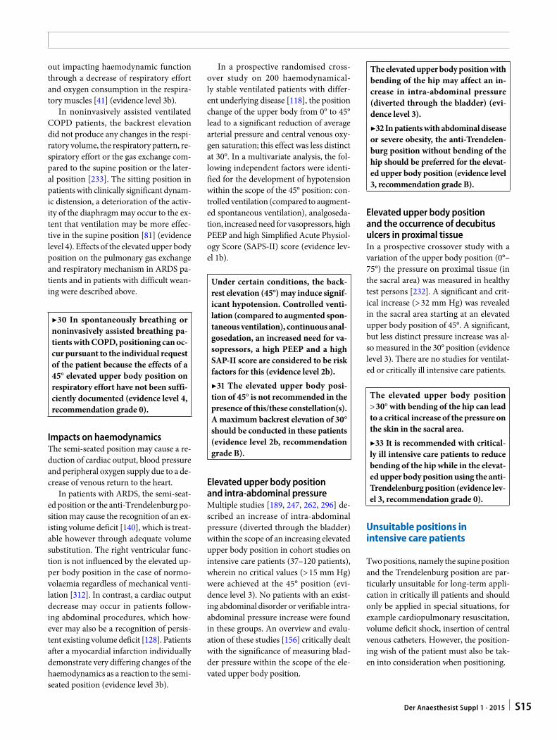
The elevated upper body position is im-plemented in various ways in different trials—there is no universal definition. Various positions are studied, which can range between the classic sitting position with bent hip and knee joints on one hand and tilting of the entire, flat-lying patient (called the anti-Trendelenburg position) on the other hand. This likewise includes the so-called ‘reclined seated position’, for which there is no date regarding its ef-fects on haemodynamics and lung func-tion. The semi-seated position refers to a position, in which—with bent hip and extended or bent knee joints—the upper body and the head of the patient are ele-vated by a certain degree as opposed to the flat-lying lower extremities (see . Fig. 1).
What all modifications of the elevated upper body share in common is that the upper body is positioned above the level of the trunk, wherein the angle is at least 30° [75].
Effect mechanisms of the backrest elevation
As a goal of the clinical trials, the gravita-tionally dependent effects of the elevated upper body position were studied. In this regard, the prevention of passive regur-gitation (pulmonary aspiration of gastric contents) [63, 113] and the reduction of in-tracerebral blood volume (reducing intra-cranial pressure) were of primary focus. The remaining described effects of the el-evated upper body position on haemody-namics (modified orthostatic reaction) and the pulmonary gas exchange (change
of diaphragm position) were considered to be gravitationally dependent [24].
Effects and impacts of backrest elevation on the lungs
Impacts on gastroesophageal reflux and pulmonary aspirationThe aspiration of secretion contaminated with bacteria in the gastrointestinal tract and the pharynx is generally perceived as a risk factor and trigger for the develop-ment of nosocomial and ventilator-associ-ated pneumonia (VAP). Consequentially, measures that lead to decrease of gastroin-testinal reflux and a reduction of the oro-pharyngeal secretion volume should ac-company a lower incidence of nosocomi-al pneumonia and VAP [7, 142, 213] (evi-dence level 3).
Studies are available that have been conducted on patients with orotracheal intubation, who do not have known risk factors for gastroesophageal reflux. All patients were supplied with a nasogas-tric tube; some were fed enterally. Stress bleeding prophylaxis was conducted and the endotracheal cuff pressure was mon-itored (> 25 cm H2O). A 45° elevated up-per body position in these patients lead to a delay of gastroesophageal reflux and to a decrease, though not a complete preven-tion, of pulmonary aspiration of pharyn-geal secretion compared to a flat supine position [219, 290] (evidence level 2b).
In two prospective randomised trials [78, 117], a substantial reduction of VAP was observed through the application of a 45° backrest elevation compared to the su-pine position (evidence level 2b), howev-er, both of these studies were heavily crit-icised with respect to their design and the method [213]. A small randomised pilot study observed a trend for reducing VAP with this position (evidence level 3) [154].
Further studies regarding feasibility and the effect of 45° position [19, 20, 37, 214, 231, 248, 249, 250] revealed that precise compliance with the position in clinical practice is normally not feasible and a tar-get angle of 45° could not be achieved (ev-idence level 2a). To improve practical im-plementation, numerous technical appli-cations (angle measuring systems, train-ings programmes for nursing staff) were recommended and implemented, which (with substantial effort) contributed to the increase of the precise execution [20, 37, 182, 311, 314] (evidence level 2b).
A systematic analysis and evaluation of the three randomised trials regarding the impact of the backrest elevation on VAP incidence by means of the Delphi meth-od [213] did not reveal any clear evidence for the application of a 45° elevated up-per body position due to the heterogene-ity of the studies. Considering undesired accompanying effects, this expert consen-sus recommended that the elevated upper body position (20°–45°; more than 30°if possible) be used as a preferred position with reference to numerous limitations in ventilated patients (evidence level 2a). Despite the weakness of the Delphi rec-ommendation, which is due to the weak-ness of the analysed studies, the guideline group supports this recommendation as it appears practical for clinical use and re-flects the limited evidence.
Impacts on pulmonary gas exchangeEven in those with healthy lungs, anaes-thesia and mechanical ventilation lead to a change of the regional ventilation with the development of atelectasis, particular-ly in the dorsal and diaphragm areas of the lungs. This effect is likely more distinctive in patients with increased intra-abdom-inal pressure (e.g. severe obesity, exten-
Fig. 1 8 Modifications of the elevated upper body position
▶23 The preferred principle position for intubated patients is the backrest elevation position of 20°–45°, prefer-ably ≥ 30°, considering the limitations (evidence level 3, recommendation grade B).For patients with elevated intracrani-al pressure, specific recommendations will be announced (see ▶ 27–29).
S13Der Anaesthesist Suppl 1 · 2015 |
sive surgical procedures on the abdomen, peritonitis) because the mobility of the di-aphragm is limited and situated in crani-al orientation. Even with ARDS, the im-paired lung function leads to ventilation disorders and the formation of atelectasis. We must assume that actions for prevent-ing diaphragm dislocation reduce the for-mation of atelectasis and thus contribute to an improvement of the gas exchange.
In one prospective crossover trial in 40 ARDS patients, the backrest eleva-tion (20°–45°) leads to an increase of ox-ygenation in 32 % of the patients studied (> 20 % compared to the flat supine posi-tion) and to an increase of the lung vol-ume [72] (evidence level 2b). In a simi-lar crossover trial in 24 ventilated patients with difficult weaning, the 45° position lead to a significant reduction of respira-tory effort. Patients found the comfort lev-el in this position to be the highest; no im-pact on the reduction of the weaning pro-cess was observed [74] (evidence level 2b).
In postoperative patients without AR-DS, the semi-seated or sitting position lead to contradicting results with respect to the gas exchange compared to the su-pine position. In patients who were not characterised in more detail with pre-ex-isting pulmonary diseases, the sitting po-sition had no effect on capillary blood gas-es as opposed to the flat position regard-less of age [212].
The effects of an intraoperative, semi-seated position on the gas exchange are al-so studied in neurosurgical patients [66]. The small amount of available data re-vealed an improvement of oxygenation in these patients. However, due to the fact that the intraoperative position was pri-marily determined by the surgery, a tar-geted, therapeutic application is not rele-vant (evidence level 4).
Backrest elevation in the case of obesity
In one prospective cohort study on 30 ventilated patients with obesity (BMI > 35 kg/m2), a significant reduction of ex-piratory flow limitation (= improvement of the gas flow) and a reduction of auto PEEP was revealed while in the sitting po-sition (> 45°) compared to the lying posi-tion. These effects were not demonstrable in a control cohort (15 patients with BMI < 30 kg/m2) [173] (evidence level 2 b).
Impacts on other organ system
Intracerebral pressure (ICP) and cerebral perfusion pressure (CPP)The elevated upper body position has been in treating ICP for a long time. Due to gravitationally dependent shifting, the cerebral blood and fluid volume are re-duced and ICP decreases. However, the semi-seated position may also lead to an impact on haemodynamics and thus to a reduction of CPP. In patients with normal and elevated ICP, the elevated upper body position normally leads to a reduction of ICP depending on the angle [87]. An ac-companying reduction of CPP can be ob-served more frequently with an elevat-ed upper body position of 30° and great-er. However, the breadth of the individual
reaction through interactions with other parameters, such as ventilation pressure, sympathetic stimulation, haemodynamic function, volume status and level of seda-tion is vast and thus not predictable [44, 83, 89, 155, 180, 251, 313] (evidence level 3).
Impacts on respiratory effortBackground: The most frequent postop-erative complications after thoracic proce-dures are of a pulmonary nature caused by partial respiratory insufficiency as well as postoperative hypermetabolism with in-creased O2 consumption. Increased respi-ratory effort must be made by changing the lung volume particularly in patients with chronic obstructive pulmonary dis-ease (COPD). Regarding the effects of the position, however, differences can be ex-pected between patients with a chron-ic gas exchange disorder and those with acute exacerbation.
In patients following a thoracotomy, the semi-seated position resulted in a re-duction of energy consumption with-
▶24 The elevated upper body position (20°–45°) may contribute to an im-provement of oxygenation and the re-spiratory mechanics in patients with ARDS (evidence level 2b, recommen-dation grade B).
▶25 Within the scope of the difficult weaning of mechanical ventilation (without the presence of COPD), the elevated upper body (45°) should be used to reduce respiratory effort and to increase the comfort level of the pa-tient (evidence level 2b, recommenda-tion grade B).
▶26 The flat supine position should be avoided in patients with severe obesi-ty (evidence level 4, expert consen-sus). The backrest elevation position (> 45°) may contribute to an improve-ment of the respiratory mechanics in ventilated patients with severe obesi-ty (BMI > 35 kg/m2) (evidence level 2b, recommendation grade 3). Regarding contraindications for the elevated up-per body position—see ▶28 and ▶31
▶27 The application of an elevated up-per body position of 15°–30° is sensi-ble in patients with increased intra-cranial pressure and may contribute to a reduction of intracerebral pres-sure (evidence level 2b, recommenda-tion grade B)▶28 A 45° backrest elevation cannot be recommended without limitation in patients with suspicion of increased intracranial pressure due to the fact that cerebral perfusion pressure can become critically degraded with an in-creasingly elevated position (evidence level 2b, recommendation grade B).▶29 With respect to the treatment of patients with elevated intracranial pressure, please refer to the S1 guide-line intracranial pressure (AWMF reg-istry no. 030/105, valid until 12/2015): ‘If possible, an elevated upper body po-sition should be aimed for. The indi-vidually optimised upper body posi-tion should be regularly evaluated with ICP and CPP controls in the 0° (not in the case of the risk of aspiration or with ventilation), 15° and 30° position. Ve-nous return flow should not be prevent-ed by bending the head’ [11].
S14 | Der Anaesthesist Suppl 1 · 2015
Guidelines and recommendations
out impacting haemodynamic function through a decrease of respiratory effort and oxygen consumption in the respira-tory muscles [41] (evidence level 3b).
In noninvasively assisted ventilated COPD patients, the backrest elevation did not produce any changes in the respi-ratory volume, the respiratory pattern, re-spiratory effort or the gas exchange com-pared to the supine position or the later-al position [233]. The sitting position in patients with clinically significant dynam-ic distension, a deterioration of the activ-ity of the diaphragm may occur to the ex-tent that ventilation may be more effec-tive in the supine position [81] (evidence level 4). Effects of the elevated upper body position on the pulmonary gas exchange and respiratory mechanism in ARDS pa-tients and in patients with difficult wean-ing were described above.
Impacts on haemodynamicsThe semi-seated position may cause a re-duction of cardiac output, blood pressure and peripheral oxygen supply due to a de-crease of venous return to the heart.
In patients with ARDS, the semi-seat-ed position or the anti-Trendelenburg po-sition may cause the recognition of an ex-isting volume deficit [140], which is treat-able however through adequate volume substitution. The right ventricular func-tion is not influenced by the elevated up-per body position in the case of normo-volaemia regardless of mechanical venti-lation [312]. In contrast, a cardiac output decrease may occur in patients follow-ing abdominal procedures, which how-ever may also be a recognition of persis-tent existing volume deficit [128]. Patients after a myocardial infarction individually demonstrate very differing changes of the haemodynamics as a reaction to the semi-seated position (evidence level 3b).
In a prospective randomised cross-over study on 200 haemodynamical-ly stable ventilated patients with differ-ent underlying disease [118], the position change of the upper body from 0° to 45° lead to a significant reduction of average arterial pressure and central venous oxy-gen saturation; this effect was less distinct at 30°. In a multivariate analysis, the fol-lowing independent factors were identi-fied for the development of hypotension within the scope of the 45° position: con-trolled ventilation (compared to augment-ed spontaneous ventilation), analgoseda-tion, increased need for vasopressors, high PEEP and high Simplified Acute Physiol-ogy Score (SAPS-II) score (evidence lev-el 1b).
Elevated upper body position and intra-abdominal pressureMultiple studies [189, 247, 262, 296] de-scribed an increase of intra-abdominal pressure (diverted through the bladder) within the scope of an increasing elevated upper body position in cohort studies on intensive care patients (37–120 patients), wherein no critical values (> 15 mm Hg) were achieved at the 45° position (evi-dence level 3). No patients with an exist-ing abdominal disorder or verifiable intra-abdominal pressure increase were found in these groups. An overview and evalu-ation of these studies [156] critically dealt with the significance of measuring blad-der pressure within the scope of the ele-vated upper body position.
Elevated upper body position and the occurrence of decubitus ulcers in proximal tissueIn a prospective crossover study with a variation of the upper body position (0°–75°) the pressure on proximal tissue (in the sacral area) was measured in healthy test persons [232]. A significant and crit-ical increase (> 32 mm Hg) was revealed in the sacral area starting at an elevated upper body position of 45°. A significant, but less distinct pressure increase was al-so measured in the 30° position (evidence level 3). There are no studies for ventilat-ed or critically ill intensive care patients.
Unsuitable positions in intensive care patients
Two positions, namely the supine position and the Trendelenburg position are par-ticularly unsuitable for long-term appli-cation in critically ill patients and should only be applied in special situations, for example cardiopulmonary resuscitation, volume deficit shock, insertion of central venous catheters. However, the position-ing wish of the patient must also be tak-en into consideration when positioning.
▶30 In spontaneously breathing or noninvasively assisted breathing pa-tients with COPD, positioning can oc-cur pursuant to the individual request of the patient because the effects of a 45° elevated upper body position on respiratory effort have not been suffi-ciently documented (evidence level 4, recommendation grade 0).
Under certain conditions, the back-rest elevation (45°) may induce signif-icant hypotension. Controlled venti-lation (compared to augmented spon-taneous ventilation), continuous anal-gosedation, an increased need for va-sopressors, a high PEEP and a high SAP-II score are considered to be risk factors for this (evidence level 2b).▶31 The elevated upper body posi-tion of 45° is not recommended in the presence of this/these constellation(s). A maximum backrest elevation of 30° should be conducted in these patients (evidence level 2b, recommendation grade B).
The elevated upper body position with bending of the hip may affect an in-crease in intra-abdominal pressure (diverted through the bladder) (evi-dence level 3).▶32 In patients with abdominal disease or severe obesity, the anti-Trendelen-burg position without bending of the hip should be preferred for the elevat-ed upper body position (evidence level 3, recommendation grade B).
The elevated upper body position > 30° with bending of the hip can lead to a critical increase of the pressure on the skin in the sacral area.▶33 It is recommended with critical-ly ill intensive care patients to reduce bending of the hip while in the elevat-ed upper body position using the anti-Trendelenburg position (evidence lev-el 3, recommendation grade 0).
S15Der Anaesthesist Suppl 1 · 2015 |
Flat supine position
Definition. The supine position refers to a position, in which the patient lies flat and horizontally on his back.
If someone with a normal weight lies in the flat supine position, an increased venous return flow to the heart will oc-cur. Cardiac output, pulmonary blood flow and arterial blood pressure increase, the functional residual capacity (FRC) de-creases, the diaphragm is compromised by the abdomen and limited in its mobil-ity. Anaesthesia, analgosedation or mus-cle relaxants increase the undesired effects [239]. The reduced FRC will also lead to the collapse of small respiratory tracts, to the formation of atelectasis and to a limit-ed pulmonary gas exchange [69].
The flat supine position can be dan-gerous particularly for obese patients. It can lead to acute heart failure, respirato-ry arrest and pronounced pulmonary gas exchange disorders [165, 172, 292, 315]. Death in the extremely obese due to the flat supine position is referred to as ‘obesi-ty supine death syndrome’ [292].
Expiratory flow impediments, the de-velopment of an auto PEEP as well as a collapse of small respiratory tracts oc-curred regularly in mechanically ventilat-ed obese patients in the flat supine posi-tion if an external ZEEP (zero endexpira-tory pressure) or too low of a PEEP level was selected [174].
If there is a combination of COPD and obesity, a tracheomalacia can only be ex-pected in rare cases (3 %) based on a dif-ferential diagnosis, which becomes symp-tomatic in the flat supine position [133, 165].
Trendelenburg position
Definition. The Trendelenburg position is a variation of the flat supine position, in which the head is at the lowest position of the body through the inclined positioning of the bed. It was used regularly starting in 1880 by the surgeon, Friedrich Trendelen-burg (*1844, †1924), during urological and gynaecological procedures and remained widely popular in the following decades [196, 197].
The Trendelenburg position is an ex-treme strain on the respiratory and car-diovascular system of the critically ill pa-tient. Blood is channelled from the low-er parts of the body toward the heart and causes a right heart overload. The ab-dominal organs and—in the case of the obese—the abdominal fat masses press the diaphragm upward and compromise the lungs. The Trendelenburg position leads to a variety of physiological/patho-physiological changes: an increase in the stroke volume of the heart, the pressure on the central veins and pulmonary arter-ies, the resistance of the vascular system, the right and left ventricular end systol-ic volume index, cardiac output and in-trathoracic blood volume as well as to re-duced cerebral blood flow, to reduced sys-temic oxygenation and an increase of ar-terial carbon dioxide partial pressure. The FRC decreases; atelectasis formation oc-curs [129].
The Trendelenburg position is the most hazardous position for the obese [191, 264]. It should not be applied in spontaneously breathing, awake, obese patients. For anaesthesiological and in-tensive care interventions (e.g. applying a central venous catheter, etc.), the obese patient should not be placed in the Tren-delenburg position.
Early mobilisation
Definition of mobilisation
The term mobilisation describes mea-sures involving the patient, which intro-duce and/or assist passive or active move-ment exercises and which aim to promote and/or maintain mobility. In contrast, po-sitioning refers to the change of bodily po-sitions with the goal of influencing gravi-ty-related effects [3, 121, 153, 176].
Elements of mobilisation
Methods for mobilisation are classified in three areas: passive mobilisation, assisted active mobilisation and active mobilisa-tion [3, 9, 80, 84, 85, 130, 176, 153, 230, 258, 259, 319]. These three areas can be struc-tured as follows:
Passive mobilisation: 5 Passive motions of all extremities in all physiological directions 5 Passive cycling (bed pedal exerciser) 5 Passive vertical mobilisation (tilting table, standing frame) 5 Passive transfer to rehabilitation chair
Assisted active mobilisation: 5 Active movement exercises in the su-pine position with manual support 5 Independent mobilisation in bed (sit-ting down upright, turning) 5 Balance training 5 Assisted cycling
▶34 The flat supine position should not be applied in critically ill patients due to the numerous unfavourable ef-fects on haemodynamics and pulmo-nary gas exchange (evidence level 3, recommendation grade B).If the application of the flat supine po-sition is absolutely necessary for spe-cial medical or nursing measures, it should be limited to the shortest pos-sible period (evidence level 4, recom-mendation grade A).
▶35 The Trendelenburg position should not be applied in critically ill patients due to numerous unfavour-able effects on haemodynamics, pul-monary gas exchange and the respira-tory system (evidence level 3, recom-mendation grade B).
If the application of the Trendelen-burg position is absolutely necessary for special medical or nursing mea-sures, it should be limited to a brief period (evidence level 4, recommen-dation grade A).▶36 The Trendelenburg position should principally be avoided in obese patients (evidence level 3a, recom-mendation grade A).
Early mobilisation refers to the begin-ning of mobilisation within 72 h after admittance to intensive care.
S16 | Der Anaesthesist Suppl 1 · 2015
Guidelines and recommendations
Active mobilisation: 5 Sitting on the edge of the bed, tor-so control 5 Active mobilisation to the status 5 Attempting to stand up, walking exer-cises while standing 5 Walking with and without walking aids 5 Active cycling 5 Isotonic movement exercises with walking aids
Goals of mobilisation
The general goals of mobilisation are to promote and maintain mobility as well as to prevent and/or reduce the effects of im-mobilisation. Immobilisation refers to the idle position of the bodily parts or the en-tire body for the purpose of treatment or for rest (bed rest). Undesired effects of im-mobilisation are a general deconditioning, the development of a weakness, rapid fa-tigue and atrophy of the muscular respira-tory pumps and the skeletal muscles, the development of psycho-cognitive deficits and delirium, the emergence of position-ing-related skin and soft tissue damage as well as the reduction of haemodynamic responsiveness [47, 179].
The specific goals of mobilisation con-sist in improving/maintaining skeletal and respiratory muscle function, increas-ing haemodynamic responsiveness, im-proving central and peripheral perfusion and muscle metabolism, increasing cog-nitive competence and mental wellbeing, reducing incidence and duration of de-lirium, reducing positioning-related skin ulcers and—compared to patients, who were not mobilised early—improving the subsequent health-related quality of life [3, 9, 121, 153, 176].
Effects of early mobilisation on treatment success
When recording and assessing the effects of early mobilisation on the outcome, various relevant parameters are included. These include bodily function outcomes, peripheral muscle strength and function of the muscular respiratory pump, neu-rocognitive competence, ventilator-free days, ICU stay, hospitalisation, mortali-
ty, quality of life and discharge from the hospital.
The following prospective randomised trials are suitable for an analysis: Morris et al. [199] discovered a substantially short-er treatment period in the intensive care unit and in the hospital as well as a trend for shorter treatment costs in early mobil-ised patients. In Burtin et al. [48], signifi-cantly higher muscle strength in the quad-riceps as well as a significantly higher state of functional independence (SF-36) after discharge was observed following early mobilisation. Schweickert et al. [259] de-scribe a substantially longer walking dis-tance after intensive care treatment, a sig-nificantly higher Barthel index, a signifi-cantly higher state of functional indepen-dence (SF-36), a shorter ventilation peri-od during intensive care treatment and a trend toward greater probability of dis-charge in the early mobilisation group (all—evidence level 2b). Other prospec-tive randomised trials with limited qual-ity [35, 52, 55, 64, 82, 208] underscore other outcome findings—Chen et al. [ 55] detected a lower one-year mortality rate among a very small group of patients in the early mobilisation group. Cuesy et al. [64] observed a significant reduction of incidences of nosocomial pneumo-nia among stroke patients in the patient group that experienced passive mobili-sation at an early stage (‘turn-mob’). Na-va et al. [208] studied the effect of early mobilisation in patients with COPD dur-ing a six-minute walk. The patients of the group with early mobilisation walked a significantly longer distance. Bezbaruah et al. [35] recognised a substantially short-er ICU treatment duration in early mobil-isation patients in a small study (all—ev-idence level 3).
Patient-related requirements/suitability for mobilisation
Patient-related requirements and suitabil-ity for mobilisation was reviewed in mul-
tiple observational studies [15, 40, 56, 103, 121, 139, 152, 168, 181, 201, 317]. Bourdin et al. [40] systematically compiled 275 in-terventions, for which 33 % of ventilated patients were mobilised. Getting up out of the chair (56 % of actions) was linked to a substantial reduction in heart and re-spiratory rate; average arterial blood pres-sure and arterial oxygen saturation (pulse oxymetry) remained unchanged. Contin-ued standing (25 %) and walking (11 %) resulted in a heart and respiratory rate in-crease and a significant decrease of arteri-al oxygen saturation (evidence level 2b). Kasotakis et al. [152] presented a surgical intensive care unit optimal mobility score, which captured the exclusion of serious organ function disorders and the suit-ability for mobilisation prior to mobilisa-tion. In one prospective study, this score proved to be better suited—compared to other general scores (comorbidity index, APACHE)—to determine suitability for mobilisation (evidence level 3).
▶37 In principle, early mobilisation should be conducted in all patients treated in intensive care, for whom no exclusion criteria apply (evidence level 2b, recommendation grade A).
▶38 Regarding early mobilisation, the following requirements should be present or established: 5 Customised, score-controlled (e.g. RASS) symptom control of pain, fear, agitation and delirium ac-cording to the S3 guideline revi-sion ‘Analgosedation’ (German Society of Anesthesia and Inten-sive Care) 5 Sufficient respiratory reserve 5 Sufficient cardiovascular reserve
The following serve as reference points for this: average arterial blood pres-sure > 65 or < 110 mm Hg, systolic blood pressure < 200 mm Hg, heart rate > 40 or < 130/min, arterial oxygen saturation (pulse oxymetry) ≥ 88 %, no higher-dosage vasopressor therapy.If cardiopulmonary instability devel-ops during ongoing mobilisation, the exercise unit should be discontinued until stabilisation returns or conduct-ed to an adapted extent (evidence level 2b, recommendation grade A).
S17Der Anaesthesist Suppl 1 · 2015 |

Criteria for checking the feasibility/contraindications or cancellation criteria for (early) mobilisation
Clearly defined exclusion criteria for ear-ly mobilisation are not designated in the literature. However, the requirement for mobilisation should be evaluated for cer-tain acute situations in a symptom-adapt-ed manner. The following examples are described in the literature [9, 97, 109, 150, 159, 218, 268, 269, 281, 289, 318]: 5 increased intracranial pressure 5 active bleeding 5 acute myocardial ischaemia 5 agitated delirium
Preparation/monitoring
The preparation for mobilisation and monitoring of the patient during the ac-tion is described in various observational studies or randomised trials [15, 40, 103, 139, 152, 168, 181, 201, 317]. This includes the information of the patient, the provi-sioning of sufficient staff and the secur-
ing/extension of structures of the mechan-ical respiratory tract, the infusion lines or other drainages as well as the monitoring of vital parameters during the procedure.
Duration and intensity of mobilisation
In the prospective randomised studies [84, 85, 121, 130, 192, 270] deemed suitable for the meta-analyses [3, 121, 176], early mobilisation was started within 72 h after
admittance to intensive care with a grad-ual increase. The actions were conducted on average or at least 20 min twice daily.
Safety aspects/complications and cancellation criteria within the scope of mobilisation
The following complications are de-scribed in individual cases within the scope of mobilisation: orthostatic dys-regulation, patient fall, disconnection of catheters/airway, cardiac dysrhythmias, respiratory fatigue/dyspnoea and agita-tion/stress [103, 150, 268, 269]. In a sys-tematic of overall four studies, no seri-ous complications were detected within the scope of mobilisation, which involved further intervention other than the ter-mination of the action [150]. Mobilisation should be cancelled in the event of the fol-lowing signs of intolerance: SaO2 < 88 %,
Table 5 Components for an ‘early mobilisation’ algorithm. The essential initial conditions of the patient, the aid to be used, the suitable proce-dure and formulation of objectives are listed without clear allocation. The allocation is the result of available staff resources and aids of the respec-tive intensive therapy unit. The stated actions are examples without claim of completeness. Further information can be found at the German Early Mobilisation Network (www.frühmobilisierung.de)
Patient Aid Method Goal
Limited vigilance (RASS ≥ − 3) – Passive motion Prophylaxis of joint contractions and muscle lossPassive cycling
Increasing vigilance (RASS − 3 to − 1) Mobilisation chairTilting table
Activated sitting in bed Prophylaxis of ‘deconditioning’ and delirium
Moving the extremities against gravity
Vertical mobilisation
Passive cycling
(Passive) transfer to mobilisation chair
Return of vigilance(RASS ≥ 0)
Mobilisation chair Active cycling Prophylaxis of deconditioning, delirium and pulmonary function disorders(Active) transfer to mobilisation chair
No serious haemodynamic instability Mobilisation chair Standing in front of the bed Prophylaxis of deconditioning, delirium and pulmonary function disordersWalking exercises while standing
Walking aids Walking with and without walking aid Prophylaxis of deconditioning, delirium and pulmonary function disorders
RASS Richmond Agitation Sedation Scale.
▶39 The decision to conduct limited forms of mobilisation (passive or ac-tive with assistance) with the specified relative contraindications should be considered in individual cases in light of the benefits and risks (evidence lev-el 2b, recommendation grade A).
▶40 The preparation of early mobil-isation comprises the information of the patient, the provisioning of suf-ficient staff and the securing/exten-sion of structures of the mechanical respiratory tract, the infusion lines or other drainages. During mobili-sation, the heart rate, blood pressure and arterial oxygen saturation should be continuously/closed recorded for the monitoring of the vital parame-ters (evidence level 2b, recommenda-tion grade A).▶41 In ventilated patients, the venti-lation parameters should be contin-uously featured (tidal volume, inspi-ratory pressure, respiratory rate, re-spiratory minute volume; in the case of invasively ventilated patients cap-nometry) (evidence level 3, recom-mendation grade B)
▶42 Treatment should begin no later than 72 h after admittance to inten-sive care and be conducted twice dai-ly with a duration of at least 20 min for the length of stay in intensive care. A gradual approach should be aimed for starting with passive mobilisation (. Table 5). In this regard, the develop-ment of an algorithm specific to a unit or hospital is recommended (evidence level 3, recommendation grade B)
S18 | Der Anaesthesist Suppl 1 · 2015
Guidelines and recommendations
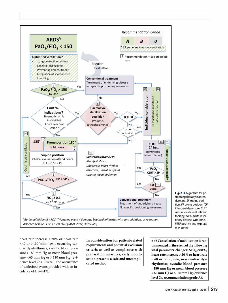
heart rate increase > 20 % or heart rate < 40 or > 130/min, newly occurring car-diac dysrhythmias, systolic blood pres-sure > 180 mm Hg or mean blood pres-sure < 65 mm Hg or > 110 mm Hg (evi-dence level 2b). Overall, the occurrence of undesired events provided with an in-cidence of 1.1–4.4 %.
Fig. 2 9 Algorithm for po-sitioning therapy in inten-sive care. SP supine posi-tion, PP prone position, ICP intracranial pressure, CLRT continuous lateral rotation therapy, ARDS acute respi-ratory distress syndrome, PEEP positive end-expirato-ry pressure
In consideration for patient-related requirements and potential exclusion criteria as well as compliance with preparation measures, early mobili-sation presents a safe and uncompli-cated method.
▶43 Cancellation of mobilisation is rec-ommended in the event of the following vital parameter changes: SaO2 < 88 %, heart rate increase > 20 % or heart rate < 40 or > 130/min, new cardiac dys-rhythmias, systolic blood pressure > 180 mm Hg or mean blood pressure < 65 mm Hg or > 110 mm Hg (evidence level 2b, recommendation grade A).
S19Der Anaesthesist Suppl 1 · 2015 |
Structure/organisation/personnel/expense/protocol
Early mobilisation represents an interdis-ciplinary, targeted approach in improving the results of intensive care. The establish-ment of a concept for this tiered, specif-ic approach in consideration for safety as-pects was classified as beneficial in multi-ple publications [9, 16, 64, 67, 80, 84, 130, 192, 230, 259, 319]. A standard of care is recommended [230], which enables a tiered, customised increase of mobilisa-tion in four phases, particularly in ven-tilated patients [259] as well. Appropri-ate personnel and spatial requirements are integrated in this standard of care. In a prospective observation study, the regu-lar integration of a physiotherapist in early mobilisation proved to have a better effect on outcome parameters compared to ear-ly mobilisation without physiotherapeutic aid [131, 270] (evidence level 2b).
Outlook: early muscle activation through electrical muscle stimulation
A few observational studies describe that the application of electrical muscle stim-ulation in critically ill intensive care pa-tients has a positive effect on the preserva-tion of muscle mass and muscle strength [112, 151, 253, 246, 308]. Very different pa-tient groups were studied at various times while in intensive care in these studies. Comparative, randomised studies do not exist. It is currently unclear, which pa-
tients benefited at which point from elec-trical muscle stimulation while in inten-sive care [305].
At the moment, no recommendation for the use of electrical muscle stimulation in intensive care patients can be expressed due to the incomplete data (. Fig. 2).
Corresponding address
Prof. Dr. Th. BeinClinic for AnaesthesiologyUniversity Hospital Regensburg 93042 [email protected]
Compliance with ethical guidelines
Conflicts of interests. Information regarding con-flicts of interests was requested with the AWMF form. Based on a self-assessment on the part of the partici-pants, there were no relevant conflicts of interest. A discussion among the guideline group regarding the evaluation of and dealing with conflicts of interest at the onset of the initial guideline meeting found that the systematic search for and evaluation of evidence and the additionally structured coordination of recommendations was considered adequate under neutral moderation for avoiding risks for bias.
Open Access This article is distributed under the terms of the Creative Commons Attribution 4.0 International License (http://creativecommons.org/licenses/by/4.0/), which permits unrestricted use, dis-tribution, and reproduction in any medium, provided you give appropriate credit to the original author(s) and the source, provide a link to the Creative Com-mons license, and indicate if changes were made.
References
1. Abroug F, Ouanes-Besbes L, Dachraoui F et al (2011) An updated study-level meta-analysis of randomised controlled trials on proning in ARDS and acute lung injury. Crit Care 15:R6
2. Abroug F, Ouanes-Besbes L, Elatrous S et al (2008) The effect of prone positioning in acute respiratory distress syndrome or acute lung inju-ry: a meta-analysis. Areas of uncertainty and rec-ommendations for research. Intensive Care Med 34:1002–1011
3. Adler J, Malone D (2012) Early mobilization in the intensive care unit: a systematic review. Cardio-pulm Phys Ther J 23:5–13
4. Ahrens T, Kollef M, Stewart J et al (2004) Effect of kinetic therapy on pulmonary complications. Am J Crit Care 13(5):376–383
5. Albert RK, Hubmayr RD (2000) The prone posi-tion eliminates compression of the lungs by the heart. Am J Respir Crit Care Med 161:1660–1665
6. Albert RK, Leasa D, Sanderson M et al (1987) The prone position improves arterial oxygenation and reduces shunt in oleic-acid-induced acute lung injury. Am Rev Respir Dis 135:628–633
7. Alexiou VG, Ierodiakonou V, Dimopoulos G et al (2009) Impact of patient position on the inci-dence of ventilator-associated pneumonia: a me-ta-analysis of randomized controlled trials. J Crit Care 24:515–522
8. Alsaghir AH, Martin CM (2008) Effect of prone positioning in patients with acute respiratory dis-tress syndrome: a meta-analysis. Crit Care Med 36:603–609
9. Amidei C (2012) Mobilisation in critical care: a concept analysis. Intensive Crit Care Nurs 28:73–81
10. Anzueto A, Peters JI, Seidner SR et al (1997) Ef-fects of continuous bed rotation and prolonged mechanical ventilation on healthy, adult ba-boons. Crit Care Med 25:1560–1564
11. Arbeitsgemeinschaft der Wissenschaftlichen Medizinischen Fachgesellschaften (AWMF) (2012)—Leitlinie Intrakranieller Druck (ICP)—Entwicklungsstufe: S1, Stand: September 2012, Gültig bis: Dezember 2015, AWMF-Registernum-mer: 030/105
12. Aries MJH, Aslan A, Elting JWJ et al (2012) Intra-arterial blood pressure reading in intensive care unit patients in the lateral position. J Clin Nurs 21:1825–1830
13. Athota KP, Millar D, Branson RD, Tsuei BJ (2014) A practical approach to the use of prone therapy in acute respiratory distress syndrome. Expert Rev Respir Med 8:453–458
14. Badia JR, Sala E, Rodriguez-Roisin R (1998) Posi-tional changes and drug interventions in acute respiratory failure. Respirology 3:103–106
15. Bahadur K, Jones G, Ntoumenopoulos G (2008) An observational study of sitting out of bed in tracheostomised patients in the intensive care unit. Physiotherapy 94:300–305
16. Balas MC (2000) Prone positioning of patients with acute respiratory distress syndrome: apply-ing research to practice. Crit Care Nurse 20:24–36
17. Balas M, Buckingham R, Braley T et al (2013) Ex-tending the ABCDE bundle to the post-intensive care unit setting. J Gerontol Nurs 39:39–51
18. Ball C, Adams J, Boyce S et al (2001) Clinical guidelines for the use of the prone position in acute respiratory distress syndrome. Intensive Crit Care Nurs 17:94–104
19. Ballew C, Buffmire MV, Fisher C et al (2011) Fac-tors associated with the level of backrest eleva-tion in a thoracic cardiovascular intensive care unit. Am J Crit Care 20:395–399
20. Balonov K, Miller AD, Lisbon A et al (2007) A nov-el method of continuous measurement of head of bed elevation in ventilated patients. Intensive Care Med 33:1050–1054
21. Banasik JL, Emerson RJ (1996) Effect of lateral po-sition on arterial and venous blood gases in post-operative cardiac surgery patients. Am J Crit Care 5:121–126
22. Banasik JL, Emerson RJ (2001) Effect of lateral po-sitions on tissue oxygenation in the critically ill. Heart Lung 30:269–276
23. Baraka A, Moghrabi R, Yazigi A (1987) Unilateral pulmonary oedema/atelectasis in the lateral de-cubitus position. Anaesthesia 42:171–174
24. Baydur A, Sassoon CS, Carlson M (1996) Measure-ment of lung mechanics at different lung vol-umes and esophageal levels in normal subjects: effect of posture change. Lung 174:139–151
25. Bein T, Metz C, Eberl P et al (1994) Acute pulmo-nary and cardiovascular effects of continuous ax-ial rotation (kinetic therapy) in respiratory failure. Schweiz Med Wochenschr 124:2167–2172
▶44 A protocol-based approach is recommended for implementing ear-ly mobilisation. Active mobilisation should be conducted by at least two qualified staff members; a physiother-apist should be regularly integrated. Sufficient spatial requirements and resources should be kept.▶45 Early mobilisation should be in-corporated into a set of measures, which includes the strategy for adapt-ed symptom monitoring of pain, fear, agitation and delirium, as well as for the daily assessment of spontaneous breathing (evidence level 2b, recom-mendation level A).
S20 | Der Anaesthesist Suppl 1 · 2015
Guidelines and recommendations
26. Bein T, Metz C, Keyl C et al (1996) Effects of ex-treme lateral posture on hemodynamics and plasma atrial natriuretic peptide levels in critical-ly ill patients. Intensive Care Med 22:651–655
27. Bein T, Ploner F, Ritzka M et al (2010) No change in the regional distribution of tidal volume dur-ing lateral posture in mechanically ventilated pa-tients assessed by electrical impedance tomogra-phy. Clin Physiol Funct Imaging 30:234–240
28. Bein T, Reber A, Metz C et al (1998) Acute effects of continuous rotational therapy on ventilation-perfusion inequality in lung injury. Intensive Care Med 24:132–137
29. Bein T, Ritzka M, Schmidt F et al (2007) Position-ing therapy in intensive care medicine in Ger-many. Results of a national survey. Anaesthesist 56:226–231
30. Bein T, Sabel K, Scherer A et al (2004) Comparison of incomplete (135 degrees) and complete prone position (180 degrees) in patients with acute re-spiratory distress syndrome. Results of a prospec-tive, randomised trial. Anaesthesist 53:1054–1060
31. Bein T, Zimmermann M, Schiewe-Langgartner F et al (2012) Continuous lateral rotational therapy and systemic inflammatory response in posttrau-matic acute lung injury: results from a prospec-tive randomised study. Injury 43:1892–1897
32. Bein T, Reber A, Ploner F (2007) Continuous axi-al rotation and pulmonary fluid balance in acute lung injury. Clin Intensive Care 11:307–310
33. Beitler JR, Shaefi S, Montesi SB et al (2014) Prone positioning reduces mortality from acute respi-ratory distress syndrome in the low tidal volume era: a meta-analysis. Intensive Care Med 40:332–341
34. Beuret P, Carton M, Nourdine K et al (2002) Prone position as prevention of lung injury in comatose patients: a prospective, randomized, controlled study. Intensive Care Med 28:564–569
35. Bezbaruah P, Swaminathan N, D’silva C et al (2012) Effect of graded early mobilization versus routine physiotherapy on the length of intensive care unit stay in mechanically ventilated patients: a randomized controlled study. Intern J Health Allied Sciences 1:172–177
36. Blanch L, Mancebo J, Perez M et al (1997) Short-term effects of prone position in critically ill pa-tients with acute respiratory distress syndrome. Intensive Care Med 23:1033–1039
37. Bloos F, Muller S, Harz A et al (2009) Effects of staff training on the care of mechanically venti-lated patients: a prospective cohort study. Br J Anaesth 103:232–237
38. Bodmann KF, Lorenz J, Bauer TT et al (2003) No-sokomiale Pneumonie: Prävention, Diagnostik und Therapie. Chemother J 12:33–44
39. Borelli M, Lampati L, Vascotto E et al (2000) He-modynamic and gas exchange response to in-haled nitric oxide and prone positioning in acute respiratory distress syndrome patients. Crit Care Med 28:2707–2712
40. Bourdin G, Barbier J, Burle J et al (2010) The fea-sibility of early physical activity in intensive care unit patients: a prospective observational one-center study. Respir Care 55:400–407
41. Brandi LS, Bertolini R, Janni A et al (1996) Ener-gy metabolism of thoracic surgical patients in the early postoperative period. Effect of posture. Chest 109:630–637
42. Breiburg AN, Aitken L, Reaby L et al (2000) Effi-cacy and safety of prone positioning for patients with acute respiratory distress syndrome. J Adv Nurs 32:922–929
43. Brettner F, Tsekos E, Schmidt A et al (1999) Splen-ic rupture as a complication of ventilation in the prone position and pneumococcal sepsis as a late complication. Anasthesiol Intensivmed Not-fallmed Schmerzther 34:789–792
44. Brimioulle S, Moraine JJ, Norrenberg D et al (1997) Effects of positioning and exercise on in-tracranial pressure in a neurosurgical intensive care unit. Phys Ther 77:1682–1689
45. Broccard A, Shapiro RS, Schmitz LL et al (2000) Prone positioning attenuates and redistributes ventilator-induced lung injury in dogs. Crit Care Med 28:295–303
46. Broccard AF, Shapiro RS, Schmitz LL et al (1997) Influence of prone position on the extent and distribution of lung injury in a high tidal volume oleic acid model of acute respiratory distress syn-drome. Crit Care Med 25:16–27
47. Brower RG (2009) Consequences of bed rest. Crit Care Med 37(10 Suppl):422–428
48. Burtin C, Clerckx B, Robbeets C et al (2009) Ear-ly exercise in critically ill patients enhances short-term functional recovery. Crit Care Med 37:2499–2505
49. Cason CL, Holland CL, Lambert CW et al (1990) Effects of backrest elevation and position on pul-monary artery pressures. Cardiovasc Nurs 26:1–6
50. Chan M, Jensen L (1992) Positioning effects on arterial oxygen and relative pulmonary shunt in patients receiving mechanical ventilation after CABG. Heart Lung 21:448–456
51. Chandy D, Sahityani R, Aronow WS et al (2007) Impact of kinetic beds on the incidence of atel-ectasis in mechanically ventilated patients. Am J Ther 14:259–261
52. Chang MY, Chang LY, Huang YC et al (2011) Chair-sitting exercise intervention does not improve re-spiratory muscle function in mechanically ven-tilated intensive care unit patients. Respir Care 56:1533–1538
53. Chang SC, Chang HI, Shiao GM et al (1993) Ef-fect of body position on gas exchange in patients with unilateral central airway lesions. Down with the good lung? Chest 103:787–791
54. Chatte G, Sab JM, Dubois JM et al (1997) Prone position in mechanically ventilated patients with severe acute respiratory failure. Am J Respir Crit Care Med 155:473–478
55. Chen S, Su C, Wu Y et al (2011) Physical training is beneficial to functional status and survival in pa-tients with prolonged mechanical ventilation. J Formos Med Assoc 110:572–579
56. Cheung N, To K, Dickinson S et al (2012) Early mobility (EM) protocol in a surgical intensive care unit (SICU): impact on risk factors for pressure ul-cer (PU) development. Crit Care Med 40:249–255
57. Chipman JG, Taylor JH, Thorson M et al (2006) Kinetic therapy beds are associated with more complications in patients with thoracolumbar spinal column injuries. Surg Infect 7:513–518
58. Chiumello D, Cressoni M, Racagni M et al (2006) Effects of thoraco-pelvic supports during prone position in patients with acute lung injury/acute respiratory distress syndrome: a physiological study. Crit Care 10:87–96
59. Choe KH, Kim YT, Shim TS et al (2000) Closing vol-ume influences the postural effect on oxygen-ation in unilateral lung disease. Am J Respir Crit Care Med 161:1957–1962
60. Clemmer TP, Green S, Ziegler B et al (1990) Effec-tiveness of the kinetic treatment table for pre-venting and treating pulmonary complications in severely head-injured patients. Crit Care Med 18:614–617
61. Colmenero-Ruiz M, Pola-Gallego de Guzman D, Jimenez-Quintana MM et al (2001) Abdomen re-lease in prone position does not improve oxy-genation in an experimental model of acute lung injury. Intensive Care Med 27:566–573
62. Cornejo RA, Diaz JC, Tobar EA et al (2013) Effects of prone positioning on lung protection in pa-tients with acute respiratory distress syndrome. Am J Respir Crit Care Med 188:440–448
63. Craven DE, Steger KA (1998) Ventilator-associat-ed bacterial pneumonia: challenges in diagnosis, treatment, and prevention. New Horiz 6:30–45
64. Cuesy PG, Sotomayor PL, Pina JOT (2010) Reduc-tion in the incidence of poststroke nosocomial pneumonia by using the “turn-mob” program. J Stroke Cerebrovasc Dis 19:23–28
65. Curley MA, Thompson JE, Arnold JH (2000) The effects of early and repeated prone positioning in pediatric patients with acute lung injury. Chest 118:156–163
66. Dalrymple DG, MacGowan SW, MacLeod GF (1979) Cardiorespiratory effects of the sitting po-sition in neurosurgery. Br J Anaesth 51:1079–1082
67. Davis J, Crawford K, Wierman H et al (2013) Mobi-lization of ventilated older adults. J Geriatr Phys Ther 36:162–166
68. Davis K Jr, Johannigman JA, Campbell RS et al (2001) The acute effects of body position strat-egies and respiratory therapy in paralyzed pa-tients with acute lung injury. Crit Care 5:81–87
69. Dean E (1985) Effect of body position on pulmo-nary function. Phys Ther 65:613–618
70. deBoisblanc BP, Castro M, Everret B et al (1993) Effect of air-supported, continuous, postural os-cillation on the risk of early ICU pneumonia in nontraumatic critical illness. Chest 103:1543–1547
71. Delaney A, Gray H, Laupland KB et al (2006) Ki-netic bed therapy to prevent nosocomial pneu-monia in mechanically ventilated patients: a systematic review and meta-analysis. Crit Care 10:R70
72. Dellamonica J, Lerolle N, Sargentini C et al (2013) Effect of different seated positions on lung vol-ume and oxygenation in acute respiratory dis-tress syndrome. Intensive Care Med 39:1121–1127
73. Deniz MN, Erakgun A, Sertoz N et al (2013) The effect of head rotation on intraocular pressure in prone position: a randomized trial. Braz J Anes-thesiol 63:209–212
74. Deye N, Lellouche F, Maggiore SM et al (2013) The semi-seated position slightly reduces the ef-fort to breathe during difficult weaning. Inten-sive Care Med 39:85–92
75. Dillon A, Munro CL, Grap MJ (2002) Nurses’ accu-racy in estimating backrest elevation. Am J Crit Care 11:34–37
76. Doering LV (1993) The effect of positioning on hemodynamics and gas exchange in the critically ill: a review. Am J Crit Care 2:208–216
77. Dolovich M, Rushbrook J, Churchill E et al (1998) Effect of continuous lateral rotational therapy on lung mucus transport in mechanically ventilated patients. J Crit Care 13:119–125
78. Drakulovic MB, Torres A, Bauer TT et al (1999) Su-pine body position as a risk factor for nosocomial pneumonia in mechanically ventilated patients: a randomised trial. Lancet 354:1851–1858
S21Der Anaesthesist Suppl 1 · 2015 |
79. Dreyfuss D, Djedaini K, Lanore JJ et al (1992) A comparative study of the effects of almitrine bis-mesylate and lateral position during unilateral bacterial pneumonia with severe hypoxemia. Am Rev Respir Dis 146:295–299
80. Drolet A, DeJuilio P, Harkless S et al (2013) Move to improve: the feasibility of using an early mo-bility protocol to increase ambulation in the in-tensive and intermediate care settings. Phys Ther 93:197–207
81. Druz WS, Sharp JT (1982) Electrical and mechan-ical activity of the diaphragm accompanying body position in severe chronic obstructive pul-monary disease. Am Rev Respir Dis 125:275–280
82. Dull JL, Dull WL (1983) Are maximal inspiratory breathing exercises or incentive spirometry bet-ter than early mobilization after cardiopulmo-nary bypass? Phys Ther 63:655–659
83. Durward QJ, Amacher AL, Del Maestro, RF et al (1983) Cerebral and cardiovascular responses to changes in head elevation in patients with intra-cranial hypertension. J Neurosurg 59:938–944
84. Engel HJ, Needham DM, Morris PE et al (2013) ICU early mobilization: from recommendation to implementation at three medical centers. Crit Care Med 41:S69–80
85. Engels PT, Beckett AN, Rubenfeld GD et al (2013) Physical rehabilitation of the critically ill trauma patient in the ICU. Crit Care Med 41:1790–1801
86. Erhard J, Waydhas C, Ruchholtz S et al (1998) Ef-fect of kinetic therapy on the treatment outcome in patients with post-traumatic lung failure. Un-fallchirurg 101:928–934
87. Fan J (2004) Effect of backrest position on intra-cranial pressure and cerebral perfusion pressure in individuals with brain injury: a systematic re-view. J Neurosci Nurs 36:278–288
88. Farag E, Sessler DI, Kovaci B et al (2012) Effects of crystalloid versus colloid and the α-2 agonist bri-monidine versus placebo on intraocular pressure during prone spine surgery: a factorial random-ized trial. Anesthesiology 116:807–815
89. Feldman Z, Kanter MJ, Robertson CS et al (1992) Effect of head elevation on intracranial pres-sure, cerebral perfusion pressure, and cerebral blood flow in head-injured patients. J Neurosurg 76:207–211
90. Ferguson ND, Fan E, Camporota L et al (2012) The Berlin definition of ARDS: an expanded rationale, justification, and supplementary material. Inten-sive Care Med 38:1573–1582
91. Fernandez R, Trenchs X, Klamburg J et al (2008) Prone positioning in acute respiratory distress syndrome: a multicenter randomized clinical tri-al. Intensive Care Med 34:1487–1491
92. Fichele S, Woodhouse N, Swift AJ et al (2004) MRI of helium-3 gas in healthy lungs: posture related variations of alveolar size. J Magn Reson Imaging 20:331–335
93. Fink MP, Helsmoortel CM, Stein KL et al (1990) The efficacy of an oscillating bed in the preven-tion of lower respiratory tract infection in critical-ly ill victims of blunt trauma. A prospective study. Chest 97:132–137
94. Fischer JA (2000) How to promote pulmonary health with kinetic therapy. Nurs Manage 31:38–40
95. Fishman AP (1981) Down with the good lung. N Engl J Med 304:537–538
96. Flaatten H, Aardal S, Hevroy O (1998) Improved oxygenation using the prone position in patients with ARDS. Acta Anaesthesiol Scand 42:329–334
97. Flanders SA, Harrington L, Fowler RJ (2009) Falls and patient mobility in critical care: keeping pa-tients and staff safe. Adv Crit Care 20:267–276
98. Fleegler B, Grimes C, Anderson R et al (2009) Continuous lateral rotation therapy for acute hy-poxemic respiratory failure: the effect of timing. DCCN 28:283–287
99. Fletcher SJ (2006) The effect of prone ventilation on intra-abdominal pressure. Clin Intensive Care 17:109–112
100. Fridrich P, Krafft P, Hochleuthner H et al (1996) The effects of long-term prone positioning in pa-tients with trauma-induced adult respiratory dis-tress syndrome. Anesth Analg 83:1206–1211
101. Gainnier M, Michelet P, Thirion X et al (2003) Prone position and positive end-expiratory pres-sure in acute respiratory distress syndrome. Crit Care Med 31:2719–2726
102. Galiatsou E, Kostanti E, Svarna E et al (2006) Prone position augments recruitment and pre-vents alveolar overinflation in acute lung injury. Am J Respir Crit Care Med 174:187–197
103. Garzon-Serrano J, Ryan C, Waak K et al (2011) Early mobilization in critically ill patients: pa-tients’ mobilization level depends on health care provider’s profession. PM R 3:307–313
104. Gattinoni L, Pelosi P, Vitale G et al (1991) Body po-sition changes redistribute lung computed-to-mographic density in patients with acute respira-tory failure. Anesthesiology 74:15–23
105. Gattinoni L, Pesenti A, Carlesso E (2013) Body po-sition changes redistribute lung computed-to-mographic density in patients with acute respi-ratory failure: impact and clinical fallout through the following 20 years. Intensive Care Med 39:1909–1915
106. Gattinoni L, Tognoni G, Pesenti A et al (2001) Ef-fect of prone positioning on the survival of pa-tients with acute respiratory failure. N Engl J Med 345:568–573
107. Gattinoni L, Vagginelli F, Carlesso E et al (2003) Decrease in PaCO2 with prone position is predic-tive of improved outcome in acute respiratory distress syndrome. Crit Care Med 31:2727–2733
108. Gawlinski A, Dracup K (1998) Effect of position-ing on SvO2 in the critically ill patient with a low ejection fraction. Nurs Res 47:293–299
109. Genc A, Ozyurek S, Koca U et al (2012) Respirato-ry and hemodynamic responses to mobilization of critically ill obese patients. Cardiopulm Phys Ther J 23:14–18
110. Gentilello L, Thompson DA, Tonnesen AS et al (1988) Effect of a rotating bed on the incidence of pulmonary complications in critically ill pa-tients. Crit Care Med 16:783–786
111. Germann P, Poschl G, Leitner C et al (1998) Ad-ditive effect of nitric oxide inhalation on the ox-ygenation benefit of the prone position in the adult respiratory distress syndrome. Anesthesiol-ogy 89:1401–1406
112. Gerovasili V, Stefanidis K, Vitzilaios K et al (2009) Electrical muscle stimulation preserves the mus-cle mass of critically ill patients: a randomized study. Crit Care 13:R161
113. Gianakis A, McNett M, Belle J et al (2015) Risk fac-tors for ventilator-associated pneumonia: among trauma patients with and without brain injury. J Trauma Nurs 22:125–131
114. Gillart T, Bazin JE, Cosserant B et al (1998) Com-bined nitric oxide inhalation, prone positioning and almitrine infusion improve oxygenation in severe ARDS. Can J Anaesth 45:402–409
115. Gillespie DJ, Rehder K (1987) Body position and ventilation-perfusion relationships in unilateral pulmonary disease. Chest 91:75–79
116. Girard R, Baboi L, Ayzac L et al (2014) The impact of patient positioning on pressure ulcers in pa-tients with severe ARDS: results from a multicen-tre randomised controlled trial on prone posi-tioning. Intensive Care Med 40:397–403
117. Girou E, Buu-Hoi A, Stephan F et al (2004) Airway colonisation in long-term mechanically ventilat-ed patients. Effect of semi-recumbent position and continuous subglottic suctioning. Intensive Care Med 30:225–233
118. Gocze I, Strenge F, Zeman F et al (2013) The ef-fects of the semirecumbent position on hemody-namic status in patients on invasive mechanical ventilation—prospective randomized multivari-able analysis. Crit Care 17:R80
119. Goettler CE, Pryor JP, Reilly PM (2002) Brachi-al plexopathy after prone positioning. Crit Care 6:540–542
120. Goldhill DR, Imhoff M, McLean B et al (2007) Ro-tational bed therapy to prevent and treat respira-tory complications: a review and meta-analysis. Am J Crit Care 16:50–61
121. Gosselink R, Bott J, Johnson M et al (2008) Phys-iotherapy for adult patients with critical illness: recommendations of the European Respiratory Society and European Society of Intensive Care Medicine Task Force on Physiotherapy for Crit-ically Ill Patients. Intensive Care Med 34:1188–1199
122. Grant GP, Szirth BC, Bennett HL et al (2010) Ef-fects of prone and reverse trendelenburg posi-tioning on ocular parameters. Anesthesiology 112:57–65
123. Guerin C (2014) Prone position. Curr Opin Crit Care 20:92–97
124. Guerin C, Baboi L, Richard JC (2014) Mechanisms of the effects of prone positioning in acute re-spiratory distress syndrome. Intensive Care Med 40:1634–1642
125. Guerin C, Badet M, Rosselli S et al (1999) Effects of prone position on alveolar recruitment and oxygenation in acute lung injury. Intensive Care Med 25:1222–1230
126. Guerin C, Gaillard S, Lemasson S et al (2004) Ef-fects of systematic prone positioning in hypox-emic acute respiratory failure: a randomized con-trolled trial. JAMA 292:2379–2387
127. Guerin C, Reignier J, Richard J et al (2013) Prone positioning in severe acute respiratory distress syndrome. N Engl J Med 368:2159–2168
128. Gui D, Tazza L, Boldrini G et al (1982) Effects of su-pine versus sitting bedrest upon blood gas ten-sions, cardiac output, venous admixture and ven-tilation-perfusion ratio in man after upper ab-dominal surgery. Int J Tissue React 4:67–72
129. Halm MA (2012) Trendelenburg position: “put to bed” or angled toward use in your unit? Am J Crit Care 21:449–452
130. Hanekom S, Gosselink R, Dean E et al (2011) The development of a clinical management algo-rithm for early physical activity and mobilization of critically ill patients: synthesis of evidence and expert opinion and its translation into practice. Clin Rehabil 25:771–787
131. Hanekom SD, Louw Q, Coetzee A (2012) The way in which a physiotherapy service is structured can improve patient outcome from a surgical in-tensive care: a controlled clinical trial. Crit Care 16:R230
S22 | Der Anaesthesist Suppl 1 · 2015
Guidelines and recommendations
132. Hasan FM, Beller TA, Sobonya RE et al (1982) Ef-fect of positive end-expiratory pressure and body position in unilateral lung injury. J Appl Physiol Respir Environ Exerc Physiol 52:147–154
133. Hasegawa I, Boiselle PM, Raptopoulos V et al (2003) Tracheomalacia incidentally detected on CT pulmonary angiography of patients with sus-pected pulmonary embolism. Am J Roentgenol 181:1505–1509
134. Hering R, Vorwerk R, Wrigge H et al (2002) Prone positioning, systemic hemodynamics, hepatic in-docyanine green kinetics, and gastric intramuco-sal energy balance in patients with acute lung in-jury. Intensive Care Med 28:53–58
135. Hering R, Wrigge H, Vorwerk R et al (2001) The effects of prone positioning on intraabdominal pressure and cardiovascular and renal function in patients with acute lung injury. Anesth Analg 92:1226–1231
136. Hess DR (2005) Patient positioning and ventila-tor-associated pneumonia. Respir Care 50:892–898
137. Hojlund J, Sandmand M, Sonne M et al (2012) Ef-fect of head rotation on cerebral blood veloci-ty in the prone position. Anesthesiol Res Pract 2012:647258
138. Holden J, Dawson J, Horsfield C (2000) Proning patients in intensive care. Nurs Crit Care 5:125–129
139. Hopkins RO, Spuhler VJ, Thomsen GE (2007) Transforming ICU culture to facilitate early mobil-ity. Crit Care Clin 23:81–96
140. Hoste, Eric AJ, Roosens, Carl DVK, Bracke S et al (2005) Acute effects of upright position on gas exchange in patients with acute respiratory dis-tress syndrome. J Intensive Care Med 20:43–49
141. Hough A (1984) The effect of posture on lung function. Physiotherapy 70:101–104
142. Ibanez J, Penafiel A, Raurich JM et al (1992) Gas-troesophageal reflux in intubated patients re-ceiving enteral nutrition: effect of supine and semirecumbent positions. J Parenter Enteral Nutr 16:419–422
143. Ibanez J, Raurich JM, Abizanda R et al (1981) The effect of lateral positions on gas exchange in pa-tients with unilateral lung disease during me-chanical ventilation. Intensive Care Med 7:231–234
144. Johannigman JA, Davis K Jr, Miller SL et al (2000) Prone positioning for acute respiratory distress syndrome in the surgical intensive care unit: who, when, and how long? Surgery 128:708–716
145. Johannigman JA, Davis K Jr, Miller SL et al (2001) Prone positioning and inhaled nitric oxide: syner-gistic therapies for acute respiratory distress syn-drome. J Trauma 50:589–595
146. Jolliet P, Bulpa P, Chevrolet JC (1998) Effects of the prone position on gas exchange and hemo-dynamics in severe acute respiratory distress syn-drome. Crit Care Med 26:1977–1985
147. Jones AT, Hansell DM, Evans TW (2001) Pulmo-nary perfusion in supine and prone positions: an electron-beam computed tomography study. J Appl Physiol 90:1342–1348
148. Jones, Alice YM, Dean E (2004) Body position change and its effect on hemodynamic and met-abolic status. Heart Lung 33:281–290
149. Jozwiak M, Teboul J, Anguel N et al (2013) Ben-eficial Hemodynamic Effects of Prone Position-ing in Patients with Acute Respiratory Distress Syndrome. Am J Respir Crit Care Med 188:1428–1433
150. Kalisch BJ, Dabney BW, Lee S (2013) Safety of mobilizing hospitalized adults: review of the lit-erature. J Nurs Care Qual 28:162–168
151. Karatzanos E, Gerovasili V, Zervakis D et al (2012) Electrical muscle stimulation: an effective form of exercise and early mobilization to preserve mus-cle strength in critically ill patients. Crit Care Res Pract 2012:432752
152. Kasotakis G, Schmidt U, Perry D et al (2012) The surgical intensive care unit optimal mobility score predicts mortality and length of stay. Crit Care Med 40:1122–1128
153. Kayambu G, Boots R, Paratz J (2013) Physical therapy for the critically ill in the ICU: a system-atic review and meta-analysis. Crit Care Med 41:1543–1554
154. Keeley L (2007) Reducing the risk of ventilator-acquired pneumonia through head of bed eleva-tion. Nurs Crit Care 12:287–294
155. Kenning JA, Toutant SM, Saunders RL (1981) Up-right patient positioning in the management of intracranial hypertension. Surg Neurol 15:148–152
156. Keulenaer BL de, Waele JJ de, Powell B et al (2009) What is normal intra-abdominal pressure and how is it affected by positioning, body mass and positive end-expiratory pressure? Intensive Care Med 35:969–976
157. Kiefer P, Morin A, Putzke C et al (2001) Influ-ence of prone position on gastric mucosal-arteri-al PCO2 gradients. Intensive Care Med 27:1227–1230
158. Kipping V, Weber-Carstens S, Lojewski C et al (2013) Prone position during ECMO is safe and improves oxygenation. Int J Artif Organs 36:821–832
159. Kirkeby-Garstad I, Sellevold OFM, Stenseth R et al (2005) Mixed venous oxygen desaturation dur-ing early mobilization after coronary artery by-pass surgery. Acta Anaesthesiol Scand 49:827–834
160. Kirschenbaum L, Azzi E, Sfeir T et al (2002) Effect of continuous lateral rotational therapy on the prevalence of ventilator-associated pneumonia in patients requiring long-term ventilatory care. Crit Care Med 30:1983–1986
161. Klingstedt C, Hedenstierna G, Baehrendtz S et al (1990) Ventilation-perfusion relationships and at-electasis formation in the supine and lateral po-sitions during conventional mechanical and dif-ferential ventilation. Acta Anaesthesiol Scand 34:421–429
162. Kommission für Krankenhaushygiene und In-fektionsprävention (KRINKO) (2013) Prävention der nosokomialen beatmungsassoziierten Pneu-monie. Bundesgesundheitsbl 56:1578–1590
163. Kopterides P, Siempos II, Armaganidis A (2009) Prone positioning in hypoxemic respiratory fail-ure: meta-analysis of randomized controlled tri-als. J Crit Care 24:89–100
164. Kredel M, Bischof L, Wurmb T et al (2013) Com-bination of positioning therapy and venovenous extracorporeal membrane oxygenation in ARDS patients. Perfusion 29:171–177
165. Kurnutala LN, Joshi M, Kamath H et al (2014) A surprising cause of wheezing in a morbidly obese patient: a case report. Int Med Case Rep J 7:143–145
166. Lamm WJ, Graham MM, Albert RK (1994) Mech-anism by which the prone position improves ox-ygenation in acute lung injury. Am J Respir Crit Care Med 150:184–193
167. Langer M, Mascheroni D, Marcolin R et al (1988) The prone position in ARDS patients. A clinical study. Chest 94:103–107
168. Leditschke IA, Green M, Irvine J et al (2012) What are the barriers to mobilizing intensive care pa-tients? Cardiopulm Phys Ther J 23:26–29
169. Lee DL, Chiang H, Lin S et al (2002) Prone-posi-tion ventilation induces sustained improvement in oxygenation in patients with acute respirato-ry distress syndrome who have a large shunt. Crit Care Med 30:1446–1452
170. Lee K, Kim M, Yoo J et al (2010) Clinical mean-ing of early oxygenation improvement in severe acute respiratory distress syndrome under pro-longed prone positioning. Korean J Intern Med 25:58–65
171. Legras A, Dequin PF, Hazouard E et al (1999) Right-to-left interatrial shunt in ARDS: dramat-ic improvement in prone position. Intensive Care Med 25:412–414
172. Lemyze M, Guerry M, Mallat J et al (2012) Obesi-ty supine death syndrome revisited. Eur Respir J 40:1568–1569
173. Lemyze M, Mallat J, Duhamel A et al (2013) Ef-fects of Sitting Position and Applied Positive End-Expiratory Pressure on Respiratory Mechanics of Critically Ill Obese Patients Receiving Mechanical Ventilation. Crit Care Med 41:2592–2599
174. Lemyze M, Taufour P, Duhamel A et al (2014) De-terminants of noninvasive ventilation success or failure in morbidly obese in patients in acute re-spiratory failure. Plos One 9:e97563
175. L’Her E, Renault A, Oger E et al (2002) A prospec-tive survey of early 12-h prone positioning ef-fects in patients with the acute respiratory dis-tress syndrome. Intensive Care Med 28:570–575
176. Li Z, Peng X, Zhu B et al (2013) Active mobiliza-tion for mechanically ventilated patients: a sys-tematic review. Arch Phys Med Rehab 94:551–561
177. Lim CM, Kim EK, Lee JS et al (2001) Compari-son of the response to the prone position be-tween pulmonary and extrapulmonary acute re-spiratory distress syndrome. Intensive Care Med 27:477–485
178. Linn DD, Beckett RD, Foellinger K (2014) Admin-istration of enteral nutrition to adult patients in the prone position. Intensive Crit Care Nurs 31:38–43
179. Lipshutz AKM, Gropper MA (2013) Acquired neu-romuscular weakness and early mobilization in the intensive care unit. Anesthesiology 118:202–215
180. Lodrini S, Montolivo M, Pluchino F et al (1989) Positive end-expiratory pressure in supine and sitting positions: its effects on intrathoracic and intracranial pressures. Neurosurgery 24:873–877
181. Lord RK, Mayhew CR, Korupolu R et al (2013) ICU early physical rehabilitation programs: financial modeling of cost savings. Crit Care Med 41:717–724
182. Lyerla F, LeRouge C, Cooke DA et al (2010) A nurs-ing clinical decision support system and poten-tial predictors of head-of-bed position for pa-tients receiving mechanical ventilation. Am J Crit Care 19:39–47
183. Mackenzie CF (2001) Anatomy, physiology, and pathology of the prone position and postural drainage. Crit Care Med 29:1084–1085
184. Maclntyre NR, Helms M, Wunderink R (1999) Au-tomated rotational therapy for the prevention of respiratory complications during mechanical ventilation. Respir Care 44:1447–1451
S23Der Anaesthesist Suppl 1 · 2015 |
185. Mancebo J, Fernandez R, Blanch L et al (2006) A multicenter trial of prolonged prone ventilation in severe acute respiratory distress syndrome. Am J Respir Crit Care Med 173:1233–1239
186. Martinez M, Diaz E, Joseph D et al (1999) Im-provement in oxygenation by prone position and nitric oxide in patients with acute respiratory dis-tress syndrome. Intensive Care Med 25:29–36
187. Matejovic M, Rokyta R Jr, Radermacher P et al (2002) Effect of prone position on hepato-splanchnic hemodynamics in acute lung injury. Intensive Care Med 28:1750–1755
188. McAuley DF, Giles S, Fichter H et al (2002) What is the optimal duration of ventilation in the prone position in acute lung injury and acute respi-ratory distress syndrome? Intensive Care Med 28:414–418
189. McBeth PB, Zygun DA, Widder S et al (2007) Ef-fect of patient positioning on intra-abdominal pressure monitoring. Am J Surg 193:644–647
190. McCormick J, Blackwood B (2001) Nursing the ARDS patient in the prone position: the experi-ence of qualified ICU nurses. Intensive Crit Care Nurs 17:331–340
191. Meininger D, Zwissler B, Byhahn C et al (2006) Impact of overweight and pneumoperitone-um on hemodynamics and oxygenation dur-ing prolonged laparoscopic surgery. World J Surg 30:520–526
192. Mendez-Tellez PA, Needham DM (2012) Early physical rehabilitation in the ICU and ventilator liberation. Respir Care 57:1663–1669
193. Mentzelopoulos SD, Roussos C, Zakynthinos SG (2005) Prone position reduces lung stress and strain in severe acute respiratory distress syn-drome. Eur Respir J 25:534–544
194. Mentzelopoulos SD, Sigala J, Roussos C et al (2006) Static pressure–volume curves and body posture in severe chronic bronchitis. Eur Respir J 28:165–174
195. Messerole E, Peine P, Wittkopp S et al (2002) The pragmatics of prone positioning. Am J Respir Crit Care Med 165:1359–1363
196. Meyer W (1914) Der Siegeszug der Beckenho-chlagerung. Deutsche Z Chirurgie 129:306–320
197. Meyer W (1885) Ueber die Nachbehandlung des hohen Steinschnittes sowie uber Verwendbarkeit desselben zur Operation von Blasenscheiden-fisteln. Archiv für Chirurgie 31:494–525
198. Michelet P, Roch A, Gainnier M et al (2005) Influ-ence of support on intra-abdominal pressure, he-patic kinetics of indocyanine green and extravas-cular lung water during prone positioning in pa-tients with ARDS: a randomized crossover study. Crit Care 9:R251–257
199. Morris PE, Goad A, Thompson C et al (2008) Early intensive care unit mobility therapy in the treat-ment of acute respiratory failure. Crit Care Med 36:2238–2243
200. Mounier R, Adrie C, Francais A et al (2010) Study of prone positioning to reduce ventilator-asso-ciated pneumonia in hypoxaemic patients. Eur Respir J 35:795–804
201. Mulamula A, Winter SM, Schweizer K et al (2010) Implementation of a program of early mobiliza-tion of icu patients without additional staff re-sources using a capacity assessment and exercise prescription tool and culture change. Am J Respir Crit Care Med 181: A6717
202. Mullins CD, Philbeck TE Jr, Schroeder WJ, Thomas SK (2002) Cost effectiveness of kinetic therapy in preventing nosocomial lower respiratory tract in-fections in patients suffering from trauma. Man-ag Care Interface 15:35–40
203. Mure M, Lindahl SG (2001) Prone position im-proves gas exchange–but how? Acta Anaesthesi-ol Scand 45:150–159
204. Mure M, Domino KB, Lindahl SG et al (2000) Re-gional ventilation-perfusion distribution is more uniform in the prone position. J Appl Physiol 88:1076–1083
205. Mure M, Glenny RW, Domino KB et al (1998) Pul-monary gas exchange improves in the prone po-sition with abdominal distension. Am J Respir Crit Care Med 157:1785–1790
206. Mure M, Martling CR, Lindahl SG (1997) Dramat-ic effect on oxygenation in patients with severe acute lung insufficiency treated in the prone po-sition. Crit Care Med 25:1539–1544
207. Murray TA, Patterson LA (2002) Prone position-ing of trauma patients with acute respiratory dis-tress syndrome and open abdominal incisions. Crit Care Nurse 22:52–56
208. Nava S (1998) Rehabilitation of patients admit-ted to a respiratory intensive care unit. Arch Phys Med Rehab 79:849–854
209. Nekludov M, Bellander B, Mure M (2006) Oxygen-ation and cerebral perfusion pressure improved in the prone position. Acta Anaesthesiol Scand 50:932–936
210. Nelson LD, Anderson HB (1989) Physiologic ef-fects of steep positioning in the surgical inten-sive care unit. Arch Surg 124:352–355
211. Nelson LD, Choi SC (1992) Kinetic therapy in crit-ically ill trauma patients. Clin Intensive Care 3:248–252
212. Nielsen KG, Holte K, Kehlet H (2003) Effects of posture on postoperative pulmonary function. Acta Anaesthesiol Scand 47:1270–1275
213. Niël-Weise BS (2011) An evidence-based recom-mendation on bed head elevation for mechani-cally ventilated patients. Critical Care 15:R111
214. Nieuwenhoven CA van, Vandenbroucke-Grauls C, Tiel FH van et al (2006) Feasibility and effects of the semirecumbent position to prevent ventila-tor-associated pneumonia: a randomized study. Crit Care Med 34:396–402
215. Nyren S, Mure M, Jacobsson H et al (1999) Pul-monary perfusion is more uniform in the prone than in the supine position: scintigraphy in healthy humans. J Appl Physiol 86:1135–1141
216. Nyren S, Radell P, Lindahl SGE et al (2010) Lung ventilation and perfusion in prone and supine postures with reference to anesthetized and me-chanically ventilated healthy volunteers. Anes-thesiology 112:682–687
217. Offner PJ, Haenel JB, Moore EE et al (2000) Com-plications of prone ventilation in patients with multisystem trauma with fulminant acute respi-ratory distress syndrome. J Trauma 48:224–228
218. Olkowski BF, Devine MA, Slotnick LE et al (2013) Safety and feasibility of an early mobilization program for patients with aneurysmal subarach-noid hemorrhage. Phys Ther 93:208–215
219. Orozco-Levi M, Torres A, Ferrer M et al (1995) Semirecumbent position protects from pulmo-nary aspiration but not completely from gastro-esophageal reflux in mechanically ventilated pa-tients. Am J Respir Crit Care Med 152:1387–1390
220. Papazian L, Bregeon F, Gaillat F et al (1998) Re-spective and combined effects of prone position and inhaled nitric oxide in patients with acute re-spiratory distress syndrome. Am J Respir Crit Care Med 157:580–585
221. Papazian L, Paladini M, Bregeon F et al (2002) Can the tomographic aspect characteristics of patients presenting with acute respiratory dis-tress syndrome predict improvement in oxygen-ation-related response to the prone position? Anesthesiology 97:599–607
222. Pape HC, Regel G, Borgmann W et al (1993) Effect of continuous change in axial position in treat-ment of post-traumatic lung failure (ARDS). A clinical study. Unfallchirurgie 19:329–338
223. Pape HC, Regel G, Borgmann W et al (1994) The effect of kinetic positioning on lung function and pulmonary haemodynamics in posttraumatic ARDS: a clinical study. Injury 25:51–57
224. Pape HC, Remmers D, Weinberg A et al (1998) Is early kinetic positioning beneficial for pulmo-nary function in multiple trauma patients? Injury 29:219–225
225. Pappert D, Rossaint R, Slama K et al (1994) Influ-ence of positioning on ventilation-perfusion rela-tionships in severe adult respiratory distress syn-drome. Chest 106:1511–1516
226. Pedersen T, Moller AM (2001) How to use evi-dence-based medicine in anaesthesiology (re-view). Acta Anaesthesiol Scand 45:267–274
227. Pelosi P, Bottino N, Chiumello D et al (2003) Sigh in supine and prone position during acute respi-ratory distress syndrome. Am J Respir Crit Care Med 167:521–527
228. Pelosi P, Brazzi L, Gattinoni L (2002) Prone posi-tion in acute respiratory distress syndrome. Eur Respir J 20:1017–1028
229. Pelosi P, Tubiolo D, Mascheroni D et al (1998) Ef-fects of the prone position on respiratory me-chanics and gas exchange during acute lung in-jury. Am J Respir Crit Care Med 157:387–393
230. Perme C, Chandrashekar R (2009) Early mobili-ty and walking program for patients in intensive care units: creating a standard of care. Am J Crit Care 18:212–221
231. Peterlini MAS, Rocha PK, Kusahara DM et al (2006) Subjective assessment of backrest eleva-tion: magnitude of error. Heart Lung 35:391–396
232. Peterson M, Schwab W, McCutcheon K et al (2008) Effects of elevating the head of bed on interface pressure in volunteers. Crit Care Med 36:3038–3042
233. Porta R, Vitacca M, Clini E et al (1999) Physio-logical effects of posture on mask ventilation in awake stable chronic hypercapnic COPD pa-tients. Eur Respir J 14:517–522
234. Prebio M, Katz-Papatheophilou E, Heindl W et al (2005) Reduction of pressure sores during prone positioning of ventilated intensive care patients by the prone-head support system: a pilot study. Wien Klin Wochenschr 117:98–105
235. Prokocimer P, Garbino J, Wolff M et al (1983) In-fluence of posture on gas exchange in artificially ventilated patients with focal lung disease. Inten-sive Care Med 9:69–72
236. Protti A, Chiumello D, Cressoni M et al (2009) Relationship between gas exchange response to prone position and lung recruitability dur-ing acute respiratory failure. Intensive Care Med 35:1011–1017
237. Rance M (2005) Kinetic therapy positively influ-ences oxygenation in patients with ALI/ARDS. Nurs Crit Care 10:35–41
238. Raoof S, Chowdhrey N, Feuerman M et al (1999) Effect of combined kinetic therapy and percus-sion therapy on the resolution of atelectasis in critically ill patients. Chest 115:1658–1666
S24 | Der Anaesthesist Suppl 1 · 2015
Guidelines and recommendations
239. Rehder K (1998) Postural changes in respiratory function. Acta Anaesthesiol Scand Suppl 113:13–16
240. Reignier J, Thenoz-Jost N, Fiancette M et al (2004) Early enteral nutrition in mechanically ventilat-ed patients in the prone position. Crit Care Med 32:94–99
241. Reinprecht A, Greher M, Wolfsberger S et al (2003) Prone position in subarachnoid hemor-rhage patients with acute respiratory distress syndrome: effects on cerebral tissue oxygenation and intracranial pressure. Crit Care Med 31:1831–1838
242. Reutershan J, Schmitt A, Dietz K et al (2006) Al-veolar recruitment during prone position: time matters. Clin Sci 110:655–663
243. Rialp G, Betbese AJ, Perez-Marquez M et al (2001) Short-term effects of inhaled nitric oxide and prone position in pulmonary and extrapulmo-nary acute respiratory distress syndrome. Am J Respir Crit Care Med 164:243–249
244. Rivara D, Artucio H, Arcos J et al (1984) Positional hypoxemia during artificial ventilation. Crit Care Med 12:436–438
245. Robak O, Schellongowski P, Bojic A et al (2011) Short-term effects of combining upright and prone positions in patients wi th ARDS: a pro-spective randomized study. Critical Care 15:230
246. Rodriguez PO, Setten M, Maskin LP et al (2012) Muscle weakness in septic patients requiring me-chanical ventilation: protective effect of transcu-taneous neuromuscular electrical stimulation. J Crit Care 27:319.e1–8
247. Rooban N, Regli A, Davis WA et al (2012) Compar-ing intra-abdominal pressures in different body positions via a urinary catheter and nasogastric tube: a pilot study. Ann Intensive Care 2:S11
248. Rose L, Baldwin I, Crawford T (2009) Recumben-cy with continuous measurement using beddials, education and teamwork (Recumbent). Crit Care Med 37:273–279
249. Rose L, Baldwin I, Crawford T et al (2010) Semi-recumbent positioning in ventilator-dependent patients: a multicenter, observational study. Am J Crit Care 19:e100–e108
250. Rose L, Baldwin I, Crawford T (2010) The use of bed-dials to maintain recumbent positioning for critically ill mechanically ventilated patients (The RECUMBENT study): multicentre before and af-ter observational study. Int J Nurs Stud 47:1425–1431
251. Rosner MJ, Coley IB (1986) Cerebral perfusion pressure, intracranial pressure, and head eleva-tion. J Neurosurg 65:636–641
252. Ross DJ, Wu P, Mohsenifar Z (1997) Assessment of postural differences in regional pulmonary per-fusion in man by single-photon emission com-puterized tomography. Clin Sci 92:81–85
253. Routsi C, Gerovasili V, Vasileiadis I et al (2010) Electrical muscle stimulation prevents critical ill-ness polyneuromyopathy: a randomized parallel intervention trial. Crit Care 14:R74
254. Rowe C (2004) Development of clinical guide-lines for prone positioning in critically ill adults. Nurs Crit Care 9:50–57
255. Saez de la Fuente I, Saez de la Fuente J, Quin-tana Estelles MD et al (2014) Enteral Nutrition in Patients Receiving Mechanical Ventilation in a Prone Position. J Parent Ent Nutr 20:1–6
256. Schellongowski P, Losert H, Locker GJ et al (2007) Prolonged lateral steep position impairs respira-tory mechanics during continuous lateral rota-tion therapy in respiratory failure. Intensive Care Med 33:625–631
257. Schmitz TM (1991) The semi-prone position in ARDS: five case studies. Crit Care Nurse 11:25–26
258. Schweickert WD, Kress JP (2011) Implementing Early Mobilization Interventions in Mechanical-ly Ventilated Patients in the ICU. Chest 140:1612–1617
259. Schweickert WD, Pohlman MC, Pohlman AS et al (2009) Early physical and occupational therapy in mechanically ventilated, critic ally ill patients: a randomised controlled trial. Lancet 373:1874–1882
260. Seaton-Mills D (2000) Prone positioning in AR-DS: a nursing perspective. Clin Intensive Care 11:203–208
261. Servillo G, Roupie E, Robertis E de et al (1997) Ef-fects of ventilation in ventral decubitus position on respiratory mechanics in adult respiratory dis-tress syndrome. Intensive Care Med 23:1219–1224
262. Shuster MH, Sekula LK, Kern JC et al (2011) Mea-suring intrabladder pressure with the head of the bed elevated 30 degrees. Evidence to support a change in practice. Am J Crit Care 20:e80–e89
263. Simonis G, Steiding K, Schaefer K et al (2012) A prospective, randomized trial of continuous lat-eral rotation (“kinetic therapy”) in patients with cardiogenic shock. Clin Res Cardiol 101:955–962
264. Sprung J, Whalley DG, Falcone T et al (2003) The effects of tidal volume and respiratory rate on oxygenation and respiratory mechanics during laparoscopy in morbidly obese patients. Anesth Analg 97:268–274
265. Staudinger T, Bojic A, Holzinger U et al (2010) Continuous lateral rotation therapy to prevent ventilator-associated pneumonia. Crit Care Med 38:486–490
266. Staudinger T, Kofler J, Mullner M et al (2001) Comparison of prone positioning and contin-uous rotation of patients with adult respirato-ry distress syndrome: results of a pilot study. Crit Care Med 29:51–56
267. Stiletto R, Gotzen L, Goubeaud S (2000) Kinet-ic therapy for therapy and prevention of post-traumatic lung failure. Results of a prospective study of 111 polytrauma patients. Unfallchirurg 103:1057–1064
268. Stiller K (2007) Safety issues that should be con-sidered when mobilizing critically ill patients. Crit Care Clin 23:35–53
269. Stiller K (2013) Physiotherapy in intensive care: an updated systematic review. Chest 144:825–847
270. Stiller K, Phillips AC, Lambert P (2004) The safety of mobilisation and its effect on haemodynamic and respiratory status of intensive care patients. Physiother Theory Pract 20:175–185
271. Stocker R, Neff T, Stein S et al (1997) Prone pos-tioning and low-volume pressure-limited ventila-tion improve survival in patients with severe AR-DS. Chest 111:1008–1017
272. Sud S, Friedrich JO, Adhikari NK, Mancebo J et al (2014) Effect of prone positioning during me-chanical ventilation on mortality among pa-tients with acute respiratory distress syndrome: a systematic review and meta-analysis. CMAJ 186:381–390
273. Sud S, Friedrich JO, Taccone P et al (2010) Prone ventilation reduces mortality in patients with acute respiratory failure and severe hypoxemia: systematic review and meta-analysis. Intensive Care Med 36:585–599
274. Sud S, Sud M, Friedrich JO et al (2008) Effect of mechanical ventilation in the prone position on clinical outcomes in patients with acute hypox-emic respiratory failure: a systematic review and meta-analysis. CMAJ 178:1153–1161
275. Sud S, Sud M, Friedrich JO et al (2008) Effect of prone positioning in patients with acute respira-tory distress syndrome and high Simplified Acute Physiology Score II. Crit Care Med 36:2711–2722
276. Sullivan J (2000) Positioning of patients with se-vere traumatic brain injury: research-based prac-tice. J Neurosci Nurs 32:204–209
277. Summer WR, Curry P, Haponik EF et al (1989) Continuous mechanical turning of intensive care unit patients shortens length of stay in some di-agnostic-related groups. J Crit Care 4:45–53
278. Suter PM (2006) Reducing ventilator-induced lung injury and other organ injury by the prone position. Crit Care 10:139
279. Swadener-Culpepper L, Skaggs RL, Vangilder CA (2008) The impact of continuous lateral rotation therapy in overall clinical and financial outcomes of critically ill patients. Crit Care Nurs Q 31:270–279
280. Taccone P, Pesenti A, Latini R et al (2009) Prone positioning in patients with moderate and severe acute respiratory d istress syndrome: a random-ized controlled trial. JAMA 302:1977–1984
281. Talley CL, Wonnacott RO, Schuette JK et al (2013) Extending the benefits of early mobility to criti-cally ill patients undergoing continuous renal re-placement therapy: the Michigan experience. Crit Care Nurs Q 36:89–100
282. Tanskanen P, Kytta J, Randell T (1997) The effect of patient positioning on dynamic lung compli-ance. Acta Anaesthesiol Scand 41:602–606
283. Thelandersson A, Cider A, Nellgard B (2006) Prone position in mechanically ventilated pa-tients with reduced intracranial compliance. Acta Anaesthesiol Scand 50:937–941
284. Thomas AR, Bryce TL (1998) Ventilation in the pa-tient with unilateral lung disease. Crit Care Clin 14:743–773
285. Thomas PJ, Paratz JD (2007) Is there evidence to support the use of lateral positioning in inten-sive care? A systematic review. Anaesth Intensive Care 35:239–255
286. Thomas PJ, Paratz JD, Lipman J et al (2007) Lat-eral positioning of ventilated intensive care pa-tients: a study of oxygenation, respiratory me-chanics, hemodynamics, and adverse events. Heart Lung 36:277–286
287. Tillett JM, Marmarou A, Agnew JP et al (1993) Ef-fect of continuous rotational therapy on intra-cranial pressure in the severely brain-injured pa-tient. Crit Care Med 21:1005–1011
288. Tiruvoipati R, Bangash M, Manktelow B et al (2008) Efficacy of prone ventilation in adult pa-tients with acute respiratory failure: a meta-anal-ysis. J Crit Care 23:101–110
289. Titsworth WL, Hester J, Correia T et al (2012) The effect of increased mobility on morbidity in the neurointensive care unit. J Neurosurg Spine 116:1379–1388
290. Torres A, Serra-Batlles J, Ros E et al (1992) Pul-monary aspiration of gastric contents in patients receiving mechanical ventilation: the effect of body position. Ann Intern Med 116:540–543
291. Traver GA, Tyler ML, Hudson LD et al (1995) Con-tinuous oscillation: outcome in critically ill pa-tients. J Crit Care 10:97–103
292. Tsueda K, Debrand M, Zeok SS et al (1979) Obe-sity supine death syndrome: reports of two mor-bidly obese patients. Anesth Analg 58:345–347
S25Der Anaesthesist Suppl 1 · 2015 |
293. Valenza F, Guglielmi M, Maffioletti M et al (2005) Prone position delays the progression of ventila-tor-induced lung injury in rats: does lung strain distribution play a role? Crit Care Med 33:361–367
294. van der Voort PH, Zandstra DF (2001) Enteral feeding in the critically ill: comparison between the supine and prone positions: a prospective crossover study in mechanically ventilated pa-tients. Crit Care 5:216–220
295. Varpula T, Jousela I, Niemi R et al (2003) Com-bined effects of prone positioning and airway pressure release ventilation on gas exchange in patients with acute lung injury. Acta Anaesthesi-ol Scand 47:516–524
296. Vasquez DG, Berg-Copas GM, Wetta-Hall R (2007) Influence of semi-recumbent position on in-tra-abdominal pressure as measured by bladder pressure. J Surg Res 139:280–285
297. Venet C, Guyomarc’h S, Pingat J et al (2003) Prog-nostic factors in acute respiratory distress syn-drome: a retrospective multivariate analysis in-cluding prone positioning in management strat-egy. Intensive Care Med 29:1435–1441
298. Vieillard-Baron A, Charron C, Caille V et al (2007) Prone positioning unloads the right ventricle in severe ARDS. Chest 132:1440–1446
299. Vieillard-Baron A, Rabiller A, Chergui K et al (2005) Prone position improves mechanics and alveolar ventilation in acute respiratory distress syndrome. Intensive Care Med 31:220–226
300. Voggenreiter G, Aufmkolk M, Stiletto RJ et al (2005) Prone positioning improves oxygenation in post-traumatic lung injury–a prospective ran-domized trial. J Trauma 59:333–341
301. Voggenreiter G, Neudeck F, Aufmkolk M et al (1999) Intermittent prone positioning in the treatment of severe and moderate posttraumatic lung injury. Crit Care Med 27:2375–2382
302. Voggenreiter G, Neudeck F, Obertacke U et al (1995) Intermittent dorso-ventral positioning in therapy of severe post-traumatic lung failure. Un-fallchirurg 98:72–78
303. Vollman KM (1997) Prone positioning for the AR-DS patient. Dimens Crit Care Nurs 16:184–193
304. Vollman KM (2004) Prone positioning in the pa-tient who has acute respiratory distress syn-drome: the art and science. Crit Care Nurs Clin North Am 16:319–336
305. Wageck B, Nunes GS, Silva FL et al (2014) Appli-cation and effects of neuromuscular electrical stimulation in critically ill patients: systematic re-view. Med Intensiva 38:444–454
306. Ward NS (2002) Effects of prone position venti-lation in ARDS. An evidence-based review of the literature. Crit Care Clin 18:35–44
307. Watanabe I, Fujihara H, Sato K et al (2002) Bene-ficial effect of a prone position for patients with hypoxemia after transthoracic esophagectomy. Crit Care Med 30:1799–1802
308. Weber-Carstens S, Schneider J, Wollersheim T et al (2013) Critical illness myopathy and GLUT4: significance of insulin and muscle contraction. Am J Respir Crit Care Med 187:387–396
309. Weig T, Janitza S, Zoller M et al (2014) Influence of abdominal obesity on multiorgan dysfunction and mortality in acute respiratory distress syn-drome patients treated with prone positioning. J Crit Care 29:557–561
310. Whiteman K, Nachtmann L, Kramer D et al (1995) Effects of continuous lateral rotation therapy on pulmonary complications in liver transplant pa-tients. Am J Crit Care 4:133–139
311. Williams Z, Chan R, Kelly E (2008) A simple de-vice to increase rates of compliance in maintain-ing 30-degree head-of-bed elevation in ventilat-ed patients. Crit Care Med 36:1155–1157
312. Wilson AE, Bermingham-Mitchell K, Wells N et al (1996) Effect of backrest position on hemo-dynamic and right ventricular measurements in critically ill adults. Am J Crit Care 5:264–270
313. Winkelman C (2000) Effect of backrest position on intracranial and cerebral perfusion pressures in traumatically brain-injured adults. Am J Crit Care 9:373–380
314. Wolken RF, Woodruff RJ, Smith J et al (2012) Ob-servational study of head of bed elevation adher-ence using a continuous monitoring system in a medical intensive care unit. Respir Care 57:537–543
315. Yamaguchi S, Hirakawa K, Kitamura J (2013) A case of ventilation disorder and poor oxygen-ation after changing position from prone to su-pine. Masui 62:87–91
316. Yanase LR, Baraban E, Stuchiner TL et al (2013) A safe early mobility protocol for stroke patients. Stroke 2013:44–50
317. Zanni JM, Korupolu R, Fan E et al (2010) Rehabili-tation therapy and outcomes in acute respiratory failure: an observational pilot project. J Crit Care 25:254–262
318. Zeppos L, Patman S, Berney S et al (2007) Physio-therapy in intensive care is safe: an observational study. Aust J Physiother 53:279–283
319. Zomorodi M, Topley D, McAnaw M (2012) Devel-oping a mobility protocol for early mobilization of patients in a surgical/trauma ICU. Crit Care Res Pract 2012:964547
S26 | Der Anaesthesist Suppl 1 · 2015
Guidelines and recommendations