s u p p l e m e n t t o n a v c c l i n i c i a n ’...
TRANSCRIPT

Canine OsteoarthritisO V E R V I E W, T H E R A P I E S , & N U T R I T I O N
S U P P L E M E N T T O N A V C C L I N I C I A N ’ S B R I E F ®
APRIL 2005 SUPPLEMENT TO NAVC CLINICIAN’S BRIEF®

PATHOPHYSIOLOGY
Osteoarthritis (OA) is a disease characterized by joint pain, tenderness, limitation ofmovement, crepitus, occasional effusion, and variable degrees of local inflammation.1
Osteoarthritis occurs when injury or cellular damage disrupts the normal homeostasis of thejoint, releasing inflammatory mediators and degradative enzymes in a recurring degenerative cycle.
THE NORMAL JOINTComposed of articularcartilage, subchondral bone, asynovial layer, joint capsule,and supporting ligaments andtendons, the normal jointallows for stable motion andtransfer of body weight loadsduring walking, running,jumping, climbing, sitting, andstanding. The normal joint isenergy-efficient and pain free.
Chondrocytes (5%) and amostly-water extracellular matrix make up articularcartilage. Articular cartilage is aneural, avascular, and alymphatic. Chondrocytes produce collagen andproteoglycan that are continuously modified bydegradative enzymes. Collagen fibrils combine with proteoglycans to form a meshwork, providingstructural support and compressive stiffness. In health, there is a normal slow turnover of the cartilage matrix.
Canine Osteoarthritis
2 c l i n i c i a n ’ s u p d a t e ™
! Healthy articular cartilageconsists of chondrocytes andextracellular matrix (collagen,proteoglycans, and water).
! Injury to the joint may disruptnormal homeostasis resulting ina cycle of inflammation anddegradation.
! Deterioration of articularcartilage, periarticular changes,and localized inflammation leadto chronic pain and disability.
Spencer A. Johnston, VMD, DACVSUpstate Veterinary SpecialistsGreenville, SC
++ +
++
+++
+++
+
+–
– ––
– – – ––
– – –
––
– –
––
– – – ––
–
– ––
––
–––
– – –
–
– – – – – – – – – –
+
++
+ +
+ +
+ +
+
+ +
+ +
+
+
++
+
+
++Water
Water
Collagan Fibrils
Core Protein
Water
Water Water
Water
WaterWater
Water
WaterWater Water
Water
Water Water
Water
Water
Water
Water
Water
Load-InducedCompression(Reversible)
Electric FieldStreaming Potential
Currents
Fluid Flow
GAG Chains(Fixed Negative
Charges)
KEY POINTSWhy Cartilage Maintains its Shape When Load is Applied
Donnan Equilibrium
Risk Factors! Advanced age! Large size! Fast growth! Genetic predisposition! Working dogs, athletes! Obesity/overweight! Trauma
The process of local
inflammation,
degeneration, and
mechanical dysfunction
becomes a vicious cycle
leading to progressive
change.
Healthy hip joint Arthritic hip joint
GAG = Glycosaminoglycan
c l i n i c i a n ’ s b r i e f ® 3
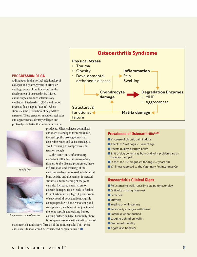
PROGRESSION OF OAA disruption in the normal relationship ofcollagen and proteoglycans in articularcartilage is one of the first events in thedevelopment of osteoarthritis. Injuredchondrocytes produce inflammatorymediators, interleukin-1 (IL-1) and tumornecrosis factor alpha (TNF-!), whichstimulates the production of degradativeenzymes. These enzymes, metalloproteinasesand aggrecanases, destroy collagen andproteoglycans faster than new ones can be
produced. When collagen destabilizes and loses its ability to form crosslinks, the hydrophilic proteoglycans startabsorbing water and cause cartilage toswell, reducing its compressive and tensile strength.
At the same time, inflammatorymediators influence the surroundingtissues. As the disease progresses, thereis fibrillation and fissuring of thecartilage surface, increased subchondralbone activity and thickening, increasedstiffness, and thickening of the jointcapsule. Increased shear stress onalready damaged tissue leads to furtherloss of articular cartilage. A progressionof subchondral bone and joint capsulechanges produces bone remodeling andosteophytes (new bone at the junction ofthe joint capsule and existing bone),causing further damage. Eventually, thereis complete loss of cartilage with areas of
osteonecrosis and severe fibrosis of the joint capsule. This severeend-stage situation could be considered “organ failure.” !
Fragmented coronoid process
Osteoarthritis Clinical Signs! Reluctance to walk, run, climb stairs, jump, or play
! Difficulty in rising from rest
! Lameness
! Stiffness
! Yelping or whimpering
! Personality changes, withdrawal
! Soreness when touched
! Lagging behind on walks
! Decreased mobility
! Aggressive behavior
Prevalence of Osteoarthritis2,3,4,5
! #1 cause of chronic pain in dogs
! Affects 20% of dogs >1 year of age
! Affects quality & length of life
! 31% of dog owners say bone and joint problems are anissue for their pet
! In the “Top 10” diagnoses for dogs >7 years old
! #7 illness reported to the Veterinary Pet Insurance Co.
Osteoarthritis Syndrome
Healthy joint

MULTIMODAL THERAPY
4 c l i n i c i a n ’ s u p d a t e ™
Chronic Pain Management
Steven C. Budsberg, DVM, MS, DACVS
Professor of Surgery College of Veterinary MedicineUniversity of GeorgiaAthens, Georgia
Options for Osteoarthritis Pain Therapy
NSAID Most common medication,effective, palliative
N-methyl-D-aspartate(NMDA) inhibitors
Opioid receptor agonist
Corticosteroids
Chondromodulating agents
Nutritional support
Physical therapy, exercisemodification
Weight reduction
• Carprofen• Etodolac• Meloxicam• Deracoxib• Tepoxalin
• Amantadine
• Tramadol
• Hydrocortisone
• Chondroitin sulfate• Glucosamine • Hyaluronic acid• Doxycycline• Polysulfated
glycosaminoglycan
• Omega-3 fatty acids(EPA)
–
–
• COX-1 and/or COX-2 inhibitors
• Combined COX & LOX inhibition
• Chronic NMDA-receptor stimulation can produce a “wind-up”effect, a state of chronic CNS sensitization
• In combination, may aid in prevention of chronic pain
• Synthetic derivative of codeine that acts on the µ-opioidreceptor, facilitating the descending serotinergic system
• Can slow early stages of osteoarthritis• CAVEAT – May enhance disease progression long-term
• Support or enhance macromolecular synthesis and synthesisof hyaluronate
• Inhibit degenerative enzymes or inflammatory mediators• Remove or prevent formation of fibrin, thrombi, and plaque
• Controls inflammation, interrupts signal (mRNA) that promptsproduction of degradative enzymes
• Joint manipulation, no-force activities, prosthetic devices
• Limits opportunity for further injury
Class Drug Action! Pain receptors and pathways
become sensitized withpersistent inflammation.
! Treatment plans include weightreduction, nutritional support,exercise modification/physical therapy, andpharmacologic management.
! NSAIDs remain the most commonmedication for chronic pain.
! Encourage client compliance
! Ensure optimal dosing
! Adapt therapy to patientrequirements
! Give lowest effective dose
! Observe for toxicity
! Screen patients for potential risk
KEY POINTS
MONITORING NSAID USE
Sources of pain in osteoarthritis are the synovium, periarticular tissues, periosteum, andsubchondral bone. With chronic conditions, pain receptors and pathways become sensitizedthrough persistent inflammation. Eventually, even minimal noxious or normal stimulations
can cause pain. Interruption of the pathways to the central nervous system (CNS) is the first stepin controlling chronic pain.
Although NSAIDs remain the most common medicationsprescribed for the treatment of osteoarthritis, they do notsignificantly alter the progression of the disease.Pharmacologic management is only one aspect of treatment.Several drug classes in addition to NSAIDs can be helpful andmay slow early stages of OA — preventing, retarding, and insome cases reversing cartilage damage. Drug effectiveness(and side effects) can depend on individual response, socare must be taken to monitor and adapt therapy to eachpatient’s needs. Exercise and physical therapy can also assistin ameliorating pain and restoring quality of life. Recentinformation stresses the role of nutritional management andthe role of omega-3 fatty acids. Nutritional supplements(nutraceuticals), while still controversial, are being usedwith good results in combination therapy by many and areaccepted by clients. !
CNS sensitization
Combination therapy achieves
the goal of relieving pain and
discomfort and improving
quality of life.

c l i n i c i a n ’ s b r i e f ® 5
Denis J. Marcellin-Little, DEDV, DACVS, DECVS, CCRP
North Carolina State UniversityRaleigh, North Carolina
Physical therapy treatments are aimed at addressing the secondary effects of osteoarthritis — mainly pain and loss
of muscle strength — and promoting repair ofdamaged tissues, improving quality of life, andslowing progression of the disease. !
PHYSICAL THERAPY FOR CANINE OSTEOARTHRITISTherapy Options
Physical Therapy Options*
Temperature
Electrical stimulation
Magnetic therapy
Acupuncture
Stretching
Exercises
ACUTECold therapy decreases blood flow, inflammation, muscle spasm, and pain. Cartilage-degrading enzymes are inhibited below 30° C. Superficial ice decreases skintemperature by 16° C and joint temperature by 6° C.8
CHRONICHeat therapy increases blood flow, enzymatic activity, collagen extensibility, andmuscle relaxation. Superficial heat increases skin temperature by 8° C and jointtemperature by 2° C.8
In both heat and cold therapy studies, temperatures returned to pretreatment levelsafter three hours.8
Transcutaneous neuromuscular electrical therapy stimulates the large cutaneousnerve fibers which transmit sensory impulses faster than pain fibers.
Neuromuscular electrical stimulation may strengthen atrophied muscle fibers.
In people, pulse electromagnetic field therapy has been used to treat OA.
Two studies showed benefit after 18 half-hour treatment periods. The benefits lastedfor more than one month.9
While it may be effective as an adjunctive therapy, there are no clear proven benefits.
1 to 3 stretching sessions daily (10 to 15 repetitions of stretching for 20 to 40 seconds)may be beneficial in arthritic joints with limited range of motion.
Cold can be applied with ice, gel packs, and CO2 deliverydevices.
Heat can be applied with heating pads or therapeuticultrasound.
Transcutaneous electrical nerve stimulation can bedelivered by handheld machines ($150 to $500 throughmedical supply vendors).
Optimal treatment duration = ~40 minutes
Isotonic exercises performed at low or high intensity
Resistance provided by water or elastic bands; walkingon an underwater treadmill
Modality Action Method
* Developed with Joanna Freeman, BSc PT, BSc Kine, CSCS, Animal Rehabilitationand Wellness Hospital, Raleigh, North Carolina
Walking or trottingWalking with resistance Sit-to-stand exercisesSwimming
Isometric Exercises - Muscle contractionswithout a change in muscle length or joint motion Isokinetic Exercises - Dynamic exercises withconstant joint velocity requiring the use of amachine to control themIsotonic Exercises - Dynamic exercises using aconstant load. This is the most practical form ofexercise for companion animals
! Physical means can be usedtherapeutically to ameliorate clinical signsof osteoarthritis and improve quality of life.
! Low-impact exercises can help increaserange of motion of arthritic joints.
! Low-intensity exercises are as beneficial ashigh-intensity exercises.6
! The environment can be modified to assistpatient independence.
! Multimodal drug and nondrug therapiesincrease likelihood of adequate treatment.7
! Undertreatment of pain has seriousnegative consequences.
KEY POINTS
Exercise and passive therapies
benefit canine patients and
increase the likelihood of
adequate treatment.
Bren
da B
unch

OPTIMAL OSTEOARTHRITIS DIET
Most dogs are at risk for several diseases as they ageand more than 50% of dogs 10 years of age orolder have osteoarthritis.2 Because it is too painful
to exercise normally, osteoarthritis may increase the risk ofobesity — a major concern for dogs with osteoarthritis as itincreases stress on joints.
HEALTH ISSUES FOR AGING DOGSRisk of kidney disease and osteoarthritis increasessteadily from youth and older dogs are also at risk forcognitive decline. Obesity can start as a problem in youngdogs and continue into later years or it can develop in anarthritic dog that can no longer exercise normally. Thecycle of inflammation, degradation, and chondrocytedamage in osteoarthritis can be promoted by joint stressbecause of excess body weight. Obesity also increases the likelihood of other diseases in additionto osteoarthritis. A diet with a senior nutrient profile is a good place to start to treat and preventthe infirmities of old age in the dog.
WEIGHT MANAGEMENT Because energy requirements depend on activity levels, reducing arthritis pain and inflammation willprovide exercise benefits for maintaining appropriate body weight in an arthritic dog. If the patient isonly slightly overweight, a food designed to disrupt the cycle of inflammation may increase activity, whichin turn allows weight to remain constant or normalize. Foods with increased levels of omega-3 fattyacids, in particular eicosapentaenoic acid (EPA), reduce the degradative enzymes that cause cartilagedamage. Most overweight dogs can benefit from eating a therapeutic food specially formulated for theamelioration of arthritis signs, at least until their joints are better. As their activity levels increase, patientscan be reevaluated to see if a weight loss program with more restrictive calorie levels is warranted. !
6 c l i n i c i a n ’ s u p d a t e ™
Nutritional Management
Phillip W. Toll, DVM, MSAssociate Director,
Nutrition TechnologyPet Nutrition CenterTopeka, Kansas
! Risk of osteoarthritis and otherdiseases increases with age.
- Dogs 1 year of age or older areat risk for osteoarthritis.
- Middle-aged dogs are at risk forobesity.
! Distribution of weight in very olddogs is bimodal — some are too fatand some are too thin.
! A successful nutritional profile forosteoarthritis should target older dogsand their common issues.
KEY POINTS
Optimal Osteoarthritis Diet! Senior nutrient profile
! High omega-3 fatty acids
! High ratio of omega-3 toomega-6 fatty acids
! High level of EPA
! High level of !-linolenic acid
! Chondroprotectives(glucosamine & chondroitin)
! Carnitine
! Antioxidants
Hill’s® Prescription Diet® Canine j/d™
Energy, kcal/kg
Protein, %
Fat, %
Total omega-3, %
EPA, %
Omega-6:Omega-3
Carnitine, mg/kg
Vitamin E, IU/kg
Glucosamine & Chondroitin
3704
20.1
14.3
3.51
0.44
0.7:1
351
851
Included
4190
19.6
19.3
4.24
0.85
0.7:1
319
698
Included
Dry Canned
Managing osteoarthritis
means stopping
the degradative
process and the
inflammatory cycle.
Managing theOsteoarthritis Cycle! Stress
- Correct abnormal forces(weight, conformation)
- Strengthen cartilage matrix
! Inflammation- Medications- Omega-3 fatty acids
! Degradation- EPA
! Cartilage matrix damage- Chondroprotectives
(glucosamine & chondroitin)

c l i n i c i a n ’ s b r i e f ® 7
The breakdown of cartilage isperformed by the cartilageproteinases —
aggreganase (ADAMT-4 & -5),collagenase, and metalloproteinase(MMP-13). In osteoarthritis, theseproteinases are important in cartilage proteoglycan catabolismand release. When proteoglycans and collagen are released,monoclonal antibody testing (e.g., Western Blot) can be usedto evaluate the metabolites. This has enabled the creation of amodel culture system for drug or nutraceutical evaluation. !
! In normal adult cartilage, there is a balance(homeostasis) between synthesis anddegradation of cartilage matrix.
! In pathologic cartilage (arthritis),degradation outstrips synthesis.
! Adult cartilage matrix turnover is relativelyslow compared with that of other tissuesand organs. Normal adult cartilage turnoveris measured in years (1 to 2). Normalcartilage proteoglycan turnover ismeasured in months (80 to 100 days).
! Cartilage turnover is performed byproteolytic enzymes called matrixproteinases (aggrecanases, collegenases,and gelatinases), which are active in theextracellular matrix.
! Cartilage degradation starts with loss ofcartilage aggrecan followed by loss ofcartilage collagens, resulting in loss ofability to resist compressive forces duringjoint movement.
! This lab has developed models thatmimic canine cartilage degradation inthe arthritic state. These models can beused to test the potential benefit ofdietary supplementation in canineosteoarthritis.
KEY POINTS
PHYSIOLOGY OF CARTILAGE TURNOVER — A MODEL SYSTEM10
Canine Cartilage
Bruce Caterson, PhDAssociate Director,School of Biosciences & CardiffInstitute of Tissue Engineering & RepairCardiff UniversityCardiff, Wales, UK
In Vitro Model
Canine articularcartilage is harvestedfrom stifle joints, thecells incubated for 72hours, then culturedfor 4 days withcatabolic agents orcontrol. UsingWestern Blot (shown),chondrocytes areexposed to catabolicagents to determinewhich cause cartilagedegeneration.
Measuring Progressive CartilageDegeneration in OsteoarthritisGlycosaminoglycan (GAG) that has been released byproteoglycan catabolism can be measured in caninesynovial fluids (SF). They increase early in osteoarthritis (OA)and then decrease with progressive cartilage degeneration.
GAG Normal SF Early Elbow Early Stifle Late Stifle
Mean (SD) 22.8 (30.7) 31.6 (16.0) 48.4 (21.5) 8.9 (3.0)
Median 10.5 27.1 51.6 8.2
Range 2.2 - 83.6 11.0 - 74.1 21.1 - 69.2 5.5 - 15.3
Aggrecan proteoglycan model showinglarge macromolecular aggregates ofaggrecan monomers. Aggrecan bindswater strongly and becomes entrapped bythe larger collagen fibrils in cartilagetissue to form a meshwork that maintainstension and high osmotic pressure,enabling the joint to function normally.
Cartilage is in a constant state of turnover.
When the system becomes pathologic
(osteoarthritis), the degradation process
outstrips the synthesis of new matrix.
This analysis (using mAb BC-3that detects aggrecanase-generated degradationproducts) shows evidence forsome weak aggrecanaseactivity in control cultures thathave been either untreated orexposed to EPA or ALA. Incontrast, in cultures exposedto inflammatory cytokines suchas oncostatin-M (Osm-treated,lane 6) that induce increasedcartilage degradation, there isevidence for a large increasein aggrecanase activity.However, this increasedactivity is reduced whencultures are exposed to EPA(OSM-treated lanes 7 & 8; butnot effected by ALA exposure,lanes 9 & 10).
Control
100 300 100 300 100 300 100 300
Osm-treated
1 2 3 4 5 6 7 8 9 10
150k Oa-
100k Oa-75k Oa-
50k Oa-37k Oa-
EPAC Osm
ALA EPA ALA
BC-3 Western Blot
EPA = Eicosapentaenoic acid; ALA = !-Linolenic acid

FATTY ACID METABOLISM & INFLAMMATION
8 c l i n i c i a n ’ s u p d a t e ™
Canine Cartilage
William D. Schoenherr, PhDPrincipal Nutrition ScientistPet Nutrition CenterTopeka, Kansas
! Fatty acids are an important part ofcell membranes.
! Omega-3 fatty acids were able toreduce inflammation in dogchondrocytes.
! Omega-3s are incorporated intocanine cartilage in ~3 days.
! When the omega-3 fatty acid EPAreplaces arachidonic acid (AA) in cellmembranes, the inflammatorycascade is decreased.
! Dog chondrocyte membranesselectively store EPA and not otheromega-3 fatty acids.
! EPA is the only omega-3 fatty acid that decreases aggrecandegradation, preventingfragmentation of GAGs.
! EPA turns off signal mRNA thatprompts production of degradativeenzymes.
KEY POINTS
• !-Linolenic Acid (ALA) – Leafy green vegetables, flaxseed, canola oil
• Eicosapentaenoic Acid (EPA)– Fish oil
• Docosahexaenoic Acid (DHA)– Fish oil
• Linoleic Acid (LA) – Soy, corn, safflower oils
• Arachidonic Acid (AA) 20:4 n-6– Animal Fat
• Oleic Acid (OA) – Olives, olive & canola oil– Walnuts
• Eicosatrienoic Acid (ETA)– Animal Fat, Fish oil
Omega-3 (n-3) Fatty Acids Omega-6 (n-6) Fatty Acids Omega-9 (n-9) Fatty Acids
Polyunsaturated Fatty Acids (PUFAs)
STUDY 1 OMEGA-3 FATTY ACIDS IN CANINE IN VITRO CHONDROCYTE MEMBRANE
! Chondrocytes were enzymatically released (explants) from canine articular cartilage and thencultured for 24 hours in serum-free Dulbecco’sModification of Eagle’s Medium (DMEM).
! Monolayers were then cultured for 3 to 6 days inserum-free DMEM with either 100 µg/ml PUFA, 300µg/ml PUFA, or no PUFA.
! Fatty acids were extracted and the amount of PUFA thatwas incorporated into chondrocyte membranes wasevaluated with gas liquid chromatography.
RESULTS! Incorporation of omega-3 PUFAs into canine
chondrocyte membranes required much longer (>3days) than in bovine or human cells (12 to 24 H).
! No difference was found between 3- and 6-dayexposure to PUFAs for incorporation in monolayer.
! At doses and times tested, EPA, ALA, and AA were incorporated into canine chondrocyte membranes.! None of the PUFAs had a detrimental effect on chondrocyte metabolism after 3 to 6 days’ incubation
as determined by media lactate.
STUDY 2 PUFA MODULATION OF IN VITRO CANINE CARTILAGE DEGENERATION
! Chondrocyte explants were cultured for 5days in serum-free DMEM with or without100 or 300 µg/ml PUFAs.
! 50 ng/ml oncostatin M (OSM) or 10-6 Mretinoic acid (RA) was added to cultures.
! Release of proteoglycans was measured.
RESULTS†
! EPA (300 µg/ml) significantly decreased OSM& RA-induced glycosaminoglycans (GAGs).
*See page 10 for study grade explanation.†In another, larger study, when cells derived from donor dogswere maintained on a standard diet for 6 months prior tocollection, results were even better.11
µg/m
l of F
A
14
12
10
8
6
4
2
0Control ALA EPA DHA
ALA EPA DHA
PUFA Incorporation IntoChondrocyte Membranes
Cell
Mea
n fo
r GAG
µg/
mg
25
22.5
20
17.5
15
12.5
10
7.5
5
2.5
0
cc
+ AL
A 10
0c
+ AL
A 30
0c
+ DH
A 10
0c
+ DH
A 30
0c
+ EP
A 10
0c
+ EP
A 30
0os
mos
m +
ALA
100
osm
+ A
LA 3
00os
m +
DHA
100
osm
+ D
HA 3
00os
m +
EPA
100
osm
+ E
PA 3
00 rara
+ A
LA 1
00ra
+ A
LA 3
00ra
+ D
HA 1
00ra
+ D
HA 3
00ra
+ E
PA 1
00ra
+ E
PA 3
00
*
*
*
*
Proteoglycan Release From Cartilage
EPA controls the
production of the
degradative enzyme,
aggrecanase, at the
gene level.
IVGRADE*
Dog chondrocyte membranes selectively store EPA and not other omega-3 fatty acids,positively moderating pathologic canine cartilage catabolism. Food with high levels ofomega-3 fatty acids also decreases inflammation and helps improve clinical signs of OA
— especially difficulty in rising from a resting position, walking, running, and playing. !
IVGRADE*

c l i n i c i a n ’ s b r i e f ® 9
Nutritional management using a food with a high level of omega-3 fatty acids helped improveclinical signs of osteoarthritis in dogs as measured by clinical examination and analysis ofground reaction forces. !
! A high level of omega-3 fatty acids arebeneficial for osteoarthritic joints.
! Metabolism of omega-3 fatty acidsproduces eicosanoids that are less potentinflammatory mediators than thoseproduced by omega-6 fatty acids.
! Force plate analysis is an objectivemethod of measuring limb groundreaction forces.
! Dogs with OA that were fed Hill’s®Prescription Diet® Canine j/d™ were 7.48times more likely to have improved peakvertical force (PVF) than dogs fed thecontrol food.
! In a similar study with NSAIDs, dogs wereonly 3.3 times more likely to haveimproved PVF.13
KEY POINTS
OMEGA-3 FATTY ACID EFFECTS ONFORCE PLATE ANALYSIS & CLINICAL SIGNS12
University Clinical Trials
James K. Roush, DVM, MS, DACVSProfessor & Section HeadSmall Animal SurgeryKansas State UniversityManhattan, Kansas
[Study sidebar box]
A 90-day prospective, randomized, double-masked, controlled study at Kansas State University andUniversity of Florida
ELIGIBLE DOGS! 38 client-owned dogs
completed the trial! >1 year of age! !25 pounds! Free of systemic disease! Radiographic evidence of
osteoarthritis! Clinical signs of lameness! Consuming AAFCO standard
dry food
FOODS TESTED! 22 dogs received test food (Hill’s® Prescription
Diet® Canine j/d™)! 16 dogs received control food
CLINICAL EVALUATION PARAMETERS! Lameness! Weight bearing! Range of motion! Reluctance to stand on limb with
contralateral elevation ! Pain on palpation of affected joint
GROUND FORCE ANALYSIS Lameness analyses using a computerized biomechanical force plate were conducted at the beginningof the study, 45, and 90 days after feeding the control food or the test food. Five valid force plate trialswere obtained for each dog for the most severely affected and contralateral limbs. All forces werenormalized with respect to body weight. Data from valid trials for each limb were averaged to obtain amean value for each time period.The key parameter measured was peak vertical force (PVF).
CLINICAL RESULTSA significantly greater percentage of dogs consuming the test food were evaluated as improvedversus those consuming the control food. Also, at the end of the 90-day trial, more dogs in the testfood group had a reduction in pain when the joint was palpated.
GROUND FORCE ANALYSIS RESULTSThere was no significant change in mean peak vertical force (PVF) in the control group. Mean PVFincreased significantly in the lame leg (5.35%) in the dogs consuming the test food. Dogs with OA fedtest food were 7.48 times more likely to have improved PVF on the lame leg (82% of dogs improvedover the course of the study) than dogs fed the control food (31% improved over the study). In a studyof dogs with osteoarthritis given carprofen, dogs were only 3.3 times more likely to have improvedtheir PVF as the dogs receiving placebo.13
UNIVERSITY FORCE PLATE CLINICAL TRIALS
Perc
ent o
f Dog
s
Control Test
50%
40%
30%
20%
10%
0%2 1 0 -1 -2 -3
Metabolism of omega-3
fatty acids produces
eicosanoids that are not
as inflammatory as
eicosanoids produced
from omega-6 fatty acids.
IGRADE
Change in Combined Assessments(0-90 days)
Mantel-Haenszel Chi-Square: P = 0.0263

THE ROLE OF NUTRITION INOSTEOARTHRITIS — 3 CLINICAL STUDIES
Evidence-based medicine (EBM) isthe integration of the best researchevidence and clinical expertise.14
Using the scientific method for clinicaldecisions requires a systematic,rigorous, disciplined approach toevaluating research and recognizes thatnot all studies are equal. By consideringthe total body of knowledge, bettertherapeutic decisions can be made. !
Evidence-Based Medicine
10 c l i n i c i a n ’ s u p d a t e ™
! Evidence-based medicine appliesthe best available evidence usinga systematic approach.
! Clinical trials for PrescriptionDiet® Canine j/d™ included 3Grade I studies.
! Study 1 – Dogs with OA in 6-month feeding trial showedimproved ability to rise from rest,walk, run, and play.
! Study 2 – Serum EPA levelsincreased in 3-month dose-titration feeding trials and dogswith OA improved.
! Study 3 – In a 3-month trial, 43%of dogs with OA consumingCanine j/d were able to sustain decreased NSAID doses.
! Study 4* – In 90-day universityforce plate clinical trials, dogs fedhigh omega-3 Canine j/d were7.48 times as likely to improve.
Timothy A. Allen, DVM, DACVIMDirector & Chief Medical OfficerPet Nutrition CenterTopeka, Kansas
KEY POINTS
Study Grades† in Evidence-Based Medicine
Grade I Well-designed, properly randomized and controlled clinical trial that utilizespatients with naturally occurring disease.! Prospective studies
GRADE II Well-designed and controlled laboratory studies in the target species withnaturally occurring disease.
GRADE III Evidence obtained from one of the following ! Well-designed nonrandomized clinical trial ! Cohort- or case-controlled epidemiologic studies! Studies using an acceptable disease model ! Case series ! Dramatic results from uncontrolled studies
GRADE IVEvidence obtained from one of thefollowing! Bench-top in vivo laboratory studies! Opinions based on clinical
experience! Descriptive studies! Studies conducted in
another species! Pathophysiologic justification! Reports of expert committees
†Quality of evidence guidelines are adapted from the U.S. Preventive Services Task Force.
RandomizedControlled Trials
Epidemiologic Studies
Models of Disease
Case Series
Case Reports
Research in Other Species
Pathophysiologic Rationale
Editorials, Opinions
Evidence-Based Clinical Nutrition Pyramid
Clinical trials for Hill’s® Prescription
Diet® Canine j/d™ included 4 Grade I
studies.* This is the only food
developed for the management of
osteoarthritis utilizing this level
of research.
*See also: Roush university study, page 9 of this supplement.
IGRADE
IIGRADE
IIIGRADE
IVGRADE

c l i n i c i a n ’ s b r i e f ® 11
STUDY 1 OMEGA-3 FATTY ACIDS IN CANINE OSTEOARTHRITIS15
! Prospective; 6-month feeding period! Randomized, controlled! Double masked (pet owner and veterinarian)! Multicenter (18 general practices, 131 dogs)! Data collected at 0, 6, 12, and 24 wk
RESULTS! Dogs fed the test food had significantly improved ability to rise from a resting
position, run, and play at 6 wk.! Improvements were seen in walking at 12 and 24 wk compared to control dogs.! Serum EPA levels were significantly higher in dogs fed the test food at 45 days.
STUDY 2 DOSE-TITRATION EFFECTS OF OMEGA-3 FATTY ACIDS FED TO OSTEOARTHRITIC DOGS16
! Prospective; 3-month feeding period! Randomized, controlled (3 dietary treatments – dose titration)
• Dry food EPA values of 0.5%, 1.2%, or 1.7% dry matter basis (DMB)• Canned food EPA values of 0.4%, 0.9%, or 1.4% DMB
! Double masked (veterinarian and pet owner)! Multicenter (28 general practices, 177 dogs)! Data collected at 0, 3, 6, and 12 wk
RESULTS! Pet owner evaluations – All foods resulted in significant improvement in pet
owner evaluations.There were no statistically significant differences.! Clinician evaluations – Dogs consuming the highest concentrations displayed
the greatest improvements. Progression of disease was reduced in the highestEPA level group.
STUDY 3 PROSPECTIVE 3-MONTH FEEDING PERIOD17
Objective: Determine whether a therapeutic food alters NSAID dose requiredto control clinical signs in dogs with OA! Randomized, controlled (drug/dietary treatment)! Double masked (veterinarian and pet owner)! Multicenter (35 general practices, 193 dogs)! Standardized drug-dose period! Examination and dose adjustments at 3, 6, and 12 wk
RESULTS! Pet owners reported a decrease in severity on 10 of 15 parameters
during the first 21 days of feeding Canine j/d.! Pet owners observed significantly greater pain reduction in dogs
consuming the therapeutic food compared to the control food.! NSAID dose reduction was possible for 43% of dogs consuming the
therapeutic food compared to 32% of dogs eating the control food(significantly greater reduction in the dogs eating the therapeutic food).
! NSAID dose increases were needed in 11% of dogs consuming thecontrol food compared to 2% of dogs consuming the therapeutic food.
Hill’s® Prescription Diet® Canine j/d™ Clinical Trials
General inclusion criteria! Dogs >1 year old! Consuming dry food! Clinical diagnosis of OA! Radiographic evidence of OA! Otherwise healthy! Consistent dosing of
medications and/orsupplements
General exclusioncriteria! Acute traumatic
injury! Systemic illness! Planned surgery
during the study! Recent intraarticular
injection orarthrocentesis
Cumulative Rimadyl Dose Change
Therapeutic Food and Drug in Dogs with Osteoarthritis
mg
/ lb
body
wei
ght
0.100
0.000
-0.100
-0.200
-0.300
-0.400
-0.500
-0.600
0.064
Day 21
Control
Day 42 Day 63 Day 84
-0.038
-0.116
-0.229 -0.205 -0.215
-0.495
-0.423
Test
Radiograph ofosteoarthritis
IGRADE
IGRADE
IGRADE

© 2005 Educational Concepts LLC P-7014
This summary is based on presentations by Drs. Steven C. Budsberg, Bruce Caterson, Denis J. Marcellin-Little, and William D. Schoenherr at the NAVC 2005 Symposium, Canine Osteoarthritis as well as additional presentations by Drs. Spencer A. Johnston, Philip W. Toll, James K. Roush, and
Tim A. Allen at the Hill’s Canine j/d™ Symposium in Orlando, Florida, also included here. Additional data on file at Hill’s Pet Nutrition, Inc.
Hill’s® Prescription Diet® Canine j/d™ Highlights
Osteoarthritis is the most common form of canine joint and musculoskeletal disease, affectingup to 20% of dogs over age one.2 The concept of managing arthritic dogs with nutritionalsupplementation of omega-3 fatty acids is relatively new in veterinary medicine. Canine j/d isthe only clinically proven nutritional product for management of dogs with arthritis. EBM(evidence-based medicine) Level 1 research showed that dogs with osteoarthritis run better,play better, and rise more easily on Canine j/d. They also experience less stiffness and more easeof movement, including walking better and climbing stairs more easily.
! Clinically proven to reduce pain in dogs with osteoarthritis.12,16
! Helps dogs with OA walk, run, play, and climb stairs more easily.16,17
! 82% of dogs fed Prescription Diet® Canine j/d™ experienced improvementin weight-bearing ability as measured by limb peak vertical force.12
! EPA, an omega-3 fatty acid component, works to turn off the genes thatcause cartilage damage.10
! Prescription Diet® j/d™ has highest levels of EPA.18
! Contains highest18 levels of omega-3 fatty acids and lowest18 levels ofomega-6, helping to reduce inflammatory mediators that cause inflammation.
! Contains appropriate levels of nutrients for long-term feeding for both adult and senior dogs
REFERENCESPage 2 & 3 - Johnston1. Workshop on etiopathogenesis of osteoarthritis. Brandt KD, Mankin HJ,
Shulman LE. J Rheumatol 13:126-160, 1986. 2. Pfizer Inc. research, www.rimadyl.com. Data on file at Hill’s Pet Nutrition, Inc.3. Hill’s market research, 2003. Data on file at Hill’s Pet Nutrition, Inc.4. Veterinary Pet Insurance Corp., 1999. Data on file at Hill’s Pet Nutrition, Inc.5. Health status and population characteristics of dogs and cats examined at
private veterinary practices in the United States. Lund EM, Armstrong PJ, KirkCA, et al. JAVMA 214:1336-1341, 1999.
Page 5 – Marcellin-Little6. Intensity of exercise for the treatment of osteoarthritis. Brosseau L, MacLeay
L, Robinson VA, et al. Cochrane Database Syst Rev, 2003.7. Pain in osteoarthritis, rheumatoid arthritis and juvenile chronic
arthritis, 2nd ed. Simon LS, Lipman AG, Jacox AK, et al. Glenview IL — AmericanPain Society (APS), 2002, p 179.
8. Effects of local heat and cold treatment on surface and articulartemperature of arthritic knees. Oosterveld FGJ, Rasker JJ. Arthritis Rheum37:1578-1582,1994.
9. A double blind trial of the clinical effects of pulsed electromagnetic fieldsin osteoarthritis. Trock DH, Bollet AJ, Dyer RH Jr, et al. J Rheumatol 20:456-460, 1993.
Page 7 - Caterson10. Cartilage physiology — unique aspects of canine articular cartilage.
Caterson B. NAVC Symposium, Canine Osteoarthritis, 2005.
Page 8 - Schoenherr11. Hill’s Pet Nutrition, Inc., Data on file.Page 9 - Roush12. University study: Effects of feeding omega-3 fatty acids on force plate gait
analysis in dogs with osteoarthritis, 3-month feeding study, 2003. Roush JK,Cross A. Data on file, Hill’s Pet Nutrition, Inc.
13. Randomized, controlled trial of the efficacy of carprofen, a nonsteroidalanti-inflammatory drug, in the treatment of osteoarthritis in dogs. VasseurPB, Johnson AL, Budsberg SC, et al. JAVMA 206:807-811, 1995.
Page 10 - Allen14. Application of evidence-based medicine to veterinary clinical nutrition.
Roudebush P, Allen TA, Dodd CE, and Novotny BJ. JAVMA, 224:1713-1888, 2004.15. Omega-3 fatty acids in canine osteoarthritis: A randomized, double-
masked, practice-based, 6-month feeding study, 2003. Data on file, Hill’s PetNutrition, Inc.
16. Dose titration effects of omega-3 fatty acids fed to osteoarthritic dogs, 3-month feeding study, 2003. Data on file, Hill’s Pet Nutrition, Inc.
17. A multicenter practice-based study of a therapeutic food and drug in dogswith osteoarthritis, 3-month feeding study. Data on file, Hill’s Pet Nutrition, Inc.
Page 1218. Based on published data as labeled on therapeutic pet foods for canine osteoarthritis
and data on file at Hill’s Pet Nutrition, Inc., 2004.