s alivary gland diseases. reactive lesions mucocele mucus retention cysts sialolithiasis chronic...
TRANSCRIPT

Salivary gland diseases
Reactive lesions• Mucocele
• Mucus retention cysts
• Sialolithiasis
• Chronic sclerosing sialadenitis

Salivary infections• Acute parotitis
• Viral endemic parotitis (Mumps)
• Bacterial sialadenitis

Salivary gland tumorsBENIGN• Pleomorphic adenoma
• Monomorphic adenoma
• Papillary cystadenoma
• Oncocytoma

Salivary gland tumorsMALIGNANT• Mucoepidermoid carcinoma.
• Adenoid cystic carcinoma
• Acinic cell carcinoma
• Adenocarcinomas
Salivary Gland Neoplasms
• Diverse histopathology
• Relatively uncommon– 2% of head and neck neoplasms
• Distribution– Parotid: 80% overall; 80% benign– Submandibular: 15% overall; 50% benign– Sublingual/Minor: 5% overall; 40% benign

Pleomorphic Adenoma
• Most common of all salivary gland neoplasms• 70% of parotid tumors
• 50% of submandibular tumors
• 45% of minor salivary gland tumors
• 6% of sublingual tumors
• 4th-6th decades
• F:M = 3-4:1

Pleomorphic Adenoma
• Slow-growing, painless mass
• Parotid: 90% in superficial lobe
• Minor salivary gland: submucosal mass

Pleomorphic Adenoma
• Gross pathology– Smooth
– Well-demarcated
– Solid
– Cystic changes
– Myxoid stroma
Pleomorphic Adenoma
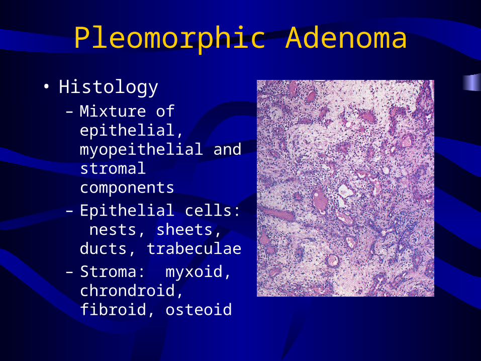
• Histology– Mixture of epithelial,
myopeithelial and stromal components
– Epithelial cells: nests, sheets, ducts, trabeculae
– Stroma: myxoid, chrondroid, fibroid, osteoid

Pleomorphic Adenoma
• Treatment: complete surgical excision– Parotidectomy with facial nerve preservation– Submandibular gland excision– Wide local excision of minor salivary gland
Warthin’s Tumor
• Papillary cystadenoma lymphomatosum
• 6-10% of parotid neoplasms
• Older, Caucasian, males
• 10% bilateral or multicentric
• Presentation: slow-growing, painless mass

Warthin’s Tumor• Gross pathology
– Encapsulated
– Smooth/lobulated surface
– Cystic spaces of variable size, with viscous fluid, shaggy epithelium
– Solid areas with white nodules representing lymphoid follicles
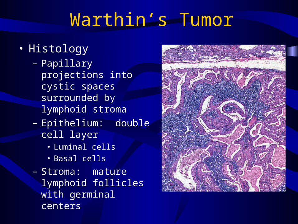
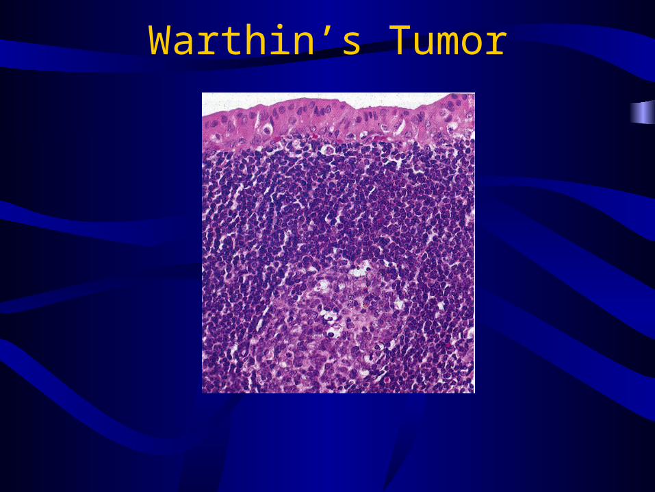
Warthin’s Tumor
• Histology– Papillary projections into
cystic spaces surrounded by lymphoid stroma
– Epithelium: double cell layer
• Luminal cells
• Basal cells
– Stroma: mature lymphoid follicles with germinal centers
Warthin’s Tumor

Oncocytoma
• 6th decade
• M:F = 1:1
• Parotid: 78%
• Submandibular gland: 9%
• Minor salivary glands
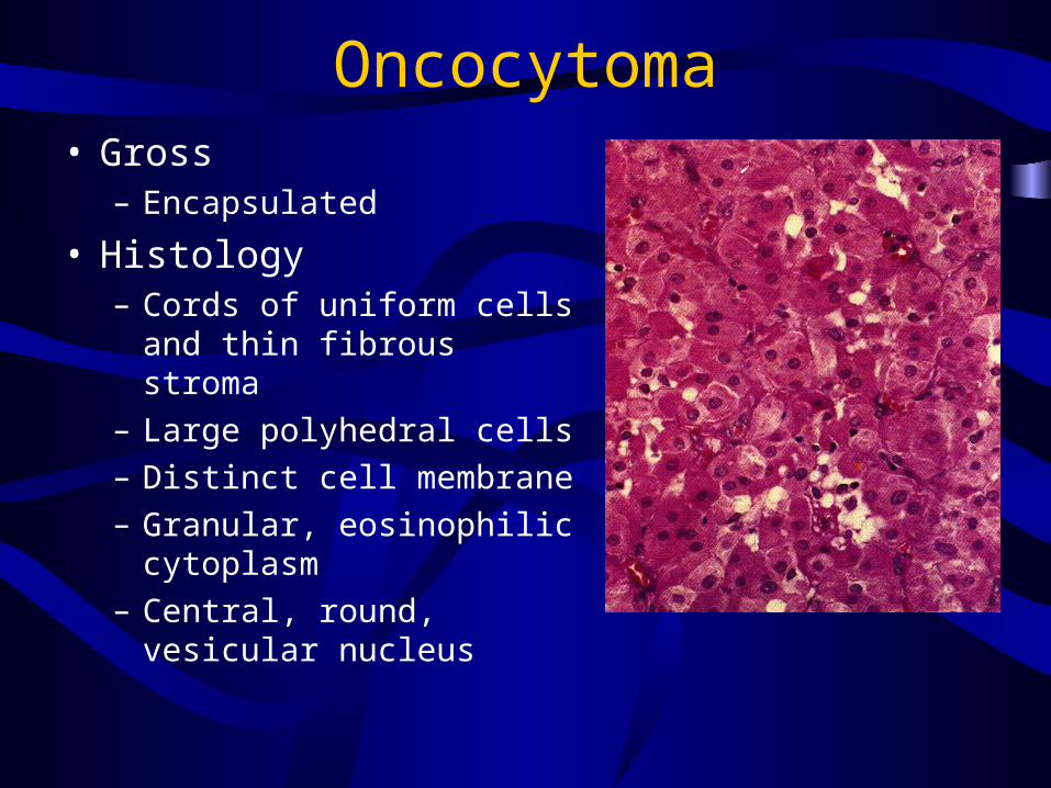
Oncocytoma• Gross
– Encapsulated
• Histology– Cords of uniform cells and
thin fibrous stroma
– Large polyhedral cells
– Distinct cell membrane
– Granular, eosinophilic cytoplasm
– Central, round, vesicular nucleus

Malignant tumours
Mucoepidermoid Carcinoma
• Most common salivary gland malignancy
• 5-9% of salivary neoplasms
• Parotid 45-70% of cases
• Palate 18%
• 3rd-8th decades, peak in 5th decade
• F>M
• Caucasian > African American

Mucoepidermoid Carcinoma
• Presentation– Low-grade: slow growing, painless mass– High-grade: rapidly enlarging, +/- pain

Mucoepidermoid Carcinoma
• Gross pathology– Well-circumscribed to
partially encapsulated to unencapsulated
– Solid tumor with cystic spaces

Mucoepidermoid Carcinoma
• Histology—Low-grade– Mucus cell > epidermoid
cells
– Prominent cysts
– Mature cellular elements

Mucoepidermoid Carcinoma
• Histology—Intermediate- grade– Mucus = epidermoid
– Fewer and smaller cysts
– Increasing pleomorphism and mitotic figures

Mucoepidermoid Carcinoma
• Histology—High-grade– Epidermoid > mucus
– Solid tumor cell proliferation
Adenoid Cystic Carcinoma
• Overall 2nd most common malignancy
• Most common in submandibular, sublingual and minor salivary glands
• M = F
• 5th decade
• Presentation– Asymptomatic enlarging mass– Pain, paresthesia, facial weakness/paralysis

Adenoid Cystic Carcinoma
• Gross pathology– Well-circumscribed
– Solid, rarely with cystic spaces
– infiltrative

Adenoid Cystic Carcinoma
• Histology—cribriform pattern– Most common
– “swiss cheese” appearance
Adenoid Cystic Carcinoma
• Treatment– Complete local excision
• Prognosis– Local recurrence: 40%– Distant metastasis: lung– Indolent course: 5-year survival 75%
Acinic Cell Carcinoma
• 2nd most common parotid malignancy
• 5th decade
• F>M
• Bilateral parotid disease in 3%
• Presentation– Solitary, slow-growing, often painless mass
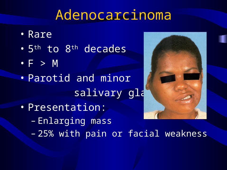
Adenocarcinoma• Rare
• 5th to 8th decades
• F > M
• Parotid and minor
salivary glands
• Presentation:– Enlarging mass– 25% with pain or facial weakness
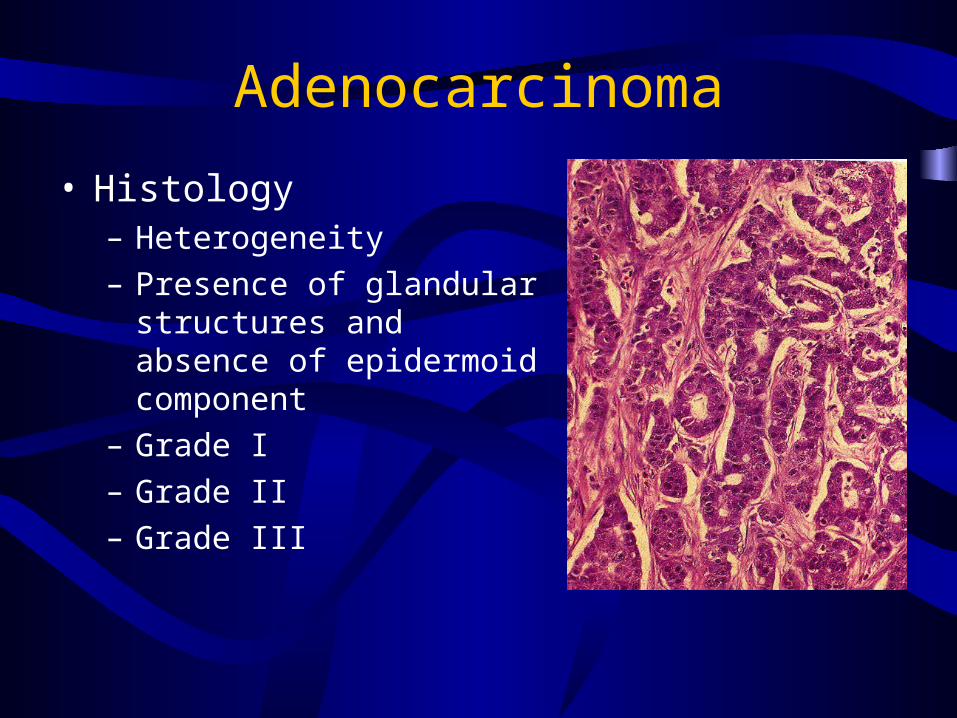
Adenocarcinoma
• Histology– Heterogeneity
– Presence of glandular structures and absence of epidermoid component
– Grade I
– Grade II
– Grade III
Malignant Mixed Tumors
• Carcinoma ex-pleomorphic adenoma• Carcinoma developing in the epithelial component
of preexisting pleomorphic adenoma
• Carcinosarcoma• True malignant mixed tumor—carcinomatous and
sarcomatous components
Fine-Needle Aspiration Biopsy
• Efficacy, Accuracy, Sensitivity, Specificity
• Safe, well tolerated