running head : poplar glutathione peroxidase-like … · nicolas navrot1, valérie collin2, josé...
TRANSCRIPT

Running head : Poplar glutathione peroxidase-like proteins
Corresponding author : Nicolas Rouhier, Unité Mixte de Recherches 1136 IaM
INRA-UHP Nancy I. Université Henri Poincaré, 54506 Vandoeuvre-lès-Nancy Cedex
France.
email. [email protected], telephone.++33383684225
Research area/Associate editor : Biochemical processes and Macromolecular
Structures/John Ohlrogge
Plant Physiology Preview. Published on October 27, 2006, as DOI:10.1104/pp.106.089458
Copyright 2006 by the American Society of Plant Biologists
www.plantphysiol.orgon August 21, 2018 - Published by Downloaded from Copyright © 2006 American Society of Plant Biologists. All rights reserved.

2
Plant glutathione peroxidases are functional peroxiredoxins distributed in several
subcellular compartments and regulated during biotic and abiotic stresses
Nicolas Navrot1, Valérie Collin2, José Gualberto3, Eric Gelhaye1, Masakazu Hirasawa4,
Pascal Rey2, David B. Knaff4, Emmanuelle Issakidis5, Jean-Pierre Jacquot1, Nicolas
Rouhier1,6
1 Unité Mixte de Recherche INRA-UHP 1136, Interactions Arbres/Micro-organismes, IFR
110 GEEF, Université Henri Poincaré, Faculté des Sciences, BP 239 54506 Vandoeuvre
Cedex, France. 2 CEA/Cadarache, DSV, DEVM, Laboratoire d’Ecophysiologie Moléculaire des Plantes,
13108 Saint-Paul-lez-Durance Cedex, France 3 Institut de Biologie Moléculaire des Plantes, CNRS 67084 Strasbourg Cedex. 4 Department of Chemistry and Biochemistry, and Center for Biotechnology and
Genomics, Texas Tech University, Lubbock, Texas 79409-1061, USA. 5 Institut de Biotechnologie des Plantes, UMR 8618, Université de Paris Sud 91405 Orsay
Cedex, France.
6 Corresponding author : Nicolas Rouhier, Unité Mixte de Recherches 1136 IaM INRA-
UHP Nancy I. Université Henri Poincaré, 54506 Vandoeuvre-lès-Nancy Cedex France.
email. [email protected], telephone.++33383684225
Footnotes
The portion of this work carried out at Texas Tech University was supported by a grant (D-
0710 to DBK) from the Robert A. Welch Foundation. Financial support from CEA
(program "Toxicologie Nucléaire Environnementale") is acknowledged. N.Navrot is
supported by MENRT grant and N. Rouhier, J.P. Jacquot and E. Gelhaye by BQR Région
Lorraine fundings.
www.plantphysiol.orgon August 21, 2018 - Published by Downloaded from Copyright © 2006 American Society of Plant Biologists. All rights reserved.

3
Abstract
We provide here an exhaustive overview of the glutathione peroxidase (Gpx)
family of Populus trichocarpa. Although these proteins were initially defined as
glutathione dependent, in fact they use only reduced thioredoxin (Trx) for their
regeneration and do not react with glutathione or glutaredoxin, constituting a fifth
class of peroxiredoxins. The two chloroplastic Gpxs display a marked selectivity
toward their electron donors, being exclusively specific for thioredoxins of the y type
for their reduction. In contrast, poplar Gpxs are much less specific with regard to
their electron-accepting substrates, reducing hydrogen peroxide and more complex
hydroperoxides equally well. Site-directed mutagenesis indicates that the catalytic
mechanism and the Trx-mediated recycling process involve only two (Cys 107 and
Cys 155) of the three conserved cysteines, which form a disulfide bridge with an
oxidation-redox midpoint potential of -295 mV. The reduction/formation of this
disulfide is detected both by a shift on SDS PAGE or by measuring the intrinsic
tryptophan fluorescence of the protein. The six genes identified coding for Gpxs are
expressed in various poplar organs and two of them are localized in the chloroplast,
with one co-localizing in mitochondria, suggesting a broad distribution of Gpxs in
plant cells. The abundance of some of Gpxs is modified in plants subjected to
environmental constraints, generally increasing during fungal infection, water deficit
and metal stress, and decreasing during photooxidative stress, showing that Gpx
proteins are involved in the response of both biotic and abiotic stress conditions.
www.plantphysiol.orgon August 21, 2018 - Published by Downloaded from Copyright © 2006 American Society of Plant Biologists. All rights reserved.

4
Introduction
In plant cells, aerobic reactions such as photosynthesis or respiration lead to
reactive oxygen species (ROS) production. These ROS, such as superoxide radicals,
hydroxyl radicals, or hydrogen peroxide, can damage biological molecules, including
nucleic acids, lipids and proteins. Rates of ROS generation and cellular ROS levels both
increase greatly when plants are subjected to environmental or biotic stresses. Plants have
developed several non-enzymatic and enzymatic systems to protect against oxidative
damage caused by these ROS. Carotenoids, tocopherols, glutathione and ascorbate are the
major non-enzymatic antioxidant compounds (Noctor and Foyer, 1998). Enzymatic
systems include superoxide dismutases, catalases, ascorbate peroxidases and
peroxiredoxins (Prxs) also named thioredoxin peroxidases.
Usually, plant Prxs are classified to belonging to one of four subgroups, called 2-
Cys Prx, 1-Cys Prx, type II Prx and Prx Q. They were initially identified by sequence
analysis (i.e., on the number and position of conserved cysteines) and on the catalytic
mechanism used for peroxide reduction (Rouhier and Jacquot, 2002). All Prxs display a
conserved catalytic cysteine, located in the N-terminal portion of the protein, which is first
transformed into a sulfenic acid after peroxide reduction. The main difference between the
different groups of plant Prxs is the mechanism of regeneration of the reduced enzyme. For
the 2-Cys Prx class, the sulfenic acid is reduced via the formation of an intermolecular
disulfide bridge using a “resolving” cysteine found on another subunit, whereas for the Prx
Q class, the “resolving” cysteine is located on the same subunit and forms an
intramolecular disulfide bridge (Rouhier and Jacquot, 2002). In most cases, these disulfides
are then reduced via the thioredoxin (Trx) system. For the two other classes, the sulfenic
acid is likely to be reduced without formation of a disulfide bond, as there is no other
strictly conserved cysteine involved in catalysis. One major difference between these two
classes of plant Prxs is that type II Prxs are reduced by the glutaredoxin (Grx) or the Trx
system or both (Rouhier et al., 2001; Brehelin et al., 2003; Finkemeier et al., 2005),
whereas the 1-Cys Prxs seem to be reduced only by the Trx system (Pedrajas et al., 2000).
Recent studies have shown that some plant and yeast glutathione peroxidases (Gpx)
can reduce peroxides, much more efficiently or sometimes exclusively, by using the
thioredoxin (Trx) system rather than glutathione (GSH) as a reductant (Jung et al., 2002;
Herbette et al., 2002; Tanaka et al., 2005). In the unicellular parasite Plasmodium
falciparum, a Gpx-like protein, closely related to plant Gpxs, has also been shown to be
specific for Trx as a reductant (Sztajer et al., 2001). Thus, in a classification based on
www.plantphysiol.orgon August 21, 2018 - Published by Downloaded from Copyright © 2006 American Society of Plant Biologists. All rights reserved.

5
biochemical feature, rather than on phylogenetic linkage, the plant Gpxs constitute a fifth
group of peroxiredoxins (Rouhier and Jacquot, 2005).
Most of the Gpxs found in animal cells are well-characterized selenium-containing
enzymes. Because of the high reactivity of the active site selenocysteine, enzymes of this
family are among the most efficient antioxidant systems in animal cells (Maiorino et al.,
1990). These selenium-containing enzymes have also been found in an unicellular
photosynthetic organism, the green alga Chlamydomonas reinhardtii (Fu et al., 2002,
Novoselov et al., 2002). In higher plants, the Gpx-like proteins identified so far possess a
cysteine instead of a selenocysteine at their active site. Some studies showing that plant
Gpxs are far less reactive than their animal counterparts have attributed the lower activity
of plant Gpxs to the lower reactivity of cysteine, compared to selenocysteine (Eshdat et al.,
1997). However, replacement of the active-site cysteine with selenocysteine did not
significantly increase the glutathione-dependent peroxidase activity of a citrus Gpx
(Hazebrouck et al., 2002). Thus, at least in some cases, other differences between animal
and plant enzymes exist and produce different biochemical properties. Avery et al. (2004)
have shown some gaps in the amino acid sequences of yeast Gpxs, compared to
mammalian Gpxs, raising the possibility of some important biochemical differences
between these two types of Gpxs. It should also be noted that yeast and plant Gpxs may
play a role distinct from that of their animal counterparts, and could be part of an
alternative pathway that scavenges peroxides, especially phospholipid hydroperoxides
(Eshdat et al., 1997).
The level of Gpx mRNA from various organisms is affected by stress conditions
(Criqui et al.,1992; Holland et al., 1993; Sugimoto and Sakamoto, 1997; Depege et al.,
1998; Roeckel-Drevet et al., 1998; Li et al., 2000; Agrawal et al., 2002; Rodriguez Milla et
al., 2003; Kang et al., 2004). Other studies have followed changes in Gpx activity in stress
conditions (Ali et al., 2006; Aravind and Prasad, 2005; Gajewska and Sklodowska, 2006;
Kuzniak and Sklodowska, 2004). In these studies, the Gpx expression or activity is
generally up-regulated in response to stress. However, there are exceptions to this pattern.
For example, while the expression of two Gpx isoforms increases in barley under osmotic
or paraquat-induced stress, a third Gpx isoform (HVGPH3) is down-regulated under these
conditions (Churin et al., 1999). These proteins may thus have different functions in plant
cells, with one or more isoforms functioning in a signal transduction pathway while other
isoforms serve as enzymes involved in catalyzing modification of potentially harmful
stress-related chemical agents. A signal-transducing role for a Gpx has already been
www.plantphysiol.orgon August 21, 2018 - Published by Downloaded from Copyright © 2006 American Society of Plant Biologists. All rights reserved.

6
demonstrated in yeast, which possesses 3 plant-type Gpxs, where Gpx3 functions as a
redox sensor and is involved in gene activation by regulating the AP1 transcription factor
(Delaunay et al., 2002).
The annotation of the first release of the Populus trichocarpa genome indicates the
presence of 6 complete Gpx genes. To understand why so many thioredoxin-dependent
peroxidases are present in plant cell compartments, we first focused our attention on the
specificity of Gpx proteins toward different electron donors and substrates. All of them use
Trx but not glutathione as a donor, but most importantly, a specific interaction was found
between chloroplastic Gpxs and thioredoxins y. No specificity was observed with regard to
substrates. Moreover, we have demonstrated that only two of the three conserved cysteines
present in plant Gpxs participate in the catalytic cycle and in Trx-mediated regeneration.
The subcellular localization of two Gpx isoforms was characterized by Green Fluorescent
Protein (GFP) fusions in two distinct subcellular compartments and the gene expression
and protein abundance were analyzed in various plant tissues and stress conditions. The
amount of some Gpxs is modified upon pathogen infection and in response to water deficit,
photooxidative and metal stresses, revealing their participation in stress responses.
www.plantphysiol.orgon August 21, 2018 - Published by Downloaded from Copyright © 2006 American Society of Plant Biologists. All rights reserved.

7
Results
Sequence and genome analysis
Five different isoforms of Gpx, termed PtrcGpx 1 to 5 (for Populus trichocarpa
glutathione peroxidase) were initially identified in the different poplar EST databases
available. With the recent release of the first version of the Populus trichocarpa genome
sequence by the Department of Energy (DOE) Joint Genome Institute, a sixth isoform,
very similar to PtrcGpx3, was identified and these two isoforms were named PtrcGpx3.1
and PtrcGpx3.2. The only obvious difference between the two sequences is the presence of
an N-terminal extension in PtrcGpx3.2, otherwise they are 90% identical and display a
99% functional homology in their predicted mature form. The protein sequences of all
poplar Gpxs identified are shown in Fig. 1 and compared with Gpxs from yeast and
human. The percentage of strict identity ranges from 62 to 90% among all poplar Gpxs,
and from 20 to 48% vs yeast and mammalian Gpxs. The lengths of the proteins vary from
170 to 238 amino acids, depending on the presence of N-terminal extensions in some
isoforms.
From this amino acid sequence comparison, it appears that poplar Gpxs are more
closely related to yeast Gpxs than to human Gpxs. Indeed, three cysteines (residues
generally assumed to be essential for Gpx catalysis) are not only strictly conserved in all
poplar Gpxs but also in all higher plant and yeast sequences (Fig. 1 and data not shown).
This is in clear contrast to animal Gpxs, which display either two conserved cysteine
residues, or in some isoforms a cysteine and a selenocysteine. The difference in the number
of conserved cysteines could result in different reactivities for plant and yeast Gpxs
compared to mammalian enzymes. The closest human homolog to plant and yeast Gpxs is
Gpx4, also known as PHGpx (phospholipid hydroperoxide glutathione peroxidase). Some
residues are highly conserved in all these sequences, especially in the neighbourhood of the
two first conserved cysteines (the respective consensus sequences are VASx[C/U]G and
FPCNQF, with U being the symbol for selenocysteine). The three residues, Gly, Gln and
Trp denoted with asterisks in Fig. 1, which are involved in the catalytic mechanism of the
mammalian seleno-Gpxs (Prabakhar et al., 2005), are also conserved in plant and yeast
sequences. However, no information is available about the role of these residues in plant or
yeast Gpxs.
www.plantphysiol.orgon August 21, 2018 - Published by Downloaded from Copyright © 2006 American Society of Plant Biologists. All rights reserved.

8
Another interesting feature, arising from the poplar genome release, is that the gpx
gene structure is very much conserved in P. trichocarpa. All the genes contain 6 exons of
almost the same length, whereas the size of the intron sequences varies greatly. The major
differences reside in the first exon, since it includes putative targeting sequences for some
isoforms (supplementary Table I). Another difference is the slightly smaller size of exon 6
for PtrcGpx1 (30 nucleotides instead of 33 in the other Gpx sequences, resulting in a
PtrcGpx1 sequence shortened by one amino acid in the C-terminus). This organization is
similar in A. thaliana, where the 8 genes also contain 6 exons. In Arabidopsis, the last exon
is also shorter for some genes, in particular for the homologs of PtrcGpx1 (Rodriguez-
Milla et al., 2003 and data not shown).
Some characteristics of poplar Gpxs are summarized in Table I. The consensus
subcellular predictions obtained with different software (Predotar, TargetP, Mitoprot and
Psort) are as follows : PtrcGpx1 carries a putative plastidic transit peptide, PtrcGpx3.2 a
mitochondrial or plastidic one, PtrcGpx2 and PtrcGpx5 a peptide presumably directing the
proteins into the secretory pathways, whereas PtrcGpx4 and PtrcGpx3.1 should be
localized in the cytosol.
We then built a phylogenetic tree using most of the plant Gpx sequences present in
the databases with an emphasis on the 8 and 5 Gpxs identified in Arabidopsis thaliana and
Oryza sativa genomes, respectively (Fig. 2). Although the tree was constructed using
sequences devoid of N-terminal extensions, five subgroups of Gpx, which are likely to
correspond to the predicted intracellular protein localization, can be defined. This means
that there are probably some amino acid signatures specific for each subgroup. It also
appears that some organisms possess two members of the same group. For example, A.
thaliana has two putative chloroplastic and two cytosolic Gpxs, explaining the higher gene
content in this plant. In contrast, the subgroup including the predicted secreted proteins is
not found in Oryza sativa.
Biochemical characterization
We have cloned five of the six Gpx sequences, minus their N-terminal extensions,
in the pET-3d plasmid for subsequent expression in E. coli. Because of the high similarity
between PtrcGpx3.1 and PtrcGpx3.2, we have only expressed the mature form of
PtrcGpx3.2, which was the most abundant in terms of ESTs and also the first identified.
The sizes of the mature forms that we have expressed range from 164 to 171 amino acids.
www.plantphysiol.orgon August 21, 2018 - Published by Downloaded from Copyright © 2006 American Society of Plant Biologists. All rights reserved.
9
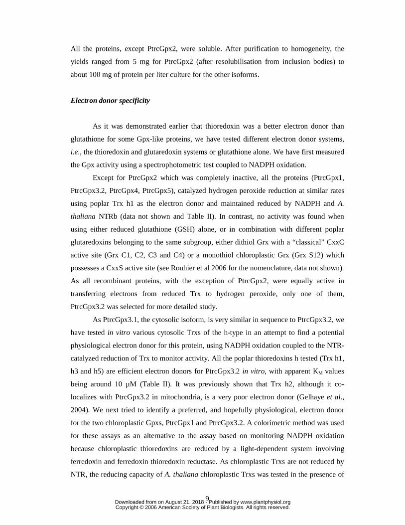
All the proteins, except PtrcGpx2, were soluble. After purification to homogeneity, the
yields ranged from 5 mg for PtrcGpx2 (after resolubilisation from inclusion bodies) to
about 100 mg of protein per liter culture for the other isoforms.
Electron donor specificity
As it was demonstrated earlier that thioredoxin was a better electron donor than
glutathione for some Gpx-like proteins, we have tested different electron donor systems,
i.e., the thioredoxin and glutaredoxin systems or glutathione alone. We have first measured
the Gpx activity using a spectrophotometric test coupled to NADPH oxidation.
Except for PtrcGpx2 which was completely inactive, all the proteins (PtrcGpx1,
PtrcGpx3.2, PtrcGpx4, PtrcGpx5), catalyzed hydrogen peroxide reduction at similar rates
using poplar Trx h1 as the electron donor and maintained reduced by NADPH and A.
thaliana NTRb (data not shown and Table II). In contrast, no activity was found when
using either reduced glutathione (GSH) alone, or in combination with different poplar
glutaredoxins belonging to the same subgroup, either dithiol Grx with a “classical” CxxC
active site (Grx C1, C2, C3 and C4) or a monothiol chloroplastic Grx (Grx S12) which
possesses a CxxS active site (see Rouhier et al 2006 for the nomenclature, data not shown).
As all recombinant proteins, with the exception of PtrcGpx2, were equally active in
transferring electrons from reduced Trx to hydrogen peroxide, only one of them,
PtrcGpx3.2 was selected for more detailed study.
As PtrcGpx3.1, the cytosolic isoform, is very similar in sequence to PtrcGpx3.2, we
have tested in vitro various cytosolic Trxs of the h-type in an attempt to find a potential
physiological electron donor for this protein, using NADPH oxidation coupled to the NTR-
catalyzed reduction of Trx to monitor activity. All the poplar thioredoxins h tested (Trx h1,
h3 and h5) are efficient electron donors for PtrcGpx3.2 in vitro, with apparent KM values
being around 10 µM (Table II). It was previously shown that Trx h2, although it co-
localizes with PtrcGpx3.2 in mitochondria, is a very poor electron donor (Gelhaye et al.,
2004). We next tried to identify a preferred, and hopefully physiological, electron donor
for the two chloroplastic Gpxs, PtrcGpx1 and PtrcGpx3.2. A colorimetric method was used
for these assays as an alternative to the assay based on monitoring NADPH oxidation
because chloroplastic thioredoxins are reduced by a light-dependent system involving
ferredoxin and ferredoxin thioredoxin reductase. As chloroplastic Trxs are not reduced by
NTR, the reducing capacity of A. thaliana chloroplastic Trxs was tested in the presence of
www.plantphysiol.orgon August 21, 2018 - Published by Downloaded from Copyright © 2006 American Society of Plant Biologists. All rights reserved.

10
500 µM DTT, a reagent that will reduce these thioredoxins but does not support the
peroxidase activity of Gpxs during the relatively short time used for the assay. Of all the
chloroplastic Trxs tested, only the two A. thaliana y-type Trxs (Trx y1 and Trx y2) were
able to reduce Gpx to a significant extent. Similar results were obtained for PtrcGpx1 and
PtrcGpx3.2, with the reduced y-type Trxs leading to an 80% decrease of the H2O2 content
with both Gpxs (Fig. 3 and data not shown). The other isoforms tested did not support Gpx
activity. The cytosolic poplar Trx h1, present at the same concentration as the chloroplastic
Trxs, was approximately as effective as the y-type Trxs in supporting H2O2 reduction
catalyzed by either PtrcGpx1 or PtrcGpx3.2. Despite the similar reactivity of the
cytoplasmic Trx h1, the fact that the two y type Trxs present in the poplar genome co-
localize with PtrcGpx1 and PtrcGpx 3.2 in chloroplasts suggest that the two Trx y may
well constitute in vivo reductants for these two Gpxs.
Peroxide specificity
The substrate specificity of PtrcGpx3.2 was tested under steady-state conditions
using Trx h1 as reductant and different types of peroxides, ranging from hydrogen
peroxide to more complex molecules, such as tert-butyl hydroperoxide or cumene
hydroperoxide as electron acceptors. The KM values for the different substrates and the
catalytic efficiencies of the enzymes are summarized in Table II. Although slightly
different (KM ranging from 239 µM to 1.41 mM), the KM values for the three substrates are
all of the same order of magnitude, lying in the millimolar range. In terms of catalytic
efficiency (kcat/ KM), PtrcGpx3.2 is slightly more efficient in reducing cumene
hydroperoxide (5.3x10⁴ M-1 s-1) compared to H2O2 (20x103 M-1 s-1) and to tert-butyl
hydroperoxide (6.4x10³ M-1 s-1).
We then examined the stoichiometry of the reaction catalyzed by Gpxs, by mixing
known concentrations of reduced enzyme, with various known concentrations of tert-butyl
hydroperoxide in the absence of reductants. After completion of the reaction, the remaining
peroxide content was measured using the FOX colorimetric method. All the enzymes
tested (PtrcGpx1, 3.2 and 5) can reduce one mole substrate per mole enzyme, indicating
that Gpxs only use one or two cysteines, but certainly not three.
It is known that, for enzymes which use sulfenic acid chemistry for their catalysis,
the stoichiometry depends on the number of cysteines involved. When one or two cysteines
are involved, the stoichiometry of the reaction is 1 mol of enzyme oxidized per mol of
www.plantphysiol.orgon August 21, 2018 - Published by Downloaded from Copyright © 2006 American Society of Plant Biologists. All rights reserved.

11
peroxide reduced (Boschi Muller et al., 2000). In one case, the first “peroxidatic” cysteine,
attacks the substrate and is converted to a sulfenic acid, with the reaction stopping at this
point. More commonly, a second “resolving” cysteine attacks the sulfenic acid, leading to
the formation of a disulfide bridge. When a third cysteine is involved in the catalytic
mechanism, as it is the case for some methionine sulfoxide reductases, the stoichiometry of
the reaction is 2 (Boschi Muller et al., 2005). In this case, the third cysteine (or the second
resolving cysteine) reduces the first disulfide bridge formed between the peroxidatic
cysteine and the first resolving cysteine, leading to a new intramolecular bridge. Then, the
peroxidatic cysteine would be free and able to attack another molecule of peroxide, leading
to a consumption of two moles peroxide per mole of reduced enzyme.
Catalytic mechanism
In order to understand the mechanism used by PtrcGpxs for peroxide reduction and
for the Trx-dependent regeneration, site-directed mutagenesis was used to generate four
variants of PtrcGpx3.2 (PtrcGpx3.2 C107S, C136S, C155 or C136/155S) in which cysteine
residues were replaced by serines. The activity of the mutated proteins was tested in the
presence of the Trx system. As expected, the Cys 107S mutant is totally inactive. While
PtrcGpx3.2 C155S or the double mutant PtrcGpx3.2 C136/155S are able to reduce
peroxides in “single-turnover” experiments (i.e., pre-reduced samples of these two variants
can reduce an equal amount of peroxide), they are completely inactive with the Trx
reducing system, regardless of the peroxide substrate used. This indicates that Trx is
probably not able to directly reduce the sulfenic acid formed on Cys107. In contrast,
PtrcGpx3.2 C136S displays almost identical enzymatic properties to PtrcGpx3.2 (Table II).
The only difference is a small modification of the KM value for peroxides which results in
a decreased catalytic efficiency. These results indicate unequivocally that Cys107 and
Cys155 are required for thioredoxin regeneration, whereas Cys136, albeit strictly
conserved and found in a very conserved motif, does not have a catalytic role.
Redox and oligomerization states
We examined the behaviour of PtrcGpx3.2 and its various cysteine/serine variants
during denaturing polyacrylamide gel electrophoresis (SDS PAGE) under different
conditions. Under reducing conditions (30 mM DTT), all the proteins migrated at their
www.plantphysiol.orgon August 21, 2018 - Published by Downloaded from Copyright © 2006 American Society of Plant Biologists. All rights reserved.

12
expected molecular masses, around 19 kDa (Fig. 4A, lanes 2,4,6,8,10). Under oxidizing
conditions (30 mM H2O2), the migration of PtrcGpx3.2, as well as of its C107S and C136S
variants is shifted to a lower apparent molecular mass (around 13 kDa) whereas the
migration of C155S and C136S/155S mutated proteins was not altered (Fig. 4A, lanes
1,3,5,7,9). The change in the migration profile under oxidizing conditions for all the
proteins in which Cys155 has not been mutated suggests that an intramolecular disulfide
bond involving Cys155 can be formed. The disulfide, formed between Cys 107 and Cys
155 in the case of the C136S mutant and between Cys 136 and Cys 155 in the case of the
C107S mutant, apparently modifies the overall structure of the proteins sufficiently to alter
the migration behavior during electrophoresis. It should also be pointed out that a covalent
dimer, involving Cys 107 of two subunits, can be observed for the double mutant under
non-reducing conditions but not under reducing conditions, as has been previously
observed for mutated proteins in which only the catalytic cysteine remains (Rouhier et al.,
2004a). These results were confirmed by following changes in intrinsic tryptophan
fluorescence emission of the proteins in reduced or oxidized conditions (Fig. 4B). Only
one tryptophan found in position 196 in the WNF motif and conserved among organisms,
is present in PtrcGpx3.2. The behaviour of PtrcGpx3.2 is shown in Fig. 4B as an example,
but all Gpxs behave similarly. While the protein is in the oxidized form after purification
(the number of detectable thiol groups is generally close to zero), adding 250 µM DTT
changed the emission fluorescence spectrum of the protein, with a decrease of
approximatively 3-fold in the fluorescence intensity and a shift of the maximum of
fluorescence emission from 350 to 316 nm. The addition of 250 µM peroxide in the
cuvette completely restored the initial spectrum suggesting that changes of tryptophan
environment and thus of fluorescence emission are linked to changes in oxidation state.
Among the cysteinic mutated proteins, only PtrcGpx3.2 C136S showed similar differences
in the fluorescence properties of the oxidized and reduced forms. All other mutated
proteins (PtrcGpx3.2 C107S, PtrcGpx3.2 C155S, PtrcGpx3.2 C136S/155S) did not share
this property (data not shown). As observed under SDS PAGE, where it abolished the shift
between oxidized and reduced states, the mutation of Cys 155 prevents modification of
tryptophan fluorescence and thus of the formation of the disulfide bond. No change of
fluorescence was observed for PtrcGpx3.2 C107S, while a shift, attributed to the formation
of a Cys136-Cys155 disulfide bond, was visible on SDS PAGE. Altogether, these results
indicate that tryptophan fluorescence is modified only when the peroxidatic cysteine (Cys
www.plantphysiol.orgon August 21, 2018 - Published by Downloaded from Copyright © 2006 American Society of Plant Biologists. All rights reserved.

13
107) and the resolving cysteine (Cys 155) are involved in the formation of a disulfide
bond.
Gel-filtration experiments performed with reduced or untreated PtrcGpx3.2 showed
that it consistently elutes with an apparent molecular mass of 38 kDa, corresponding to an
homodimer (data not shown). As the protein migrates as a monomer (around 19 kDa)
under both reducing and non-reducing conditions during denaturing SDS PAGE, the dimer
observed during gel filtration cannot involve an intermolecular disulfide and must be
stabilized by non-covalent forces (e.g., electrostatic or hydrophobic interactions).
Redox titrations of PtrcGpx3.2 using redox equilibration buffers composed either of
defined mixtures of oxidized and reduced DTT or of defined mixtures of GSH plus GSSG
are most readily interpreted in terms of a single disulfide/dithiol redox couple per
monomer, with a midpoint redox potential (Em) value of 295 ± 10 mV at pH 7.0 (Fig. 5).
Redox titrations (not shown) over a more positive range than that shown in Fig. 5 (i.e., Eh
values ranging from -250 mV to -80 mV), in which defined mixtures of GSH plus GSSG
were used as redox equilibration buffers, demonstrated that no additional more positive
dithiol/disulfide couple is present in the protein. As Cys136 is not involved in catalysis and
the protein is not present as a disulfide-linked covalent dimer, one can conclude that the
value observed corresponds to an intramolecular disulfide bridge linking Cys107 to
Cys155.
Expression of Prx in plant organs and in stress conditions
We first performed an in silico analysis based on ESTs found in the DOE JGI
database and found 191 ESTs coding for poplar Gpxs (Table I). The different isoforms are
not equally represented among these ESTs : PtrcGpx3.2 seems to be the most frequently
expressed gene, with 85 corresponding ESTs (i.e. approximately 45%). We also found 48
ESTs for PtrcGpx1, 10 for PtrcGpx2, 23 for PtrcGpx3.1, 17 for PtrcGpx4 and and 8 for
PtrcGpx5. These results were combined with a semi-quantitative RT-PCR approach. As
these genes belong to the Prx family, we have studied the transcript expression pattern of
all Gpxs together with the 9 Prxs existing in poplar in seven different tissues (roots, young
leaves, expanded leaves, fruits, stems, petioles and stamen). For most genes, we have been
able to find specific hybridizing areas. However, we were sometimes unable to distinguish
some closely related sequences (i.e., Prx Q1 and 2 or 2-Cys Prx A and B). In these two
cases, we used non-specific primers able to amplify both transcripts (Prx Q1 and Q2 or 2-
www.plantphysiol.orgon August 21, 2018 - Published by Downloaded from Copyright © 2006 American Society of Plant Biologists. All rights reserved.
14
Cys Prx A and B) but we were also able to define one primer to specifically amplify one
gene (Prx Q1 and 2-Cys Prx A). From the difference between these two amplifications, the
expression of the second gene can be deduced. For example, as the 2-Cys Prx A is only
expressed in expanded leaves, we can estimate from the 2-Cys Prx A and B expression
profile that 2-Cys Prx B is expressed in fruits, stems, petioles and stamens and possibly in
expanded leaves. All the genes exhibit different expression patterns but there is at least one
gene expressed in each organ tested (Fig. 6A). These data, coupled with the in silico
analysis, are indicative of a broad distribution and expression of Prxs in plants at least at
the transcript level. Nevertheless, the multiplicity of Prx genes can be explained by some
specificity in the expression pattern. For example, out a total of 17 genes, only three
(coding for PtrcGpx2, PtrcGpx 3.1 and PtrcPrx IIE), are expressed at detectable levels in
roots under the growth conditions used. One gene (PtrcPrx IIE) encodes a plastidic protein,
another (PtrcGpx 3.1) appears to encode a cytosolic Gpx and the third (PtrcGpx2) appears
to encode a secreted Gpx. In contrast, all the genes are expressed in expanded leaves.
Given the specificity of the two chloroplastic Gpxs (PtrcGpx 1 or PtrcGpx 3.2) for a Trx of
the y-type as an electron donor, it was also of interest to examine the expression patterns of
these two poplar thioredoxins. Both Trx y1 and y2 were found to be expressed in all tissues
tested and thus their role cannot simply be restricted to serving as specific reductants for
plastidial Gpxs.
In order to get a deeper view of the expression of Gpx genes, we used both
microarray data available using Genevestigator and the results presented by Rodriguez-
Milla and colleagues to follow the expression pattern of the different Arabidopsis Gpx
genes in the whole plant (Figure 6B) (Zimmermann et al., 2004; Rodriguez-Milla et al.,
2003). Based on the results of Rodriguez Milla and colleagues, AtGpxs1 to 7 are expressed
in all tissues analyzed, except AtGpx2 which does not seem to be expressed in dried seeds.
AtGpx4 and 7 are the less expressed genes as they were only detected by RT PCR and few
or no ESTs are found for these genes (Rodriguez-Milla et al., 2003). Using Genevestigator,
we observed that AtGpx1, AtGpx2 and AtGpx6, respectively homologous to PtrcGpx1,
PtrcGpx2 and PtrcGpx3.2, are highly expressed in all organs or tissues analyzed (i.e. in
young, expanded and senescent leaves, as well as in stems, roots and flowers), while
AtGpx3, AtGpx5, AtGpx7 and AtGpx8 and especially AtGpx4 generally show a low
expression level in these organs. Nevertheless, as expected for a chloroplastic protein,
AtGpx1 is preferentially expressed in green tissues (young and expanded leaves, stems and
flowers). The main difference between the two sources concerns the expression of AtGpx6,
www.plantphysiol.orgon August 21, 2018 - Published by Downloaded from Copyright © 2006 American Society of Plant Biologists. All rights reserved.

15
which was found to be low in all organs except dry seeds by northern blot, while
microarray data indicates a strong expression in all organs.
Using an antibody raised against purified recombinant PtrcGpx3.2, we first checked
its specificity against the recombinant proteins. It reacts with all five recombinant proteins
(which incidentally all migrate at slightly different positions on SDS-PAGE) but only
faintly with PtrcGpx 1 and 5 and more strongly with PtrcGpx2, 3.2 and 4 (Fig. 7A). This
antibody is thus specific for Gpx as a whole, including the sixth protein, PtrcGpx3.1, which
is very similar to PtrcGpx3.2, but does not discriminate between the isoforms. We could
however differentiate at least two isoforms with two slightly different molecular masses in
poplar protein extracts. The higher molecular mass band seems specific to roots, and since
the signals obtained with the extracts from all the other organs tested have a similar size,
they could thus correspond to a single isoform which we cannot identify for the reasons
explained above (Fig. 7B).
In A. thaliana (Fig. 7C), two bands of different sizes arising from two or more
different isoforms have been detected in almost all organs, except roots. The protein(s) of
smaller molecular mass is most easily detected in leaves and flowers while in contrast the
protein(s) of higher molecular mass is present in stems and roots and to a lesser extent in
leaves and flowers. Using proteins extracted from purified leaf chloroplasts, only the lower
band was detected (data not shown). Given the fact that AtGpx7 is weakly expressed at the
transcriptional level, only the other two predicted chloroplastic proteins (i.e., AtGpx1 and
AtGpx6) are likely to be detectable. Thus, we conclude that the lower band consists
predominantly of AtGpx 1 and/or AtGpx6. The upper band is likely to arise from the
presence of AtGpx2, as it is not a chloroplastic protein and because AtGpx3, AtGpx4,
AtGpx5 and AtGpx8 also seem to be weakly expressed at the transcriptional level.We next
investigated the abundance of these proteins in stress situations (Fig. 8). Gpx abundance as
a function of time was followed after infection of poplar by the agent of poplar rust, the
fungus Melampsora larici-populina, and using two different isolates, a virulent one that
induces a compatible reaction, and an avirulent one, that induces an incompatible reaction
(Fig 8A). As is the case for untreated leaf extracts, a single band is visible. During the
incompatible reaction, the expression of Gpx increased greatly 2h after infection and even
more after 48h. The pattern of expression during the compatible reaction is strikingly
different with a lower protein level observed at both 2h after infection and then again at
24h after infection, but with levels very similar to those seen prior to infection found at all
other times. Because this expression pattern is surprising, we have analysed the expression
www.plantphysiol.orgon August 21, 2018 - Published by Downloaded from Copyright © 2006 American Society of Plant Biologists. All rights reserved.

16
of Trx h1, which is constitutively expressed and was never found to vary with stress, as a
control (Rouhier, unpublished results). As expected, the Trx h1 abundance is not
dependent on the pathogen infection, regardless of which the fungal isolate was used.
In addition, we followed Gpx levels in young leaves from A. thaliana or tobacco
plants submitted to various abiotic stresses. After 6 days of water deficit, the intensity of
the lower band decreased, while that of the upper band increased (Fig 8B). This was even
more pronounced in young tobacco leaves subjected to the same conditions, with an
increased intensity of the Gpx signal, which could correspond to the presence of one or
more Gpx isoforms (Fig. 8B). Similar profiles were observed using expanded leaves (data
not shown). In plants subjected to photooxidative stress (Fig. 8C), the amount of the two
proteins varied in the opposite way, the intensity the upper band increased slightly under
stress conditions in expanded leaves, whereas the intensity of the lower band decreased
after 3 or 8 days of treatment.
In plants exposed to metal stresses induced by application of various concentrations
of cadmium or copper for 2 and 7 days, only the amount of the lower molecular weight
protein(s) detected is modified. It decreased at 150 µM copper after 2 days (Fig. 8E) but
increased strongly after 7 days at cadmium concentrations of 50 and 75 µM (Fig. 8D) and
at copper concentrations of 25, 75 and 150 µM (Fig. 8E).
Subcellular localization of PtrcGpx1 and PtrcGpx3.2
PtrcGpx2 and PtrcGpx5 are predicted to be secreted, but were not studied here as
we were unable to amplify the 5’ end coding for their N-terminal extensions from either
cDNA libraries or genomic DNA. Although A. thaliana and O. sativa homologs possess
this extension, it is not yet clear whether these extensions really belong to the cDNA or are
annotation or assembly errors. PtrcGpx4 and PtrcGpx3.1 contain neither an N-terminal
extension nor known C-terminal signals, suggesting that the proteins are cytosolic. In order
to determine the subcellular localization of PtrcGpx1 and PtrcGpx3.2, the 5’ portions of
the ORF encoding only the N-terminal parts were fused to the sequence coding for a green
fluorescent protein (GFP). The recombinant pCK GFP S65C plasmids were then used to
transiently transform tobacco cells by bombardment. In the guard cells shown here, it
appears that Gpx1 is specifically targeted to chloroplasts, and that Gpx3.2 is directed both
to chloroplasts and to mitochondria (Fig. 9). Similar results were obtained in mesophyll
cells (data not shown).
www.plantphysiol.orgon August 21, 2018 - Published by Downloaded from Copyright © 2006 American Society of Plant Biologists. All rights reserved.

17
Discussion
As more and more genomes are sequenced and annotated, it is now clear that
multigenic families are very common in organisms. Here we describe the existence of 6
Gpx isoforms in the model tree Populus trichocarpa. By comparison, 8 and 5 Gpx
isoforms are present in the genome of Arabidopsis thaliana and Oryza sativa, respectively.
The number of Gpx genes thus varies among species, probably because of gene duplication
events. A continuing challenge is to understand why so many genes or proteins with
similar functions are needed in plants. To answer this question, both the abundance and
localization of the proteins and the substrate specificities of these enzymes were studied.
As a result of our study, it is now clear that the poplar enzymes originally named
glutathione peroxidases are reduced by thioredoxins rather than by glutathione and belong
to the peroxiredoxin family. This large family thus includes 15 different peroxiredoxins in
Populus trichocarpa.
Substrate and electron donor specificities
The poplar Gpxs do not exhibit any difference in the reduction of hydrogen
peroxide compared to complex hydroperoxides (COOH and tBOOH), as the catalytic
efficiencies (kcat/ KM) vary only over a 20-fold range (i.e., from 2.6 to 53x103 M-1 s-1). The
reduction of complex peroxides is consistent with a role for Gpxs (and for Prxs in general)
that compliments other systems, like catalases or ascorbate peroxidases, that specifically
reduce H2O2. The catalytic efficiencies of poplar Gpxs lie in the range previously described
for other peroxiredoxins. Rouhier et al. (2004a) determined kinetics parameters for poplar
chloroplastic Prx Q, with kcat around 1 s-1 and kcat/ KM of 103 to 104 M-1 s-1, depending on
the substrate considered. Horling and co-workers (2002) reported an activity of Prx IIC
that is in the same range. Previous studies (Jung et al., 2002, Herbette et al., 2002 and
Tanaka et al., 2005) also indicate that plant or yeast Gpxs may display kinetic parameters
similar to those of the other classes of the broad Prx family. The catalytic efficiencies
reported to date for Gpxs and Prxs is far lower than those of catalases or ascorbate
peroxidases, but they are likely to reduce a broader range of substrates than these two
enzymes. Gpxs may also provide the graduated response, necessary for the metabolic fine
tuning in case of oxidative stress, as has been demonstrated in the interaction with
www.plantphysiol.orgon August 21, 2018 - Published by Downloaded from Copyright © 2006 American Society of Plant Biologists. All rights reserved.

18
transcription factors (Delaunay et al., 2002). All these data are consistent with a possible
role for Gpxs and Prxs both in ROS detoxication in plant cells and, as has been
demonstrated in other organisms in redox signalling (Rhee et al., 2005, Delaunay et al.,
2002, Vivancos et al., 2005).
In so far as the electron donor specificity of poplar Gpxs is concerned, these
enzymes seem to behave like previously characterized plant Gpx in that they can be
reduced by thioredoxins, as earlier demonstrated for some plant Gpx (Herbette et al., 2002,
Jung et al., 2002), but not at all by glutathione. We have also demonstrated that a
glutaredoxin, which can provide a pathway for disulfide reduction alternative to the
thioredoxin pathway, is unable to reduce poplar Gpxs. However, because only five
glutaredoxins belonging to the same subgroup were tested, the possibility, that one or more
other members of the large Grx might serve as an effective electron donor to Gpx, exist.
The oxidation-reduction titrations carried out in this study (see Fig. 5) provide a
thermodynamic basis for this donor specificity. The intramolecular Cys107/Cys155
disulfide present in the oxidized PtrcGpx3.2 was shown to have a Em value at pH 7.0 of -
295 ± 10 mV. While reduction of this PtrcGpx3.2 disulfide by thioredoxins, which have Em
values ranging from -275 mV to -330 mV (Collin et al., 2003; Brehelin et al., 2003) would
be expected to be thermodynamically favourable, the more positive Em values of
glutathione (Em = -245 mV at pH 7.0) and plant glutaredoxins (Em values around -180 mV,
Rouhier et al. unpublished results) would make reduction of the PtrcGpx3.2 disulfide
thermodynamically unfavourable.
The question of which Trx is the actual physiological electron donor for different
Gpxs remains unsettled. Taking the Trx h family as an example, we were unable to
establish a preference between Trx h1, h3, and h5 in in vitro assays. It was previously
demonstrated that neither a poplar Trx h2 nor an A. thaliana Trx o were able to serve as
reductants to PtrcGpx3.2 (Gelhaye et al., 2004) and thus the identity of the physiological
reductant for this mitochondrial Gpx remains unclear.
Considering the putative secreted isoforms, namely PtrcGpx2 and PtrcGpx5, we
found that Trx h5 is able to reduce PtrcGpx5 in a catalytic manner (data not shown). Juarez
and co-workers (2005) recently shown that an homolog of poplar Trx h5 was secreted by
Nicotiana alata cells. Assuming that PtrcGpx5 is actually secreted, these two proteins
could be part of an extracellular redox signaling system. Pignocchi and Foyer (2003)
suggested that changes in the apoplastic redox state could be sensed by a system involving
dithiol-disulfure exchanges. Secreted Gpxs may be candidates as apoplastic redox sensors.
www.plantphysiol.orgon August 21, 2018 - Published by Downloaded from Copyright © 2006 American Society of Plant Biologists. All rights reserved.

19
There are at least 11 chloroplastic Trxs in A. thaliana (NtrC, CDSP32, Trx x, Trx
y1 and y2, Trx f1 and f2, Trx m1, m2, m3 and m4) and even more in Populus trichocarpa
(unpublished results). Although we did not test the efficiency of the bimodular Trxs (NtrC
and CDSP32), it appears that only the two Trxs y are able to efficiently support the activity
of PtrcGpx1 and PtrcGpx3.2. These chloroplastic Trxs all have very similar Em and so the
specificities observed must arise from properties of the Trxs other than their Em values (for
example, the distributions of surface charges and of surface hydrophobic patches). In a
previous study, it was found in Arabidopsis that only Trx x is able to reduce 2-Cys Prx,
and Trx y is the most efficient electron donor to Prx Q (Collin et al., 2004). It is then likely
that the major function of x-type and y-type chloroplastic Trxs is the reduction of
chloroplastic Prxs (this family includes 2-Cys Prxs, Prx Q, Prx IIE and Gpxs). The f-type
Trxs are specifically needed in the regulation of chloroplastic metabolism (in particular,
through the activation of FBPase and of the CF1 ATP synthase), but they can also reduce
2-Cys Prxs and Prx Q. The m-type Trxs may play a dual role, regulating metabolism
through control of NADP-dependent malate dehydrogenase, and also serving as possible
reductants for 2-Cys Prx and Prx Q (Collin et al., 2003). Overall, this would constitute a
system with partial redundancy, that could be linked to each subcellular compartment
redox state.
Using the Digital Northern function of Genevestigator online database
(https://www.genevestigator.ethz.ch) (Zimmermann et al., 2004), we noticed that the
mRNA profiles of the A. thaliana ortholog of PtrcGpx1 (AtGpx1, Accession number:
At2g25080) are more closely related to that of AtTrx y1 (Accession number: At1g76760),
than to that of AtTrx y2 (Accession number: At1g43560). This may indicate which of the
two Trxs of the y-type is likely to be the favored electron donor to this enzyme in vivo. As
there are also two isoforms of Trx y in poplar, the situation might be similar. RT-PCR
experiments showed that the two genes (Trx y1 and y2) are expressed in all the organs
tested, and thus at the moment one cannot identify specific and distinct roles for the two
isoforms.
Catalytic mechanism and Trx-mediated recycling process
When starting this work, one question was to determine whether one, two or three
cysteines were involved in the reactions catalyzed by plant Gpxs. Although yeast Gpx2 had
been shown to possess a Trx-dependent activity, involving only the two cysteines
www.plantphysiol.orgon August 21, 2018 - Published by Downloaded from Copyright © 2006 American Society of Plant Biologists. All rights reserved.

20
equivalent to Cys107 and Cys155 in poplar (Tanaka et al., 2005), Jung et al. (2002) had
previously suggested a catalytic mechanism involving three conserved cysteine residues
for a Chinese cabbage Gpx. This proposal was based on mass spectrometry data that
showed that a disulfide bridge could be formed between the cysteines corresponding to
poplar Cys136 and Cys155 (Jung et al., 2002) and on their observation that the mutated
protein equivalent to PtrcGpx C136S retained only 30% of the activity displayed by the
wild-type enzyme. Here, we show that, although three cysteines are absolutely conserved
in all Gpxs from higher plants, only two are involved both in catalysis and in the Trx-
dependent regeneration. Indeed, three observations support this conclusion: (i) The
stoichiometry of the reaction catalyzed by the wild-type enzyme is consistently 1:1 (in
terms of peroxide reduced to enzyme oxidized) and never 2:1; (ii) the mutated PtrcGpx3.2
C136S is as active as PtrcGpx3.2; and (iii) we have only detected one disulfide bridge
when titrating PtrcGpx3.2. Another characteristic of these enzymes is a shift during
migration under SDS-PAGE, similar to that previously reported by Tanaka et al. (2005) for
yeast Gpx2, as the redox state of the protein is altered. Both the redox-dependent shift in
SDS-PAGE migration properties and the redox-dependent changes in the tryptophan
fluorescence parameters of the proteins clearly indicate that Cys155 is required for the
formation of an intramolecular disulfide with Cys 107. One additional piece of evidence
that Gpx belongs to the Prx family, is its formation of non-covalent dimers. Indeed, the
only plant Prx for which a structure is known is a homodimer with a conserved interface
found in other non-plant Prx structures (Echalier et al., 2005). A structural study, designed
to determine the structural determinants responsible for substrate binding, for dimerization
and to elucidate a possible role for the highly conserved Cys136, is currently underway.
Unlike the case for many other eukaryotic Prx characterized so far, we did not
observe the formation of “over-oxidized” forms of Gpx. Indeed, the stoichiometry of the
reaction catalyzed by Gpxs is consistently 1, the activity measured in the presence of the
Trx system is linear with time and H2O2-treated proteins do not undergo the mass
increment expected for sulfinic or sulfonic formation (data not shown). We thus propose a
three-step reaction mechanism for plant Gpxs similar to the one used by Prx Q with 2
cysteines forming an intramolecular disulfide bridge in the oxidized state (Fig. 10). These
steps are: (i) a nucleophilic attack of the catalytic cysteine (Cys107) on the peroxide with
the release of an alcohol and the concomitant formation of a sulfenic acid; (ii) an attack of
the sulfenic acid by Cys155 and formation of an intramolecular disulfide bridge between
Cys107 and Cys155; and (iii) a reduction of the disulfide bridge by Trx.
www.plantphysiol.orgon August 21, 2018 - Published by Downloaded from Copyright © 2006 American Society of Plant Biologists. All rights reserved.

21
Expression and localization of Gpx in plants
We analyzed the expression of all the Prxs in poplar organs. All the members of this
family are expressed at the transcriptional level in at least one of the organs tested, with a
specific expression pattern for each of them. Gpxs are likely to be present in all organs and in
nearly all cell compartments, suggesting a redundancy with the peroxiredoxins identified
previously. Interestingly, many isoforms are expressed in flowers or in fruits but their precise
physiological function is yet unknown. Nevertheless, to explain the need for proteins with
identical functions, we can imagine that some genes could be expressed in specific conditions
under stress for example or could be temporally regulated.
When poplar leaves are subjected to a biotic stress (infection by the rust fungus M.
larici populina), the abundance of these proteins is modified. This is in line with previous
observations that the levels of Prx Q, Prx IIC, Prx IIF but not of 2-Cys Prx are modified in
this situation (Rouhier et al 2004a, Gama et al 2006). Abiotic stress conditions (water deficit,
photooxidative and metal stresses) also induce modifications of the Gpx levels. The amount
of isoform(s) of lower molecular masses is especially modified, generally decreasing in water
deficit and photo-oxidative stress but strongly increasing in the presence of toxic metals, such
as cadmium or copper. In the case of photooxidative stress, it was previously shown that
among members of the Prx family, only the amount of Prx Q increased in this situation,
whereas the level of Prx IIE and 2-Cys Prx remained unchanged (Havaux et al., 2005). In our
case, the amount of chloroplastic Gpx(s) decreased, suggesting there is a fine control and a
functional specialization among members of the same family. It was previously demonstrated
at the transcript levels that AtGpx2, AtGpx5 and AtGpx6 were strongly and rapidly increased
in Arabidopsis plants treated with 200 µM of FeSO4 or CuSO4 (Rodriguez Milla et al., 2003).
Other studies provided evidence for an increased Gpx activity in plants treated with copper
(Ali et al., 2006) or nickel (Gajewska and Sklodowska), but decreased activity in the presence
of cadmium (Aravind et al., 2006). The conclusion, based on our results and those of others
that glutathione peroxidases are thioredoxin-dependent enzymes, suggests that the
glutathione-dependent peroxidase activity found in these studies corresponds to the
peroxidase activity of glutathione S-transferases. Taken as a whole, the results presented
above provide evidence at the protein level that supports the conclusion arrived at based on
previous observations mRNA studies, that Gpx are involved in stress responses and especially
in response to metals. For example, it was previously demonstrated that overexpression of a
www.plantphysiol.orgon August 21, 2018 - Published by Downloaded from Copyright © 2006 American Society of Plant Biologists. All rights reserved.
22
Gpx from C. reinhardtii both in the cytosol and in the chloroplast and overexpression of a
tomato Gpx in yeast and plant increased tolerance to oxidative stress (Chen et al., 2004;
Yoshimura et al., 2004).
Another possible explanation for the redundancy of thioredoxin peroxidases as a
whole could be linked to their subcellular distribution. Using GFP-fusion, we have
demonstrated that two Gpxs (PtrcGpx 1 and 3.2) are localized in chloroplast, one of these
(PtrcGpx 3.2) being also targeted to mitochondria. This dual targeting is not unusual, having
already been observed for some other antioxidant proteins (glutathione reductase,
monodehydroascorbate reductase, ascorbate peroxidase) (Chew et al., 2003). The plastidic
localization of PtrcGpx1 was expected, as a pea Gpx homolog had been previously been
shown to be imported into chloroplasts (Mullineaux et al., 1998). Only one other plant Gpx
had previously been localized experimentally. This protein, from Lycopersicon esculentum,
called GPXle-1 and homologous to PtrcGpx 3.1, was found in the cytosol near the plasma
membrane and to a lesser extent in the cell wall of some leaf cells (Herbette et al., 2004).
Adding the results of this investigation to the previously available data on distribution
of Prxs, 8 poplar Prxs are now known to be expressed in plastids (Prx IIE, 2-Cys Prx A and B,
Prx Q 1 and 2, PtrcGpx1 and PtrcGpx3.2), two in mitochondria (Prx IIF, PtrcGpx3.2), 3 in the
cytosol (Prx IIB, Prx IIC and PtrcGpx3.1), one in the nucleus (Prx 1-Cys) and two are
predicted to be directed to the secretory pathways (PtrcGpx2 and PtrcGpx5), assuming that
these proteins indeed contain an N-terminal extension. This situation is slightly different from
A. thaliana, in which there are only one PrxQ, but an additional cytosolic Prx II and 8 Gpxs
instead of the 6 found in poplar.
www.plantphysiol.orgon August 21, 2018 - Published by Downloaded from Copyright © 2006 American Society of Plant Biologists. All rights reserved.

23
Materials and Methods
Materials
Hydrogen peroxide (H2O2), tert-butyl hydroperoxide (tBOOH), cumene hydroperoxide
(COOH), NADPH, GSH and glutathione reductase (GR) from yeast were purchased from
Sigma (St. Louis, USA).
Expression of poplar Gpxs in various organs
RT-PCR
Semi-quantitative RT-PCR experiments were used to estimate the distribution of Gpx,
Prx and y-type Trx transcripts in various poplar organs. Total RNA isolation was performed
with the RNeasy Plant Mini kit (Qiagen, Courtaboeuf, France) from approximately 100 mg of
various frozen tissues obtained from 4-month-old Populus trichocarpa plants (young and
expanded leaves, roots, stems, petioles) or from older Populus trichocarpa trees (fruits,
stamen). To remove contaminating DNA, the samples were treated with DNAse I (Qiagen,
Courtaboeuf, France). Except for the stem (for which we used 300 ng), a total of 1 µg of total
RNA was used to generate cDNAs by reverse transcription using the Omniscript RT kit
(Qiagen, Courtaboeuf, France). These products were used as templates for subsequent PCR
reactions using the primers described in supplementary Table II. The program used was as
follows : 94°C for 3 min and 35 cycles of 94°C for 30 s, 54°C for 45 s, and 72°C for 1 min 45
s. The primers used were sometimes identical to those used for the cloning experiments
(Gpx1, 2 and 4; Prx 1-Cys). When two ORFs were very similar (e.g., Gpx 3.1 or 3.2, Prx IIC,
D and E, Prx Q1 and Q2, Prx 2-Cys A and B, Trx y 1 and 2), we used one or two new primers
hybridizing in the 3’ or 5’ part of mRNAs, sometimes in combination with one of the cloning
primers. When no selective amplification was possible for one isoform out of a pair of very
close sequences, primers that can hybridize the two forms were used. In the case of Gpx5, the
amplification was done with two specific internal primers. The Trx h1 gene from poplar,
which is expressed in all tissues tested, was amplified simultaneously and used as a control.
The PCR fragments were then loaded on a 0.8 % agarose gel.
www.plantphysiol.orgon August 21, 2018 - Published by Downloaded from Copyright © 2006 American Society of Plant Biologists. All rights reserved.

24
Western blotting
Infection of poplar leaves with either avirulent or virulent fungal isolates of the rust
fungus, Melampsora larici-populina was carried out as described in Rouhier et al (2004a). A.
thaliana plants subjected to water deficit, photo-oxidative or metal stresses were treated as
described in Vieira dos Santos et al., 2005 and Gama et al., in press. 30 µg of proteins were
loaded per lanes on 15% denaturing SDS PAGE, then transferred onto PVDF membranes
(Millipore, Billerica, USA), when using poplar protein extracts, or onto nitrocellulose
membranes (Pall, Pensacola, USA) for A. thaliana extracts. Antibodies raised against
PtrcGpx3.2 were diluted 1500 x. Detection of proteins recognized by antibodies on PVDF
membranes was done by autoradiography and using the “Odyssey Infrared Imager” from
LICOR in the case of nitrocellulose membranes.
Cloning and site-directed mutagenesis
After identification in the databases (see results), the nucleotide sequences of
PtrcGpx2, 3.2 and 4 were then amplified by PCR using the primers described in
supplementary Table II from a Populus tremula X trichocarpa cv. Beaupre poplar root cDNA
library (Kohler et al., 2003) and PtrcGpx1 and PtrcGpx5 were amplified directly from isolated
E. coli colonies containing the cDNA of interest (this was a gift from Dr Gunnar Wingsle,
University of Umeå, Sweden). Each of the 3 cysteines of the mature PtrcGpx3.2, at positions
107, 136, and 155 respectively, were replaced individually by serines using site-directed
mutagenesis following a procedure described in Jacquot et al. (1997). The double mutant
PtrcGpx3.2 C136S, C155S, which only contains the first cysteine, was also generated and
cloned. After digestion with the NcoI and BamHI restriction enzymes, PCR fragments were
inserted into the pET-3d expression plasmid.
Production and purification of recombinant Gpxs
The recombinant plasmids obtained were used to transform the BL21(DE3) pSBET
strain of E. coli (Schenk et al., 1995). The bacteria were then grown to a final volume of ca.
3.2 L at 37 °C, and protein production was induced during exponential phase by adding 100
µM isopropyl-β-D-thiogalactoside (IPTG). For PtrcGpx3.2, the amount of soluble protein
increased when bacteria were grown overnight at 30°C, without induction by IPTG. The
www.plantphysiol.orgon August 21, 2018 - Published by Downloaded from Copyright © 2006 American Society of Plant Biologists. All rights reserved.

25
bacteria were harvested by centrifugation at 5000 rpm for 20 minutes and then resuspended in
TE buffer (Tris-HCl 30 mM pH 8.0, EDTA 1 mM). Cells were broken by sonication and all
Gpxs were found in the soluble fraction after high speed centrifugation, except for PtrcGpx2,
which was present mostly in inclusion bodies.
The insoluble portion of PtrcGpx2 was first denatured by resuspending the pellet in
TE buffer containing 8 M urea. The soluble proteins obtained after centrifugation at 16000
rpm for 30 minutes were then renatured slowly by two successive dialyses of 6 hours, first
against TE buffer containing 0.5 M urea and then against TE buffer alone. A centrifugation
step (30 minutes at 16000 rpm) was performed after each dialysis, to remove denatured
proteins. For the soluble isoforms, ammonium sulfate fractionation was used as the first step
in the purification procedure. For all of these soluble Gpxs, most of the protein precipitated
between 40 and 80% saturation.
The next purification step, used for all Gpxs, was gel filtration carried out on ACA44
(Biosepra, Cergy-Saint-Christophe, France), equilibrated in TE Buffer containing 200 mM
NaCl. After removing NaCl by ultrafiltration, the Gpx fractions were loaded onto a DEAE-
Sephacel ion exchange column (Amersham, Uppsala, Sweden). A gradient ranging from 0 to
400 mM NaCl was used for the elution of PtrcGpx2, 4 and 5, whereas PtrcGpx1 and 3.2
passed through the column. After dialysis and concentration, the proteins were analysed for
purity by SDS PAGE and stored frozen at –20 °C in TE buffer.
Biochemical characterization of poplar Gpx
NADPH-coupled spectrophotometric method
These experiments were performed at 30°C, and were based on monitoring the
decrease in absorbancy at 340 nm arising from NADPH oxidation. All Gpxs were tested using
this method and showed similar catalytic parameters. The catalytic properties of PtrcGpx3.2
and of its cysteinic mutants (PtrcGpx3.2 C107S, C136S, C155S and C136/155S) (200 nM)
were investigated in more detail and were tested using either a reduced Trx generating system
(3 µM A. thaliana recombinant NTRb and various concentrations of Trx h1, h3 and h5 from
poplar, or the Grx system (0.5 U GR, 1 mM GSH and 100 µM Grx C4), or GSH alone (0.5 U
GR, 2 mM GSH) as possible electron donors. These assays were carried out in a total volume
of 500 µL in 30 mM Tris HCl pH 8 buffer, EDTA 1 mM and 200 µM NADPH. The catalytic
parameters for one substrate were measured by varying its concentration at saturating
www.plantphysiol.orgon August 21, 2018 - Published by Downloaded from Copyright © 2006 American Society of Plant Biologists. All rights reserved.

26
concentrations of the other substrate (typically between 1 and 100 µM for Trx, and between
40 µM to 12 mM for the peroxides (H2O2, tert-butyl hydroperoxide or cumene
hydroperoxide)).
FOX colorimetric method
The reduction of peroxides by PtrcGpx1 and PtrcGpx3.2, the two chloroplastic
isoforms of poplar, was also followed using the FOX (ferrous oxidation of xylenol orange)
colorimetric method (Wolff, 1994). All the mono-modular chloroplastic Trxs from
Arabidopsis thaliana were assayed as possible electron donors. A 50 µL reaction mixture
contained 2 µM of recombinant PtrcGpx1 or 3.2, 500 µM DTT and 10 µM of Trx f1, f2, m1,
m2, m3, m4, x, y1, or y2 recombinant proteins from A. thaliana, purified as described in
Collin et al. (2003). The reaction was started by adding the peroxide (either H2O2, or tBOOH,
or COOH) to a final concentration of 400 µM. At different times, 5 µL of the reaction mixture
were added to 495 µL of the colorimetric reagent and the absorbancy read at 560 nm after 30
min incubation in the dark.
Stoichiometry of the reaction
The stoichiometry of the reaction was also measured using the FOX method with a
known concentration of reduced enzyme (typically 100 µM or 200 µM), in the absence of any
other reductants and in the presence of 400 or 500 µM peroxides. The residual amount of
peroxide remaining after reduction by a known amount of protein was titrated after
completion of the reaction (30 min). The enzyme was reduced with a large excess of DTT (10
mM), which was removed, prior to the assay, by two successive dialyses, each time against
1L of TE buffer.
Oligomerization and redox state of Gpx
The redox state of PtrcGpx3.2 and of the mutated proteins was analysed by incubating
the proteins with either 30 mM DTT or 30 mM H2O2 for 30 min, before separating them on
14% acrylamide SDS-PAGE gels. It was also analyzed by following intrinsic tryptophan-
emission fluorescence using a Cary Eclipse fluorimeter (Varian). Measurements were
recorded at room temperature in a 1 × 1 cm cuvette. The excitation wavelength was set to 290
www.plantphysiol.orgon August 21, 2018 - Published by Downloaded from Copyright © 2006 American Society of Plant Biologists. All rights reserved.

27
nm (5 nm bandpass). Emitted light was detected at 90° between 300 and 500 nm. Spectra of a
1 mL solution containing 4 µM of various PtrcGpxs were recorded before adding successively
250 µM DTT and then 250 µM H2O2.
The native oligomerization state of PtrcGpx3.2 was determined both in oxidized (i.e.,
as isolated) or reduced (30 minutes incubation with 20 mM DTT) conditions using an ACA
44 gel-filtration column (5X75 cm) and proteins of known molecular masses as standards
(bovine serum albumin, ovalbumin, and chymotrypsinogen from Sigma (St. Louis, USA)).
Intracellular localization via Green Fluorescent Protein (GFP) fusion
The nucleotide sequences corresponding to the predicted N-terminal extension of
PtrcGpx1 and PtrcGpx3.2 were cloned into the NcoI and BamHI sites of pCK-GFP S65C
using the primers detailed in supplementary Table II as PtrcGpx1 FOR GFP, PtrcGpx1 REV
GFP, PtrcGpx3.2 FOR GFP and PtrcGpx3.2 REV GFP. These primers were designed to
amplify the sequences corresponding to the predicted signal peptides of Gpx1 (from MASLPF
to TEKSVH) and Gpx3.2 (from M(A)LTSRS to SQSSPQ) (see Fig. 1). The PCR fragments
were fused to GFP at the BamHI site, resulting in chimeric proteins where the N-terminal
extensions of the two Gpxs are present at the N-terminus of eGFP (enhanced Green
Fluorescent Protein), under the control of a double 35S promoter (Menand et al., 1998).
Nicotiana benthamiana cells were then transfected by bombardment of leaves with tungsten
particles coated with plasmid DNA and images were obtained with a Zeiss LSM510 confocal
microscope.
Redox potential determination
Oxidation-reduction titrations, using the fluorescence of the adduct formed between
the protein and mBBr to monitor the thiol content of the protein, were carried out at ambient
temperature as described previously (Krimm et al., 1998; Hirasawa et al., 1999). The reaction
mixtures contained 100 µg of protein in 100 mM MOPS buffer (pH 7.0) containing defined
mixtures of oxidized and reduced DTT to set the ambiant potential (Eh). Identical titrations
were obtained at total DTT concentrations of 1 mM, 2 mM and 4 mM. The titration behavior
was also independent of redox equilibration time over the range from 1.5 to 3 hours.
Titrations at more positive (Eh) values were carried out in a similar fashion except that
glutathione replaced DTT in the redox equilibration buffer total GSH plus GSSG
www.plantphysiol.orgon August 21, 2018 - Published by Downloaded from Copyright © 2006 American Society of Plant Biologists. All rights reserved.

28
concentration of 2 mM. The ambient potential was set by varying the ratio of GSSG to GSH
as described previously (Setterdahl et al., 2000). Redox equilibration times of either 2 or 3
hours were used.
www.plantphysiol.orgon August 21, 2018 - Published by Downloaded from Copyright © 2006 American Society of Plant Biologists. All rights reserved.

29
Literature cited
Agrawal GK, Rakwal R, Jwa NS, Agrawal VP (2002) Effects of signaling molecules, protein
phosphatase inhibitors and blast pathogen (Magnaporthe grisea) on the mRNA level of a rice
(Oryza sativa L.) phospholipid hydroperoxide glutathione peroxidase (OsPHGPX) gene in
seedling leaves. Gene 283: 227-236
Ali MB, Hahn EJ, Paek KY (2006) Copper-induced changes in the growth, oxidative
metabolism, and saponin production in suspension culture roots of Panax ginseng in
bioreactors. Plant Cell Rep in press
Aravind P, Prasad MN (2005) Modulation of cadmium-induced oxidative stress in
Ceratophyllum demersum by zinc involves ascorbate-glutathione cycle and glutathione
metabolism. Plant Physiol Biochem 43: 107-116
Avery AM, Willetts SA, Avery SV (2004) Genetic dissection of the phospholipid
hydroperoxidase activity of yeast gpx3 reveals its functional importance. J Biol Chem 279:
46652-46658
Boschi-Muller S, Azza S, Sanglier-Cianferani S, Talfournier F, Van Dorsselear A, Branlant G
(2000) A sulfenic acid enzyme intermediate is involved in the catalytic mechanism of peptide
methionine sulfoxide reductase from Escherichia coli. J Biol Chem 275: 35908-35913
Boschi-Muller S, Olry A, Antoine M, Branlant G (2005) The enzymology and biochemistry
of methionine sulfoxide reductases. Biochim Biophys Acta 1703: 231-238
Brehelin C, Meyer EH, de Souris JP, Bonnard G, Meyer Y (2003) Resemblance and
dissemblance of Arabidopsis type II peroxiredoxins: similar sequences for divergent gene
expression, protein localization, and activity. Plant Physiol 132: 2045-2057
Chen S, Vaghchhipawala Z, Li W, Asard H, Dickman MB (2004) Tomato phospholipid
hydroperoxide glutathione peroxidase inhibits cell death induced by Bax and oxidative
stresses in yeast and plants. Plant Physiol 135:1630-1641
www.plantphysiol.orgon August 21, 2018 - Published by Downloaded from Copyright © 2006 American Society of Plant Biologists. All rights reserved.

30
Chew O, Whelan J, Millar AH (2003) Molecular definition of the ascorbate-glutathione cycle
in Arabidopsis mitochondria reveals dual targeting of antioxidant defenses in plants. J Biol
Chem 278: 46869-46877
Churin Y, Schilling S, Borner T (1999) A gene family encoding glutathione peroxidase
homologues in Hordeum vulgare (barley). FEBS Lett 459: 33-38
Collin V, Issakidis-Bourguet E, Marchand C, Hirasawa M, Lancelin JM, Knaff DB, Miginiac-
Maslow M (2003) The Arabidopsis plastidial thioredoxins: new functions and new insights
into specificity. J Biol Chem 278: 23747-23752
Collin V, Lamkemeyer P, Miginiac-Maslow M, Hirasawa M, Knaff DB, Dietz KJ, Issakidis-
Bourguet E (2004) Characterization of plastidial thioredoxins from Arabidopsis belonging to
the new y-type. Plant Physiol 136: 4088-4095
Criqui MC, Jamet E, Parmentier Y, Marbach J, Durr A, Fleck J (1992) Isolation and
characterization of a plant cDNA showing homology to animal glutathione peroxidases. Plant
Mol Biol 18: 623-627
Delaunay A, Pflieger D, Barrault MB, Vinh J, Toledano MB (2002) A thiol peroxidase is an
H2O2 receptor and redox-transducer in gene activation. Cell 111: 471-481
Depege N, Drevet J, Boyer N (1998) Molecular cloning and characterization of tomato
cDNAs encoding glutathione peroxidase-like proteins. Eur J Biochem 253: 445-451
Echalier A, Trivelli X, Corbier C, Rouhier N, Walker O, Tsan P, Jacquot JP, Aubry A, Krimm
I, Lancelin JM (2005) Crystal structure and solution NMR dynamics of a D (type II)
peroxiredoxin glutaredoxin and thioredoxin dependent: a new insight into the peroxiredoxin
oligomerism. Biochemistry 44: 1755-1767
Eshdat Y, Holland D, Faltin Z, Ben-Hayyim G (1997) Plant glutathione peroxidases. Physiol
Plant 100: 234-240
www.plantphysiol.orgon August 21, 2018 - Published by Downloaded from Copyright © 2006 American Society of Plant Biologists. All rights reserved.

31
Finkemeier I, Goodman M, Lamkemeyer P, Kandlbinder A, Sweetlove LJ, Dietz KJ (2005)
The mitochondrial type II peroxiredoxin F is essential for redox homeostasis and root growth
of Arabidopsis thaliana under stress. J Biol Chem 280: 12168-12180
Fu LH, Wang XF, Eyal Y, She YM, Donald LJ, Standing KG, Ben-Hayyim G (2002) A
selenoprotein in the plant kingdom. Mass spectrometry confirms that an opal codon (UGA)
encodes selenocysteine in Chlamydomonas reinhardtii gluthathione peroxidase. J Biol Chem
277: 25983-25991
Gajewska E, Sklodowska M (2006) Effect of nickel on ROS content and antioxidative
enzyme activities in wheat leaves. Biometals, in press.
Gama F, Keech O, Eymery F, Finkemeier I, Gelhaye E, Gardeström P, Dietz KJ, Rey P,
Jacquot JP, Rouhier N (2006) The mitochondrial type II peroxiredoxin from poplar. Physiol
Plant, in press.
Gelhaye E, Rouhier N, Gerard J, Jolivet Y, Gualberto J, Navrot N, Ohlsson PI, Wingsle G,
Hirasawa M, Knaff DB, Wang H, Dizengremel P, Meyer Y, Jacquot JP (2004) A specific
form of thioredoxin h occurs in plant mitochondria and regulates the alternative oxidase. Proc
Natl Acad Sci USA 101: 14545-14550
Havaux M, Eymery F, Porfirova S, Rey P, Dormann P (2005) Vitamin E protects against
photoinhibition and photooxidative stress in Arabidopsis thaliana. Plant Cell 17: 3451-3469
Hazebrouck S, Camoin L, Faltin Z, Strosberg AD, Eshdat Y (2000) Substituting
selenocysteine for catalytic cysteine 41 enhances enzymatic activity of plant phospholipid
hydroperoxide glutathione peroxidase expressed in Escherichia coli. J Biol Chem 275: 28715-
28721
Herbette S, Lenne C, Leblanc N, Julien JL, Drevet JR, Roeckel-Drevet P (2002) Two GPX-
like proteins from Lycopersicon esculentum and Helianthus annuus are antioxidant enzymes
with phospholipid hydroperoxide glutathione peroxidase and thioredoxin peroxidase
activities. Eur J Biochem 269: 2414-2420
www.plantphysiol.orgon August 21, 2018 - Published by Downloaded from Copyright © 2006 American Society of Plant Biologists. All rights reserved.

32
Herbette S, Brunel N, Prensier G, Julien JL, Drevet JR, Roeckel-Drevet P (2004)
Immunolocalization of a plant glutathione peroxidase-like protein. Planta 219:784-789
Hirasawa M, Schürmann P, Jacquot JP, Manier W, Jacquot P, Keryer E, Hartman FC, Knaff
DB (1999) Oxidation-reduction properties of chloroplast thioredoxins, ferredoxin:thioredoxin
reductase, and thioredoxin f –regulated enzymes, Biochemistry 38: 5200-5205
Holland D, Faltin Z, Perl A, Ben-Hayyim G, Eshdat Y (1994) A novel plant glutathione
peroxidase-like protein provides tolerance to oxygen radicals generated by paraquat in
Escherichia coli. FEBS Lett 337: 52-55
Horling F, König J, Dietz KJ (2002) Type II peroxiredoxin C, a member of the peroxiredoxin
family of Arabidopsis thaliana: Its expression and activity in comparison with other
peroxiredoxins. Plant Physiol and Biochem 40: 491-499
Jacquot JP, Stein M, Suzuki A, Liottet S, Sandoz G, Miginiac-Maslow M (1997) Residue
Glu-91 of Chlamydomonas reinhardtii ferredoxin is essential for electron transfer to
ferredoxin-thioredoxin reductase. FEBS Lett 400: 293-296
Juarez-Diaz JA, McClure B, Vazquez-Santana S, Guevara-Garcia A, Leon-Mejia P, Marquez-
Guzman J, Cruz-Garcia F (2005) A novel thioredoxin h is secreted in Nicotiana alata and
reduces S-RNase in vitro. J Biol Chem 281: 3418-3424
Jung BG, Lee KO, Lee SS, Chi YH, Jang HH, Kang SS, Lee K, Lim D, Yoon SC, Yun DJ,
Inoue Y, Cho MJ, Lee SY (2002) A chinese cabbage cDNA with high sequence identity to
phospholipid hydroperoxide glutathione peroxidases encodes a novel isoform of thioredoxin-
dependent Peroxidase. J Biol Chem 277: 12572-12578
Kang SG, Jeong HK, Suh HS (2004) Characterization of a new member of the glutathione
peroxidase gene family in Oryza sativa. Mol Cells 17: 23-28
Kohler A, Delaruelle C, Martin D, Encelot N, Martin F (2003) The poplar root transcriptome:
analysis of 7000 expressed sequence tags. FEBS Lett 542: 37-41
www.plantphysiol.orgon August 21, 2018 - Published by Downloaded from Copyright © 2006 American Society of Plant Biologists. All rights reserved.

33
Krimm I, Lemaire S, Ruelland E, Miginiac-Maslow M, Jacquot J-P, Hirasawa M, Knaff DB,
Lancelin J-M (1998) The single mutation W35A in the 35-40 redox site of Chlamydomonas
reinhardtii thioredoxin h affects its biochemical activity and the pH dependence of C36-C39 1H-13C NMR. Eur J Biochem 255: 185-195
Kuzniak E, Sklodowska M (2005) Fungal pathogen-induced changes in the antioxidant
systems of leaf peroxisomes from infected tomato plants. Planta 222: 192-200
Li WJ, Feng H, Fan JH, Zhang RQ, Zhao NM, Liu JY (2000) Molecular cloning and
expression of a phospholipid hydroperoxide glutathione peroxidase homolog in Oryza sativa.
Biochim Biophys Acta 1493: 225-230
Maiorino M, Gregolin C, Ursini F (1990) Phospholipid hydroperoxide glutathione peroxidase.
Methods Enzymol 186: 448-457
Menand B, Marechal-Drouard L, Sakamoto W, Dietrich A, Wintz H (1998) A single gene of
chloroplast origin codes for mitochondrial and chloroplastic methionyl-tRNA synthetase in
Arabidopsis thaliana. Proc Natl Acad Sci USA 95: 11014-11019
Mullineaux PM, Karpinski S, Jimenez A, Cleary SP, Robinson C, Creissen GP (1998)
Identification of cDNAs encoding plastid-targeted glutathione peroxidase. Plant J 13: 375-379
Noctor G and Foyer CH (1998) Ascorbate and glutathione : keeping active oxygen under
control. Annu Rev Plant Physiol Plant Mol Biol 49: 249-279
Novoselov SV, Rao M, Onoshko NV, Zhi H, Kryukov GV, Xiang Y, Weeks DP, Hatfield
DL, Gladyshev VN (2002) Selenoproteins and selenocysteine insertion system in the model
plant cell system, Chlamydomonas reinhardtii. EMBO J 21: 3681-3693
Pedrajas JR, Miranda-Vizuete A, Javanmardy N, Gustafsson JA, Spyrou G (2000)
Mitochondria of Saccharomyces cerevisiae contain one-conserved cysteine type
peroxiredoxin with thioredoxin peroxidase activity. J Biol Chem 275: 16296-16301
www.plantphysiol.orgon August 21, 2018 - Published by Downloaded from Copyright © 2006 American Society of Plant Biologists. All rights reserved.

34
Pignocchi C, Foyer CH (2003) Apoplastic ascorbate metabolism and its role in the regulation
of cell signalling. Curr Opin Plant Biol 6: 379-389
Prabhakar R, Vreven T, Morokuma K, Musaev DG (2005) Elucidation of the mechanism of
selenoprotein glutathione peroxidase (GPx)-catalyzed hydrogen peroxide reduction by two
glutathione molecules: a density functional study. Biochemistry 44: 11864-11871
Rhee SG, Kang SW, Jeong W, Chang TS, Yang KS, Woo HA (2005) Intracellular messenger
function of hydrogen peroxide and its regulation by peroxiredoxins. Curr Opin Cell Biol 17:
183-189
Rodriguez Milla MA, Maurer A, Rodriguez Huete A, Gustafson JP (2003) Glutathione
peroxidase genes in Arabidopsis are ubiquitous and regulated by abiotic stresses through
diverse signaling pathways. Plant J 36: 602-615
Roeckel-Drevet P, Gagne G, Tourvieille de Labrouhe D, Dufaure JP, Nicolas P, Drevet JR
(1998) Molecular characterization, organ distribution and stress-mediated induction of two
glutathione peroxidase-encoding mRNAs in sunflower (Helianthus annuus). Physiol Plant
103: 385-394
Rouhier N, Gelhaye E, Sautiere PE, Brun A, Laurent P, Tagu D, Gerard J, de Fay E, Meyer Y,
Jacquot JP (2001) Isolation and characterization of a new peroxiredoxin from poplar sieve
tubes that uses either glutaredoxin or thioredoxin as a proton donor. Plant Physiol 127: 1299-
1309
Rouhier N, Jacquot JP (2002) Plant peroxiredoxins: alternative hydroperoxide scavenging
enzymes. Photosynth Res 74: 259-268
Rouhier N, Gelhaye E, Gualberto JM, Jordy MN, De Fay E, Hirasawa M, Duplessis S,
Lemaire SD, Frey P, Martin F, Manieri W, Knaff DB, Jacquot JP (2004a) Poplar
peroxiredoxin Q. A thioredoxin-linked chloroplast antioxidant functional in pathogen defense.
Plant Physiol 134: 1027-1038
www.plantphysiol.orgon August 21, 2018 - Published by Downloaded from Copyright © 2006 American Society of Plant Biologists. All rights reserved.

35
Rouhier N, Gelhaye E, Jacquot JP (2004b) Plant glutaredoxins: still mysterious reducing
systems. Cell Mol Life Sci 61: 1266-1277
Rouhier N, Jacquot JP (2005) The plant multigenic family of thiol peroxidases. Free Radic
Biol Med 38: 1413-1421
Rouhier N, Couturier J, Jacquot JP (2006) Genome-wide analysis of plant glutaredoxin
systems. J Exp Bot 57: 1685-1696
Schenk PM, Baumann S, Mattes R, Steinbiss HH (1995) Improved high-level expression
system for eukaryotic genes in Escherichia coli using T7 RNA polymerase and rare Arg
tRNAs. Biotechniques 19: 196-198
Setterdahl AT, Goldman BS, Hirasawa M, Jacquot P, Smith AJ, Kranz RG, Knaff DB (2000)
Oxidation-reduction properties of disulfide-containing proteins of the Rhodobacter capsulatus
cytochrome c biogenesis system. Biochemistry 39: 10172-10176
Sugimoto M, Sakamoto W (1997) Putative phospholipid hydroperoxide glutathione
peroxidase gene from Arabidopsis thaliana induced by oxidative stress. Genes Genet Syst 72:
311-316
Sztajer H, Gamain B, Aumann KD, Slomianny C, Becker K, Brigelius-Flohe R, Flohe L
(2001) The putative glutathione peroxidase gene of Plasmodium falciparum codes for a
thioredoxin peroxidase. J Biol Chem 276: 7397-7403
Tanaka T, Izawa S, Inoue Y (2005) GPX2, encoding a phospholipid hydroperoxide
glutathione peroxidase homologue, codes for an atypical 2-Cys peroxiredoxin in
Saccharomyces cerevisiae. J Biol Chem 280: 42078-42087
Vieira Dos Santos C, Cuine S, Rouhier N, Rey P (2005) The Arabidopsis plastidic methionine
sulfoxide reductase B proteins. Sequence and activity characteristics, comparison of the
expression with plastidic methionine sulfoxide reductase A, and induction by photooxidative
stress. Plant Physiol 138: 909-922
www.plantphysiol.orgon August 21, 2018 - Published by Downloaded from Copyright © 2006 American Society of Plant Biologists. All rights reserved.

36
Vivancos AP, Castillo EA, Biteau B, Nicot C, Ayte J, Toledano MB, Hidalgo E (2005) A
cysteine-sulfinic acid in peroxiredoxin regulates H2O2-sensing by the antioxidant Pap1
pathway. Proc Natl Acad Sci USA 102: 8875-8880
Wolff SP (1994) Ferrous ion oxidation of ferric ion indicator xylenol orange for measurement
of hydroperoxides. Methods Enzymol 233: 182-189
Yoshimura K, Miyao K, Gaber A, Takeda T, Kanaboshi H, Miyasaka H, Shigeoka S (2004)
Enhancement of stress tolerance in transgenic tobacco plants overexpressing Chlamydomonas
glutathione peroxidase in chloroplasts or cytosol. Plant J 37: 21-33
Zimmermann P, Hirsch-Hoffmann M, Hennig L, Gruissem W (2004) GENEVESTIGATOR.
Arabidopsis Microarray Database and Analysis Toolbox. Plant Physiol 136: 2621-2632
www.plantphysiol.orgon August 21, 2018 - Published by Downloaded from Copyright © 2006 American Society of Plant Biologists. All rights reserved.

37
Figure legends
Fig. 1. Amino acid sequence alignment of Gpxs from Populus trichocarpa, Saccharomyces
cerevisiae, and Homo sapiens.
Poplar Gpx gene models from DOE JGI Populus trichocarpa v1.0 database
(http://genome.jgi-psf.org/Poptr1/Poptr1.home.html): PtrcGpx1: grail3.0013048601,
PtrcGpx2: estExt_Genewise1_v1.C_LG_VII0868, PtrcGpx3.1:
estExt_Genewise1_v1.C_LG_I8960, PtrcGpx3.2: fgenesh4_kg.C_LG_III000037, PtrcGpx4:
estExt_fgenesh4_kg.C_LG_XIV0026, PtrcGpx5 : eugene3.00010913. Accession numbers of
yeast and human Gpx: ScGpx1 : P36014; ScGpx2 : P38143; ScGpx3 : NP012303; HsGpx1 :
P07203; HsGpx2 : AAH05277; HsGpx3 : P22352; HsGpx4 : NP_002076; HsGpx5 :
NP_001500 ; HsGpx6 : NP_874360. Strictly conserved amino acid are on black background,
functionally conserved amino acids are on grey background. Arrows show the three conserved
cysteines, in position 107, 136, and 155 (PtrcGpx1 numbering). Asterisks figure three
residues involved in the catalytic mechanism in human selenoenzyme. U indicates
selenocysteine in human enzymes.
Fig. 2. Phylogenetic tree of plant Gpxs.
The gene models used for P. trichocarpa Gpxs are indicated in Fig. 1. The other accession
numbers (full-length cDNAs or ESTs) are as follows: Arabidopsis thaliana : AtGpx1 :
At2g25080; AtGpx2 : At2g31570; AtGpx3 : At2g43350; AtGpx4 : At2g48150; AtGpx5 :
At3g63080; AtGpx6 : At4g11600; AtGpx7 : At4g31870; AtGpx8 : At1g63460; Oryza sativa :
OsGpx1 : AY100689; OsGpx2 : CB666847; OsGpx3 : CF306402; OsGpx4 : CF281628;
OsGpx5 : AAX96834 ; Helianthus annuus : Ha1 : CAA74775 ; Ha2 : CAA75009 ;
Lycopersicon esculentum : Le1 : BI209257; Le2 : AI898013; Brassica napus: AF411209; Ca
: Cicer arietinum, CAD31839; Hv : Hordeum vulgare, BJ448530 ; Mc : Momordica
charantia, AF346906 ; Nt : Nicotiana tabacum, CK286614 ; St : Solanum tuberosum,
BM404021 ; Vv : Vitis vinifera, CB978870 ; Za : Zantedeschia aethiopica, AAC78466; Zm :
Zea mays, BI319059. Only the predicted mature sequences were used for tree building.
Fig. 3. Time-course dependent H2O2 reduction by PtrcGpx1 in the presence of chloroplastic
Trxs.
Activity of PtrcGpx1 was tested using different chloroplastic Arabidopsis thaliana Trxs by
following H2O2 consumption in function of time. Reactions contain 2 µM PtrcGpx1, 500 µM
DTT, 400 µM H2O2, and 10 µM of the different A. thaliana Trx. From top to bottom: control
www.plantphysiol.orgon August 21, 2018 - Published by Downloaded from Copyright © 2006 American Society of Plant Biologists. All rights reserved.

38
without addition of Trx (�), Trx m3 (♦, dotted line), Trx m1 (♦), Trx m4(◊, dotted line), Trx
m2(◊),Trx f1 (■), Trx f2 (�), Trx x (x), poplar Trx h1(●), Trx y2 (▲) and Trx y1 (�).
Fig. 4. Effects of redox conditions on PtrcGpx 3.2
A. The proteins were either oxidized with 30 mM H2O2 (lanes 1, 3, 5, 7, 9) or reduced with 30
mM DTT (lanes 2, 4, 6, 8, 10) and separated on 14% SDS-PAGE. Lanes 1 & 2 : Gpx3.2,
lanes 3 & 4 : Gpx3.2 C107S, lanes 5 & 6 : Gpx3.2 C136S, lanes 7 & 8 : Gpx3.2 C155S, lanes
9 & 10 : Gpx3.2 C136S/155S. 1 µg of protein was loaded per lane.
B. Fluorescence emission spectrum of PtrcGpx3.2 in different redox conditions. Excitation
was set at 290 nm. All spectra were recorded for 4 µM PtrcGpx3.2 concentration in 30 mM
Tris pH 8, EDTA 1 mM at room temperature. Untreated sample spectrum is figured in
continuous line, spectrum for sample treated for 10 minutes with 250 µM DTT is figured in
discontinuous line, and sample treated with DTT then incubated for 1 minute with 250 µM
H2O2 is figured in dotted line. Maximum emission peaks are indicated.
Fig. 5. Redox titration of PtrcGpx3.2. The titration was carried out as described in Methods
using a total DTT concentration of 2 mM in the redox buffer and with a redox equilibration
time of 2 hours.
Fig. 6. Prx transcript expression profiles.
A. Expression of Gpx, Prx and Trx y transcripts. RT-PCR reactions were performed with
RNA extracted from various poplar organs (Ro : root ; Yl : young leaf ; El : expanded leaf ;
Fr : fruit ; St : stem ; Pe : petiole ; S : stamen), loaded on 0.8% agarose gel and stained with
EtBr.
B. AtGpx genes expression pattern in the different plant organs, as compiled from microarray
data available using the GeneAtlas function of Genevestigator (Zimmermann et al., 2004).
Accession numbers : AtGpx1 : At2g25080; AtGpx2 : At2g31570; AtGpx3 : At2g43350;
AtGpx4 : At2g48150; AtGpx5 : At3g63080; AtGpx6 : At4g11600; AtGpx7 : At4g31870;
AtGpx8 : At1g63460.
Fig. 7. Gpx Expression in poplar and Arabidopsis organs.
A. Specificity of PtrcGpx3.2 antibodies. 30 ng of each recombinant poplar Gpx (PtrcGpx1 to
5) were used. B. Distribution of Gpx expression in different poplar organs. L: leaf, S: stem, R:
root, P: petiole (30 µg of total proteins loaded). C. Distribution of Gpx expression in different
www.plantphysiol.orgon August 21, 2018 - Published by Downloaded from Copyright © 2006 American Society of Plant Biologists. All rights reserved.

39
arabidopsis organs. YL: young leaf, EL: expanded leaf, SL: senescent leaf, Fl: flower, S:
stem, R: root (30 µg of total proteins loaded).
Fig. 8. Gpx protein expression in poplar and arabidopsis subjected to biotic or abiotic stresses.
A. Abundance of poplar Gpxs during infection by the rust fungus Melampsora larici-
populina, between 0 and 96h after a compatible or an incompatible reaction. The variation of
Trx h1 amount was used as a control. B. Abundance of Gpxs in young leaves of A. thaliana or
Nicotiana tabacum plants exposed to a water deficit of 6 days (S) compared to control plants
(C). C. Abundance of Gpxs in expanded leaves of A. thaliana plants exposed to
photooxidative stress (low temperature (8 °C) combined to high light (1400 µE m-2 s-1) (S) for
3 or 8 days compared to control plants (C). D. Abundance of Gpxs in expanded leaves of A.
thaliana plants subjected to various concentrations of cadmium (0, 25, 50 or 75 µM added to
hydroponic plants) for 2 or 7 days. E. Abundance of Gpxs in expanded leaves of A. thaliana
plants subjected to various concentrations of copper (0, 25, 75 or 150 µM added to
hydroponic plants) for 2 or 7 days. Each western blot (30 µg of protein were loaded per lanes)
was done at least in duplicates, with two independent treatments.
Fig. 9. Subcellular localization of PtrcGpx1 (A) and PtrcGpx3.2 (B).
N-terminal extensions of the two Gpxs were fused to the GFP and used to bombard tobacco
cells. The pictures presented here are guard cells.
1 : Visible light ; 2 : Autofluorescence of chlorophyll (blue) and mitochondrial marker (red) ;
3 : Fluorescence of the construction ; 4 : Merged images.
Fig.10. Proposed catalytic and Trx-dependent recycling mechanisms for poplar Gpx : (i)
nucleophilic attack of Cys107 on peroxide (ROOH) leading to the formation of a sulfenic acid
and the concomitant release of an alcohol; (ii) formation of an intramolecular disulfide bridge
between Cys107 and Cys155 ; (iii) reduction of the intramolecular disulfide bridge by
thioredoxin leading to a reduced enzyme and an oxidized thioredoxin.
www.plantphysiol.orgon August 21, 2018 - Published by Downloaded from Copyright © 2006 American Society of Plant Biologists. All rights reserved.

40
Protein Gene
Model
length
(AA)
Molecular
mass (Da)
EST
number
Predicted
localization
Predicted
TP length
A.thaliana homolog O. sativa homolog
PtrcGpx1 grail3.0013048601 164
(232)
18250
(25359)
48 Chloroplastic 71 AtGPX1 (At2g25080),
AtGPX7 (At4g31870)
LOC_Os06g08670
PtrcGpx2 estExt_Genewise1_
v1.C_LG_VII0868
170
(203)
18842
(22850)
10 Secreted 38 AtGPX2 (At2g31570),
AtGPX3 (At2g43350)
LOC_Os11g18170
PtrcGpx3.1 estExt_Genewise1_
v1.C_LG_I8960
168 18691 23 Cytosolic - AtGPX6 (At4g11600) LOC_Os04g46960
LOC_Os02g44500
PtrcGpx3.2 fgenesh4_kg.C_LG
_III000037
168
(238)
18550
(26306)
85 Mitochondrial
Chloroplastic
70 AtGPX6 (At4g11600) LOC_Os04g46960
LOC_Os02g44500
PtrcGpx4 estExt_fgenesh4_kg
.C_LG_XIV0026
171 18950 17 Cytosolic - AtGPX4 (At2g48150),
AtGPX5 (At3g63080)
LOC_Os03g24380
PtrcGpx5 eugene3.00010913 169
(203)
19250
(21498)
8 Secreted 31 AtGPX8 (At1g63460) -
Table I. Characteristics of poplar Gpxs.
The molecular mass of the recombinant mature protein are indicated, in brackets the mass of
the full-length protein including signal peptide. Poplar genes models were found in the DOE
JGI Populus trichocarpa v1.0 database. Arabidopsis homologs are named according to
Rodriguez-Milla et al., 2004, except for AtGpx8, which was not described in this paper. Rice
homologs were found using the TIGR rice genome annotation database
(http://www.tigr.org/tdb/e2k1/osa1/index.shtml). The localization predictions are the
consensus from results obtained with Predotar (http://genoplante-
info.infobiogen.fr/predotar/predotar.html), TargetP (http://www.cbs.dtu.dk/services/TargetP/),
Mitoprot (http://ihg.gsf.de/ihg/mitoprot.html) and WoLF Psort (http://wolfpsort.seq.cbrc.jp/)
softwares. Ptrc stands for Populus trichocarpa.
www.plantphysiol.orgon August 21, 2018 - Published by Downloaded from Copyright © 2006 American Society of Plant Biologists. All rights reserved.

41
KM (mM) kcat (s-1)
kcat / KM
(M-1/s-1)
x 103
PtrcGpx3.2 0.545 11 20
H2O
2
PtrcGpx3.2 C136S 1.388 13 12
PtrcGpx3.2 1.41 9.08 6.4
tBO
OH
PtrcGpx3.2 C136S 4.66 12 2.6
PtrcGpx3.2 0.239 13 53
CO
OH
PtrcGpx3.2 C136S 0.454 16 35
PtrcGpx3.2 KM (µM)
Trx h1 12
Trx h3 13
tBO
OH
Trx h5 10.3
Table II. Steady-state kinetic parameters of PtrcGpx3.2 and PtrcGpx3.2 C136S. Values for
the different peroxides were obtained using Trx h1 as electron donor. 200 nM of enzyme
were used in the tests. Ptrc Gpx C107S, C155S and C136/155S mutants are inactive with
the thioredoxin system.
www.plantphysiol.orgon August 21, 2018 - Published by Downloaded from Copyright © 2006 American Society of Plant Biologists. All rights reserved.

www.plantphysiol.orgon August 21, 2018 - Published by Downloaded from Copyright © 2006 American Society of Plant Biologists. All rights reserved.

www.plantphysiol.orgon August 21, 2018 - Published by Downloaded from Copyright © 2006 American Society of Plant Biologists. All rights reserved.

www.plantphysiol.orgon August 21, 2018 - Published by Downloaded from Copyright © 2006 American Society of Plant Biologists. All rights reserved.

B
A
www.plantphysiol.orgon August 21, 2018 - Published by Downloaded from Copyright © 2006 American Society of Plant Biologists. All rights reserved.

www.plantphysiol.orgon August 21, 2018 - Published by Downloaded from Copyright © 2006 American Society of Plant Biologists. All rights reserved.

0
3000
6000
9000
12000
15000
young mature senescent stem root flower
A.U
.
AtGpx1AtGpx2AtGpx3AtGpx4AtGpx5AtGpx6AtGpx7AtGpx8
B
A
www.plantphysiol.orgon August 21, 2018 - Published by Downloaded from Copyright © 2006 American Society of Plant Biologists. All rights reserved.

1 2 3 4 5
L S R P
C
A
B
YL EL SL Fl S R
www.plantphysiol.orgon August 21, 2018 - Published by Downloaded from Copyright © 2006 American Society of Plant Biologists. All rights reserved.

T0 C3 S3 C8 S8
0 25 50 75
2d
Trx h1
Trx h1
Gpx
Gpx
0 2 6 12 24 48 96h
comp
incomp
A
C
D
E
A. thaliana
C S C S
N.tabacum
B
0 25 50 75
7d
0 25 50 75
2d
0 25 50 75
7d
www.plantphysiol.orgon August 21, 2018 - Published by Downloaded from Copyright © 2006 American Society of Plant Biologists. All rights reserved.

www.plantphysiol.orgon August 21, 2018 - Published by Downloaded from Copyright © 2006 American Society of Plant Biologists. All rights reserved.

www.plantphysiol.orgon August 21, 2018 - Published by Downloaded from Copyright © 2006 American Society of Plant Biologists. All rights reserved.