rsc cp c3cp44653k 3
TRANSCRIPT

This journal is c the Owner Societies 2013 Phys. Chem. Chem. Phys., 2013, 15, 12551--12557 12551
Cite this: Phys. Chem.Chem.Phys.,2013,15, 12551
Hydrogen bonding involving side chain exchangeablegroups stabilizes amyloid quarternary structure
Vipin Agarwal,wa Rasmus Linser,za Muralidhar Dasari,abc Uwe Fink,a
Juan-Miguel Lopez del Amoyabc and Bernd Reif*abc
The amyloid b-peptide (Ab) is the major structural component of amyloid fibrils in the plaques of brains
of Alzheimer’s disease patients. Numerous studies have addressed important aspects of secondary and
tertiary structure of fibrils. In electron microscopic images, fibrils often bundle together. The
mechanisms which drive the association of protofilaments into bundles of fibrils are not known. We
show here that amino acid side chain exchangeable groups like e.g. histidines can provide useful
restraints to determine the quarternary assembly of an amyloid fibril. Exchangeable protons are only
observable if a side chain hydrogen bond is formed and the respective protons are protected from
exchange. The method relies on deuteration of the Ab peptide. Exchangeable deuterons are substituted
with protons, before fibril formation is initiated.
Alzheimer’s disease (AD) is a slowly progressive neurologicaldisorder which is associated with memory loss, cognitiveimpairment and finally dementia and death. The amyloidb-peptide (Ab) is the major structural component of amyloidfibrils in the plaques of brains of Alzheimer’s disease patients.Ab is a cleavage product resulting from Alzheimer PrecursorProtein (APP) processing.1 The g-secretase cut yields Ab fragmentsof different lengths of which Ab40 and Ab42 are most abundant.2,3
In vitro, the Ab peptide aggregates over time into oligomers andfibrillar structures.4 In the past, several Ab fibril structuremodels have been suggested. These are based on MAS solid-state NMR experiments,5–11 and cryo-electron microscopicimage reconstructions.12 Even though there is some contro-versy concerning the conformational space that Ab fibrils canadopt, all published models, including the 2-fold7,10 and 3-fold9
symmetric Ab1–40 fibril structure suggested by Tycko, andBertini and co-workers, agree on the basic building block which
involves a b-sheet (b1, residues 12–24), a turn, and a secondb-sheet (b2, residues 28–40). Biophysical studies indicate acertain degree of conformational plasticity for the N-terminalb-sheet.13,14 Whereas b2 is present in all the studies, b1 can betruncated or does not adopt a regular b-sheet structure.Depending on the preparation conditions, amide protons inthe region of the peptide where the first b-strand is expectedshow differential H/D protection factors.13 Similarly, a certaindegree of conformational variability in the N-terminus issuggested using cryo-electron microscopy.14 In all structuralmodels, Ab peptides are arranged in a parallel fashion. Anantiparallel arrangement is only observed for the Iowa mutantAb-D23N,15 and for truncated forms of the peptide such asAb11–25.16 The fibril structure is stabilized in particular byhydrogen bonding interactions. Therefore, observation ofexchangeable and titratable groups is of particular interest toidentify hydrogen bonding patterns. We and others could showin the past that high resolution proton spectra can be obtainedfor crystalline proteins,17–22 as well as for amyloid fibrils andmembrane proteins.23,24 Due to the fact that magnetizationtransfer via dipolar interactions is very efficient, quicklyexchanging side chain hydroxyl protons can be observed andassigned easily.25 At the same time, scalar coupling based experi-ments yield reliable resonance assignments in the solid-state.26–29
Fibril samples prepared from deuterated Ab peptides allow us tocollect long range distance restraints which will help to refinethe quarternary structure of the fibril.30,31 In the long run, thislabeling scheme will allow an artifact-free quantification ofdynamics in the solid-state without spin diffusion as a potential
a Leibniz-Institut fur Molekulare Pharmakologie (FMP), Robert-Rossle-Str. 10,
13125 Berlin-Buch, Germany. E-mail: [email protected] Munich Center for Integrated Protein Science (CIPSM) at Department Chemie,
Technische Universitat Munchen (TUM), Lichtenbergstr. 4, 85747 Garching,
Germanyc Helmholtz-Zentrum Munchen (HMGU), Deutsches Forschungszentrum fur
Gesundheit und Umwelt, Ingolstadter Landstr. 1, 85764 Neuherberg, Germany
† Current Address: ETH Zurich, Laboratorium fur Physikalische Chemie, Wolfgang-Pauli-Str. 10, CH-8093 Zurich, Switzerland.‡ Current Address: Harvard Medical School, 240 Longwood Avenue, Boston, MA02115, USA.§ Current Address: CIC Energigune, Albert Einstein 48, 01510 Minano (Alava),Spain.
Received 22nd December 2012,Accepted 7th May 2013
DOI: 10.1039/c3cp44653k
www.rsc.org/pccp
PCCP
PAPER
Publ
ishe
d on
08
May
201
3. D
ownl
oade
d by
Max
Pla
nck
Inst
itut f
uer
on 0
2/04
/201
4 12
:51:
29.
View Article OnlineView Journal | View Issue

12552 Phys. Chem. Chem. Phys., 2013, 15, 12551--12557 This journal is c the Owner Societies 2013
source of error.21,32 The registry of monomers which are con-catenated in the amyloid fibril via backbone hydrogen bonds isrestrained employing backbone–backbone correlations fromXHHY (X, Y = 13C or 15N) type experiments.33,34 To differentiatebetween intramolecular and intermolecular contacts, exclusively15N labeled protein is mixed with a selectively 13Ca labeledsample.35 These experiments yield the structure of the mono-meric fibril building unit, and the arrangement of the peptidestrands along the fibril axis. To determine the orientation ofdifferent protofilaments with respect to one another, side chaininteractions need to be analyzed. This information can beobtained e.g. from HMQC-RFDR type experiments.20 In the past,we have shown that for perdeuterated proteins it is possible toidentify the donor and acceptor moieties of side chains involvedin hydrogen bonds using a sufficiently long CP.25 For Ab,hydrophobic interactions in the C-terminus yield an antiparallelarrangement of the protofilaments.7 The other face of theprotofilament is more hydrophilic, and is not involved in tertiarycontacts in the models presented so far.
We show here that exchangeable side chain protons canassist in defining the quarternary structure of the fibril assembly.Side chain exchangeable protons are only observable if they areinvolved in a hydrogen bond such that the respective proton isprotected from exchange. The chemical shifts of these imidazoleprotons together with geometric information from dipolarcoupling measurements will yield further information on thenature of these hydrogen bonds.
Experimental procedures
Expression of uniformly [2D, 15N, 13C]-labeled Ab1–40 was achieved byrecombinant expression in E. coli (BL21 DE3), using a p28a vector(Novagen) carrying an insert encoding the Ab1–40 sequence. Expres-sion tests were performed in LB, subsequent expression of labelledprotein was done in isotopically enriched minimal medium(1.0 g L�1 15NH4Cl, 2 g L�1 13C glucose) containing 50 mg L�1
kanamycin. Cells were grown to an OD600 of 0.6 at 37 1C andinduced using 1 mM IPTG. Cells were harvested after 4 hby centrifugation. The pellet was resuspended and lysed bysonication. Inclusion bodies were purified using a differentialcentrifugation-detergent wash procedure,36,37 with repeatedwashing steps (resuspension of the pellet by sonication andcentrifugation) in a buffer containing 50 mM Tris-HCl pH 7.5,100 mM NaCl, 1 mM EDTA, 0.1% NaN3, and 0.5% triton X-100.In order to obtain a monomeric peptide solution, we employedthe protocols developed by Teplow38 and Hou et al.,39 withminor modifications. In brief, the peptide was dissolved in20 mM NaOH, sonicated and passed through a filter (0.22 mmpore size). The peptide solution was diluted in Tris-buffer(pH 7.2) (final concentration of Ab1–40: 150 mM), seeded withpreformed sonicated fibrils (12 generations of seeding), andincubated at room temperature under agitation for one week.Seeds and protocols were the same as used for the preparationof fibrils of the protonated Ab peptide described previously.11
For fibril formation, the employed buffer contained H2O andD2O at mixing ratios of H2O/D2O = 0.5. To exploit PRE
(Paramagnetic Relaxation Enhancement), Cu(edta) was addedto the monomeric Ab1–40 peptide, prior to fibrilization, at aconcentration of 75 mM.40,41 Growth and quality of the fibrilswere monitored using EM. For each sample, typically 10 mg ofAb1–40 fibrils were packed into a 3.2 mm MAS solid-state NMRrotor. Proton detected 1H, 15N and carbon detected 1H, 13C MASsolid-state NMR experiments were carried out as describedpreviously.25,42 In the 1H, 13C correlation experiments, a CPcontact time of 2.2 ms has been employed. Longer mixing times(up to 10.0 ms) result in spectra with reduced intensities. Noadditional peaks were observed under these conditions. The RFfield on the carbon channel was ramped and adjusted to match the(n = �1) Hartmann–Hahn conditions. The rf carrier frequencies onthe 1H and the 13C channel were set to water and to 100 ppm,respectively. 1H, 15N and 2H scalar decoupling during acquisitionwas achieved using WALTZ-16. The decoupling rf field strength onprotons and nitrogen was set to 2–2.5 kHz, respectively, while an rffield on the order of 1.5 kHz was employed on the deuteriumchannel.43 13C detected 1H, 13C correlation experiments at 5 1C wereperformed for approximately 37 hours, while the experiment at 27 1Cwas performed over 70 hours. The 1H, 15N correlation experimentshave been performed using a CP contact time of 1.0 ms. Theexperiments were performed using a Bruker 700 MHz quadrupleresonance probe in which a deuterium coil was mounted onto thestandard triple resonance setup for locking and deuterium decou-pling.44 In all experiments, the MAS frequency was set to 24 kHz.
Results
In electron microscopic images, often bundles of amyloid fibrils areobserved (Fig. 1A and B). So far, it has not been clear how specificthese interactions are. In Ab structural models, hydrophobic inter-actions in the C-terminus yield an antiparallel arrangement of theprotofilaments.7 The other face of the protofilament is morehydrophilic and is not involved in tertiary contacts in all modelspresented so far. 1H detected 1H, 15N correlation spectra recordedfor a perdeuterated sample of fibrils formed by Ab1–40 yield addi-tional correlation peaks outside the amide backbone spectralregion of 7–10 ppm and 100–130 ppm in the 1H and 15N chemicalshift dimension, respectively (Fig. 1C). The chemical shifts in the1H, 15N correlation suggest that these resonances are due tohistidine and lysine side chain chemical groups. Amide backboneresonances have been assigned previously.23 In this manuscript,emphasis is put on the characterization of side chain exchangeablegroups, in particular on the histidine resonances.
The histidine spin systems of Ab can be unambiguouslyidentified in a 13C detected 1H, 13C correlation experiment (Fig. 1D).In this experiment, a long 1H, 13C cross polarization mixing step isemployed which allows us to transfer magnetization from theexchangeable imidazole proton to closely spaced carbon atomswhich are not directly bonded.25 The aromatic region of thespectrum is shown enlarged in Fig. 2A. Two out of the threehistidines in Ab are protected from exchange. Their imidazoleprotons (Hd1/He2) have a proton chemical shift which is distinctfrom water. The respective 15N imidazole ring chemical shifts are onthe order of 160–180 ppm which is consistent with a charged
Paper PCCP
Publ
ishe
d on
08
May
201
3. D
ownl
oade
d by
Max
Pla
nck
Inst
itut f
uer
on 0
2/04
/201
4 12
:51:
29.
View Article Online

This journal is c the Owner Societies 2013 Phys. Chem. Chem. Phys., 2013, 15, 12551--12557 12553
imidazole ring.45,46 The histidine proton resonance which is corre-lated to the carbon resonance with a 13Ce1 chemical shift of 136 ppmis assigned to His-6, as this imidazole proton is quickly exchangedwith the solvent. This interpretation is consistent with the observa-tion that the N-terminus of the peptide in the amyloid fibril is notstructured.11,23,47 Additional correlations involving the hydroxylgroup of Ser-26 and arginine/tyrosine residues are observed, indicat-ing that also these side chains are involved in tertiary contacts.
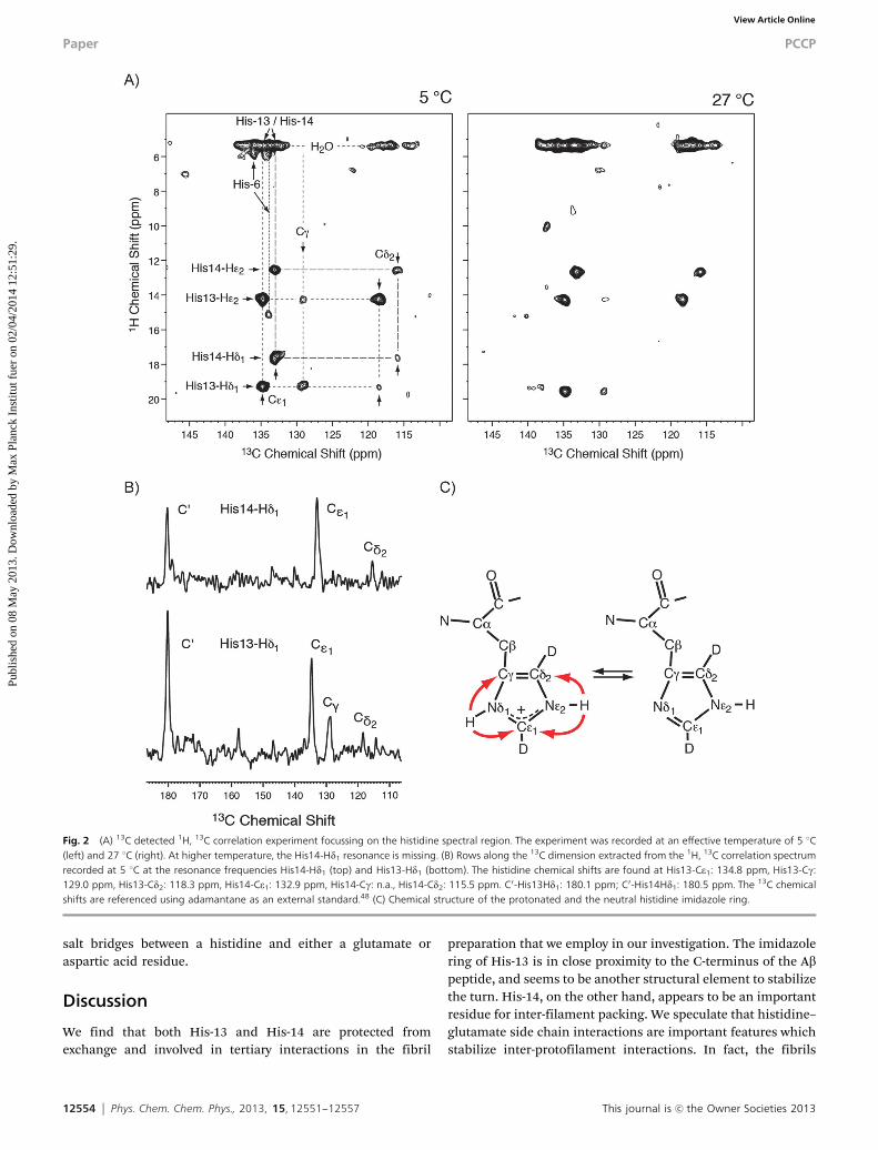
Cross peaks in the carbonyl and the aliphatic region of the 13Cdetected 1H, 13C correlation spectrum (Fig. 1D) yield the assignmentof the hydrogen bonding acceptor. For His13-Hd1, correlations to aCO (180.1 ppm) and a Ca (55.2 ppm) resonance are observed. Thecarboxyl correlation can be assigned to a contact between theimidazole proton and the carboxylic group at the C-terminus ofthe Ab peptide. This cross peak is consistent with the publishedAb(1–40) fibril models,7–10 in which a distance of 3–4 Å between theVal-40 carboxylic group and the His-13 imidazole ring was found.The assignment of the histidine aromatic carbon chemical shifts isin agreement with a recent study in which it was found that His-13,
His-40 and the Val-40 carboxylic group are involved in binding to aCu(II) metal ion.49 Assuming that His-13 and His-14 are located in ab-sheet secondary structure element, His-14 is facing the solvent andcannot be involved in hydrogen bonds within one protofilament.The i, i � 2 and i, i + 2 neighboring residues Val-12 and Lys-16 arenot potential hydrogen bonding partners. A potential acceptor forthe imidazole proton of His-14 is Glu-22 (Fig. 3). In the 1H, 13Ccorrelation spectra (Fig. 1), His14-Hd1 yields a contact with analiphatic carbon with a chemical shift of 28.0 ppm. This shift isconsistent with the Cg side chain chemical shift of a glutamate. Thepresence of this cross peak suggests that His-14 is involved in aninteraction with another protofilament.
To generate the filamentous bundle model in Fig. 3, a C2z fold-symmetric Ab structure has been assumed.7 Formation of interfila-mentous bundles, however, is not restricted to this particular fibrilpolymorph. C2x and C3z-fold9 symmetric Ab structures, as well asdifferent quarternary arrangements which differ with respect to theside chain contacts at the internal interface (F19/M35 and F20/M35in Petkova et al.7) would be compatible with the formation of
Fig. 1 (A and B) Electron microscopic images of Alzheimer’s disease Ab1–40 amyloid fibrils employed in the MAS solid-state NMR experiments. (C) 1H detected 1H, 15Nand (D) 13C detected 1H, 13C MAS solid-state NMR correlation spectra of peptide Ab1–40 fibrils using a perdeuterated peptide sample in which 50% of all exchangeableprotons are deuterated. The resonance of His14-Hd1 is not observable in the 1H, 15N correlation experiment. Both experiments are recorded setting the effectivesample temperature to 5 1C.
PCCP Paper
Publ
ishe
d on
08
May
201
3. D
ownl
oade
d by
Max
Pla
nck
Inst
itut f
uer
on 0
2/04
/201
4 12
:51:
29.
View Article Online
12554 Phys. Chem. Chem. Phys., 2013, 15, 12551--12557 This journal is c the Owner Societies 2013
salt bridges between a histidine and either a glutamate oraspartic acid residue.
Discussion
We find that both His-13 and His-14 are protected fromexchange and involved in tertiary interactions in the fibril
preparation that we employ in our investigation. The imidazolering of His-13 is in close proximity to the C-terminus of the Abpeptide, and seems to be another structural element to stabilizethe turn. His-14, on the other hand, appears to be an importantresidue for inter-filament packing. We speculate that histidine–glutamate side chain interactions are important features whichstabilize inter-protofilament interactions. In fact, the fibrils
Fig. 2 (A) 13C detected 1H, 13C correlation experiment focussing on the histidine spectral region. The experiment was recorded at an effective temperature of 5 1C(left) and 27 1C (right). At higher temperature, the His14-Hd1 resonance is missing. (B) Rows along the 13C dimension extracted from the 1H, 13C correlation spectrumrecorded at 5 1C at the resonance frequencies His14-Hd1 (top) and His13-Hd1 (bottom). The histidine chemical shifts are found at His13-Ce1: 134.8 ppm, His13-Cg:129.0 ppm, His13-Cd2: 118.3 ppm, His14-Ce1: 132.9 ppm, His14-Cg: n.a., His14-Cd2: 115.5 ppm. C0-His13Hd1: 180.1 ppm; C0-His14Hd1: 180.5 ppm. The 13C chemicalshifts are referenced using adamantane as an external standard.48 (C) Chemical structure of the protonated and the neutral histidine imidazole ring.
Paper PCCP
Publ
ishe
d on
08
May
201
3. D
ownl
oade
d by
Max
Pla
nck
Inst
itut f
uer
on 0
2/04
/201
4 12
:51:
29.
View Article Online

This journal is c the Owner Societies 2013 Phys. Chem. Chem. Phys., 2013, 15, 12551--12557 12555
that we observe in our preparation often appear to be bundlesof protofilaments.
As a consequence of conformational plasticity of b1,13,14
resonances in this region can be missing due to chemicalexchange broadening. Or, side chains might exist in differentrotameric states. Under these pre-conditions, His-13 might behydrogen bonded to Val-40 in one rotameric state. In anotherconformer, His-13 might be interacting with Glu-22. (In thisscenario, His-14 would be exchange broadened.) Differentrotamers could either be populated in different fibril poly-morphous forms. This explanation, however, seems unlikelyas the intensities of both correlations are rather similar, andthe seeding protocol should result in the enrichment of a singlefibril polymorph. Alternatively, different rotamers might bepopulated within the same polymorph. Our assignments forHis-13Ce1 and His-14Ce1 are consistent with the assignmentreported by Parthasarathy et al.,49 excluding this interpretation.The carboxyl carbons for which we observe correlations invol-ving histidines in the 13C detected 1H, 13C correlation experi-ment have rather similar chemical shifts. Previously, a 2 ppmchemical shift difference for Glu-22 CO2� and Val-40 CO2
� wasobserved.11,49 The observed carboxylic carbon chemical shifts(180.1 ppm/180.5 ppm) correspond rather to Glu-22 CO2� thanto Val-40 CO2
�. An alternative interpretation thus might imply thatboth His-13 and His-14 are both hydrogen bonded to Glu-22,potentially in different fibril polymorphs. The correlation betweenHis-13Hd1 and a Ca resonance, and the missing correlations to sidechain Cb/Cg carbons do not fit into this picture. However, dueto the low signal-to-noise ratio for the aliphatic correlation peaks(S/N C 3 : 1), this interpretation cannot be totally ruled out. Higher-dimensional experiments including an additional chemical shiftdimension need to be carried out in order to unambiguouslyresolve this question.
Recently, it was shown that His-13, His-14, Val-40 carboxyland Glu side chains are involved in Cu(II) binding.49 We suggest
that Cu(II) might compete for the hydrogen bonding interactions,resulting in a destabilization of the fibril structure. This mightexplain why addition of Cu(II) to monomeric Ab results in theformation of amorphous amyloid aggregates, and adds to theconformational variability observed for the N-terminus asdescribed above.
The hydrogen bond involving His-14 seems to be less stable,as the correlation peak in the 1H, 13C correlation disappears athigher temperature. Also, the correlation peak involving Cg ismissing for His-14 in all spectra. Similarly, the cross peakinvolving His14-Hd1 is typically very weak in the 1H, 15Ncorrelation experiments. This in agreement with the assump-tion that His-14 is more dynamic as it is involved in inter-filament interactions. Cross polarization (CP) is employed formagnetization transfer. Therefore, the intensities in the carbondetected 1H, 13C MAS solid-state NMR experiments (Fig. 2B) canbe quantitatively related to the distances between the protonin the hydrogen bonding donor and the hydrogen bondingacceptor, after taking into account a potential scaling of thedipolar interaction which can be affected due to dynamicprocesses. The His-13 imidazole-carboxyl cross peak intensitiesare stronger in comparison to the cross peaks observed withinthe imidazole ring. This is different to what we have observedpreviously for a hydrogen bond involving a tyrosine hydroxyland a carboxylic group.25 In that case, the cross peak intensitiesof the donor carbons were approximately two-fold stronger incomparison to the intensities of the acceptor carbon. Thedownfield shifted 1H imidazole resonance indicates that theproton is delocalized between the donor and the acceptorgroup, which is consistent with what has been found for modelcompounds by Limbach and co-worker.50
The pH of the fibril sample is strictly controlled at pH 7.0. Itis therefore surprising to see that both imidazole protons of thetwo histidines are observable. He2 can potentially be stabilizedby a serine or tyrosine hydroxyl group. However, there are no
Fig. 3 Left: structural model of Ab40 fibrils with C2z local symmetry, according to Petkova et al.7 (PDB code: 2LMN). Right: hypothetical arrangement of adi-protofilamentous bundle stabilized by hydrogen bonding interactions between His-14 and Glu-22. Interfilamentous bundling is compatible with fibrilpolymorphism, as the formation of salt bridges between a histidine and either a glutamate or aspartic acid residue requires only that the sheet b1 is located onthe surface of a protofilament.
PCCP Paper
Publ
ishe
d on
08
May
201
3. D
ownl
oade
d by
Max
Pla
nck
Inst
itut f
uer
on 0
2/04
/201
4 12
:51:
29.
View Article Online

12556 Phys. Chem. Chem. Phys., 2013, 15, 12551--12557 This journal is c the Owner Societies 2013
long range correlations detectable for either His-13 He2 or His-14He2, indicating that the imidazole ring might be stabilized by awater molecule. This is confirmed by correlations between waterand Ce1 and Cd2 in the 13C detected 1H, 13C correlation experi-ments. At this end, it is not possible to differentiate if thesepeaks are due to direct correlations between water and theprotein, or whether the cross peaks are exchange mediated.The positive charge of the imidazole ring is largely compensatedby the negatively charged glutamic acid carboxylic group.Additionally, the imidazole ring might experience charge com-pensation by buffer anions,51 which seems plausible given theabove discussed water accessibility.
We use here exactly the same fibril preparation protocol asin Lopez del Amo et al.11 The fibrils show a very similar morphologyusing electron microscopy. However, spectra are not easily super-imposable even taking the deuterium isotope induced chemicalshift changes into account. As in the case for assignment of thebackbone amide resonances in perdeuterated Ab fibrils,23 only oneset of resonances is observed. We attribute this observation to adifferential thermodynamic stability of protonated and deuteratedamyloid fibrils. In fact, substitution of protons with deuterium atexchangeable sites induces formation of slightly stronger hydrogenbonds.52 As amyloid aggregates are stabilized by hydrogen bonds,D2O should shift the equilibrium towards the aggregated form ofthe protein.53 At the same time, C–D bonds are approximately 10�stronger in comparison to C–H bonds, resulting in a slightly shorterbond length in the case of C–D. Introduction of deuterium at non-exchangeable hydrogen positions appears to decrease non-polarinteractions.54 This is in agreement with the observation that theperdeuterated Ab peptide elutes slightly earlier on a reverse phasecolumn in comparison to the protonated peptide (data not shown).The same seeds and seeding protocols have been employed togenerate the protonated and the deuterated amyloid fibril sample.All other experimental parameters are tightly controlled and keptthe same in both preparations. Deuteration of exchangeablesites thus yields an increase in stability, whereas deuteration ofnon-exchangeable aliphatic side chains results in a destabiliza-tion of the aggregate. The relative contribution of both effects isdifficult to estimate quantitatively. It seems likely, however, thateven small differences in the folding energy landscape can resultin a change in the amyloid fibril morphology.
Taken together, we have shown that histidine–glutamatehydrogen bonding interactions can be an important driving forcefor Ab fibril inter-filament packing. More experiments are neededto define the exact geometry around the histidines involved inquarternary contacts. In the future, it will be of particular interestto see how the hydrogen bonding pattern is changed if Aboligomeric assemblies are investigated.55–57 These experimentsare currently in progress in our laboratory.
Acknowledgements
We are grateful to Robert Tycko for providing us with the pdbcoordinate file of the Ab(1–40) fibril structure prior to publication inthe PDB. This work was performed in the framework of SFB 1035(German Research Foundation DFG), Sonderforschungsbereich
1035, Projekt B07. R. L. was a Kekule scholar and acknowledgesfinancial support by the Verband der ChemischenIndustrie (VCI).
References
1 D. J. Selkoe, Nature, 1999, 399, A23–A31.2 Y. Qi-Takahara, M. Morishima-Kawashima, Y. Tanimura,
G. Dolios, N. Hirotani, Y. Horikoshi, F. Kametani, M. Maeda,T. C. Saido, R. Wang and Y. Ihara, J. Neurosci., 2005, 25, 436–445.
3 G. Zhao, M. Z. Cui, G. Mao, Y. Dong, J. Tan, L. Sun andX. Xu, J. Biol. Chem., 2005, 280, 37689–37697.
4 C. S. Goldsbury, S. Wirtz, S. A. Muller, S. Sunderji, P. Wicki,U. Aebi and P. Frey, J. Struct. Biol., 2000, 130, 217–231.
5 P. T. Lansbury Jr., P. R. Costa, J. M. Griffiths, E. J. Simon,M. Auger, K. Halverson, D. A. Kocisko, A. S. Hendsch,T. T. Ashburn, R. G. S. Spenser, B. Tidor and R. G. Griffin,Nat. Struct. Biol., 1995, 2, 990–998.
6 T. S. Burkoth, T. L. S. Benzinger, V. Urban, D. M. Morgan,D. M. Gregory, P. Thiyagarajan, R. E. Botto, S. C. Meredithand D. G. Lynn, J. Am. Chem. Soc., 2000, 122, 7883–7889.
7 A. T. Petkova, W.-M. Yau and R. Tycko, Biochemistry, 2006,45, 498–512.
8 R. Tycko, Q. Rev. Biophys., 2006, 39, 1–55.9 A. K. Paravastu, R. D. Leapman, W.-M. Yau and R. Tycko,
Proc. Natl. Acad. Sci. U. S. A., 2008, 105, 18349–18354.10 I. Bertini, L. Gonnelli, C. Luchinat, J. Mao and A. Nesi, J. Am.
Chem. Soc., 2011, 133, 16013–16022.11 J. M. Lopez del Amo, M. Schmidt, U. Fink, M. Dasari,
M. Fandrich and B. Reif, Angew. Chem., Int. Ed., 2012, 51,6136–6139.
12 M. Schmidt, C. Sachse, W. Richter, C. Xu, M. Fandrich andN. Grigorieff, Proc. Natl. Acad. Sci. U. S. A., 2009, 106,19813–19818.
13 R. B. Kodali, A. D. Williams, S. Chemuru and R. Wetzel,J. Mol. Biol., 2010, 401, 503–517.
14 C. Sachse, M. Fandrich and N. Grigorieff, Proc. Natl. Acad.Sci. U. S. A., 2008, 105, 7462–7466.
15 W. Qiang, W.-M. Yau, Y. Luo, M. P. Mattson and R. Tycko,Proc. Natl. Acad. Sci. U. S. A., 2012, 109, 4443–4448.
16 A. T. Petkova, G. Buntkowsky, F. Dyda, R. D. Leapman,W. M. Yau and R. Tycko, J. Mol. Biol., 2004, 335, 247–260.
17 V. Chevelkov, K. Rehbein, A. Diehl and B. Reif, Angew.Chem., Int. Ed., 2006, 45, 3878–3881.
18 V. Agarwal and B. Reif, J. Magn. Reson., 2008, 194, 16–24.19 U. Akbey, S. Lange, T. W. Franks, R. Linser, A. Diehl,
B. J. van Rossum, B. Reif and H. Oschkinat, J. Biomol.NMR, 2010, 46, 67–73.
20 S. Asami, P. Schmieder and B. Reif, J. Am. Chem. Soc., 2010,132, 15133–15135.
21 P. Schanda, B. H. Meier and M. Ernst, J. Am. Chem. Soc.,2010, 132, 15957–15967.
22 J. R. Lewandowski, J. N. Dumez, U. Akbey, S. Lange, L. Emsleyand H. Oschkinat, J. Phys. Chem. Lett., 2011, 2, 2205–2211.
23 R. Linser, M. Dasari, M. Hiller, V. Higman, U. Fink,J.-M. Lopez del Amo, L. Handel, B. Kessler, P. Schmieder,
Paper PCCP
Publ
ishe
d on
08
May
201
3. D
ownl
oade
d by
Max
Pla
nck
Inst
itut f
uer
on 0
2/04
/201
4 12
:51:
29.
View Article Online

This journal is c the Owner Societies 2013 Phys. Chem. Chem. Phys., 2013, 15, 12551--12557 12557
D. Oesterhelt, H. Oschkinat and B. Reif, Angew. Chem., Int.Ed., 2011, 50, 4508–4512.
24 D. H. Zhou, A. J. Nieuwkoop, D. A. Berthold, G. Comellas,L. J. Sperling, M. Tang, G. J. Shah, E. J. Brea, L. R. Lemkauand C. M. Rienstra, J. Biomol. NMR, 2012, 54, 291–305.
25 V. Agarwal, R. Linser, U. Fink, K. Faelber and B. Reif, J. Am.Chem. Soc., 2010, 132, 3187–3195.
26 L. Chen, J. M. Kaiser, T. Polenova, J. Yang, C. M. Rienstraand L. J. Mueller, J. Am. Chem. Soc., 2007, 129, 10650–10651.
27 R. Linser, U. Fink and B. Reif, J. Magn. Reson., 2008, 193, 89–93.28 Y. Tian, L. Chen, D. Niks, J. M. Kaiser, J. Lai, C. M. Rienstra,
M. F. Dunn and L. J. Mueller, Phys. Chem. Chem. Phys., 2009,11, 7078–7086.
29 R. Linser, U. Fink and B. Reif, J. Biomol. NMR, 2010, 47, 1–6.30 R. Linser, B. Bardiaux, V. Higman, U. Fink and B. Reif, J. Am.
Chem. Soc., 2011, 133, 5905–5912.31 M. Huber, S. Hiller, P. Schanda, M. Ernst, A. Bockmann,
R. Verel and B. H. Meier, ChemPhysChem, 2011, 12, 915–918.32 V. Chevelkov, U. Fink and B. Reif, J. Biomol. NMR, 2009, 45,
197–206.33 A. Lange, S. Luca and M. Baldus, J. Am. Chem. Soc., 2002,
124, 9704–9705.34 B. Reif, B. J. van Rossum, F. Castellani, K. Rehbein, A. Diehl
and H. Oschkinat, J. Am. Chem. Soc., 2003, 125, 1488–1489.35 M. J. Bayro, G. T. Debelouchina, M. T. Eddy, N. R. Birkett,
C. E. MacPhee, M. Rosay, W. E. Maas, C. M. Dobson andR. G. Griffin, J. Am. Chem. Soc., 2011, 133, 13967–13974.
36 M. Carrio, N. Gonzalez-Montalban, A. Vera, A. Villaverdeand S. Ventura, J. Mol. Biol., 2005, 347, 1025–1037.
37 L. Wang, S. K. Maji, M. R. Sawaya, D. Eisenberg and R. Riek,PLoS Biol., 2008, 6, e195.
38 D. B. Teplow, Methods Enzymol., 2006, 413, 20–33.39 L. Hou, I. Kang, R. E. Marchant and M. G. Zagorski, J. Biol.
Chem., 2002, 277, 40173–40176.40 R. Linser, V. Chevelkov, A. Diehl and B. Reif, J. Magn. Reson.,
2007, 189, 209–216.41 N. P. Wickramasinghe, S. Parthasarathy, C. R. Jones,
C. Bhardwaj, F. Long, M. Kotecha, S. Mehboob,
L. W. Fung, J. Past, A. Samoson and Y. Ishii, Nat. Methods,2009, 6, 215–218.
42 R. Linser, U. Fink and B. Reif, J. Am. Chem. Soc., 2010, 132,8891–8893.
43 V. Agarwal, A. Diehl, N. Skrynnikov and B. Reif, J. Am. Chem.Soc., 2006, 128, 12620–12621.
44 M. Huber, O. With, P. Schanda, R. Verel, M. Ernst andB. H. Meier, J. Magn. Reson., 2012, 214, 76–80.
45 W. W. Bachovchin, Biochemistry, 1986, 25, 7751–7759.46 E. L. Ash, J. L. Sudmeier, E. C. D. Fabo and
W. W. Bachovchin, Science, 1997, 278, 1128–1132.47 A. T. Petkova, Y. Ishii, J. J. Balbach, O. N. Antzutkin,
R. D. Leapman, F. Delaglio and R. Tycko, Proc. Natl. Acad.Sci. U. S. A., 2002, 99, 16742–16747.
48 C. R. Morcombe and K. W. Zilm, J. Magn. Reson., 2003, 162,479–486.
49 S. Parthasarathy, F. Long, Y. Miller, Y. L. Xiao, D. McElheny,K. Thurber, B. Y. Ma, R. Nussinov and Y. Ishii, J. Am. Chem.Soc., 2011, 133, 3390–3400.
50 P. M. Tolstoy, J. Guo, B. Koeppe, N. S. Golubev,G. S. Denisov, S. N. Smirnov and H.-H. Limbach, J. Phys.Chem. A, 2010, 114, 10775–10782.
51 S. Narayanan and B. Reif, Biochemistry, 2005, 44, 1444–1452.52 A. Hattori, H. L. Crespi and J. J. Katz, Biochemistry, 1965, 4,
1213–1225.53 P. A. Baghurst, L. W. Nichol and W. H. Sawyer, J. Biol. Chem.,
1972, 247, 3199–3204.54 M. Turowski, N. Yamakawa, J. Meller, K. Kimata,
T. Ikegami, K. Hosoya, N. Tanaka and E. R. Thornton,J. Am. Chem. Soc., 2003, 125, 13836–13849.
55 S. Chimon, M. A. Shaibat, C. R. Jones, D. C. Calero, B. Aizeziand Y. Ishii, Nat. Struct. Mol. Biol., 2007, 14, 1157–1164.
56 M. Ahmed, J. Davis, D. Aucoin, T. Sato, S. Ahuja, S. Aimoto,J. I. Elliott, W. E. Van Nostrand and S. O. Smith, Nat. Struct.Mol. Biol., 2010, 17, 561–568.
57 J.-M. Lopez del Amo, M. Dasari, U. Fink, G. Grelle,E. E. Wanker, J. Bieschke and B. Reif, J. Mol. Biol., 2012,421, 517–524.
PCCP Paper
Publ
ishe
d on
08
May
201
3. D
ownl
oade
d by
Max
Pla
nck
Inst
itut f
uer
on 0
2/04
/201
4 12
:51:
29.
View Article Online