rp-hplc method development and validation of … hplc, method development, validation, reverse...
TRANSCRIPT

Yasodha A et al / Int. J. of Pharmacy and Analytical Research Vol-6(1) 2017 [018-038]
www.ijpar.com
~18~
IJPAR |Vol.6 | Issue 1 | Jan - Mar -2017 Journal Home page: www.ijpar.com
Research article Open Access
RP-HPLC method development and validation of Rilpivirine
Dr.A.Yasodha*1, J. Rani
1, G.Venkataih
1, A.Sivakumar
2
1Dhanvanthri College of Pharmaceutical Sciences, Mahabubnagar- 509002, Telangana, India.
2AurobindoPharma Limited, Unit –VII, Jadcherla, Hyderabad.
*Corresponding Author: Dr.A.Yasodha Email: [email protected]
ABSTRACT
A simple, accurate, rapid, and stability-indicating RP-HPLC method for a Rilpivirine has been developed and
subsequently validated in commercial tablets. The proposed HPLC method utilizes Develosil ODS HG-5 RP C18,
5µm, 15cmx4.6mm and mobile phase consisting of ACN : Acetate buffer (pH=4.0) = 65:35 (v/v) at a flow rate of 1.0
ml/min. Quantitation was achieved with UV detection at 260nm. The method was validated in terms of accuracy,
precision, linearity, limits of detection, limits of quantitation, and robustness. This optimized method has been
successively applied to pharmaceutical formulation and no interference from the tablet excipients was found.
Rilpivirine drug products were subjected to acid, base, neutral hydrolysis, oxidation, dry heat, and photolytic stress
conditions and the stressed samples were analyzed by the proposed method. As the proposed RP-HPLC method could
effectively separate the drugs from its degradation products, it can be employed as stability-indicating method for the
determination of instability of these drugs in bulk and pharmaceutical dosage form.
Keywords: HPLC, Method development, Validation, Reverse Phase, Rilpivirine.
INTRODUCTION
The number of drugs introduced into the market
is increasing every year. These drugs may be either
new entities or partial structural modification of the
existing one [1]. Very often there is a time lag from
the date of introduction of a drug into the market to
the date of its inclusion in pharmacopoeias. This
happens because of the possible uncertainties in the
continuous and wider usage of these drugs, reports
of new toxicities (resulting in their withdrawal
from the market), development of patient resistance
and introduction of better drugs by competitors.
Under these conditions, standards and analytical
procedures for these drugs may not be available in
the pharmacopoeias. It becomes necessary,
therefore to develop newer analytical methods for
such drugs [2-5].
Chromatographic techniques [6-8] are dynamic
processes wherein two mutually immiscible phases
are brought into contact; one phase is stationary
and the other mobile phase. A liquid mobile phase
is pumped under pressure through a stainless steel
column containing particles of stationary phase
ISSN:2320-2831
Yasodha A et al / Int. J. of Pharmacy and Analytical Research Vol-6(1) 2017 [018-038]
www.ijpar.com
~19~
with a diameter of 3-10 µm. The analyte is loaded
onto the head of the column via a loop valve and
separation of a mixture occurs according to the
relative lengths of time spent by its components in
the stationary phase. Components with the least
affinity for the stationary phase emerge or elute
first whereas the components with greater affinity
for stationary phase elute last. Monitoring of the
column effluent can be carried out with a variety of
detectors. The aim of the proposed method is to
develop simple and accurate methods for the
determination of Rilpivirine by RP-HPLC method
in pharmaceutical dosage forms. This new method
was successfully developed and validated as per
ICH guidelines [9-10], can be utilized for the
validation of Rilpivirine in pharmaceutical dosage
forms.
Rilpivirine is non-nucleoside reverse
transcriptase inhibitor (NNRTI) which is used for
the treatment of HIV-1 infections in treatment-
naive patients. It is a diarylpyrimidine, a class of
molecules that resemble pyrimidine nucleotides
found in DNA. Because of its flexible chemical
structure, resistance of rilpivirine is less likely to
develop than other NNRTI’s. FDA approved on
May 20, 2011.
IUPAC Name 4-{[4-({4-[(1E)-2-cyanoeth-1-en-1-yl]-2,6dimethylphenyl}amino)pyrimidin-2-yl]amino}
benzonitrile
Chemical Formula C22H18N6
MATERIALS AND METHODS
Table 2.1: List of chemicals and equipemnets
Chemicals Equipments
Ammonium acetate Analytical Balance
Dimethyl Sulfoxide(DMSO) Sonicator
HPLC grade Water HPLC
UV-spectrophotometer
Standard & sample preparation for UV-
spectrophotometer analysis
25 mg of Rilpivirine standard was transferred
into 25 ml volumetric flask, dissolved in mobile
phase & make up to volume with mobile phase.
Further dilution was done by transferring 4 ml of
the above solution into a 10ml volumetric flask and
make up to volume with mobile phase.
The standard & sample stock solutions were
prepared separately by dissolving standard &
sample in a solvent in mobile phase diluting with
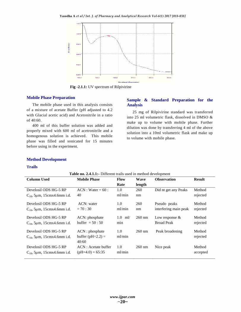
the same solvent.(After optimization of all
conditions) for UV analysis. It scanned in the UV
spectrum in the range of 200 to 400nm. This has
been performed to know the maxima of Rilpivirine,
so that the same wave number can be utilized in
HPLC UV detector for estimating the Rilpivirine.
While scanning the Rilpivirine solution we
observed the absorption maxima was 260 nm. The
UV spectrum has been recorded on Elico, corp.
make UV – Vis spectophotometer model UV-2450.
The scanned UV spectrum is attached in
Yasodha A et al / Int. J. of Pharmacy and Analytical Research Vol-6(1) 2017 [018-038]
www.ijpar.com
~20~
Fig -2.1.1: UV spectrum of Rilpivirine
Mobile Phase Preparation
The mobile phase used in this analysis consists
of a mixture of acetate Buffer (pH adjusted to 4.2
with Glacial acetic acid) and Acetonitrile in a ratio
of 40:60.
400 ml of this buffer solution was added and
properly mixed with 600 ml of acetronitrile and a
homogenous solution is achieved. This mobile
phase was filled and sonicated for 15 minutes
before using in the experiment.
Sample & Standard Preparation for the
Analysis
25 mg of Rilpivirine standard was transferred
into 25 ml volumetric flask, dissolved in DMSO &
make up to volume with mobile phase. Further
dilution was done by transferring 4 ml of the above
solution into a 10ml volumetric flask and make up
to volume with mobile phase.
Method Development
Trails
Table no. 2.4.1.1:- Different trails used in method development
Column Used Mobile Phase Flow
Rate
Wave
length
Observation Result
Develosil ODS HG-5 RP
C18, 5m, 15cmx4.6mm i.d.
ACN : Water = 60 :
40
1.0
ml/min
260
nm
Did nt get any Peaks Method
rejected
Develosil ODS HG-5 RP
C18, 5m, 15cmx4.6mm i.d.
ACN: water
= 70 : 30
1.0
ml/min
260
nm
Pseudo peaks
interfering main peak
Method
rejected
Develosil ODS HG-5 RP
C18, 5m, 15cmx4.6mm i.d.
ACN: phosphate
buffer = 50 : 50
1.0 ml/
min
260 nm Low response &
Broad Peak
Method
rejected
Develosil ODS HG-5 RP
C18, 5m, 15cmx4.6mm i.d.
ACN : phosphate
buffer (pH=2.2) =
40:60
1.0
ml/min
260 nm Peak broadening Method
rejected
Develosil ODS HG-5 RP
C18, 5m, 15cmx4.6mm i.d.
ACN : Acetate buffer
(pH=4.0) = 65:35
1.0
ml/min
260 nm Nice peak Method
accepted
Yasodha A et al / Int. J. of Pharmacy and Analytical Research Vol-6(1) 2017 [018-038]
www.ijpar.com
~21~
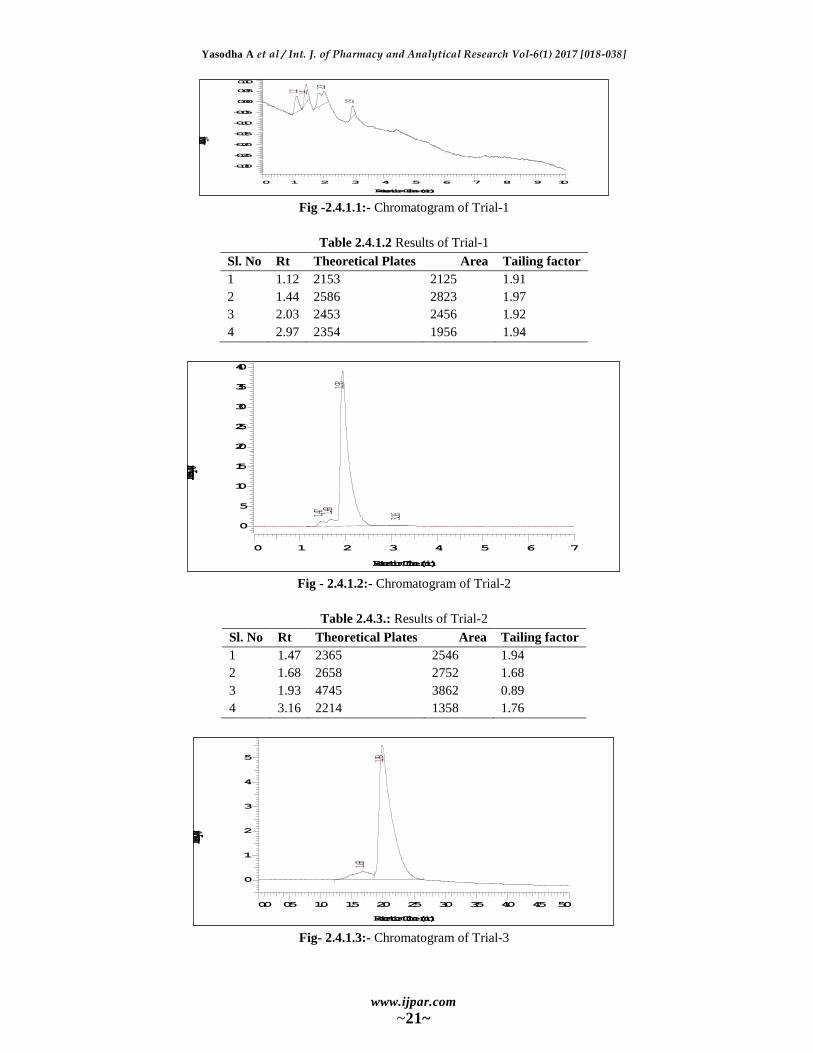
Fig -2.4.1.1:- Chromatogram of Trial-1
Table 2.4.1.2 Results of Trial-1
Sl. No Rt Theoretical Plates Area Tailing factor
1 1.12 2153 2125 1.91
2 1.44 2586 2823 1.97
3 2.03 2453 2456 1.92
4 2.97 2354 1956 1.94
Fig - 2.4.1.2:- Chromatogram of Trial-2
Table 2.4.3.: Results of Trial-2
Sl. No Rt Theoretical Plates Area Tailing factor
1 1.47 2365 2546 1.94
2 1.68 2658 2752 1.68
3 1.93 4745 3862 0.89
4 3.16 2214 1358 1.76
Fig- 2.4.1.3:- Chromatogram of Trial-3
1.12 1.44
2.03
2.97
0 1 2 3 4 5 6 7 8 9 10
Retention Time (min)
-0.30
-0.25
-0.20
-0.15
-0.10
-0.05
0.00
0.05
0.10
Intensity (mV)
1.47 1.681.93
3.16
0 1 2 3 4 5 6 7
Retention Time (min)
0
5
10
15
20
25
30
35
40
Intensity (mV)
1.68
1.99
0.0 0.5 1.0 1.5 2.0 2.5 3.0 3.5 4.0 4.5 5.0
Retention Time (min)
0
1
2
3
4
5
Intensity (mV)
Yasodha A et al / Int. J. of Pharmacy and Analytical Research Vol-6(1) 2017 [018-038]
www.ijpar.com
~22~
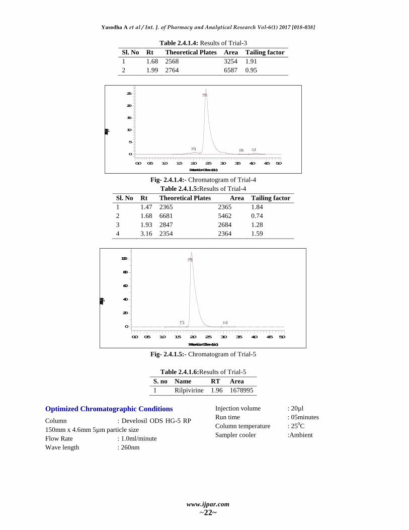
Table 2.4.1.4: Results of Trial-3
Sl. No Rt Theoretical Plates Area Tailing factor
1 1.68 2568 3254 1.91
2 1.99 2764 6587 0.95
Fig- 2.4.1.4:- Chromatogram of Trial-4
Table 2.4.1.5:Results of Trial-4
Sl. No Rt Theoretical Plates Area Tailing factor
1 1.47 2365 2365 1.84
2 1.68 6681 5462 0.74
3 1.93 2847 2684 1.28
4 3.16 2354 2364 1.59
Fig- 2.4.1.5:- Chromatogram of Trial-5
Table 2.4.1.6:Results of Trial-5
S. no Name RT Area
1 Rilpivirine 1.96 1678995
Optimized Chromatographic Conditions
Column : Develosil ODS HG-5 RP
150mm x 4.6mm 5µm particle size
Flow Rate : 1.0ml/minute
Wave length : 260nm
Injection volume : 20µl
Run time : 05minutes
Column temperature : 250C
Sampler cooler :Ambient
2.05
2.43
3.69 4.11
0.0 0.5 1.0 1.5 2.0 2.5 3.0 3.5 4.0 4.5 5.0
Retention Time (min)
0
5
10
15
20
25
Intensity (mV)
1.70
1.96
3.11
0.0 0.5 1.0 1.5 2.0 2.5 3.0 3.5 4.0 4.5 5.0
Retention Time (min)
0
20
40
60
80
100
Intensity (mV)

Yasodha A et al / Int. J. of Pharmacy and Analytical Research Vol-6(1) 2017 [018-038]
www.ijpar.com
~23~
Fig -2.4.2.1:- Chromatogram for blank
Fig-2.4.2.2: HPLC spectrum of Rilpivirine (40 ppm) in optimized conditions (RT 1.96 min.)
Table no 2.4.2.1: Peak results of optimised chromatogram
S. no Name RT Area
1 Rilpivirine 1.96 1678995
Evaluation of system suitability
Perform the blank run by injecting 5µl of
diluent and ensure that there is no interference with
the main peak retention time. Inject 5µl of the
standard solution for two times into the
chromatograph and measure the peak responses.
Calculate the % RSD for replicate injections of the
standard solution and it should be less than 2.0%.
Procedure
5µl of placebo solution is injected and ensured
that there is no interference. Then 5µl of sample
solution is injected into the chromatograph and
recorded the chromatograms. Now peak area
responses are measured and the average peak
responses are taken.
Calculations
Content of the drug =
Where,
At = Avg area responses of Rilpivirine obtained from
the Sample preparation
As = Avg area responses of Rilpivirine obtained from
the Standard preparation
Sc = Working standard concentration
Tc = Test sample concentration
LC = Label claim
RESULTS
Forced Degradation Studies
Following protocol was strictly adhered to for
forced degradation of Rilpivirine Active
Pharmaceutical Ingredient (API). The API
(Rilpivirine) was subjected to stress conditions in
various ways to observe the rate and extent of
degradation that is likely to occur in the course of
storage and/or after administration to body.
1.58
1.93
3.75
0.0 0.5 1.0 1.5 2.0 2.5 3.0 3.5 4.0 4.5 5.0
Retention Time (min)
0.0
0.2
0.4
0.6
0.8
1.0
1.2
Intensity (mV)
1.70
1.96
3.11
0.0 0.5 1.0 1.5 2.0 2.5 3.0 3.5 4.0 4.5 5.0
Retention Time (min)
0
20
40
60
80
100
Intensity (mV)
Yasodha A et al / Int. J. of Pharmacy and Analytical Research Vol-6(1) 2017 [018-038]
www.ijpar.com
~24~
This is one type of accelerated stability studies
that helps us determining the fate of the drug that is
likely to happen after along time storage, within a
very short time as compare to the real time or long
term stability testing.
The various degradation pathways studied are
acid hydrolysis, basic hydrolysis, thermal
degradation and oxidative degradation.
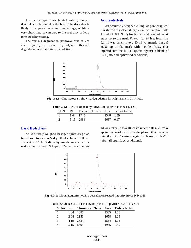
Acid hydrolysis
An accurately weighed 25 mg. of pure drug was
transferred to a clean & dry 25 ml volumetric flask.
To which 0.1 N Hydrochloric acid was added &
make up to the mark & kept for 24 hrs. from that
0.1 ml was taken in to a 10 ml volumetric flask &
make up to the mark with mobile phase, then
injected into the HPLC system against a blank of
HCl ( after all optimized conditions).
Fig -3.2.1: Chromatogram showing degradation for Rilpivirine in 0.1 N HCl
Table-3.2.1: Results of acid hydrolysis of Rilpivirine in 0.1 N HCL
Sl. No Rt Theoretical Plates Area Tailing factor
1 1.64 1745 2548 1.59
2 5.15 2934 5687 0.17
Basic Hydrolysis
An accurately weighed 10 mg. of pure drug was
transferred to a clean & dry 10 ml volumetric flask.
To which 0.1 N Sodium hydroxide was added &
make up to the mark & kept for 24 hrs. from that 4s
ml was taken in to a 10 ml volumetric flask & make
up to the mark with mobile phase, then injected
into the HPLC system against a blank of NaOH
(after all optimized conditions).
Fig -3.3.1: Chromatogram showing degradation related impurity in 0.1 N NaOH
Table-3.3.2: Results of basic hydrolysis of Rilpivirine in 0.1 N NaOH
Sl. No Rt Theoretical Plates Area Tailing factor
1 1.64 1685 2365 1.68
2 2.04 2156 2658 1.29
3 4.19 2654 2864 1.75
4 5.15 5698 4985 0.59
1.64
5.15
0 1 2 3 4 5 6 7 8 9 10
Retention Time (min)
0
10
20
30
40
50
60
70
Intensity (mV)
1.64 2.04 4.19
5.13
0 1 2 3 4 5 6 7 8 9 10
Retention Time (min)
0
10
20
30
40
50
60
70
Intensity (mV)

Yasodha A et al / Int. J. of Pharmacy and Analytical Research Vol-6(1) 2017 [018-038]
www.ijpar.com
~25~
Thermal Degradation
An accurately weighed 10 mg. of pure drug was
transferred to a clean & dry 100 ml volumetric
flask, make up to the mark with mobile phase &
was maintained at 50 0C. for 24 hrs then injected
into the HPLC system against a blank of mobile
phase ( after all optimized conditions).
Fig -3.4.1: Chromatogram showing thermal degradation studies
Table-3.4.2: Results of thermal degradation of Rilpivirine
Sl. No Rt Theoretical Plates Area Tailing factor
1 2.15 2416 2365 1.37
2 3.26 2568 2484 1.28
3 3.99 2654 2657 1.49
4 4.50 6598 6254 0.73
Photolytic degradation
Approximately 10 mg. of pure drug was taken
in a clean & dry Petridis. It was kept in a UV
cabinet at 254 nm wavelength for 24 hours without
interruption. Accurately weighed 1 mg. of the UV
exposed drug was transferred to a clean & dry 10
ml. volumetric flask. First the UV exposed drug
was dissolved in methanol & make up to the mark
than injected into the HPLC system against a blank
of mobile phase (after all optimized conditions).
Fig -3.5.1: Chromatogram showing photolytic degradation.
Table-3.5.1: Results of photolytic degradation of Rilpivirine
Sl. No Rt Theoretical Plates Area Tailing factor
1 2.15 2145 2365 1.38
2 3.26 2365 2654 1.49
3 3.99 2564 2846 1.53
4 4.50 6954 5942 0.81
2.15 3.26 3.99
4.50
0 1 2 3 4 5 6 7 8 9 10
Retention Time (min)
0
20
40
60
80
100
120
140
Intensity (mV)
2.15 3.26 3.99
4.50
0 1 2 3 4 5 6 7 8 9 10
Retention Time (min)
0
20
40
60
80
100
120
140
Intensity (mV)
Yasodha A et al / Int. J. of Pharmacy and Analytical Research Vol-6(1) 2017 [018-038]
www.ijpar.com
~26~
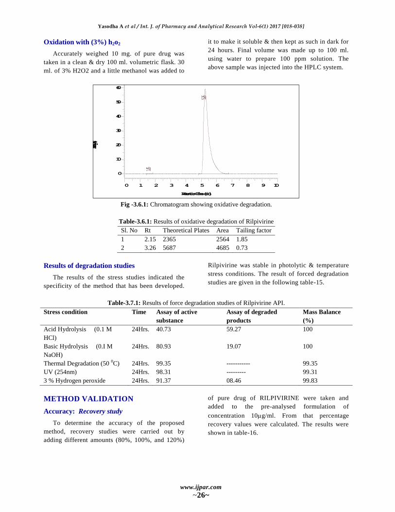
Oxidation with (3%) h2o2
Accurately weighed 10 mg. of pure drug was
taken in a clean & dry 100 ml. volumetric flask. 30
ml. of 3% H2O2 and a little methanol was added to
it to make it soluble & then kept as such in dark for
24 hours. Final volume was made up to 100 ml.
using water to prepare 100 ppm solution. The
above sample was injected into the HPLC system.
Fig -3.6.1: Chromatogram showing oxidative degradation.
Table-3.6.1: Results of oxidative degradation of Rilpivirine
Sl. No Rt Theoretical Plates Area Tailing factor
1 2.15 2365 2564 1.85
2 3.26 5687 4685 0.73
Results of degradation studies
The results of the stress studies indicated the
specificity of the method that has been developed.
Rilpivirine was stable in photolytic & temperature
stress conditions. The result of forced degradation
studies are given in the following table-15.
Table-3.7.1: Results of force degradation studies of Rilpivirine API.
Stress condition Time Assay of active
substance
Assay of degraded
products
Mass Balance
(%)
Acid Hydrolysis (0.1 M
HCl)
24Hrs. 40.73 59.27 100
Basic Hydrolysis (0.I M
NaOH)
24Hrs. 80.93 19.07 100
Thermal Degradation (50 0C) 24Hrs. 99.35 ----------- 99.35
UV (254nm) 24Hrs. 98.31 --------- 99.31
3 % Hydrogen peroxide 24Hrs. 91.37 08.46 99.83
METHOD VALIDATION
Accuracy: Recovery study
To determine the accuracy of the proposed
method, recovery studies were carried out by
adding different amounts (80%, 100%, and 120%)
of pure drug of RILPIVIRINE were taken and
added to the pre-analysed formulation of
concentration 10g/ml. From that percentage
recovery values were calculated. The results were
shown in table-16.
1.65
5.26
0 1 2 3 4 5 6 7 8 9 10
Retention Time (min)
0
10
20
30
40
50
60
Intensity (mV)
Yasodha A et al / Int. J. of Pharmacy and Analytical Research Vol-6(1) 2017 [018-038]
www.ijpar.com
~27~
Table-3.8.1: Accuracy Readings
STD Spike-1
Conc. AUC Conc AUC Diff % Recovery
8 506881 1 18 1905481 1398600 99.14396
8 506881 2 18 1900430 1393549 98.7859
8 506881 3 18 1902331 1395450 98.92066
98.95017
0.180843
0.182762
STD Spike-2
Conc. AUC Conc AUC Diff % Recovery
10 1426346 1 20 2832999 1406653 99.71482
10 1426346 2 20 2834395 1408049 99.81378
10 1426346 3 20 2833499 1407153 99.75026
99.75962
0.050139
0.05026
STD Spike-3
Conc. AUC Conc AUC Diff % Recovery
12 1999858 1 22 3401595 1401737 99.36633
12 1999858 2 22 3400499 1400641 99.28864
12 1999858 3 22 3403358 1403500 99.49131
99.38209
0.10225
0.102885
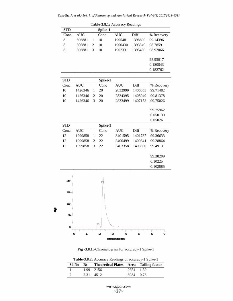
Fig -3.8.1:-Chromatogram for accuracy-1 Spike-1
Table-3.8.2: Accuracy Readings of accuracy-1 Spike-1
Sl. No Rt Theoretical Plates Area Tailing factor
1 1.99 2156 2654 1.59
2 2.31 4512 3984 0.73
1.99
2.31
0 1 2 3 4 5 6 7
Retention Time (min)
0
50
100
150
200
Intensity (mV)
Yasodha A et al / Int. J. of Pharmacy and Analytical Research Vol-6(1) 2017 [018-038]
www.ijpar.com
~28~
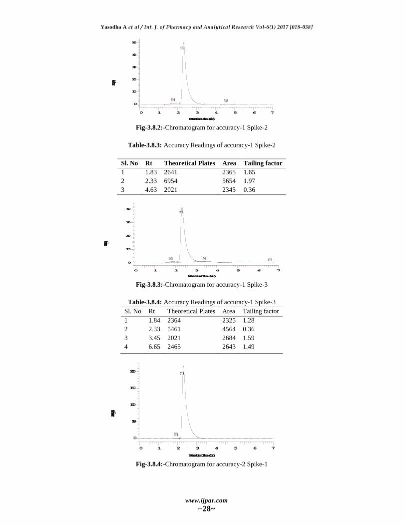
Fig-3.8.2:-Chromatogram for accuracy-1 Spike-2
Table-3.8.3: Accuracy Readings of accuracy-1 Spike-2
Sl. No Rt Theoretical Plates Area Tailing factor
1 1.83 2641 2365 1.65
2 2.33 6954 5654 1.97
3 4.63 2021 2345 0.36
Fig-3.8.3:-Chromatogram for accuracy-1 Spike-3
Table-3.8.4: Accuracy Readings of accuracy-1 Spike-3
Sl. No Rt Theoretical Plates Area Tailing factor
1 1.84 2364 2325 1.28
2 2.33 5461 4564 0.36
3 3.45 2021 2684 1.59
4 6.65 2465 2643 1.49
Fig-3.8.4:-Chromatogram for accuracy-2 Spike-1
1.83
2.33
4.63
0 1 2 3 4 5 6 7
Retention Time (min)
0
10
20
30
40
50
Intensity (mV)
1.84
2.33
3.45 6.650 1 2 3 4 5 6 7
Retention Time (min)
0
10
20
30
40
Intensity (mV)
1.99
2.31
0 1 2 3 4 5 6 7
Retention Time (min)
0
50
100
150
200
Intensity (mV)
Yasodha A et al / Int. J. of Pharmacy and Analytical Research Vol-6(1) 2017 [018-038]
www.ijpar.com
~29~
Table-3.8.5: Accuracy Readings of accuracy-2 Spike-1
Sl. No Rt Theoretical Plates Area Tailing factor
1 1.99 2685 2354 1.28
2 2.31 5684 4561 0.57
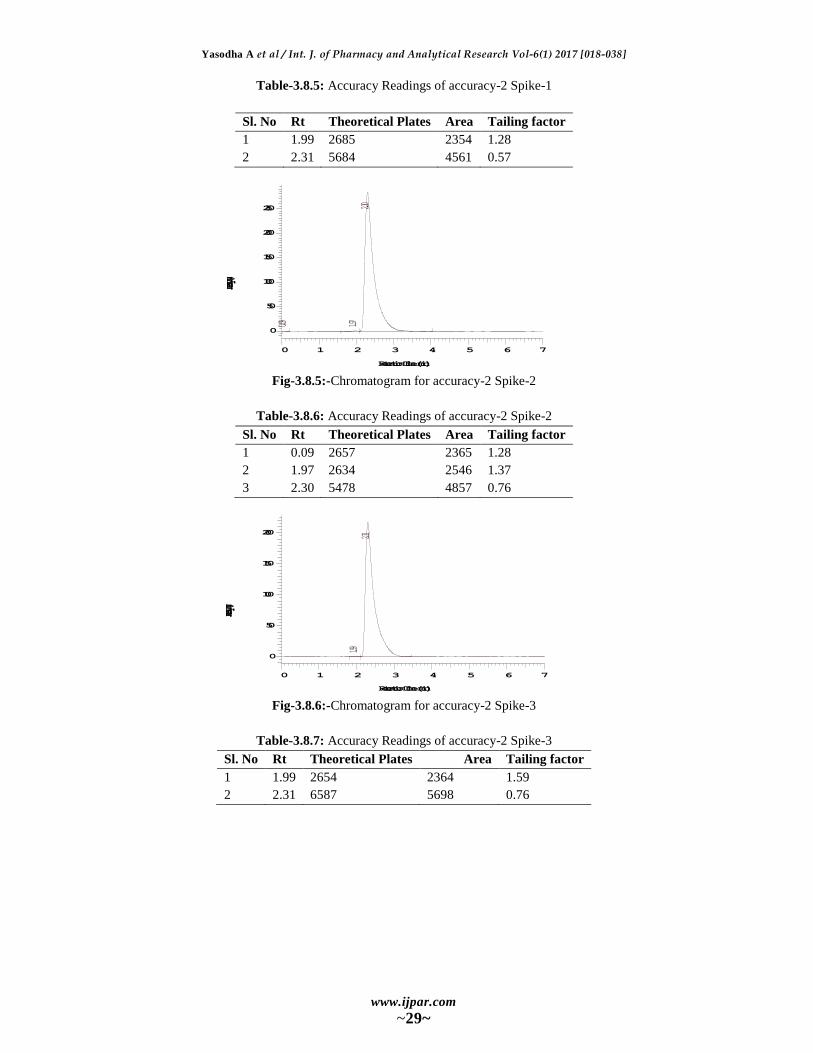
Fig-3.8.5:-Chromatogram for accuracy-2 Spike-2
Table-3.8.6: Accuracy Readings of accuracy-2 Spike-2
Sl. No Rt Theoretical Plates Area Tailing factor
1 0.09 2657 2365 1.28
2 1.97 2634 2546 1.37
3 2.30 5478 4857 0.76
Fig-3.8.6:-Chromatogram for accuracy-2 Spike-3
Table-3.8.7: Accuracy Readings of accuracy-2 Spike-3
Sl. No Rt Theoretical Plates Area Tailing factor
1 1.99 2654 2364 1.59
2 2.31 6587 5698 0.76
0.09 1.97
2.30
0 1 2 3 4 5 6 7
Retention Time (min)
0
50
100
150
200
250
Intensity (mV)
1.99
2.31
0 1 2 3 4 5 6 7
Retention Time (min)
0
50
100
150
200
Intensity (mV)
Yasodha A et al / Int. J. of Pharmacy and Analytical Research Vol-6(1) 2017 [018-038]
www.ijpar.com
~30~
Fig-3.8.7:-Chromatogram for accuracy-3 Spike-1
Table-3.8.8: Accuracy Readings of accuracy-3 Spike-1
Sl. No Rt Theoretical Plates Area Tailing factor
1 2.01 2354 2546 1.59
2 2.33 6356 5762 0.71
3 5.57 2021 2014 1.43
Fig-3.8.9:-Chromatogram for accuracy-3 Spike-2
Table-3.8.10: Accuracy Readings of accuracy-3 Spike-2
Sl. No Rt Theoretical Plates Area Tailing factor
1 2.33 6514 5642 0.89
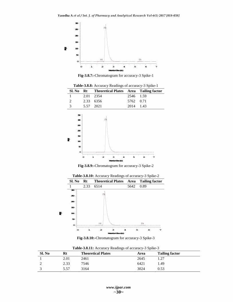
Fig-3.8.10:-Chromatogram for accuracy-3 Spike-3
Table-3.8.11: Accuracy Readings of accuracy-3 Spike-3
Sl. No Rt Theoretical Plates Area Tailing factor
1 2.01 2461 2645 1.27
2 2.33 7546 6421 1.49
3 5.57 3164 3024 0.53
2.01
2.33
5.57
0 1 2 3 4 5 6 7
Retention Time (min)
0
50
100
150
200
250
300
Intensity (mV)
2.33
0 1 2 3 4 5 6 7
Retention Time (min)
0
50
100
150
200
250
300
350
Intensity (mV)
2.01
2.33
5.57
0 1 2 3 4 5 6 7
Retention Time (min)
0
50
100
150
200
250
300
Intensity (mV)
Yasodha A et al / Int. J. of Pharmacy and Analytical Research Vol-6(1) 2017 [018-038]
www.ijpar.com
~31~
Precision
Repeatability
The precision of each method was ascertained
separately from the peak areas & retention times
obtained by actual determination of five replicates
of a fixed amount of drug. Rilpivirine (API). The
percent relative standard deviations were calculated
for Rilpivirine are presented in the table-26.
Table-3.8.2.1: Shows results of repeatability.
HPLC Injection
Replicates of Rilpivirine
Retention Time Area
Replicate – 1 2.08 833769
Replicate – 2 2.08 835768
Replicate – 3 2.08 855929
Replicate – 4 2.07 833458
Replicate – 5 2.07 848232
Average 2.076 841431.2
Standard Deviation 0.005477226 10133.98
% RSD 0.263835529 1.204374
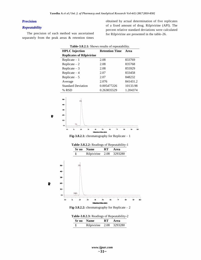
Fig-3.8.2.1: chromatography for Replicate – 1
Table-3.8.2.2: Readings of Repeatability-1
Sr no Name RT Area
1 Rilpivirine 2.08 3293280
Fig-3.8.2.2: chromatography for Replicate – 2
Table-3.8.2.3: Readings of Repeatability-2
Sr no Name RT Area
1 Rilpivirine 2.08 3293280
1.47
2.08
0 1 2 3 4 5 6 7 8 9 10
Retention Time (min)
0
10
20
30
40
50
Intensity (mV)
1.46 1.662.08
0 1 2 3 4 5 6 7 8 9 10
Retention Time (min)
0
10
20
30
40
50
Intensity (mV)
Yasodha A et al / Int. J. of Pharmacy and Analytical Research Vol-6(1) 2017 [018-038]
www.ijpar.com
~32~
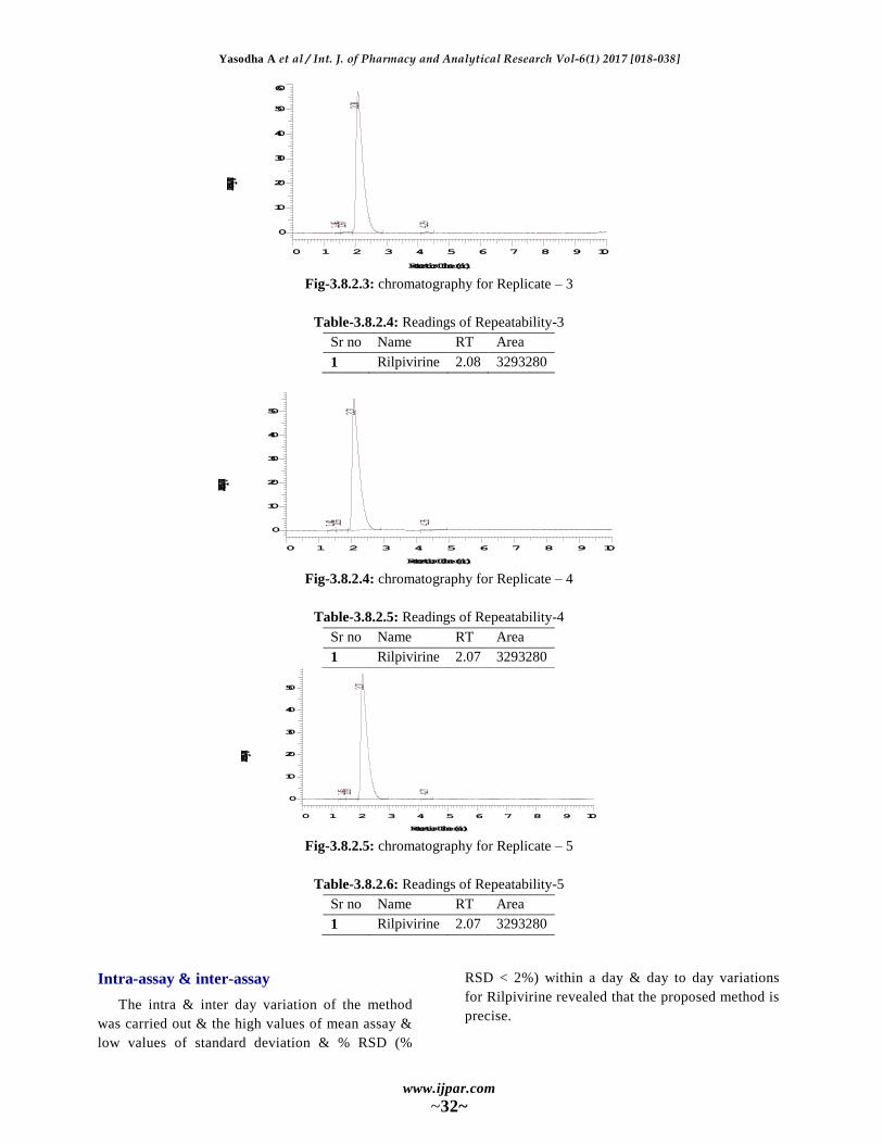
Fig-3.8.2.3: chromatography for Replicate – 3
Table-3.8.2.4: Readings of Repeatability-3
Sr no Name RT Area
1 Rilpivirine 2.08 3293280
Fig-3.8.2.4: chromatography for Replicate – 4
Table-3.8.2.5: Readings of Repeatability-4
Sr no Name RT Area
1 Rilpivirine 2.07 3293280
Fig-3.8.2.5: chromatography for Replicate – 5
Table-3.8.2.6: Readings of Repeatability-5
Sr no Name RT Area
1 Rilpivirine 2.07 3293280
Intra-assay & inter-assay
The intra & inter day variation of the method
was carried out & the high values of mean assay &
low values of standard deviation & % RSD (%
RSD < 2%) within a day & day to day variations
for Rilpivirine revealed that the proposed method is
precise.
1.46 1.672.08
4.29
0 1 2 3 4 5 6 7 8 9 10
Retention Time (min)
0
10
20
30
40
50
60
Intensity (mV)
1.44 1.65
2.07
4.35
0 1 2 3 4 5 6 7 8 9 10
Retention Time (min)
0
10
20
30
40
50
Intensity (mV)
1.45 1.63
2.07
4.27
0 1 2 3 4 5 6 7 8 9 10
Retention Time (min)
0
10
20
30
40
50
Intensity (mV)

Yasodha A et al / Int. J. of Pharmacy and Analytical Research Vol-6(1) 2017 [018-038]
www.ijpar.com
~33~
Table–3.8.2.7: Results of intra-assay & inter-assay
Conc. Of Rilpivirine (API) (µg/ml) Observed Conc. Of Rilpivirine (µg/ml) by the proposed method
Intra-Day Inter-Day
Mean (n=6) % RSD Mean (n=6) % RSD
10 9.93 0.14 10.32 0.95
30 30.63 0.78 30.14 0.16
100 99.21 0.96 99.78 0.73
Linearity & Range
The calibration curve showed good linearity in
the range of 0-25 µg/ml, for Rilpivirine (API) with
correlation coefficient (r2) of 0.994 (Fig-39). A
typical calibration curve has the regression
equation of y = 6780x + 65596 for Rilpivirine.
Fig-3.8.3.1: Calibration curve of Rilpivirine (API).
Table-3.8.3.1: Linearity Results
CONC. in ppm AUC (n=6)
0 0
50 460548
100 783769
150 1096795
200 1376884
Fig-3.8.3.2: Chromatogram for 50ppm
Table-3.8.3.2: Readings of Linearity-50 ppm
Sr no Name RT Area
1 Rilpivirine 2.07 3293280
Fig-3.8.3.3: Chromatogram for 100ppm
y = 6944x + 40998 R² = 0.9914
AU
C (
n=6
)
Conc. in ppm
Calibration curve for Rilpivirine
Series1
Linear (Series1)
1.46 1.672.08
4.29
0 1 2 3 4 5 6 7 8 9 10
Retention Time (min)
0
10
20
30
40
50
60
Intensity (mV)

Yasodha A et al / Int. J. of Pharmacy and Analytical Research Vol-6(1) 2017 [018-038]
www.ijpar.com
~34~
Table-3.8.3.3: Readings of Linearity-100 ppm
Sr no Name RT Area
1 Rilpivirine 2.08 3293280
Fig-3.8.3.4: Chromatogram for 150ppm
Table-3.8.3.4: Readings of Linearity-150 ppm
Sr no Name RT Area
1 Rilpivirine 2.07 3293280
Fig-3.8.3.5: Chromatogram for 200ppm
Table-3.8.3.5: Readings of Linearity-200 ppm
Sr no Name RT Area
1 Rilpivirine 2.05 3293280
Method Robustness
Influence of small changes in chromatographic
conditions such as change in flow rate (
0.1ml/min), Temperature (20C), Wavelength of
detection (2nm) & acetonitrile content in mobile
phase (2%) studied to determine the robustness of
the method are also in favour of (Table-38, % RSD
< 2%) the developed RP-HPLC method for the
analysis of Rilpivirine( API).
Table-3.8.4.1: Result of method robustness test
Change in parameter % RSD
Flow (1.1 ml/min) 0.06
Flow (0.9 ml/min) 0.04
Temperature (270C) 0.08
Temperature (230C) 0.11
Wavelength of Detection (202 nm) 0.03
Wavelength of detection (209 nm) 0.02
2.07
4.18
0 1 2 3 4 5 6 7 8 9 10
Retention Time (min)
0
10
20
30
40
50
60
70
Intensity (mV)
1.45
2.05
4.13 4.72
0 1 2 3 4 5 6 7 8 9 10
Retention Time (min)
0
20
40
60
80
Intensity (mV)

Yasodha A et al / Int. J. of Pharmacy and Analytical Research Vol-6(1) 2017 [018-038]
www.ijpar.com
~35~
LOD & LOQ
The Minimum concentration level at which the
analyte can be reliable detected (LOD) &
quantified (LOQ) were found to be 0.03 & 0.09
µg/ml respectively.
System Suitability Parameter
System suitability testing is an integral part of
many analytical procedures. The tests are based on
the concept that the equipment, electronics,
analytical operations and samples to be analyzed
constitute an integral system that can be evaluated
as such. Following system suitability test
parameters were established. The data are shown in
Table-39.
Table-3.8.6.1: Data of System Suitability Parameter
S.No. Parameter Limit Result
1 Resolution Rs 2 9.15
2 Asymmetry T 2 Rilpivirine=0.12
3 Theoretical plate N 2000 Rilpivirine=3246
SPECIFICITY
Preparation and running of Rilpivirine
The performance test of the method has been
conducted on market sample. As per the label
claim, each tablet contains 50mg of Rilpivirine. To
estimate this powder of the tablet equivalent to
25mg of Rilpivirine has been dissolved in 25 ml of
the mobile phase. Further dilution was done by
taking 1ml of this solution in 10ml volumetric
flask, dissolve and making up the volume upto the
mark with mobile phase by which 100ppm solution
was prepared. Again same process is repeated to
make 10ppm from 100ppm solution. To extract the
drug in the solution, it has been sonicated for 5
minutes followed by cyclo-mixing for 5 minutes.
Resulting solution was filtered by using Millipore
syringe filter (0.45 micron). Resulting clear
solution was injected in HPLC in duplicate as per
the above mentioned HPLC method.
Chromatogram obtained for the injection is shown
below with Rt of 2.69 mins.
Fig-3.8.7.1: Chromatogram for specificity sample
Table-3.8.7.1: Results for specificity sample
Sl. No Rt Theoretical Plates Area Tailing factor
1 1.54 2143 2354 1.28
2 2.69 5658 4689 1.74
3 6.08 2654 2541 0.49
1.54
2.69
6.08
0 1 2 3 4 5 6 7
Retention Time (min)
0
20
40
60
80
Intensity (mV)
Yasodha A et al / Int. J. of Pharmacy and Analytical Research Vol-6(1) 2017 [018-038]
www.ijpar.com
~36~
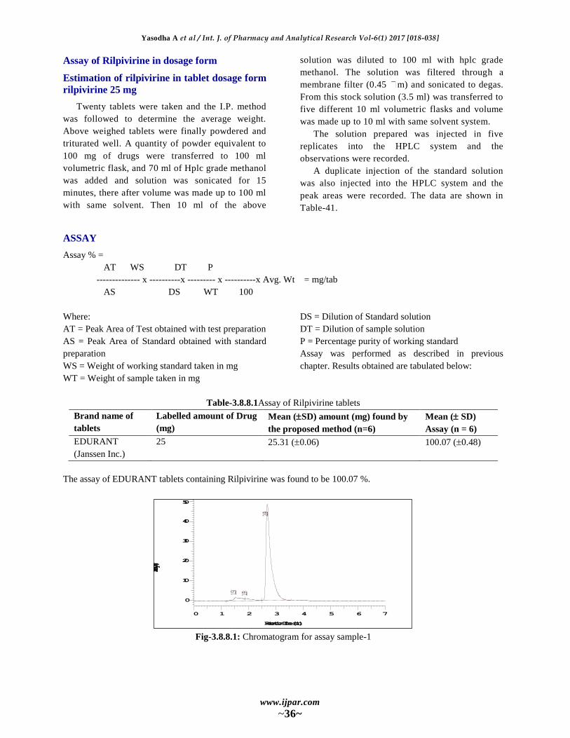
Assay of Rilpivirine in dosage form
Estimation of rilpivirine in tablet dosage form
rilpivirine 25 mg
Twenty tablets were taken and the I.P. method
was followed to determine the average weight.
Above weighed tablets were finally powdered and
triturated well. A quantity of powder equivalent to
100 mg of drugs were transferred to 100 ml
volumetric flask, and 70 ml of Hplc grade methanol
was added and solution was sonicated for 15
minutes, there after volume was made up to 100 ml
with same solvent. Then 10 ml of the above
solution was diluted to 100 ml with hplc grade
methanol. The solution was filtered through a
membrane filter (0.45 m) and sonicated to degas.
From this stock solution (3.5 ml) was transferred to
five different 10 ml volumetric flasks and volume
was made up to 10 ml with same solvent system.
The solution prepared was injected in five
replicates into the HPLC system and the
observations were recorded.
A duplicate injection of the standard solution
was also injected into the HPLC system and the
peak areas were recorded. The data are shown in
Table-41.
ASSAY
Assay % =
AT WS DT P
-------------- x ----------x --------- x ----------x Avg. Wt = mg/tab
AS DS WT 100
Where:
AT = Peak Area of Test obtained with test preparation
AS = Peak Area of Standard obtained with standard
preparation
WS = Weight of working standard taken in mg
WT = Weight of sample taken in mg
DS = Dilution of Standard solution
DT = Dilution of sample solution
P = Percentage purity of working standard
Assay was performed as described in previous
chapter. Results obtained are tabulated below:
Table-3.8.8.1Assay of Rilpivirine tablets
Brand name of
tablets
Labelled amount of Drug
(mg)
Mean (SD) amount (mg) found by
the proposed method (n=6)
Mean ( SD)
Assay (n = 6)
EDURANT
(Janssen Inc.)
25 25.31 (0.06) 100.07 (0.48)
The assay of EDURANT tablets containing Rilpivirine was found to be 100.07 %.
Fig-3.8.8.1: Chromatogram for assay sample-1
1.53 1.93
2.69
0 1 2 3 4 5 6 7
Retention Time (min)
0
10
20
30
40
50
Intensity (mV)
Yasodha A et al / Int. J. of Pharmacy and Analytical Research Vol-6(1) 2017 [018-038]
www.ijpar.com
~37~
Table-3.8.8.1: Assay chromatogram results of Rilpivirine sample-1
Sl. No Rt Theoretical Plates Area Tailing factor
1 1.53 2153 2133 1.91
2 1.93 2586 2852 1.97
3 2.69 5642 5293 0.98
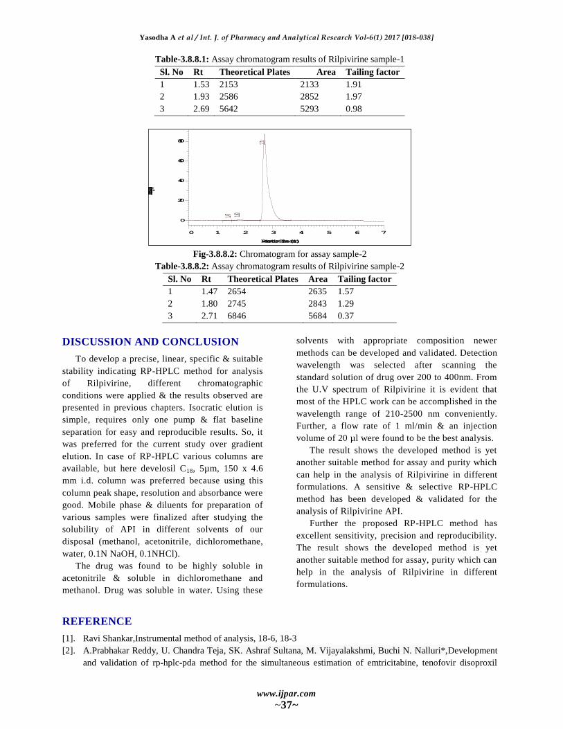
Fig-3.8.8.2: Chromatogram for assay sample-2
Table-3.8.8.2: Assay chromatogram results of Rilpivirine sample-2
Sl. No Rt Theoretical Plates Area Tailing factor
1 1.47 2654 2635 1.57
2 1.80 2745 2843 1.29
3 2.71 6846 5684 0.37
DISCUSSION AND CONCLUSION
To develop a precise, linear, specific & suitable
stability indicating RP-HPLC method for analysis
of Rilpivirine, different chromatographic
conditions were applied & the results observed are
presented in previous chapters. Isocratic elution is
simple, requires only one pump & flat baseline
separation for easy and reproducible results. So, it
was preferred for the current study over gradient
elution. In case of RP-HPLC various columns are
available, but here develosil C18, 5µm, 150 x 4.6
mm i.d. column was preferred because using this
column peak shape, resolution and absorbance were
good. Mobile phase & diluents for preparation of
various samples were finalized after studying the
solubility of API in different solvents of our
disposal (methanol, acetonitrile, dichloromethane,
water, 0.1N NaOH, 0.1NHCl).
The drug was found to be highly soluble in
acetonitrile & soluble in dichloromethane and
methanol. Drug was soluble in water. Using these
solvents with appropriate composition newer
methods can be developed and validated. Detection
wavelength was selected after scanning the
standard solution of drug over 200 to 400nm. From
the U.V spectrum of Rilpivirine it is evident that
most of the HPLC work can be accomplished in the
wavelength range of 210-2500 nm conveniently.
Further, a flow rate of 1 ml/min & an injection
volume of 20 µl were found to be the best analysis.
The result shows the developed method is yet
another suitable method for assay and purity which
can help in the analysis of Rilpivirine in different
formulations. A sensitive & selective RP-HPLC
method has been developed & validated for the
analysis of Rilpivirine API.
Further the proposed RP-HPLC method has
excellent sensitivity, precision and reproducibility.
The result shows the developed method is yet
another suitable method for assay, purity which can
help in the analysis of Rilpivirine in different
formulations.
REFERENCE
[1]. Ravi Shankar,Instrumental method of analysis, 18-6, 18-3
[2]. A.Prabhakar Reddy, U. Chandra Teja, SK. Ashraf Sultana, M. Vijayalakshmi, Buchi N. Nalluri*,Development
and validation of rp-hplc-pda method for the simultaneous estimation of emtricitabine, tenofovir disoproxil
1.47 1.80
2.71
0 1 2 3 4 5 6 7
Retention Time (min)
0
20
40
60
80
Intensity (mV)
Yasodha A et al / Int. J. of Pharmacy and Analytical Research Vol-6(1) 2017 [018-038]
www.ijpar.com
~38~
fumarate and rilpivirine hydrochloride in bulk, pharmaceutical dosage forms and in dissolution samples, Indo
American Journal of Pharmaceutical analysis, ISSN NO 2231-6876.
[3]. Uttam Prasad Panigrahy1and A. Sunil Kumar Reddy2, A novel validated RP-HPLC method for the
simultaneous estimation of Emtricitabine, Tenofovir Disoproxil Fumarate and Rilpivirine in bulk and
pharmaceutical tablet dosage forms, Scholars Research Library Der Pharmacia Lettre, 7(1), 2015, 303-314,
ISSN 0975-5071 USA CODEN: DPLEB4.
[4]. Kavitha, k. y.; geetha, g.; hariprasad, r.; venkatnarayana, r.; subramanian, g., Development and validation of rp-
hplc analytical method for simultaneous estimation of emtricitabine, rilpivirine, tenofovir disoproxil fumarate
and its pharmaceutical dosage forms, Pharmacie Globale; 1(1), 2013, 1.
[5]. Masthanamma sk* and alekhya gottumukkala, Development and validation of uv spectrophotometric methods
for estimation of rilpivirine in bulk and pharmaceutical formulation, international journal of pharmaceutical
sciences and research, ISSN (Online): 0975-8232, ISSN (Print): 2320-5148.
[6]. M. E. Swartz, journal of liquid chromatography, 28(7/8), 2005, 1253-1263
[7]. Biomedical chromatography : bmc 22(5), 2008, 469-477
[8]. Journal of chromatography .b, analytical technologies in the biomedical and life sciences. 1, 2008, 863(2), 2008,
258-265.
[9]. Ich q2b: validation of analytical procedure; methodology (international conferences on harmonization of
technical requirements for the registration of drugs for human use, Geneva, Switzerland, may 1997
[10]. Ich q2b: validation of analytical procedure; methodology (international conferences on harmonization of
technical requirements for the registration of drugs for human use, Geneva, Switzerland, 2003.