role of ns1 antibodies in the pathogenesis of acute
TRANSCRIPT

ARTICLE
Role of NS1 antibodies in the pathogenesis of acutesecondary dengue infectionDeshni Jayathilaka1, Laksiri Gomes1, Chandima Jeewandara1, Geethal.S.Bandara Jayarathna1, Dhanushka Herath1,
Pathum Asela Perera1, Samitha Fernando1, Ananda Wijewickrama2, Clare S. Hardman3, Graham S. Ogg3 &
Gathsaurie Neelika Malavige 1,3
The role of NS1-specific antibodies in the pathogenesis of dengue virus infection is poorly
understood. Here we investigate the immunoglobulin responses of patients with dengue fever
(DF) and dengue hemorrhagic fever (DHF) to NS1. Antibody responses to recombinant-NS1
are assessed in serum samples throughout illness of patients with acute secondary DENV1
and DENV2 infection by ELISA. NS1 antibody titres are significantly higher in patients with
DHF compared to those with DF for both serotypes, during the critical phase of illness.
Furthermore, during both acute secondary DENV1 and DENV2 infection, the antibody
repertoire of DF and DHF patients is directed towards distinct regions of the NS1 protein. In
addition, healthy individuals, with past non-severe dengue infection have a similar antibody
repertoire as those with mild acute infection (DF). Therefore, antibodies that target specific
NS1 epitopes could predict disease severity and be of potential benefit in aiding vaccine and
treatment design.
DOI: 10.1038/s41467-018-07667-z OPEN
1 Centre for Dengue Research, University of Sri Jayewardenepura, Nugegoda 10100, Sri Lanka. 2 National Institute of Infectious Diseases, Angoda 10250, SriLanka. 3MRC Human Immunology Unit, Weatherall Institute of Molecular Medicine, Oxford NIHR Biomedical Research Centre, Oxford OX3 9DS, UK. Theseauthors contributed equally: Deshni Jayathilaka, Laksiri Gomes. The authors jointly supervised this work: Graham S. Ogg, Gathsaurie Neelika Malavige.Correspondence and requests for materials should be addressed to G.N.M. (email: [email protected])
NATURE COMMUNICATIONS | (2018) 9:5242 | DOI: 10.1038/s41467-018-07667-z | www.nature.com/naturecommunications 1
1234
5678
90():,;

Dengue virus (DENV) is one of the most rapidly emergingviral infections worldwide, infecting 390 million indivi-duals annually. Despite such obvious need, there is cur-
rently no licensed specific drug for the treatment of thispotentially fatal disease1. A tetravalent live attenuated yellow feverand dengue chimeric vaccine has been recently licensed in somecountries, however, it has poor efficacy in naive individuals andefficacy depends on DENV serotype2. In addition, the vaccinemanufacturer recently suggested that this vaccine should beavoided in dengue naive individuals, due to the likelihood ofdisease enhancement3. One of the major challenges faced in thedevelopment of an efficacious dengue vaccine is our poorunderstanding of what constitutes a protective immune response.
DENV infections result in a spectrum of disease ranging fromsubclinical inapparent presentation, through mild dengue fever(DF) to severe dengue hemorrhagic fever (DHF), which is char-acterized by an increase in vascular permeability resulting inshock and organ dysfunction4. Understanding the molecularpathway that leads to development of vascular leak would providea major step forward in the development of effective denguetreatments. One such potential target is dengue non-structuralprotein 1 (NS1), a secreted glycoprotein used as a diagnosticmarker of dengue infection, appearing early in the serum beforeantibodies are generated5. NS1, synthesized as a monomer, formsa dimer in the ER lumen and hexamer in the serum. NS1 isthought to function as a cofactor for viral RNA replication6. NS1has been shown to trigger cytokine release and contributesdirectly to vascular leak through binding TLR4 and engaging theendothelial glycocalyx7,8. In addition, in vitro data show thatsome antibodies against NS1 cross-react with endothelial cellsand induce apoptosis, which is suggested to contribute to endo-thelial dysfunction and vascular leak9. Interestingly, NS1 isimportant for viral evasion of complement activation by bindingmannose-binding lectin and preventing neutralization of DENVvia the lectin pathway10. However, some studies have shown thatNS1 activates complement, a process that is further enhanced byNS1-specific antibodies11.
Secondary dengue infections incur an increased risk of devel-oping DHF, which supports the hypothesis that NS1-specificantibodies derived from primary infection may, upon expansionfollowing secondary challenge, play a role in diseasepathogenesis12,13. However, apoptosis of the endothelium anddeposition of immune complexes in the endothelium have notbeen demonstrated in autopsy studies of fatal dengue14. In con-trast to the data in the above studies, some investigations indengue mouse models have shown that mice injected with serafrom dengue-infected mice or monoclonal antibodies to NS1,have significantly reduced vascular leak, implying a protectiverole for antibodies to NS115. It is currently not clear if antibodiestargeted to specific regions of dengue NS1 protein may playdifferential roles in the protection or pathogenesis of dengue feverin humans.
The majority of human DENV-specific antibodies are directedagainst DENV envelope and PrM proteins, and these responseshave been relatively well-characterized16–19. However, NS1-specific antibodies have not been thoroughly investigateddespite constituting 27% of antibody responses20. There arelimited data on the target, isotype and function of anti-NS1antibodies in acute secondary dengue infections, which accountfor most cases of severe dengue infection.
Interestingly, Dengvaxia®, the only registered dengue vaccinecurrently available, does not generate DENV–NS1-specific anti-bodies21, and some have speculated that the failure to raise NS1-specific antibodies may contribute to the lower-than-expectedefficacy of this vaccine22. In order to address these questions, herewe investigate NS1-specific antibody responses in a cohort of
patients with acute secondary dengue (DENV1 and DENV2)infection and study how NS1-specific antibody responses evolveduring the course of illness in relation to clinical disease severity.We further characterize anti-NS1 antibody responses in patientsand healthy individuals with varying severity of past dengueinfection to identify epitope regions within the NS1 protein thatassociate with disease progression or protection.
ResultsDengue patient cohort. Prior to investigating the NS1 antibodyresponses in a patient cohort, we evaluated the NS1 antibodylevels in a healthy population to determine NS1 antibody levels atbaseline. The NS1 antibody levels were assessed by an in-houseELISA, in DENV-seronegative individuals (n= 20), in those whowere seropositive for DENV but had never been hospitalized for afebrile infection and were therefore considered to have past non-severe dengue (NSD) (n= 36) and those who had a past historyof DHF and were considered to have past severe dengue (SD)(n= 34). The median NS1 antibody titres of seronegative indi-viduals was 0.37 (IQR: 0.21 to 0.4 OD at 405 nm). Healthyindividuals with past SD (median 1.6, IQR: 0.78–2.2 OD at 405nm) had significantly higher antibody titres (p= 0.004 using theMann–Whitney U test (two tailed)) compared with healthyindividuals with past NSD (median 0.91, IQR: 0.65–1.36 OD at405 nm) (Fig. 1a).
To investigate NS1 antibody responses in patients with acutesecondary dengue infection, we recruited 76 patients with acutedengue, and through analysis of patient IgM:IgG antibody ratiosdetermined that 55/76 patients had a secondary dengue infection(IgM:IgG antibody ratio < 1.2)23 and were included in theanalyses of NS1 antibody responses. Twenty patients of thesecondary cohort had acute DENV1 infection, and 35 had acuteDENV2 infection. Ten patients with acute DENV1 infection hadDHF, while 10 had DF and 23 patients with an acute DENV2infection had DHF and 12 had DF (Table 1). Althoughdifferentiating primary and secondary dengue based on IgMand IgG ratios may not be optimum and may not be 100%accurate in some patients, currently, there are no T-cell-basedassays for such purpose. Although we had previously developed aT-cell-based assay to determine past infecting serotype24, wefound that this assay was not suitable to be used in patients withacute infection, due to absent or low T-cell responses related tolymphopenia25,26.
Kinetics of NS1 antibodies in acute secondary dengue infec-tion. Serum samples were collected daily from the patients withinour cohort, and levels of DENV1 (Fig. 1b) or DENV2 (Fig. 1c)NS1-specific IgG antibodies were measured. We found that NS1antibody responses in both acute secondary DENV1 and DENV2infection increased profoundly during the critical phase and weresignificantly higher in DHF patients compared with those withDF (Fig. 1b, c). In DENV1-infected DHF patients, the serumconcentration of NS1 antibodies started to rise 5.5 days after theonset of fever. NS1 antibody levels were significantly higher inpatients with DHF than in those with DF on day 6 (p= 0.003)and 7 (p= 0.002) of illness, when analysed using Holm–Sidakmethods which correct the p-value in multiple comparisons (p=0.05), which represents the critical phase (Fig. 1b). The samepattern was observed in patients with an acute secondary DENV-2 infection, although the difference in antibody titres betweenpatients with DHF and DF was less significant than in DENV1infection (day 6, p= 0.04). In this cohort, it was shown that onsetof the critical phase in patients with DENV-2 infection occurssignificantly earlier than in acute DENV1 infection27. Accord-ingly, here we find that NS1 antibody titres rise a day earlier in
ARTICLE NATURE COMMUNICATIONS | DOI: 10.1038/s41467-018-07667-z
2 NATURE COMMUNICATIONS | (2018) 9:5242 | DOI: 10.1038/s41467-018-07667-z | www.nature.com/naturecommunications

3.0
2.5
2.0
1.5
1.0
0.5
**
**
**
* *
*
0.0
NS
1 an
tibod
y(o
ptic
al d
ensi
ty -
405
nm
)N
S1
antib
ody
in D
EN
V1
(opt
ical
den
sity
- 4
05 n
m)
NS
1 an
tibod
y in
DE
NV
2(o
ptic
al d
ensi
ty -
405
nm
)Ig
M a
ntib
ody
in D
EN
V2
(pan
bio
units
)
IgM
ant
ibod
y in
DE
NV
1(p
anbi
o un
its)
IgG
ant
ibod
y in
DE
NV
1(p
anbi
o un
its)
IgG
ant
ibod
y in
DE
NV
2 (p
anbi
o un
its)
NSD SD Seronegative
3.0
2.5
2.0
1.5
1.0
0.5
0.0
DF
DF
DF DF
DF
DF
DHF
DHF DHF
DHF
DHFDHF
3 4 5 6 7 8 9
3 4 5 6 7 8 9 3 4 5 6 7 82
3 4 5 6 7 82
3 4 5 6 7 823 4 5 6 7 8 9
Day of fever Day of fever
Day of feverDay of fever
Day of fever Day of fever
3.0
2.5
2.0
1.5
1.0
0.5
0.0
70e
gf
d
b
a
c
60
40
30
10
0
20
50
70
60
40
30
10
0
20
50
70
60
40
30
10
0
20
50
70
60
40
30
10
0
20
50
Fig. 1 Kinetics of DENV NS1-specific antibody responses in acute dengue. a NS1 antibody levels in patients with past severe dengue (SD) (n= 34), non-severe dengue (NSD) (n= 36) and dengue seronegatives (n= 20). b DENV1 NS1-specific IgG antibody levels, measured in patients with an acutesecondary DENV1 infection causing DF (n= 10) or DHF (n= 10) using ELISA. c DENV2 NS1-specific IgG antibody levels were measured in patients with anacute secondary DENV2 infection causing DF (n= 12) or DHF (n= 23) using ELISA. DENV-specific IgM antibody (Panbio unit) in patients with an acutesecondary DENV1 (d) and DENV2 (e). DENV-specific IgG antibody levels were measured in patients with an acute DENV1 (f) and DENV2 (g) infection.Differences between serial values of NS1-specific antibodies and IgM and IgG in patients with DHF and DF were compared using the Holm–Sidak method.The vertical dotted line represents the day in which the patients entered the critical phase. NS1 antibody levels in patients with DHF are indicated in red,and in those with DF in blue. Error bars indicate mean and standard error of mean (SEM). *P < 0.05, **P < 0.01
NATURE COMMUNICATIONS | DOI: 10.1038/s41467-018-07667-z ARTICLE
NATURE COMMUNICATIONS | (2018) 9:5242 | DOI: 10.1038/s41467-018-07667-z | www.nature.com/naturecommunications 3

DHF patients with DENV-2 (day 4.5) than DENV1 infection(Fig. 1b, c).
We also semi-quantitatively measured DENV-specific IgM andIgG antibody levels throughout the course of illness, using thePanbio IgM and IgG capture ELISA. Both Panbio IgM and IgGELISAs use DENV envelope protein as the coating antigen, andantibody titres are expressed as Panbio units. There was nosignificant difference in DENV-specific IgM antibody titresthroughout the course of illness between patients with DHF orDF with either acute secondary DENV1 or DENV2 infection(Fig. 1d, e, respectively). Interestingly, in patients with an acutesecondary DENV1 infection, IgG antibody titres were signifi-cantly higher during the critical phase on day 6 (p= 0.019) andday 8 (p= 0.018) when analysed using the Holm–Sidak method.(Fig. 1f). However, there was no difference in the DENV-specificIgG antibody titres in patients with an acute secondary DENV2infection (Fig. 1g).
In order to measure the association of NS1-specific IgG, andDENV-specific IgM and IgG antibodies, we also correlated the NS1antigen levels, measured by Panbio NS1 capture ELISA and viralloads in these patients throughout the course of illness as previouslydescribed28. We found that NS1 antigen levels were higher inpatients with DF compared with DHF in acute DENV1(Supplementary Fig. 1a), whereas the NS1 antigen levels werehigher in patients with DHF compared with those with DF in acuteDENV2 infection (Supplementary Fig. 1b). Although viral loads inDENV1 infection showed a similar pattern to NS1 antigen(Supplementary Fig. 1c), this was not seen in acute DENV2infection, where viral loads in patients with DF and DHF weresimilar (Supplementary Fig. 1d). As expected, there was a significantinverse correlation of NS1 antibody levels with NS1 antigen levels(Spearman’s r=−0.45, p= 0.005) (Supplementary Fig. 1e), viralloads (Spearman’s r=−0.79, p < 0.0001) in patients with acuteDENV1 infection who had DF, and also with NS1 antigen levels(Spearman’s r=−0.60, p < 0.0001) (Supplementary Fig. 1f) andviral loads (Spearman’s r=−0.65, p= 0.0003), in patients withacute DENV1 infection resulting in DHF. The same association wasseen in NS1 antigen levels (Spearman’s r=−0.50, p= 0.0006)(Supplementary Fig. 1g), viral loads (Spearman’s r=−0.58, p <0.0001) in patients with acute DENV2 infection who had DF, andalso with NS1 antigen levels (Spearman’s r=−0.80, p < 0.0001)(Supplementary Fig. 1h) and viral loads (Spearman’s r=−0.72, p <0.0001), in patients with acute DENV2 infection resulting in DHF.
NS1 antibody titres of DHF and DF patients with acutesecondary DENV2 infection inversely correlated with plateletcounts (Spearman’s r=−0.3, p= 0.008 and Spearman’s r=−0.4, p= 0.009, respectively) (Fig. 2a, b). In acute secondaryDENV1 infection, NS1 antibody titres inversely correlated withthe platelet counts in patients with DHF (Spearman’s r=−0.3,p= 0.03), but there was no significant correlation in patients withDF (Spearman’s r=−0.17, p= 0.30) (Fig. 2c, d).
Characterization of antibody responses to NS1 peptides. Ourobservation that NS1 antibody responses were significantlyincreased in patients with acute secondary DENV1 and DENV2infection during the critical phase of illness, leads us to investigatethe epitopes recognized by anti-NS1 antibodies. The NS1 anti-body levels were measured at day 4 (DENV1) and day 3(DENV2), as it was felt important to measure the NS1 epitopes inpatients with DF or DHF before they proceeded to the criticalphase (while all patients had DF and had not progressed tovascular leakage). They were again measured on day 7 in patientswith DENV1 and day 6 in patients with DENV2, as this waswhen the most significant difference in NS1 antibody titres wasobserved between DF and DHF patients. We performed ELISAon patient serum samples to assess antibody recognition of aseries of overlapping peptides constituting the entire length of theNS1 protein sequence.
We found that in both acute secondary DENV1 and DENV2infection, there were significantly higher antibody titres in DFpatients (Figs. 3, 4, 5 and 6) recognizing certain regions, orpeptides, of NS1 compared with DHF patients. The epitoperegions, for which a significant difference in antibody responsesbetween DF and DHF patients was detected, were similar forDENV1 and DENV2 overlapping NS1 peptides (Figs. 3, 4, 5 and6). During the febrile phase, patients with DF had significantlyhigher responses to DENV1 NS1 peptides spanning aa 297–311(pep 53), aa 318–334 (pep 57) and aa 341–353 (pep 61) (Fig. 3g,h). In acute DENV2 infection during the febrile phase, patientswith DF had significantly higher responses to aa 62–102 (pep9–12), aa 177–210 (pep 25–27), aa 236–280 (pep 33–37) and289–306 (pep 41) (Fig. 4b, d, e, f). In comparison, whencomparing the NS1 antibody responses in the critical phase ofillness, patients with DF had significantly higher responses toDENV1 NS1 peptides spanning aa 73–107 (pep14, 15 and 17)and aa 115–137 (pep 21–22) regions and DENV2 NS1 peptides aa33–111 (pep 5–13), aa 115–141 (pep 16 and pep 18) and aa146–163 (pep 21) (Figs. 5b, 5c and 6a–c). In addition, patientswith DF had significantly higher antibody titres to the epitoperegion represented by aa 152–180 of both DENV1 and DENV2NS1 (peptide 26–31 in DENV1 NS1 and peptides 21–24 inDENV2 NS1) (Figs. 5d and 6c).
In the febrile phase in DENV1 infection, patients with DF hadsignificantly higher antibody responses to the distal C-terminalregion (aa 277–324), whereas in DENV2 infection, patients withDF also had significantly higher responses to the C-terminalregion (aa 236–280), but also to regions in the N-terminal region(aa 62–102). In the critical phase, notably patients with DF, as aresult of either DENV1 and DENV2 infection, had significantlyhigher antibody titres to the distal C-terminal region (aa230–300) represented by pep 41–48 of DENV1 and pep 33–42and pep 44–45 in DENV2 (Figs. 5f, 6e, f). In contrast, nodifferences were observed between patients with DF and DHFwith DENV1 or DENV2 to the proximal end of C-terminal NS1during the febrile or the critical phase (Figs. 5e and 6d).
Although there were similarities in recognition of NS1 antigenepitopes in patients with acute DENV1 and acute DENV2, therewere also notable differences. In order to investigate if these
Table 1 Clinical and laboratory characteristics of secondarydengue patients with DHF and DF recruited for the study
Clinical findings DHF (n= 33) DF (n= 22)
Vomiting 14 (42.42%) 5 (22.72%)Abdominal pain 21 (63.63%) 5 (22.72%)Hepatomegaly 28 (84.84%) 0Bleeding manifestations 1 (03.03%) 3 (13.63%)Pleural effusion 18 (54.54%) 0Ascites 33 (100.0%) 0Lowest platelet count
<20,000 cells/mm3 23 (69.69%) 2 (9.09%)20,000–50,000 10 (30.30%) 9 (40.90%)50,000–100,000 0 8 (36.36%)>100,000 0 3 (13.63%)
Lowest lymphocyte count< 750 21 (63.63%) 9 (40.90%)750–1500 10 (30.30%) 13 (59.09%)>1500 2 (6.06%) 0
Infecting serotype
DENV1 10 (30.30%) 10 (45.45%)DENV2 23 (69.69%) 12 (54.54%)
ARTICLE NATURE COMMUNICATIONS | DOI: 10.1038/s41467-018-07667-z
4 NATURE COMMUNICATIONS | (2018) 9:5242 | DOI: 10.1038/s41467-018-07667-z | www.nature.com/naturecommunications

differences were due to the differences in the NS1 sequences ofDENV1 and DENV2, we did multiple alignment of DENV1 andDENV2 sequences isolated in Sri Lanka during the last few yearsusing clustal omega29 (Supplementary Fig. 2a). As the DENV2 se-quence responsible for the current epidemic has not beenpublished, we only aligned DENV2 sequences of past DENV2epidemics. During the febrile phase, patients with DF due toeither DENV1 or DENV2 had significantly higher antibodyresponses than patients with DHF, directed to highly conservedregions of the NS1 (aa 297–311, aa 318–324 and aa 345–353 forDENV1 and aa 61–102, aa 170–210 and aa 236–280 in DENV2).The DENV1 and DENV2 overlapping peptides that were used totest for NS1-specific antibodies were also similar to the recentcirculating DENV1 and DENV2 NS1 antigen sequences in SriLanka (Supplementary Fig. 2b and 2c).
NS1 antibody responses to NS1 peptides throughout the courseof illness. Having observed that patients with acute secondaryDF, compared with DHF, generated more antibodies targetedtowards similar regions of DENV1 and DENV2 sequences duringthe critical phase of illness, we investigated how the antibodyresponses to these regions evolve through the course of illness.Therefore, we chose seven regions of NS1 in DENV1, representedby a combination of two or four peptides to detect antibodyspecificity, for which patients with DF made significantly higherantibody responses, to see the kinetics of the antibody responses.In both DF and DHF patients, responses to pool 2 (aa 121–142)followed by pool 7 (aa 341–353) elicited the highest antibodytitres (Fig. 7). By using the Holm–Sidak method for analysis, wefound that patients with DF had higher responses to peptidepools 2 (p= 0.02) and pool 7 (p= 0.03), representing theC-terminal end of NS1, towards the end of the critical phase than
DHF sera, suggesting that antibody responses to these immuno-dominant regions of NS1 could associate with protection.
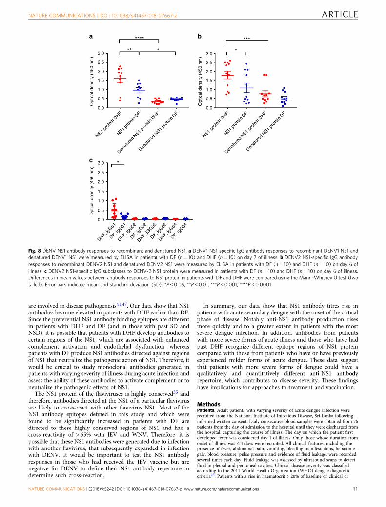
Antibody responses to recombinant and denatured NS1.Although we found that antibodies to recombinant NS1 weresignificantly higher in patients with DHF during the criticalphase, further analysis of NS1 epitopes showed that patients withDF had significantly higher antibody titres to certain peptides/regions of NS1. In order to determine whether the NS1 antibodiesbind to conformational epitopes not represented in the peptide-binding assay, we coated 96-well plates with recombinant ordenatured DENV NS1 and measured the antibody titres in thesera of patients with DF and DHF during acute secondary DENVinfection. The recombinant NS1 was denatured by heating theprotein at 95 °C for 15 min, as previously described30. The seraused for these experiments were sampled on day 7 of illness frompatients with DENV1 and day 6 from patients with DENV2. Thedetection of antibody titres significantly decreased when theDENV1 NS1 protein capture antigen was denatured in patientswith acute DENV1 infection resulting in DHF (p < 0.0001, usingthe Mann–Whitney U test) (two tailed) and DF (p= 0.008)(Fig. 8a). Similarly, a significant reduction in the antibody titreswas seen when DENV2 NS1 protein was denatured in patientswith DHF (p= 0.0004) although not significant in patients withDF (Fig. 8b).
DENV NS1 IgG subclass types in patients with acute dengue.Different IgG subclasses that form immune complexes, vary intheir ability to activate complement and have different affinitiesfor IgG receptors31. While IgG binding to FcγRI, FcγRIIA/C andFcγRIIIB results in activation of macrophages, monocytes anddendritic cells, IgG binding to FcγRIIB may inhibit immune
DENV–2 DHF
DENV–1 DHF
NS1 antibody (optical density - 405 nm) NS1 antibody (optical density - 405 nm)
NS1 antibody (optical density - 405 nm)NS1 antibody (optical density - 405 nm)
DENV–1 DF
DENV–2 DF200,000
150,000
100,000
50,000
200,000
150,000
100,000
50,000
200,000
150,000
100,000
50,000
0
200,000
150,000
100,000
50,000
0
00
0.0
Pla
tele
ts c
ount
(ce
lls/m
m3 )
Pla
tele
ts c
ount
(ce
lls/m
m3 )
Pla
tele
ts c
ount
(ce
lls/m
m3 )
Pla
tele
ts c
ount
(ce
lls/m
m3 )
0.5 1.0 1.5 2.0 2.5 3.0
a b
c d
0.0 0.5 1.0 1.5 2.0 2.5 3.0 0.0 0.5 1.0 2.0 2.5 3.01.5
0.0 0.5 1.0 1.5 2.0 2.5 3.0
Fig. 2 Correlation of NS1 antibody titres with platelet counts. Correlation of NS1 antibody titres in patients with acute secondary DENV1 and DENV2infection with platelet counts. Serum anti-NS1 antibody titres were measured throughout the course of illness by ELISA and correlated with circulatingplatelet counts in patients with (a) DHF due to DENV2, n= 23 (Spearmans’ r=−0.3, p= 0.008), (b) DF due to DENV2, n= 12 (Spearman’s r=−0.39,p= 0.009), (c) DHF due to DENV1, n= 10 (Spearman’s r=−0.3, p= 0.03), and (d) DF due to DENV1, n= 10 (Spearman’s r=−0.17, p= 0.30)
NATURE COMMUNICATIONS | DOI: 10.1038/s41467-018-07667-z ARTICLE
NATURE COMMUNICATIONS | (2018) 9:5242 | DOI: 10.1038/s41467-018-07667-z | www.nature.com/naturecommunications 5

responses32,33. IgG3 has shown to be the predominant subclassthat activated the inhibitory FcγRIIB receptors, whereas IgG1preferentially activated the activating receptors34. Therefore, weinvestigated the NS1-specific IgG subclasses in patients with acutesecondary DENV2 infection. We found that IgG1 was the pre-dominant subclass of NS1 antibody present and NS1-specific IgG1
was significantly higher in patients with DHF (p= 0.037), whencompared with patients with DF (Fig. 8c) by Mann–Whitney Utest (two tailed). In contrast, the NS1 IgG3 subclass levels showeda trend to be higher in patients with DF (median 0.04, IQR:0.03–0.06) compared with those with DHF (median 0.02, IQR:0.01–0.03) although this was not significant (p= 0.1).
3.0 *
*
**** *******
Opt
ical
den
sity
(450
nm
)O
ptic
al d
ensi
ty(4
50 n
m)
Opt
ical
den
sity
(450
nm
)O
ptic
al d
ensi
ty(4
50 n
m)
Opt
ical
den
sity
(450
nm
)O
ptic
al d
ensi
ty(4
50 n
m)
Opt
ical
den
sity
(450
nm
)O
ptic
al d
ensi
ty(4
50 n
m)
2.5
2.0
1.5
1.0
0.5
0.0
pep
01 D
HF
pep
01 D
F
pep
02 D
HF
pep
03 D
F
pep
03 D
HF
pep
02 D
F
pep
06 D
F
pep
08 D
F
pep
08 D
HF
pep
07 D
F
pep
07 D
HF
pep
06 D
HF
pep
05 D
F
pep
05 D
HF
pep
04 D
F
pep
04 D
HF
pep
09 D
HF
pep
09 D
F
pep
10 D
HF
pep
11 D
F
pep
11 D
HF
pep
10 D
F
pep
14 D
F
pep
16 D
F
pep
16 D
HF
pep
15 D
F
pep
15 D
HF
pep
14 D
HF
pep
13 D
F
pep
13 D
HF
pep
12 D
F
pep
12 D
HF
pep
17 D
HF
pep
17 D
F
pep
18 D
HF
pep
19 D
F
pep
19 D
HF
pep
18 D
F
pep
22 D
F
pep
24 D
F
pep
24 D
HF
pep
23 D
F
pep
23 D
HF
pep
22 D
HF
pep
21 D
F
pep
21 D
HF
pep
20 D
F
pep
20 D
HF
pep
41 D
HF
pep
41DF
pep
42 D
HF
pep
43 D
F
pep
43 D
HF
pep
42 D
F
pep
46 D
F
pep
48 D
F
pep
48 D
HF
pep
47 D
F
pep
47 D
HF
pep
46 D
HF
pep
45 D
F
pep
45 D
HF
pep
44 D
F
pep
44 D
HF
pep
33 D
HF
pep
33 D
F
pep
34 D
HF
pep
35 D
F
pep
35 D
HF
pep
34 D
F
pep
38 D
F
pep
40 D
F
pep
40 D
HF
pep
39 D
F
pep
39 D
HF
pep
38 D
HF
pep
37 D
F
pep
37 D
HF
pep
36 D
F
pep
36 D
HF
pep
49 D
HF
pep
49 D
F
pep
50 D
HF
pep
51 D
F
pep
51 D
HF
pep
50 D
F
pep
54 D
F
pep
56 D
F
pep
56 D
HF
pep
55 D
F
pep
55 D
HF
pep
54 D
HF
pep
53 D
F
pep
53 D
HF
pep
52 D
F
pep
52 D
HF
pep
57 D
HF
pep
57 D
F
pep
58 D
HF
pep
59 D
F
pep
59 D
HF
pep
58 D
F
pep
61 D
F
pep
61 D
HF
pep
60 D
F
pep
60 D
HF
pep
25 D
HF
pep
25 D
F
pep
26 D
HF
pep
27 D
F
pep
27 D
HF
pep
26 D
F
pep
30 D
F
pep
32 D
F
pep
32 D
HF
pep
31 D
F
pep
31 D
HF
pep
30 D
HF
pep
29 D
F
pep
29 D
HF
pep
28 D
F
pep
28 D
HF
3.0
2.5
2.0
1.5
1.0
0.5
0.0
3.0
2.5
2.0
1.5
1.0
0.5
0.0
3.0
2.5
2.0
1.5
1.0
0.5
0.0
3.0
2.5
2.0
1.5
1.0
0.5
0.0
3.0
2.5
2.0
1.5
1.0
0.5
0.0
hg
f
c
a
d
b
3.0
2.5
2.0
1.5
1.0
0.5
0.0
3.0
2.5
2.0
1.5
1.0
0.5
0.0
e
Fig. 3 DENV1 NS1 antibody responses to NS1 peptides in the febrile phase. a–h DENV1 NS1-specific IgG antibody responses to 61 DENV1 NS1 overlappingpeptides in patients with DF (n= 10) and DHF (n= 10) were measured by ELISA on day 4 of illness. Differences in mean values between antibodyresponses to overlapping peptides in patients with DF and DHF were compared using the Mann–Whitney U test (two tailed). NS1 antibody levels inpatients with DHF are indicated in red, and in those with DF in blue. Error bars indicate mean and standard deviation (SD). *P < 0.05, **P < 0.01, ***P <0.001, ****P < 0.0001
ARTICLE NATURE COMMUNICATIONS | DOI: 10.1038/s41467-018-07667-z
6 NATURE COMMUNICATIONS | (2018) 9:5242 | DOI: 10.1038/s41467-018-07667-z | www.nature.com/naturecommunications

Antibody responses to NS1 in healthy individuals past dengue.Recurrent dengue infection is thought to be a risk factor fordeveloping severe dengue. As mentioned above, we investigatedthe total NS1 antibody levels in healthy individuals who wereDENV seronegative, and those with past NSD and SD. We foundthat those with past SD had significantly higher NS1 antibodylevels than in individuals with past NSD (Fig. 1a). We thencharacterized the antibody responses to NS1 in 24 individualswith past NSD and 17 individuals with past SD. We cannot knowwhen the NSD cohort contracted DENV or whether the infectionwas primary or secondary, due to their lack of clinicalpresentation.
As observed in patients with acute dengue infection (Figs. 3–6),we found that healthy participants with past SD and NSDrecognized distinct regions of DENV NS1. We observed negligiblelevels of antibodies to the NS1 overlapping peptides in DENV-seronegative individuals (Fig. 9a–o). Patients with past SD, had
significantly higher antibody responses to regions represented bypeptides 1–4, 9–16, 22–26 and 29–31. In contrast, as observed inpatients with DF, those who had past NSD had significantlyhigher antibody responses to the distal C-terminus of NS1compared to those with past SD, represented by peptide 33–37,40–43, 45–48 and 58–60 (Fig. 9a–o). Therefore, while those withpast SD had significantly higher antibody responses to the N-terminus of the NS1 antigen, those with past NSD hadsignificantly higher antibodies to the peptides representing theC-terminus (Supplementary Fig. 3).
Cross-reactivity of DENV NS1 epitopes with other flaviviruses.Among the non-structural proteins of the flaviviruses, NS1 hasbeen shown to be the most highly conserved protein35. In orderto find out if the NS1 antibody epitopes defined in this study weredirected at conserved or non-conserved regions, we carried outmultiple alignment of the NS1 antigen of Japanese Encephalitis
2.0 *
*
**** *** ** ** * ** * * *
* * ** ** ** * *
* * * * ** ** * ** **** * *
1.5
1.0
Opt
ical
den
sity
(45
0 nm
)
Opt
ical
den
sity
(45
0 nm
)
0.0
pep
01 D
HF
pep
01 D
F
pep
02 D
HF
pep
02 D
F
pep
04 D
F
pep
04 D
HF
pep
05 D
F
pep
05 D
HF
pep
06 D
F
pep
06 D
HF
pep
07 D
F
pep
07 D
HF
pep
08 D
F
pep
08 D
HF
pep
17 D
HF
pep
17 D
F
pep
18 D
HF
pep
18 D
F
pep
19 D
HF
pep
19 D
F
pep
20 D
HF
pep
20 D
F
pep
21 D
HF
pep
21 D
F
pep
22 D
HF
pep
22 D
F
pep
23 D
HF
pep
23 D
F
pep
24 D
HF
pep
24 D
F
pep
33 D
HF
pep
33 D
F
pep
34 D
HF
pep
34 D
F
pep
35 D
HF
pep
35 D
F
pep
36 D
HF
pep
36 D
F
pep
37 D
HF
pep
37 D
F
pep
38 D
HF
pep
38 D
F
pep
39 D
HF
pep
39 D
F
pep
40 D
HF
pep
40 D
F
pep
41 D
HF
pep
41 D
F
pep
42 D
HF
pep
42 D
F
pep
43 D
HF
pep
43 D
F
pep
44 D
HF
pep
44 D
F
pep
45 D
HF
pep
45 D
F
pep
46 D
HF
pep
46 D
F
pep
47 D
HF
pep
47 D
F
pep
25 D
HF
pep
25 D
F
pep
26 D
HF
pep
26 D
F
pep
27 D
HF
pep
27 D
F
pep
28 D
HF
pep
28 D
F
pep
29 D
HF
pep
29 D
F
pep
30 D
HF
pep
30 D
F
pep
31 D
HF
pep
31 D
F
pep
32 D
HF
pep
32 D
F
pep
09 D
F
pep
09 D
HF
pep
10 D
F
pep
10 D
HF
pep
11 D
F
pep
11 D
HF
pep
12 D
F
pep
12 D
HF
pep
13 D
F
pep
13 D
HF
pep
14 D
F
pep
14 D
HF
pep
15 D
F
pep
15 D
HF
pep
16 D
F
pep
16 D
HF
0.5
2.0
1.5
1.0
Opt
ical
den
sity
(45
0 nm
)
0.0
0.5
2.0
1.5
1.0
Opt
ical
den
sity
(45
0 nm
)
0.0
0.5
2.0
1.5
1.0
Opt
ical
den
sity
(45
0 nm
)
0.0
0.5
2.0
1.5
1.0
Opt
ical
den
sity
(45
0 nm
)0.0
0.5
2.0
1.5
1.0
0.0
0.5
a b
c d
e f
Fig. 4 DENV2 NS1 antibody responses to NS1 peptides in the febrile phase. a–f DENV2 NS1-specific IgG antibody responses to 47 DENV2 NS1 overlappingpeptides were measured by ELISA in patients with DF (n= 10) and DHF (n= 10) on day 3 of illness. Differences in mean values between antibodyresponses to overlapping peptides in patients with DF and DHF were compared using the Mann–Whitney U test (two tailed). Error bars indicate the meansand standard deviation (SD). *P < 0.05, **P < 0.01, ***P < 0.001, ****P < 0.0001
NATURE COMMUNICATIONS | DOI: 10.1038/s41467-018-07667-z ARTICLE
NATURE COMMUNICATIONS | (2018) 9:5242 | DOI: 10.1038/s41467-018-07667-z | www.nature.com/naturecommunications 7

(JEV) virus and West Nile virus (WNV) with all four serotypes ofthe DENVs using clustal omega29, as these are the only tworeported flaviviruses co-circulating in Sri Lanka (SupplementaryFig. 4)36. We found that the majority of NS1 antibody epitopesthat we have described in this study, were directed against theregions that were highly conserved among DENV1, 2, 3 and 4,
JEV and WNV (Supplementary Fig. 4a). The regions were simi-larly conserved when comparing the WNV, JEV andNS1 sequences of DENV1 and DENV2 strains that recently cir-culated in Sri Lanka (Supplementary Fig. 4b). Therefore, themajority of NS1 antibodies defined in this study are likely to behighly cross-reactive.
2.0
2.5
3.0
1.5
1.0
Opt
ical
den
sity
(45
0 nm
)
0.0
pep
01 D
F
pep
01 D
HF
0.5
2.0
2.5
3.0
1.5
1.0
Opt
ical
den
sity
(45
0 nm
)
0.0
0.5
2.0
2.5
3.0
1.5
1.0
Opt
ical
den
sity
(45
0 nm
)
0.0
0.5
2.0
2.5
3.0 * * * *
****
*
*** ** * * * *
**
1.5
1.0
Opt
ical
den
sity
(45
0 nm
)
0.0
0.5
2.0
2.5
3.0
1.5
1.0
Opt
ical
den
sity
(45
0 nm
)
0.0
0.5
2.0
2.5
3.0
1.5
1.0
Opt
ical
den
sity
(45
0 nm
)
0.0
0.5
2.0
2.5
3.0
1.5
1.0
Opt
ical
den
sity
(45
0 nm
)
0.0
0.5
2.0
2.5
3.0*
1.5
1.0
Opt
ical
den
sity
(45
0 nm
)
0.0
0.5
pep
02 D
HF
pep
02 D
F
pep
04 D
F
pep
04 D
HF
pep
03 D
F
pep
03 D
HF
pep
05 D
F
pep
05 D
HF
pep
06 D
F
pep
06 D
HF
pep
07 D
F
pep
07 D
HF
pep
08 D
F
pep
08 D
HF
pep
09 D
F
pep
09 D
HF
pep
10 D
F
pep
10 D
HF
pep
11 D
F
pep
11 D
HF
pep
12 D
F
pep
12 D
HF
pep
13 D
F
pep
13 D
HF
pep
14 D
F
pep
14 D
HF
pep
15 D
F
pep
15 D
HF
pep
16 D
F
pep
16 D
HF
pep
17 D
HF
pep
17 D
F
pep
18 D
HF
pep
18 D
F
pep
19 D
HF
pep
19 D
F
pep
20 D
HF
pep
20 D
F
pep
21 D
HF
pep
21 D
F
pep
22 D
HF
pep
22 D
F
pep
23 D
HF
pep
23 D
F
pep
24 D
HF
pep
24 D
F
pep
25 D
HF
pep
25 D
F
pep
26 D
HF
pep
26 D
F
pep
27 D
HF
pep
27 D
F
pep
28 D
HF
pep
28 D
F
pep
29 D
HF
pep
29 D
F
pep
30 D
HF
pep
30 D
F
pep
31 D
HF
pep
31 D
F
pep
32 D
HF
pep
32 D
F
pep
33 D
HF
pep
33 D
F
pep
34 D
HF
pep
34 D
F
pep
35 D
HF
pep
35 D
F
pep
36 D
HF
pep
36 D
F
pep
37 D
HF
pep
37 D
F
pep
38 D
HF
pep
38 D
F
pep
39 D
HF
pep
39 D
F
pep
40 D
HF
pep
40 D
F
pep
41 D
HF
pep
41 D
F
pep
42 D
HF
pep
42 D
F
pep
43 D
HF
pep
43 D
F
pep
44 D
HF
pep
44 D
F
pep
45 D
HF
pep
45 D
F
pep
46 D
HF
pep
46 D
F
pep
47 D
HF
pep
47 D
F
pep
48 D
HF
pep
48 D
F
pep
49 D
HF
pep
49 D
F
pep
50 D
HF
pep
50 D
F
pep
51 D
HF
pep
51 D
F
pep
52 D
HF
pep
52 D
F
pep
53 D
HF
pep
53 D
F
pep
54 D
HF
pep
54 D
F
pep
55 D
HF
pep
55 D
F
pep
56 D
HF
pep
56 D
F
pep
57 D
HF
pep
57 D
F
pep
58 D
HF
pep
58 D
F
pep
59 D
HF
pep
59 D
F
pep
60 D
HF
pep
60 D
F
pep
61 D
HF
pep
61 D
F
a b
c d
e f
g h
Fig. 5 DENV1 NS1 antibody responses to NS1 peptides in the critical phase. a–h DENV1 NS1-specific IgG antibody responses to 61 DENV1 NS1 overlappingpeptides in patients with DF (n= 10) and DHF (n= 10) were measured by ELISA on day 7 of illness. Differences in mean values between antibodyresponses to overlapping peptides in patients with DF and DHF were compared using the Mann–Whitney U test (two tailed). NS1 antibody levels inpatients with DHF are indicated in red, and in those with DF in blue. Error bars indicate mean and standard deviation (SD). *P < 0.05, **P < 0.01,***P < 0.001
ARTICLE NATURE COMMUNICATIONS | DOI: 10.1038/s41467-018-07667-z
8 NATURE COMMUNICATIONS | (2018) 9:5242 | DOI: 10.1038/s41467-018-07667-z | www.nature.com/naturecommunications
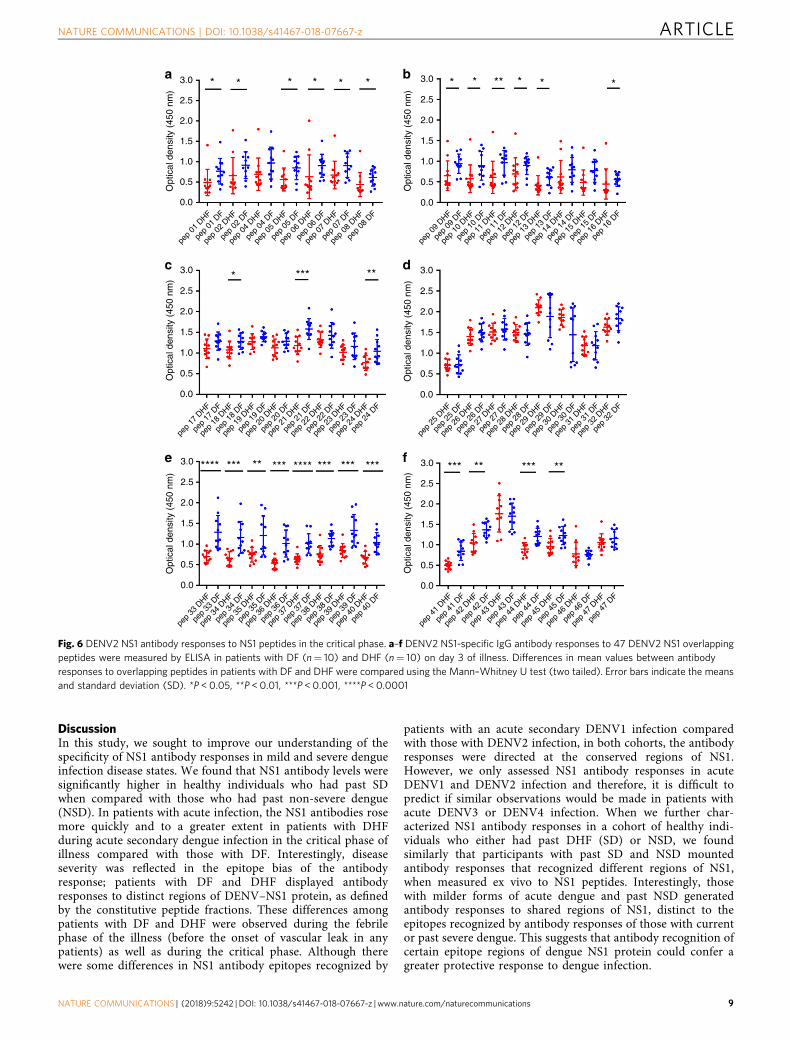
DiscussionIn this study, we sought to improve our understanding of thespecificity of NS1 antibody responses in mild and severe dengueinfection disease states. We found that NS1 antibody levels weresignificantly higher in healthy individuals who had past SDwhen compared with those who had past non-severe dengue(NSD). In patients with acute infection, the NS1 antibodies rosemore quickly and to a greater extent in patients with DHFduring acute secondary dengue infection in the critical phase ofillness compared with those with DF. Interestingly, diseaseseverity was reflected in the epitope bias of the antibodyresponse; patients with DF and DHF displayed antibodyresponses to distinct regions of DENV–NS1 protein, as definedby the constitutive peptide fractions. These differences amongpatients with DF and DHF were observed during the febrilephase of the illness (before the onset of vascular leak in anypatients) as well as during the critical phase. Although therewere some differences in NS1 antibody epitopes recognized by
patients with an acute secondary DENV1 infection comparedwith those with DENV2 infection, in both cohorts, the antibodyresponses were directed at the conserved regions of NS1.However, we only assessed NS1 antibody responses in acuteDENV1 and DENV2 infection and therefore, it is difficult topredict if similar observations would be made in patients withacute DENV3 or DENV4 infection. When we further char-acterized NS1 antibody responses in a cohort of healthy indi-viduals who either had past DHF (SD) or NSD, we foundsimilarly that participants with past SD and NSD mountedantibody responses that recognized different regions of NS1,when measured ex vivo to NS1 peptides. Interestingly, thosewith milder forms of acute dengue and past NSD generatedantibody responses to shared regions of NS1, distinct to theepitopes recognized by antibody responses of those with currentor past severe dengue. This suggests that antibody recognition ofcertain epitope regions of dengue NS1 protein could confer agreater protective response to dengue infection.
2.0
2.5
3.0 * * *
* ***
**** *** *** *** *** *** ***** ***********
**
* * * * * ** * * *
1.5
1.0O
ptic
al d
ensi
ty (
450
nm)
0.0
pep
01 D
F
pep
01 D
HF
0.5
2.0
2.5
3.0
1.5
1.0
Opt
ical
den
sity
(45
0 nm
)
0.0
0.5
2.0
2.5
3.0
1.5
1.0
Opt
ical
den
sity
(45
0 nm
)0.0
0.5
2.0
2.5
3.0
1.5
1.0
Opt
ical
den
sity
(45
0 nm
)
0.0
0.5
2.0
2.5
3.0
1.5
1.0
Opt
ical
den
sity
(45
0 nm
)
0.0
0.5
2.0
2.5
3.0
1.5
1.0
Opt
ical
den
sity
(45
0 nm
)
0.0
0.5
pep
02 D
HF
pep
02 D
F
pep
04 D
F
pep
04 D
HF
pep
05 D
F
pep
05 D
HF
pep
06 D
F
pep
06 D
HF
pep
07 D
F
pep
07 D
HF
pep
08 D
F
pep
08 D
HF
pep
09 D
F
pep
09 D
HF
pep
10 D
F
pep
10 D
HF
pep
11 D
F
pep
11 D
HF
pep
12 D
F
pep
12 D
HF
pep
13 D
F
pep
13 D
HF
pep
14 D
F
pep
14 D
HF
pep
15 D
F
pep
15 D
HF
pep
16 D
F
pep
16 D
HF
pep
17 D
HF
pep
17 D
F
pep
18 D
HF
pep
18 D
F
pep
19 D
HF
pep
19 D
F
pep
20 D
HF
pep
20 D
F
pep
21 D
HF
pep
21 D
F
pep
22 D
HF
pep
22 D
F
pep
23 D
HF
pep
23 D
F
pep
24 D
HF
pep
24 D
F
pep
25 D
HF
pep
25 D
F
pep
26 D
HF
pep
26 D
F
pep
27 D
HF
pep
27 D
F
pep
28 D
HF
pep
28 D
F
pep
29 D
HF
pep
29 D
F
pep
30 D
HF
pep
30 D
F
pep
31 D
HF
pep
31 D
F
pep
32 D
HF
pep
32 D
F
pep
33 D
HF
pep
33 D
F
pep
34 D
HF
pep
34 D
F
pep
35 D
HF
pep
35 D
F
pep
36 D
HF
pep
36 D
F
pep
37 D
HF
pep
37 D
F
pep
38 D
HF
pep
38 D
F
pep
39 D
HF
pep
39 D
F
pep
40 D
HF
pep
40 D
F
pep
41 D
HF
pep
41 D
F
pep
42 D
HF
pep
42 D
F
pep
43 D
HF
pep
43 D
F
pep
44 D
HF
pep
44 D
F
pep
45 D
HF
pep
45 D
F
pep
46 D
HF
pep
46 D
F
pep
47 D
HF
pep
47 D
F
a b
c d
e f
Fig. 6 DENV2 NS1 antibody responses to NS1 peptides in the critical phase. a–f DENV2 NS1-specific IgG antibody responses to 47 DENV2 NS1 overlappingpeptides were measured by ELISA in patients with DF (n= 10) and DHF (n= 10) on day 3 of illness. Differences in mean values between antibodyresponses to overlapping peptides in patients with DF and DHF were compared using the Mann–Whitney U test (two tailed). Error bars indicate the meansand standard deviation (SD). *P < 0.05, **P < 0.01, ***P < 0.001, ****P < 0.0001
NATURE COMMUNICATIONS | DOI: 10.1038/s41467-018-07667-z ARTICLE
NATURE COMMUNICATIONS | (2018) 9:5242 | DOI: 10.1038/s41467-018-07667-z | www.nature.com/naturecommunications 9

A previous study characterized NS1 antibody epitopes in tenchildren with DF, seven children with DHF and three with DSS,which included children with an acute primary or secondarydengue infection with DENV1, DENV2 and DENV3 serotypes,and used a similar approach as us. It was shown that the mostimmunodominant regions of NS1 were those represented by aa101–121, aa 111–131 and aa 301–32137. In our patient cohort,we found somewhat overlapping regions with the peptides thatwere identified in the previous study in paediatric patients withacute secondary DENV1 (aa121–142 and aa 291–307) andin acute secondary DENV2 (aa 297–313). However, most of theNS1 antibodies identified by us were directed at different regions.The reasons for these differences could be due to our denguecohorts being patients with secondary dengue whereas, Hertzet al. had mapped epitopes in patients with both primary andsecondary infection. In addition, we examined the antibody epi-topes in 20 adult patients with DENV1 and 22 patients withDENV2, whereas Hertz et al. had mapped antibody epitopes in 27children with DENV2 (n= 7) and DENV3 (n= 14), and there-fore the variations in the sample size, ethnicity and age could havecontributed to these differences.
Many of the studies that show NS1 antibodies are associatedwith protection have been carried out in dengue mousemodels15,38. Proteomics, sequence identity analyses and in vitrostudies have shown that the C-terminal region of NS1 containsmany cross-reactive epitopes, with sequence homology to self-antigens such as endothelial cell proteins and mediators involvedin the coagulation pathways and platelet response9,39–41. As aresult, the monoclonal antibody, which substitutes the C-terminus of dengue NS1 with that of Japanese Encephalitis NS1was found to be protective in dengue mouse models42. Our datashow that DF patients with acute secondary DENV1 and DENV2infection, had significantly higher antibody titres to the C-terminus of NS1 compared with those with severe forms ofdengue (DHF). In fact, when we studied the antibody responsekinetics to the C-terminus in patients with DHF and DF, weobserved that the levels of antibody directed at the C-terminus ofNS1, rose significantly preceding the recovery phase of DF patientillness, whereas DHF responses had declined by this time point.In addition, healthy participants who experienced past NSD alsohad significantly higher antibody titres directed towards the C-terminus of NS1. Therefore, although the C-terminus appears toshare cross-reactive regions with self-proteins, milder forms ofillness associate with higher antibody titres targeted to this region.However, it is noteworthy that in our experiments, we used NS1overlapping peptides which might not reflect the actual binding ofNS1 antibodies to the fully folded and polymeric structure. Thisindeed may account for our observation that antibody responses
to the whole recombinant NS1 protein are greater in DHF,whereas we detected higher antibody responses to NS1 peptidesin serum of patients with DF. Therefore, in order to furthercharacterize the possible protective and pathogenic antibodies toNS1, it would be important to map antibody epitopes to thedimeric and hexameric forms of NS1.
Although we observed similarities in the NS1 antibodyresponses from patients with acute secondary DENV1 andDENV2, we did observe some interesting differences. Forinstance, the difference in NS1 antibody titres during the criticalphase between patients with DF and DHF was more markedduring DENV1 infection. In addition, while those with DF, due toeither serotype, had higher antibody titres to the C-terminusregion of NS1, DHF patients with DENV2 infection had sig-nificantly higher antibody responses directed towards theN-terminus. Although all DENV serotypes are known to causesevere disease, the severity of infection and clinical features havebeen shown to vary depending on the viral serotype andgenotype27,43,44. During this study period, it was found thatpatients with DENV2 had a higher risk of developing DHF, andmore rapid progression to plasma leakage27. Therefore, the tar-gets of NS1 antibodies generated to each serotype, may play a rolein disease pathogenesis.
It has been shown that NS1 antibodies bind to and result indestruction of platelets in dengue mouse models45,46. Our datashowed a trend towards inverse correlation between NS1 anti-body levels and platelet counts in patients with DHF. Althoughthe possibility that NS1 antibodies lead to thrombocytopenia inhumans has not been investigated, one possible explanation forour observations is that NS1 antibodies could be binding toplatelets in patients with DHF, causing their elimination. Sincethe NS1 antibody repertoire appears to be different in those withDF compared with DHF, it is possible that patients with DHFmay have NS1 antibodies that preferentially bind platelets.Therefore, careful experiments should be carried out to determinewhether such pathways may contribute to disease pathogenesis.
Dengue mouse models have suggested that NS1 antibodieswere associated with protection, yet human in vitro experimentshave cast some doubt on this hypothesis. Studies of human serafrom those with mild and severe forms of dengue have shownthat the binding of NS1 antibodies to endothelial monolayers wassignificantly enhanced when using sera from patients with severedengue during the critical phase of illness, a possible route toinducing life-threatening plasma leak9. Furthermore, NS1–NS1antibody complexes have been shown to activate complement11
and these complexes are known to cross-react with platelets,which could be linked to the haemorrhage seen in severe disease.Therefore, it has been speculated that NS1 antibody complexes
2.0
1.5
1.0
0.0
0.5
Opt
ical
den
sity
(45
0 nm
)
Opt
ical
den
sity
(45
0 nm
)
2.0
1.5
1.0
0.5
0.0
Day of fever4 5 6 7 8 9 4 5 6 7 8 9
1
2
3
4
5
6
7
Day of fever
a b
Fig. 7 NS1 antibody responses to selected regions of NS1. Antibody levels were measured in ten patients with DF (a) and ten patients with DHF (b), withacute secondary DENV1 infection throughout the course of illness to seven pools of selected NS1 overlapping peptides. Pool 1 represents aa 1–33, pool 2 aa121–142, pool 3 aa 161–195, pool 4 aa 222–249, pool 5 aa 251–284, pool 6 aa 279–307 and pool 7 aa 341–353. The bars indicate the means and standarderror of mean (SEM)
ARTICLE NATURE COMMUNICATIONS | DOI: 10.1038/s41467-018-07667-z
10 NATURE COMMUNICATIONS | (2018) 9:5242 | DOI: 10.1038/s41467-018-07667-z | www.nature.com/naturecommunications
are involved in disease pathogenesis41,47. Our data show that NS1antibodies become elevated in patients with DHF earlier than DF.Since the preferential NS1 antibody binding epitopes are differentin patients with DHF and DF (and in those with past SD andNSD), it is possible that patients with DHF develop antibodies tocertain regions of the NS1, which are associated with enhancedcomplement activation and endothelial dysfunction, whereaspatients with DF produce NS1 antibodies directed against regionsof NS1 that neutralize the pathogenic action of NS1. Therefore, itwould be crucial to study monoclonal antibodies generated inpatients with varying severity of illness during acute infection andassess the ability of these antibodies to activate complement or toneutralize the pathogenic effects of NS1.
The NS1 protein of the flaviviruses is highly conserved35 andtherefore, antibodies directed at the NS1 of a particular flavivirusare likely to cross-react with other flavivirus NS1. Most of theNS1 antibody epitopes defined in this study and which werefound to be significantly increased in patients with DF aredirected to these highly conserved regions of NS1 and had across-reactivity of > 65% with JEV and WNV. Therefore, it ispossible that these NS1 antibodies were generated due to infectionwith another flavivirus, that subsequently expanded in infectionwith DENV. It would be important to test the NS1 antibodyresponses in those who had received the JEV vaccine but arenegative for DENV to define their NS1 antibody repertoire todetermine such cross-reaction.
In summary, our data show that NS1 antibody titres rise inpatients with acute secondary dengue with the onset of the criticalphase of disease. Notably anti-NS1 antibody production risesmore quickly and to a greater extent in patients with the mostsevere dengue infection. In addition, antibodies from patientswith more severe forms of acute illness and those who have hadpast DHF recognize different epitope regions of NS1 proteincompared with those from patients who have or have previouslyexperienced milder forms of acute dengue. These data suggestthat patients with more severe forms of dengue could have aqualitatively and quantitatively different anti-NS1 antibodyrepertoire, which contributes to disease severity. These findingshave implications for approaches to treatment and vaccination.
MethodsPatients. Adult patients with varying severity of acute dengue infection wererecruited from the National Institute of Infectious Disease, Sri Lanka followinginformed written consent. Daily consecutive blood samples were obtained from 76patients from the day of admission to the hospital until they were discharged fromthe hospital, capturing the course of illness. The day on which the patient firstdeveloped fever was considered day 1 of illness. Only those whose duration fromonset of illness was ≤ 4 days were recruited. All clinical features, including thepresence of fever, abdominal pain, vomiting, bleeding manifestations, hepatome-galy, blood pressure, pulse pressure and evidence of fluid leakage, were recordedseveral times each day. Fluid leakage was assessed by ultrasound scans to detectfluid in pleural and peritoneal cavities. Clinical disease severity was classifiedaccording to the 2011 World Health Organization (WHO) dengue diagnosticcriteria23. Patients with a rise in haematocrit > 20% of baseline or clinical or
Opt
ical
den
sity
(45
0 nm
)
Opt
ical
den
sity
(45
0 nm
)
3.0
2.5
2.0
1.5
1.0
0.5
0.0
3.0
2.5
2.0
1.5
1.0
0.5
0.0
3.0
2.5
2.0
1.5
1.0
0.5
0.0
*
Opt
ical
den
sity
(45
0 nm
)
*** *
*******
NS1 pr
otein
DF
Denat
ured
NS1
prot
ein D
F
DHF_igG01
DF_igG01
DHF_igG02
DHF_iGG03
DF_igG03
DHF_igG04
DF_igG04
DF_igG02
Denat
ured
NS1
prot
ein D
HF
NS1 pr
otein
DHF
NS1 pr
otein
DF
Denat
ured
NS1
prot
ein D
F
Denat
ured
NS1
prot
ein D
HF
NS1 pr
otein
DHF
a b
c
Fig. 8 DENV NS1 antibody responses to recombinant and denatured NS1. a DENV1 NS1-specific IgG antibody responses to recombinant DENV1 NS1 anddenatured DENV1 NS1 were measured by ELISA in patients with DF (n= 10) and DHF (n= 10) on day 7 of illness. b DENV2 NS1-specific IgG antibodyresponses to recombinant DENV2 NS1 and denatured DENV2 NS1 were measured by ELISA in patients with DF (n= 10) and DHF (n= 10) on day 6 ofillness. c DENV2 NS1-specific IgG subclasses to DENV-2 NS1 protein were measured in patients with DF (n= 10) and DHF (n= 10) on day 6 of illness.Differences in mean values between antibody responses to NS1 protein in patients with DF and DHF were compared using the Mann–Whitney U test (twotailed). Error bars indicate mean and standard deviation (SD). *P < 0.05, **P < 0.01, ***P < 0.001, ****P < 0.0001
NATURE COMMUNICATIONS | DOI: 10.1038/s41467-018-07667-z ARTICLE
NATURE COMMUNICATIONS | (2018) 9:5242 | DOI: 10.1038/s41467-018-07667-z | www.nature.com/naturecommunications 11

**** ****
****** *****
****
****
Opt
ical
den
sity
(45
0 nm
)
3.0
2.5
2.0
1.5
1.0
0.5
0.0
pep1
SD
pep1
Neg
pep2
Neg
pep2
SD
pep3
SD
pep1
NSD
pep2
NSD
pep3
Neg
pep4
SD
pep3
NSD
pep4
Neg
pep4
NSD
pep1
3 SD
pep1
3 Neg
pep1
4 Neg
pep1
4 SD
pep1
5 SD
pep1
3 NSD
pep1
4 NSD
pep1
5 Neg
pep1
6 SD
pep1
5 NSD
pep1
6 Neg
pep1
6 NSD
pep1
7 SD
pep1
7 Neg
pep1
8 Neg
pep1
8 SD
pep1
9 SD
pep1
7 NSD
pep1
8 NSD
pep1
9 Neg
pep2
0 SD
pep1
9 NSD
pep2
0 Neg
pep2
0 NSD
pep2
1 SD
pep2
1 Neg
pep2
2 Neg
pep2
2 SD
pep2
3 SD
pep2
1 NSD
pep2
2 NSD
pep2
3 Neg
pep2
4 SD
pep2
3 NSD
pep2
4 Neg
pep2
4 NSD
pep2
5 SD
pep2
5 Neg
pep2
6 Neg
pep2
6 SD
pep2
7 SD
pep2
5 NSD
pep2
6 NSD
pep2
7 Neg
pep2
8 SD
pep2
7 NSD
pep2
8 Neg
pep2
8 NSD
pep2
9 SD
pep2
9 Neg
pep3
0 Neg
pep3
0 SD
pep3
1 SD
pep2
9 NSD
pep3
0 NSD
pep3
1 Neg
pep3
2 SD
pep3
1 NSD
pep3
2 Neg
pep3
2 NSD
pep3
3 SD
pep3
3 Neg
pep3
4 Neg
pep3
4 SD
pep3
5 SD
pep3
3 NSD
pep3
4 NSD
pep3
5 Neg
pep3
6 SD
pep3
5 NSD
pep3
6 Neg
pep3
6 NSD
pep3
7 SD
pep3
7 Neg
pep3
8 Neg
pep3
8 SD
pep3
9 SD
pep3
7 NSD
pep3
8 NSD
pep3
9 Neg
pep4
0 SD
pep3
9 NSD
pep4
0 Neg
pep4
0 NSD
pep4
1 SD
pep4
1 Neg
pep4
2 Neg
pep4
2 SD
pep4
3 SD
pep4
1 NSD
pep4
2 NSD
pep4
3 Neg
pep4
4 SD
pep4
3 NSD
pep4
4 Neg
pep4
4 NSD
pep4
5 SD
pep4
5 Neg
pep4
6 Neg
pep4
6 SD
pep4
7 SD
pep4
5 NSD
pep4
6 NSD
pep4
7 Neg
pep4
8 SD
pep4
7 NSD
pep4
8 Neg
pep4
8 NSD
pep4
9 SD
pep4
9 Neg
pep5
0 Neg
pep5
0 SD
pep5
1 SD
pep4
9 NSD
pep5
0 NSD
pep5
1 Neg
pep5
2 SD
pep5
1 NSD
pep5
2 Neg
pep5
2 NSD
pep5
3 SD
pep5
3 Neg
pep5
4 Neg
pep5
4 SD
pep5
5 SD
pep5
3 NSD
pep5
4 NSD
pep5
5 Neg
pep5
6 SD
pep5
5 NSD
pep5
6 Neg
pep5
6 NSD
pep5
7 SD
pep5
7 Neg
pep5
7 NSD
pep5
8 SD
pep5
8 Neg
pep5
8 NSD
pep5
9 SD
pep5
9 Neg
pep5
9 NSD
pep6
0 SD
pep6
0 Neg
pep6
0 NSD
pep6
1 SD
pep6
1 Neg
pep6
1 NSD
pep5
SD
pep5
Neg
pep6
Neg
pep6
SD
pep7
SD
pep5
NSD
pep6
NSD
pep7
Neg
pep8
SD
pep7
NSD
pep8
Neg
pep8
NSD
pep9
SD
pep9
Neg
pep1
0 Neg
pep1
0 SD
pep1
1 SD
pep9
NSD
pep1
0 NSD
pep1
1 Neg
pep1
2 SD
pep1
1 NSD
pep1
2 Neg
pep1
2 NSD
Opt
ical
den
sity
(45
0 nm
)
3.0
2.5
2.0
1.5
1.0
0.5
0.0
Opt
ical
den
sity
(45
0 nm
)
3.0
2.5
2.0
1.5
1.0
0.5
0.0
3.0
2.5
2.0
1.5
Opt
ical
den
sity
(45
0 nm
)
1.0
0.5
0.0
Opt
ical
den
sity
(45
0 nm
)
3.0
2.5
2.0
1.5
1.0
0.5
0.0
Opt
ical
den
sity
(45
0 nm
)
3.0
2.5
2.0
1.5
1.0
0.5
0.0
3.0
2.5
2.0
1.5
Opt
ical
den
sity
(45
0 nm
)
1.0
0.5
0.0
Opt
ical
den
sity
(45
0 nm
)
3.0
2.5
2.0
1.5
1.0
0.5
0.0
Opt
ical
den
sity
(45
0 nm
)
3.0
2.5
2.0
1.5
1.0
0.5
0.0
3.0
2.5
2.0
1.5
Opt
ical
den
sity
(45
0 nm
)
1.0
0.5
0.0
Opt
ical
den
sity
(45
0 nm
)
3.0
2.5
2.0
1.5
1.0
0.5
0.0
Opt
ical
den
sity
(45
0 nm
)
3.0
2.5
2.0
1.5
1.0
0.5
0.0
3.0
2.5
2.0
1.5
Opt
ical
den
sity
(45
0 nm
)
1.0
0.5
0.0
Opt
ical
den
sity
(45
0 nm
)
3.0
2.5
2.0
1.5
1.0
0.5
0.0
Opt
ical
den
sity
(45
0 nm
)
3.0
2.5
2.0
1.5
1.0
0.5
0.0
********** **
***** ** **
****
*
*
**** **** **** ****
**************** *** *
****
************
**** *** **
******
**** ******
**** **** **** ****
******* ******** ***
****
**** ****
****
****
****
**** ****
****
* ****
****
**** ********
********
****
****
** ***
**** ****
****************
**** ****
*** * * ******** ******** **** **** ****
******* **** ****
************
****
********
***
****
**
**
**** ***
* **
a b c
d e f
g h i
j k l
m n o
Fig. 9 NS1-antibody responses in individuals with past infection. a–o DENV1 NS1-specific IgG antibody responses to DENV-1 NS1 overlapping peptides 1–61in seronegative individuals (Neg, n= 20), those with past non-severe dengue (NSD, n= 24) and past severe dengue (SD, n= 17). Error bars representmean and standard deviation. Differences between the groups were compared using the Mann–Whitney U test (two tailed). *P < 0.05, **P < 0.01, ***P <0.001, ****P < 0.0001
ARTICLE NATURE COMMUNICATIONS | DOI: 10.1038/s41467-018-07667-z
12 NATURE COMMUNICATIONS | (2018) 9:5242 | DOI: 10.1038/s41467-018-07667-z | www.nature.com/naturecommunications

ultrasound scan evidence of plasma leakage were classified as having DHF. Shockwas defined as having cold clammy skin, along with a narrowing of pulse pressureof 20 mmHg. As such, 36 patients were classified as DHF and 40 patients wereclassified as DF.
Healthy individuals with varying severity of past dengue. In our previousstudies, we had recruited 1689 healthy individuals attending the Family PracticeCentre, which is a primary health care facility of the University of Sri Jaye-wardenepura, Sri Lanka, providing community healthcare to over 2000 familiesliving in the suburban areas of the Colombo district48. In this cohort, we haveserum samples from the time of recruitment to the study, information regardingdengue serostatus and whether the individuals had DHF or mild or asymptomaticdengue. Seventeen serum samples were used from those who reported an episodeof DHF (past severe dengue (SD)), and 24 serum samples were used from thosewho were seropositive for dengue but had never been hospitalized due to a febrileillness. The individuals who were seropositive but had never been hospitalized dueto any febrile illness were considered to have a mild/inapparent or non-severedengue infection (past non-severe dengue infection (NSD)). We also used 20 serumsamples from individuals who were seronegative at the time of recruitment. Onlyone of them had received the Japanese encephalitis vaccine, and all individuals(including the person who received the JEV vaccine) were seronegative for JEV.
Ethics statement. Ethical approval was obtained by the Ethics Review Committeeof the Faculty of Medical Sciences, University of Sri Jayawardenapura. All patientswere recruited following informed written consent. We have complied with allrelevant ethical regulations in carrying out this study.
Serotyping of DENV and assessment of viral titre. Acute dengue infection wasconfirmed by quantitative real-time PCR, and DENV viruses were serotyped andtitres quantified by quantitative real-time PCR28. Viral RNA in serum wasextracted using QIAamp Viral RNA Mini Kit (Qiagen, USA, Cat: 52906) andtranscribed to cDNA using High Capacity cDNA reverse transcription kit (AppliedBiosystems, USA, cat: 4368814) as per the manufacturer’s protocol in 20 -µlreaction mixtures containing 1 × RT buffer, 1 × RT random primers, 10 × dNTPMix (100 mM), 50 U of MultiScribe™ Reverse Transcriptase, 20 U of RNaseInhibitor (Applied Biosystems, USA, Cat: N8080119), 10 µl of RNA and PCR-grade water (Applied Biosystems, USA, Cat: AM9935).
Multiplex quantitative real-time PCR was performed using the CDC real-timePCR assay for detection of the dengue virus49, and modified to quantify DENV.Oligonucleotide primers and a dual-labelled probe for DEN 1, 2, 3, 4 serotypeswere used (Life Technologies, India) based on published sequences49. In order toquantify viruses, a tenfold dilution series (106–00 PFU/ml) was prepared using thecDNA of reference strains for standard curves28. Real-time PCR was performedusing TaqMan® Multiplex Master Mix (Applied Biosystems, USA, Cat: 4461881).The reactions consisted of 20- μl volumes and contained the following reagents:1 × TaqMan multiplex master mix (containing Mustag Purple dye), 900 nM of eachprimer, 250 nM of each probe, 2 μl of cDNA and PCR-grade water (AppliedBiosystems, USA, Cat: AM9935). Following initial denaturation for 20 s at 95 °C,the reaction was carried out for 40 cycles of 3 s at 95 °C and 30 s at 60 °C. Thethreshold cycle value (Ct) for each reaction was determined by manually setting thethreshold limit. All assays were done in triplicate. After the primers and probeswere validated, a multiplex method was optimized to quantify the four serotypes ina single reaction and was used to determine viral loads of unknown samples thatwere determined using the optimized multiplex real-time protocol.
Analysis of dengue NS1 antigen and DENV IgM and IgG levels. The presence ofNS1 antigen was assessed using the NS1 early dengue enzyme-linked immuno-sorbent assay (ELISA) (Panbio, Brisbane, QLD, Australia). NS1 antigen levels weresemi-quantitatively assessed in serial blood samples of patients and expressed asPanbio units. Dengue antibody assays were performed using a commercial capture-IgM and IgG ELISA (Panbio, Brisbane, Australia)50,51. Based on the WHO criteria,patients with an IgM:IgG ratio of > 1.2 were considered to have a primary dengueinfection, while patients with IgM:IgG ratios < 1.2 were categorized under sec-ondary dengue infection52. The DENV-specific IgM and IgG ELISA was also usedto semi-quantitatively determine the DENV-specific IgM and IgG titres, whichwere expressed as Panbio units.
Development of an ELISA to detect NS1-specific antibody. Currently, there areno assays to measure NS1 antibody levels, and as such, we developed an ELISA tomeasure NS1 antibody levels in patient samples. Commercially available recom-binant DENV1 and DENV2 NS1 full-length recombinant proteins (Native antigen,USA) expressed from a mammalian cell line were used to coat the ELISA plates andcapture NS1-specific antibodies in serum samples. The optimal concentration ofcoating antigen, serum dilution and concentration of secondary antibody weredetermined by checkerboard titrations.
Our indirect ELISA to detect dengue NS1-specific antibodies was performed in96-well microtitre plates (PierceTM, Cat: 15031). The plates were coated with either100 µl/well DENV1 NS1 protein (Native antigen, USA) or DENV2 NS1 protein(Native antigen, USA) diluted in carbonate–bicarbonate coating buffer (pH 9.6) at
a final concentration of 1 µg/ml and incubated overnight at 4 °C. After incubation,the wells were washed three times with 300 µl/well washing buffer (phosphate-buffered saline (PBS) containing 0.05% Tween 20). The wells were blocked with300 µl/well with blocking buffer (PBS containing 0.05% Tween 20 and 1% bovineserum albumin (BSA)) for 1 h at room temperature. The wells were washed againthree times with 300 µl/well washing buffer. Serum samples were diluted 1:5000 inPBS containing 0.05% Tween 20 and 1% BSA. Diluted serum samples were addedin triplicates to the appropriate plate at 100 µl/well and incubated for 60 min atroom temperature. After incubation, wells were washed three times with 300 µl/well washing buffer. Goat anti-human IgG, biotinylated antibody (Mabtech,Sweden, Cat:3820-4-250) was diluted to 1:1000 in PBS containing 0.05% Tween 20and 1% BSA and added 100 µl/well. The plates were incubated again for 30 min atroom temperature and washed three times with washing buffer. Streptavidinalkaline phosphatase (Abcam, UK, Cat: ab64268) was diluted 1:20 with PBScontaining 0.05% Tween 20 and 1% BSA and added 100 µl/well. The plate wasincubated for 30 min at room temperature and washed five times. Para-nitrophenylphosphate (PNPP) (Thermo Fisher Scientific, USA, Cat: 37621)substrate was added at 100 µl/well and incubated for 20 min in the dark at roomtemperature. The reaction was stopped by adding 50 µl/well 2 M NaOH, and theELISA read on MPSCREEN MR-96A ELISA reader at 405-nm wavelength. Samplediluent (PBS containing 0.05% Tween 20 and 1% BSA) was used as the blank foreach assay. Using this optimized assay, we determined NS1 antibody levels inDENV1 DF (n= 10), DENV1 DHF (n= 10) and DENV-seronegative individuals(n= 20). The assay showed a significant difference in NS1 antibody levels betweenboth DHF and seronegative individuals (p < 0.0001) and DF and seronegativeindividuals (p= 0.0302). Furthermore, we determined NS1 antibody levels inDENV2 DF (n= 10), DENV2 DHF (n= 17) and DENV-seronegative individuals(n= 18). Again, the assay showed significant differences between both DHF andseronegative individuals (p < 0.0001) and DF and seronegative individuals (p=0.0001). Healthy individuals with past severe dengue (n= 34) and past non-severedengue (n= 36) too resulted in a significantly different antibody level (p < 0.0001)compared with seronegative individuals (n= 20).
NS1 antibody responses to DENV1 and DENV2 overlapping peptides. DengueNS1 peptide arrays were obtained through the NIH Biodefense and EmergingInfections Research Resources Repository, NIAID (NIH: Peptide Array, DengueVirus Type 1, Singapore/S275/1990, NS1 Protein, NR-2751) (Accession code:P33478 and NIH: Peptide Array, Dengue Virus Type 2, New Guinea C (NGC), NS1Protein, NR-508) (Accession code: AAA42941). Ninety-six-well microtitre plates(Pierce™ Cat:15031) were coated with 100 μl/well peptide preparations diluted inbicarbonate/carbonate coating buffer (pH 9.6) at a final concentration of 1 μg/100 μl. The peptides were coated individually and incubated overnight at 4 °C.After incubation, unbound peptide was washed away twice with phosphate-buffered saline (PBS, pH 7.4). The wells were blocked with 250 μl/well PBS con-taining 0.05% Tween 20 and 1% bovine serum albumin (BSA) and incubated for1.5 h at room temperature. The plates were then washed three times with 300 μl/well washing buffer (PBS containing 0.05% Tween 20). Serum samples were diluted1:250 in ELISA diluent (Mabtech, Sweden, Cat: 3652-D2). Diluted serum sampleswere added 100 μl/well in duplicates and incubated for 30 min at room tem-perature. After incubation, wells were washed three times with 300 μl/well washingbuffer. Goat anti-human IgG, biotinylated antibody (Mabtech, Sweden Cat: 3820-4-250) was diluted 1:1000 in PBS containing 1% BSA and added 100 μl/well. Plateswere then incubated for 30 min at room temperature and washed three times asdescribed previously. Streptavidin–HRP (Mabtech, Sweden, Cat: 3310-9) wasdiluted 1:1000 in PBS containing 1% BSA and added 100 μl/well. Incubation wascarried out for 30 min at room temperature, and the washing procedure wasrepeated five times. TMB ELISA substrate solution (Mabtech, Sweden, Cat: 3652-F10) was added 100 μl/well and incubated in the dark for 10 min at room tem-perature. The reaction was stopped by adding 100 μl/well 2 M H2SO4 stop solutionand absorbance values were read at 450 nm.
Analysis of antibody binding to NS1 epitopes. DENV1 NS1 and DENV2 NS1recombinant proteins (Native antigen, USA) were added into separate wells incarbonate–bicarbonate coating buffer (pH 9.6) at a final concentration of 1 µg/mland were incubated overnight at 4 °C. NS1 proteins, which were denatured byheating them for 95 °C for 15 min30, were used to compare the responses to therecombinant NS1 in this assay. The wells were blocked with 300 µl of blockingbuffer (PBS containing 1% bovine serum albumin (BSA)) for 1 h at room tem-perature. DENV1 and DENV2 serum samples were diluted 1:5000 with ELISAdiluent (Mabtech, Cat: 3652-D2). Diluted serum samples were added at 100 μl/wellinto respective wells and incubated for 60 min at room temperature. The boundNS1-specific antibodies were detected using 100 μl/well goat anti-human IgG,biotinylated antibody (Mabtech, Sweden, Cat: 3820-4-250), diluted 1:1000 in PBScontaining 1% BSA and incubated for 30 min at room temperature.Streptavidin–alkaline phosphatase (Abcam, UK, Cat: ab64268) diluted 1:20 in PBScontaining 1% BSA was added 100 μl/well and incubated for 30 min at roomtemperature. PNPP (Thermo Fisher Scientific, USA, Cat: 37621) substrate solutionwas added 100 μl/well and incubated in the dark for 20 min at room temperature.The reaction was stopped by adding 100 μl/well 2 M NaOH stop solution, andabsorbance values were read at 405 nm.
NATURE COMMUNICATIONS | DOI: 10.1038/s41467-018-07667-z ARTICLE
NATURE COMMUNICATIONS | (2018) 9:5242 | DOI: 10.1038/s41467-018-07667-z | www.nature.com/naturecommunications 13

Characterization of DENV2 NS1 IgG subtype responses. DENV2 NS1 recom-binant protein was bound to microplate wells with carbonate–bicarbonate coatingbuffer (pH 9.6) at a final concentration of 1 µg/ml and was incubated overnight at4 °C. The wells were blocked with 300 µl of blocking buffer (PBS containing 1%bovine serum albumin (BSA)) for 1 h at room temperature. DENV2 serum samplesdiluted 1:5000 in ELISA diluent (Mabtech, Sweden, Cat: 3652-D2) were added at100 μl/well and incubated for 60 min at room temperature. The bound NS1-specificIgG antibody subtypes were detected using 100 μl/well goat anti-human IgG1(Mabtech, Sweden, Cat: 3851-6-250), IgG2 (Mabtech, Sweden, Cat: 3852-6-250),IgG3 (Mabtech, Sweden, Cat: 3853-6-250) and IgG4 (Mabtech, Sweden, Cat: 3854-6-250) biotinylated antibody subtypes separately, diluted 1:1000 in PBS containing1% BSA. Plates were then incubated for 30 min at room temperature andstreptavidin–alkaline phosphatase (Abcam, UK, Cat: ab64268) diluted to 1:20 inPBS containing 1% BSA was added at 100 μl/well for 30 min at room temperature.PNPP (Thermo Fisher Scientific, USA, Cat: 37621) substrate solution was added at100 μl/well and incubated in the dark for 20 min at room temperature. The reactionwas stopped by adding 100 μl/well 2 M NaOH stop solution and absorbance valueswere read at 405 nm.
Statistical analysis. Statistical analysis was performed using GraphPad Prismversion 6. As the data were not normally distributed (as determined by the fre-quency distribution analysis of GraphPad PRISM), non-parametric tests were usedin the statistical analysis and two-sided tests were carried out in all instances.Differences in the serial values of NS1 antibodies, NS1 antigen and viral loads inpatients with DHF and DF were calculated/compared using the Holm–Sidakmethod. Corrections for multiple comparisons were completed using theHolm–Sidak method, and the statistical significant value was set at 0.05 (alpha).Differences in mean values between antibody responses to overlapping peptides inpatients with DF and DHF and in individuals with SD and NSD were comparedusing the Mann–Whitney U test (two tailed).
Data availabilityAll data generated or analyzed during this study are available included in the mainmanuscript and the supplementary files. All data are available without restrictions.
Received: 1 June 2018 Accepted: 13 November 2018
References1. Bhatt, S. et al. The global distribution and burden of dengue. Nature 496,
504–507 (2013).2. Capeding, M. R. et al. Clinical efficacy and safety of a novel tetravalent dengue
vaccine in healthy children in Asia: a phase 3, randomised, observer-masked,placebo-controlled trial. Lancet 384, 1358–1365 (2014).
3. Rouvinski, A. et al. Recognition determinants of broadly neutralizing humanantibodies against dengue viruses. Nature 520, 109–113 (2015).
4. Malavige, G. N. & Ogg, G. S. Pathogenesis of vascular leak in dengue virusinfection. Immunology 151, 261–269 (2017).
5. Paranavitane, S. A. et al. Dengue NS1 antigen as a marker of severe clinicaldisease. Bmc. Infect. Dis. 14, 570 (2014).
6. Muller, D. A. & Young, P. R. The flavivirus NS1 protein: molecular andstructural biology, immunology, role in pathogenesis and application as adiagnostic biomarker. Antivir. Res. 98, 192–208 (2013).
7. Modhiran, N. et al. Dengue virus NS1 protein activates cells via Toll-likereceptor 4 and disrupts endothelial cell monolayer integrity. Sci. Transl. Med.7, 304ra142 (2015).
8. Glasner, D. R. et al. Dengue virus NS1 cytokine-independent vascular leak isdependent on endothelial glycocalyx components. PLoS Pathog. 13, e1006673(2017).
9. Lin, C. F. et al. Antibodies from dengue patient sera cross-react withendothelial cells and induce damage. J. Med. Virol. 69, 82–90 (2003).
10. Thiemmeca, S. et al. Secreted NS1 protects dengue virus frommannose-binding lectin-mediated neutralization. J. Immunol. 197, 4053–4065(2016).
11. Avirutnan, P. et al. Vascular leakage in severe dengue virus infections: apotential role for the nonstructural viral protein NS1 and complement. J.Infect. Dis. 193, 1078–1088 (2006).
12. Lam, P. K. et al. Clinical characteristics of dengue shock syndrome invietnamese children: a 10-year prospective study in a single hospital. Clin.Infect. Dis. 57, 1577–1586 (2013).
13. Vaughn, D. W. et al. Dengue viremia titer, antibody response pattern, andvirus serotype correlate with disease severity. J. Infect. Dis. 181, 2–9 (2000).
14. Aye, K. S. et al. Pathologic highlights of dengue hemorrhagic fever in 13autopsy cases from Myanmar. Hum. Pathol. 45, 1221–1233 (2014).
15. Beatty, P. R. et al. Dengue virus NS1 triggers endothelial permeability andvascular leak that is prevented by NS1 vaccination. Sci. Transl. Med. 7,304ra141 (2015).
16. Dejnirattisai, W. et al. A new class of highly potent, broadly neutralizingantibodies isolated from viremic patients infected with dengue virus. Nat.Immunol. 16, 170–177 (2015).
17. de Alwis, R. et al. Identification of human neutralizing antibodies that bind tocomplex epitopes on dengue virions. Proc. . Natl. Acad. Sci. U. S. A. 109,7439–7444 (2012).
18. Luo, Y. Y. et al. Identification of a novel infection-enhancing epitope ondengue prM using a dengue cross-reacting monoclonal antibody. Bmc.Microbiol. 13, 194 (2013).
19. Decembre, E. et al. Sensing of immature particles produced by dengue virusinfected cells induces an antiviral response by plasmacytoid dendritic cells.PLoS Pathog. 10, e1004434 (2014).
20. Dejnirattisai, W. et al. Cross-reacting antibodies enhance dengue virusinfection in humans. Science 328, 745–748 (2010).
21. Nascimento, E. J. M. et al. Development of an anti-dengue NS1 IgG ELISA toevaluate exposure to dengue virus. J. Virol. Methods 257, 48–57 (2018).
22. Halstead, S. B. Which dengue vaccine approach is the most promising, andshould we be concerned about enhanced disease after vaccination? There isonly one true winner. Cold Spring Harb. Perspect. Biol. 10, a030700 (2018).
23. WHO. In SEARO Technical Publication Series, Vol. 60 (World HealthOrganization, SEARO, New Delhi, India, 2011).
24. Malavige, G. N. et al. Identification of serotype-specific T cell responses tohighly conserved regions of the dengue viruses. Clin. Exp. Immunol. 168,215–223 (2012).
25. Mongkolsapaya, J. et al. Original antigenic sin and apoptosis in thepathogenesis of dengue hemorrhagic fever. Nat. Med. 9, 921–927 (2003).
26. Malavige, G. N. et al. Suppression of virus specific immune responses by IL-10in acute dengue infection. PLoS. Negl. Trop. Dis. 7, e2409 (2013).
27. Wijewickrama A. F. S., et al. In Annual Conference of the Sri Lanka MedicalAssociation (SLMA, Colombo, 2017).
28. Fernando, S. et al. Patterns and causes of liver involvement in acute dengueinfection. Bmc. Infect. Dis. 16, 319 (2016).
29. Sievers, F. et al. Fast, scalable generation of high-quality protein multiplesequence alignments using Clustal Omega. Mol. Syst. Biol. 7, 539 (2011).
30. Amorim, J. H. et al. Protective immunity to DENV2 after immunization witha recombinant NS1 protein using a genetically detoxified heat-labile toxin asan adjuvant. Vaccine 30, 837–845 (2012).
31. Schroeder, H. W. Jr. & Cavacini, L. Structure and function ofimmunoglobulins. J. Allergy Clin. Immunol. 125, S41–S52 (2010).
32. Gillis, C. M. et al. Mechanisms of anaphylaxis in human low-affinity IgGreceptor locus knock-in mice. J. Allergy Clin. Immunol. 139, 1253–1265(2017). e1214.
33. Santegoets, K. C., Wenink, M. H., van den Berg, W. B. & Radstake, T. R. Fcgamma receptor IIb on GM-CSF macrophages controls immune complexmediated inhibition of inflammatory signals. PLoS One 9, e110966 (2014).
34. Vidarsson, G., Dekkers, G. & Rispens, T. IgG subclasses and allotypes: fromstructure to effector functions. Front. Immunol. 5, 520 (2014).
35. Rastogi, M., Sharma, N. & Singh, S. K. Flavivirus NS1: a multifacetedenigmatic viral protein. Virol. J. 13, 131 (2016).
36. Lohitharajah, J. et al. Viral aetiologies of acute encephalitis in a hospital-basedSouth Asian population. Bmc. Infect. Dis. 17, 303 (2017).
37. Hertz, T. et al. Antibody epitopes identified in critical regions of dengue virusnonstructural 1 protein in mouse vaccination and natural human infections. J.Immunol. 198, 4025–4035 (2017).
38. Henchal, E. A., Henchal, L. S. & Schlesinger, J. J. Synergistic interactions ofanti-NS1 monoclonal antibodies protect passively immunized mice fromlethal challenge with dengue 2 virus. J. Gen. Virol. 69(Pt 8), 2101–2107(1988).
39. Cheng, H. J. et al. Anti-dengue virus nonstructural protein 1 antibodiesrecognize protein disulfide isomerase on platelets and inhibit plateletaggregation. Mol. Immunol. 47, 398–406 (2009).
40. Cheng, H. J. et al. Proteomic analysis of endothelial cell autoantigensrecognized by anti-dengue virus nonstructural protein 1 antibodies. Exp. Biol.Med. (Maywood). 234, 63–73 (2009).
41. Chuang, Y. C. et al. Dengue virus-induced autoantibodies bind toplasminogen and enhance its activation. J. Immunol. 187, 6483–6490 (2011).
42. Wan, S. W. et al. Therapeutic effects of monoclonal antibody against denguevirus NS1 in a STAT1 knockout mouse model of dengue infection. J.Immunol. 199, 2834–2844 (2017).
43. Endy, T. P. et al. Spatial and temporal circulation of dengue virus serotypes: aprospective study of primary school children in Kamphaeng Phet, Thailand.Am. J. Epidemiol. 156, 52–59 (2002).
44. Yung, C. F. et al. Dengue serotype-specific differences in clinicalmanifestation, laboratory parameters and risk of severe disease in adults,singapore. Am. J. Trop. Med. Hyg. 92, 999–1005 (2015).
ARTICLE NATURE COMMUNICATIONS | DOI: 10.1038/s41467-018-07667-z
14 NATURE COMMUNICATIONS | (2018) 9:5242 | DOI: 10.1038/s41467-018-07667-z | www.nature.com/naturecommunications

45. Sun, D. S. et al. Antiplatelet autoantibodies elicited by dengue virus non-structural protein 1 cause thrombocytopenia and mortality in mice. J. Thromb.Haemost. 5, 2291–2299 (2007).
46. Lien, T. S. et al. Dengue virus and antiplatelet autoantibodies synergisticallyinduce haemorrhage through Nlrp3-inflammasome and FcgammaRIII.Thromb. Haemost. 113, 1060–1070 (2015).
47. Lin, Y. S. et al. Molecular mimicry between virus and host and its implicationsfor dengue disease pathogenesis. Exp. Biol. Med. (Maywood). 236, 515–523(2011).
48. Jeewandara, C. et al. Change in dengue and Japanese encephalitisseroprevalence rates in Sri Lanka. PLoS One 10, e0144799(2015).
49. Santiago, G. A. et al. Analytical and clinical performance of the CDC real timeRT-PCR assay for detection and typing of dengue virus. PLoS. Negl. Trop. Dis.7, e2311 (2013).
50. Vaughn, D. W. et al. Rapid serologic diagnosis of dengue virus infection usinga commercial capture ELISA that distinguishes primary and secondaryinfections. Am. J. Trop. Med. Hyg. 60, 693–698 (1999).
51. Sang, C. T., Cuzzubbo, A. J. & Devine, P. L. Evaluation of a commercialcapture enzyme-linked immunosorbent assay for detection ofimmunoglobulin M and G antibodies produced during dengue infection. Clin.Diagn. Lab. Immunol. 5, 7–10 (1998).
52. WHO. World health organization (2009).
AcknowledgementsFunding was provided by the Centre for Dengue Research, University of Sri Jaye-wardenapura, National Research Council, Sri Lanka (15-043), National Science Foun-dation, Sri Lanka (RPHS/2016/D-06) and by the Medical Research Council (UK).Graham Ogg receives support from the National Institute for Health Research (NIHR)Oxford Biomedical Research Centre (BRC).
Author contributionsG.N.M. and G.S.O. designed the project and experiments, D.J. and L.G. performed theNS1 antibody experiments, S.F., L.G. and C.J. did the viral loads and other antibody
experiments, C.J., P.G.S.B.J., D.H., M.A.P.A.P., S.F. and A.W. recruited the patients andcollected all clinical data, D.J., L.G. and G.N.M. analyzed and interpreted the data and D.J., G.N.M., C.S.H. and G.S.O. wrote the paper.
Additional informationSupplementary Information accompanies this paper at https://doi.org/10.1038/s41467-018-07667-z.
Competing interests: G.N.M. is a consultant to Sun Pharma Industries Ltd. Theremaining authors declare no competing interests.
Reprints and permission information is available online at http://npg.nature.com/reprintsandpermissions/
Publisher’s note: Springer Nature remains neutral with regard to jurisdictional claims inpublished maps and institutional affiliations.
Open Access This article is licensed under a Creative CommonsAttribution 4.0 International License, which permits use, sharing,
adaptation, distribution and reproduction in any medium or format, as long as you giveappropriate credit to the original author(s) and the source, provide a link to the CreativeCommons license, and indicate if changes were made. The images or other third partymaterial in this article are included in the article’s Creative Commons license, unlessindicated otherwise in a credit line to the material. If material is not included in thearticle’s Creative Commons license and your intended use is not permitted by statutoryregulation or exceeds the permitted use, you will need to obtain permission directly fromthe copyright holder. To view a copy of this license, visit http://creativecommons.org/licenses/by/4.0/.
© The Author(s) 2018
NATURE COMMUNICATIONS | DOI: 10.1038/s41467-018-07667-z ARTICLE
NATURE COMMUNICATIONS | (2018) 9:5242 | DOI: 10.1038/s41467-018-07667-z | www.nature.com/naturecommunications 15