role of neutrophils in the increased sensitivity to oxidative stress in diabetes
TRANSCRIPT

A1l28 AGA ABSTRACTS
5199
EFFECT OF ANTIOXIDATIVE AGENTS ON APOPTOSIS INDUCED BY ISCHEMIA-REPERFUSION IN RAT INTESTINALMUCOSA.Masataka Kojima, Ryuichi Iwakiri, Yudai Goto, Bin Woo, Keiji Matsunaga, Hiroyoshi Utsumi, Sadatoshi Ishibashi, Kazuma Fuj imoto, Saga MedSch, Saga, Japan.
We reported that mucosal injury in the small intestine caused by occlusionof superior mesentric artery foilwed by reperfusion was partly associatedwith induction of apoptosis. This study was to examine if antioxidativeagent could diminish mucosal apoptosis induced by ischemia-reperfusionin rat intestine for evaluation of a role of oxidative stress on mucosalapoptosis. Methods: Under halothane anesthesia, male SD rats (200 3OOg)were inserted with a duodenal tube. Antioxidative agent, DMSO, rebarnipide, glutathione were infused into each rat through the duodenal tube forIh before an ischemic insult. DMSO was administered at dose of 20mM 2oomM. Rebamipide was administered at dose of 3Ornglkg, loomglkg,dissolved in 0.5% carboxylmethyl cellulose. Glutathione was administeredat a dose of 32mM. Control ischemic animals were received with vehicles.Superior mesentric artery was occluded with a bulldog clamp for 30 min,followed by reperfusion for 60 min. Rats were sacrificed and small intestine was removed to examine apoptosis. Apoptosis was evaluated by %fragment DNA and immunohisto chemical stain. Results: Apoptosis in theintestinal mucosa increased in rats after ischemia-reperfusion(% fragmented DNA = 22.5±2.2%), compared to the value in sham-treated rats(%fragmented DNA =2.2±0.8%). Administration of DMSO had no significant effect on the increased mucosal apoptosis at any dose tested. Infusionof glutathione did not reduce intestinal apoptosis followed by ischemiareperfusion. In contrast, rebamipide reduced % fragmented DNA inducedby ischemia-reperfusion in a dose dependent manner(30mg/kg:l4 .9±2.2%, IOOmg/kg:7.4 ± 2.2%;p< 0.05). This result was confirmed byimmunohistochemical study. Conclusion: DMSO, free radical scavenger,did not diminished mucosal apoptosis caused by ischemia-reperfusion,whreas rebamipide attenuated increasing % fragmented DNA in the intestinal mucosa after ischernia-reperfusion. suggesting that the scavengingeffect of rebarnipide on htdroxyl radicals might reduce mucosal apoptosiscaused by ischemia-reperfusion. Exogenous administration of glutathione,a major cellular reductant, did not have any scavenging effect in thisexperiment. Organic source of glutathione might overbeat the scavengingeffect of exogenous glutathione, that warrants an experiment under condition that organic glutathione is depleted. This experiment suggests possibility that increased mucosal apoptosis might be caused by hydroxylradicals.
5200DETECTION OF GASTRIC KI-M9 POSITIVE CELLS AS PUTA·TIVE ACCESSORY CELLS OF A LOCAL B-CELL STIMULA·TION.Matthias Kraft, Christian Maaser, Hans-Hubert Wacker, Susanne Riedel,Torsten Kucharzik, Reza Parwaresch, Wolfram Domschke, Markus Tiemann, Norbert Luegering , Dept of Medicine B, Muenster, Germany ; Deptof Hernatopathology , Kiel, Germany.
Introduction: In low-grade MALT lymphomas occurrence of clonal expansion and somatic hypermutations of B-cells frequently can be found.Affinity maturation of B-cells by somatic hypermutation usually takesplace within germinal centres. Follicular dendritic cells (FDC) are believedto function as accessory cells and to act on B-cells in shaping theirimmunoglobuline repertoire . Only recently, Ki-M9 positive B-cell accessory sinus lining cells have been detected in lymph nodes and weredescribed as putative precursors of FDC, As somatic hypermutation ofB-cells in MALT lymphomas can regularly be found in mucosa devoid ofgerminal centers we addressed the question whether these B-cell accessorycells might also be found within gastric MALT lymphoma, We investigated 10 biopsies of chronic gastritis and MALT lymphoma by immunohistochemistry regarding the expression of Ki-M9, Methods: Ki-M9 is amonoclonal antibody (mAb) against a 70 kD protein which is selectivelyexpressed on precursors of B-cells. The mAb was generated by immunisation of Balblc mice with fragmented human lymph nodes and subsequenttransfection of myeloma cell lines. Immunohistochemistry was performedby APAAP method. Unaffected gastric mucosa was used as control tissue.Monoclonal antibodies investigated were as follows: B-cells (CD22 andCD19), T-cells (CD3, Leu 4), CD4 (OKT3) and CD8 (OKT8) as well asKi-M1 (cells of monocytoid lineage) and Ki-M4 (follicular dendrit ic cells).Results: Immunohistochemistry of low-grade MALTomas revealed Ki-M9positive cells in the subepithelial compartment of all cases investigated.Examination of biopsy specimens of chronic gastritis showed Ki-M9positive accessory cells in 5 of 10 cases, three of them in combination withIgM (s+ ) B-cells. No co-reaction of plasma cells and monocytes wasobserved. Furthermore , intraepithelial Ki-M9 positive cells could not bedetected. Conclusion: Ki-M9 positive cells frequently can be found withinthe subepithelial compartment of low-grade Malt lymphomas and in somecases of chronic gastritis with lymphatic hyperplasia. These precursors ofB-cell accessory cells, such as follicular dendrit ic cells, are surrounded byB-cells and morphologically point out a close interaction of these two cellpopulations within the subepithelial compartment. Ki-M9 positive cells ofgastric mucosa may hold immunomodulating functions by means of a Bcell accessory function by analogy with lymph nodes. The functional roleof these cells remains to be investigated yet.
GASTROENTEROLOGY Vol. 118, No.4
5201EXPRESSION OF THE C-TERMINAL TAIL OF DRA (DOWNREGULATED IN ADENOMA) AS A RECOMBINANT ANTIGENAND TO STUDY PROTEIN-PROTEIN-INTERACTIONS.Georg Lamprecht, Elena Lin Wu, Michael Gregor, Ursula Seidler , Eberhard-Karls-University, Tuebingen, Tuebingen, Germany.
Introduction: dra (down regulated in adenoma) is an intestinal anionexchanger that is involved in NaCI absorption. Mutations in the dra genecause congenital chloride diarrhea. Other ion transporters (e.g. NHE3, theintestinal Na/Il- exchanger) interact with regulatory protein s through theircytoplasmic tail. Such interactions are conceivable for dra as well and theymay be studied in transfected cells or in vitro. Aims: I) To express theentire C-terminal tail of dra as a recombinant antigen to raise a polyclonalantiserum against dra . 2) To express the recombinant C-terminal tail as aBiotin tagged construct for future studies of protein-protein-interactions.Methods: The entire open reading frame of dra DNA was cloned fromCaco-2 RNA by RT-PCR. The C-terminus beginning at aa 566 (C-dra) wasexpressed as a His- and S-tagged fusion protein (HS-C-dra) , purified onNickel agarose, and used to immunize rabbits. Full-length dra and HS-Cdra were transfected into HEK293 cells to test the antiserum for sensitivityand specificity. C-dra was also expressed as an in vivo biotinylatedconstruct (Biotin-C-dra ) using the PinPoint expression vector (Promega).Results: HS-C-dra was expressed and purified at high levels (> 1 mg per100 ml) and high purity (> 99% on Coomassie stain). The resultingantiserum recognizes HS-C -dra with a high titer (I :3000) in HEK293 cells.The sensitivity of the antiserum is about 10 x better than a commerciallyavailable probe against the S-tag. The antiserum can be further purified onthe immobilized antigen. Despite demonstration of dra mRNA in transfected cells only the HS-C-dra construct but not the full-length dra construct was detected by western blot. The in vivo biotinylated C-dra construct could be detected by HRP-conjugated streptavidin with comparablesensitivity to western blot. Summary and conclusions: We have raised apolyclonal antiserum against the C-terminus of dra that recognizes theantigen both in bacteria and in transfected HEK293 cells with high sensitivity. The antiserum needs further purification to increase specificity.Transfected full-length dra was not detected by the antiserum; this may bedue to insufficient sensitivity of the antiserum or insufficient expression orstability of the transfected full-length protein. The HS-C-dra and theBio tin-C-dra constructs will be tools to study interactions of dra with otherproteins.
5202OSTEOPENIA IN CELIAC DISEASE: DOES LACTOSE INTORANCE PLAYA MAJOR ROLE'!Francesco Lanzarotto, Francesco Crimi' , Vincenzo Villanacci, Mauro Panteghini, Franca Pagani, Alberto Lanzini, Dept Medicine, Brescia, Italy;Pathology, Brescia, lIaly; Clin Chemistry, Brescia, Italy.
Background: osteopenia is a common condition in adult untreated celiacpatients, and it may theoretically be aggravated by reduced dietary calciumintake as for patients with concomitant lactose intolerance. Aims of thisstudy were to test the hypothesis that lactose intolerance may worsenosteopenia in adult celiac patients, and to assess the effect of gluten freediet on bone mineralization and on biochemical parameters of bone remodelling. Patients and Methods: we studied a total 97 celiac patients, 36pre-gluten-free diet (MIF ratio 13/23 , mean age 36.9 ± 2.1 years, mean ±ES) and 61 on-diet for at least 1 year (MIF ratio 13/48, mean age 36.1 ±1.4 years). We measured bone mineral density (expressed as «Z" score)using dual photon X-ray absorptiometry on lumbar spine in addition toserum parameters of bone remodelling (deoxypyridinoline, osteocalcin andbone-ALP). Lactose tolerance was assessed using the H-breath test in 65patients. Results: 23/36 pre-diet patients were osteopenic «<Z" score :s - I)by comparison with 31/61 on-diet patients (NS). Mean «Z" score valueswas similar in the two groups (-1.2 ± 0.2 and -1.0 ± 0.1 respectively).Deoxypyridinoline concentration was 10.1 :!: 1.2 vs 6.8 ± 0.3 uM/Molcreatinine (p<O.OI), osteocalcin was 15.7 :!: 1.6 vs 10.1 ± + 0.7 ugIL(p<0.002) and bone-ALP was 54.0 ± 5.6 vs 29.4 ± 1.4 UIL (p<O.ooOI)in pre-diet and on-diet patients respectively. There was no relationshipbetween biochemical parameters of bone remodelling and «Z" score valuesin the two groups. 24/3 5 osteopenic patients had a positive lactose H-breathtest, by comparison with only 12/30 patients with normal «Z» score (p <0.03). 7/35 patients were osteoporotic ( «Z» score :s -2) among thoselactose intolerant by comparison with 2/30 among those lactose tolerant(NS). Conclusio ns: osteopenia is frequent in adult celiac patients, andpersists during gluten free diet despite normalisation of biochemical parameters of bone remodelling. Osteopenia is more frequent with a tendencyto be more severe in patients with lactose intolerance probably as a resultof reduced dietary intake of calcium-rich dairy products.
5203ROLE OF NEUTROPHILS IN THE INCREASED SENSITIVITYTO OXIDATIVE STRESS IN DIABETES.Steve Lawson, David T. Ward, Scott T. Rehrig, Sherry D. Fleming, DanielMulloy, Rober t Hutton, Terez Shea-Donohue , Usuhs, Bethesda , MD;Usuhs, Bethesda, DC.
We showed previously that diabetic rats exhibit a increased local andsystemic injury to normally non-injurious periods of mesenteric ischemiaAim: To determine the mechanism of diabetes-induced sensitivity to in-
April 2000
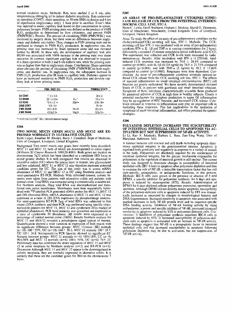
testinal oxidative stress. Methods: Rats were studied 2 or 8 wks afterstreptozotocin (60mglkg ip) to induce diabetes sensitivity. Rats underwentno operation (CONT), sham operation, or 30 min SMA occlusion and 4 hrsof reperfusion (euglycemics only). I hour prior to sacrifice, Evan's bluewas injected to assess capillary leak based on dye concentration in serum,pulmonary lavages. PMN priming state was defined as the product of PMNHZ0 2 production as determined by flow cytometry, and percent PMN(PMNIWBC). Results: The percent of circulating PMN (PMNIWBC) wasincreased by surgery alone, but there were no differences between euglycemic and diabetic rats (table). Thus, changes in PMN priming state can beattributed to changes in PMN HzOz production. In euglycemic rats, thepriming state was increased by sham operation alone and was elevatedfurther by IR240. In these rats, the development of capillary leak afer[R240 was associated with 100% increase in the priming state over shamoperation. In contrast, significant capillary leak was observed in responseto a sham operation in both 2 and 8 wk diabetic rats, while the priming statewas no higher than that in euglycemic shams. Conclusions: Sham operationalone elevates the percent of circulating PMN (PMNIWBC)in both euglycemic and diabetic rats. In euglycemia rats, the subsequent increase inPMN H20 2 production after IR leads to capillary leak. Diabetics appear tohave an increased sensitivity to PMN H20 2 production and develop capillary leak at lower priming states.
PMNIWSC(%) SAL PRIMING STATE
EU CONT 118± 06 20± 2EU SHAM 62.7 ± 6.5- 4± 1 110 ±17-EU 1R240 734±31- 20±4- 218± 36-DIASCONT 19.6±3.5 39±9-2WKSHAM 40.1±8.6- 14±2- 65±20-8WKSHAM 43.9±2.3- 16+2- 123±13-
·P,;0.05 VS EU CONT; SAL= Sronchoalveolar lavage
5204
TWO NOVEL MUCIN GENES MUCH AND MUC12 ARE EXPRESSED NORMALLY IN ULCERATIVE COLITIS.Keith Leiper, Jonathan M. Rhodes, Barry J. Campbell, Dept of Medicine,Univ of Liverpool, Liverpool, United Kingdom.
Background Two novel mucin core genes have recently been described,MUC II and MUC 12, both of which are downregulated in colon cancer(Williams SJ Cancer Research 1999;59:4083). Both genes are located onchromosome 7q22 which has been identified as a candidate 18D locus inrecent genetic studies. It is well recognised that rnucins are abnormal inulcerative colitis (UC) where the mucus layer is thinner, less glycosylatedand less sulphated. MUC II or MUC 12 may therefore represent potentialcandidate genes for UC. The aim of this study was to assess mRNAabundance of MUC II and MUC 12 in UC using Northern analysis andserni-quantatative RT-PCR. Methods With informed consent, colonic biopsies were taken from patients with ulcerative colitis and patients withnormal colon. Total RNA was extracted using a commercially available kit.For Northern analysis, 20JLg total RNA was electrophoresed and transferred onto nylon membranes. Membranes were then sequentially hybridised with 32p labelled PCR generated cDNA probes for MUC II,MUC 12and 28S ribosomal RNA. mRNA abundance of MUC II and MUC 12 wasexpressed as a ratio of 28S RNA expression by phosphorimage analysis.For semi-quantatative RT-PCR 5JLg of total RNA was subjected to firststrand eDNA synthesis and then PCR was performed using specific oligonucleotide primers for MUC II, MUC 12 and cytokeratin 20 (a marker ofepithelial abundance). PCR band intensity was quantified and expressed asa ratio of cytokeratin 20 abundance. All results were expressed as apercentage of control normal colon (100%). Results Northern analysis forMUC II and MUCI2 revealed a polydisperse signal typical of mucins.Quantification showed a wide variation in expression of both mucin withno significant difference between groups: MUC I I (mean::t:SE) normals(n=IO) 100::t:35%, UC (n=18) 144::t: 30.3, MUC 12 normals 100::t:19.7,UC 120::t: 16.8. Semiquantative PCR likewise showed no significant differences between groups: MUC II normals (n=6) lOO::t: 18%, UC (n=6)90::t:38% and MUC 12 normals (n=6) 100::t: 54%, UC (n=6) 76::t: 23.Preliminary data has confirmed the down regulation of MUC 11 and MUC12 in colon neoplasia by Northern analysis (n=2) and RT-PCR (n=2).Discussion Although MUC 11 and MUC 12 appear to be downregulated incolonic neoplasia, they are normally expressed in ulcerative colitis. It isunlikely that these are the candidate genes for IBD on the chromosome 7locus.
AGAAl129
5205AN ARRAY OF PRO-INFLAMMATORY CYTOKINES STlMU·LATE RELEASE OF CCK FROM THE INTESTINAL ENTEROEN·DOCRINE CELL LINE, STC-l.Fiona C. Leslie, Geoff Warhurst, Graham J. Dockray, David G. Thompson,Univ of Manchester, Manchester, United Kingdom; Univ of Liverpool,Liverpool, United Kingdom.
Aims: To study the effect of an array of pro-inflammatory cytokines on thecholecystokinin(CCK)-secreting cell line, STC-1. Methods: The CCKsecreting cell line STC-I was incubated with an array of pro-inflammatorycytokines IFN-'}', [L-I J3 and TNF-a at varying concentrations for 2 hours,followed by a standard 15 minute stimulation period with lauric acid (C 12).The supernatant was collected and CCK levels were measured by radioimmunoassay. Results: After incubation with IFN-'}' (1.25U/ml), lipidinduced CCK secretion was increased by 74.6 ::t: 28.4% compared tocontrol (p<O.003), with IL-IJ3 (0.125 ng/ml) by 74.9 ::t: 21.73% comparedto control (p<0.004), and with TNF-a (5 ng/ml) by 39.2 ::t: 11.86%compared to control (p<0.0002). Responses were dose dependent. Conclusions: An array of pro-inflammatory cytokines stimulate nutrient-induced CCK release from the CCK secreting cell line, STC-I. The effectsof mucosal inflammation on small intestinal enteroendocrine (EEC) cellfunction are poorly understood. We have recently demonstrated elevatedlevels of CCK in patients with giardiasis and small intestinal infection.Symptoms of these infections characteristically resemble those producedby parenteral infusion of CCK in high dose in healthy subjects. Clearly apotential mechanism for symptom production in upper GI inflammationmay be up-regulation of EEC function, and increased CCK release. Cytokines released in response to inflammation may play an important role inmediating these responses. This has implications in the mediation ofsymptoms of small intestinal inflammation, and offers potential therapeuticstrategies.
5206
POLYAMINE DEPLETION INCREASES THE SUSCEPTIBILITYOF INTESTINAL EPITHELIAL CELLS TO APOPTOSIS VIA ACTIVATION BUT NOT SUPPRESSION OF NF-KB ACTIVITY,Li Li, Rao N. Jaladanki, Barbara L. Bass, Jian-Ying Wang, Univ ofMaryland, Baltimore, MD.
A balance between cell renewal and cell death including apoptosis determines epithelial integrity in the gastrointestinal mucosa. Apoptosis isregulated both positively and negatively in response to a variety of stimuliin the body. Polyamines are absolutely required for the maintenance ofintestinal epithelial integrity, but the precise mechanism responsible forpolyamines in the regulation of mucosal growth is still unclear. The currentstudy was designed to determine changes in susceptibility of intestinalepithelial cells (IEC-6Iine) to apoptosis after polyamine depletion and thento examine the role of NF-kB, a inducible transcription factor that has celltype-speci fie, proapoptotic, or antiapoptotic functions, in this process.Methods: IEC-6 cells were grown in the presence or absence of 5 mMDFMO, a specific inhibitor for polyamine synthesis, for 6 days and apoptosis is induced by staurosporine (STS). Results: Administration ofDFMO for 6 days depleted cellular polyamines putrescine, spermidine andspermine. Although DFMO did not directly induce apoptosis, susceptibilityof the polyamine-deficient cells to apoptosis induced by STS was dramatically increased as measured by changes in morphological features andDNA fragmentation. Increased sensitivity to apoptosis was associated withmarked increases in both NF-kB protein level and its sequence-specificDNA binding activity. Inhibition of NF-kB binding activity by usingsulfasalazine, a potent and specific inhibitor of NF-kB, prevented increasedsensitivity to apoptosis induced by STS in polyamine-deficient cells. Conclusions: 1) Inhibition of polyamine synthesis sensitizes IEC-6 cells toapoptosis induced by STS. 2) Increased susceptibility of polyamine-deficient cells to apoptosis is associated with an increase in NF-kB activity.These findings suggest that NF-kB is a proapoptotic factor in intestinalepithelial cells and that increased susceptibility to apoptosis followingpolyamine depletion may be due to activation, but not suppression. ofNF-kB activity.