role of inflammation and ... - texas a&m...
TRANSCRIPT
ROLE OF INFLAMMATION AND ENDOTHELIAL DYSFUNCTION
OF CORONARY ARTERIOLES IN TYPE 2 DIABETES
A Thesis
by
JI YEON YANG
Submitted to the Office of Graduate Studies of
Texas A&M University
in partial fulfillment of the requirements for the degree of
MASTER OF SCIENCE
August 2008
Major Subject: Biomedical Sciences
ROLE OF INFLAMMATION AND ENDOTHELIAL DYSFUNCTION
OF CORONARY ARTERIOLES IN TYPE 2 DIABETES
A Thesis
by
JI YEON YANG
Submitted to the Office of Graduate Studies of
Texas A&M University
in partial fulfillment of the requirements for the degree of
MASTER OF SCIENCE
Approved by:
Co-Chairs of Committee, Cuihua Zhang
Glen A. Laine
Committee Members, Cristine L. Heaps
Cynthia J. Meininger
Head of Department, Glen A. Laine
August 2008
Major Subject: Biomedical Sciences
iii
ABSTRACT
Role of Inflammation and Endothelial Dysfunction
of Coronary Arterioles in Type 2 Diabetes. (August 2008)
Ji Yeon Yang, B.S., Dongduk Women’s University
Co-Chairs of Advisory Committee: Dr. Cuihua Zhang
Dr. Glen A. Laine
We hypothesized that the interaction between tumor necrosis factor alpha
(TNFα)/nuclear factor-kappaB (NFκB) via activation of IKKβ may amplify one another
resulting in the evolution of vascular disease and insulin resistance associated with
diabetes. The interaction between TNFα and monocyte chemoattractant protein-1 (MCP-
1) may contribute to the evolution of vascular inflammation and endothelial dysfunction
in coronary arterioles in type 2 diabetes. To test this hypothesis, endothelium-dependent
(ACh) and –independent (SNP) vasodilation of isolated, pressurized coronary arterioles
(40-100 µm) from mLeprdb
(heterozygote, normal), Leprdb
(homozygote, diabetic) and
Leprdb
mice null for TNFα (dbTNF-
/dbTNF-
) were examined. Although dilation of vessels
to SNP was not different between Leprdb
and mLeprdb
mice, dilation to ACh was reduced
in Leprdb
mice. The NFκB antagonist, MG-132, IKKβ inhibitor, sodium salicylate
(NaSal), or Anti-MCP-1 partially restored endothelium-dependent coronary arteriolar
dilation in Leprdb
mice. Protein expression of IKKα and IKKβ were higher in Leprdb
than
in mLeprdb
mice. The expression of IKKβ, but not the expression of IKKα was increased
in dbTNF-
/dbTNF-
mice. Leprdb
mice showed increased insulin resistance, but NaSal
iv
improved insulin sensitivity. Protein expression of TNFα, NFκB, phosphorylation of
IKKβ and JNK were greater in Leprdb
mice, but NaSal attenuated protein expression of
them in Leprdb
mice. The ratio of phosphorylated IRS-1 at Ser307 (pIRS-1)/IRS-1
protein expression was elevated in Leprdb
mice; both NaSal and JNK inhibitor SP600125
reduced pIRS-1/IRS-1 in Leprdb
mice. MG-132 or neutralization of TNFα reduced
superoxide production in Leprdb
mice. Anti-MCP-1 attenuated superoxide production
and protein expression of nitrotyrosine (N-Tyr), which is an indicator of peroxynitrite
production, in isolated coronary arterioles of Leprdb
mice. Immunostaining results
showed that expression of MCP-1 and vascular cellular adhesion molecule-1 (VCAM) is
co-localized with endothelial cells and macrophages. Anti-TNFα or anti-MCP-1
markedly reduced macrophage infiltration and the number of MCP-1 positive cells.
Neutralization of TNFα or anti-MCP-1 reduced the expression of adhesion molecules. In
conclusion, our results indicate that the interaction between NFκB and TNFα signaling
induces activation of IKKβ. In addition, TNFα and TNFα-related signaling, including
the expression of MCP-1 and adhesion molecules, further exacerbates oxidative stress
leading to endothelial dysfunction in type 2 diabetes.

v
DEDICATION
To mom, dad and my husband, Min Jae Song. This project is dedicated to you.

vi
ACKNOWLEDGEMENTS
I would like to acknowledge those individuals who have given me motivation to
succeed, financial assistance, and most of all their friendship. First, my advisor,
Professor Cuihua Zhang, for the guidance and help I received during my master's thesis
research. I wish to express my warm and sincere thanks to Professor Louise Abbott for
her detailed and constructive comments. Also I would like to appreciate Professor Glen
A. Laine, Cristine L. Heaps and Cindy Meininger for agreeing to serve as my committee
co-chair and members. Without doubt, I could not have completed this step of my life
without my many friends, family, and mentors who have assisted me in many forms and
manners. Mom and Dad, you are truly the best. I am indebted to you for all your love
and encouragement. Thanks to my wonderful and beautiful sister and brother. My lab
members: Yoonjung Park, Hanarui Zhang, Sewon Kim, Darcey Klaahsen, XiuPing
Chen, Xue Gao, Stefano Capobianco, Yuan Gao. I thank you for the positive things,
large and small, that you have added to my life. This study was supported by an
Atorvastatin Research Award from Pfizer (2004-37), an American Heart Association
SDG (110350047A), and NIH grants (RO1-HL077566 and RO1-HL085119).
vii
TABLE OF CONTENTS
Page
ABSTRACT .............................................................................................................. iii
DEDICATION .......................................................................................................... v
ACKNOWLEDGEMENTS ...................................................................................... vi
TABLE OF CONTENTS .......................................................................................... vii
LIST OF FIGURES................................................................................................... x
LIST OF TABLES .................................................................................................... xi
CHAPTER
I INTRODUCTION................................................................................... 1
A. Feed-forward Signaling of TNFα and NFκB via IKKβ Pathway
Induces Insulin Resistance and Coronary Arteriolar Dysfunction in
Type 2 Diabetic Mice ........................................................................ 1
B. Role of MCP-1 in TNFα-induced Endothelial Dysfunction in
Type 2 Diabetic Mice ........................................................................ 3
II METHODS ............................................................................................. 5
A. Animal Treatment............................................................................. 5
1. Animal Model............................................................................... 5
2. Neutralization and Treatment ....................................................... 5
B. Measurement of Blood Parameters................................................... 6
1. Measurement of Glucose.............................................................. 6
2. Serum Cholesterol Level .............................................................. 7
3. Serum Insulin Level ..................................................................... 7
4. HOMA-IR..................................................................................... 7
C. Functional Study............................................................................... 7
1. Preparation of Coronary Microvessels ......................................... 7
2. Data Analysis................................................................................ 9
3. Insulin Tolerance Test .................................................................. 9
D. Molecular Study ............................................................................... 9
1. Electron Paramagnetic Resonance (EPR) Spectroscopy.............. 9
2. Immunofluorescence .................................................................... 10
3. Immunoblotting ............................................................................ 11

viii
CHAPTER Page
4. Quantitative Reverse Transcriptase-Polymerase Chain Reaction
(RT-PCR) ..................................................................................... 12
III DATA ANALYSIS I.............................................................................. 13
A. Roles of IKKα and IKKβ in Type 2 Diabetes .................................. 13
B. Activation of IKKβ, NFκB and Degradation of IκBα-induced
by TNFα ........................................................................................... 15
C. Role of Oxidative Stress in Type 2 Diabetes-induced Vascular
Dysfunction ...................................................................................... 16
D. Effect of NaSal on Improvement of Insulin Sensitivity ................... 17
E. Role of NaSal in Vascular Dysfunction in Type 2 Diabetes............. 18
F. Insulin Resistance Enhanced Activity of JNK and IKKβ in Type 2
Diabetes ............................................................................................ 20
IV DATA ANALYSIS II ............................................................................ 24
A. TNFα and MCP-1 Amplification of Signaling in Coronary
Arterioles in Type 2 Diabetes........................................................... 24
B. Cellular Source of MCP-1 Expression in Type 2 Diabetes .............. 25
C. Role of MCP-1 in Type 2 Diabetes-Induced Vascular Dysfunction 28
D. TNFα and MCP-1-induced Macrophage Recruitment and
Activation ......................................................................................... 29
E. TNFα and MCP-1-induced Adhesion Molecules and Macrophage
Infiltration......................................................................................... 31
V SUMMARY AND CONCLUSION ...................................................... 33
A. Roles of Interaction of TNFα and NFκB Signaling on Impaired
Coronary Arteriolar Responses in Type 2 Diabetes ......................... 34
B. The Role of IKKβ in Vascular Dysfunction in Type 2 Diabetes...... 36
C. Link between Inflammation-induced Insulin Resistance and
Vascular Dysfunction ....................................................................... 38
D. Roles of TNFα and MCP-1 in Type 2 Diabetes............................... 42
E. The Role of MCP-1 in ROS Production in Coronary Arterioles
in Type 2 Diabetes............................................................................ 43
F. Enhancement of Adhesion Molecules by MCP-1 and TNFα in
Endothelial Cells in Diabetes ........................................................... 45

ix
Page
REFERENCES.......................................................................................................... 49
VITA.......................................................................................................................... 57

x
LIST OF FIGURES
FIGURE Page
1 Isolated vessel set-up for coronary arteries ................................................ 8
2 Analysis of IKK subunit expression........................................................... 14
3 IKKβ activity.............................................................................................. 15
4 Analysis of IκBa and NFκB expression ..................................................... 16
5 NFκB induces O2˙¯ production .................................................................. 17
6 Insulin tolerance test................................................................................... 18
7 NaSal improves arteriolar function ............................................................ 19
8 Effect of NaSal on IKKβ and JNK activity................................................ 21
9 Protein expression of IRS-1 ser307............................................................ 22
10 Putative TNFα/ NFκB and insulin signaling pathways ............................. 23
11 Interaction between TNF and MCP-1 ........................................................ 25
12 Co-localization of MCP-1 in microvessels ................................................ 27
13 Role of MCP-1 on endothelial function ..................................................... 28
14 Effects of neutralizing MCP-1 in Leprdb
on oxidative stress ..................... 29
15 Co-localization of MCP-1 in macrophages ................................................ 30
16 Analysis of adhesion molecule expression ................................................ 32
17 Potential pathway of tyrosine nitration oxidation ...................................... 43
18 Interactions among TNF, MCP-1 and adhesion molecules in endothelium 48

xi
LIST OF TABLES
TABLE Page
1 Serum parameters in mice with NaSal ....................................................... 13
2 Serum parameters in mice with anti-MCP-1.............................................. 24
1
CHAPTER I
INTRODUCTION
A. Feed-forward Signaling of TNFα and NFκB via IKKβ Pathway Induces Insulin
Resistance and Coronary Arteriolar Dysfunction in Type 2 Diabetic Mice
The prevalence of obesity and diabetes has led to a significant increase in the
complications associated with cardiovascular disease. Many of these complications can
be linked to a dysregulation of the body's immune system.1,2
Tumor necrosis factor alpha
(TNFα) is a pro-inflammatory cytokine that has been implicated in cardiovascular
diseases.3,4
We previously found that increases in TNFα expression induces activation of
NAD(P)H oxidase and the production of reactive oxidative species (ROS), leading to
endothelial dysfunction in type 2 diabetes.5 Importantly, TNFα activates the
transcription nuclear factor-kappaB (NFκB), which regulates the expression of genes
involved in inflammation, oxidative stress and endothelial dysfunction.5 TNFα also
initiates the signaling cascades via the inhibitor of NFκB (IκB) kinase (IKK) complex,
which contains IKKα and IKKβ. The inhibitory protein IκBα is phosphorylated,
ubiquitinated and degraded by proteasomes, releasing NFκB to translocate into the
____________ This thesis follows the style of Circulation Research.

2
nucleus. Under normal physiological conditions, the inflammatory response is
terminated by binding NFκB with the inhibitory protein IκB.6,7
Investigation of TNFα-
mediated NFκB pathway is needed to identify specific pro-inflammatory agents in type
2 diabetes.
Salicylates, including aspirin and sodium salicylate (NaSal) are common
nonsteroidal anti-inflammatory drugs (NSAIDs), which have anti-platelet and anti-
inflammatory effects. Aspirin inhibits prostaglandin production, but NaSal has a
prostaglandin-independent effect. NaSal inhibits the activity of IKKβ, which is required
for the activation of NFκB. IKKβ, the molecular target of NaSal, is associated with the
rise of insulin resistance.8,9
Recent work suggests that specific antagonism of the NFκB
inflammatory pathway through the inhibition of IKKβ reduces acute myocardial damage
following ischemia-reperfusion injury10
and insulin action plays a critical role in
reducing myocardial infarct size by increasing cardiac myocyte metabolism.11-13
We
previously found that TNFα-activated Jun N-terminal kinase (JNK), which mediates
O2˙¯ production and impairs endothelium-dependent vasodilation in coronary
arterioles.14
NaSal prevents TNFα-induced JNK activation from blocking insulin
signaling via a serine phosphorylation.15
Therefore, we evaluated 1) whether reciprocal
interactions between TNFα and NFκB via activation of IKKβ accentuate the evolution
of vascular disease in type 2 diabetes; and 2) whether NaSal restores endothelial
dysfunction and insulin resistance by inhibiting IKKβ/NFκB activity thereby preventing
JNK activation in coronary arterioles from type 2 diabetes (Leprdb
), type 2 diabetic mice
null for TNFα (dbTNF-
/dbTNF-
) and lean control (mLeprdb
) mice.

3
B. Role of MCP-1 in TNFα-induced Endothelial Dysfunction in Type 2 Diabetic Mice
Diabetes is accompanied by a maturing inflammatory response that consists of
increased TNFα and monocyte chemoattractant protein-1 (MCP-1) expression in
adipocytes and the lumen of vessels.16
At the tissue level, diabetic inflammation
enhances expression of adhesion molecules, which facilitates macrophage
infiltration.17~19
The progression of vascular disease in type 2 diabetes is caused by the
complex interaction of many factors. We previously found that increases in TNFα
expression induces activation of NAD(P)H oxidase and production of reactive oxidative
species (ROS), leading to endothelial dysfunction in type 2 diabetes.5
Additionally,
TNFα is reported to enhance MCP-1 expression;20
thus the interaction between these
two systems may contribute significantly to the evolution of vascular disease in diabetes.
MCP-1 is a major component of adipose tissue that contributes to abnormal lipid
metabolism21
and is expressed by a variety of activated cells (e.g., endothelial cells,
monocytes, and smooth muscle cells). MCP-1 has been thought to be essential in
defending the host against foreign organisms and is an important mediator in chronic
inflammation.22
Many studies suggest that MCP-1 plays a detrimental role in the
development of vascular disease through recruitment of monocytes that accumulate
in/on vascular endothelium and may augment vascular disease at the inflammatory
site.23-25
Monocytes that migrate from the peripheral vasculature into the myocardium
undergo maturation to macrophages, which then release proteolytic enzymes and
damage the myocardium.26
The action of MCP-1 on the stages of monocyte recruitment
(rolling and/or adhesion) is associated with the activation of specific cell surface proteins.

4
In this regard, adhesion molecules (e.g.,vascular cell adhesion molecule-1 [VCAM],
intracellular adhesion molecule-1 [ICAM], and E-selectin as well as MCP-1) are
responsible for myocardial injury and cardiac contractile dysfunction.27
Thus, the goal of this study was to document the cellular and molecular
mechanisms involved in MCP-1 in the coronary microcirculation in TNFα-induced
endothelial dysfunction in type 2 diabetes. We examined the role of MCP-1 in type 2
diabetes, and tested whether the interaction of TNFα and MCP-1 amplifies the signaling
of adhesion molecules and macrophage infiltration. Endothelial function and the
production of ROS were determined in isolated coronary arterioles (40-100 µm) from
genetically modified mice with type 2 diabetes (Leprdb
) with and without treatment with
MCP-1 neutralizing antibody. We also tested the co-localization of MCP-1, CD68 and
adhesion molecules in type 2 diabetes using immunohistochemical analysis.

5
CHAPTER II
METHODS
A. Animal Treatment
1. Animal Model
The procedures followed were in accordance with approved guidelines set by the
Laboratory Animal Care and Use Committee at Texas A&M University. Heterozygote
controls (C57BLKS/J, mLeprdb
), homozygote type 2 diabetes (C57BLKS/J, Leprdb
) and
Leprdb
null for TNFα (dbTNF-
/dbTNF-
) mice were purchased from Jackson Laboratory and
maintained on a normal rodent chow diet. Our studies utilized 12-16 week-old, 20-30 g
mLeprdb
, 40-60 g Leprdb
and dbTNF-
/dbTNF-
mice of either sex. The cross (dbTNF-
/dbTNF-
)
of Leprdb
with TNFKO is heterozygous for Lepr
db and homozygous for TNF KO
(TNF−/−).The db
TNF-/db
TNF- mice show the phenotypes of hyperglycemia and obesity that
is consistent with diabetes and the penetrance of the leptin receptor mutation. The obese
mice from the second round of breeding of Leprdb
and TNF−/− mice were used in
experimentation.
2. Neutralization and Treatment
The neutralizing antibody to TNFα28
is 2E2 monoclonal antibody (2E2 MAb.
94021402, NCI Biological Resources Branch). At 12-16 weeks of age, all mice received
anti-TNF (2E2 MAb. 0.625 mg/ml/kg/day, 3 days, i.p.); dosage was based on our
estimates of TNFα expression, which was estimated to be in the low ng or pg range.
This amount was able to neutralize 10-100 fold more than the estimated levels of TNFα.

6
MG132 tripeptide (Z-Leu-Leu-Leu- aldehyde, Sigma, dissolved in dimethyl
sulfoxide [DMSO]) is a peptide aldehyde proteasome inhibitor. Ultimately preventing
IκBα degradation by the proteasome restricts NFκB activity in vitro.7 Lepr
db mice were
injected (10 mg/kg/day for 3 days, i.p.).29
The contribution of NAD(P)H oxidase in
generating O2˙¯ was assessed by treating mice with the NAD(P)H oxidase inhibitor
apocynin (100 mg/kg/day, i.p. for 3 days). We administered NaSal (Calbiochem; 6
mg/ml in drinking water for 7-10 days, pH 7) to Leprdb
and mLeprdb
mice to determine
whether blocking IKKβ affects their blood glucose level. To determine whether
blockage of JNK affects insulin receptor substrate-1 (IRS-1) activity, we also
administered the JNK inhibitor, SP600125 (CalBiochem, 10 mg/kg/day in 0.05 ml
DMSO, i.p. for 7 days) to Leprdb
mice.30
Mice were treated with the neutralizing
antibody to MCP-1 (anti-MCP-1; 200 mg/kg/day, i.p. for 3 days, MURINE; Biovision)
to block MCP-1 signaling.
B. Measurement of Blood Parameters
Blood was obtained from the vena cava after anesthesia with sodium
pentobarbital (50 mg/kg; i.p.) and exposure of the vein. Whole blood samples were spun
at 3,000 rpm for 10 minutes, and serum was stored at -80°C until analysis.
1. Measurement of Glucose
We used a OneTouch Ultramini glucometer (LifeScan) for measuring blood
glucose in mLeprdb
, Leprdb
and Leprdb
mice treated with NaSal at the same time (8:00-
10:00AM) throughout the protocol.
7
2. Serum Cholesterol Level
Serum cholesterol level was measured with the Cholesterol/Cholesteryl Ester
Quantitation Kit (Biovision) using spectrophotometry (Multiskan MCC, Fisher
Scientific). A large portion of cholesterol in blood is in the form of cholesteryl esters.
Cholesterol esterase hydrolized cholesteryl ester into cholesterol. Cholesterol is then
oxidized by cholesterol oxidase to yield H2O2. The produced H2O2 interacts with a
sensitive cholesterol probe.
3. Serum Insulin Level
Insulin was measured with the use of a commercial kit, insulin (Mouse)
Ultrasensitive EIA (ALPCO Diagnostics). Insulin concentration was measured by
spectrophotometry (Multiskan MCC, Fisher Scientific).
4. HOMA-IR
Insulin resistance was determined by utilizing the homeostasis model assessment;
HOMA-IR using the following formula:
HOMA-IR= ((non-fasting glucose [mmol/L]) × (non-fasting insulin [mU/L]))/22.5.
C. Functional Study
1. Preparation of Coronary Microvessels
The techniques for identification and isolation of coronary microvessels were described
in detail previously.31,32
Coronary arterial microvessels (<100 µm) were carefully
dissected out for in vitro study. After isolation, the microvessel was cannulated with two
glass micropipettes filled with filtered albumin-PSS, and the outside of the microvessel

8
was securely tied to the pipettes with 11-O ophthalmic suture. The preparation was
transferred to the stage of an inverted microscope coupled to a CCD camera and video
micrometer (Figure 1). To determine whether NFκB, IKKβ, or MCP-1 was playing a
role in endothelial injury in type 2 diabetes, the endothelium-dependent vasodilator
acetylcholine (ACh, 0.1 nmol/L to 10 µmol/L), the endothelial independent vasodilator
sodium nitroprusside (SNP, 0.1 nmol/L to 10 µmol/L)-induced, and flow-induced
vasodilation (NO-mediated, endothelial dependent but agonist independent; 4 to 60 cm
H2O) were assessed in coronary arterioles in mLeprdb
, Leprdb
, and Leprdb
mice treated
with MG132, NaSal, or anti-MCP-1. Flow was established by the production of a
pressure drop across the vessel, and linearly related to the pressure drop (∆P).
Figure 1. Isolated vessel set-up for coronary arteries.
9
2. Data Analysis
At the end of each experiment, the vessel was relaxed with 100 µmol/L SNP to
obtain its maximal diameter at 60 cm H2O intraluminal pressure.32
All diameter changes
to pharmacological agonists were normalized to the control diameter and expressed as a
percentage of control diameter. Functional data are presented as mean±SEM, except as
specifically stated (e.g. as mean±SD for molecular study). Statistical comparisons of
vasomotor responses under various treatments were performed with two-way ANOVA
and intergroup differences were tested with Bonferonni Inequality. Significance was
accepted at P<0.05.
3. Insulin Tolerance Test
The tail was nicked with a fresh razor blade by horizontal cut of the tip and the
OneTouch Ultramini glucometer was used to measure baseline blood glucose. A total of
0.75 units per kg body weight of diluted insulin from porcine pancreas (Sigma) was
injected into the intraperitoneal cavity. Blood samples were taken for glucose
determinations 0, 30, 60, and 120 minutes later. Blood was sampled from the tail of each
mouse by gently massaging a small drop of blood onto the glucometer strip.
D. Molecular Study
1. Electron Paramagnetic Resonance (EPR) Spectroscopy
Homogenates of (4-6 isolated coronary arterioles) were prepared in a 50 mmol/L
phosphate buffer containing 0.01 mmol/L EDTA. The homogenate was then subjected to
low-speed centrifugation (1000 g) for 10 minutes to remove unbroken cells and tissue
debris. The supernatants containing 2 mmol/L CPH (1-hydrox-3 carboxypyrrolidine)

10
were incubated for 30 minutes at 37 °C and frozen quickly in liquid nitrogen. EPR
spectroscopy was performed at room temperature using a Bruker EMX spectrometer and
1 mm diameter capillaries. Superoxide quantitation from the EPR spectra was
determined by double integration of the peaks, with reference to a standard curve from
horseradish peroxidase generation of the anion from standard solutions of hydrogen
peroxide, using p-acetamidophenol as the co-substrate,33,31
then normalized by protein
concentration.
2. Immunofluorescence
Mouse heart was perfused with PBS and then perfused again with 4 %
paraformaldehyde. Isolated heart tissue was fixed in 4 % paraformaldehyde diluted in
PBS 0.1 M (overnight at 4 °C). Fixed heart tissue was perfused in 20 % sucrose diluted
in PBS 0.1 M (overnight at 4 °C). Once the heart was completely perfused, the tissue
was embedded in OCT and snap-frozen at –60 °C and stored at –80 °C. Frozen tissues
were sectioned at 5 µm thick, using cryostat (Leica) and slides allowed to dry at room
temperature; then the tissues were incubated in 0.3 % Triton-100 for 30 minutes and
washed in PBS 3 times. 5 % goat serum was used for 30 minutes to block nonspecific
binding. Anti-rat primary MCP-1, VCAM-1 antibodies (Abcam) and anti-rabbit primary
α-actin, von Wilibrand factor (VWF), anti-mouse primary CD68 (Abcam) as
monocyte/macrophage marker were incubated sequentially overnight at 4 °C. Sections
were washed 3 times in PBS. Appropriate secondary FITC-(green color) and Texas
Red-(red color) conjugated antibodies were incubated with sections for 4 hours and
washed off. Sections were finally mounted in an anti fading agent (Slowfade gold with
11
DAPI, Molecular Probes), and then the slides were observed and analyzed with the use
of a fluorescence microscope (Zeiss Axioplan microscope with a 40X objective or a
Nikon microscope with a 63X objective).
3. Immunoblotting
Coronary arterioles (4-6 vessels per sample) or heart tissues were separately
homogenized and sonicated in lysis buffer (Cellytic™ MT Mammalian Tissue
Lysis/Extraction Reagent, Sigma). Protein concentrations were assessed using a BCA™
Protein Assay Kit (Pierce), and equal amounts of protein were separated by SDS-PAGE
and transferred to nitrocellulose or PVDF membranes (Pierce Biotechnology). IKKα
(Santa Cruz), IKKβ (Abcam), p-IKKβ (Cell Signal), IκBα (Santa Cruz), p-IκBα (Santa
Cruz), NFκB (Santa Cruz), TNFα (Santa Cruz), N-Tyr (Abcam), JNK, p-JNK (Abcam),
IRS-1, and pIRS-1 (Millipore) antibodies were utilized to assess protein expression in
mLeprdb
, Leprdb
, and Leprdb
mice treated with anti-TNF (0.625 mg/ml/kg/day, i.p. for 3
days), NAD(P)H oxidase inhibitor apocynin (100 mg/kg/day, i.p. for 3 days),34
NFκB
antagonist MG132 (10 mg/kg/day, i.p. for 3 days), NaSal (6 mg/ml in drinking water), or
SP600125 (10 mg/kg/day in 0.05 ml DMSO, i.p. for 7 days). MCP-1 (Abcam), N-Tyr
(Abcam, an indicator for peroxynitrite-mediated tissue injury), VCAM, ICAM, and E-
selectin (Santa Cruz) protein expression were determined with mLeprdb
, Leprdb
, and
Leprdb
mice treated with anti-TNFα (0.625 mg/ml/kg/day, i.p. for 3 days). Signals were
visualized by enhanced chemiluminescence (ECL, Amersham), and quantified by Fuji
film imaging software.

12
4. Quantitative Reverse Transcriptase-Polymerase Chain Reaction (RT-PCR)
RNA is first reverse transcribed into cDNA using a reverse transcriptase as
described. The resulting cDNA is used as templates for subsequent PCR amplification
using primers specific for one or more genes. RT-PCR can also be carried out as one-
step RT-PCR in which all reaction components are mixed in one tube prior to starting
the reactions.
Total RNA was extracted from heart tissue using Trizol reagent (Life
Technologies Inc), and was processed directly to cDNA using the SuperScript™ III
Reverse Transcriptase (Life Technologies Inc). The primers of IKKα and IKKβ were
designed (Beacon software) and synthesized (Invitrogen). cDNA was amplified using a
qRT-PCR Kit with SYBR® Green (Life Technologies Inc). Data were calculated by 2-
∆∆CT method and presented as fold change of transcripts for IKKα and IKKβ gene in
Leprdb
mice normalized to β-actin, compared with mLeprdb
mice (defined as 1.0 fold).
∆∆CT=(CT-target-CT- β-actin) Leprdb
- (CT-target-CT- β-actin) mLeprdb

13
CHAPTER III
DATA ANALYSIS I
Table 1. Serum parameters in mice with NaSal.
Body weight and serum parameters were measured at 12-16 weeks for the different
strains of mice (Table 1). Leprdb
mice were treated with NaSal for 7-10 days and serum
was collected from non-fasting mice in the morning. Non-fasting measurements were
required since the Leprdb
murine model was intolerant to a fasting protocol. Data
represent mean ± SD, n=8. *p<0.05 vs mLeprdb
, #
p<0.05 vs Leprdb
.
A. Roles of IKKα and IKKβ in Type 2 Diabetes
To evaluate the roles of IKKα and IKKβ in diabetes, Leprdb
mice were treated
with anti-TNF, NAD(P)H oxidase inhibitor apocynin, and NFκB antagonist MG132.
Protein expression of IKKα and IKKβ were increased in Leprdb
mice (Figure 2). IKKβ
expression was significantly attenuated in Leprdb
mice treated with MG-132 or apocynin,

14
or in dbTNF-
/dbTNF-
mice vs. Leprdb
mice. However, the alteration of IKKα expression
was not observed in Leprdb
mice treated with neutralizing antibody to TNFα, or in dbTNF-
/dbTNF-
mice. These findings suggest a link among IKKβ, NFκB, NAD(P)H oxidase and
TNFα in coronary arterioles from mice with type 2 diabetes.
Figure 2. Analysis of IKK subunit expression. (a) Anti-TNF attenuated IKKβ, not IKKα
protein expression in Leprdb
mice. (b) mRNA expression of IKKα and IKKβ are
presented as relative (i.e., -fold) changes in Leprdb
compared with control mice. Data are
normalized to β-actin expression levels. Data represent mean±SD, n=4. * p<0.05 vs.
mLeprdb
; # p<0.05 vs. Leprdb
.

15
B. Activation of IKKβ, NFκB and Degradation of IκBα-induced by TNFα
Phosphorylation of IKKβ activates IκBα to decrease NFκB inhibitory action of IκBα.
In Leprdb
mice heart, protein expression of p-IKKβ was higher, but expression of p-
IKKβ was attenuated by anti-TNF, MG132, apocynin or in dbTNF-
/dbTNF-
mice (Figure
3). In Leprdb
mice heart, protein expression of IκBα was lower (Figure 4a) vs. mLeprdb
mice. However, protein expression of p-IκBα was higher in Leprdb
vs. mLeprdb
mice.
Protein expression of p-IκBα was attenuated in dbTNF-
/dbTNF-
and Leprdb
mice treated
anti-TNF, apocynin or MG-132 vs. mLeprdb
mice (Figure 4b). In Leprdb
mice heart,
protein expression of NFκB was higher, but apocynin, MG-132 or dbTNF-
/dbTNF-
mice
presented a reduced protein expression of NFκB (Figure 4c).
Figure 3. IKKβ activity. Protein expression of p-IKKβ was higher in Leprdb
vs. mLeprdb
.
Data represent mean±SD, n=4. *p<0.05 vs. mLeprdb
; #p<0.05 vs. Leprdb
.
16
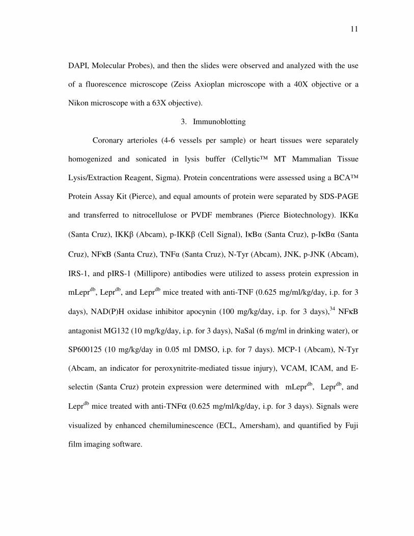
Figure 4. Analysis of IκBa and NFκB expression. Data represent mean±SD, n=4.
*p<0.05 vs. mLeprdb
; #p<0.05 vs. Leprdb
.
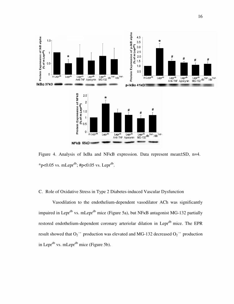
C. Role of Oxidative Stress in Type 2 Diabetes-induced Vascular Dysfunction
Vasodilation to the endothelium-dependent vasodilator ACh was significantly
impaired in Leprdb
vs. mLeprdb
mice (Figure 5a), but NFκB antagonist MG-132 partially
restored endothelium-dependent coronary arteriolar dilation in Leprdb
mice. The EPR
result showed that O2˙¯ production was elevated and MG-132 decreased O2˙¯ production
in Leprdb
vs. mLeprdb
mice (Figure 5b).
17
Figure 5. NFκB induces O2˙¯ production. (a) Leprdb
mice impaired but MG132 treatment
significantly restored ACh-induced endothelial-dependent vasodilation. (b) O2˙¯
production from isolated coronary arterioles was higher in Leprdb
mice vs. mLeprdb
mice,
and MG132 attenuated O2˙¯ production in Leprdb
mice. Values were normalized to the
mean value of mLeprdb
, n=5. *p<0.05 vs. mLeprdb
; #p<0.05 vs. Leprdb
.
D. Effect of NaSal on Improvement of Insulin Sensitivity
Blood glucose, body weight, abdominal girth, lipid levels and insulin levels were
higher in Leprdb
vs. mLeprdb
mice. There were no significant differences in body weight,
abdominal girth, lipid levels and insulin levels before and after treatment with NaSal in
Leprdb
mice (See Table 1). Lower values of glucose concentration and HOMA-insulin
resistance were found in Leprdb
mice treated with NaSal. The average body weight loss
of Leprdb
mice was 2.3 ± 2.5 mg after 7 days administration with 6 mg/ml of NaSal in
drinking water. Insulin tolerance test (Figure 6) showed a significant difference in the

18
glucose clearance rate at 120 minutes after insulin administration in Leprdb
mice treated
with NaSal compared to Leprdb
mice.
Figure 6. Insulin tolerance test. Insulin was injected (0.75 U/kg, i.p.) and blood samples
were taken for glucose determinations 0, 30, 60, and 120 minutes later. (a) Insulin
tolerance test showed that the glucose clearance rate at 120 minutes after insulin
administration in Leprdb
mice treated with NaSal was significantly higher compared to
Leprdb
mice. The value was normalized to the basal glucose level. (b) Actual glucose
level. Data represent mean±SEM, n=6-8. *p<0.05 vs. mLeprdb
; #p<0.05 vs. Leprdb
.
E. Role of NaSal in Vascular Dysfunction in Type 2 Diabetes
To establish the role of IKKβ in the signaling pathway, we studied whether the
blockade of IKKβ improves endothelium-dependent vasodilation in Leprdb
mice.

19
Figure 7. NaSal improves arteriolar function. (a) ACh-induced vasodilation in isolated
mice coronary arterioles was blunted in Leprdb
vs. mLeprdb
mice, and NaSal partially
restored this dilation in Leprdb
mice. (b) NaSal partially restored flow-induced
vasodilation in Leprdb
mice. (c) SNP-induced dilation in coronary arterioles was
identical between mLeprdb
and Leprdb
mice. NaSal did not affect SNP-induced
vasodilation in Leprdb
mice. n=5; *p<0.05 vs. mLeprdb
; #p<0.05 vs. Leprdb
.

20
Functional results (Figure 7) showed that NaSal partially restored the ACh- and
flow-induced endothelium-dependent vasodilation in Leprdb
mice. SNP-induced
vasodilation was equivalent in mLeprdb
and Leprdb
mice, indicating that function of
smooth muscle was preserved.
F. Insulin Resistance Enhanced Activity of JNK and IKKβ in Type 2 Diabetes
Western blotting (Figure 8) showed that protein expression of TNFα, NFκB,
phosphorylation of IKKβ and phosphorylation of JNK were greater in Leprdb
mice heart,
but NaSal attenuated the protein expression of TNFα, NFκB, phosphorylation of IKKβ
and phosphorylation of JNK in Leprdb
mice heart. Phosphorylation of insulin-receptor
substrate-1 on serine site blocks insulin signaling transduction in diabetes. Protein
expression of IRS-1 (Figure 9a) was significantly reduced in Leprdb
, Leprdb
treated with
NaSal and Leprdb
treated with JNK inhibitor SP600125 vs. mLeprdb
mice. The ratio of
pIRS-1 and IRS-1 was significantly higher in Leprdb
, Leprdb
treated with NaSal and
Leprdb
treated with JNK inhibitor SP600125 vs. mLeprdb
mice. However, protein
expression of phosphorylation of IRS-1 (pIRS-1, Ser307) was unaffected (Figure 9b) in
Leprdb
, Leprdb
treated with NaSal and Leprdb
treated with JNK inhibitor SP600125 vs
mLeprdb
.

21
Figure 8. Effect of NaSal on IKKβ and JNK activity. Protein expression of p-IKKβ, p-
JNK, NFκB and JNK were greater in Leprdb
mice vs. mLeprdb
mice, but NaSal decreased
protein expression of p-IKKβ, p-JNK, NFκB and JNK, without affecting protein
expression of IKKβ and JNK. Data represent mean±SD, n=5. *p<0.05 vs. mLeprdb
;
#p<0.05 vs. Leprdb
.

22
Figure 9. Protein expression of IRS-1 ser307. (a) Total insulin receptor substrate-1 (IRS-
1) protein expression was decreased in Leprdb
vs. mLeprdb
mice. NaSal and SP600125
treatment did not increase IRS-1 expression. (b) p-IRS-1 (Ser307) protein expression
was elevated in Leprdb
compared with mLeprdb
control mice and both NaSal and
SP600125 treatment appeared to reduce p-IRS-1 protein expression although there was
no statistically significant difference between groups. (c) Ratio of p-IRS-1 to total IRS-1
was significantly higher in Leprdb
vs. mLeprdb
mice. NaSal and SP600125 treatment
reduced p-IRS-1/IRS1. Data represent mean±SD, n=8. *P<0.05 vs. mLeprdb
; # P<0.05
vs. Leprdb
.

23
Figure 10. Putative TNFα/ NFκB and insulin signaling pathways. This schematic shows
the correlative and causative relationship between inflammation and insulin resistance
and depicts our proposed mechanism(s) by which NaSal sensitizes insulin signaling.
TNFα triggers the activation of IKKβ (rather than IKKα) to initiate processes leading to
inflammation. NFκB is free to translocate and bind to DNA on the target genes when
IκBα is phosphorylated by IKKβ, ubiquitinated and degraded by S26 proteasome. NFκB
is involved in many aspects of the inflammatory response, e.g. induction of TNFα,
MCP-1, and adhesion molecules. NFκB itself is induced by stimuli such as pro-
inflammatory cytokines. On the other hand, JNK and IKKβ induce IRS-1 serine
phosphorylation to block insulin signaling in type 2 diabetes.

24
CHAPTER IV
DATA ANALYSIS II
Table 2. Serum parameters in mice with anti-MCP-1.
Glucose concentration and cholesterol level are higher in Lepr
db and Lepr
db mice
treated with anti-MCP-1(0.2 g/kg/day, 3 days) than in mLeprdb
control mice (P<0.05,
n=8). Abdominal girth was higher in Leprdb
and Leprdb
mice treated with anti-MCP-1 vs.
mLeprdb
on the day of surgery (n=8). * p<0.05 vs. mLeprdb
; # p<0.05 vs. Leprdb
.
A. TNFα and MCP-1 Amplification of Signaling in Coronary Arterioles in Type 2
Diabetes
We determined if TNFα and MCP-1 interact in their protein expressions. Protein
expression of TNFα and MCP-1 from isolated coronary arterioles was analyzed in
mLeprdb
, Leprdb
and Leprdb
mice treated with anti-TNFα or anti-MCP-1. Western blot
analysis (Figure 11) revealed that anti-TNFα markedly decreased MCP-1 expression and
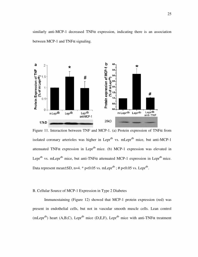
25
similarly anti-MCP-1 decreased TNFα expression, indicating there is an association
between MCP-1 and TNFα signaling.
Figure 11. Interaction between TNF and MCP-1. (a) Protein expression of TNFα from
isolated coronary arterioles was higher in Leprdb
vs. mLeprdb
mice, but anti-MCP-1
attenuated TNFα expression in Leprdb
mice. (b) MCP-1 expression was elevated in
Leprdb
vs. mLeprdb
mice, but anti-TNFα attenuated MCP-1 expression in Leprdb
mice.
Data represent mean±SD, n=4. * p<0.05 vs. mLeprdb
; # p<0.05 vs. Leprdb
.
B. Cellular Source of MCP-1 Expression in Type 2 Diabetes
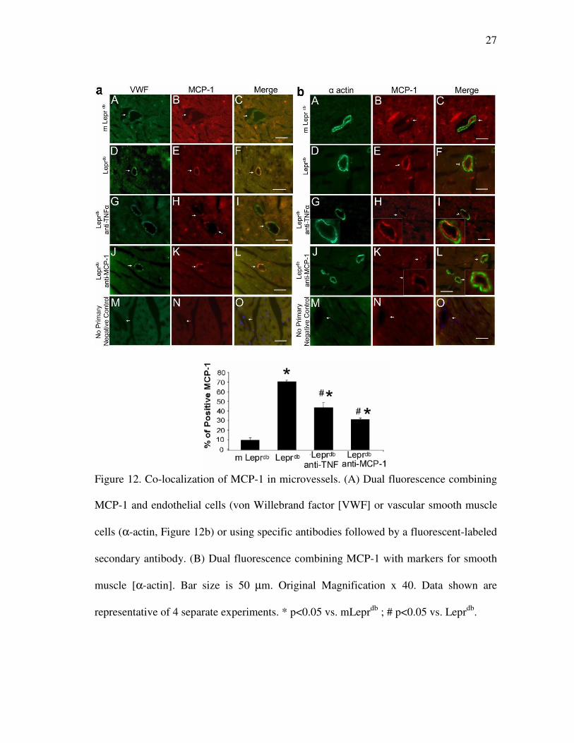
Immunostaining (Figure 12) showed that MCP-1 protein expression (red) was
present in endothelial cells, but not in vascular smooth muscle cells. Lean control
(mLeprdb
) heart (A,B,C), Leprdb
mice (D,E,F), Leprdb
mice with anti-TNFα treatment

26
(G,H,I) and Leprdb
mice with anti-MCP-1 treatment (J,K,L). The expression of MCP-1
was decreased in Leprdb
mice with anti-TNFα and anti-MCP-1 treatment. Negative
control (M,N,O) shows an absence of staining in vessels with the secondary antibodies.
Additionally, MCP-1 expression was higher in Leprdb
than in mLeprdb
mice, but anti-
TNFα or anti-MCP-1 decreased MCP-1 expression in Leprdb
mice. Leprdb
heart yielded
a high percentage (70 %) of MCP-1 positive staining in the vessels, but a lower
percentage of MCP-1 positive staining was presented in Leprdb
mice treated with anti-
MCP-1 (30 %) or anti-TNFα (40 %) comparable to that in mLeprdb
mice (15%). We
quantified MCP-1 expression in vessels by counting the specific stained MCP-1 per
section and normalized to mean value of mLeprdb
mice (data not shown). Experiments
were performed without the primary antibodies to test whether or not staining was
related to the non specific binding of the secondary antibodies, which showed no
staining in heart sections, indicating that the signals were due to specific binding of the
primary antibody.
27
Figure 12. Co-localization of MCP-1 in microvessels. (A) Dual fluorescence combining
MCP-1 and endothelial cells (von Willebrand factor [VWF] or vascular smooth muscle
cells (α-actin, Figure 12b) or using specific antibodies followed by a fluorescent-labeled
secondary antibody. (B) Dual fluorescence combining MCP-1 with markers for smooth
muscle [α-actin]. Bar size is 50 µm. Original Magnification x 40. Data shown are
representative of 4 separate experiments. * p<0.05 vs. mLeprdb
; # p<0.05 vs. Leprdb
.

28
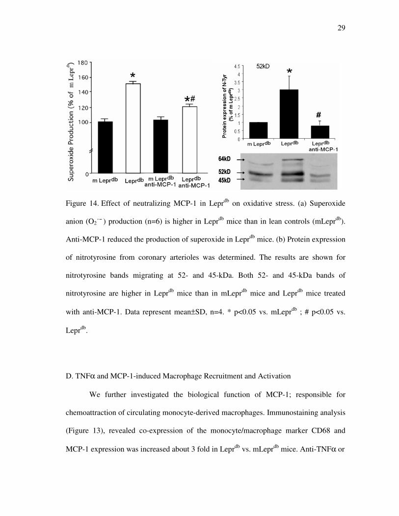
C. Role of MCP-1 in Type 2 Diabetes-Induced Vascular Dysfunction
Vasodilation to the endothelium-dependent vasodilator Ach was impaired in Leprdb
mice compared to mLeprdb
mice (Figure 13). Conversely, neutralizing antibody to MCP-
1 partially restored coronary arteriolar dilation-induced by Ach in Leprdb
mice. To
address whether over-expression of MCP-1 influences enhanced oxidative stress in
Leprdb
mice, we analyzed protein expression of N-Tyr (Figure 14b) to infer the
concentration of peroxinitrite (ONOO¯ ), and measured O2˙¯ production (Figure 14a).
Multiple bands revealed that protein expression of N-Tyr was higher in Leprdb
mice vs
mLeprdb
mice, but neutralization of MCP-1 attenuated protein expression of N-Tyr in
Leprdb
mice. The EPR result showed that O2˙¯ production was elevated and
neutralization of MCP-1 decreased O2˙¯ production in Leprdb
mice.
Figure 13. Role of MCP-1 on endothelial function. Neutralizing antibodies to MCP-1
restored coronary arteriolar dilation to Ach in Leprdb
mice, but did not affect the
vasodilation to Ach in mLeprdb
mice. * p<0.05 vs. mLeprdb
; # p<0.05 vs. Leprdb
.
29
Figure 14. Effect of neutralizing MCP-1 in Leprdb
on oxidative stress. (a) Superoxide
anion (O2˙¯ ) production (n=6) is higher in Leprdb
mice than in lean controls (mLeprdb
).
Anti-MCP-1 reduced the production of superoxide in Leprdb
mice. (b) Protein expression
of nitrotyrosine from coronary arterioles was determined. The results are shown for
nitrotyrosine bands migrating at 52- and 45-kDa. Both 52- and 45-kDa bands of
nitrotyrosine are higher in Leprdb
mice than in mLeprdb
mice and Leprdb
mice treated
with anti-MCP-1. Data represent mean±SD, n=4. * p<0.05 vs. mLeprdb
; # p<0.05 vs.
Leprdb
.
D. TNFα and MCP-1-induced Macrophage Recruitment and Activation
We further investigated the biological function of MCP-1; responsible for
chemoattraction of circulating monocyte-derived macrophages. Immunostaining analysis
(Figure 13), revealed co-expression of the monocyte/macrophage marker CD68 and
MCP-1 expression was increased about 3 fold in Leprdb
vs. mLeprdb
mice. Anti-TNFα or

30
Figure 15. Co-localization of MCP-1 in macrophages. Heart sections from lean control
(mLeprdb
) (A,B,C), Leprdb
(D,E,F), and Leprdb
mice with anti-TNFα treatment (G,H,I)
and Leprdb
mice with anti-MCP-1 treatment (J,K,L). Macrophage infiltration is
significantly increased in Leprdb
mice compared to mLeprdb
mice and decreased after
anti-TNFα and anti-MCP-1 treatment to Leprdb
mice. Negative control (M,N,O) shows
an absence of staining in vessels with only the secondary antibodies. Original
magnification 16X. Data shown are representative of 4 separate experiments. * p<0.05
vs. mLeprdb
; # p<0.05 vs. Leprdb
.

31
anti-MCP-1 treatment reduced infiltration of macrophages by 2 and 1.5 fold,
respectively, in Leprdb
mice, which suggests that coronary arteriolar dysfunction might
be associated with macrophage influx. We quantified macrophage infiltration by
counting the specifically stained macrophages per equal unit area and normalized to the
mean value of mLeprdb
mice.
E. TNFα and MCP-1-induced Adhesion Molecules and Macrophage Infiltration
Dual fluorescence combining VCAM with markers for endothelial cells (von
Willebrand factor [VWF]) (A~F), vascular smooth muscle cells [α-actin] (G~O) or
macrophages [CD68] (P~R) using specific antibodies followed by a fluorescent labeled
secondary antibody. Arrows show co-localization of VCAM in endothelial cells and
specific stain of VCAM inside vascular smooth muscle cells or co-localization of
VCAM in infiltrated macrophage in diabetic mice (Leprdb
). No primary antibody control
(S~U) shows an absence of staining in vessels with secondary antibodies. Figure 16a
shows increased protein expression of VCAM-1 (red) in the heart of Leprdb
vs mLeprdb
mice, which is consistent with the elevated protein expression detected by western blot
analysis (Figure 16 b-d). We analyzed protein expression of adhesion molecules; VCAM,
ICAM, and E-selectin to test whether they are correlated with increased expression of
cytokines (e.g., TNFα and MCP-1) in Leprdb
mice. The concentration of adhesion
molecules was elevated in Leprdb
mice vs. mLeprdb
mice but attenuated after
administration with anti-TNFα.

32
Figure 16. Analysis of adhesion molecule expression. (a) Bar size is 50µm. Original
magnification: 63X. Data shown are representative of 4 separate experiments. (b) Protein
expression of VCAM, (c) ICAM, and (d) E-selectin from coronary arterioles was
determined. Data represent mean±SD, n=4. * p<0.05 vs. mLeprdb
; # p<0.05 vs. Leprdb
.

33
CHAPTER V
SUMMARY AND CONCLUSION
First, our results in feed-forward Signaling of TNFα and NFκB via IKKβ
Pathway suggest that the interaction of NFκB and TNFα signaling induces activation of
IKKβ and amplifies oxidative stress, leading to endothelial dysfunction in coronary
arterioles of Leprdb
mice, a model for obesity and type 2 diabetes. Importantly, our
findings also support the concept that feed-forward signaling of TNFα and NFκB via the
IKKβ pathway induces insulin resistance and coronary arteriolar dysfunction in type 2
diabetes based on the following observations: NFκB antagonist MG-132, or IKKβ
inhibitor NaSal, restored endothelium-dependent coronary arteriolar dilation in Leprdb
mice, but the responses in mLeprdb
mice were unaffected. Protein expression of IKKα
and IKKβ were higher in Leprdb
than in mLeprdb
mice; the expression of IKKβ, but not
the expression of IKKα, were attenuated by MG-132, NAD(P)H oxidase inhibitor
apocynin or in dbTNF-
/dbTNF-
mice. Insulin resistance was increased in Leprdb
mice and
NaSal improved insulin sensitivity. Protein expression of TNFα, NFκB, phosphorylation
of IKKβ and JNK were greater in Leprdb
mice, but NaSal attenuated protein expression
of TNFα, NFκB, phosphorylation of IKKβ and JNK in Leprdb
mice. The pIRS-1/IRS-1
ratio was elevated in Leprdb
compared with mLeprdb
mice. Both NaSal and the JNK
inhibitor SP600125 reduced the pIRS-1/IRS-1 ratio in Leprdb
mice. MG-132 (Figure 5b)
or anti-TNFα5 reduced O2˙¯ production in Lepr
db mice. NFκB induces TNFα signaling
to accentuate oxidative stress and endothelial dysfunction via an IKKβ dependent

34
mechanism, which may be associated with inflammatory and insulin signaling pathways
in type 2 diabetes. Our findings are consistent with NFκB involvement, by interacting
with TNFα, thereby inducing IKKβ and NAD(P)H oxidase, in endothelial dysfunction
in type 2 diabetes. The present molecular results further support our previous
physiological observations that increases in TNFα expression induce activation of
NAD(P)H oxidase and production of ROS, leading to endothelial dysfunction in type 2
diabetes.5
A. Roles of Interaction of TNFα and NFκB Signaling on Impaired Coronary Arteriolar
Responses in Type 2 Diabetes
The inducible transcription factor NFκB regulates the expression of genes
encoding oxidants, cytokines, chemokines and adhesion molecules, which are associated
with inflammation and activated by gene products of NFκB, e.g., a feed-forward
interaction.5,35-37
These pro-inflammatory agents play a detrimental role in vascular
pathology and TNFα initiates signaling cascades that converge on the IKK complex via
the NFκB signaling pathway. We previously found that TNFα signaling leads to
oxidative stress via NAD(P)H oxidase activation and perhaps via activating NFκB,
which in turn may lead to further increases in TNFα expression leading to endothelial
dysfunction in type 2 diabetic mice.5
TNFα is an inducer of IKKβ and IκBα in the
classical NFκB pathway. Thus, TNFα initiates signaling cascades predominantly acting
through IKKβ.38,39
In this study, we examined whether feed-forward interaction between
TNFα and NFκB, via the IKKβ pathway, amplify one another toward the evolution of
35
vascular disease and insulin resistance in type 2 diabetes. Our western blotting results
showed that protein expression of IKKα and IKKβ were higher in Leprdb
than in mLeprdb
mice. MG-132, NAD(P)H oxidase inhibitor, apocynin, or dbTNF-
/dbTNF-
mice attenuated
the expression of IKKβ, but not the expression of IKKα. This points to a linkage among
IKKβ, NFκB, NAD(P)H oxidase and TNFα in contributing to type 2 diabetes.
Biological activity of NFκB is controlled mainly by IκBα protein, which binds to
NFκB in the cytoplasm and inhibits DNA binding activity. IκB blocks a nuclear
translocation signal to inactivate NFκB in the cytoplasm and removes NFκB bound to
promoters in the nucleus.6 MG-132 has been shown to inhibit the degradation of IκBα.
Our results showed that protein expression of IκBα was lower. However, protein
expression of p-IκBα was higher in Leprdb
mice compared to mLeprdb
mice. In dbTNF-
/dbTNF-
and Leprdb
mice treated with anti-TNF, apocynin or MG-132, protein expression
of p-IκBα was attenuated. In Leprdb
mice, protein expression of NFκB was higher, but
apocynin, MG-132 or dbTNF-
/dbTNF-
mice reduced protein expression of NFκB,
indicating that TNFα, NAD(P)H oxidase and IκBα degradation may increase the activity
of NFκB. Our functional results showed that ACh-induced vasodilation was impaired in
Leprdb
mice vs. mLeprdb
mice, but NFκB antagonist MG-132 partially restored ACh-
induced coronary arteriolar dilation in Leprdb
mice. The EPR results showed that O2˙¯
production was elevated and MG-132 decreased O2˙¯ production in Leprdb
mice, which
indicates that blocking NFκB transcriptional action markedly attenuated O2˙¯ production
in isolated coronary arterioles in Leprdb
mice. This supports the concept that diabetes

36
increases NAD(P)H oxidase activity through the interaction of TNFα/NFκB signaling,
which in turn accentuates oxidative stress and induces endothelial dysfunction via an
IKKβ activation. Our results are consistent with the concept that IKKβ is a putative
activator of NFκB, which is thought to alter subsequent gene expression in this
pathway.35-37
B. The Role of IKKβ in Vascular Dysfunction in Type 2 Diabetes
NaSal is an IKKβ inhibitor that interrupts the cascade of molecular events
leading to inflammation. Yuan et al. showed that reduced signaling through the IKKβ
pathway inhibition by NaSal in obese mice is accompanied by improved insulin
sensitivity.9 Recent studies noted that NFκB blockade (by inhibiting IKKβ activity)
decreased myocardial injury and preserved cardiac function following ischemia-
reperfusion,10
which supports the concept that the inflammatory response participates in
the development of heart failure.39
Our results showed that phosphorylation of IKKβ
increases in diabetes, but blockade of NFκB and NAD(P)H oxidase and dbTNF-
/dbTNF-
mice attenuated phosphorylation of IKKβ, suggesting that 1) IKKβ activity increases in
diabetes; and 2) phosphorylation of IKKβ increases in diabetic mice by activation of
NAD(P)H oxidase and TNFα. This indicates that TNFα-induced phosphorylation of
IKKβ is followed by phosphorylation of IκBα to release NFκB into the nucleus.
The pathogenesis of endothelial dysfunction in diabetes is initially related to a
decrease in NO synthesis or inactivation of NO due to increased endothelial production
of ROS.41
An underlying mechanism proposed for TNFα induced endothelial
37
dysfunction is that TNFα signaling leads to oxidative stress via NAD(P)H activation,
which in turn may lead to reduced NO bioactivity.5,34
We identified a link between
vascular dysfunction and insulin resistance in that treatment with NaSal in coronary
microcirculation resulted in an improvement of endothelial dysfunction in diabetic
Leprdb
mice. The NO donor SNP induced an identical vasodilation in Leprdb
and
mLeprdb
mice, but ACh-induced vasodilation was impaired in isolated coronary
arterioles in diabetic Leprdb
mice, which indicates that endothelium-dependent
vasodilation is impaired in diabetes. Most importantly, NFκB antagonist MG132 or
IKKβ inhibitor NaSal partially restored endothelium-dependent coronary arteriolar
dilation in type 2 diabetes. These results further support our hypothesis that TNFα and
NFκB signaling via an IKKβ dependent mechanism plays a key role in endothelial
dysfunction in diabetes.
Blood glucose, body weight, abdominal girth, lipid level and insulin level were
higher in Leprdb
vs. mLeprdb
mice, and there were no significant differences in body
weight, abdominal girth, lipid level and insulin level before and after treatment with
NaSal. However, lower values of glucose concentration and HOMA-insulin resistance
found in Leprdb
mice treated with NaSal indicate that NaSal improved insulin sensitivity
in Leprdb
mice (Table 1). Insulin tolerance test in Figure 6 shows that there was a
significant difference in the glucose clearance rate at 120 minutes after insulin
administration in Leprdb
mice treated with NaSal compared to Leprdb
mice. Our results
indicate that hyperglycemia and insulin resistance in Leprdb
mice were amended by a
short-term treatment with NaSal via a downregulation of IKKβ and NFκB signaling.

38
Increased phosphorylation of IKKβ may also contribute to the development or
progression of the vascular disease and hyperglycemia in type 2 diabetes.
C. Link between Inflammation-induced Insulin Resistance and Vascular Dysfunction
Coronary artery disease causes acute myocardial infarction, which is associated
with significant morbidity and mortality in patients with diabetes. Diabetes is associated
with alterations in cardiac energy metabolism, characterized by reduced glucose
utilization, and increased utilization of fatty acids as a metabolic substrate.42
The data
show that insulin may also modulate cardiac myocyte metabolism through paracrine
mechanisms by activating insulin receptors in the heart,11
thereby decreasing myocardial
infarct size and increasing cardiac contractility.12,13
Human type 2 diabetes is currently
characterized by defects in both insulin action and insulin secretion, which lack the focus
needed to identify a single molecular abnormality underlying these features, if indeed
one exists. Insulin-receptor substrates (IRS proteins) may be involved in type 2
diabetes43
and tyrosine phosphorylation activates insulin IRS-1, which further leads to
the translocation of glucose transporters (GLUT4) to the cell surface.44
Impaired
transcription of the major cardiac glucose transporter GLUT4 is seen within 4 days after
the induction of diabetes.45
IKKβ may affect glucose metabolism by altering NFκB
transcriptional action to express GLUT4.46
JNK is a main regulatory molecule that
contributes to coronary arteriolar dysfunction and insulin resistance.14,47
IRS-1 tyrosine
phosphorylation by Akt induces insulin signal transduction.15
Serine phoshphorylation
by JNK can downregulate or inactivate IRS-1. The impaired activation of Akt and
enhanced activation of JNK by TNFα is correlated with insulin resistance.47
Sholeson et

39
al. reported that inflammation-induced insulin resistance and noted that two signaling
pathways are linked to the proinflammatory effects of obesity and type 2 diabetes. Both
NFκB/IKKβ and JNK pathways are activated by TNFα in liver or adipose tissue.
Genetic disruption of NFκB and JNK signaling pathways has been shown to improve
insulin resistance.1 Consistent with the findings described above, inhibition of IKKβ or
JNK activity by NaSal significantly reduced blood glucose level and augmented insulin
signaling in cardiac tissue. Our results showed that protein expression of TNFα and
NFκB were higher in diabetic mice, but NaSal attenuated protein expression of TNFα
and NFκB. Specifically, NaSal attenuated phosphorylation of IKKβ and JNK without
altering protein expression of IKKβ and JNK. This suggests that NaSal interrupts
phosphorylation of IKKβ, which may in turn decrease phosphorylation of JNK. TNFα
activates the IKKβ and JNK signaling pathway and, conversely, the expression of the
TNFα gene is regulated by NFκB through activation of IKKβ. IKKβ and c-JNK
phosphorylate specific serine sites (e.g., Ser307) on the insulin receptor and /or insulin
receptor substrate-1 (IRS-1). Serine phosphorylation of IRS-1 blocks insulin-stimulated
tyrosine phosphorylation at IRS-1 and reduces downstream insulin signaling, resulting in
reduced GLUT4 translocation and insulin resistance.48,49
To further elucidate the signaling pathway involved in the recovery of insulin
action in cardiac tissue, we tested whether insulin receptor substrate-1 serine
phosphorylation is attenuated by SP600125 and NaSal. Phosphorylated IRS-1 (pIRS-1)
protein expression was elevated in Leprdb
compared with mLeprdb
mice; both NaSal and
JNK inhibitor SP600125 reduced pIRS-1 protein expression in Leprdb
mice. Our results

40
indicate that TNFα, phosphorylation of JNK and IKKβ, and NFκB contribute to insulin
resistance and vascular dysfunction probably via activation of IKKβ in type 2 diabetes.
Interestingly, NaSal and JNK inhibitor SP600125 sensitize insulin signaling by
preventing serine phosphorylation on IRS-1 from IKKβ and JNK. This indicates NaSal
inhibits phosphorylation of IKKβ and JNK in insulin resistant diabetic murine hearts.
The pIRS-1/IRS-1 ratio was elevated in Leprdb
compared with mLeprdb
mice; both NaSal
and JNK inhibitor SP600125 reduced the pIRS-1/IRS-1 ratio in Leprdb
mice. The
postulated mechanism is that activation of IKKβ may directly stimulate JNK activation
or cause an increase in TNFα expression. Consequently, this activation could aggravate
insulin resistance by further limiting insulin signaling.
In summary, our results demonstrated that TNFα was correlated with IKKβ, but
not IKKα, to induce NFκB activation. Inhibition of IKKβ restored ACh-induced
endothelium-dependent vasodilation in coronary arterioles in diabetes. Diabetic mice
treated with NaSal showed a decrease in glucose level and a reversal of insulin
resistance. Both IKKβ and JNK activation were increased in Leprdb
mice and NaSal
attenuated their activation without altering their expression levels. Overall, our
molecular and functional results show that IKKβ is a crucial component of the
biochemical pathway responsible for vascular dysfunction, inflammation, and insulin
resistance in type 2 diabetic murine hearts. As noted above, diabetic mice treated with
MG132 or NaSal showed partially restored ACh-induced vascular dysfunction.
Blockade of IKKβ activity not only preserved coronary arteriolar vasodilation, but also
prevented insulin resistance in type 2 diabetic mice. Although the current study

41
confirmed that NaSal amended whole body insulin sensitivity and demonstrated that
IKKβ and NFκB signaling is associated with insulin resistance and inflammation in
diabetic murine hearts, such a proposal needs to further validate how direct insulin
signaling to coronary arterioles affects vessel function. Nevertheless, anti-diabetic
effects of NaSal in vivo were observed in our study. Understanding these mechanisms
regulating heart disease processes will provide new therapies to resolve these problems.
Second, our results in role of MCP-1 in TNFα-induced endothelial dysfunction
suggest that MCP-1 interacts with TNFα to amplify adhesion molecules and oxidative
stress, thereby contributing to vascular dysfunction in coronary microcirculation in type
2 diabetes. Our molecular evidence indicated that the expression of TNFα and MCP-1
was significantly increased in Leprdb
mice; but, anti-MCP-1 decreased TNFα protein
expression and anti-TNFα attenuated MCP-1 expression. Antibody neutralization of
MCP-1 prevented coronary endothelial dysfunction and reduced ONOO¯ and O2˙¯
generation and formation of N-Tyr in Leprdb
mice. Administration of anti-TNFα or anti-
MCP-1 to Leprdb
mice resulted in attenuation in cardiac macrophage accumulation
comparable to that in the control, suggesting that an interaction of MCP-1 and TNFα
may accentuate the process of macrophage accumulation. Most importantly, blockade of
TNFα or MCP-1 attenuated the expression of adhesion molecules (e.g., VCAM, ICAM,
and E-selectin) in isolated coronary vessels in diabetes. We conclude that interactions of
TNFα and MCP-1 signaling contribute to oxidative stress by increasing inflammatory
cell accumulation and induce endothelial dysfunction in coronary arterioles in diabetic
Leprdb
mice.
42
D. Roles of TNFα and MCP-1 in Type 2 Diabetes
The important manifestation of diabetes-induced microvascular injury in the
heart is a diminution in coronary blood flow. This is emphasized by clinical data
showing ~25% of patients with acute myocardial infarction do not have adequate
restoration of myocardial tissue perfusion in the infarct region, despite coronary artery
recanalization.39
We previously found that increases in TNFα expression induces
activation of NAD(P)H oxidase and production of ROS, leading to endothelial
dysfunction in type 2 diabetes.5
The endothelium-derived NO is an important
endogenous vasodilator that regulates microvascular tone.50
Consistent with our previous
studies, our present study shows that neutralization of MCP-1 decreased TNFα protein
expression and prevented coronary endothelial dysfunction in Leprdb
mice. Our western
blot results indicated that the expression of TNFα and MCP-1 were significantly
increased in Leprdb
mice; but, anti-MCP-1 decreased TNFα protein expression and anti-
TNFα attenuated MCP-1 expression. The evidence from our immunostaining in Leprdb
mice showed that endothelial cells expressed MCP-1 (over 4.6-fold in microvessels), but
Leprdb
mice with anti-TNFα had attenuated MCP-1 expression (over 2.6-fold) in
endothelial cells.
MCP-1 is an important mediator in response to acute tissue injury that plays a
putative role in host defense.27
Recent evidence suggests that MCP-1 also plays a role in
the evolution of vascular disease through inducing adhesion molecule expression on/in
endothelium and recruiting monocytes/macrophages at the inflammatory site.22-24
Our
results in Table 2 show that anti-MCP-1 did not decrease insulin levels, but slightly

43
affected lipid metabolism by decreasing circulating cholesterol levels. The results
suggest that MCP-1 plays a role in upregulating cholesterol level in blood. Despite the
similarities in glucose, body weight, cholesterol level, insulin level and abdominal girth
in diabetic animals, endothelial function was better in Leprdb
mice treated with anti-
MCP-1. This suggests that MCP-1 signaling plays a pivotal role in attenuating TNFα
protein expression, and TNFα is the key cytokine that induces endothelial dysfunction in
type 2 diabetes.5
E. The Role of MCP-1 in ROS Production in Coronary Arterioles in Type 2 Diabetes
The link between diabetes and inflammation leading to vascular disease is associated
with enhanced oxidative stress.5 Nitration of proteins is caused by peroxynitrite, a ROS
formed by the reaction of nitric oxide (NO) with O2˙¯ ,51
which further consumes NO and
increases oxidative stress. Numerous studies have noted that nitration of tyrosine occurs
at high levels of peroxynitrite (ONOO¯ ) during the pathogenesis of human
atherosclerosis (Figure 17).40,52
Figure 17. Potential pathway of tyrosine nitration oxidation.

44
In particular, decrease in expression of constitutive nitric oxide synthase (NOS)
and inactivation of NO has been implicated in the endothelial dysfunction seen in type 2
diabetes.40
Previous work detailed the role of TNFα, NFκB, and ROS in inflamed heart
and vessels and suggested that they trigger NFκB activation to induce TNFα protein
expression.5,53
The interaction between activated macrophages and endothelial cells
produces an oxidative stress that affects the activation state of transcription factor
NFκB.24
To test if MCP-1 enhances macrophage infiltration and induces ROS,
macrophages derived from circulating monocytes were characterized by CD68 staining,
which specifically labels macrophages and monocytes. Our immunostaining results
showed that Leprdb
mice expressed MCP-1 in macrophage (over 3-fold in heart sections),
but anti-TNFα attenuated MCP-1 expression (2-fold in heart sections), which suggest
that TNFα up-regulates MCP-1 expression in macrophages. Antibody neutralization of
MCP-1 reduced ONOO¯ and O2˙¯ generation and formation of N-Tyr in Leprdb
mice.
This result suggests that TNFα initiates the interaction of macrophages and endothelial
cells via upregulation of MCP-1 signaling, which results in macrophage infiltration and
production of ROS. Based on the evidence that MCP-1 contains a kB site upstream from
its promoters,24,54
NFκB may regulate both TNFα and MCP-1 gene expression in
endothelial cells, thereby possibly inducing macrophage infiltration leading to vascular
disease.
One of the major events of endothelial dysfunction is vascular inflammation,
which is associated with impaired vascular control. It appears that MCP-1 residing in the
endothelium contributes to the regulation of vasodilation under diabetic conditions
45
because anti-MCP-1 significantly enhanced endothelium-dependent coronary arteriolar
dilations and decreased O2˙¯ production in isolated blood vessels from diabetic mice.
Our results support an endogenous role of vascular MCP-1 in regulating NO-mediated
dilation of coronary microvessels in type 2 diabetes. Consistently, our molecular results
showed that anti-MCP-1 decreased TNFα protein expression; anti-TNFα attenuated
MCP-1 expression; and antibody neutralization of MCP-1 reduced ONOO¯ and O2˙¯
generation and formation of N-Tyr in Leprdb
mice. Of particular importance to this study
is that MCP-1 and TNFα signaling plays a pivotal role in endothelial dysfunction,
consistent with what might be expected in an inflammatory disease in coronary arterioles.
F. Enhancement of Adhesion Molecules by MCP-1 and TNFα in Endothelial Cells
in Diabetes
Endothelial cells exert broad functions to maintain vessel homeostasis. The
modification of the pattern of gene expression in the endothelial cells is a critical step
during the progression of vascular disease. In endothelial cells, NFκB regulates the
inducible expression of genes encoding chemokines and adhesion molecules.55
In our
studies, the inflammatory cytokine TNFα has been explored as a factor in initiation or
progression of vascular disease.5,28
Inflammatory mediators, MCP-1 and adhesion
molecules, have also been implicated in the inflammatory processes related to TNFα in
the development of vascular disease in type 2 diabetes.55
Elevated adhesion molecules in
the vessels have been thought to be significantly associated with an increase in the risk
of coronary arterial disease.16
E-selectin is important in monocyte arrest or the transition
from slow rolling to firm adhesion. VCAM and ICAM are critical for firm attachment of

46
monocyte in the recruitment cascade.17
Immunostaining data showed that macrophages
and endothelial cells express adhesion molecules and western blot analysis revealed that
TNFα and MCP-1 increased expression of adhesion molecules (e.g., VCAM, ICAM,
and E-electin), which suggest that elevated expression of adhesion molecules is the
result of the release of cytokines, TNFα and MCP-1, from endothelial cells and
macrophages in Leprdb
mice. The actions of adhesion molecules are favored in small
vessels (~ 50µm) compared to that seen in the large coronary arteries under normal
conditions because of the lower flow and shear stress in diabetes.
Monocyte-derived macrophages are mainly inflammatory cells. Macrophages
contribute to the oxidation of LDL and oxidized LDL acts on mature macrophages to
form macrophage foam cells.21,56
Initial low expression of adhesion molecules results in
low affinity to ligands, and a signal from MCP-1 is essential to mobilize adhesion
molecules for firm adhesion.22
Inflammatory mediators such as MCP-1 and adhesion
molecules have been implicated in the inflammatory processes related to TNFα in the
development of vascular disease in type 2 diabetes. Co-localization of MCP-1 and
VCAM-1 with macrophages was observed in the myocardium, suggesting that MCP-1
and cell adhesion molecules play a role in monocyte transmigration into the heart.
ICAM-1 and E-selectin also co-exist in endothelial cells and macrophages (data not
shown).
Taken together, MCP-1 may play a critical role in inflammation-induced
endothelial dysfunction in coronary arterioles in diabetes by increasing adhesion
molecule expression in endothelial cells, thereby inducing macrophage infiltration at the

47
inflammatory site (Figure 18) without influencing the development of obesity or insulin
resistance.
In this study, we found that beyond monocyte recruitment, MCP-1 is critical in
TNFα-induced endothelial dysfunction in type 2 diabetes. The postulated mechanism is
that MCP-1-induced the production of O2˙¯ and ONOO¯ in vascular endothelial cells,
which then attenuated NO bioavailability and reduced NO mediated vasodilation in the
coronary microcirculation in type 2 diabetes. Furthermore, inflammatory cell infiltration
was enhanced via overexpression of TNFα and MCP-1-induced upregulation of cell
adhesion molecules in coronary arterioles in type 2 diabetes. Our findings demonstrate
that the endothelial dysfunction occurring in type 2 diabetes is the result of the effects of
the inflammatory cytokine TNFα and TNFα−related signaling, including the expression
of MCP-1, which also exacerbates the oxidative stress.

48
Figure 18. Interactions among TNF, MCP-1 and adhesion molecules in endothelium. We
illustrate a new aspect of the regulation of TNF-dependent signal transduction. Serine
site phosphorylation by Akt is blocked and JNK phosphorylate at tyrosine site of insulin
receptor substrate, leading to insulin resistance in type 2 diabetes. Interaction of TNF
and MCP-1 induces expression of adhesion molecules, leading to increase of
macrophage infiltration and oxidative stress, which further exacerbate vascular
dysfunction.
49
REFERENCES
1. Shoelson SE, Herrero L, Naaz A. Obesity, inflammation and insulin resistance.
Gastroenterology. 2007; 132:2169-2180.
2. Hamilton SJ, Chew GT, Watts GF. Therapeutic regulation of endothelial
dysfunction in type 2 diabetes mellitus. Diab Vasc Dis Res. 2007; 4:89-102.
3. Fried SK, Bunkin DA, Greenberg AS. Omental and subcutaneous adipose tissues
of obese subjects release interleukin-6: depot difference and regulation by
glucocorticoid. J Clin Endocrinol Metab. 1998; 83:847-850.
4. Steppan CM, Bailey ST, Bhat S, Brown EJ, Banerjee RR, Wright CM, Lazar MA.
The hormone resistin links obesity to diabetes. Nature. 2001; 409:307-312.
5. Gao X, Zhang C. Tumor necrosis factor-α induces endothelial dysfunction in
Leprdb
mice. Circulation. 2007; 115:245-254.
6. Tran K, Dimitris T. Distinct functional properties of IκBα and IκBβ. Molecular
and Cellular Biology. 1997; 17:5386-5399.
7. Lee E, Boone D, Ma A. Failure to regulate TNF-induced NFκB and cell death
responses in A20-deficient mice. Science. 2000; 289:2350-2354.
8. Cai D, Yuan M, Sheolson SE. Local and systemic insulin resistance resulting
from hepatic activation of IKKβ and NFκB. Nature Medicine. 2005; 11:183-190.
9. Yuan M, Konstantopoulos N, Sheolson SE. Reversal of obesity- and diet-induced
insulin resistance with salicylates or targeted distruption of IKKβ. Science. 2001;
293:1673-1677.
50
10. Moss NC, Stansfield WE, Willis MS, Tang RH, Selzman CH. Inhibitory kappa B
kinase beta (IKKβ) inhibition attenuates myocardial injury and dysfunction
following acute ischemia-reperfusion injury. Am J Physiol Heart Circ Physiol.
2007; 293(4): H2248-53.
11. Belke DD, Betuing S, Tuttle MJ, Graveleau C, Young ME. Insulin signaling
coordinately regulates cardiac size, metabolism, and contractile protein isoform
expression. J Clin Invest. 2002; 109(5):629-639.
12. Ren J, Sowers JR, Walsh MF, Brown RA. Reduced contractile response to
insulin and IGF-I in ventricular myocytes from genetically obese Zucker rats. Am
J Physiol Heart Circ Physiol. 2000; 279:H1708–H1714.
13. Chai W, Wu Y, Li G, Cao W, Yang Z, Liu Z. Activation of p38 mitogen-
activated protein kinase abolishes insulin-mediated myocardial protection against
ischemia-reperfusion injury. Am J Physiol Endocrinol Metab. 2008;
294(1):E183-189.
14. Zhang C, Hein TW, Wang W, Ren Y, Shipley RD, Kuo L. Activation of JNK
and Xanthine Oxidase by TNFα- Impairs Nitric Oxide-Mediated Dilation of
Coronary Arterioles. J Mol Cell Cardiol. 2006; 40(2):247-257.
15. Guoqiang J. Bei BZ. Salicylic acid reverses phorbol 12-Myristate-13-Acetate
(PMA)- and Tumor Necrosis Factor α (TNFα)-induced Insulin receptor substrate
1(IRS1) serine 307 phosphorylation and insulin resistance in human embryonic
kidney 293 (HEK293) cells. J Biol Chem. 2003; 278:180-186.
51
16. Ross R. Atherosclerosis: and inflammatory disease. N Engl J Med.1999;
340:115-126.
17. Ley K. Molecular mechanisms of leukocyte recruitment in the inflammatory
process. Cardiovasc Res. 1999; 32:733-742.
18. Carvalho D, Savage C. Cytokines, Adhesion molecules, antiendothelial cell
autonatibodies and vascular disease. Cadiovasc. Pathol. 1997; 6:61-78.
19. Pedro R. Moreno, Alvaro M Murcia, John T. Fallon. Coronary composition and
macrophage infiltration in atheroctomy specimens from patients with diabetes
mellitus. Circulation. 2000; 102:2180-2184.
20. Murao K, Ohyama T, Imachi H. TNF-stimulation of MCP-1 expression is
mediated by the Akt/PKB signal transduction pathway in vascular endothelial
cells. Biochem Biophys Res Commun. 2000; 276:791-796.
21. Hotamisligil GS, Shargill NS, Spiegelman BM. Adipose expression of tumor
necrosis factor-alpha: direct role in obesity-linked insulin resistance. Science.
1993; 259:87-91.
22. Leonard E.J., and T. Yoshimura. Human monocyte chemoattractant protein-
1(MCP-1). Immunol Today. 1990; Today 11:97-101.
23. Constantin G, Majeed M, Giagulli C, Piccio L, Laudanna C. Chemokines trigger
immediate beta 2 integrin affinity and mobility changes: differential regulation
and roles in lymphocyte arrest under flow. Immunity. 2000; 13:759-769.

52
24. Rimbach G, Valacchi G, Canali R, Virgili F. Macrophages stimulated with IFN-γ
activate NFκB and induce MCP-1 gene expression in primary human endothelial
cells. Mol Cell Biol Res Commun. 2000; 3:238-242.
25. Wang G.P, Deng Z.D, Ni J, Qu Z.L. Oxidized low density lipoprotein and very
low density lipoprotein enhance expression of monocyte chemoattractant protein-
1 in rabbit peritoneal exudate macrophage. Atherosclerosis. 1997; 133:31-36.
26. Hoffmeyer MR, Scalia R, Ross CR, Jones SP, and Lefer DJ. PR-39, a potent
neutrophil inhibitor, attenuates myocardial ischemia-reperfusion injury in mice.
Am J Physiol Heart Circ Physiol. 2000; 279:H2824-H2822.
27. Mascareno E, El-Shafei M, Maulik N, Sato M, Guo Y, Das DK, Siddiqui MA.
JAK/STAT signaling is associated with cardiac dysfunction during ischemia and
reperfusion. Circulation. 2001; 104:325-329.
28. Lattime EC SO. Thymic lymphomas mediate non-MHC-restricted, TNF-
dependent lysis of the murine sarcoma WEHI-164. Cell Immunol. 1991; 136:69-
79.
29. Letoha T, Somlai C, Penke B. The proteasome inhibitor MG132 protects against
acute pancreatitis. Free Radic Biol Med. 2005; 39:1142-1151.
30. Assi K, Pillai R, Gómez-Muñoz A, Owen D, Salh B. The GTPase Rac regulates
the proliferation and invasion of fibroblast-like synoviocytes from rheumatoid
arthritis patients. Mol Med. 2007; 13(5-6):297-304.

53
31. Zhang C, Xu X, Potter BJ, Wang W, Kuo L, Michael L, Bagby GJ, Chilian MW.
TNF-alpha Contributes to Endothelial Dysfunction in Ischemia/Reperfusion
Injury. Arterioscler Thromb Vasc Biology. 2006; 26:475-480.
32. Kuo L, Davis MJ, Chilian WM. Myogenic activity in isolated subepicardial and
subendocardial coronary arterioles. Am J Physiol. 1998; 255:H1558-H1562.
33. Salvemini D, Cuzzocrea S. Superoxide, superoxide dismutase and ischemic
injury. Curr Opin Investig Drugs. 2002; 3(6):886-895.
34. Picchi A, Zhang C. Tumor Necrosis Factor α induces endothelial dysfunction in
the prediabetic metabolic syndrome. Circ Res. 2006; 99:69-77.
35. Dela Paz NG, Simeonidis S, Collins T. Regulation of NFκB-dependent gene
expression by the pou domain transcription factor OCT-1. J Biol Chem. 2007;
282(11):8424-34.
36. Rimbach G, Valacchi G, Canali R, Virgili F. Macrophages stimulated with IFN-γ
activate NFκB and induce MCP-1 gene expression in primary human endothelial
cells. Mol Cell Bio Res Commun. 2000; 3:238-242.
37. K Ashok. Takada Y. Boriek AM, Nucrear factor κB: its role in health and disease.
J Mol Med. 2004; 82:434-448.
38. Bonizzi G, Bebien M, Karin M. Activation of IKKα target genes depends on
recognition of specific IκB binding sites by RelB:p52 dimers. EMBO J. 2004;
23:4202-4210.
39. Lawrence T, Bebien M, Karin M. IKKα limits macrophage NFκB activation and
contributes to the resolution of inflammation. Nature. 2005; 434:1138-1143.

54
40. Wong SC, Fukuchi M, Melnyk P, Rodger I, Giaid A. Induction of
cyclooxygenase-2 and activation of nuclear factor kappaB in myocardium of
patients with congestive heart failure. Circulation. 1998; 98:100-103.
41. Russo G, Leopold JA, Loscalzo J. Vasoactive substances: Nitric oxide and
endothelail dysfunction in atherosclerosis. Vascul Pharmacol. 2002; 38:259-269.
42. Chatham, JC, Gao, ZP, Bonen, A, and Forder, JR. Preferential inhibition of
lactate oxidation relative to glucose oxidation in the rat heart following diabetes.
Cardiovasc. Res. 1999; 43:96–106.
43. Withers DJ, Gutierrez JS, Towery H, Burks DJ, Ren JM, Previs S, Zhang Y,
Bernal D, Pons S, Shulman GI, Bonner-Weir S, White MF. Disruption of IRS-2
causes type 2 diabetes in mice. Nature. 1998; 391(6670):900-904.
44. Virkamak A, Uherek M, Kahn C. Protein-protein interaction in insulin signaling
and the molecular mechanisms of insulin resistance. J Clin Invest. 1999; 103:
931-943.
45. Olson AL, Pessin JE. Transcriptional regulation of the human GLUT4 gene
promoter in diabetic transgenic mice. J. Biol. Chem. 1995; 270:23491–23495.
46. Austin RL, Rune A, Bouzakri K, Zierath JR, Krook A. siRNA-mediated
reduction of IKK{beta} prevents TNF-{alpha}-induced insulin resistance in
human skeletal muscle. Diabetes. 2008; 0:db07-0763v1-0.
47. Sugita M, Sugita H, Kaneki M. Increased insulin receptor substrate 1 serine
phosphorylation and stress-activated protein kinase/c-Jun N-Terminal Kinase
55
activation Associated with vascular insulin resistance in spontaneously
hypertensive rats. Hypertension. 2004; 44:484-489.
48. Hirosumi J, Tuncman G, Chang L, Gorgun CZ, Uysal KT, Maeda K, Karin M,
Hotamisligil GS. A central role for JNK in obesity and insulin resistance. Nature.
2002; 420(6913):333-336.
49. Aguirre V, Werner ED, Giraud J, Lee YH, Shoelson SE, White MF.
Phosphorylation of Ser307 in insulin receptor substrate-1 blocks interactions with
the insulin receptor and inhibits insulin action. J Biol Chem. 2002; 277(2):1531-
1537.
50. Hein TW, Kuo L. cAMP-independent dilation of coronary arterioles to
adenosine: Role of nitric oxide, G proteins, and KATP channels. Circ Res. 1999;
85:634-642.
51. Beckman J.S, Oxidative damage and tyosine nitration from peroxinitrite. Chem
Res Toxicol. 1996; 9:836-844.
52. Beckman J.S, Ye, Y.Z, Anderson P.G, Chen J, Accavitti M.A., Tarpey M.M,
White C.R. Extensive nitration of protein tyrosines in human atherosclerosis
detected by immunohistochemistry. Biol Chemi Hoppe Seyler. 1994; 375: 81-88.
53. Yang J, Zhang C. Feed-forward signaling of TNFα and NFκB via IKKβ pathway
induced insulin resistance and coronary arteriolar dysfunction in type 2 diabetic
mice. In preparation.

56
54. Shyy YJ, Li YS, Kolattakudy PE. Structure of human monocyte chemoattractic
protein gene and its regulation by TPA. Biochem Biophys Res Commun. 1990;
169(2):346-351.
55. Lefer AM, Ma XL. Cytokines and growth factors in endothelial dysfunction. Crit
Care Med. 1993; 21(Suppl):S9-S14.
56. Tangirala RK, Murao K, Quehenberger O. Regulation of expression of the
human monocyte chemotactic protein-1 receptor (hCCR2) by cytokines. J Biol
Chem. 1997; 272:8050-8056.
57
VITA
Ji Yeon Yang [email protected]
Address : 6-501 KyeongNam APT, Banpo2-dong, Seoucho-Gu, Seoul, 137-765, Korea
Education
M.S., BMIS, Texas A&M University. Spring 2007 – Summer 2008
Genetics, Texas A&M University. Spring 2006 – Fall 2006
B.S., Nutrition, Dongduk Women’s University, Korea Mar. 1997 – Feb. 2001
Positions and Employment
Research Assistant Jan. 2007 – May. 2008
Dept. of Veterinary Physiology & Pharmacology, Texas A&M University
Teaching Assistant, Genetics Laboratory Fall 2006
Dept. of Biochemistry & Biophysics, Texas A&M University
Technical Representative, Technical Support Aug. 2001 – Jun. 2003
KOMED Co., Ltd., South Korea (ROK), Biotechnology Company
Academic Awards and Honors
1998 Academic Year Top Honor Scholarships
1999 Scholarship for Academic Excellence
1999 Merits Scholarship
Membership
Student member, American Heart Association