role of hrct in evaluation of bronchiectasis jyothi-.pdf · jemds.com original research article j....
TRANSCRIPT
Jemds.com Original Research Article
J. Evolution Med. Dent. Sci./eISSN- 2278-4802, pISSN- 2278-4748/ Vol. 5/ Issue 77/ Sept. 26, 2016 Page 5728
ROLE OF HRCT IN EVALUATION OF BRONCHIECTASIS M. Vijaya Kumari1, J. S. Aswini Jyothi2, K. Sapna3 1Associate Professor, Department of Radiology, Osmania Medical College/Hospital, Hyderabad. 2Assistant Professor, Department of Radiology, Osmania Medical College/Hospital, Hyderabad. 3Postgraduate, Department of Radiology, Kakatiya Medical College/MGM General Hospital, Warangal.
ABSTRACT
BACKGROUND
Bronchiectasis is defined as a localised, irreversible dilatation of the bronchial tree. Bronchiectasis is a chronic and progressive
lung disease and can lead to chronic respiratory failure. HRCT has emerged as the imaging modality of choice for evaluating
suspected cases of bronchiectasis. HRCT is excellent in characterisation of the type of bronchiectasis and extent of lung involvement.
AIMS
1. To study the role of HRCT in diagnosis of bronchiectasis. 2. To study regional distribution of bronchiectasis on HRCT. 3. To characterise bronchiectasis morphologically and to determine aetiologies of bronchiectasis wherever possible.
MATERIAL AND METHODS
A prospective study of 50 patients was done in the Department of Radiology and Imaging at Kakatiya Medical College and MGM
General Hospital, Warangal.
PATIENT SELECTION
Patients of all age groups were selected in whom clinically bronchiectasis was suspected. Ours was a random study for a period
of three years and no specific criteria were laid down for patient selection. From May 2005 – April 2008, 50 patients clinically
suspected to have bronchiectasis were subjected to chest radiograph and HRCT examination.
RESULTS
In our study by co-relating HRCT findings and clinical history an accurate diagnosis was made in 46 cases, i.e. 76.6% which was
confirmed by laboratory investigations. In our study 23 patients (46%) had pulmonary tuberculosis, in 8 patients (16%) no cause
was found, 16 patients (32%) had history of recurrent childhood infections, 2 patients (4%) had bronchial asthma and COPD and 1
patient (2%) had allergic bronchopulmonary aspergillosis. In this study incidence was common in the age group of 21-30 years and
is more common in males than females. Bilateral involvement is more common than unilateral involvement.
CONCLUSION
In a given clinical setting suggestive of bronchiectasis, HRCT serves as the best modality in confirming the diagnosis of
bronchiectasis. HRCT is definitely superior to chest radiographs in the detection of bronchiectasis.
KEYWORDS
HRCT bronchiectasis, Cylindrical, Cystic, Varicose Bronchiectasis.
HOW TO CITE THIS ARTICLE: Kumari MV, Jyothi JSA, Sapna K. Role of HRCT in evaluation of bronchiectasis. J. Evolution Med. Dent. Sci. 2016;5(77):5728-5737, DOI: 10.14260/jemds/2016/1292
INTRODUCTION
Bronchiectasis is defined as a localised, irreversible dilatation
of the bronchial tree. Although, a wide variety of disorders
have been associated with bronchiectasis, it most commonly
results from acute, chronic or recurrent infections and cystic
fibrosis. Bronchiectasis is commonly acquired during
childhood, the condition may rarely result from a gross
Financial or Other, Competing Interest: None. Submission 18-07-2016, Peer Review 05-08-2016, Acceptance 11-08-2016, Published 26-09-2016. Corresponding Author: Dr. J. S. Aswini Jyothi, Flat: 204, Vasavi Bhuvana Apartments, H: 8-3-981/1, 3,4,6,8,10,11 Srinagar Colony, Hyderabad-500037, Telangana. E-mail: [email protected] DOI: 10.14260/jemds/2016/1292
congenital developmental anomaly or may be predisposed by
some other inherited defect either ultra-structural (e.g. ciliary
dyskinesia which includes Kartagener’s and Young’s
syndrome) or related to a generalised metabolic defect (e.g.
Cystic fibrosis and Alpha-1 antitrypsin deficiency) or due to an
immunodeficiency syndrome (e.g.
hypogammaglobulinaemia). Post infective is the most
common cause and may result from measles, necrotising
bacterial pneumonias, granulomatous disease, allergic
bronchopulmonary aspergillosis or from bronchial
obstruction as in cases of inhaled foreign body.
The presentation includes recurrent respiratory tract
infections, productive cough, dyspnoea and occasionally
haemoptysis.
Treatment
Is with antibiotics and surgical resection is indicated. Surgery
in patients with bronchiectasis is indicated when medical
treatment fails if there is obstructing tumour or foreign body,

Jemds.com Original Research Article
J. Evolution Med. Dent. Sci./eISSN- 2278-4802, pISSN- 2278-4748/ Vol. 5/ Issue 77/ Sept. 26, 2016 Page 5729
if life-threatening complications occur such as uncontrolled
haemorrhage.
Imaging
With conventional radiography, the accuracy is 65% to 80%.
Documentation of the disease has traditionally relied on
bronchography, which is rarely performed now.
HRCT has largely eliminated the need for bronchography
in the diagnosis of bronchiectasis. HRCT is currently the most
sensitive tool for non-invasive imaging of the lung
parenchyma. It allows acquisition of in vivo images with
spatial resolution comparable to direct visualisation of the
lung slices. HRCT techniques have evolved over the last 10
years together with advance in CT technology. These have
resulted in increased spatial resolution and decreased scan
time leading to marked improvements in image quality. The
unique high contrast between lung parenchyma and air
provides an ideal situation for HRCT scanning. Although, its
use was initially controversial, volumetric CT with thin
sections is superior to conventional high-resolution CT
technique in the detection of bronchiectasis. According to
recent study, volumetric CT with thin sections is superior to
conventional high-resolution CT technique in the detection of
bronchiectasis.
However, when using the same exposure factors MDCT
results in considerably greater radiation dose (August 2006
edition of the American Journal of Roentgenology). The first
use of the term high resolution CT (HRCT) has been attributed
to Todo et al who in 1982, described the potential use of this
technique for assessing lung disease. The first reports of HRCT
in English, date to 1985, including landmark descriptions of
HRCT findings by Nakata, Naidich and Zerhouni. The HRCT
technique has resulted in: 1) Increased spatial resolution, 2)
Decreased scan time leading to, 3) Marked improvement in
image quality. The unique high contrast between lung
parenchyma and air provides an ideal situation for HRCT
scanning.
MATERIALS AND METHODS
A prospective study of 50 patients was done in the Department
of Radiology and Imaging at Kakatiya Medical College and
MGM General Hospital, Warangal.
Patient Selection
Patients of all age groups were selected in whom clinically
bronchiectasis was suspected. Ours was a random study and
no specific criteria were laid down for patient selection. From
May 2005 – April 2008, 50 patients clinically suspected to have
bronchiectasis were subjected to chest radiograph and HRCT
examination.
Technical Consideration
All HRCT scans were performed at our hospital on the Toshiba
Asteion CT Scanner. The patients were placed supine and no
gantry tilt was given. Scout films were taken routinely in all
patients before starting the scan. Scanning commenced from
lung apices to lung bases. Scans were performed in suspended
inspiration. Lung window setting was used with window
width of 1500 to 1600 HU and window level of – 600 to -700
HU. HRCT was performed obtaining 1 mm intervals. A high
algorithm was used with kvp of 120 and mA of 150.
OBSERVATIONS AND RESULTS
We did a random study in 50 patients in our institute to
evaluate the HRCT findings of bronchiectasis. All these
patients were clinically suspected to be suffering from this
disease.
Out of a total of 50 patients there were 29 (58%) males and
21 (41%) females, the average age groups being 21-30 years.
Age in Years Male Female Total 0-10 - 2 2
11-20 2 2 4 21-30 12 8 20 31-40 10 2 12 41-50 2 5 7 51-60 1 2 3 61-70 2 0 2 Total 29 21 50
Table 1: Age & Sex Incidence
We thus concluded that the maximum number of patients
(n=20) were in the age group of 21-30 years (40%). There
were only 2 patients below the age of 10 and above 60 yrs.
Majority of male and female patients in our study were in
the age group of 21-30 years.
No. of Patients % Tuberculosis 23 46
Recurrent Infections 16 32 Aspergillosis 1 2
Bronchial Asthma 2 4 Idiopathic 8 16
Table 2: Possible Aetiological Factors in our Study
Tuberculosis was identified as the cause of bronchiectasis
in 23 patients (46%) and is the major case of bronchiectasis in
our study.
The Lobar Distribution was as Follows
15 patients (30%) had unilateral disease and 35 (70%) had
disease in both lungs.
Right Left No. of Patients %
Unilateral 2 13 15 30%
Bilateral 35 70%
Table 3: Localisation of Bronchiectasis
Jemds.com Original Research Article
J. Evolution Med. Dent. Sci./eISSN- 2278-4802, pISSN- 2278-4748/ Vol. 5/ Issue 77/ Sept. 26, 2016 Page 5730
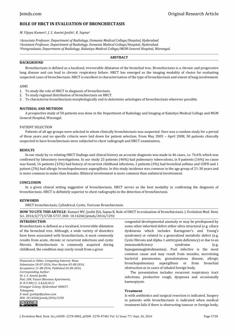
Location Right Left Total Upper Lobe 26 21 47
Middle (Lingual) 29 25 54 Lower lobe 27 41 68
Total 82 87 169 Table 4: Regional (Lobar) Distribution of Bronchiectasis
There was thus a predominance of lower lobe affliction
(40.2%) in our study.
Site Lobe Segment Number Percent Right Upper Apical 9 18
Anterior 25 50 Posterior 6 12 Middle Medial 28 56 Lateral 17 34 Lower Superior 12 24 Medial Basal 13 26 Posterior Basal 12 24 Lateral Basal 5 10 Anterior Basal 16 32
Left Upper Apicoposterior 7 14 Anterior 17 34 Lingual Superior 25 50 Inferior 20 40 Lower Superior 25 50 Posterior Basal 36 72 Lateral Basal 17 34 Anterior Basal 20 40
Table 5: Segmental Distribution
Posterior basal segment of left lower lobe was most
commonly affected (72%) followed by medial segment of right
middle lobe (56%).
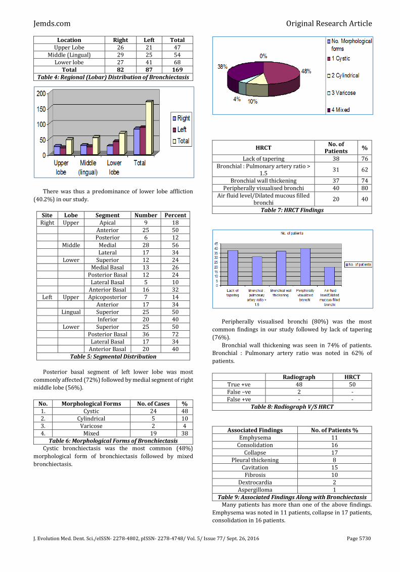
No. Morphological Forms No. of Cases % 1. Cystic 24 48 2. Cylindrical 5 10 3. Varicose 2 4 4. Mixed 19 38
Table 6: Morphological Forms of Bronchiectasis Cystic bronchiectasis was the most common (48%)
morphological form of bronchiectasis followed by mixed
bronchiectasis.
HRCT No. of
Patients %
Lack of tapering 38 76 Bronchial : Pulmonary artery ratio >
1.5 31 62
Bronchial wall thickening 37 74 Peripherally visualised bronchi 40 80
Air fluid level/Dilated mucous filled bronchi
20 40
Table 7: HRCT Findings
Peripherally visualised bronchi (80%) was the most
common findings in our study followed by lack of tapering
(76%).
Bronchial wall thickening was seen in 74% of patients.
Bronchial : Pulmonary artery ratio was noted in 62% of
patients.
Radiograph HRCT True +ve 48 50 False –ve 2 - False +ve - -
Table 8: Radiograph V/S HRCT
Associated Findings No. of Patients % Emphysema 11
Consolidation 16 Collapse 17
Pleural thickening 8 Cavitation 15
Fibrosis 10 Dextrocardia 2 Aspergilloma 1
Table 9: Associated Findings Along with Bronchiectasis Many patients has more than one of the above findings.
Emphysema was noted in 11 patients, collapse in 17 patients,
consolidation in 16 patients.

Jemds.com Original Research Article
J. Evolution Med. Dent. Sci./eISSN- 2278-4802, pISSN- 2278-4748/ Vol. 5/ Issue 77/ Sept. 26, 2016 Page 5731
DISCUSSION
Bronchiectasis has been defined as localised, irreversible
abnormal dilatation of the bronchial tree. Although, a wide
variety of disorders have been associated with it,
bronchiectasis most commonly occurs from acute, chronic or
recurrent infections.
In general a clinical diagnosis of bronchiectasis is possible
only in the most severely affected patients and even in this
setting differentiation from chronic bronchitis may be difficult.
Most patients present with purulent sputum production and
recurrent pulmonary infections. Haemoptysis is also frequent
and may be the only clinical finding.
Although, traditionally considered the “gold standard,” the
reliability of bronchography in the diagnosis of bronchiectasis
has been called into question. Currie et al (1987) in a study of
27 patients with chronic sputum production evaluated
bronchographically showed that there was a significant inter-
observer variability when studies were interpreted by two
well-trained bronchographers. Agreement was reached only
in 19 out of 27 (70%) by one radiologist only. These findings
suggest that bronchography may be more limited in its utility
than previously thought.1
We did a random study in 50 patients in our hospital who
were clinically suspected to have bronchiectasis. HRCT’s were
performed on these patients on Toshiba sub-second Spiral CT
Scan.
Scans were performed by obtaining 1 mm sections at 10
mm interval from the thoracic inlet to diaphragm using a high
algorithm. This was in accordance with Webb et al who
suggested that in patients in whom there are no specific
clinical or radiological signs to help localise the disease, 1 mm
or 1.5 mm high resolution images should be obtained every 10
mm from the lung apices to bases. Despite the lack of
contiguous scanning, this technique allows adequate
assessment of the interstitium in nearly all cases.
This approach can be modified to reflect various clinical
presentations. For example, in patients presenting with
haemoptysis it is usually necessary to rule out occult central
endobronchial lesions in addition to detecting bronchiectasis.
This is accomplished by obtaining 1 mm to 1.5 mm thick
sections every 10 mm through the upper and lower lung zones
and contiguous 5 mm thick sections from carina to level of
inferior pulmonary vein.
A high algorithm and a scan time of 1-2 seconds and 120
KV tube voltage and MA of 150 were used in our study.
While performing the HRCT’s, we used lung window
settings with a window width of 1500-1700 HU and Window
level of – 600 --- 700 HU.
The Data of the Present Studies are Discussed as follows
Age Incidence
In the present series, age of patients varied between as young
as 9 yrs. to as old as 70 yrs. The maximum number of cases
(20) 40% were in the age group of 21-30 yrs.
Year Age Group
Present series 2007, 21 – 30 yrs.
Fazul Gela et al 1999, 31 – 40 yrs.
Davies, et al 1991, 21 – 30 yrs.
The present study matches with the study of Davies et al. It
has been seen that with advancing years, the incidence is
increasing in the younger individuals. This could be due to
increasing pollution and also because of early presentation of
patients because of increased awareness and early detection
due to better imaging modalities.
Sex Incidence
In the present series, there were 21 (42%) females and 29
(58%) males.
Year Male Female Present Series 2007 29 21
Pasteur MC et al 2000 56 94 Fazul Gelal et al 1999 5 11 Zaleska M et al 1999 24 45
Symptoms
Cough was the major symptom and was seen in all 60 (100%)
cases. Most of the patients except 5 had productive cough.
In the study done by Frey HR and Russi et al (1997),2 daily
sputum production was the most common symptom. In a
clinical study done by Guleria in 1996, 92% of patients had
cough and sputum as presenting symptoms. Total daily
sputum production has been used to characterise severity of
bronchiectasis. According to Issac Hassan et al (2003)
production of less than 10 mL/day is defined as mild
bronchiectasis, 10-15 mL/day is defined as moderate
bronchiectasis and more than 15 mL/day is defined as severe
bronchiectasis.
In studies conducted by Bindra (1987) and Guleria (1996)
haemoptysis was noted in 58 and 60%, respectively. In our
study, 41.6% of patients presented with haemoptysis.
Haemoptysis occurs in 50-70% of cases and can be due to
bleeding from friable, inflamed airway mucosa. More
significant massive bleeding is often a consequence of bleeding
from hypertrophied bronchial arteries. These hypertrophied
bronchial arteries can be detected as nodular and tubular
structures in the mediastinum and around the central airways
on thin section CT scans.
Breathlessness was seen in 35% and fever was seen in
33.3%.
The triad of cough, fever and haemoptysis is a reliable
indicator of bronchiectasis.
Jemds.com Original Research Article
J. Evolution Med. Dent. Sci./eISSN- 2278-4802, pISSN- 2278-4748/ Vol. 5/ Issue 77/ Sept. 26, 2016 Page 5732
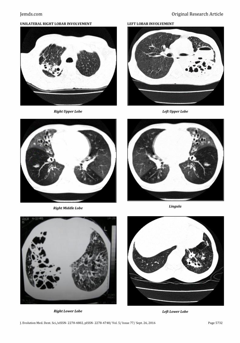
UNILATERAL RIGHT LOBAR INVOLVEMENT
Right Upper Lobe
Right Middle Lobe
Right Lower Lobe
LEFT LOBAR INVOLVEMENT
Left Upper Lobe
Lingula
Left Lower Lobe

Jemds.com Original Research Article
J. Evolution Med. Dent. Sci./eISSN- 2278-4802, pISSN- 2278-4748/ Vol. 5/ Issue 77/ Sept. 26, 2016 Page 5733
BILATERAL INVOLVEMENT
Upper Lobe
Middle Lobe/Lingula
Lower Lobe
HRCT FINDINGS
Bronchial: Pulmonary arterial ratio >1.5 (Signet Ring
Appearance)
Lack of tapering of bronchi, peripherally visualised
bronchi.

Jemds.com Original Research Article
J. Evolution Med. Dent. Sci./eISSN- 2278-4802, pISSN- 2278-4748/ Vol. 5/ Issue 77/ Sept. 26, 2016 Page 5734
Bronchial Wall Thickening
Possible Aetiological Factors: Utility of HRCT in Different
Causes of Bronchiectasis
In our study by co-relating HRCT findings and clinical history, an
accurate diagnosis was made in 46 cases, i.e. 76.6% which was
confirmed by laboratory investigations.
In our study 23 patients (46%) had pulmonary
tuberculosis, in 8 patients (16%) no cause was found, 16
patients (32%) had history of recurrent childhood infections, 2
patients (4%) had bronchial asthma and COPD and 1 patient
(2%) had allergic bronchopulmonary aspergillosis.
Although, the underlying cause of bronchiectasis is
identified in 60% - 80% of cases, HRCT findings in a number of
disease entities have been described. The reliability of HRCT for
distinguishing between these is still debated.
Lee et al (1995) in a study of HRCT scans in 108 patients,
who had bronchiectasis from a variety of causes, found that a
correct first choice diagnosis was made by 3 experienced
observers in only 45% of cases. Further interobserver
agreement was poor, leading these observers to conclude that
HRCT was of little value in diagnosing specific aetiologies of
bronchiectasis. It should be emphasised that HRCT scans were
interpreted in absence of clinical data.3
Cartier et al (1999) reported slightly better results in a
retrospective study of 82 patients who head bronchiectasis with
documented aetiologies. These authors noted that a correct
diagnosis was reached by 2 independent observers in 61% of
cases of cystic fibrosis, 67% of cases with tuberculosis and 56%
of cases with allergic bronchopulmonary aspergillosis. Bilateral
upper lobe distribution was most commonly seen in patients
who had cystic fibrosis and allergic bronchopulmonary
aspergillosis, whereas unilateral upper lobe distribution was
most common in patients who had tuberculosis and a lower lobe
distribution was most often seen in patients after recurrent
childhood viral infections.4
In our study, 23 patients (46%) had pulmonary
tuberculosis. This is a significant number leading to the
conclusion that bronchiectasis is an important and common
sequel to tuberculosis. The parenchymal fibrosis and retraction
that occur in tuberculosis result in bronchial enlargement.
Another factor contributing to bronchiectasis in patients of
tuberculosis is the infection of bronchial wall and endobronchial
occlusion causing distal bronchiectasis. In study by
Palwaywichai A (2002), the most common identifiable aetiology
was tuberculosis. The study of 168 patients by Reiff et al (1995)
had a large proportion of patients having post-tuberculosis
bronchiectasis.
Our study co-related well with the findings of Cartier et al.
Upper lobe bronchiectasis was seen in patients suffering from
allergic bronchopulmonary aspergillosis. Predominantly lower
lobe distribution, especially posterior basal segment of left
lower lobe was affected in patients with history of recurrent
childhood infections. Bronchiectasis as a result of tuberculosis
was seen in upper lobes as well as in right middle lobe and
lingual in our study.
Recurrent childhood infections as a cause of bronchiectasis
could be obtained in 16 patients (32%) in our study. In a review
of 123 patients done by Nicotra (1995) who had documented
bronchiectasis, an antecedent potentially causative event,
usually pneumonia could be identified in 70% of cases.
In our study, in patients (16%) there was no identifiable
cause of bronchiectasis. In study by Frey et al (1997), 25% of
cases had no identifiable cause of bronchiectasis. In study by

Jemds.com Original Research Article
J. Evolution Med. Dent. Sci./eISSN- 2278-4802, pISSN- 2278-4748/ Vol. 5/ Issue 77/ Sept. 26, 2016 Page 5735
Reiff et al (1995), the cause of bronchiectasis could not be
ascertained in 63% of cases.
In our study, 2 out of 60 patients (3.23%) had bronchial
asthma. Hansell et al (1994) showed an association between
bronchial asthma and bronchiectasis. In study by Grenier et al
(1996), bronchiectasis in asthmatic patients was found in 28.5%
of patients. Bronchial asthma leads to small airway involvement
consistent with obliterative bronchiolitis, which in turn leads to
bronchiectasis.
In our study, allergic bronchopulmonary aspergillosis was
identified as the cause of bronchiectasis in 1 patient (1.6%). In a
retrospective study of 82 patients of bronchiectasis, Cartier et al
(1999) found allergic bronchopulmonary aspergillosis in 5 cases
(6.9%). Central bronchiectasis in association with bronchial
occlusion due to mucous plugging, air fluid levels in dilated
cystic airways and bronchial wall thickening are usually seen in
bronchiectasis due to allergic bronchopulmonary aspergillosis.
Of particular interest is the finding of high attenuation mucoid
impaction in the dilated bronchi. First described in association
with chronic fungal sinusitis, high density mucous presumably
represents the presence of calcium ions, metallic ions or both
within viscous mucous. The prevalence of this finding has been
noted to be as high as 28% in one series and when present
should be considered characteristic.
Morphological Forms of Bronchiectasis
We based on our classification of bronchiectasis based on Reid’s
classification, i.e. cystic, cylindrical and varicose bronchiectasis.
A total of 24 patients (58%) in our study had cystic
bronchiectasis, while 5 patients (10%) had cylindrical variety, 2
patients (4%) had varicose bronchiectasis and 19 patients
(38%) had mixed variety. There have been varied reports
regarding the preponderance of cystic and cylindrical
bronchiectasis; however, in majority of the studies the least
number of patients had varicose bronchiectasis. This was a
finding in our study also.5
MORPHOLOGICAL FORMS OF BRONCHIECTASIS
Cylindrical Bronchiectasis
Cystic Bronchiectasis
Varicose Bronchiectasis
Mixed Bronchiectasis
Localisation of Bronchiectasis
With the advent of HRCT, accurate lobar and segmental
localisation is possible. A total of 169 lobes were found to be
Jemds.com Original Research Article
J. Evolution Med. Dent. Sci./eISSN- 2278-4802, pISSN- 2278-4748/ Vol. 5/ Issue 77/ Sept. 26, 2016 Page 5736
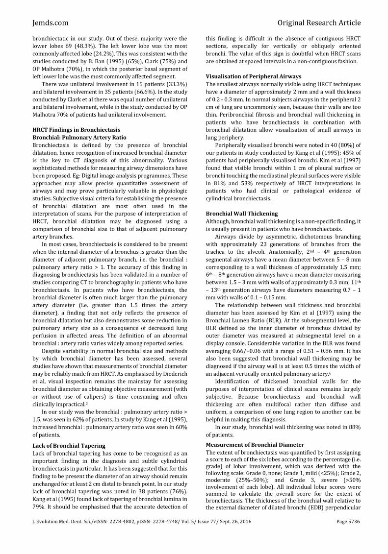
bronchiectatic in our study. Out of these, majority were the
lower lobes 69 (48.3%). The left lower lobe was the most
commonly affected lobe (24.2%). This was consistent with the
studies conducted by B. Ban (1995) (65%), Clark (75%) and
OP Malhotra (70%), in which the posterior basal segment of
left lower lobe was the most commonly affected segment.
There was unilateral involvement in 15 patients (33.3%)
and bilateral involvement in 35 patients (66.6%). In the study
conducted by Clark et al there was equal number of unilateral
and bilateral involvement, while in the study conducted by OP
Malhotra 70% of patients had unilateral involvement.
HRCT Findings in Bronchiectasis
Bronchial: Pulmonary Artery Ratio
Bronchiectasis is defined by the presence of bronchial
dilatation, hence recognition of increased bronchial diameter
is the key to CT diagnosis of this abnormality. Various
sophisticated methods for measuring airway dimensions have
been proposed. Eg: Digital image analysis programmes. These
approaches may allow precise quantitative assessment of
airways and may prove particularly valuable in physiologic
studies. Subjective visual criteria for establishing the presence
of bronchial dilatation are most often used in the
interpretation of scans. For the purpose of interpretation of
HRCT, bronchial dilatation may be diagnosed using a
comparison of bronchial size to that of adjacent pulmonary
artery branches.
In most cases, bronchiectasis is considered to be present
when the internal diameter of a bronchus is greater than the
diameter of adjacent pulmonary branch, i.e. the bronchial :
pulmonary artery ratio > 1. The accuracy of this finding in
diagnosing bronchiectasis has been validated in a number of
studies comparing CT to bronchography in patients who have
bronchiectasis. In patients who have bronchiectasis, the
bronchial diameter is often much larger than the pulmonary
artery diameter (i.e. greater than 1.5 times the artery
diameter), a finding that not only reflects the presence of
bronchial dilatation but also demonstrates some reduction in
pulmonary artery size as a consequence of decreased lung
perfusion in affected areas. The definition of an abnormal
bronchial : artery ratio varies widely among reported series.
Despite variability in normal bronchial size and methods
by which bronchial diameter has been assessed, several
studies have shown that measurements of bronchial diameter
may be reliably made from HRCT. As emphasised by Diederich
et al, visual inspection remains the mainstay for assessing
bronchial diameter as obtaining objective measurement (with
or without use of calipers) is time consuming and often
clinically impractical.2
In our study was the bronchial : pulmonary artery ratio >
1.5, was seen in 62% of patients. In study by Kang et al (1995),
increased bronchial : pulmonary artery ratio was seen in 60%
of patients.
Lack of Bronchial Tapering
Lack of bronchial tapering has come to be recognised as an
important finding in the diagnosis and subtle cylindrical
bronchiectasis in particular. It has been suggested that for this
finding to be present the diameter of an airway should remain
unchanged for at least 2 cm distal to branch point. In our study
lack of bronchial tapering was noted in 38 patients (76%).
Kang et al (1995) found lack of tapering of bronchial lumina in
79%. It should be emphasised that the accurate detection of
this finding is difficult in the absence of contiguous HRCT
sections, especially for vertically or obliquely oriented
bronchi. The value of this sign is doubtful when HRCT scans
are obtained at spaced intervals in a non-contiguous fashion.
Visualisation of Peripheral Airways
The smallest airways normally visible using HRCT techniques
have a diameter of approximately 2 mm and a wall thickness
of 0.2 - 0.3 mm. In normal subjects airways in the peripheral 2
cm of lung are uncommonly seen, because their walls are too
thin. Peribronchial fibrosis and bronchial wall thickening in
patients who have bronchiectasis in combination with
bronchial dilatation allow visualisation of small airways in
lung periphery.
Peripherally visualised bronchi were noted in 40 (80%) of
our patients in study conducted by Kang et al (1995); 45% of
patients had peripherally visualised bronchi. Kim et al (1997)
found that visible bronchi within 1 cm of pleural surface or
bronchi touching the mediastinal pleural surfaces were visible
in 81% and 53% respectively of HRCT interpretations in
patients who had clinical or pathological evidence of
cylindrical bronchiectasis.
Bronchial Wall Thickening
Although, bronchial wall thickening is a non-specific finding, it
is usually present in patients who have bronchiectasis.
Airways divide by asymmetric, dichotomous branching
with approximately 23 generations of branches from the
trachea to the alveoli. Anatomically, 2nd – 4th generation
segmental airways have a mean diameter between 5 – 8 mm
corresponding to a wall thickness of approximately 1.5 mm;
6th – 8th generation airways have a mean diameter measuring
between 1.5 – 3 mm with walls of approximately 0.3 mm, 11th
– 13th generation airways have diameters measuring 0.7 – 1
mm with walls of 0.1 – 0.15 mm.
The relationship between wall thickness and bronchial
diameter has been assessed by Kim et al (1997) using the
Bronchial Lumen Ratio (BLR). At the subsegmental level, the
BLR defined as the inner diameter of bronchus divided by
outer diameter was measured at subsegmental level on a
display console. Considerable variation in the BLR was found
averaging 0.66/+0.06 with a range of 0.51 – 0.86 mm. It has
also been suggested that bronchial wall thickening may be
diagnosed if the airway wall is at least 0.5 times the width of
an adjacent vertically oriented pulmonary artery.6
Identification of thickened bronchial walls for the
purposes of interpretation of clinical scans remains largely
subjective. Because bronchiectasis and bronchial wall
thickening are often multifocal rather than diffuse and
uniform, a comparison of one lung region to another can be
helpful in making this diagnosis.
In our study, bronchial wall thickening was noted in 88%
of patients.
Measurement of Bronchial Diameter
The extent of bronchiectasis was quantified by first assigning
a score to each of the six lobes according to the percentage (i.e. grade) of lobar involvement, which was derived with the
following scale: Grade 0, none; Grade 1, mild (<25%); Grade 2, moderate (25%–50%); and Grade 3, severe (>50% involvement of each lobe). All individual lobar scores were summed to calculate the overall score for the extent of bronchiectasis. The thickness of the bronchial wall relative to the external diameter of dilated bronchi (EDB) perpendicular

Jemds.com Original Research Article
J. Evolution Med. Dent. Sci./eISSN- 2278-4802, pISSN- 2278-4748/ Vol. 5/ Issue 77/ Sept. 26, 2016 Page 5737
to the transverse plane was evaluated in each lobe. This score was determined with the following scale: Grade 0, normal thickness; Grade 1, thickness greater than 20% and less than 50% EDB; Grade 2, thickness greater than 50% EDB; and Grade 3, complete obliteration of the bronchial lumen. If there was a range of bronchial wall thickening noted in each lobe assessed, a mean score was calculated per lobe whereby the number of scores assigned was the denominator for the sum of all scores calculated. The sum of individual lobar bronchial wall thickening scores was the overall score for each patient.
HRCT vs. Chest Radiograph
HRCT has revolutionised the detection and localisation of
bronchiectasis, because of its axial sections and superior
resolution compared to chest radiographs. In chest
radiographs, 70% of lung tissue is obscured by the bony cage.
However, this problem is not seen in HRCT. The bronchus can
be followed from origin to periphery with the help of
contiguous sections. Chest radiograph is neither sensitive nor
specific for the diagnosis of bronchiectasis. The detection rate
of HRCT in comparison to chest radiographs is better. Out of
the 60 cases studied, Chest radiographs detected
bronchiectatic changes in 55 cases; 5 cases were interpreted
as normal by chest radiograph which were abnormal on HRCT.
Even among radiographs which showed unilateral
bronchiectasis, HRCT revealed the disease to be bilateral.
Exact localisation of disease to a particular anatomical
segment of lungs was possible with HRCT, which was not
possible with chest radiographs. In a study by Vincent
Mysliwiee et al (1999) chest radiographs were abnormal in
only 87% of cases. In a study conducted by Palwaywichai et al
(2002), abnormal chest radiographs were seen in 9% of cases.
In study of Boehm T et al (2003), chest radiograph was found
to have a sensitivity of only 37% for detection of
bronchiectasis.
In a study of 84 patients done by Van Der Bruggen (1996),
47 patients had abnormal radiographs. Out of these 36 had
bronchiectasis on HRCT and the other 11 patients had normal
scans. The sensitivity of radiograph to detect bronchiectasis
was 87.8% and the specificity was 74.4%. They found a
significant linear relationship between the severity of
bronchiectasis at HRCT and abnormalities as seen on chest
radiograph. In our study, the sensitivity of chest radiographs
in detecting bronchiectasis was 94%. This high sensitivity rate
as compared to other studies could be due to the fact that most
of our patients had severe bronchiectasis, which could be
easily detected on radiographs.7
In the study by Issac Hassan et al (2003), HRCT had a
sensitivity of 96% and specificity of 93%. In our study, HRCT
had 100% sensitivity and specificity. In study by Boehm T et al
(2003), HRCT has been shown to have a sensitivity of 96 – 98%
and a specificity of 93-99% in detection of bronchiectasis.
Using HRCT, Grenier et al (1996) found a sensitivity of 96%
and a specificity of 93% as compared to bronchography for
diagnosing bronchiectasis. Young et al also assessed the
reliability of HRCT in assessment of bronchiectasis. Their
study showed a sensitivity of 98% and a sensitivity of 99%.
The first study in evaluating bronchiectasis by CT was done
by Naidich DP et al in 1982.
CONCLUSIONS
In a given clinical setting suggestive of bronchiectasis,
HRCT serves as the best modality in confirming the
diagnosis of bronchiectasis. HRCT is definitely superior to
chest radiographs in the detection of bronchiectasis.
Regional distribution of bronchiectasis can be well
visualised by HRCT. Left lower lobe, especially posterior
basal segment is most commonly involved.
In this study, incidence is most common in the age group
of 21-30 years and is more common in males than females.
Bilateral involvement is more common than unilateral
involvement.
Peripherally visualised bronchi was the most common
finding in our study followed by lack of tapering.
Cystic bronchiectasis is the most common form of
bronchiectasis followed by mixed variety. Varicose
bronchiectasis is the least common form.
HRCT is useful in identifying the aetiology of
bronchiectasis in most patients. Tuberculosis is the most
common aetiological factor of bronchiectasis.
Other co-existent lung pathologies can also be diagnosed
by HRCT.
REFERENCES
1. Currie DC, Cooke JC, Morgan AD, et al. Interpretation of
bronchograms and chest radiographs in patients with
chronic sputum production. Thorax 1987;42(4):278-84.
2. Frey HR, Russi EW. Bronchiectasis–current aspects of an old disease. Schweiz Med Wochenschr 1997;127(6):219-30.
3. Lee PH, Carr DH, Rubens MB, et al. Accuracy of CT in
predicting the cause of bronchiectasis. Clin Radiol
1995;50(12):839–41.
4. Cartier Y, Kavanagh PV, Johkoh T, et al. Accuracy of high
resolution ct in differentiation of specific diseases. AJR Am
J Roentgenol 1999;173(1):47–52.
5. Reid LM. Reduction in bronchial subdivision in
bronchiectasis. Thorax 1950;5(3):233-47.
6. Kim JS, Im JG, Kim IO, et al. Normal bronchial and
pulmonary arterial diameters measured by thin section ct.
J Comput Assist Tomogr 1995;19(3):365–9.
7. Van Der Bruggen-Bogaarts BA, van Der Bruggen HM, van
Waes PM, et al. Screening for bronchiectasis: a
comparative study between chest radiography and high-
resolution CT. Chest 1996;109(3):608-11.