role and function of nonmuscle alpha-actinin-1 and -4 in
TRANSCRIPT

Role and function of nonmuscle alpha-actinin-1 and -4 in regulating distinct subcategories of actin stress fibers in
mammalian cells
Bianca Kovac Master’s thesis Biotechnology MBIOT University of Helsinki March 2010

HELSINGIN YLIOPISTO HELSINGFORS UNIVERSITET UNIVERSITY OF HELSINKI Tiedekunta/Osasto Fakultet/Sektion Faculty
Agriculture and Forestry
Laitos Institution Department
MBIOT Master’s Degree Programme in Biotechnology
Tekijä Författare Author
Bianca Kovac Työn nimi Arbetets titel Title
Role and function of nonmuscle alpha-actinin-1 and -4 in regulating distinct subcategories of actin stress fibers in mammalian cells Oppiaine Läroämne Subject
Biotechnology Työn laji Arbetets art Level
Master’s Thesis
Aika Datum Month and year
March 2010
Sivumäärä Sidoantal Number of pages
36
Tiivistelmä Referat Abstract
Actin stress fibers are dynamic structures in the cytoskeleton, which respond to mechanical stimuli and affect cell motility, adhesion and invasion of cancer cells. In nonmuscle cells, stress fibers have been subcategorized to three distinct stress fiber types: dorsal and ventral stress fibers and transverse arcs. These stress fibers are dissimilar in their subcellular localization, connection to substratum as well as in their dynamics and assembly mechanisms. Still uncharacterized is how they differ in their function and molecular composition. Here, I have studied involvement of nonmuscle alpha-actinin-1 and -4 in regulating distinct stress fibers as well as their localization and function in human U2OS osteosarcoma cells. Except for the correlation of upregulation of alpha-actinin-4 in invasive cancer types very little is known about whether these two actinins are redundant or have specific roles. The availability of highly specific alpha-actinin-1 antibody generated in the lab, revealed localization of alpha-actinin-1 along all three categories of stress fibers while alpha-actinin-4 was detected at cell edge, distal ends of stress fibers as well as perinuclear regions. Strikingly, by utilizing RNAi-mediated gene silencing of alpha-actinin-1 resulted in specific loss of dorsal stress fibers and relocalization of alpha-actinin-4 to remaining transverse arcs and ventral stress fibers. Unexpectedly, aberrant migration was not detected in cells lacking alpha-actinin-1 even though focal adhesions were significantly smaller and fewer. Whereas, silencing of alpha-actinin-4 noticeably affected overall cell migration. In summary, as part of my master thesis study I have been able to demonstrate distinct localization and functional patterns for both alpha-actinin-1 and -4. I have identified alpha-actinin-1 to be a selective dorsal stress fiber crosslinking protein as well as to be required for focal adhesion maturation, while alpha-actinin-4 was demonstrated to be fundamental for cell migration. Avainsanat Nyckelord Keywords
Actin stress fibers, nonmuscle, alpha-actinin, RNAi-mediated gene silencing, focal adhesion, cell migration Säilytyspaikka Förvaringsställe Where deposited
HELDA Muita tietoja Övriga uppgifter Further information
Supervisor: MD PhD Tea Vallenius

TABLE OF CONTENTS
ROLE AND FUNCTION OF NONMUSCLE ALPHA-ACTININ-1 AND -4 IN REGULATING DISTINCT SUBCATEGORIES OF ACTIN STRESS FIBERS IN MAMMALIAN CELLS......................................................................................... 1
1 INTRODUCTION ............................................................................................. 5
1.1 Actin filaments and their function in nonmuscle cells ........................ 5 1.2 Actin stress fibers are sarcomeric-like structures............................... 6 1.3 Diverse actin stress fiber types ............................................................. 7 1.4 Alpha-actinins as structural components of actin filaments .............. 9 1.5 Nonmuscle alpha-actinin-1 and -4 as actin stress fiber regulators.. 10
2 OBJECTIVES OF MASTER THESIS ............................................................ 12
3 MATERIAL AND METHODS......................................................................... 13
3.1 Cell culture............................................................................................. 13 3.2 Antibodies.............................................................................................. 13 3.3 RNAi-mediated gene silencing............................................................. 14 3.4 Sodium dodecyl sulfate polyacrylamide gel electrophoresis (SDS-PAGE) ........................................................................................................... 14 3.5 Western blotting .................................................................................... 15 3.6 Immunofluorescence analysis ............................................................. 15 3.7 Confocal microscope imaging and analysis ...................................... 16 3.8 Analysis of focal adhesions ................................................................. 17 3.9 Wound healing assay and analysis ..................................................... 17
4 RESULTS ...................................................................................................... 18
4.1 Alpha-actinin-1 and -4 differ in their localization on actin stress fibers............................................................................................................. 18 4.2 Downregulation of alpha-actinin-1 results in specific loss of dorsal stress fibers ................................................................................................. 21 4.3 Alpha-actinin-4 relocalizes prominently to transverse arcs and ventral stress fibers following alpha-actinin-1 downregulation ............. 23 4.4 Depletion of alpha-actinin-1 results in smaller and fewer focal adhesions..................................................................................................... 25 4.5 Alpha-actinin-4 is required for cell motility ........................................ 27

4
5 DISCUSSION................................................................................................. 29
5.1 Analysis of the role and function of alpha-actinin-1 and -4 .............. 29 5.2 Involvement and function of alpha-actinin-1 in nonmuscle cells..... 30 5.3 Alpha-actinin-4 as a cell motility regulator ......................................... 31
6 CONCLUSION............................................................................................... 32
7 ACKNOWLEDGEMENTS ............................................................................. 32
8 REFERENCES .............................................................................................. 33

5
1 INTRODUCTION
A network of protein filaments known as the cytoskeleton spatially organizes the
cytoplasm of eukaryotic cells. Cytoskeleton is a complex system of protein filaments
involved in a range of functions such as cell attachment, cell locomotion, intracellular
trafficking, signal transduction as well as generating spatial organization and
mechanical robustness (Mizuno et al., 2007). The major building blocks of the
cytoskeleton are three types of filaments, each assembling into characteristic structures
and carrying typical functions. Microtubules are widely recognized from their
involvement in spindle formation and star-like configuration during mitosis and are
responsible for the positioning and intra-cellular transport of cell organelles (Kaverina,
1998). Intermediate filaments provide mechanical stability and resistance to shear stress
and pressure (Kreplak and Fudge, 2007). Actin filaments form bundles and networks,
which determine the cell shape and enable cell locomotion with the help of surface
protrusions as well as enable cytokinesis (Pollard and Borisy, 2003). Each of the three
major filament classes has a large dedicated subset of accessory proteins that modify the
dynamics and structure of cytoskeletal filaments (Miano et al., 2007).
1.1 Actin filaments and their function in nonmuscle cells
A network of actin filaments is involved in regulating specific structural changes and
formation of structures such as filopodia, lamellipodia and stress fibers as well as
maintenance of morphology within cells and cellular regions (Nemethova et al., 2008;
Pollard et al., 2000). In muscle cells, actin filaments function primarily as part of the
contractile machinery whereas in nonmuscle cells actin filaments are involved in cell
motility, cytokinesis, polarity and cell spreading (Pellegrin, 2007).
Actin filaments are formed through head-to-tail polymerization of actin monomers (G-
actin) and exist as two helical interlaced strands of filamentous actin subunits (F-actin)
(Schleicher, 2008). Actin filaments have a fast-growing barbed end (+ end) and a slow-
growing pointed end (- end). Actin filament elongation or polymerization occurs mostly
at the barbed end, and shortening or depolymerization at the pointed end (Welch and
Mullins, 2002). Furthermore, actin filament network is an exceptionally dynamic
organization, which is to a great extent determined by variety of actin binding proteins

6
associated with its polymeric structure. Numerous actin binding proteins regulate
dynamics of actin filaments through actin polymerization/depolymerization,
crosslinking or bundling of actin filaments (Pollard et al., 2000).
In cultured cells, actin filaments are prominent and well-studied filaments. In vivo, actin
filaments have been detected and found essential in endothelial cells regulating blood
flow tension and mechanical stress, in platelet activation in case of endothelial injury as
well as in wound healing (Tanaka, 1998; van Nieuw Amerongen, 2001).
1.2 Actin stress fibers are sarcomeric-like structures
Actin stress fibers also known as actomyosin fibers are formed when a cell is stably
connected to the substrate and arranged in parallel with myosin and actin binding
protein to build long, straight, contractile fibers (Chrzanowska-Wodnicka and Burridge,
1996). Based on protein composition, interacting proteins and ability to contract, actin
stress fibers resemble sarcomeric-like structure found in the muscle cells (Clark et al.,
2002). Stress fibers are extremely dynamic structures and can easily be detected in
immunofluorescence analysis by phalloidin staining.
To date, most well characterized regulator of stress fiber assembly is RhoA, small
GTPases family member (Pellegrin, 2007). The GTP bound form of RhoA activates
Rho-associated kinase (ROCK) which in turn phosphorylates myosin light chain (MLC)
therefore promoting stress fiber formation (Riento and Ridley, 2003). Activation of
RhoA promotes both bundling of actin filaments into stress fibers and clustering of
integrins and associated proteins to form focal adhesions (Bershadsky et al., 2006).
Focal adhesions are anchoring cell junctions, which anchor the cell and stress fibers to
the extracellular matrix thus promoting mechanotransduction forces between
extracellular matrix and intracellular tension (Clark et al., 2007; Endlich et al., 2007).
In general, stress fibers are crucial for cell function by enabling cells to sense and
respond to mechanical stimuli and affecting cell motility, adhesion, apoptosis and
invasion of cancer cells (Lu et al., 2008).
7
1.3 Diverse actin stress fiber types
Actin stress fibers have been subcategorized to three distinct stress fiber types: dorsal
and ventral stress fibers and transverse arcs, based on their subcellular localization and
association with focal adhesions in mouse fibroblasts (Small et al., 1998). More
recently, this view has been expanded and stress fibers have been further characterized
according to their connection to substratum as well as in their dynamics and assembly
mechanisms (Hotulainen and Lappalainen, 2006).
Dorsal stress fibers (Figure 1; indicated in red) are connected to the substrate via a focal
adhesion at the leading edge of the cell. Dorsal stress fibers elongate from a focal
adhesion and is assembled through an actin nucleation promoting formin called mDIA1
and actin polymerization. In comparison, transverse arcs are not directly associated with
a focal adhesion. Transverse arcs (Figure 1; indicated in blue) are contractile and
myosin rich arc structures with a suggested function in cell contraction. Suggested
model for transverse arcs formation is through endwise annealing between myosin and
actin-related protein (Arp2/3) complex of nucleated actin bundles (Hotulainen and
Lappalainen, 2006). Third type of stress fibers – ventral stress fibers (Figure 1;
indicated in green) – is connected from both ends to a focal adhesion thus proposing
that ventral stress fibers could be important in cell adhesion.

8
Figure 1. Diverse stress fiber types found in a migrating cell. Direction of cell migration is indicated as well as leading edge and trailing edge of the cell. Cell nucleus is located near the trailing edge of the cell. Dorsal stress fibers (indicated in red) elongate from a focal adhesion (black circles) at the leading edge towards the cell center where they connect with transverse arcs (indicated in blue). Ventral stress fibers (indicated in green) are the third type of stress fibers, which are formed between two distinct focal adhesions at both ends.
Even though stress fibers have been subcategorized to separate stress fiber types, it still
remains uncharacterized how distinct stress fiber types differ in their function and
molecular composition. At this point, it is essential to investigate involvement of stress
fiber interacting proteins such as alpha-actinins and their function as well as regulation
of distinct stress fiber types.

9
1.4 Alpha-actinins as structural components of actin filaments
Alpha-actinin is conserved and ubiquitously expressed protein with the exception of
plants and prokaryotes, which has originally been identified as actin crosslinking protein
(Ebashi and Ebashi, 1964). Alpha-actinin is a long, narrow, rod shaped antiparallel,
dimer formed of two 100kDa alpha-actinin monomers (Lorenzi and Gimona, 2008).
Alpha-actinin belongs to a family of structurally related proteins, including spectrin,
dystrophin and utrophin, which regulate the organization of actin cytoskeleton in a cell
type specific fashion (Bois et al., 2005). All members of the family as illustrated in
Figure 2, possess a N-terminal actin binding domain (ABD), an EF-hand calcium
binding motif at the C-terminal and a central rod domain containing a varying number
of spectrin repeats where the dimerization of alpha-actinin is mediated (Virel and
Backman, 2007).
Figure 2. Alpha-actinin as a structural component of actin filaments. (A) A closer look at the alpha-actinin antiparallel, dimer structure where actin binding domain (ABD), four spectrin-like repeats (R1-R4) and C-terminal EF hands are indicated. (B) Interlaced strands of actin filaments (indicated in red) and alpha-actinin dimer (indicated in green) crosslinking the two actin filaments.

10
In humans, four alpha-actinin genes have been identified and grouped into two distinct
classes: muscle and nonmuscle cytoskeletal alpha-actinins (Blanchard et al., 1989).
Alpha-actinin-2 and -3 can be found in striated, cardiac and smooth muscle cells mainly
at the Z-disks of sarcomeres and analogous dense bodies, where they form a lattice like
structure and stabilize the muscle contractile apparatus (Sjöblom et al., 2008). Alpha-
actinin-2 is a major protein in the cardiac and oxidative skeletal muscle whereas alpha-
actinin-3 is largely expressed in glycolytic skeletal muscle fibers (Mills et al., 2001). In
nonmuscle cells, alpha-actinin-1 and -4 are primarily located on stress fibers, focal
adhesions and cell-cell contact sites, cellular protrusions, lamellipodia and stress fiber
dense regions (Otey and Carpen, 2004). In addition, alpha-actinin-1 and -4 have an
exceptional large number of interacting molecular partners. However, specificity to
which of the nonmuscle alpha-actinins an interacting protein binds has not clearly been
recognized.
1.5 Nonmuscle alpha-actinin-1 and -4 as actin stress fiber regulators
Nonmuscle alpha-actinin-1 and -4 share over 80% homology but display diverse roles in
cytoskeletal organization, and cell motility as well as in subcellular localization and
binding partners (Bolshakova et al., 2007; Youssoufian et al., 1990). Nonmuscle alpha-
actinin-1 was first identified (Lazarides, 1975) followed by much later identification of
alpha-actinin-4 (Honda et al., 1998). Based on previous knockdown studies, alpha-
actinin-1 has been demonstrated to primarily localize along stress fibers, at focal
adhesion plaques as well as taking part in bundling of actin filaments (Craig et al.,
2008). It also associates with several cytoskeletal and membrane associated proteins
such as integrins, intercellular adhesion molecules and vinculin (Bois et al., 2005;
Vallenius et al., 2000).
In comparison, alpha-actinin-4 is less clearly concentrated to stress fibers and is neither
detected in focal adhesions nor cell contacts but it is enriched at the leading edge of
invading cells and in cytoplasmic regions with sharp cell extensions (Honda et al.,
2005). Alpha-actinin-4 is a cell motility enhancer and is associated with invasion and
metastasis of cancer cells (Honda et al., 1998). Alpha-actinin-4 is overexpressed in
various human epithelial carcinomas, including colorectal, breast and ovarian
carcinomas (Barbolina et al., 2008). Hence, interest in studying role and function of

11
alpha-actinin-4 has increased enormously over the past years. Futhremore, mutations in
alpha-actinin-4 gene cause familial focal segmental glomerulosclerosis (FSGS), an
autosomal dominant disease with abnormal protein secretion to the urine and as a result
gradually causing renal insufficiency (Kaplan et al., 2000). Mouse studies where alpha-
actinin-4 has been deleted show kidney failure, progressive proteinuria and severe
glomerular disease (Dandapani et al., 2007; Kos et al., 2003), therefore suggesting a role
for alpha-actinin-4 in regulating multiple cellular processes and animal development.
Both alpha-actinin-1 and -4 are most commonly known as structural components of
stress fibers and their ability to crosslink actin filaments. Nevertheless, from previous
studies it is evident that alpha-actinin-1 and -4 possess wider roles and functions in the
cell (Sen et al., 2009). Now it is important to establish if there are any localization or
functional differences between alpha-actinin-1 and -4 such as in the assembly and
crosslinking ability, stability and regulation of stress fibers in mammalian cells.

12
2 OBJECTIVES OF MASTER THESIS
Objectives of my master thesis study:
1) Determine localization of nonmuscle alpha-actinin-1 and -4 in human U2OS
osteosarcoma cells by utilizing specific antibodies generated in the lab
2) Investigate how alpha-actinin-1 and -4 regulate distinct stress fiber types
previously characterized in nonmuscle cells thus determine how the individual
stress fiber types differ in their functional and molecular composition
3) Exploring functional situations, such as cell motility, where alpha-actinin-1 and
alpha-actinin-4 could differ
Understanding functional differences between alpha-actinin-1 and -4 would further
enable investigation of how they regulate distinct stress fiber types and therefore overall
cell function in mammalian cells as well as in cancer.

13
3 MATERIAL AND METHODS
3.1 Cell culture
Human U2OS osteosarcoma cell line was used during this master thesis study. U2OS
cells were propagated in Dulbecco’s modified Eagle’s medium (DMEM) supplemented
with 10% fetal calf serum, penicillin and streptomycin and glutamine at 37°C in 5%
CO2 incubator. U2OS cells were cultured on 10 cm culture dishes and passaged every
four days. Prior cell passaging, culture dishes were rinsed twice with sterile PBS
followed by 5 min incubation with trypsin-EDTA resulting in cell detachment.
Detached cells were re-suspended in a fresh growth media and re-plated for further
experiments.
3.2 Antibodies
Following primary antibodies were used: the rabbit polyclonal antibody to detect alpha-
actinin-1 (SNO341), which was specifically generated in the lab (dil. 1:4000 for
Western blotting and dil. 1:400 for immunofluorescence), the rabbit polyclonal antibody
to detect alpha-actinin-4 (ALX-210-356) from Alexis Biochemicals (dil. 1:2000 for
Western blotting and dil. 1:300 for immunofluorescence), the mouse monoclonal anti-
vinculin antibody (V9131) from Sigma to detect focal adhesions (dil. 1:400 for
immunofluorescence). Filamentous actin was stained by using Alexa Fluor® 488
phalloidin from Invitrogen (dil. 1:50). For secondary antibodies in Western Blotting
anti-rabbit-HRP (Chemicon International) was used (dil. 1:5000). For
immunofluorescence, Alexa Fluor® anti-rabbit 594 (dil. 1:500) and Alexa Fluor® anti-
mouse 647 (dil. 1:500) from Invitrogen were used.

14
3.3 RNAi-mediated gene silencing
For RNAi-mediated gene silencing Lipofectamine™ 2000 transfection reagent
(Invitrogen) was used together with the pooled short interfering RNA (siRNA) oligos L-
011195 (siACTN1), L-011988 (siACTN4) and D-001206-13 (siNT) (Dharmacon). One
day prior to transfection, U2OS cells were plated, 70 000 cells per six-well plate
chamber to reach 40-50% confluence on the day of transfection. On the first day of
transfection, each transfection sample was prepared by adding diluted 20 pmol siRNA
to Opti-MEM I Reduced Serum Medium to form a complex. Lipofectamine™ 2000
was also mixed with Opti-MEM I Reduced Serum Medium to form a separate
complex. Both complexes were mixed gently and let to settle for 5 min separately. After
5 min incubation, diluted siRNAs were combined with diluted Lipofectamine™ 2000
and incubated for 20 min at room temperature. Following the 20 min incubation period,
siRNA - Lipofectamine™ 2000 complexes were added to each plate containing 40-50%
confluent U2OS cells together with penicillin and streptomycin free media. Penicillin
and streptomycin free media was changed to Dulbecco’s modified Eagle’s medium
(DMEM) supplemented with 10% fetal calf serum, penicillin and streptomycin and
glutamine after 4-6 hours of transfection. Same transfection protocol was conducted
also on the following day to reach the highest knockdown efficiency. Cells were
incubated at 37°C in 5% CO2 incubator for a total of 96 h.
3.4 Sodium dodecyl sulfate polyacrylamide gel electrophoresis (SDS-PAGE)
SDS-PAGE is a widely used technique to separate proteins according to their
electrophoretic mobility. For detection of alpha-actinin-1 and -4 total protein levels
following siRNA-mediated transfection, cells were lysed using SDS-boiling buffer
(2.5% SDS, 0.25 M Trizma base including 50 mM NaF, 10 mM β-glycerophosphate,
0.5 mM DTT, 0.5 mM PMSF, 2.5 µg/ml Aprotinin and 1 µg/ml Leupeptin). Preceding
cell lysis, each culture dish was rinsed twice with sterile PBS to ensure the absence of
growth media from the protein lysates. SDS-lysis buffer was heated to 95°C and then
added over the cells. Cells were carefully scraped from the culture dishes by using a
policeman and collected to separate eppendorf tubes. SDS-lysed samples were needled
with a 25G needle 10 times and centrifuged for 15 min at 13 000 rpm, +4°C.

15
Supernatant was separated from the pellet and stored by freezing at -20°C or proceeded
directly to protein concentration measurements. Protein concentrations were measured
by using Bio-Rad DC protein assay kit (Bio-Rad Laboratories) at the wavelength of 595
nm with a spectrophotometer. 10-30 µg protein lysates were prepared for the SDS-
PAGE gel run according to BSA standard protein concentration measurements. Protein
lysates were finalized by addition of 1 x Laemmli buffer and further diluted with
supplementary SDS-lysis buffer. Prepared lysates were heated for 5 min at +95°C,
cooled down and run on a SDS-PAGE gel or alternatively stored at -20°C.
3.5 Western blotting
SDS-PAGE samples were run on a 10% SDS-PAGE gel with a 35 mA current for a
period of 2,5 hours. Following SDS-PAGE run, proteins were transferred on a
nitrocellulose filter for a period of 1 h 45 min by using 400 mA current and finally
detected by a Ponceau staining. 5% milkpowder – TBS – 0,05% Tween buffer was used
for blocking the nitrocellulose filter as well as primary and secondary antibody
incubations. Nitrocellulose filter was blocked for a period of 1 h, followed by an
overnight incubation with a primary antibody at +4°C. Primary antibody was washed 3
x 10 min with TBS – 0,05% Tween buffer followed by 40 min secondary antibody
(peroxidase conjugated anti-rabbit) incubation. Secondary antibody was washed
following the same protocol as with primary antibody. For the protein detection,
commercially available ECL reagents (SuperSignal West Femto Maximum sensitivity
Substrate, PIERCE) were used in equal amounts in PBS.
3.6 Immunofluorescence analysis
Immunofluorescence analysis was used for detection of both alpha-actinin-1 and -4 as
well as stress fibers. U2OS cells were re-plated 3 h prior fixation on 10 µg/ml
fibronectin (734-0101, from VWR International) pre-coated coverslips. For part of the
coverslips the 0.5% Triton X-100 extraction was performed prior to fixation. Coverslips
were fixed with 4% paraformaldehyde (PFA) for 15 minutes and washed three times
with PBS. Fixed coverslips were further permeabilized with 0.1% Triton X-100 for 5
minutes. 5% goat serum in PBS buffer was used for blocking the cells as well as
diluting the primary and secondary antibodies. Cells were blocked with 5% goat serum

16
in PBS for 30 minutes, labeled with primary antibody for 30 minutes, washed three
times with PBS, labeled with secondary antibody for 30 minutes, washed three times in
PBS. Nucleus was labeled with 0,5 µm/ml Hoechst, washed two times with PBS and
finally stained coverslips were mounted with Immu-mount (Thermo Scientific) and
stored at +4°C until further analysis.
3.7 Confocal microscope imaging and analysis
As part of my master thesis study, confocal microscope use was extensive and essential
for observing and understanding my results. I received my advanced confocal
microscope training in the molecular imaging unit (MIU) in Meilahti. MIU is an
imaging core facility responsible for the training and maintenance of the confocal
microscope and offers also other state-of-the-art imaging facilities. Stained coverslips
from the immunofluorescence analysis were analyzed and imaged by using Zeiss LSM
510 Meta laser scanning confocal microscope equipped with 63x/Plan-
Apochromat/1.40/DIC objective. While acquiring images with the confocal microscope
the following lasers were used Diode 25 mW, Argon 30 mW, Helium-Neon 1 mW and
Helium-Neon 5 mW to detect 405 nm, 488 nm, 543 nm and 633 nm wavelengths,
respectively, thus enabling visualization of up to four different immunofluorescence
stainings simultaneously. LSM 3.2 software was used for the images acquired by the
confocal microscope and by using Adobe photoshop CS4 imaging software final images
were generated.

17
3.8 Analysis of focal adhesions
Anti-vinculin stained focal adhesions were quantified by ImageJ software by
determining the threshold for each image to clearly distinguish individual focal
adhesions, followed by measurement of the amount and size of focal adhesion present in
the image (see Results for detail). Focal adhesions were quantified according to their
size and amount from 20 different cells of transfected pooled non-targeting, ACTN1 or
ACNT4 siRNA oligos.
3.9 Wound healing assay and analysis
A scratch wound assay was generated to a confluent monolayer of U2OS cells with a
sterile tip 72 hours after transfection of pooled non-targeting, ACTN1 or ACNT4
siRNA oligos. Wound areas were marked to the bottom of each plate to enable image
acquisition of the same are using Olympus CKX42 microscope equipped with UplanFL
4x/0,13 PhP objective and Canon DS6041 EOS 300D digital camera. Wound closure
was monitored up to 24 hours and images where acquired at 0 h, 8 h and 24 h
timepoints. For image analysis and quantification ImagePro Plus software was used to
calculate and measure remaining open areas of each wound from four independent
experiments.
18
4 RESULTS
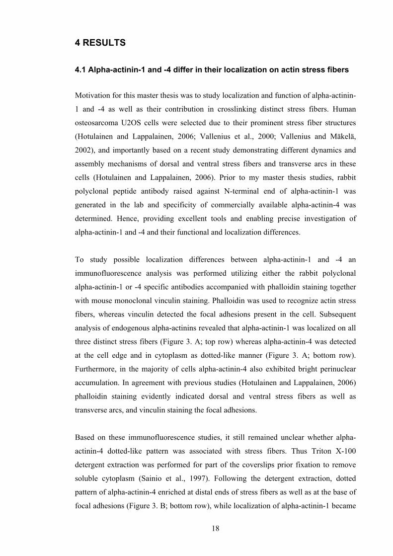
4.1 Alpha-actinin-1 and -4 differ in their localization on actin stress fibers
Motivation for this master thesis was to study localization and function of alpha-actinin-
1 and -4 as well as their contribution in crosslinking distinct stress fibers. Human
osteosarcoma U2OS cells were selected due to their prominent stress fiber structures
(Hotulainen and Lappalainen, 2006; Vallenius et al., 2000; Vallenius and Mäkelä,
2002), and importantly based on a recent study demonstrating different dynamics and
assembly mechanisms of dorsal and ventral stress fibers and transverse arcs in these
cells (Hotulainen and Lappalainen, 2006). Prior to my master thesis studies, rabbit
polyclonal peptide antibody raised against N-terminal end of alpha-actinin-1 was
generated in the lab and specificity of commercially available alpha-actinin-4 was
determined. Hence, providing excellent tools and enabling precise investigation of
alpha-actinin-1 and -4 and their functional and localization differences.
To study possible localization differences between alpha-actinin-1 and -4 an
immunofluorescence analysis was performed utilizing either the rabbit polyclonal
alpha-actinin-1 or -4 specific antibodies accompanied with phalloidin staining together
with mouse monoclonal vinculin staining. Phalloidin was used to recognize actin stress
fibers, whereas vinculin detected the focal adhesions present in the cell. Subsequent
analysis of endogenous alpha-actinins revealed that alpha-actinin-1 was localized on all
three distinct stress fibers (Figure 3. A; top row) whereas alpha-actinin-4 was detected
at the cell edge and in cytoplasm as dotted-like manner (Figure 3. A; bottom row).
Furthermore, in the majority of cells alpha-actinin-4 also exhibited bright perinuclear
accumulation. In agreement with previous studies (Hotulainen and Lappalainen, 2006)
phalloidin staining evidently indicated dorsal and ventral stress fibers as well as
transverse arcs, and vinculin staining the focal adhesions.
Based on these immunofluorescence studies, it still remained unclear whether alpha-
actinin-4 dotted-like pattern was associated with stress fibers. Thus Triton X-100
detergent extraction was performed for part of the coverslips prior fixation to remove
soluble cytoplasm (Sainio et al., 1997). Following the detergent extraction, dotted
pattern of alpha-actinin-4 enriched at distal ends of stress fibers as well as at the base of
focal adhesions (Figure 3. B; bottom row), while localization of alpha-actinin-1 became

19
more evident along all three stress fiber types (Figure 3. B; top row). These results
indicate distinct distribution between alpha-actinin-1 and -4 due to alpha-actinin-1
localization on all type of stress fibers in comparison to alpha-actinin-4 localization only
on a subset of stress fibers as well as cell membrane.
20
Figure 3. Nonmuscle alpha-actinin-1 and -4 localize differently on stress fibers. Immunofluorescence analysis of U2OS cells using alpha-actinin-1 and -4 specific antibodies together with phalloidin and vinculin antibodies as shown on top. (A) Alpha-actinin-1 is localized on dorsal (red arrowhead) and ventral stress fibers (white arrowhead) and transverse arcs (purple arrowhead). Distinct stress fibers are clearly indicated by phalloidin staining and focal adhesions by vinculin staining. In comparison alpha-actinin-4 is localized on cell edge and has a cytoplasmic dotted appearance. (B) Similar analysis as in (A) but following a Triton X-100 extraction. Localization of alpha-actinin-1 remained noticeably on all three stress fibers, while alpha-actinin-4 localization changed to the distal ends of stress fibers as well as to the base of focal adhesions. Scale bar 10 µm.

21
4.2 Downregulation of alpha-actinin-1 results in specific loss of dorsal stress fibers
Observed differences in subcellular localization between alpha-actinin-1 and -4
suggested that these two alpha-actinins might differently crosslink distinct stress fibers,
thus prompted to further investigate consequences following downregulation of alpha-
actinin-1 and -4 in U2OS cells. To this end RNAi-mediated gene silencing of alpha-
actinin-1 and -4 was conducted where short interfering RNA (siRNA) were used to
interfere with the expression of alpha-actinin-1 and -4 genes by silencing. Subsequent
Western blotting analysis indicated over 90% downregulation of both alpha-actinin
proteins (Figure 4. A). Here note of mentioning, that over 90% downregulation of
alpha-actinin-1 required optimization, and it was achieved by a double siRNA
transfection over a period of 96 h (see Material and Methods for detail). This could be
due to differences in protein levels between alpha-actinin-1 and -4 in U2OS cells, where
it is known that alpha-actinin-1 is more abundant than alpha-actinin-4 (Vallenius et al.,
2000) or even different protein half-life periods. Thus indicating that alpha-actinin-1
requires a longer siRNA treatment for complete gene silencing.
Next, immunofluorescence analysis of phalloidin stained control, alpha-actinin-1 and -4
downregulated cells was performed. Strikingly, in alpha-actinin-1 depleted cells, lack of
dorsal stress fibers became evident without disturbing the formation of transverse arcs
and ventral stress fibers (Figure 4. B; middle panel). However, depletion of alpha-
actinin-4 maintained all three stress fibers (Figure 4. B; right panel), thus indicating that
alpha-actinin-1 is selectively required for dorsal stress fibers.

22
Figure 4. Loss of dorsal stress fibers following alpha-actinin-1 silencing. RNAi-mediated gene silencing technique was adapted to downregulate both alpha-actinin-1 and -4. (A) Western blotting analysis was performed to observe the silencing efficiency. Protein levels of alpha-actinin-1 and -4 were detected by using specific rabbit polyclonal alpha-actinin-1 and -4 antibodies, respectively. (B) Immunofluorescence analysis was conducted with phalloidin staining on control, alpha-actinin-1 and -4 silenced cells. Control cell illustrates all three stress fibers present; dorsal (red arrowhead) and ventral stress fibers (white arrowhead) and transverse arcs (purple arrowhead) (B; left panel). Strikingly, in alpha-actinin-1 depleted cells lack of dorsal stress fibers became evident and only transverse arcs (purple arrowheads) and ventral stress fibers (white arrowheads) remained (B; middle panel). In alpha-actinin-4 depleted cells all three types of stress fibers were evident (B; right panel). Scale bar 10 µm.

23
4.3 Alpha-actinin-4 relocalizes prominently to transverse arcs and ventral stress fibers following alpha-actinin-1 downregulation
Further investigating localization and functional differences between alpha-actinin-1
and -4 it was essential to observe whether alpha-actinin-1 and -4 could compensate each
other when either of the alpha-actinins is lost. Alpha-actinin-1 staining was performed
on control, alpha-actinin-1 and -4 depleted cells prior and following Triton X-100
detergent extraction to observe possible localization or compensation changes.
Importantly, localization of alpha-actinin-1 was not altered during alpha-actinin-4
silencing, suggesting alpha-actinin-1 having a primary role in crosslinking dorsal stress
fibers (Figure 5. A). These findings were further confirmed when an alpha-actinin-4
staining was performed in the same manner as for alpha-actinin-1 mentioned above. In
alpha-actinin-1 silenced cells, alpha-actinin-4 was found to loose its cell edge
localization prior Triton X-100 extraction (Figure 5. B; arrow) and furthermore
prominently relocalized to remaining transverse arcs and ventral stress fibers following
Triton X-100 extraction (Figure 5. B; arrowheads). These results demonstrate evident
relocalization of alpha-actinin-4 to transverse arcs and ventral stress fibers subsequent
Triton X-100 extraction and alpha-actinin-1 silencing but not the ability to fully
compensate each other’s localizations. Hence, suggesting a primary role for alpha-
actinin-1 in selectively crosslinking dorsal stress fibers.

24
Figure 5. Alpha-actinin-4 relocalizes to transverse arcs and ventral stress fibers upon alpha-actinin-1 depletion. (A) Immunofluorescence analysis with alpha-actinin-1 specific antibody prior and following Triton X-100 treatment of control and silenced alpha-actinin-1 and -4 U2OS cells. As illustrated in both conditions, alpha-actinin-1 localization is not affected by the loss of alpha-actinin-4 when compared to the control cell. (B) Similar analysis as in (A) but cells were stained by alpha-actinin-4 specific antibody. Surprisingly, depletion of alpha-actinin-1 prior Triton X-100 treatment resulted in a loss of cell edge localization (white arrow) and became apparent on distal ends of stress fibers. Furthermore, relocalization of alpha-actinin-4 became more evident following Triton X-100 treatment where alpha-actinin-4 was clearly relocalized to transverse arcs and ventral stress fibers (white arrowheads). Scale bar 10 µm.

25
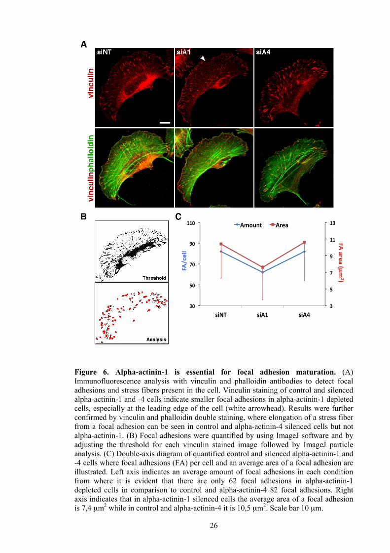
4.4 Depletion of alpha-actinin-1 results in smaller and fewer focal adhesions
Previous stress fiber studies demonstrate that formation and elongation of stress fibers
require mature focal adhesions as well as RhoA induced contractility (Chrzanowska-
Wodnicka and Burridge, 1996). Therefore it was of a great interest to determine focal
adhesions in the cells. 96 hours after knockdown, vinculin staining was performed on
alpha-actinin-1 depleted cells from which it became evident that focal adhesions,
particularly at the leading edge of the cell, were smaller (Figure 6. A; top row;
arrowhead). Strikingly, focal adhesions formed between ventral stress fibers appear to
be mature which was further indicated from double-immunofluorescence analysis
between phalloidin and vinculin (Figure 6. A; bottom row; second panel). Hence,
indicating that loss of dorsal stress fibers following depletion of alpha-actinin-1 has an
affect on focal adhesion maturation. In comparison, focal adhesion maturation in either
control or alpha-actinin-4 depleted cells was not affected which was also indicated by
vinculin staining (Figure 6. A; top row). Results were further confirmed by merge
image of phalloidin and vinculin stained control and alpha-actinin-4 depleted cells
where stress fiber formation was normal and dorsal as well as ventral stress fibers were
elongating from a focal adhesion (Figure 6. A; bottom row).
To quantify this obvious difference I used ImageJ software where the threshold was
adjusted for each image to distinguish individual focal adhesions, followed by
measurement of the amount and size of focal adhesion present in the cell (Figure 6. B).
This analysis indicate focal adhesions being 27% smaller as well as 20 focal adhesions
less per cell in cells lacking alpha-actinin-1, whereas loss of alpha-actinin-4 was not
significantly changed in comparison to the control cells (Figure 6. C). Results suggest
that alpha-actinin-1 is required for a part of focal adhesion maturation at the leading
edge of migrating cells where loss of dorsal stress fibers was detected.
26
Figure 6. Alpha-actinin-1 is essential for focal adhesion maturation. (A) Immunofluorescence analysis with vinculin and phalloidin antibodies to detect focal adhesions and stress fibers present in the cell. Vinculin staining of control and silenced alpha-actinin-1 and -4 cells indicate smaller focal adhesions in alpha-actinin-1 depleted cells, especially at the leading edge of the cell (white arrowhead). Results were further confirmed by vinculin and phalloidin double staining, where elongation of a stress fiber from a focal adhesion can be seen in control and alpha-actinin-4 silenced cells but not alpha-actinin-1. (B) Focal adhesions were quantified by using ImageJ software and by adjusting the threshold for each vinculin stained image followed by ImageJ particle analysis. (C) Double-axis diagram of quantified control and silenced alpha-actinin-1 and -4 cells where focal adhesions (FA) per cell and an average area of a focal adhesion are illustrated. Left axis indicates an average amount of focal adhesions in each condition from where it is evident that there are only 62 focal adhesions in alpha-actinin-1 depleted cells in comparison to control and alpha-actinin-4 82 focal adhesions. Right axis indicates that in alpha-actinin-1 silenced cells the average area of a focal adhesion is 7,4 µm2 while in control and alpha-actinin-4 it is 10,5 µm2. Scale bar 10 µm.
27
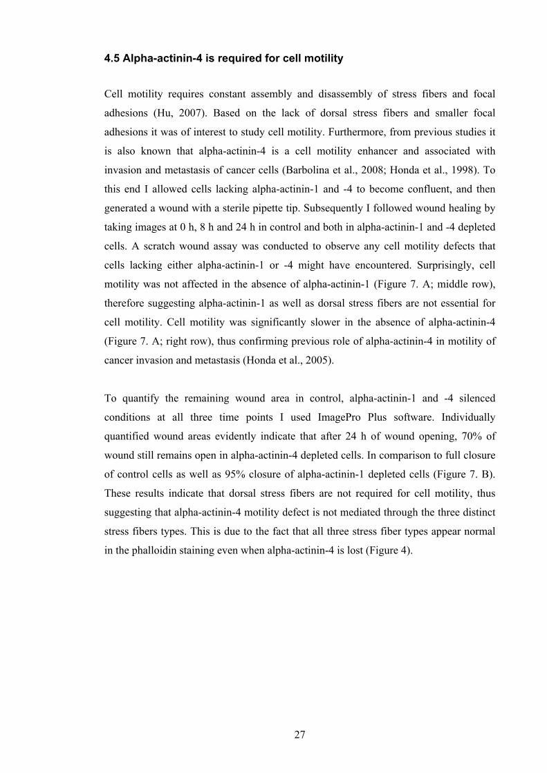
4.5 Alpha-actinin-4 is required for cell motility
Cell motility requires constant assembly and disassembly of stress fibers and focal
adhesions (Hu, 2007). Based on the lack of dorsal stress fibers and smaller focal
adhesions it was of interest to study cell motility. Furthermore, from previous studies it
is also known that alpha-actinin-4 is a cell motility enhancer and associated with
invasion and metastasis of cancer cells (Barbolina et al., 2008; Honda et al., 1998). To
this end I allowed cells lacking alpha-actinin-1 and -4 to become confluent, and then
generated a wound with a sterile pipette tip. Subsequently I followed wound healing by
taking images at 0 h, 8 h and 24 h in control and both in alpha-actinin-1 and -4 depleted
cells. A scratch wound assay was conducted to observe any cell motility defects that
cells lacking either alpha-actinin-1 or -4 might have encountered. Surprisingly, cell
motility was not affected in the absence of alpha-actinin-1 (Figure 7. A; middle row),
therefore suggesting alpha-actinin-1 as well as dorsal stress fibers are not essential for
cell motility. Cell motility was significantly slower in the absence of alpha-actinin-4
(Figure 7. A; right row), thus confirming previous role of alpha-actinin-4 in motility of
cancer invasion and metastasis (Honda et al., 2005).
To quantify the remaining wound area in control, alpha-actinin-1 and -4 silenced
conditions at all three time points I used ImagePro Plus software. Individually
quantified wound areas evidently indicate that after 24 h of wound opening, 70% of
wound still remains open in alpha-actinin-4 depleted cells. In comparison to full closure
of control cells as well as 95% closure of alpha-actinin-1 depleted cells (Figure 7. B).
These results indicate that dorsal stress fibers are not required for cell motility, thus
suggesting that alpha-actinin-4 motility defect is not mediated through the three distinct
stress fibers types. This is due to the fact that all three stress fiber types appear normal
in the phalloidin staining even when alpha-actinin-4 is lost (Figure 4).
28
Figure 7. Cell motility requires alpha-actinin-4. (A) Images taken from the scratch wound assay at 0 h and 24 h time points of control and silenced alpha-actinin-1 and -4 U2OS cells. Following 24 h of wound closure, it is evident that alpha-actinin-4 silenced cells remain open in comparison to control and alpha-actinin-1 silenced cells. (B) Wound area from 0 h, 8 h and 24 h were measured and quantified from four subsequent experiments. The diagram illustrates percentage of wound healing process following 8 h and 24 h wound closure in control and silenced alpha-actinin-1 and -4 cells. A remarkable motility defect is noticed after 24 h of cell migration in alpha-actinin-4 depleted cells, where wound healing has occurred only 72% in comparison to control and alpha-actinin-1 silenced cells where wound healing was 100% and 94%, respectively.

29
5 DISCUSSION
During my master thesis study I have been able to identify alpha-actinin-1 as a selective
dorsal stress fiber crosslinking protein as well as to be required for focal adhesion
maturation, while alpha-actinin-4 was demonstrated to be fundamental for cell
migration.
5.1 Analysis of the role and function of alpha-actinin-1 and -4
Thus far, human osteosarcoma (U2OS) cell line together with fibroblasts have been
documented to contain subcategories of stress fibers; dorsal and ventral stress fibers and
transverse arcs (Hotulainen and Lappalainen, 2006; Small et al., 1998). As actin
cytoskeleton is an extremely dynamic network, having a cell line model that enables
investigation of defined subcategories of stress fibers and their regulation is essential.
My work during the master thesis study using U2OS cells provides the first evidence
that these distinct stress fibers might be crosslinked by different proteins. These results
were obtained due to the specific antibodies generated in the lab. Hence, for the first
time being able to provide an excellent tool to investigate alpha-actinin-1 and -4
differences.
So far, alpha-actinin-1 and -4 have been studied separately but comparison studies
between the two alpha-actinins have been rare (Bolshakova et al., 2007). During my
thesis work, a study was published demonstrating that both alpha-actinin-1 and -4 are
critical in contributing to the invasiveness of glioblastoma multiforme, a malignant
astrocytic tumor (Sen et al., 2009). Nevertheless, this study did not explore role of
alpha-actinin-1 and -4 in correlation to the stress fiber subcategories defined previously.
From now on functional, localization as well as expression differences between alpha-
actinin-1 and -4 can be investigated by using the specific tools available. Thus,
extending this kind of analysis to fibroblasts as well as other cell or tissue types would
be extremely informative. A starting point could be investigation of differences between
alpha-actinin-1 and -4 expression patterns found in different tissues and compare how
they correlate with already established results. On the other hand, trying to identify
alpha-actinin-1 selective dorsal stress fibers in other cells or tissues through contractility

30
or associated proteins would enable understanding of the physiological or pathological
circumstances where these types of alpha-actinin-1 specific fibers are advantageous.
5.2 Involvement and function of alpha-actinin-1 in nonmuscle cells
Previous studies suggest that alpha-actinin-1 contributes to cell contractility (Lu et al.,
2008). My results, which indicate that alpha-actinin-1 is localized along all three
detected stress fiber types further supports contractility idea. Lack of dorsal stress fibers
accompanied by smaller focal adhesions upon alpha-actinin-1 loss proposes that this is
due to lack of contractile function for dorsal stress fibers. This can be addressed by
determining phosphorylated myosin light chain levels in the cell, which is increased
when stress fibers are contractile. Stress fiber assembly and cell contractility are
activated by myosin light chain phosphorylation which in turn is activated by the ROCK
kinase (Riento and Ridley, 2003). Indeed, investigation of such a contractility assay is
of my future interest. Alpha-actinin-1 has previously been demonstrated also to
modulate pressure-induced colon cancer cell adhesion (Craig et al., 2007), thus future
cell adhesion as well as cell spreading studies are essential for further characterizing
alpha-actinin-1 function. Cell adhesion and cell spreading are cellular events that
require constant assembly of focal adhesions as well as stress fiber formation (Partridge
and Marcantonio, 2006). Furthermore, use of GFP-tagged alpha-actinin-1 and -4
plasmids in live cell imaging could also provide further functional as well as
localization information in live cells. Here however, it is essential to confirm with the
help of specific antibodies whether such overexpressed proteins localize as endogenous
proteins.
Interestingly smaller and fewer focal adhesions in alpha-actinin-1 downregulated cells
were more frequently detected at the leading edge. This correlates extremely well with
the noted loss of dorsal stress fibers. In the future it is highly important to quantify this
piece of interesting data. One way to do it could be by observing focal adhesion
maturation at the leading edge in comparison to the trailing edge of the cell where
ventral stress fibers are noted to elongate from mature focal adhesions. For this master
thesis study all the focal adhesions present in the cell were categorized under the same
measurement therefore resulting in high standard deviations when measuring focal
adhesion area. In general, cell attachment to the extracellular matrix is mediated by

31
variety of transmembrane proteins such as integrins, which are further linked through a
range of other proteins to the stress fibers in the cytoplasm (Huveneers and Danen,
2009). Investigation of other adhesion components such as integrins and their
involvement in focal adhesion maturation in alpha-actinin-1 and -4 depleted cells is an
essential future study.
5.3 Alpha-actinin-4 as a cell motility regulator
Directional motility is essential in various cellular processes such as wound healing,
embryonic as well as tissue development (Pollard and Borisy, 2003). From the scratch
wound healing experiment used in this master thesis study, it was obvious that alpha-
actinin-4 had an effect on cell motility but alpha-actinin-1 seemed not to have. Alpha-
actinin-4 cell motility defect was expected due to previous studies implicating
overexpression of alpha-actinin-4 increasing invasion of cancer cells (Honda et al.,
2005). Thus provided a good positive control for my experiments. Based on the results
obtained from this study it can be concluded that alpha-actinin-1 has not a major role in
scratch wound healing process in U2OS cells. Importantly my data indicates that dorsal
stress fibers are neither required for cell motility. Still remaining question is what is the
role of these fibers. Obvious follow-up experiments in addition to determine their
contractility is to study their involvement in other known stress fiber functions such as
cell spreading and polarity.
Furthermore my studies strongly suggest that the noted migration defect of alpha-
actinin-4 cannot be compensated by alpha-actinin-1 and does not involve any of the
three subcategorized stress fiber types detected by phalloidin. In comparison, alpha-
actinin-4 is relocalized only to transverse arcs and ventral stress fibers following alpha-
actinin-1 downregulation. Hence, lack of complete compensation between the two
alpha-actinins further confirms distinct functions for alpha-actinin-1 and 4 in U2OS
cells. Results obtained during this master thesis study suggest that cell motility is
mediated through other possible adhesion proteins that have not been addressed in this
study (Geiger et al., 2009) or possibly through different cell migration modes that can
occur in a cell (Friedl and Wolf, 2010).

32
6 CONCLUSION
In summary, as part of my master thesis study I have been able to demonstrate distinct
localization as well as functions for nonmuscle alpha-actinin-1 and -4. Identify alpha-
actinin-1 as a selective crosslinking protein for dorsal stress fibers without alpha-
actinin-4 being able to compensate the crosslinking ability when alpha-actinin-1 is lost.
In addition, alpha-actinin-1 is required for focal adhesion maturation whereas alpha-
actinin-4 for overall cell migration.
7 ACKNOWLEDGEMENTS
The greatest thanks goes to my supervisor Tea Vallenius for giving me the opportunity
to work on such an interesting project and for the significant guidance, patience and
support I have received. I would also like to thank Tomi Mäkelä for giving me the
opportunity to do my master thesis and work in such a successful lab. I want to express
my appreciation also to the entire lab for creating a friendly and enjoyable working
atmosphere. Most significantly, I would like to thank Peter for always offering me his
unconditional support and motivating me.

33
8 REFERENCES
Barbolina, M.V., B.P. Adley, D.L. Kelly, A.J. Fought, D.M. Scholtens, L.D. Shea, and M.S. Stack. 2008. Motility-related actinin alpha-4 is associated with advanced and metastatic ovarian carcinoma. Lab Invest. 88:602-14.
Bershadsky, A.D., C. Ballestrem, L. Carramusa, Y. Zilberman, B. Gilquin, S. Khochbin,
A.Y. Alexandrova, A.B. Verkhovsky, T. Shemesh, and M.M. Kozlov. 2006. Assembly and mechanosensory function of focal adhesions: experiments and models. Eur J Cell Biol. 85:165-73.
Blanchard, A., V. Ohanian, and D. Critchley. 1989. The structure and function of alpha-
actinin. J Muscle Res Cell Motil. 10:280-9. Bois, P.R., R.A. Borgon, C. Vonrhein, and T. Izard. 2005. Structural dynamics of alpha-
actinin-vinculin interactions. Mol Cell Biol. 25:6112-22. Bolshakova, A., O. Petukhova, L. Turoverova, D. Tentler, V. Babakov, K.E.
Magnusson, and G. Pinaev. 2007. Extra-cellular matrix proteins induce re-distribution of alpha-actinin-1 and alpha-actinin-4 in A431 cells. Cell Biol Int. 31:360-5.
Chrzanowska-Wodnicka, M., and K. Burridge. 1996. Rho-stimulated contractility drives
the formation of stress fibers and focal adhesions. J Cell Biol. 133:1403-15. Clark, K., M. Langeslag, C.G. Figdor, and F.N. van Leeuwen. 2007. Myosin II and
mechanotransduction: a balancing act. Trends Cell Biol. 17:178-86. Clark, K.A., A.S. McElhinny, M.C. Beckerle, and C.C. Gregorio. 2002. Striated muscle
cytoarchitecture: an intricate web of form and function. Annu Rev Cell Dev Biol. 18:637-706.
Craig, D.H., C. Downey, and M.D. Basson. 2008. SiRNA-mediated reduction of alpha-
actinin-1 inhibits pressure-induced murine tumor cell wound implantation and enhances tumor-free survival. Neoplasia. 10:217-22.
Craig, D.H., B. Haimovich, and M.D. Basson. 2007. Alpha-actinin-1 phosphorylation
modulates pressure-induced colon cancer cell adhesion through regulation of focal adhesion kinase-Src interaction. Am J Physiol Cell Physiol. 293:C1862-74.
Dandapani, S.V., H. Sugimoto, B.D. Matthews, R.J. Kolb, S. Sinha, R.E. Gerszten, J.
Zhou, D.E. Ingber, R. Kalluri, and M.R. Pollak. 2007. Alpha-actinin-4 is required for normal podocyte adhesion. J Biol Chem. 282:467-77.
Ebashi, S., and F. Ebashi. 1964. A New Protein Factor Promoting Contraction of
Actomyosin. Nature. 203:645-6. Endlich, N., C.A. Otey, W. Kriz, and K. Endlich. 2007. Movement of stress fibers away
from focal adhesions identifies focal adhesions as sites of stress fiber assembly in stationary cells. Cell Motil Cytoskeleton. 64:966-76.

34
Friedl, P., and K. Wolf. 2010. Plasticity of cell migration: a multiscale tuning model. J Cell Biol. 188:11-9.
Geiger, B., J.P. Spatz, and A.D. Bershadsky. 2009. Environmental sensing through focal
adhesions. Nat Rev Mol Cell Biol. 10:21-33. Honda, K., T. Yamada, R. Endo, Y. Ino, M. Gotoh, H. Tsuda, Y. Yamada, H. Chiba,
and S. Hirohashi. 1998. Actinin-4, a novel actin-bundling protein associated with cell motility and cancer invasion. J Cell Biol. 140:1383-93.
Honda, K., T. Yamada, Y. Hayashida, M. Idogawa, S. Sato, F. Hasegawa, Y. Ino, M.
Ono, and S. Hirohashi. 2005. Actinin-4 increases cell motility and promotes lymph node metastasis of colorectal cancer. Gastroenterology. 128:51-62.
Hotulainen, P., and P. Lappalainen. 2006. Stress fibers are generated by two distinct
actin assembly mechanisms in motile cells. J Cell Biol. 173:383-94. Hu, K., Ji, L., Applegate, K.T., Danuser, G. and Waterman-Storer, C.M. 2007.
Differential transmission of actin motion within focal adhesions. Science. 315:111-5.
Huveneers, S., and E.H. Danen. 2009. Adhesion signaling - crosstalk between integrins,
Src and Rho. J Cell Sci. 122:1059-69. Kaplan, J.M., S.H. Kim, K.N. North, H. Rennke, L.A. Correia, H.Q. Tong, B.J. Mathis,
J.C. Rodriguez-Perez, P.G. Allen, A.H. Beggs, and M.R. Pollak. 2000. Mutations in ACTN4, encoding alpha-actinin-4, cause familial focal segmental glomerulosclerosis. Nat Genet. 24:251-6.
Kaverina, I., Rottner, K. and Small, J.V. 1998. Targeting, capture, and stabilization of
microtubules at early focal adhesions. J Cell Biol. 142:181-90. Kos, C.H., T.C. Le, S. Sinha, J.M. Henderson, S.H. Kim, H. Sugimoto, R. Kalluri, R.E.
Gerszten, and M.R. Pollak. 2003. Mice deficient in alpha-actinin-4 have severe glomerular disease. J Clin Invest. 111:1683-90.
Kreplak, L., and D. Fudge. 2007. Biomechanical properties of intermediate filaments:
from tissues to single filaments and back. Bioessays. 29:26-35. Lazarides, E.a.B., K. 1975. Alpha-actinin: immunofluorescent localization of a muscle
structural protein in nonmuscle cell. Cell. 6:289-98. Lorenzi, M., and M. Gimona. 2008. Synthetic actin-binding domains reveal
compositional constraints for function. Int J Biochem Cell Biol. 40:1806-16. Lu, L., Y. Feng, W.J. Hucker, S.J. Oswald, G.D. Longmore, and F.C. Yin. 2008. Actin
stress fiber pre-extension in human aortic endothelial cells. Cell Motil Cytoskeleton. 65:281-94.
Miano, J.M., X. Long, and K. Fujiwara. 2007. Serum response factor: master regulator
of the actin cytoskeleton and contractile apparatus. Am J Physiol Cell Physiol. 292:C70-81.
35
Mills, M., N. Yang, R. Weinberger, D.L. Vander Woude, A.H. Beggs, S. Easteal, and K. North. 2001. Differential expression of the actin-binding proteins, alpha-actinin-2 and -3, in different species: implications for the evolution of functional redundancy. Hum Mol Genet. 10:1335-46.
Mizuno, D., C. Tardin, C.F. Schmidt, and F.C. Mackintosh. 2007. Nonequilibrium
mechanics of active cytoskeletal networks. Science. 315:370-3. Nemethova, M., S. Auinger, and J.V. Small. 2008. Building the actin cytoskeleton:
filopodia contribute to the construction of contractile bundles in the lamella. J Cell Biol. 180:1233-44.
Otey, C.A., and O. Carpen. 2004. Alpha-actinin revisited: a fresh look at an old player.
Cell Motil Cytoskeleton. 58:104-11. Partridge, M.A., and E.E. Marcantonio. 2006. Initiation of attachment and generation of
mature focal adhesions by integrin-containing filopodia in cell spreading. Mol Biol Cell. 17:4237-48.
Pellegrin, S.a.M., H. 2007. Actin stress fibers. J Cell Sci. 120:3491 - 3499. Pollard, T.D., L. Blanchoin, and R.D. Mullins. 2000. Molecular mechanisms controlling
actin filament dynamics in nonmuscle cells. Annu Rev Biophys Biomol Struct. 29:545-76.
Pollard, T.D., and G.G. Borisy. 2003. Cellular motility driven by assembly and
disassembly of actin filaments. Cell. 112:453-65. Riento, K., and A.J. Ridley. 2003. Rocks: multifunctional kinases in cell behaviour. Nat
Rev Mol Cell Biol. 4:446-56. Sainio, M., F. Zhao, L. Heiska, O. Turunen, M. den Bakker, E. Zwarthoff, M.
Lutchman, G.A. Rouleau, J. Jaaskelainen, A. Vaheri, and O. Carpen. 1997. Neurofibromatosis 2 tumor suppressor protein colocalizes with ezrin and CD44 and associates with actin-containing cytoskeleton. J Cell Sci. 110 ( Pt 18):2249-60.
Schleicher, M.a.J., B.M. 2008. Actin: its cumbersome pilgrimage through cellular
compartments. Histochem Cell Biol. 129:695-704. Sen, S., M. Dong, and S. Kumar. 2009. Isoform-specific contributions of alpha-actinin
to glioma cell mechanobiology. PLoS One. 4:e8427. Sjöblom, B., A. Salmazo, and K. Djinovic-Carugo. 2008. Alpha-actinin structure and
regulation. Cell Mol Life Sci. 65:2688-701. Small, J.V., K. Rottner, I. Kaverina, and K.I. Anderson. 1998. Assembling an actin
cytoskeleton for cell attachment and movement. Biochim Biophys Acta. 1404:271-81.
Tanaka, K.a.I., K. 1998. Reorganization of stress fiber-like structures in spreading
platelets during surface activation. J Struct Biol. 124:13-41.

36
Vallenius, T., K. Luukko, and T.P. Mäkelä. 2000. CLP-36 PDZ-LIM protein associates with nonmuscle alpha-actinin-1 and alpha-actinin-4. J Biol Chem. 275:11100-5.
Vallenius, T., and T.P. Mäkelä. 2002. Clik1: a novel kinase targeted to actin stress
fibers by the CLP-36 PDZ-LIM protein. J Cell Sci. 115:2067-73. van Nieuw Amerongen, G.P.a.v.H., V.W. 2001. Cytoskeletal effects of rho-like small
guanine nucleotide-binding proteins in the vascular system. Arterioscler Thromb Vasc Biol. 2:300-11.
Welch, M.D., and R.D. Mullins. 2002. Cellular control of actin nucleation. Annu Rev
Cell Dev Biol. 18:247-88. Virel, A., and L. Backman. 2007. A comparative and phylogenetic analysis of the alpha-
actinin rod domain. Mol Biol Evol. 24:2254-65. Youssoufian, H., M. McAfee, and D.J. Kwiatkowski. 1990. Cloning and chromosomal
localization of the human cytoskeletal alpha-actinin gene reveals linkage to the beta-spectrin gene. Am J Hum Genet. 47:62-72.