rock and nuclear factor- b–dependent activation of...
TRANSCRIPT

Molecular Biology of the CellVol. 14, 3041–3054, July 2003
ROCK and Nuclear Factor-�B–dependent Activationof Cyclooxygenase-2 by Rho GTPases: Effects onTumor Growth and Therapeutic ConsequencesSalvador Aznar Benitah,* Pilar F. Valeron*† and Juan Carlos Lacal‡
Department of Molecular and Cellular Biology of Cancer, Instituto de Investigaciones Biomedicas,Consejo Superior de Investigaciones Cientificas, Madrid, Spain
Submitted August 28, 2002; Revised March 6, 2003; Accepted March 6, 2003Monitoring Editor: Richard Assoian
Rho GTPases are overexpressed in a variety of human tumors contributing to both tumorproliferation and metastasis. Recently, several studies demonstrate an essential role of transcrip-tional regulation in Rho GTPases-induced oncogenesis. Herein, we demonstrate that RhoA, Rac1,and Cdc42 promote the expression of cyclooxygenase-2 (COX-2) at the transcriptional level by amechanism that is dependent on the transcription factor nuclear factor-�B (NF-�B), but not Stat3,a transcription factor required for RhoA-induced tumorigenesis. With respect to RhoA, this effectis dependent on ROCK, but not PKN. Treatment of RhoA-, Rac1-, and Cdc42-transformedepithelial cells with Sulindac and NS-398, two well-characterized nonsteroid antiinflammatorydrugs (NSAIDs), results in growth inhibition as determined by cell proliferation assays. Accord-ingly, tumor growth of RhoA-expressing epithelial cells in syngeneic mice is strongly inhibited byNS-398 treatment. The effect of NSAIDs over RhoA-induced tumor growth is not exclusivelydependent on COX-2 because DNA-binding of NF-�B is also abolished upon NSAIDs treatment,resulting in complete loss of COX-2 expression. Finally, treatment of RhoA-transformed cells withBay11-7083, a specific NF-�B inhibitor, leads to inhibition of cell proliferation. We suggest thattreatment of human tumors that overexpress Rho GTPases with NSAIDs and drugs that targetNF-�B could constitute a valid antitumoral strategy.
INTRODUCTION
Rho GTPases are a multimember family of proteins involvedin diverse cellular functions that relate to cell growth, de-velopment, apoptosis, tumorigenesis, and metastasis (VanAelst and D’Souza-Schorey, 1997; Bar-Sagi and Hall, 2000;Aznar and Lacal, 2001a,b, 2003; Ridley, 2001; Schmitz et al.2002). Rho proteins regulate transcription via several tran-scription factors that include SRF, NF-�B, E2F, Stat3, Stat5a,Pax6, FHL-2, Estrogen Receptor �/�, ELK, PEA3, ATF2,MEF2A, Max, and CHOP/GADD153 (Aznar and Lacal,2001b).
When overexpressed, Rho GTPases are tumorigeneic andtransform murine fibroblast to promote in vivo tumorgrowth and distant lung metastasis in syngeneic mice (Pe-rona et al., 1993; van Leeuwen et al., 1995; del Peso et al.,1997). As well, they mediate many aspects of the oncogenic-ity of several oncogenes such as Ras, Met, EGFR, and IGFR
(Qiu et al., 1995a,b; Nur-E-Kamal et al., 1999; Boerner et al.,2000; Sachdev et al., 2001). Overexpression or deregulationof the GTPase or some element of the Rho pathway has beenreported for human breast, colon, head and neck squamouscarcinomas; and testicular germ, ovarian, leukemias, osteo-sarcomas, gastric, thyroid papillary, prostate, and hepatocel-lular cancer, among others (reviewed in Aznar and Lacal,2003).
The role of transcription in promoting the tumoral andmetastatic phenotype of Rho GTPases is acquiring increasedattention. We have recently described that activation of Stat3is involved in transformation of human fibroblasts by onco-genic RhoA (Benitah et al., 2003). Furthermore, we haveidentified Stat5a as an essential component of RhoA-in-duced epithelial to mesenchymal transition and cell motility(Aznar et al., 2002). Transcription of cyclin D1 and the proto-oncogene c-myc takes place by a Rho-dependent mechanismthat permits G1 entry (Danen et al., 2000; Chiariello et al.,2001; Welsh et al., 2001). Finally, an indirect role for bothnuclear factor-�B (NF-�B) in RhoGEF-mediated tumorigen-esis, and for FHL2 in Rho-dependent tumor progression ofprostate cancer, has been proposed (Whitehead et al., 1999;Muller et al., 2002). With respect to the metastatic phenotype,transcription and expression of the uPAR gene is dependent
DOI: 10.1091/mbc.E03–01–0016.* These authors have contributed equally to this study.† Present address: Departamento de Bioquımica, Universidad de
Las Palmas de Gran Canaria, Islas Canarias, Spain 28029.‡ Corresponding author. E-mail address: [email protected].
© 2003 by The American Society for Cell Biology 3041

on RhoA upon integrin signaling, and SRF is regulated bychanges in actin dynamics to promote transcription of vin-culin and actin, both necessary for the cytoskeletal changesessential to motility and invasion (Sotiropoulos et al., 1999;Muller et al., 2000, 2002; Psichari et al., 2002). However, littleis known on the target genes regulated by these transcrip-tion factors that allow proper tumor progression in thecontext of Rho GTPases.
Herein, we demonstrate that Rho GTPases induce cyclo-oxygenase-2 (COX-2) expression in epithelial cells by a NF-�B–dependent mechanism. COX-1 and COX-2 catalyze thesynthesis of prostaglandins (Gupta and Dubois, 2001).Whereas COX-1 is constitutively expressed in most tissuesand maintains housekeeping prostaglandin synthesis,COX-2 is inducible upon proinflammatory cytokines,growth factors, and oncogenes (Dubois, 2001; Gupta andDubois, 2001). Accordingly, tumor cells that express COX-2secrete proangiogenic factors stimulating tube formationand endothelial migration, contributing to the vasculariza-tion and growth of the tumor (Tsujii et al., 1998; Cao andPrescott, 2002). COX-2 is tumorigenic because its overex-pression in the mammary glands itself causes malignantgrowth and metastasis in transgenic mice (Liu et al., 2001).At last, several human tumors including colon, breast, pan-creas, lung, and squamous cell carcinoma of the head andneck display high levels of COX-2 protein (Tegeder et al.,2001).
COX-2 has acquired great interest as a potential target forthe prevention and treatment of several human cancers.Nonsteroidal anti-inflammatory drugs (NSAIDs) that inhibitCOX-2 are potent antitumoral and antimetastatic agents invivo against several tumor models (Tegeder et al., 2001).However, these drugs display COX-2 independent effectsthat mainly affect activator protein-1, mitogen-activated pro-tein kinase, and NF-�B (Tegeder et al., 2001). Consequently,the promiscuity of NSAIDs has led to the development ofnew COX-2 inhibitors, termed Coxibs (celecoxib and rofe-coxib) with very selective COX-2 inhibitory capacity, albeit,their specificity has been recently challenged (Jones et al.,1999; Tegeder et al., 2001).
Herein, we provide evidence that indicates that treatmentof Rho-bearing tumors with NSAIDs and drugs that targetNF-�B may constitute a valid cancer therapy.
MATERIALS AND METHODS
Cell Culture, Transfections, and NSAIDs TreatmentMadin-Darby canine kidney (MDCK) epithelial cells, human colo-rectal carcinoma cells HT29 and DLD1 were cultured in DMEMsupplemented with 10% fetal bovine serum and 1 mM glutamine.NIH3T3 fibroblasts were cultured in DMEM supplemented with 5%newborn calf serum and 1 mM glutamine. For transient expressionassays, 2 � 105 cells were transfected in six-well dishes by Lipo-fectAMINE Plus method as described by the manufacturer (Invitro-gen, Carlsbad, CA). The amount of plasmidic DNA was kept con-stant at 3–5 �g/33-mm plate with the corresponding empty vector,and 0.5 �g of reporter was transfected where indicated. For stableexpression, cells were transfected as indicated above and 48 hposttransfection selection was added. For pcDNAIIIb, RhoAQL,Rac1QL, Cdc42QL, Rac1N17, and Cdc42N17, selection was carriedout with 750 �g/ml G418 (Sigma-Aldrich, St. Louis, MO). Sulindacand NS-398 were purchased from LKT Laboratories (Ann Arbor,MI) and Cayman Chemical (Ann Arbor, MI), respectively. Both
NSAIDs were diluted to the indicated concentrations and mediumwas added fresh each 48 h. A 50 mM stock solution of Bay11-7083(Calbiochem, San Diego, CA) was prepared and later used at a finalconcentration of 10 �M in DMEM.
PlasmidsPCDNAIIIB plasmid (Invitrogen) and derived expression vectorsencoding for constitutively activated RhoA (QL), Rac1 (QL), andCdc42Hs (QL) proteins and their wild-type versions have beendescribed previously (Aznar et al., 2001). The HIV-Luc reporter thatcontains NF-�B-responsive elements has been described (Aznar etal., 2001). PRCCMV-p65 and pRCCMV-I�B A32/36S, wild-type anddominant negative pCEFL-Stat3 constructs, have been describedpreviously (Aznar et al., 2001). COX-2-Luc reporter vector contain-ing the promoter sequence spanning from nucleotide �1778 to �107of human COX-2 gene was kindly provided by Dr. Munoz Salas(Diaz-Cazorla et al., 1999). Wild-type and dominant negative(deltaF3) pRCCMV-FLAG-PKN constructs were kindly provided byDr. Ono (Biosignal Research Center and Graduate School of Scienceand Technology, Kobe University, Japan). Wild-type and dominantnegative (KD-IA) pCAG-myc-ROCK constructs were a kind gift ofDr. Narumiya (Department of Pharmacology, Kyoto University,Faculty of Medicine, Kyoto, Japan).
Gene Expression AnalysisCells (2 � 105) were transfected with the indicated plasmids. Forty-eight hours after transfection protein extracts were prepared by lysiswith the commercially available Reporter lysis buffer (Promega,Madison, WI). Protein (0.5–2 �g) was assayed for luciferase activityby using a commercial kit as described by the manufacturer (Pro-mega). Transfection efficiencies were corrected by detection of theexpressed proteins by Western immunoblotting and with a consti-tutive RSV5-CAT reporter vector as indicated previously (Aznar etal., 2001).
Western Blot Assays and AntibodiesFor protein expression assays, cells were transfected with the cor-responding plasmids and incubated in DMEM 0.5% fetal bovineserum or 10% fetal bovine serum where indicated for the next 48 h.Preparation of the samples was carried out as described previously(Benitah et al., 2003). After transfer of proteins to Immobilon-Ppolyvinylidene difluoride membrane (Millipore, Bedford, MA), theblots were incubated with the corresponding antibodies, and im-munocomplexes were visualized by enhanced chemiluminescencedetection (Amersham Biosciences, Piscataway, NJ) by using eitheran anti-rabbit and anti-mouse antibody conjugated to peroxidase(Santa Cruz Biotechnology, Santa Cruz, CA). �-COX-2 and �-Cdc42monoclonal antibodies were purchased to BD Biosciences (San Jose,CA). �-COX1, �-p65, and �-RhoA were purchased to Santa CruzBiotechnology. Anti-Rac1 was purchased to Upstate Biotechnology(Lake Placid, NY). Anti-Stat3 and anti-phosphoStat3 (Tyr 705) werepurchased to Cell Signaling Technology (Beverly, MA). Mousemonoclonal anti-phospho-p44/42 mitogen-activated protein kinase(Thr202/Tyr204) and phospho-MEK1 were purchased from NewEngland Biolabs (Beverly, MA). Anti-FLAG and anti-myc antibodiesto detect the expression of PKN and ROCK were purchased fromSanta Cruz Biotechnology.
Electrophoretic Mobility Shift Assays (EMSAs)For EMSA assays, cells were either transfected with the correspond-ing plasmids or indicated treatments and incubated in appropriatemedium for 24–36 h. Nuclear extracts were obtained as describedpreviously (Benitah et al., 2003). Briefly, 2 �g of nuclear protein wasincubated with 0.1 ng of �B probe (5000 cpm) or with unlabeledprobe and subjected to electrophoresis (80 V, 45 min) on a nonde-naturing 4% acrylamide/bisacrylamide gel (29:1) (Bio-Rad, Her-
S.A. Benitah et al.
Molecular Biology of the Cell3042
cules, CA). For gel supershift analysis, the nuclear extract wasincubated for 10 min (room temperature) with anti-p65 or anti-p50(Santa Cruz Biotechnology) in ice before addition of the labeledprobe. For nonspecific competition, Stat3-binding element hSIEfrom the c-fos promoter was used.
Anchorage-independent Growth in Soft AgarCells (3 � 103; MDCK or RhoAQL stable clones) in 60-mm dishes weretrypsinized and resuspended in fresh medium. Anchorage-indepen-dent growth assay was performed as described previously by plating
5000 cells/60-mm dish (Aznar et al., 2001). After 3 wk of incubation themedium was absorbed, 500 �l of 0.005% crystal violet was added andincubated for 1 h at 37°C. Plates were then washed once with 1�phosphate-buffered saline and visualized under a microscope.
Cell Cytometry and Cell Proliferation AssaysFor cell proliferation assays, 1500 cells were seeded in 24-well dishesand 24 h later the indicated drugs were added to fresh medium. Atthe indicated time points, cells were washed, fixed on 1% glutaral-dehyde (500 �l) for 30 min, and washed three times with 1�
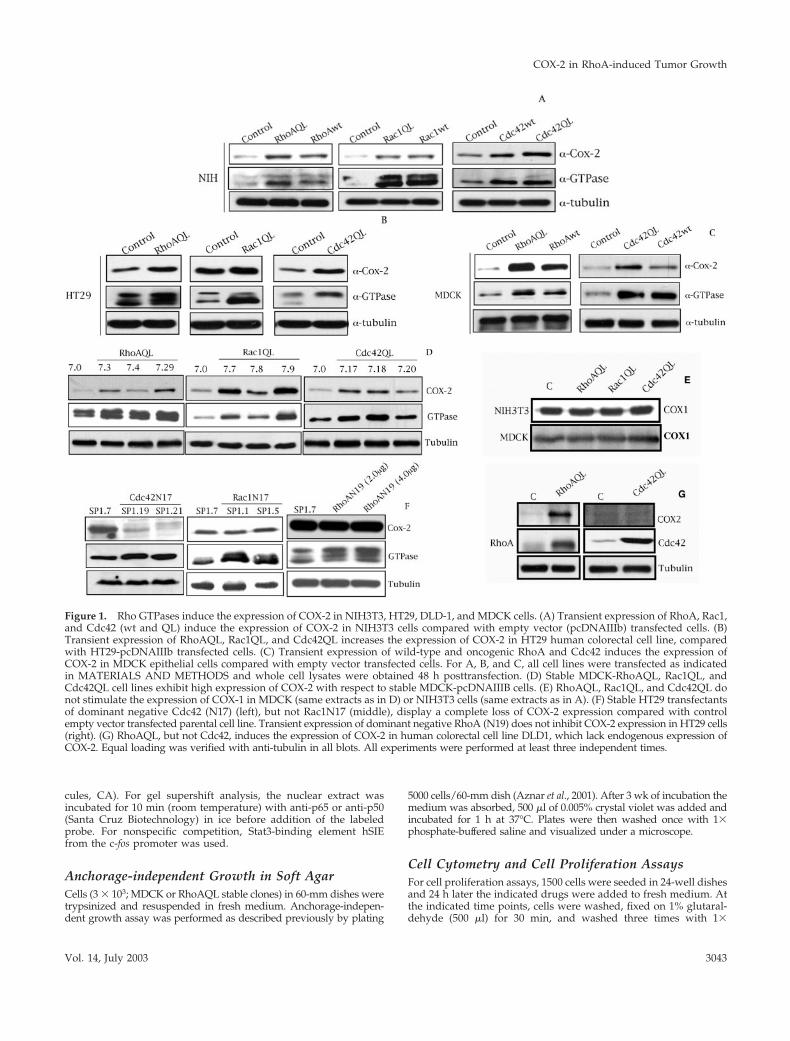
Figure 1. Rho GTPases induce the expression of COX-2 in NIH3T3, HT29, DLD-1, and MDCK cells. (A) Transient expression of RhoA, Rac1,and Cdc42 (wt and QL) induce the expression of COX-2 in NIH3T3 cells compared with empty vector (pcDNAIIIb) transfected cells. (B)Transient expression of RhoAQL, Rac1QL, and Cdc42QL increases the expression of COX-2 in HT29 human colorectal cell line, comparedwith HT29-pcDNAIIIb transfected cells. (C) Transient expression of wild-type and oncogenic RhoA and Cdc42 induces the expression ofCOX-2 in MDCK epithelial cells compared with empty vector transfected cells. For A, B, and C, all cell lines were transfected as indicatedin MATERIALS AND METHODS and whole cell lysates were obtained 48 h posttransfection. (D) Stable MDCK-RhoAQL, Rac1QL, andCdc42QL cell lines exhibit high expression of COX-2 with respect to stable MDCK-pcDNAIIIB cells. (E) RhoAQL, Rac1QL, and Cdc42QL donot stimulate the expression of COX-1 in MDCK (same extracts as in D) or NIH3T3 cells (same extracts as in A). (F) Stable HT29 transfectantsof dominant negative Cdc42 (N17) (left), but not Rac1N17 (middle), display a complete loss of COX-2 expression compared with controlempty vector transfected parental cell line. Transient expression of dominant negative RhoA (N19) does not inhibit COX-2 expression in HT29 cells(right). (G) RhoAQL, but not Cdc42, induces the expression of COX-2 in human colorectal cell line DLD1, which lack endogenous expression ofCOX-2. Equal loading was verified with anti-tubulin in all blots. All experiments were performed at least three independent times.
COX-2 in RhoA-induced Tumor Growth
Vol. 14, July 2003 3043
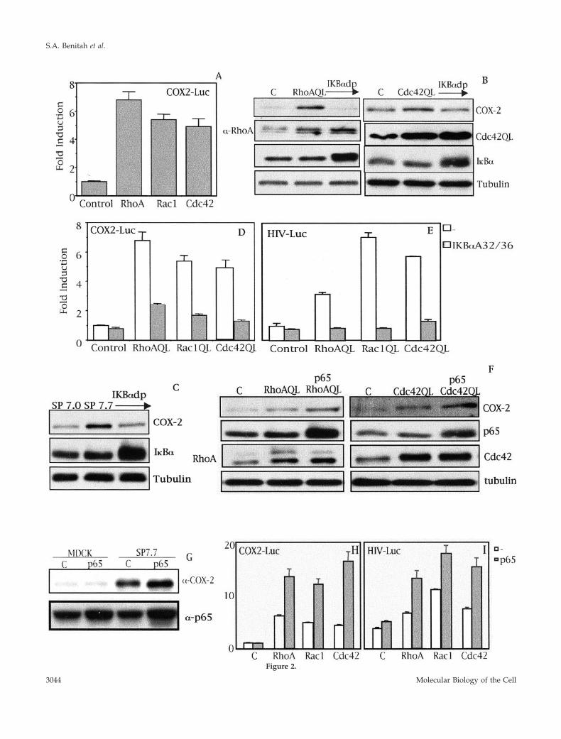
Figure 2.
S.A. Benitah et al.
Molecular Biology of the Cell3044
phosphate-buffered saline. Once all time points were collected, 500�l of 0.1% crystal violet was added to cells for 30 min and thenwashed as described above. To obtain the incorporated crystal violet500 �l of 10% acetic acid was added for 10 min and was latercollected and read at a wavelength of 595 nm. For cell cytometryanalysis, 2 � 105 cells were plated on 60-mm dishes and weretreated with sulindac or NS-398 for the indicated time. For FACS-SCAN analysis, the protocol was followed as described previously(Embade et al., 2000). Adhered cells were trypsinized and the cellmembrane was permeabilized with 70% ethanol, spun, and resus-pended in propidium iodide.
In Vivo Tumorigenic Assay and NSAIDs TreatmentCells (2 � 106) were trypsinized and resuspended in 100 �l of freshDMEM medium. Cells were injected subcutaneously in the limb andtumor growth was monitored twice a week for 90 d. Tumor volumewas determined using the following equation: V � (Dxd2)/2. Whentumors had reached a volume of 0.1 cm3, 3 mg/kg NS-398 was
injected intraperitoneally three times a week during 9 wk. Tumorvolume was measured at 2-d intervals during the treatment.
Prostaglandin E2 (PGE2) QuantificationCells (5000) were plated on 24-well dishes and 24 h later they weretreated with Bay11-7083 (10 �M) at the indicated time intervals (6,8, 12, and 24 h) to collect all the supernatants at the same time ofanalysis. The amount of PGE2 was measured using the commercialkit PGE2 EIA kit-monoclonal (Cayman Chemical) as described bythe manufacturer. Medium (50 �l) was collected and mixed in aPGE2 monoclonal antibody-coated 96-well dish and incubated over-night for 18 h at 4°C. The wells were then washed five times andincubated with Ellman’s reagent in the dark for 90 min at roomtemperature. The assay was read at a single wavelength of 405 nm.
RESULTS
Oncogenic RhoA, Rac1, and Cdc42 Induce theExpression of COX-2 in NIH3T3, HT29, and MDCKCellsWe ectopically expressed pCDNAIIIb or derived expressionvectors encoding for RhoA, Rac1, and Cdc42 in NIH3T3fibroblasts, HT29 human colorectal carcinoma cells, andMDCK epithelial cells. Wild-type and oncogenic version ofall three GTPases induced high levels of COX-2 in NIH3T3with respect to pcDNAIIIb control transfected cells (Figure1A). In addition, oncogenic RhoA, Rac1, and Cdc42 (QL)increase the high endogenous level of COX-2 in HT29 com-pared with empty vector transfected cells (Figure 1B). Withrespect to MDCK cells, transient expression of both RhoAand Cdc42 (QL and wt) induced COX-2 expression whencompared with MDCK-pcDNAIIIb cells (Figure 1C). How-ever, we were not able to obtain significant transient expres-sion of Rac1QL in MDCK cells as determined by Westernimmunoblot, nor Rac1-dependent effects such as SRF orNF-�B transcriptional activation. Thus, we sought to verifywhether Rac1QL regulates COX-2 levels in MDCK cells bystable expression. In this sense, we generated MDCK stabletransfectants of pcDNAIIIb or its derived vector encodingfor Rac1QL, and verified the level of COX-2 expression. Wewere able to select six independent Rac1QL-expressingclones that exhibited increased levels of Rac1QL and thatinduced high COX-2 expression of which three representa-tive ones are shown (Figure 1D). Additionally, we generatedMDCK stable transfectants of vectors encoding for RhoAand Cdc42QL, and tested for the level of COX-2 expressionfor three independent clones of each GTPase. Two represen-tative RhoAQL clones SP7.3, SP7.4, and a mass culture(SP7.29) that express different amounts of oncogenic RhoA,exhibit differential expression of COX-2 with respect toMDCK control cells, SP7.0. The same effect was observedwith two independent Cdc42-expressing clones, SP7.18 andSP7.17, and a mass culture (SP7.20). Thus, RhoA, Rac1, andCdc42 (QL) induce high levels of COX-2 also in MDCK cells.
In addition, we verified that this effect was specific toCOX-2 and not the constitutively expressed isoform COX-1(Figure 1E). SP7.0 (MDCK-pcDNAIIIb control) and cells thatexpress high levels of each GTPase, SP7.29 (RhoAQL), SP7.9(Rac1QL), and SP7.18 (Cdc42QL) were used to verify COX-1expression. As seen in Figure 1E the levels of COX-1 remainunchanged upon Rho GTPases expression in MDCK cells.The same results were obtained with all RhoAQL-, Rac1QL-,
Figure 2 (facing page). Rho GTPase-dependent expression ofCOX-2 is at the transcriptional level and dependent on NF-�B. (A)RhoA, Rac1, and Cdc42 (QL) induce the transcription of the proxi-mal region of the cox-2 promoter (�1772 to �106) in MDCK cells.Stable cell lines of RhoAQL, Rac1QL, and Cdc42QL (7.3, 7.9, and7.18, respectively) were transfected with COX2-Luc reporter vector(0.5 �g), and luciferase activity was measured 48 h posttransfection.Data shown represents a single experiment performed in tripli-cate � SD. (B) I�B�S32/36A inhibits RhoA- and Cdc42QL-inducedCOX-2 expression. Transient transfection of 2 �g of I�B�S32/36A(I�Bdp) together with 1 �g of pcDNAIIIb, RhoAQL, or Cdc42QLwas carried out in MDCK cells and extracts for Western blot anal-ysis were obtained 48 h posttransfection. (C) I�B�S32/36A inhibitsRac1QL-induced COX-2 expression in MDCK cells. MDCK-pcD-NAIIIb or clone SP7.9 (MDCK-Rac1QL) were transiently transfectedwith I�B�S32/36A (2 �g), and Western blot analysis was carried out48 h posttransfection. Equal loading was verified with an anti-tubulin antibody. (D) I�B�S32/36A inhibits transcription of thecox-2 promoter induced by RhoAQL, Rac1QL, and Cdc42QL. COX2-Luc (0.5 �g) and I�B�S32/36A (1.0 �g) were transiently cotrans-fected in 7.0 (MDCK-pcDNAIIIb), 7.3 (MDCK-RhoAQL), 7.9(MDCK-Rac1QL), and 7.18 (MDCK-Cdc42QL), and luciferase activ-ity was measured 48 h posttransfection (E) Expression of I�B�S32/36A leads to a functional inhibition of NF-�B transcriptional activityin MDCK cells. Same experiment as in D was carried out with 0.5 �gof HIV-luc reporter instead of COX-2-luc, and luciferase activitywas measured 48 h posttransfection. Data shown in D and E rep-resent a single experiment performed in triplicate � SD. (F) Over-expression of p65 augments COX-2 expression in RhoAQL- andCdc42QL-expressing cells. MDCK cells were transfected with 2.0 �gof p65 together with pcDNAIIIB (1.0 �g) (referred as control cells)RhoAQL (1.0 �g) or Cdc42QL (1.0 �g), and extracts were obtained48 h posttransfection. (G) p65 potentiates COX-2 expression in SP7.7(Rac1QL-MDCK) clone without any effect in parental MDCK cells.p65 (2.0 �g) was transfected in MDCK-pcDNAIIIB control cells, orin SP7.7 cells and extracts were obtained 48 h posttransfection.Expression of p65 was verified in F and G with an anti-p65 anti-body. Equal loading was determined with anti-tubulin. (H) Expres-sion of p65 produces a synergism in Rho-mediated transcription ofthe cox-2 promoter. (I) Expression of p65 in MDCK cells togetherwith RhoAQL, Rac1QL, or Cdc42QL leads to a functional increase ofNF-�B transcriptional activity. For parts H and I transfection wascarried out as indicated in parts D and E, but cotransfecting p65rather than I�B�S32/36A. Transfection efficiencies in D, E, H, and Iwere normalized using an RSV5-CAT reporter (0.5 �g) transfectedalong with the indicated plasmids. All experiments were performedfour times with similar results.
COX-2 in RhoA-induced Tumor Growth
Vol. 14, July 2003 3045
and Cdc42QL-expressing clones and mass cultures withidentical results (our unpublished data). As well, no changein COX-1 expression was observed upon transient expres-sion of RhoAQL, Rac1QL, or Cdc42QL in NIH3T3 cellscompared with empty vector transfected cells (Figure 1E).
As shown in Figure 1B, HT29 human colorectal carcinomacells show high endogenous level of COX-2 compared withother cell systems. We next verified whether Rho GTPasesare involved in the expression of COX-2 in the human colo-rectal cancer-derived cell line HT29. To that end, we at-tempted to generate HT29 stable transfectants that expresseither dominant negative Rac1 (N17), Cdc42 (N17), RhoA(N19), or control empty vector (pcDNAIIIb). As observed inFigure 1F, expression of Cdc42N17 induced a drastic reduc-tion of COX-2 expression, whereas Rac1N17 expression hadno effect on COX-2 levels. The same result was obtainedwith transient expression of Cdc42N17 in HT29 cells, al-though due to a transfection efficiency of �35%, we did notobserved a full inhibition of COX-2 expression (our unpub-lished data). Although HT29 cells can transiently expresshigh levels of dominant negative RhoA (N19), we were notable to obtain viable clones that expressed dominant nega-tive RhoA (N19) in a stable manner. Thus, we expressedRhoA in transient transfection experiments. RhoAN19 didnot affect the expression of COX-2 in HT29 cells (Figure 1F).As controls of dominant negative activity for each GTPase,we verified that expression of RhoAN19, Rac1N17 andCdc42N17 in HT29 cells inhibited the activation of NF-�Bactivity by Ost, Vav1, and Dbl, respectively, as describedpreviously (Montaner et al. (1998)) (our unpublished data).
Because HT29 have a high level of endogenous COX-2expression, we next investigated whether Rho GTPases wereable to regulate COX-2 expression in another human colo-rectal cancer-derived cell line such as DLD-1, with low levelsof expression of Rho GTPases and which completely lacksendogenous COX-2 expression. As shown in Figure 1G,RhoA efficiently induced the expression of COX-2 in DLD1cells when expressed ectopically. In contrast, Cdc42 (Figure1G), and Rac1 (our unpublished data) failed to do so. Thus,these results suggest that Rho GTPases can modulate COX-2expression in human colon cancer. However, each GTPaseanalyzed in our work seems to have differential contributionor mechanisms to effect regulation of COX-2.
Rho-A-, Rac1-, and Cdc42-induced Expression ofCOX-2 Is Dependent on the NF-�B TranscriptionFactorAnalysis of the promoter of human COX-2 revealed severalputative binding sites for transcription factors whose activ-ity is modulated by Rho GTPases. These include NF-�B,SRF, C/EBP�, AP-1, c-Myc, and STATs. To quantify theextent of transcription of the cox-2 gene under Rho signalinga reporter vector termed COX2-Luc containing the cox-2promoter region spanning from bases �1772 to �106 wasgenerated (Diaz-Cazorla et al., 1999). Transient transfectionof 0.5 �g of COX2-Luc into 7.0 (MDCK-pcDNAIIIB), 7.3(MDCK-RhoAQL), 7.9 (MDCK-Rac1QL), and 7.18 (MDCK-Cdc42QL) clones was carried out and 48 h posttransfectionluciferase activity was measured. All three RhoA, Rac1, andCdc42 (QL) induced transcription of the cox-2 promotercompared with empty vector transfected cells (Figure 2A).
It has been reported that NF-�B regulates COX-2 expres-sion under a variety of circumstances such as inflammation,hypoxia, bacterial infections, or colorectal cancers (Croffordet al., 1997; Schmedtje et al., 1997; Lim et al., 2001). Thus, weverified whether this transcription factor played any role inthe induction of COX-2 by Rho GTPases. First, we tran-siently expressed dominant positive I�B� that carries serineresidues 32 and 35 mutated to alanine (I�B�A32/A36), whoseexpression leads to a very efficient inhibition of NF-�B, andverified COX-2 levels under Rho signaling in MDCK cells(Karin et al., 2002). The induction of COX-2 by ectopic ex-pression of both RhoAQL and Cdc42QL was drasticallyinhibited by I�B�A32/A36 (Figure 2B). The same result wasobtained with two independent stable clones for RhoAQL(SP7.3 and SP7.29) and Cdc42QL (SP7.17 and SP7.18) (ourunpublished data). As well, Rac1-dependent induction ofCOX-2 relies on the NF-�B pathway, because ectopic expres-sion of dominant positive I�B� in SP7.7 cells inhibitedCOX-2 expression (Figure 2C). These results were also ob-tained with another clone, SP7.9 (our unpublished data). Asexpected, inhibition of Rho GTPases-induced COX-2 expres-sion by I�B�A32/A36 was at the level of transcription be-cause it abrogated COX2-Luc transcription when expressedin stable clones of each GTPase (Figure 2D). As a control ofdominant positive I�B�A32/A36 activity, we verified that itsexpression led to inhibition of NF-�B activity and DNA-binding induced by Rho GTPases (Figure 2E; our unpub-lished data).
To further test whether NF-�B is involved in the inductionof COX-2 by Rho GTPases, we next expressed the p65 sub-unit of NF-�B together with RhoAQL and Cdc42QL or con-trol vector in MDCK cells. As seen in Figure 2F, transientcoexpression of p65 with either RhoAQL or Cdc42QL inMDCK cells potentiated COX-2 expression. The same effectwas observed when p65 was transiently transfected into twoRac1QL-expressing clones, SP7.7 and SP7.9, whereas over-expression of NF-�B alone in MDCK cells did not cause asignificant elevation of COX-2 expression (Figure 2G; ourunpublished data). Expression of p65 in RhoA, Rac1, orCdc42QL stable clones led to an increase in cox-2 promoteractivity by more than threefold compared with their respec-tive controls (Figure 2H). Accordingly, coexpression of p65increased NF-�B transcriptional activity induced by all threeGTPases (Figure 2I). Thus, NF-�B mediates the induction ofCOX-2 by oncogenic RhoA, Rac1, and Cdc42 at the tran-scriptional level.
Induction of COX-2 by RhoGTPases Is Not viaStat3Activation of Stat3 by members of the family of RhoGTPases,such as RhoA and Rac has been described previously (Simon etal., 2000; Aznar et al., 2001; Faruqi et al., 2001). Furthermore,Stat3 is necessary for RhoA-induced anchorage independentgrowth (Aznar et al., 2001). Because the cox-2 promoter containsputative Stat-binding elements, we sought to verify whetherStat3 might act downstream of Rho GTPases to induce COX-2expression.
To that end, we expressed wild-type Stat3 (wt) or a dom-inant negative Stat3 with a mutated transactivation domain(Stat3D), in RhoAQL-, Rac1QL-, and Cdc42QL-expressingclones SP7.29, SP7.9, and SP7.17 (Figure 3). RhoA QL, Rac1QL, and Cdc42QL efficiently induced tyrosine-705 phos-
S.A. Benitah et al.
Molecular Biology of the Cell3046
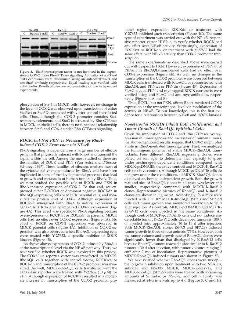
phorylation of Stat3 in MDCK cells; however, no change inthe level of COX-2 was observed upon transfection of eitherStat3wt or Stat3D compared with vector control transfectedcells. Thus, although the COX-2 promoter contains Stat-responsive elements, and Stat3 is activated by Rho GTPasesin MDCK epithelial cells, there is no functional relationshipbetween Stat3 and COX-2 under Rho GTPases signaling.
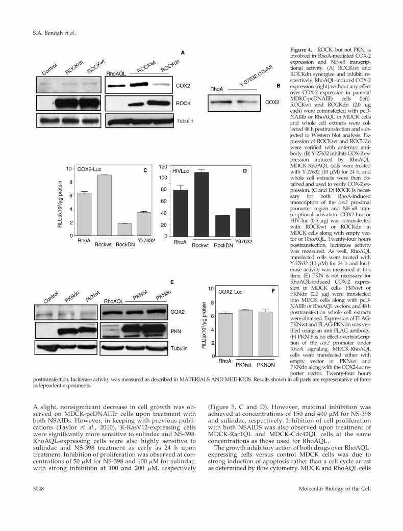
ROCK, but Not PKN, Is Necessary for RhoA-induced COX-2 Expression via NF-�BRhoA signaling is dependent on a large number of effectorproteins that physically interact with RhoA and transmit thesignal within the cell. Among the most studied of these arethe families of ROCK and PKN (Van Aelst and D’Souza-Schorey, 1997). These families of effectors mediate most ofthe cytoskeletal changes induced by RhoA and have beenimplicated in some of the developmental processes that leadto growth and metastasis of human tumors by RhoA. Thus,we next studied the possible role of ROCK and PKN inRhoA-induced expression of COX-2. To that end, we ex-pressed either ROCKwt or dominant negative ROCKdn inRhoAQL-expressing cells or MDCK parental cells, and mea-sured the protein level of COX-2. Although expression ofROCKwt synergized with RhoA to induce expression ofCOX-2, ROCKdn greatly impaired COX-2 expression (Fig-ure 4A). This effect was specific to RhoA signaling becauseoverexpression of ROCKwt or ROCKdn in parental MDCKcells had no effect over COX-2 expression (Figure 4A). Noeffect of ROCK on COX-2 expression was observed inMDCK parental cells (Figure 4A). Inhibition of COX-2 ex-pression was also observed when RhoAQL-expressing cellswere treated with Y-27632, a specific inhibitor of ROCKkinases (Figure 4B).
As shown above, expression of COX-2 induced by RhoA isat the transcriptional level via the NF-�B pathway. Thus, wenext verified whether ROCK was involved in this process.The COX2-Luc reporter vector was transfected in MDCK-RhoAQL cells together with control vector, ROCKwt, orROCKdn and transcription of the COX-2 promoter was mea-sured. As well, MDCK-RhoAQL cells transfected with theCOX2-Luc reporter were treated with Y-27632 (10 �M) for24 h. Although expression of ROCKwt resulted in a moder-ate increase in transcription of the COX-2 proximal pro-
moter region, expression ROCKdn or treatment withY-27632 inhibited such transcription (Figure 4C). The sametype of experiment was carried out with the NF-�B–respon-sive reporter vector HIV-luc, to verify whether ROCK hadany effect over NF-�B activity. Surprisingly, expression ofROCKwt or ROCKdn, or treatment with Y-27632 had thesame effect over NF-�B activity than COX-2 promoter tran-scription.
The same experiments as described above were carriedout with respect to PKN. However, expression of PKNwt ofPKNdn in RhoAQL-transformed cells had no effect overCOX-2 expression (Figure 4E). As well, no changes in thetranscription of the COX-2 promoter were observed betweenMDCK cells transfected with RhoAQL or cotransfected withRhoAQL and PKNwt or PKNdn (Figure 4F). Expression ofFLAG-tagged PKN and myc-tagged ROCK constructs wereverified using anti-FLAG and anti-myc antibodies, respec-tively (Figure 4, A and E).
Thus, ROCK, but not PKN, affects RhoA-mediated COX-2expression at the transcriptional level via modulation of theactivity of NF-�B. To our knowledge, this is the first evi-dence for a relationship between NF-�B and ROCK kinases.
Nonsteroidal NSAIDs Inhibit Both Proliferation andTumor Growth of RhoAQL Epithelial CellsGiven the implication of COX-2 and Rho GTPases overex-pression in tumorigenesis and metastasis of human tumors,the above-mentioned results suggest that COX-2 might playa role in RhoA-mediated tumorigenesis. First, we analyzedthe tumorigenic potential of stable MDCK-RhoAQL trans-fectants. Four different RhoAQL expressing clones wereplated on soft agar to determine their capacity to growunder anchorage-independent conditions compared withMDCK-pcDNAIIIb (negative control) and MDCK-K-RasV12cells (positive control). Although MDCK-pcDNAIIIb cells donot grow under these conditions, all MDCK-RhoAQL clonesdisplayed anchorage-independent growth. Both the growthrate and size of RhoAQL clones in all cases was lower andsmaller, respectively, compared with MDCK-K-RasV12clones. Representative pictures of RhoAQL and K-RasV12clones are shown in Figure 5A. Accordingly, four mice wereinjected with 2 � 106 MDCK-RhoAQL (SP7.3 and SP7.29)cells and tumor growth was monitored weekly up to 90 dafter injection. As controls, MDCK-pcDNAIIIb and MDCK-KrasV12 cells were injected in the same conditions. Al-though control MDCK-pcDNAIIIb cells did not induce anydetectable tumor, K-RasV12 cells developed tumors in 100%of injected mice approximately 10 to 15 d after injection.Both MDCK-RhoAQL clones (SP7.3 and SP7.29) inducedtumor growth in three of four animals (75%). However, boththe tumor volume and growth rate of RhoAQL clones weresignificantly lower than that displayed by K-RasV12 cellsbecause RhoAQL tumors reached a size similar to K-RasV12tumors �30 d after injection, with tumor volumes ranging 1cm3 after 2 mo of inoculation. Representative pictures ofMDCK-RhoAQL induced tumors are shown in Figure 5B.
We next verified whether RhoAQL clones were suscepti-ble to growth inhibition upon treatment with two NSAIDs,sulindac and NS-398. MDCK, MDCK-K-RasV12, andMDCK-RhoAQL (SP7.29) cells were treated with increasingamounts of Sulindac or NS-398, and cell viability wasmeasured at 24-h intervals up to 4 d (Figure 5, C and D).
Figure 3. Stat3 transcription factor is not involved in the expres-sion of COX-2 under Rho GTPases signaling. Activation of Stat3 andStat3 expression were determined using an anti-Stat3PY-694 andanti-Stat3 antibody respectively. Equal loading was verified withanti-tubulin. Results shown are representative of five independentexperiments.
COX-2 in RhoA-induced Tumor Growth
Vol. 14, July 2003 3047
A slight, nonsignificant decrease in cell growth was ob-served on MDCK-pcDNAIIIb cells upon treatment withboth NSAIDs. However, in keeping with previous publi-cations (Taylor et al., 2000), K-RasV12-expressing cellswere significantly more sensitive to sulindac and NS-398.RhoAQL-expressing cells were also highly sensitive tosulindac and NS-398 treatment as early as 24 h upontreatment. Inhibition of proliferation was observed at con-centrations of 50 �M for NS-398 and 100 �M for sulindac,with strong inhibition at 100 and 200 �M, respectively
(Figure 5, C and D). However, maximal inhibition wasachieved at concentrations of 150 and 400 �M for NS-398and sulindac, respectively. Inhibition of cell proliferationwith both NSAIDS was also observed upon treatment ofMDCK-Rac1QL and MDCK-Cdc42QL cells at the sameconcentrations as those used for RhoAQL.
The growth inhibitory action of both drugs over RhoAQL-expressing cells versus control MDCK cells was due tostrong induction of apoptosis rather than a cell cycle arrestas determined by flow cytometry. MDCK and RhoAQL cells
Figure 4. ROCK, but not PKN, isinvolved in RhoA-mediated COX-2expression and NF-�B transcrip-tional activity. (A) ROCKwt andROCKdn synergize and inhibit, re-spectively, RhoAQL-induced COX-2expression (right) without any effectover COX-2 expression in parentalMDKC-pcDNAIIIb cells (left).ROCKwt and ROCKdn (2.0 �geach) were cotransfected with pcD-NAIIIb or RhoAQL in MDCK cellsand whole cell extracts were col-lected 48 h posttransfection and sub-jected to Western blot analysis. Ex-pression of ROCKwt and ROCKdnwere verified with anti-myc anti-body. (B) Y-27632 inhibits COX-2 ex-pression induced by RhoAQL.MDCK-RhoAQL cells were treatedwith Y-27632 (10 �M) for 24 h, andwhole cell extracts were then ob-tained and used to verify COX-2 ex-pression. (C and D) ROCK is neces-sary for both RhoA-inducedtranscription of the cox2 proximalpromoter region and NF-�B tran-scriptional activation. COX2-Luc orHIV-luc (0.5 �g) was cotransfectedwith ROCKwt or ROCKdn inMDCK cells along with empty vec-tor or RhoAQL. Twenty-four hoursposttransfection, luciferase activitywas measured. As well, RhoAQLtransfected cells were treated withY-27632 (10 �M) for 24 h and lucif-erase activity was measured at thistime. (E) PKN is not necessary forRhoAQL-induced COX-2 expres-sion in MDCK cells. PKNwt orPKNdn (2.0 �g) were transfectedinto MDCK cells along with pcD-NAIIIb or RhoAQL vectors, and 48 hposttransfection whole cell extractswere obtained. Expression of FLAG-PKNwt and FLAG-PKNdn was ver-ified using an anti-FLAG antibody.(F) PKN has no effect overtranscrip-tion of the cox2 promoter underRhoA signaling. MDCK-RhoAQLcells were transfected either withempty vector or PKNwt andPKNdn along with the COX2-luc re-porter vector. Twenty-four hours
posttransfection, luciferase activity was measured as described in MATERIALS AND METHODS. Results shown in all parts are representative of threeindependent experiments.
S.A. Benitah et al.
Molecular Biology of the Cell3048

were treated with sulindac (400 �M) and NS-398 (150 �M)for 120 h and analysis of propidium iodide incorporation byflow cytometry was carried out at 24-h intervals. Figure 5Eshows a representative histogram at 96 h of NS-398 treat-ment of MDCK and MDCK-RhoAQL cells. Although controlMDCK cells exhibited a residual 13.79% of apoptosis at 96 hof treatment with NS-398, �45% of RhoAQL cells wereundergoing apoptosis. Although a higher toxicity was ob-served with sulindac in MDCK control cells, a strong induc-tion of apoptosis was observed in RhoAQL expressingclones upon sulindac treatment (our unpublished data).
In addition, the capacity of NS-398 to inhibit RhoAQL-induced tumor growth in vivo was studied. To this end, 12mice were injected with MDCK-RhoAQL (SP7.29) cells and
when tumors had reached a mean volume of 0.05–0.1 cm3
(approximately 1 mo after inoculation), mice were treatedintraperitoneally with either NS-398 (3 mg/kg, 3 times aweek during 4 wk) or vehicle, and tumor growth was com-pared between both populations. As observed in Figure 6A,a strong tumor growth inhibition was obtained in NS-398–treated mice that was statistically significant after 1 wk oftreatment (p � 0.05). A slight decrease in tumor volume wasobserved at this time, yet tumor size was maintained allthroughout the time of treatment after this first week. Arepresentative picture of a treated and a nontreated mouse isdepicted in Figure 6B. Therefore, NS-398 is a very efficientantitumoral agent against tumors that arise as a consequenceof RhoAQL overexpression.
Figure 5. Sulindac and NS-398 inhibit proliferation and induce apoptosis of RhoAQL-, Rac1QL-, Cdc42QL-, and KrasV12-expressingepithelial cells. (A) RhoAQL and KrasV12 promote anchorage independent growth in MDCK cells. (B) RhoAQL promotes tumor growth invivo. MDCK or MDCK-RhoAQL cells (2 � 106) were inoculated subcutaneously in athymic mice, and tumor volume was monitored at 2-dintervals. Pictures were taken 60 d upon inoculation. (C and D) Indicated concentrations of sulindac or NS-398 inhibit proliferation of MDCKcells transformed with RhoAQL, Rac1QL, Cdc42QL, and MDCK-RasV12 cells with no significant effect on MDCK parental cells. (E) NS-398induces apoptosis of MDCK-RhoAQL but not MDCK parental cells. NS-398 (150 �M) was added to MDCK-pcDNAIIIb or MDCK-RhoAQL,and cells were treated for 96 h. At this point, cells were collected by trypsin treatment and were stained with propidium iodide forfluorescence-activated cell sorting analysis (see MATERIALS AND METHODS).
COX-2 in RhoA-induced Tumor Growth
Vol. 14, July 2003 3049

Overexpression of members of the family of Rho GTPasesin diverse human tumors has been described. Moreover, inseveral tumoral models, Rho GTPases have been found to beessential either for tumor growth or metastasis. Thus, wenext evaluated whether inhibition of Rho GTPases in humancolorectal carcinoma-derived cell line HT29, which overex-press both Cdc42 and RhoA, would have any effect overtheir capacity to promote tumor growth in vivo. As men-tioned above, we could not obtain viable stable RhoAN19-HT29 clones; however, we established two HT29-Cdc42N17clones (SP1.19 and SP1.21), which have completely lost ex-pression of COX-2 (Figure 1F). Both HT29-Cdc42N17 stablecell lines showed an approximate 50% reduction with re-spect to vector transfected HT29 cells in their capability togrow under anchorage independent conditions in soft agar(Figure 6C). Furthermore, both SP1.19 and SP1.21 cloneswere injected each in four nude mice and tumor growth wasmonitored at 3-d intervals compared with control vectortransfected HT29 cells (SP1.7). Tumor growth of HT29-Cdc42N17 cells (clones SP1.19) was significantly delayedcompared with that of parental HT29 cells (SP1.7), withstatistical significance (p �0.05) (Figure 6D). Thus, Cdc42 isan important signaling component that contributes to tumorgrowth of HT29 human colorectal carcinoma cells.
NF-�B Activity Is Affected by Both Sulindac andNS-398 Treatment in RhoAQL-expressing Cells andIs Necessary for Cell ProliferationSeveral works have shown that different NSAIDs elicit theirantiinflammatory and antitumoral effects by COX-2–inde-
pendent mechanisms (Tegeder et al., 2001). In fact, sulindacand NS-398 have been shown to affect NF-�B activity as wellas other proteins in different cell types that would accountfor some of their specific effects (Yamamoto et al., 1999; Shaoet al., 2000; Mack et al., 2001). This is of particular interest inour system where both proteins, COX-2 and NF-�B, aredirectly connected. Thus we sought to determine the effect ofsulindac and NS-398 treatment on NF-�B activity underRhoAQL signaling. To that end, two RhoAQL expressingclones, SP7.3 and SP7.29, were treated with sulindac (400�M) or NS-398 (150 �M) for 72 h, and DNA-binding ofNF-�B studied using a �B consensus element. As shown inFigure 7A, NF-�B DNA-binding was significantly impairedupon treatment with both drugs. Accordingly, the level ofnuclear p65 subunit was verified to be lower in treatedversus untreated cells. Whole cell lysates were obtainedunder the same conditions and the total level of cellular p65was determined to remain constant (Figure 7A). Thus, inhi-bition of NF-�B by sulindac and NS-398 is not due to re-duced NF-�B synthesis but rather to a specific inhibition ofits nuclear migration and DNA-binding activity.
Expression of COX-2 by RhoAQL is dependent on NF-�B.Therefore, we hypothesized that inhibition of NF-�B uponsulindac and NS-398 treatment would lead to a reduction inthe level of expressed COX-2. Whole cell extracts were ob-tained from SP7.3 and SP7.29 cells treated during 24 h with400 �M sulindac or 150 �M NS-398, and COX-2 expressionwas determined by Western immunoblotting (Figure 7B).Interestingly, the level of COX-2 was significantly reducedin sulindac-treated cells and complete loss of expression was
Figure 6. NS-398 inhibits tumorgrowth of RhoAQL-transformedMDCK cells. (A) NS-398 inhibits tu-mor growth promoted by oncogenicRhoA. Cells (2 � 106) were inocu-lated subcutaneously in nu/nu miceand when tumors had reached amean volume of 0.05–0.1 cm3, micewere treated intraperitoneally withNS-398 (3 mg/kg body weight) at3-d intervals. Statistical significancewas achieved at day 8 of treatmentand was maintained throughout therest of the treatment (p � 0.05). (B)Representative pictures of NS398-treated and vehicle-treated mice in-oculated with MDCK-RhoAQL cellsat 2 wk of treatment. (C) Inhibition ofCdc42 in HT29 cells results in a 50%reduction of anchorage-independentgrowth in soft agar. Clones werestained with crystal violet and werequantified 1 mo after seeding of cells.(D) Tumor growth of HT29-Cdc42N17 (SP1.19) in syngeneicmice is delayed with respect toempty vector transfected cells HT29cells (SP1.7). Cells (2 � 106; SP1.7 andSP1.19) were inoculated subcutane-ously in immunosuppressed miceand tumor volume was monitored at2-d intervals. All experiments shownwere performed three independenttimes with similar results.
S.A. Benitah et al.
Molecular Biology of the Cell3050

observed upon NS-398 treatment. This effect was not due toa general loss of expression of cellular proteins because thelevel of endogenous p65 was unaffected upon treatmentwith both drugs. A similar effect was observed with 50 �MNS-398, although the effect was not as drastic (our unpub-lished data). Thus, both sulindac and NS-398 affect bothCOX-2 and NF-�B activities.
Last, we verified whether treatment of MDCK-pcDNAIIBand MDCK-RhoAQL cells with a specific inhibitor of NF-�B,termed Bay11-7083, would confirm these results. As shownin Figure 7C, treatment of RhoAQL-expressing cells withBay11-7083 resulted in inhibition of both COX-2 expressionand NF-�B DNA-binding. We next verified if exposure ofMDCK-RhoAQL cells to Bay11-7083 would lead to inhibi-
Figure 7. Sulindac and NS-398inhibit both NF-�B DNA-bindingand COX-2 expression inducedby RhoAQL. (A) Sulindac andNS-398 inhibit NF-�B DNA-bind-ing without affecting p65 expres-sion. MDCK-RhoQL cells (clonesSP7.3 and SP7.29) were treatedwith NS-398 and sulindac at theindicated concentrations for 72 h,and nuclear and whole cell ex-tracts were obtained. EMSA anal-ysis was carried out using a �Bconsensus sequence, and p65 ex-pression was verified using ananti-p65 antibody. (B) Sulindacand NS-398 treatment inhibitCOX-2 expression induced byRhoAQL. Same whole cell ex-tracts as in A were used to verifyCOX-2 expression with an anti-COX-2 antibody. (C) MDCK-RhoAQL cells (SP7.3) weretreated with Bay11-7083 (10 �M)for 72 h and nuclear and wholecell extracts were obtained.EMSA analysis and COX-2 ex-pression were performed as in Aand B. (D) Bay11-7083 inhibitsMDCK-RhoAQL cell prolifera-tion with a minor effect overempty vector transfected MDCKcells. MDCK-pcDNAIIIb andMDCK-RhoAQL cells wereplated on 24-well dishes andwere treated with Bay11-7083 (10�M) at the indicated time points.Cell viability was determined bythe crystal violet method. (E)Bay11-7083 does not affect the en-zymatic activity of COX-2 (PGE2production) in MDCK cells trans-formed with RhoAQL. MDKC-pcDNAIIIb and MDCK-RhoAQL(SP7.3) were plated in 24-welldishes and were treated withBay11-7083 (10 �M) for the indi-cated period of time (0, 6, 8, and24 h). Supernatant was collectedand PGE2 concentration wasmeasured as described in MATE-RIALS AND METHODS. As apositive control, NS-398 (100 �M)was added for 8 h and PGE2 syn-thesis was measured as describedabove. Ordinate indicate fold induction of PGE2 relative to control cells. (F) Bay11-7083 inhibits RhoAQL-induced COX-2 expression inMDCK cells at 12 h of treatment. The same experiment as in E was carried in parallel and whole cell extracts were obtained and subjectedto Western blot analysis by using an anti-COX-2 antibody. All experiments were performed three times with similar results.
COX-2 in RhoA-induced Tumor Growth
Vol. 14, July 2003 3051

tion of cell proliferation. MDCK-pcDNAIIIb and MDCK-RhoAQL cells were treated with Bay11-7083 (10 �M) during96 h, and cell viability was measured at 24-h intervals (Fig-ure 7D). Although treatment of MDCK cells with Bay11-7083produced an inhibition of cell proliferation, this inhibitionwas drastically increased in MDCK-RhoAQL cells. Thus,inhibition of NF-�B with Bay11-708 drastically interfereswith cell proliferation driven by oncogenic RhoA, in keepingwith the results shown above on a similar effect induced bythe COX-2 inhibitors sulindac and NS-398.
To discriminate the effects on NF-�B from those of COX-2,we next analyzed the possible inhibitory effect of Bay11-7083directly on COX-2 activity at early stages of treatment (Fig-ure 7E). For this, we measured synthesis of PGE2 in bothMDCK control cells (transfected with the empty vector) andin RhoAQL transformed cells. Bay11-7083 had no significanteffect on COX-2 activity up to 8 h of treatment, whereas apartial inhibition of PGE2 production could be observed at12 h of treatment. This effect was due to inhibition of COX-2expression rather than a direct inhibition over its enzymaticactivity (Figure 7F). As well, as a positive control we treatedMDCK-RhoAQL cells with NS-398 (100 �M) for 8 h in thesame experiment and verified that there was a much moreefficient inhibition of COX-2 enzymatic activity. Thus, NF-�Bactivity is necessary for cell proliferation induced by RhoA.
DISCUSSION
An emerging interest in Rho GTPases signaling as plausibletargets for the development of antitumoral strategies is aconsequence of the overwhelming evidence that relates theoverexpression of either the GTPase itself or the deregula-tion of some downstream signaling component in a highnumber of human cancers (Aznar and Lacal, 2001, 2003;Sahai and Marshall, 2002). Many works have delineated thedownstream effectors to RhoA, Rac1, and Cdc42 that con-tribute to the tumoral phenotype. On the other hand, there isa significant lack of knowledge about the physiological tar-get genes whose expression is modulated under persistentRho signaling contributing to tumor development and me-tastasis. In this sense, cyclin D1, c-Myc, p21 (Cip1), and p27(Kip1) are among the few known targets that might enableaberrant growth stimulated by Rho GTPases (Pruitt and Der,2001).
In this work, we identify COX-2 as a target gene that istranscriptionally regulated by Rho GTPases in several celllines of murine, canine, and human origin. Activation ofCOX-2 by Rho GTPases may be cell specific, as many otherRho-dependent signaling pathways (reviewed in Aznar andLacal, 2001b). Thus, overexpression of COX-2 in humancolon cancer HT29 cells is completely dependent on Cdc42,because stable expression of dominant negative Cdc42N17led to complete loss of COX-2 expression. However, al-though transient expression of oncogenic RhoAQL orRac1QL in HT29 results in an increase in COX-2 expression,inhibition of endogenous Rac1 or RhoA through expressionof their dominant negative inhibitory mutants did not affectCOX-2 expression in the cell line. However, DLD1 cells thatexpress low levels of Rho GTPases and completely lackendogenous COX-2 expression, express COX-2 upon RhoA,but not Rac1 and Cdc42 overexpression. These results sug-gest that Rho GTPases might be involved in the expressionof COX-2 in a cell type- and tumor-specific manner.
Some works had previously suggested that Rho GTPasesmight trigger transcription of the cox-2 promoter, with re-porter assays and chemical inhibitor experiments (Slice et al.,1999, 2000; Hahn et al., 2002). Slice et al. (2000) have shownthat RhoA and Rac1 but not Cdc42 induce transcriptionalactivation of a reporter vector that contains the cox-2 pro-moter region spanning from �963 to �50 in NIH3T3 fibro-blasts. Herein, we provide evidence that either wild-type orconstitutively active forms of Cdc42 stimulate expression ofendogenous COX-2 via the NF-�B pathway to a similarextent to that found for RhoA and Rac1. This discrepancymight be due to differences in the selected promoter regionused for the reporter vector, because it lacked all putative �Belements present in the endogenous cox-2 promoter. Inter-estingly, in the same work it was determined that the ele-ments located in the cox-2 promoter between �80 and �40were critical for Rac- and Rho-induced transcription of thereporter vector. In addition, a CRE/ATF element was shownto be essential for transcriptional stimulation of the reporterby Rac1, but not for RhoA. In our work, we have observedthat inhibition of NF-�B leads to complete loss of COX-2expression. Although, this does not exclude the possibilitythat other cis-acting elements might be relevant for the phys-iological expression of COX-2 under Rho signaling. Interest-ingly, although Stat3 is involved in Rho-mediated anchorageindependent growth, and the cox-2 promoter contains atleast two STAT putative binding elements, we have notobserved any effect of Stat3 signaling over Rho GTPases-induced expression of COX-2.
In terms of the molecular mechanism involved, we haveidentified ROCK as one of the RhoA effector proteins in-volved in the expression of COX-2. The role of ROCK inRho-mediated tumorigenesis has been extensively studied.However, besides their profound effects over cell cytoarchi-tecture and motility, both of which have been related to itscapability to promote tumor invasion, little is known as towhether this family of kinases regulates transcriptionalpathways that promote tumor growth. Herein, we demon-strate that ROCK is necessary for RhoA-induced expressionof COX-2 at the transcriptional level and that ROCK mod-ulates the transcriptional activity of NF-�B.
In keeping with the possible functional relationship be-tween Rho GTPases and COX-2, there are many similaritiesbetween the expression profiles of both proteins in humantumors. For instance, both proteins are up-regulated andnecessary for aberrant epidermal growth factor receptor sig-naling and tumor growth induced by the receptor. The sameholds true for tumor growth induced by oncogenic Ras,activation of c-Myc by growth factors, or signaling from thePI3K/Akt pathway (Taylor et al., 2000; Sheng et al., 2001a,b;Murga et al., 2002; Pai et al., 2002). Furthermore, overexpres-sion of COX-2 and members of the family of Rho GTPaseshas been detected in same human tumors such as breast,colon, pancreas, and head and neck squamous carcinomas.
The relationship between the NF-�B/COX-2 pathway andRho proteins in neoplastic transformation might provide anadditional way to treat tumors where Rho proteins are im-plicated. Several inhibitors of Rho signaling are availablethat exhibit antitumoral and antimetastatic activities (Aznarand Lacal, 2001b, 2003). In identifying the ROCK/NF-�B/COX-2 pathway as a physiological Rho target, new antitu-moral approaches can be made with respect to tumors
S.A. Benitah et al.
Molecular Biology of the Cell3052
where Rho GTPases are an issue. MDCK cells transformedwith RhoA, which are tumorigenic as determined by anchor-age-independent growth and in vivo tumor growth studies,are susceptible to efficient inhibition of proliferation by su-lindac and NS-398. Thus, NSAIDs inhibit cell proliferation,induce apoptosis, and prevent tumor growth of cells trans-formed with Rho proteins with little effects over parentaluntransformed cells. The differences between MDCK andMDCK-RhoAQL cells upon NSAIDs treatment are probablydue to the differential activities of signaling pathways, in-cluding COX-2 and NF-�B. Indeed, the inhibitory effects ofboth NSAIDs over Rho-transformants cannot be solely re-lated to COX-2 inhibition, because both drugs have beenreported to affect several other pathways (Tegeder et al.,2001). These pathways regulated by Rho would presumablyset the different behavior of the parental cells versus RhoA-transformed cells upon drug exposure. Of particular interestis the inhibitory action of sulindac and NS-398 over NF-�Bactivation. More importantly, COX-2 expression is com-pletely inhibited upon 24 h of NS-398 treatment of the cells.Thus, given that the half-life of COX-2 ranges from 3.5 to 8 h,the antitumoral effect of both NSAIDs over RhoAQL trans-formed cells might take place via inhibition of the preexist-ing COX-2 at early stages of treatment and to sustainedNF-�B inhibition during early and late stages of the treat-ment. This idea is further strengthened by the fact thatBAY11-7083, an inhibitor of NF-�B, drastically blocks pro-liferation of RhoAQL transformed cells with no direct effecton COX-2 enzymatic activity upon early stages of treatment.In addition, these observations suggest that treatment oftumors induced by Rho GTPases with conventional NSAIDsmight be equally valid with respect to treatment with yet tobe synthesized specific COX-2 inhibitors.
Last, we have provided evidence that inhibition of endo-genously overexpressed Cdc42 in HT29 cells leads to a sig-nificant delay of tumor growth in vivo, further potentiatingthe knowledge of an important role of Rho GTPases inhuman cancer. Thus, overall, these results suggest that in-hibition of Rho GTPases signaling via COX-2, NF-�B, orROCK may constitute a plausible strategy to inhibit tumorgrowth and open a new alley for the development of a novelantitumoral strategy against human tumors where RhoGTPases play an important role.
ACKNOWLEDGMENTS
This work was supported by grants SAF2001-2042 and SAF2002-2437 from Ministerio de Ciencia y Tecnologıa. S.A.B. is a fellow fromFondo deInvestigacion Sanitaria (Instituto deSalud Carlos III).
REFERENCES
Aznar, S., and Lacal, J.C. (2001a). Rho signals to cell growth andapoptosis. Cancer Lett. 165, 1–10.
Aznar, S., and Lacal, J.C. (2001b). Searching new targets for antican-cer drug design: the families of Ras and RhoGTPases and theireffectors. Prog. Nucleic Acid Res. Mol. Biol. 67, 193–234.
Aznar, S., and Lacal, J.C. (2003). Rho GTPases in human carcino-genesis: a tale of excess. Rev. Oncol. (in press).
Aznar, S., Valeron, P.F., del Rincon, S.V., Perez, L.F., Perona, R., andLacal, J.C. (2001). Simultaneous tyrosine and serine phosphorylation
of stat3 transcription factor is involved in rho a GTPase oncogenictransformation. Mol. Biol. Cell 12, 3282–3294.
Bar-Sagi, D., and Hall, A. (2000). Ras and Rho GTPases: a familyreunion. Cell 103, 227–238.
Boerner, J.M., Danielsen, A.J., McManus, M.J., and Maihle, N.J.(2000). Activation of Rho is required for ligand-independent onco-genic signaling by a mutant EGF receptor. J. Biol. Chem. 10, 3691–3695.
Cao, Y., and Prescott, S.M. (2002). Many actions of cyclooxygenase-2in cellular dynamics and in cancer. J. Cell. Physiol. 190, 279–286.
Crofford, L.J., Tan, B., McCarthy, C.J., and Hla, T. (1997). Involve-ment of nuclear factor kappa B in the regulation of cyclooxygen-ase-2 expression by interleukin-1 in rheumatoid synoviocytes. Ar-thritis Rheum. 40, 226–236.
Chiariello, M., Marinissen, M.J., and Gutkind, S. (2001). Regulationof c-myc expression by PDGF through Rho GTPases. Nat. Cell Biol.3, 580–586.
Danen, E.H., Sonneveld, P., Sonnenberg, A., and Ymamda, K.M.(2000). Dual stimulation of Ras/mitogen-activated protein kinaseand RhoA by cell adhesion to fibronectin supports growth factor-stimulated cell cycle progression. J. Cell Biol. 151, 1413–1422.
del Peso, L., Hernandez-Alcoceba, R., Embade, N., Carnero, A.,Esteve, P., Paje, C., and Lacal, J.C. (1997). Rho proteins inducemetastatic properties in vivo. Oncogene 15, 3047–3057.
Diaz-Cazorla, M., Perez-Sala, D., Ros, J., Jimenez, W., Fresno, M.,and Lamas, S. (1999). Regulation of cyclooxygenase-2 expression inhuman mesangial cells-transcriptional inhibition by IL-13. Eur.J. Biochem. 260, 268–274.
Dubois, R.N. (2001). New paradigms for cancer prevention. Carci-nogenesis 22, 691–692.
Embade, N., Valeron, P.F., Aznar, S., Lopez-Collazo, E., and Lacal,J.C. (2000). Apoptosis induced by Rac GTPase correlates with in-duction of FasL and ceramides production. Mol. Biol. Cell 11, 4347–4358.
Faruqi, T.R., Gomez, D., Bustelo, X.R., Bar-Sagi, D., and Reich, N.C.(2001). Rac1 mediates Stat3 activation by autocrine IL-6. Proc. Natl.Acad. Sci. USA 98, 9014–9019.
Gupta, R.A., and Dubois, R.N. (2001). Colorectal cancer preventionand treatment by inhibition of cyclooxygenase 2. Nat. Rev. Cancer 1,11–21.
Hahn, A., Barth, H., Kress, M., Mertens, P.R., and Goppelt-Struebe,M. (2002). Role of Rac and Cdc42 in lysophosphatidic acid-mediatedcyclo-oxygenase-2 gene expression. Biochem. J. 362, 33–40.
Jones, M.K., Wang, H., Peskar, B.M., Levin, E., Itani, R.M., Sarfeh,I.J., and Tarnawski, A.S. (1999). Inhibition of angiogenesis by non-steroidal anti-inflammatory drugs: insight into mechanisms andimplications for cancer growth and ulcer healing. Nat. Med. 5,1418–1423.
Karin, M., Cao, Y., Greten, F.R., and Li, Z.W. (2002). NF-�B incancer: from innocent bystander to major culprit. Nat. Rev. Cancer2, 301–310.
Lim, J.W., Kim, H., and Kim, K.H. (2001). Nuclear factor �B regu-lates cyclooxygenase-2 expression and cell proliferation in humangastric cancer cells. Lab. Investig. 81, 349–360.
Liu, C.H., Chang, S-H., Narko, K., Trifan, O.C., Wu, M-T., Smith, E.,Haudenschild, C., Lane, T.F., and Hla, T. (2001). Overexpression ofcyclooxygenase-2 is sufficient to induce tumorigenesis in transgenicmice. J. Biol. Chem. 276, 18563–18569.
Mack Strong, V.E., Mackrell, P.J., Concannon, E.M., Mestre, J.R.,Smyth, G.P., Schaefer, P.A., Stapleton, P.P., and Daly, J.M. (2001).
COX-2 in RhoA-induced Tumor Growth
Vol. 14, July 2003 3053
NS-398 treatment after trauma modifies NF-�B activation and im-proves survival. J. Surg. Res. 98, 40–46.
Muller, J.M., Metzger, E., Greschik, H., Bosserhoff, A.K., Mercep, L.,Buettner, R., and Schule, R. (2002). The transcriptional coactivatorFHL2 transmits Rho signals from the cell membrane into the nu-cleus. EMBO J. 21, 736–748.
Muller, S.M., Okan, E., and Jones, P. (2000). Regulation of urokinasereceptor transcription by Ras- and Rho-family GTPases. Biochem.Biophys. Res. Commun. 270, 892–898.
Murga, C., Zohar, M., Teramoto, H., and Gutkind, J.S. (2002). Rac1and RhoG promote cell survival by the activation of PI3K and Akt,independently of their ability to stimulate JNK and NF-�B. Onco-gene 21, 207–216.
Nur-E-Kamal, M.S.A., Kamal, J.M., Qureshi, M.M., and Maruta, H.(1999). The CDC42-specific inhibitor derived from ACK-1 blocksv-Ha-Ras-induced transformation. Oncogene 18, 7787–7793.
Pai, R., Sorenghan, B., Szabo, I.L., Pavelka, M., Baatar, D., andTarnawski, A.S. (2002). Prostaglandin E2 transactivates EGF recep-tor: a novel mechanism for promoting colon cancer growth andgastrointestinal hypertrophy. Nat. Med. 8, 289–293.
Perona, R., Esteve, P., Jimenez, B., Ballestero, R.P., Ramon y Cajal, S.,and Lacal, J.C. (1993). Tumorigenic activity of rho genes from Aply-sia californica. Oncogene 8, 1285–1292.
Pruitt, K., and Der, C.J. (2001). Ras and Rho regulation of the cellcycle and oncogenesis. Cancer Lett. 17, 1–10.
Psichari, E., Balmain, A., Plows, D., Zoumpourlis, V., and Pintzas,A. (2002). High activity of serum response factor in the mesenchy-mal transition of epithelial tumor cells is regulated by RhoA signal-ing. J. Biol. Chem. 277, 29490–29495.
Qiu, R.G., Chen, J., Kirn, D., McCormick, F., and Symons, M.(1995b). An essential role for Rac in Ras transformation. Nature 37,457–459.
Qiu, R.G., Chen, J., McCormick, F., and Symons, M. (1995a). A rolefor Rho in Ras transformation. Proc. Natl. Acad. Sci. USA 92, 11781–11785.
Ridley, A.J. (2001). Rho family proteins: coordinating cell responses.Trends Cell Biol. 11, 471–477.
Sachdev, P., Jiang, X.Y., Li, W., Miki, T., Nur-E-Kamal, M.S., Wang,L.H. (2001). Differential requirement for Rho family GTPases in anoncogenic insulin-like growth factor-I receptor-induced cell trans-formation. J. Biol. Chem. 276, 26461–26471.
Sahai, E. and Marshall, C.J. (2002) Rho-GTPases and cancer. Rev.Cancer 2, 133–142.
Schmedtje, J.F., Ji, Y.S., Lui, W.L., DuBois, R.N., and Runge, M.S.(1997). Hypoxia induces cyclooxygenase-2 via NF-�B p65 transcrip-tion factor in human vascular endothelial cells. J. Biol. Chem. 272,601–608.
Schmitz, A.A.P., Govek, E-E., Bottner, B., and Van Aelst, L. (2002).Rho GTPases: signaling, migration, and invasion. Exp. Cell Res. 261,1–12.
Shao, J., Sheng, H., Inoue, H., Morrow, J.D., and DuBois, R.N. (2000).Regulation of constitutive cyclooxygenase-2 expression in coloncarcinoma cells. J. Biol. Chem. 275, 33951–33956.
Sheng, H., Shao, J., and Dubois, R.N. (2001a). K-Ras-mediated in-crease in cyclooxygenase 2 mRNA stability involves activation ofthe protein kinase B1. Cancer Res. 61, 2670–2675.
Sheng, H., Shao, J., Washington, M.K., and DuBois, R.N. (2001b).Prostaglandin E2 increases growth and motility of colorectal carci-noma cells. J. Biol. Chem. 276, 18075–18081.
Simon, A.R., Vikis, H.G., Stewart, S., Fanburg, B.L., Cochran, B.H.,and Guan, K.L. (2000). Regulation of STAT3 by direct binding to theRac1 GTPase. Science 290, 144–147.
Slice, L.W., Bui, L., Mak, C., and Walsh, J.H. (2000). Differentialregulation of COX-2 transcription by Ras- and Rho-family ofGTPases. Biochem. Biophys. Res. Commun. 276, 406–410.
Slice, L.W., Walsh, J.H., and Rozengurt, E. (1999). G� 13 stimulatesRho-dependent activation of the cyclooxygenase-2 promoter. J. Biol.Chem. 274, 27562–27566.
Sotiropoulos, A., Gineitis, D., Copeland, J., and Treisman, R. (1999).Signal-regulated activation of serum response factor is mediated bychanges in actin dynamics. Cell 98, 159–169.
Taylor, M.T., Lawson, K.R., Ignatenko, N.A., Marek, S.E., Stringer,D.E., Skovan, B.A., and Gerner, E.W. (2000). Sulindac sulfone inhib-its K-ras-dependent cyclooxygenase-2 expression in human coloncancer cells. Cancer Res. 60, 6607–6610.
Tegeder, I., Pfeilschifter, and Geisslinger, G. (2001). Cyclooxygen-ase-independent actions of cyclooxygenase inhibitors. FASEB J. 15,2057–2072.
Tsujii, M., Kawano, S., Tsuji, S., Sawaoka, H., Hori, M., and Dubois,R.N. (1998). Cyclooxygenase regulates angiogenesis induced by co-lon cancer cells. Cell 93, 705–716.
Van Aelst, L., and D’Souza-Schorey, C. (1997). Rho GTPases andsignaling network. Genes Dev. 11, 2295–2322.
van Leeuwen, F.N., van der Kammen, R.A., Habets, G.G., andCollard, J.G. (1995). Oncogenic activity of Tiam1 and Rac1 inNIH3T3 cells. Oncogene 11, 2215–2221.
Welsh, C.F., Roovers, K., Villanueva, J., Liu, Y., Schwartz, M.A., andAssoian, R.K. (2001). Timing of Cyclin D1 expression within G1phase is controlled by Rho. Nat. Cell Biol. 3, 950–957.
Whitehead, I.P., Lambert, Q.T., Glaven, J.A., Abe, K., Rossman, K.L.,Mahon, G.M., Trzaskos, J.M., Kay, R., Campbell, S., and Der, C.J.(1999). Dependence of Dbl and Dbs transformation on MEK andNF-�B activation. Mol. Cell. Biol. 19, 7759–7770.
Yamamoto, Y., Yin, M.-J., Lin, K.-M., and Gaynor, R. (1999). Sulin-dac inhibits activation of the NF-�B pathway. J. Biol. Chem. 274,27307–27314.
S.A. Benitah et al.
Molecular Biology of the Cell3054