robotic-assisted vs conventional surgery in medial
TRANSCRIPT

RESEARCH ARTICLE Open Access
Robotic-assisted vs conventional surgery inmedial unicompartmental kneearthroplasty: a clinical and radiologicalstudyRoberto Negrín1 , Jaime Duboy1 , Magaly Iñiguez1 , Nicolás O. Reyes1* , Maximiliano Barahona2 ,Gonzalo Ferrer1 , Carlos Infante1,2 and Nicolás Jabes1
Abstract
Background: The use of unicompartmental knee arthroplasty (UKA) has increased and new technologies havebeen developed to improve patient survival and satisfaction, soft tissue balance, alignment, and component size.Robot-assisted systems offer an increase in surgical precision and accuracy. The purpose of this study is to evaluatethe precision of component position using five radiological parameters in conventional and robotic-assisted medialUKA using the NAVIO system.
Methods: A cohort study was designed for patients who underwent medial UKA between April 2017 and March2019 in a single center. Patients were allocated in the conventional (UKA-C) or robotic-assisted (UKA-R) group. Thevariables analyzed were age, gender, affected knee side, length of hospital stay, surgical time, and radiologicalmeasurements such as anatomical medial distal femoral angle (aMDFA), anatomical medial proximal tibial angle(aMPTA), tibial slope, the sagittal femoral angle, and the component size. A target was defined for eachmeasurement, and a successful UKA was defined if at least four radiological measures were on target after surgery.Also, patients’ reported outcomes were evaluated using the Oxford Knee Score (OKS) and a numeric rating scale(NRS) for pain.
Results: Thirty-four patients were included, 18 of them underwent UKA-R. The success rate for UKA in the UKA-Rgroup was 87%; meanwhile, in the UKA-C group this was 28%, this difference was significant and powered (Fisher’sexact test, p = 0.001; 1 − β = 0.95). Also, a 5-point difference in favor of the UKA-R group in the median OKS (p =0.01), and a significantly lower median NRS for pain (p < 0.000) were found after surgery.
Conclusions: UKA-R achieved more precision in the radiological parameters’ measure in this study. Also, UKA-R hasa trend towards a better OKS and a lower NRS for pain at short-term follow-up.
Keywords: Unicompartmental knee, Robotic-assisted surgery
© The Author(s). 2021 Open Access This article is licensed under a Creative Commons Attribution 4.0 International License,which permits use, sharing, adaptation, distribution and reproduction in any medium or format, as long as you giveappropriate credit to the original author(s) and the source, provide a link to the Creative Commons licence, and indicate ifchanges were made. The images or other third party material in this article are included in the article's Creative Commonslicence, unless indicated otherwise in a credit line to the material. If material is not included in the article's Creative Commonslicence and your intended use is not permitted by statutory regulation or exceeds the permitted use, you will need to obtainpermission directly from the copyright holder. To view a copy of this licence, visit http://creativecommons.org/licenses/by/4.0/.The Creative Commons Public Domain Dedication waiver (http://creativecommons.org/publicdomain/zero/1.0/) applies to thedata made available in this article, unless otherwise stated in a credit line to the data.
* Correspondence: [email protected] of Orthopedics and Traumatology, Clinica Las Condes, Estoril450, Las Condes, Santiago, ChileFull list of author information is available at the end of the article
Knee Surgery & Related Research
Negrín et al. Knee Surgery & Related Research (2021) 33:5 https://doi.org/10.1186/s43019-021-00087-2
BackgroundKnee osteoarthritis is a prevalent disease affecting up to19% of the population over 45 years of age, causingchronic pain, disability, and lower quality of life [1–8].Unicompartmental knee arthroplasty (UKA) is a cost-
effective treatment for femorotibial unicompartmentalknee osteoarthritis. Over the last few years, the propor-tion of UKA had been increasing according to worldwidenational registers [9–14]. Patients undergoing UKA hada lower rate of complications, faster functional recovery,and better satisfaction compared to total knee arthro-plasty (TKA) [15–18]. Nevertheless, UKA has its difficul-ties. To achieve an adequate position of the components– crucial in avoiding early aseptic loosening – is morechallenging than in TKA. Robotic assistance may play anessential role in decreasing the rate of malposition of thecomponents in UKA [19, 20], theoretically leading tobetter clinical results and longer survival rates [21–27].Few publications describe the radiological findings
after robotic-assisted UKA using NAVIO (Blue BeltTechnologies, Plymouth, MN, USA) [28]. This studyaims to evaluate the precision of component positionusing five radiological parameters in conventional androbotic-assisted medial UKA using the NAVIO system.The hypothesis is that the robotic-assisted technique hasa better rate of success compared to conventional UKA.Secondary aims were to compare short-term patient-reported outcomes and pain between groups and toanalyze whether an adequate component position relatedto better short-term results in UKA.
MethodsPatient selectionA cohort study was designed for patients who under-went medial UKA between April 2017 and March 2019in a single center. The local Ethical Committee approvedthe study, and all patients signed, written informed con-sent before enrollment.All patients’ clinical records were reviewed, and their
pre- and post-operative radiographs were measured. Allpatients undergoing medial UKA, having undergone apre- and post-operative radiological study were included.Patients were excluded if they had an incomplete docu-mentation or refused to participate.Patients were allocated in the conventional (UKA-C)
or robotic-assisted (UKA-R) group according to the sur-geon’s and patient’s preference and the availability of therobot at the time of the surgery. In all patients, the Jour-ney UNI implant (Smith & Nephew Inc., Cordova, TN,USA) was used. All procedures were performed underspinal anesthesia by two senior knee surgeons (JD, R.N.).UKA-C was defined as the surgery performed withoutrobotic assistance, and UKA-R as the surgery performedwith the assistance of the NAVIO Robotic System (Blue
Belt Technologies, Plymouth, MN, USA). With the useof a tourniquet, an anterior approach and a medial para-patellar arthrotomy were performed in all surgeries. Nowound drain was used.The variables analyzed were age, gender, length of hos-
pital stay, surgical time, and radiological measurements.The time of surgery in the UKA-C group ranges fromincision to wound closure; meanwhile, in the UKA-Rgroup, it was from the positioning of the pins (beforethe surgical incision) to wound closure.
Radiological measurementsRadiological measurements were performed using theimmediate post-operative radiographs. An anteroposter-ior (AP) and a lateral knee radiograph were performedin all patients on day 1 after surgery. Two blinded ortho-pedic surgeons (NR, NJ) carried out the measurements.The same protocol used in the study by Iñiguez et al.
[19] was used (Fig. 1). The anatomic medial distal fem-oral angle (aMDFA) was defined as the angle between aline through the anatomical axis of the femur and a linethat joins the most distal point of the lateral condyle andthe medial femoral component. The sagittal femoralangle (SFA) was defined as the angle between a linethrough the anterior cortex of the metaphyseal-diaphyseal junction and a line through the posterior pegof the femoral component.The anatomical medial proximal tibial angle (aMPTA)
was defined as the angle between a line through the ana-tomical axis of the tibia and a line through the tibialcomponent. The tibial slope was defined as the angle be-tween a line through the sagittal mechanical axis of thetibia and a line through the tibial component.The femoral and tibial component size was assessed in
the lateral knee radiograph, according to the designerclassification: Oversized was defined when the implantprotrudes more than 2mm and undersized when the im-plant does not achieve adequate coverage. The target foreach measurement was defined as follows: tibial slope5 ± 3°, aMDFA 98 ± 3°, aMPTA 87 ± 3°, SFA 45 ± 3° andan adequate component size.
Clinical measurementsAll patients were evaluated pre-surgery, and then theywere contacted 6 months after surgery by a blindedevaluator to request that they complete the Oxford KneeScore (OKS) and to evaluate knee pain at rest using anumeric rating scale (NRS) from 0 to 10, with 0 beingno pain.
StatisticsThe frequency, proportions, median, range, and inter-quartile range were used to describe the sample of thestudy. The nonparametric median test was used to
Negrín et al. Knee Surgery & Related Research (2021) 33:5 Page 2 of 7

compare the continuous variables: age, affected kneeside, length of hospital stay, surgery time, OKS, NRS,and radiographic measurements. Fisher’s exact test wasused to compare categorical variables: gender, side, andcomponent size. Multivariate logistic discrimination ana-lysis was conducted to assess the capacity to recognizewhether the UKA was robotic-assisted or conventionalusing the radiological parameters as predictor variables.The frequency of success in each radiological measure-
ment was compared between groups using a Fisher’sexact test. A successful UKA was considered if the targetwas achieved in at least four of the radiographic parame-ters. The proportion of successful UKAs was comparedbetween groups using Fisher’s exact test. The power ofthe estimation (1 − β) was reported and consideredunderpowered if 1 − β was lower than 0.8. Also, toanalyze the relevance of a successful UKA, a nonpara-metric median test was used to compare the medianOKS and pain NRS between successful and failed UKAs.Confidence intervals of ±95% were built, and a signifi-
cance level of 0.05 was used. The data were processedusing Stata 11.2 version (StataCorp LP, College Station,TX, USA).
ResultsA total of 34 patients were included, of whom 18 wereUKA-R patients (Table 1). No significant differenceswere found between the two groups in terms of gender,age, affected knee side, or pre-operative OKS or painNRS (Tables 1 and 2). Also, no differences were foundin pre-operative radiological measurements. The median
total surgery time was significantly lower in UKA-C pa-tients (p < 0.01) (Table 2).The post-operative radiographic measurements are
summarized in Table 2. Only the SFA reached a signifi-cant difference (nonparametric median test, p = 0.02) be-tween groups. The ability to discriminate betweengroups using the radiological parameters was high, beingthe error classification error 0.11. Only one UKA-R wasmisclassified as UKA-C, but two UKA-Cs were misclas-sified as UKA-R.The rate of success to achieve the desired target in
each radiological measurement is summarized in Table 3.Successful UKA was achieved in 14 (88%) of the UKA-Rpatients, but only five (28%) of the UKA-C patients(Table 4). The proportion of successful outcomes wasstatistically significant and powered (Fisher exact test,p = 0.001; 1 − β = 0.95).A significantly better OKS was achieved in the UKA-R
group (nonparametric median test, p = 0.01) (Fig. 2).Yet, the median difference between groups was only 5points, so it may not be clinically relevant (Table 2). The
Fig. 1 Radiological measures in the anteroposterior and lateral knee x-ray. aMDFA anatomical medial distal femoral angle, aMPTA anatomicalmedial proxial tibial angle, SFA sagital femoral angle
Table 1 Comparison of demographics variables betweengroups
UKA-R UKA-C Total P
N 16 (47%) 18 (53%) 34 N/A
Male 7 (44%) 12 (67%) 19 (56%) 0.16*
Age 66 (56 to 82) 65 (41 to 76) 66 (41 to 82) 0.60**
Right side 10 (63%) 8 (44%) 16 (47%) 0.32*
UKA-R robotic-assisted unicompartmental knee arthroplasty UKA-Cconventional unicompartmental knee arthroplasty, N number*Fisher’s exact test, **nonparametric test median
Negrín et al. Knee Surgery & Related Research (2021) 33:5 Page 3 of 7
median pain NRS in the UKA-R was 1 (range, 0 to 3)and 4 in the UKA-C (Fig. 3); this difference reached stat-istical significance (nonparametric median test, p <0.000).Successful UKA had a trend towards a higher OKS
(nonparametric median test, p = 0.088; 1 − β = 0.51) anda significantly lower pain NRS (nonparametric mediantest, p = 0.036; 1 − β = 0.57), but both findings wereunderpowered.There was only one complication in the UKA-C group;
this was an arthrofibrosis that required mobilizationunder anesthesia. No complications were found in theUKA-R group.
DiscussionThe main finding of this study is that UKA-R yielded asignificantly higher rate of radiological successful UKA.Also, there was a trend towards a better functionalpatient-reported outcome and less pain during rest.Moreover, the definition of successful UKA by the radio-logical measurements used in this study was significantly
related to better functional outcomes and lower painlevels during rest, yet this finding was underpowered.This study is a continuation of the clinical phase in thecadaveric pilot study published by our team, whichshowed consistent results of greater accuracy in relationto implant position in robotic-assisted surgery, com-pared to the conventional technique [19].Published studies have shown that robotic-assisted
surgery allows for greater accuracy and improved im-plant position [29–33].In the radiological assessment of the cases studied, we
considered five imaging parameters and we defined suc-cess as the fulfillment of at least four out of five of therequirements since we believe that combining all theradiological parameters achieved a more comprehensiveway of measuring accuracy. We have no references toprevious publications in which this way of assessing
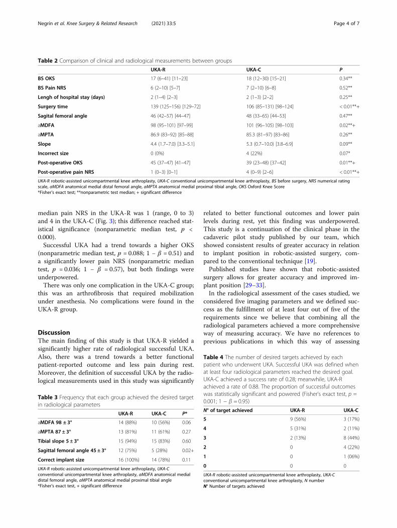
Table 2 Comparison of clinical and radiological measurements between groups
UKA-R UKA-C P
BS OKS 17 (6–41) [11–23] 18 (12–30) [15–21] 0.34**
BS Pain NRS 6 (2–10) [5–7] 7 (2–10) [6–8] 0.52**
Lengh of hospital stay (days) 2 (1–4) [2–3] 2 (1–3) [2–2] 0.25**
Surgery time 139 (125–156) [129–72] 106 (85–131) [98–124] < 0.01**+
Sagital femoral angle 46 (42–57) [44–47] 48 (33–65) [44–53] 0.47**
aMDFA 98 (95–101) [97–99] 101 (96–105) [98–103] 0.02**+
aMPTA 86.9 (83–92) [85–88] 85.3 (81–97) [83–86] 0.26**
Slope 4.4 (1.7–7.0) [3.3–5.1] 5.3 (0.7–10.0) [3.8–6.9] 0.09**
Incorrect size 0 (0%) 4 (22%) 0.07*
Post-operative OKS 45 (37–47) [41–47] 39 (23–48) [37–42] 0.01**+
Post-operative pain NRS 1 (0–3) [0–1] 4 (0–9) [2–6] < 0.01**+
UKA-R robotic-assisted unicompartmental knee arthroplasty, UKA-C conventional unicompartmental knee arthroplasty, BS before surgery, NRS numerical ratingscale, aMDFA anatomical medial distal femoral angle, aMPTA anatomical medial proximal tibial angle, OKS Oxford Knee Score*Fisher’s exact test; **nonparametric test median; + significant difference
Table 3 Frequency that each group achieved the desired targetin radiological parameters
UKA-R UKA-C P*
aMDFA 98 ± 3° 14 (88%) 10 (56%) 0.06
aMPTA 87 ± 3° 13 (81%) 11 (61%) 0.27
Tibial slope 5 ± 3° 15 (94%) 15 (83%) 0.60
Sagittal femoral angle 45 ± 3° 12 (75%) 5 (28%) 0.02+
Correct implant size 16 (100%) 14 (78%) 0.11
UKA-R robotic-assisted unicompartmental knee arthroplasty, UKA-Cconventional unicompartmental knee arthroplasty, aMDFA anatomical medialdistal femoral angle, aMPTA anatomical medial proximal tibial angle*Fisher’s exact test, + significant difference
Table 4 The number of desired targets achieved by eachpatient who underwent UKA. Successful UKA was defined whenat least four radiological parameters reached the desired goal.UKA-C achieved a success rate of 0.28; meanwhile, UKA-Rachieved a rate of 0.88. The proportion of successful outcomeswas statistically significant and powered (Fisher’s exact test, p =0.001; 1 − β = 0.95)
N° of target achieved UKA-R UKA-C
5 9 (56%) 3 (17%)
4 5 (31%) 2 (11%)
3 2 (13%) 8 (44%)
2 0 4 (22%)
1 0 1 (06%)
0 0 0
UKA-R robotic-assisted unicompartmental knee arthroplasty, UKA-Cconventional unicompartmental knee arthroplasty, N numberN° Number of targets achieved
Negrín et al. Knee Surgery & Related Research (2021) 33:5 Page 4 of 7

accuracy is used, and considering the prosthesis as awhole and not analyzing each radiological variable inparticular seem to provide a more comprehensiveevaluation.By analyzing the results of the radiological measure-
ments in both groups separately, there are only
statistically significant differences among them in termsof SFA. However, when considering the objective of suc-cess with the predefined parameters, the robotic groupreaches 88% versus 28% for the conventional group (p =0.001). This correlates with the logistic discriminant ana-lysis of both groups, which shows an error rate of 0.11,
Fig. 2 Distribution of Oxford Knee Score (OKS) among groups. Robotic-assisted unicompartmental knee arthroplasty (UKA-R) had a trend tobetter OKS than conventional unicompartmental knee arthroplasty (UKA-C) (nonparametric median test, p = 0.01)
Fig. 3 Distribution of numerical rating scale (NRS) for pain among groups. Robotic-assisted unicompartmental knee arthroplasty (UKA-R) had atrend to lower NRS for pain than conventional unicompartmental knee arthroplasty (UKA-C) (nonparametric median test, p < 0.000)
Negrín et al. Knee Surgery & Related Research (2021) 33:5 Page 5 of 7

with only one UKA-R wrongly categorized as conven-tional. We defined an interval of ±3° as it was the inter-val that maximized the difference between the groups.Bell et al. published a difference with an interval of ±2°[34]; however, in our study using an interval of ±1° morein width we found a significant difference.The first published data on the accuracy of implant
positioning using the NAVIO system were promising.Batailler et al. published the first clinical study compar-ing robotic-assisted UKA versus the conventional tech-nique [24]. The authors conclude that there is asignificant improvement in the accuracy of implant posi-tioning with robotic-assisted surgery in the coronal aswell as in the sagittal plane, thus reducing the numberof outliers, but no significant difference was found infunctional results among the groups studied. Our resultswere similar to those published by Batallier et al. regard-ing the accuracy of implant positioning, although we didfind better functional and post-operative pain results inthe UKA-R group compared to the UKA-C group at 1-year follow-up [24], which was similar to results pub-lished in other series [20] of robotic-assisted surgery.In this series, we did not find any complications in ei-
ther group, with just one case of manipulation underanesthesia in the conventional group that could explainthe difference in the OKS and NRS in favor of the ro-botic group. This is different from what St. Mart re-ported; in his study he had a higher rate of revision inthe robotic group because of early infection [35]. Thefact that we did not find any complications in the ro-botic group makes us believe that this is a safe proced-ure, even though these were our first cases, which wassimilar to what Mergenthaler [36] published in his case-control series.The limitations of this study include its small sample
size and short-term follow-up of 6 months, and not the2-year follow-up that most studies report, which pre-vented us from adequately demonstrating some of thetrends observed and does not necessarily represent long-term differences. There also may be a bias for the painNRS score, and this may explain why the results of theNRS in conventional surgery are higher than those re-ported in the literature. Another limitation is the waythat accuracy is measured with x-rays, which does notallow us to assess the rotational effects on implants. Inthe study design, we rejected the use of computed tom-graphy scans due to the need for patient exposure to ra-diation which is equivalent to 48 chest x-rays, as Ponzioet al. [37] pointed out, and also due to the high cost as-sociated with it, cost being one of the advantages of theimageless robotic NAVIO system used.Future studies in larger population groups and long-
term follow-ups are necessary to confirm the trend ob-served in the favorable results of our study.
ConclusionsRobotic-assisted UKA with the NAVIO system offersgreater accuracy of femoral implant positioning in thesagittal plane, and it is more accurate in achieving clin-ical and radiological success compared to conventionalsurgery.
AcknowledgementsTo the Department of Orthopaedics and Traumatology for its support.
Authors’ contributionsRN: conceptualization, methodology, resources, supervision, projectadministration. JD: conceptualization, resources, supervision, projectadministration. MI: methodology, data curation, writing – review and editing.NOR: methodology, validation, writing – review and editing, visualization.MB: methodology, formal analysis. CI: methodology, resources, writing –review and editing. GF: conceptualization, supervision. NJ: investigation,writing – original draft, visualization. The author(s) read and approved thefinal manuscript.
FundingNot applicable
Availability of data and materialsAll data generated or analyzed during this study are included in thispublished article and its supplementary information files.
Ethics approval and consent to participateThe local Ethical Committee approved the study, and all patients signedwritten, informed consent before enrollment.
Consent for publicationNot applicable
Competing interestsThe authors declare that they have no competing interests related to thisstudy.
Author details1Department of Orthopedics and Traumatology, Clinica Las Condes, Estoril450, Las Condes, Santiago, Chile. 2Department of Orthopaedic Surgery,Hospital Clinico de la Universidad de Chile, Santiago, Chile.
Received: 3 August 2020 Accepted: 12 January 2021
References1. Lawrence RC, Felson DT, Helmick CG, Arnold LM, Choi H, Deyo RA, Gabriel
S, Hirsch R, Hochberg MC, Hunder GG, Jordan JM, Katz JN, Kremers HM,Wolfe F, National Arthritis Data Workgroup (2008) Estimates of theprevalence of arthritis and other rheumatic conditions in the United States:part II. Arthritis Rheum 58:26–35
2. Vos T, Flaxman AD, Naghavi M et al (2012) Years lived with disability (YLDs)for 1160 sequelae of 289 diseases and injuries 1990-2010: a systematicanalysis for the Global Burden of Disease Study 2010. Lancet 380:2163–2196
3. Bijlsma JWJ, Berenbaum F, Lafeber FPJG (2011) Osteoarthritis: an updatewith relevance for clinical practice. Lancet 377:2115–2126
4. Nguyen US, Zhang Y, Zhu Y, Niu J, Zhang B, Felson DT (2011) Increasingprevalence of knee pain and symptomatic knee osteoarthritis: survey andcohort data. Ann Intern Med 155(11):725–732
5. Felson DT, Lawrence RC, Dieppe PA, Hirsch R, Helmick CG, Jordan JM,Kington RS, Lane NE, Nevitt MC, Zhang Y, Sowers M, McAlindon T, SpectorTD, Poole AR, Yanovski SZ, Ateshian G, Sharma L, Buckwalter JA, Brandt KD,Fries JF (2000) Osteoarthritis: new insights. Part 1: the disease and its riskfactors. Ann Intern Med 133(8):635–646
6. Loeser RF, Collins JA, Diekman BO (2016) Ageing and the pathogenesis ofosteoarthritis. Nat Rev Rheumatol 12:412–420
7. Abbott JH, Usiskin IM, Wilson R, Hansen P, Losina E (2017) The quality-of-lifeburden of knee osteoarthritis in New Zealand adults: a model-basedevaluation. PLoS One 12(10):e0185676
Negrín et al. Knee Surgery & Related Research (2021) 33:5 Page 6 of 7

8. Wallace IJ, Worthington S, Felson DT, Jurmain RD, Wren KT, Maijanen H,Woods RJ, Lieberman DE (2017) Knee osteoarthritis has doubled inprevalence since the mid-20th century. Proc Natl Acad Sci U S A 114(35):9332–9336
9. Bolognesi MP, Greiner MA, Attarian DE, Watters TS, Wellman SS, Curtis LH,Berend KR, Setoguchi S (2013) Unicompartmental knee arthroplasty andtotal knee arthroplasty among Medicare beneficiaries, 2000 to 2009. J BoneJoint Surg Am 95(22):e174
10. Swank ML, Alkire M, Conditt M, Lonner JH (2009) Technology and cost-effectiveness in knee arthroplasty: computer navigation and robotics. Am JOrthop (Belle Mead NJ) 38(2 Suppl):32–36
11. Australian Orthopaedic Association National Joint Registry. Hip and kneearthroplasty annual report 2018. https://aoanjrr.sahmri.com/annual-reports-2018
12. New Zealand Joint Registry. The New Zealand Registry annual report. 2018.https://nzoa.org.nz/system/files/DH8152_NZJR_2018_Report_v6_4Decv18.pdf
13. Swedish Knee Arthroplasty Register. Annual report - Swedish Knee ArthroplastyRegister 2018. https://www.researchgate.net/publication/329566953_The_Swedish_Knee_Arthroplasty_Register_-_Annual_Report_2018
14. National Joint Registry for England, Wales and Northern Ireland. 15th annualreport 2018. https://www.hqip.org.uk/wp-content/uploads/2018/11/NJR-15th-Annual-Report-2018.pdf. Accessed 15 July 2019.
15. Schwab PE, Lavand'homme P, Yombi JC, Thienpont E (2015) Lower bloodloss after unicompartmental than total knee arthroplasty. Knee Surg SportsTraumatol Arthrosc 23(12):3494–3500
16. Watanabe T, Abbasi AZ, Conditt MA, Christopher J, Kreuzer S, Otto JK, BanksSA (2014) In vivo kinematics of a robot-assisted uni- and multi-compartmental knee arthroplasty. J Orthop Sci 19(4):552–557
17. Larsen K, Sørensen OG, Hansen TB, Thomsen PB, Søballe K (2008)Accelerated perioperative care and rehabilitation intervention for hip andknee replacement is effective: a randomized clinical trial involving 87patients with 3 months of follow-up. Acta Orthop 79(2):149–159
18. McAllister CM (2008) The role of unicompartmental knee arthroplasty versustotal knee arthroplasty in providing maximal performance and satisfaction. JKnee Surg 21(4):286–292
19. Iñiguez M, Negrín R, Duboy J, Reyes NO, Díaz R (2019) Robot-assistedunicompartmental knee arthroplasty: increasing surgical accuracy? Acadaveric study. J Knee Surg. https://doi.org/10.1055/s-0039-1698771[published online ahead of print, 2019 Oct 22]
20. van der List JP, Chawla H, Joskowicz L, Pearle AD (2016) Current state ofcomputer navigation and robotics in unicompartmental and total kneearthroplasty: a systematic review with meta-analysis. Knee Surg SportsTraumatol Arthrosc 24(11):3482–3495
21. Barbadoro P, Ensini A, Leardini A, d'Amato M, Feliciangeli A, Timoncini A,Amadei F, Belvedere C (2014) Giannini S Tibial component alignment and risk ofloosening in unicompartmental knee arthroplasty: a radiographic andradiostereometric study. Knee Surg Sports Traumatol Arthrosc 22(12):3157–3162
22. Jenny JY, Boeri C (2003) Unicompartmental knee prosthesis implantationwith a non-image-based navigation system: rationale, technique, case-control comparative study with a conventional instrumented implantation.Knee Surg Sports Traumatol Arthrosc 11(1):40–45
23. Pearle AD, van der List JP, Lee L, Coon TM, Borus TA, Roche MW (2017)Survivorship and patient satisfaction of robotic-assisted medialunicompartmental knee arthroplasty at a minimum two-year follow-up.Knee 24(2):419–428
24. Batailler C, White N, Ranaldi FM, Neyret P, Servien E, Lustig S (2019)Improved implant position and lower revision rate with robotic-assistedunicompartmental knee arthroplasty. Knee Surg Sports Traumatol Arthrosc27(4):1232–1240
25. Kleeblad LJ, Borus TA, Coon TM, Dounchis J, Nguyen JT, Pearle AD (2018)Midterm survivorship and patient satisfaction of robotic-arm-assisted medialunicompartmental knee arthroplasty: a multicenter study. J Arthroplasty33(6):1719–1726
26. Winnock de Grave P, Barbier J, Luyckx T, Ryckaert A, Gunst P, Van denDaelen L (2018) Outcomes of a fixed-bearing, medial, cementedunicondylar knee arthroplasty design: survival analysis and functional scoreof 460 cases. J Arthroplasty 33(9):2792–2799
27. Lombardi AV Jr, Berend KR, Walter CA, Aziz-Jacobo J, Cheney NA (2009) Isrecovery faster for mobile-bearing unicompartmental than total kneearthroplasty? Clin Orthop Relat Res 467(6):1450–1457
28. Fu J, Wang Y, Li X et al (2018) Robot-assisted vs. conventionalunicompartmental knee arthroplasty: systematic review and meta-analysis.Roboterassistierte vs. konventionelle unikompartimentäre knieendoprothese:systematisches review und metaanalyse. Orthopade 47(12):1009–1017
29. Robinson PG, Clement ND, Hamilton D, Blyth MJG, Haddad FS, Patton JT(2019) A systematic review of robotic-assisted unicompartmental kneearthroplasty: prosthesis design and type should be reported. Bone Joint J101-B(7):838–847
30. Cobb J, Henckel J, Gomes P, Harris S, Jakopec M, Rodriguez F, Barrett A,Davies B (2006) Hands-on robotic unicompartmental knee replacement: aprospective, randomised controlled study of the acrobot system. J BoneJoint Surg Br 88(2):188–197
31. Weber P, Crispin A, Schmidutz F, Utzschneider S, Pietschmann MF, JanssonV, Müller PE (2013) Improved accuracy in computer-assisted unicondylarknee arthroplasty: a meta-analysis. Knee Surg Sports Traumatol Arthrosc21(11):2453–2461
32. Lonner JH, Smith JR, Picard F, Hamlin B, Rowe PJ, Riches PE (2015) Highdegree of accuracy of a novel image-free handheld robot for unicondylarknee arthroplasty in a cadaveric study. Clin Orthop Relat Res 473(1):206–212
33. Blyth MJG, Anthony I, Rowe P, Banger MS, MacLean A, Jones B (2017)Robotic arm-assisted versus conventional unicompartmental kneearthroplasty: exploratory secondary analysis of a randomised controlled trial.Bone Joint Res 6(11):631–639
34. Bell SW, Anthony I, Jones B, MacLean A, Rowe P, Blyth M (2016) Improvedaccuracy of component positioning with robotic-assisted unicompartmentalknee arthroplasty: data from a prospective, randomized controlled study. JBone Joint Surg Am 98(8):627–635. https://doi.org/10.2106/JBJS.15.00664
35. St Mart JP, de Steiger RN, Cuthbert A, Donnelly W (2020) The three-yearsurvivorship of robotically assisted versus non-robotically assistedunicompartmental knee arthroplasty. Bone Joint J 102-B(3):319–328
36. Mergenthaler G, Batailler C, Lording T, Servien E, Lustig S (2020) Is robotic-assisted unicompartmental knee arthroplasty a safe procedure? A casecontrol study. Knee Surg Sports Traumatol Arthrosc. https://doi.org/10.1007/s00167-020-06051-z
37. Ponzio DY, Lonner JH (2015) Preoperative mapping in unicompartmentalknee arthroplasty using computed tomography scans is associated withradiation exposure and carries high cost. J Arthroplasty 30(6):964–967
Publisher’s NoteSpringer Nature remains neutral with regard to jurisdictional claims inpublished maps and institutional affiliations.
Negrín et al. Knee Surgery & Related Research (2021) 33:5 Page 7 of 7