risk factors for infection and the effect of an
TRANSCRIPT

J Neurosurg Pediatr Volume 15 • February 2015
PEDIATRICS cliNical articleJ Neurosurg Pediatr 15:156–160, 2015
IntraventrIcular hemorrhage (IVH) from germi-nal matrix hemorrhage is a frequent and problematic consequence of premature delivery. Historic rates of
hemorrhage have ranged from 15% to 39% of preterm births in neonates weighing less than 1.5 kg.2 Advances in neonatal care in the last decade have reduced this rate to 15%–20%.3,8 Neonates with IVH suffer from neurological insults, one of the most life-threatening of which is pro-
gressive posthemorrhagic ventricular dilation (PPHVD), which affects 25%–50% of infants with IVH.8 This com-plication usually occurs within the first 10–20 days after initial hemorrhage and is manifested clinically by increas-ing head circumference, diastasis of cranial sutures, and full, bulging fontanelles. This dilation often requires neu-rosurgical intervention with the placement of a ventricular access device (VAD) to allow serial drainage of CSF to
abbreviatioNs HCRN = Hydrocephalus Clinical Research Network; IT = intrathecal; IVH = intraventricular hemorrhage; NEC = necrotizing enterocolitis; PCH = Primary Children’s Hospital; PPHVD = progressive posthemorrhagic ventricular dilation; VAD = ventricular access device; VP = ventriculoperitoneal. submitted May 2, 2014. accePted September 4, 2014.iNclude wheN citiNg Published online December 5, 2014; DOI: 10.3171/2014.9.PEDS14215.disclosure The authors report no conflict of interest concerning the materials or methods used in this study or the findings specified in this paper.
Risk factors for infection and the effect of an institutional shunt protocol on the incidence of ventricular access device infections in preterm infantsheather s. spader, md,1 dean a. hertzler, md,2 John r. w. Kestle, md,3 and Jay riva-cambrin, md, msc3
1Department of Neurosurgery, Rhode Island Hospital, Brown University, Providence, Rhode Island; 2Department of Neurosurgery, Joe DiMaggio Children’s Hospital, Hollywood, Florida; and 3Department of Neurosurgery, Division of Pediatric Neurosurgery, Primary Children’s Hospital, University of Utah, Salt Lake City, Utah
obJect Intraventricular hemorrhage in premature infants often leads to progressive ventricular dilation and the need for ventricular reservoir placement. Unfortunately, these reservoirs have a higher rate of infection than ventriculoperi-toneal shunts in premature babies. The authors analyzed the risk factors for infection in this population and studied whether the implementation of an institutional protocol for shunt placement had a corollary effect on ventricular access device (VAD) infection rates in premature neonates with intraventricular hemorrhage.methods The authors conducted a retrospective cohort review of consecutive premature neonates in whom VADs were inserted in the operating room at Primary Children’s Hospital between June 2003 and June 2011 to identify risk factors for infection. Medical records were reviewed for information on infection (culture proven or eroded hardware at 90 days), gestational age at birth, weight, gestational age at surgery, intrathecal antibiotics, hemorrhage, death, and surgeon. The institution used a pilot protocol for shunt infection reduction in 2006–2007, and then the full Hydrocephalus Clinical Research Network protocol from June 2007 to 2011, and the rates of infection during these periods were ana-lyzed. Confounding factors such as sepsis, necrotizing enterocolitis, and a history of meningitis were also analyzed.results The overall infection rate was 10.5% (11 patients) in the 105 patients identified. Gestational age at procedure was a significant risk factor for infection (p = 0.05). Meningitis was significantly associated with infection, with 63% of the infected group having had prior meningitis compared with 7% for the noninfected group (p < 0.001). Concurrent with the implementation of the protocol to reduce shunt infection, the VAD infection rate decreased from 14.7% to 5.4% (p = 0.2).coNclusioNs Gestational age at procedure and previous meningitis were significant risk factors for VAD infections. In addition, the implementation of an institutional standardized shunt protocol for ventriculoperitoneal shunts may have altered the operating room team’s behavior, indicated by a nonmandated use of intrathecal antibiotics in VAD surgeries, contributing to a reduced VAD infection rate. Although the observed difference was not statistically significant with the small sample size, the authors believe that these findings deserve further study.http://thejns.org/doi/abs/10.3171/2014.9.PEDS14215Key words infection; hydrocephalus; ventricular hemorrhage; ventricular access device
156 ©AANS, 2015
Unauthenticated | Downloaded 11/17/21 10:51 PM UTC

risk factors for vad infections in neonates
J Neurosurg Pediatr Volume 15 • February 2015
control head growth and to treat the hydrocephalus. The VAD is used until the premature infant demonstrates a need for a permanent ventriculoperitoneal (VP) shunt and is large enough to tolerate the placement procedure.
Infections of VADs are common, and the prevalence has been reported in the literature at rates from approxi-mately 5%–39%, with most studies reporting an approxi-mate 5%–15% rate of infection.1,2,7,9 Infections of VADs can delay permanent shunt implantation and decrease the functional outcome in these infants. One study that exam-ined preoperative antibiotic use found that VAD infection rates decreased 20% with the introduction of preoperative antibiotics.2 Another study looking at serial taps of VADs found that additional taps do not necessarily correlate with increased infection rates.5 A recent study by Kestle et al.4 showed that the institution of a protocol for placement of VP shunts decreased infection rates from 8.8% to 5.7% (p = 0.0028) across multiple institutions. In this study, we hypothesized that the implementation of a shunt insertion protocol at our institution in 2007 would have a paral-lel effect of decreasing infections with other ventricular implants such as VADs as a result of institutional culture change.
methodsstudy design
This was a single-center retrospective cohort study undertaken with the approval of the Primary Children’s Hospital (PCH) Privacy Board and the University of Utah Institutional Review Board, with a waiver of consent. All data were obtained from PCH paper and electronic charts. Ten percent of charts were reviewed by 2 reviewers to en-sure the accuracy of data abstraction.
study PopulationNeonates in whom VADs were placed in the operat-
ing room during the period from June 1, 2003, to June 1, 2011, were identified. All VADs were preconnected by the manufacturer and none were impregnated with antibiotics. The cohort was assembled by interrogating the pediatric neurosurgical case database and cross-referencing with a billing database to ensure a consecutive sample of infants who underwent the insertion of a VAD at our institution. Preterm (less than 37 weeks gestation) infants with Grade III or IV IVH and PPHVD on imaging who underwent the implantation of a VAD, otherwise known as a ventricu-lar reservoir, were included. Infants were excluded if they were temporized with a subgaleal shunt or died within 1 month of birth.
outcome variablesThe primary outcome variable was infection of the
VAD within 90 days of placement. All patient records were reviewed for 1 year after insertion to ensure detection of any delayed infections or complications. Infection was de-fined as a positive CSF culture sampled from the implant, a positive open implant wound culture, or exposed hardware. In all cases of confirmed infection, the VAD was surgically removed and serial lumbar punctures were used to control PPHVD until the infection was clinically cleared.
Additional variables in the analysis were gestational age (in weeks), birth weight (in grams), weight at procedure (in grams), gestational age (in weeks) at VAD insertion, postoperative hemorrhage as seen on imaging, death, sur-geon, and 90-day infection outcome. In addition, patients with documented sepsis at any time during their hospi-talizations, medical and surgical necrotizing enterocolitis (NEC) as documented by the neonatologists, and menin-gitis treated with antibiotics were also noted. All patients who had meningitis received a full course of antibiotics and had a negative CSF culture prior to VAD insertion.
Infections were also analyzed as they related to the im-plementation of a shunt insertion protocol at PCH. Prior to 2006, PCH had no specific infection control protocol for shunt surgery or VAD insertion. In 2006, the pediatric neurosurgeons self-mandated a pilot protocol that includ-ed the use of intrathecal (IT) antibiotics at the conclusion of all shunt surgeries, although not necessarily VAD in-sertions. A more extensive protocol was adopted by the Hydrocephalus Clinical Research Network (HCRN) on June 1, 2007. The HCRN protocol similarly included IT antibiotics as well as other infection prevention behaviors. Although neither shunt insertion protocol was required for VAD surgery, we hypothesized that the surgical and operating room teams might have changed their behavior as a result of their shunt infection protocol experience and applied some of the same methods when doing VAD sur-gery. Because IT antibiotics were not used during VAD surgery prior to adoption of either protocol, we recorded their use during VAD surgery as a marker of change in behavior. The use of IT antibiotics was obtained from the anesthesia record. In analyzing the effect of a shunt inser-tion protocol on VAD infections, the data were divided into 3 eras: preprotocol (before 2006), PCH pilot protocol (2006–2007), and post-HCRN protocol adoption (begin-ning in June 2007).
statistical methodsUnivariate results were expressed as counts and per-
centages for categorical variables and as means and SDs for continuous variables. The chi-square and Fisher exact tests were used for bivariate analyses for categorical vari-ables, whereas t-tests were used for bivariate analyses for the continuous variables. The p values were reported for all of these analyses. Multivariate logistic regression was constructed to predict VAD infection by using significant bivariate factors as well as the variable of interest (era of infection protocol). Results from the final model were reported as p values and adjusted odds ratios with 95% confidence intervals. Statistical analyses were conducted using SAS (version 9.2).
resultsA total of 105 patients were identified who met our cri-
teria: 34 patients before 2006, 34 during 2006–2007, and 37 after June 1, 2007. Their average gestational age at birth (mean ± SD) was 27.9 ± 3.3 weeks, the mean gestational age at procedure was 31.9 ± 16.2 weeks, the mean birth weight was 1195 ± 497 g, and the mean weight at proce-dure was 1600 ± 559 g; 65% were male (Table 1). A total
157
Unauthenticated | Downloaded 11/17/21 10:51 PM UTC

h. s. spader et al.
J Neurosurg Pediatr Volume 15 • February 2015
of 12 infants had NEC (11.4%), of whom 7 (58%) required abdominal surgery. In our overall sample, 49% were treat-ed for presumed sepsis during hospitalization, and 13.5% had a history of meningitis. There were 3 deaths and no new hemorrhages after placement. The use of IT antibi-otics started at 0%, was 87.5% by 2007, and was 100% by 2009 (Fig. 1). This correlates with our time frame of preprotocol, implementation of protocol, and post-HCRN protocol (Table 2).
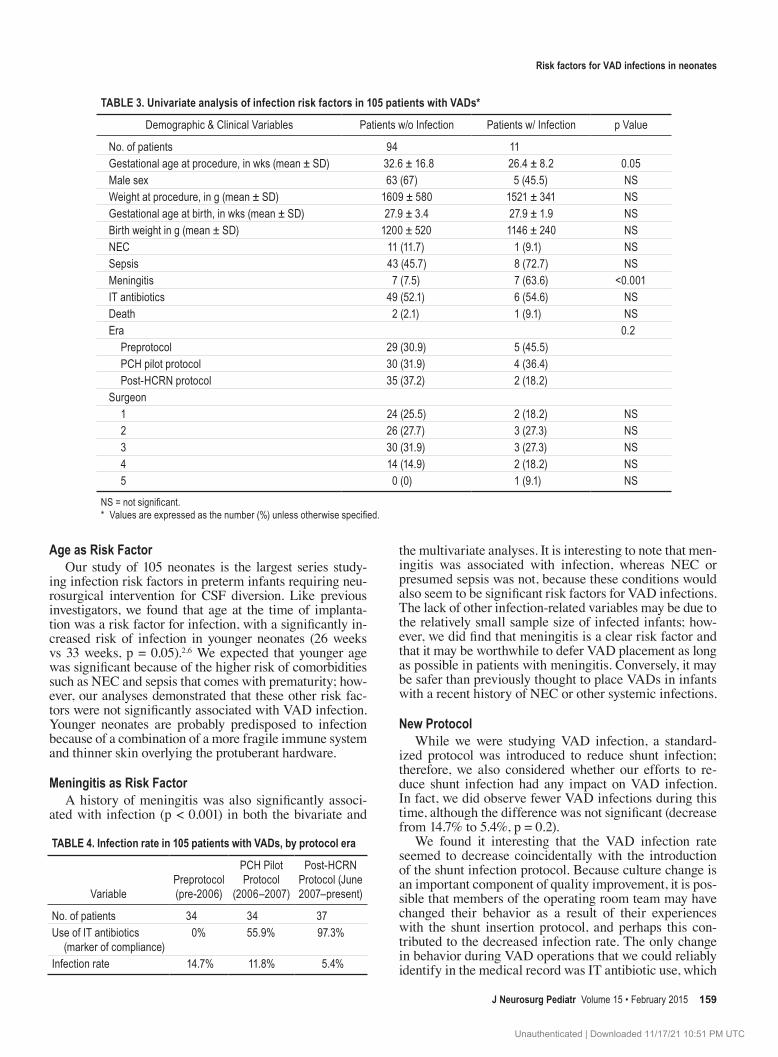
The overall infection rate was 11 of 105 (10.5%). Ges-tational age at procedure was a significant risk factor for VAD infection, with the average age of 26.4 weeks in the infected cohort compared with a mean age of 32.6 weeks for the noninfected group (p = 0.05). Meningitis was a sig-nificant risk factor for infection; 63% of the infected group had prior meningitis, whereas only 7% of the uninfected group had meningitis previously (p < 0.001). Meningitis was also significantly associated with death (p = 0.006). All the other variables were not significantly associated with VAD infection (Table 3).
Infection rates by protocol era were as follows: 14.7% preprotocol, 11.8% during implementation, and 5.4% post-HCRN shunt infection protocol (p = 0.2). The absolute risk reduction was 9.3% in the preprotocol group com-pared with the postprotocol group, and the relative risk reduction was 63.3% (Table 4).
When gestational age at procedure, a prior history of meningitis, and protocol era were analyzed using multi-variate logistic regression, only a history of meningitis was independently associated with VAD infection (OR 21.5, 95% CI 5.0–91.7).
discussionWe present a retrospective cohort study of 105 neonates
in whom VADs were placed for PPHVD. Eleven of these
patients subsequently experienced a VAD infection, for an overall infection rate of 10.5%. Bivariate analyses showed that gestational age at procedure (p = 0.05) and meningitis (p < 0.001) were significant risk factors for VAD infection. In addition, meningitis was also significantly associated with death (p = 0.006).
Two previous studies have examined risk factors for infection in VADs and VP shunts in infants. The first, by Pople et al.6 in 1992, found a VP shunt infection rate of 15.7% for infants less than 6 months old. This study found that age was the only major factor influencing the infec-tion rate. A more recent study by Bruinsma et al.2 found an infection rate of 33.3% in preterm infants with VADs or VP shunts. Of note, this study enrolled 39 preterm infants and is the largest study of infections associated with pre-term infants with VADs/VP shunts in the literature. The authors concluded that prematurity and extremely low birth weight were significant factors contributing to VAD/VP shunt infections.
table 1. description of overall cohort of 105 patients with vads*
Demographic & Clinical Variables Study CohortAge at procedure, in wks (mean ± SD) 31.9 ± 16.2Gestational age in wks (mean ± SD) 27.9 ± 3.3Male sex 68 (65)Birth weight in g (median ± IQR) 1195 ± 497Weight at procedure, in g (median ± IQR) 1600 ± 559Surgeon 1 26 (24.8) 2 29 (27.6) 3 33 (31.4) 4 16 (15.2) 5 1 (1)NEC 12 (11.4)Presumed sepsis 51 (49)Meningitis 14 (13.5)Postprocedural hemorrhage 0 (0)Death 3 (2.9)VAD infection 11 (10.5)
IQR = interquartile range.* Values are expressed as the number (%) unless otherwise specified.
Fig. 1. Graph showing use of antibiotics as a marker for compliance with the HCRN protocol. Figure is available in color online only.
table 2. annual antibiotic compliance with the shunt and infection protocol
Yr VAD Placed
No. of VADs Placed
IT Antibiotic Compliance (%) Protocol Era
2003 6 0 Preprotocol2004 10 0 Preprotocol2005 18 0 Preprotocol2006 18 27.8 PCH pilot protocol2007 16 87.5 PCH pilot protocol/post-
HCRN protocol2008 10 90 Post-HCRN protocol2009 12 100 Post-HCRN protocol2010 13 100 Post-HCRN protocol2011 2 100 Post-HCRN protocol
158
Unauthenticated | Downloaded 11/17/21 10:51 PM UTC
risk factors for vad infections in neonates
J Neurosurg Pediatr Volume 15 • February 2015
age as risk FactorOur study of 105 neonates is the largest series study-
ing infection risk factors in preterm infants requiring neu-rosurgical intervention for CSF diversion. Like previous investigators, we found that age at the time of implanta-tion was a risk factor for infection, with a significantly in-creased risk of infection in younger neonates (26 weeks vs 33 weeks, p = 0.05).2,6 We expected that younger age was significant because of the higher risk of comorbidities such as NEC and sepsis that comes with prematurity; how-ever, our analyses demonstrated that these other risk fac-tors were not significantly associated with VAD infection. Younger neonates are probably predisposed to infection because of a combination of a more fragile immune system and thinner skin overlying the protuberant hardware.
meningitis as risk FactorA history of meningitis was also significantly associ-
ated with infection (p < 0.001) in both the bivariate and
the multivariate analyses. It is interesting to note that men-ingitis was associated with infection, whereas NEC or presumed sepsis was not, because these conditions would also seem to be significant risk factors for VAD infections. The lack of other infection-related variables may be due to the relatively small sample size of infected infants; how-ever, we did find that meningitis is a clear risk factor and that it may be worthwhile to defer VAD placement as long as possible in patients with meningitis. Conversely, it may be safer than previously thought to place VADs in infants with a recent history of NEC or other systemic infections.
New ProtocolWhile we were studying VAD infection, a standard-
ized protocol was introduced to reduce shunt infection; therefore, we also considered whether our efforts to re-duce shunt infection had any impact on VAD infection. In fact, we did observe fewer VAD infections during this time, although the difference was not significant (decrease from 14.7% to 5.4%, p = 0.2).
We found it interesting that the VAD infection rate seemed to decrease coincidentally with the introduction of the shunt infection protocol. Because culture change is an important component of quality improvement, it is pos-sible that members of the operating room team may have changed their behavior as a result of their experiences with the shunt insertion protocol, and perhaps this con-tributed to the decreased infection rate. The only change in behavior during VAD operations that we could reliably identify in the medical record was IT antibiotic use, which
table 3. univariate analysis of infection risk factors in 105 patients with vads*
Demographic & Clinical Variables Patients w/o Infection Patients w/ Infection p Value
No. of patients 94 11Gestational age at procedure, in wks (mean ± SD) 32.6 ± 16.8 26.4 ± 8.2 0.05Male sex 63 (67) 5 (45.5) NSWeight at procedure, in g (mean ± SD) 1609 ± 580 1521 ± 341 NSGestational age at birth, in wks (mean ± SD) 27.9 ± 3.4 27.9 ± 1.9 NSBirth weight in g (mean ± SD) 1200 ± 520 1146 ± 240 NSNEC 11 (11.7) 1 (9.1) NSSepsis 43 (45.7) 8 (72.7) NSMeningitis 7 (7.5) 7 (63.6) <0.001IT antibiotics 49 (52.1) 6 (54.6) NSDeath 2 (2.1) 1 (9.1) NSEra 0.2 Preprotocol 29 (30.9) 5 (45.5) PCH pilot protocol 30 (31.9) 4 (36.4) Post-HCRN protocol 35 (37.2) 2 (18.2)Surgeon 1 24 (25.5) 2 (18.2) NS 2 26 (27.7) 3 (27.3) NS 3 30 (31.9) 3 (27.3) NS 4 14 (14.9) 2 (18.2) NS 5 0 (0) 1 (9.1) NS
NS = not significant. * Values are expressed as the number (%) unless otherwise specified.
table 4. infection rate in 105 patients with vads, by protocol era
VariablePreprotocol (pre-2006)
PCH Pilot Protocol
(2006–2007)
Post-HCRN Protocol (June 2007–present)
No. of patients 34 34 37Use of IT antibiotics (marker of compliance)
0% 55.9% 97.3%
Infection rate 14.7% 11.8% 5.4%
159
Unauthenticated | Downloaded 11/17/21 10:51 PM UTC

h. s. spader et al.
J Neurosurg Pediatr Volume 15 • February 2015
increased from 0% to 87.5% to 100%. Although antibiotic use alone did not impact infection rates in shunt surgery,4 it does appear to indicate a change in behavior during VAD procedures. This leads us to believe that a standard protocol for shunt surgery highlighted the infection issue and emphasized infection prevention, and may have con-tributed to reduced infections.
limitations of the studyThe main limitation of this study is that it is a retro-
spective cohort study and not a prospective trial. Another significant limitation is that there were only 11 infec-tions (outcomes) in this cohort, decreasing the power of this study. In this retrospective review, we were not able to assess operating room team behaviors accurately, and therefore our suggestion that the team’s behavior changed as a result of the infection protocol is only a hypothesis. Nevertheless, we believe that the trends seen in this study warrant further multicenter prospective studies.
conclusionsNeonates with PPHVD are at high risk for infection.
Our study found that gestational age (less than 26 weeks) and a recent history of meningitis were statistically signif-icant risk factors for VAD infection. In addition, we found that the implementation of a shunt insertion protocol at the institution may have had a clinical effect on the reduction of infections in this other patient population because of a change in the operating room culture that led to greater attention to infection prevention. We recommend a further multiinstitutional study on the effect of a protocol to de-crease VAD infections in the premature infant population.
references 1. Brockmeyer DL, Wright LC, Walker ML, Ward RM: Man-
agement of posthemorrhagic hydrocephalus in the low-birth-weight preterm neonate. Pediatr Neurosci 15:302–308, 1989
2. Bruinsma N, Stobberingh EE, Herpers MJ, Vles JS, Weber BJ, Gavilanes DA: Subcutaneous ventricular catheter res-ervoir and ventriculoperitoneal drain-related infections in preterm infants and young children. Clin Microbiol Infect 6:202–206, 2000
3. Hintz SR, Poole WK, Wright LL, Fanaroff AA, Kendrick DE, Laptook AR, et al: Changes in mortality and morbidities among infants born at less than 25 weeks during the post-surfactant era. Arch Dis Child Fetal Neonatal Ed 90:F128–F133, 2005
4. Kestle JR, Riva-Cambrin J, Wellons JC III, Kulkarni AV, Whitehead WE, Walker ML, et al: A standardized protocol to reduce cerebrospinal fluid shunt infection: the Hydro-cephalus Clinical Research Network Quality Improvement Initiative. J Neurosurg Pediatr 8:22–29, 2011
5. Kormanik K, Praca J, Garton HJ, Sarkar S: Repeated tap-ping of ventricular reservoir in preterm infants with post-hemorrhagic ventricular dilatation does not increase the risk of reservoir infection. J Perinatol 30:218–221, 2010
6. Pople IK, Bayston R, Hayward RD: Infection of cerebrospi-nal fluid shunts in infants: a study of etiological factors. J Neurosurg 77:29–36, 1992
7. Richard E, Cinalli G, Assis D, Pierre-Kahn A, Lacaze-Mas-monteil T: Treatment of post-haemorrhage ventricular dilata-tion with an Ommaya’s reservoir: management and outcome of 64 preterm infants. Childs Nerv Syst 17:334–340, 2001
8. Robinson S: Neonatal posthemorrhagic hydrocephalus from prematurity: pathophysiology and current treatment con-cepts. J Neurosurg Pediatr 9:242–258, 2012
9. Wellons JC III, Shannon CN, Kulkarni AV, Simon TD, Riva-Cambrin J, Whitehead WE, et al: A multicenter retrospective comparison of conversion from temporary to permanent cerebrospinal fluid diversion in very low birth weight infants with posthemorrhagic hydrocephalus. J Neurosurg Pediatr 4:50–55, 2009
author contributionsConception and design: Riva-Cambrin. Acquisition of data: all authors. Analysis and interpretation of data: all authors. Drafting the article: Spader, Hertzler. Critically revising the article: all authors. Reviewed submitted version of manuscript: all authors. Approved the final version of the manuscript on behalf of all authors: Riva-Cambrin. Study supervision: Riva-Cambrin, Kestle.
correspondenceJay Riva-Cambrin, Department of Neurosurgery, Division of Pediatric Neurosurgery, Primary Children’s Hospital, University of Utah, 100 Mario Capecchi Dr., Salt Lake City, UT 84113. email: [email protected].
160
Unauthenticated | Downloaded 11/17/21 10:51 PM UTC