rheumatoid factor-producing cells detected by direct hemolytic
TRANSCRIPT

Rheumatoid Factor-Producing Cells Detected by Direct
Hemolytic Plaque Assay
JoHN H. VAUGHAN, TADAO CmIHARA, TERRY L. MooRE, DicK L. ROBBINS,KiYoAxi TANIMOTO, JoHN S. JOHNSON, and ROBERT McMIAN
From the Division of Rheumatology, Department of Clinical Research, ScrippsClinic and Research Foundation, La Jolla, California 92037
A B S T R A C T Lymphocytes secreting anti-IgG anti-bodies, rheumatoid factors (RF), can be detected inthe peripheral bloods, synovial fluids, and bone mar-rows of patients with seropositive rheumatoid arthritisby using a direct plaque-forming cell (PFC) assay withsheep erythrocytes sensitized with reduced and alkylatedrabbit IgG hemolysin. The autospecific nature of theRF produced by RF-PFC was indicated by inhibitionstudies in which the order of potency was human IgG >rabbit IgG > bovine IgG. In metabolic studies puro-mycin, cycloheximide, and vinblastine suppressed RF-PFC. Cyclic AMP and cyclic GMP were without effect.A need was recognized for using full tissue culturemedia during the cell separation and plaquing proce-dures to optimize detection of the RF-PFC.RF-PFC may appear in the blood of patients inter-
mittently despite their continuing presence in the bonemarrow. They have been found in the peripheral blood,especially during acutely exacerbating polyarticular sy-novitis, generalized vasculitis, or generally active, ag-gressive disease. RF-PFC were found in synovial effu-sions of new or recrudescent acute synovitis. RF-PFCwere observed to disappear from the peripheral circula-tion and the bone marrow during therapy with cyto-toxic drugs.The data are consistent with the hypothesis that the
appearance of RF-PFC in the peripheral blood repre-sents an anamnestic response to transiently appearingantigen. The nature of the antigen is not specified. Thebone marrow may be a site of origin of RF-PFC.
INTRODUCTIONSeveral methods have been reported to detect rheuma-toid factor (RF) ' in, or associated with, lymphoid cells.
Received for publication 23 January 1976 and in rezisedform 7 June 1976.
1Abbreviation used in this paper: BM, bone marrow;BSS, balanced salt solution; FBS, fetal bovine serum;
These have included staining with fluorescent-labeledaggregated human IgG (1-4), mixed agglutination infixed tissues (5), and production of rosettes with sensi-tized erythrocytes mixed with lymphocytes (6, 7).None of these procedures, however, assesses RF pro-duction. Culture of mixed cell suspensions in the pres-ence of radio-labeled amino acids (8) has indicated RFproduction by yielding radiolabeled RF, but the numbersor types of cells involved in the production was notrevealed by this method.Some of our recent observations brought forth the
possibility that we could detect RF production by singlecells. We have shown (9) that: (a) IgM RF fixes andactivates guinea pig and human complement via theclassical complement pathway; (b) IgM RF preparessheep erythrocytes coated with reduced and alkylatedrabbit IgG hemolysin for lysis by complement; and (c)the percentage of hemolysis is directly proportional toRF agglutination titer.These observations led us to attempt to use the
direct hemolytic plaque assay described by Jerne et al.(10) to identify lymphoid cells producing RF. Withsheep erythrocytes (SRC) sensitized with reduced andalkylated rabbit IgG hemolysin, we have examined lym-phocyte preparations from peripheral blood, synovialfluid, and bone marrow and found cells secreting RFplaque-forming cells (PFC) in all three sites, but inhighest numbers in the bone marrow.The presence of RF-PFC in the peripheral blood cor-
relates with active, exacerbating, or aggressive disease.
METHODSSubjects. The patient populations studied included an
initial group of 72 patients with seropositive rheumatoid
LFT, latex fixation test; PBL, peripheral blood lympho-cytes; PFC, plaque-forming cell; RA, rheumatoid arthritis;RF, rheumatoid factor; SCAT, sensitized sheep cell agglu-tination test; SRC, sheep erythrocytes.
The Journal of Clinical Investigation Volume 58 October 1976-9339491933

arthritis (RA) randomly selected from our clinics, 14 withseronegative RA, 14 non-RA patients with a variety ofrheumatic diseases, and 24 normal individuals who wereRF negative. The patients with RA were all classical ordefinite RA according to the American Rheumatism Asso-ciation criteria (11). A second series included 16 seroposi-tive RA patients most of whom were on a drug trial withNaprosyn (Syntex Laboratories, Inc., Palo Alto, Calif.).They were selected as a group because they had generallymild-to-moderate disease and were not on corticosteroids.A third group consisted of 18 patients selected for studybecause of the clinical impression of more active or aggres-sive disease in them.Most patients were examined in our ambulatory clinics.
Some were hospitalized in our Clinical Research Center formore prolonged and intensive investigations. The patientson the drug study were followed in the ambulatory facilityof our Clinical Research Center, where more detailedclinical observation could be made than in our regularclinics. All patients were fully informed of the nature ofthe studies undertaken and gave willing consent.
Clinical characteristics. RA patients with any evidenceof generalized vasculitis were assigned to the vasculitisgroup. Manifestations of vasculitis ranged from nail foldinfarcts to mononeuritis multiplex and typical vasculiticcutaneous ulcerations. In this report no distinction is madebetween patients with minimal and severe vasculitis.
Patients were regarded as having an acute flare if theyhad acute synovitis of less than a 2-wk duration. A jointalready exhibiting synovial hypertrophy and effusion wasconsidered the site of recrudescent acute synovitis only inthe event of sudden increase in joint volume, tenderness,pain, erythema, or heat. Patients with two or more acutelyinflamed joints were considered to have polyarticular syno-vitis.
All patients who were seen in our Clinical ResearchCenter were evaluated serially with the American Rheuma-tism Association Database (12) as the form of clinicalassessment.
Preparation of lymphocytes. Lymphocytes from periph-eral blood and synovial fluid were generally purified bydifferential centrifugation on Ficoll-Hypaque (13), or oc-casionally by dextran sedimentation with iron lysine (14,15). Preparations from peripheral blood consisted of cellsappearing morphologically greater than 92% viable smalllymphocytes. Lesser degrees of purity of the lymphocyteswere encountered with some preparations from synovialeffusions.Bone marrow was aspirated from the posterior iliac
crest, or in some instances from the sternum, 5 ml beingtaken into a plastic syringe containing 10 mg of heparin.The cells were washed in buffer containing 10% fetal bovineserum (FBS), and then submitted to hypotonic lysis ofthe erythrocytes for 10 min at 37'C in 0.83% NH4Cl. Theywere then washed twice further in RPMI-1640 (GrandIsland Biological Co., Grand Island, N. Y.) with 10%oFBS and used for RF-PFC.
Sensitized sheep cells. Rabbit anti-SRC hemolysin pur-chased from Baltimore Biological Laboratories (Divisionof BioQuest, Cockeysville, Md.) was separated into IgMand IgG fractions by Sephadex G-200 (Pharmacia FineChemicals, Inc., Piscataway, N. J.) gel filtration. IgGhemolysin was reduced and alkylated and used to sensitizeSRC as reported previously (9).Measurement of RF activity. The RF activity of sera
and synovial effusions was measured by the slide latexfixation test (LFT, RA test, Hyland Laboratories, Los
Angeles, Calif.) and by the sensitized sheep cell agglutina-tion tests (SCAT) (16). The latter was carried out bysettling patterns in microtiter plates.
Soutrce of comnplenent. Lyophilized guinea pig serum(Hyland Laboratories) was reconstituted with the diluentprovided. For some experiments natural antibody to SRCwas absorbed by repeated incubation at 0'C with packedSRC as reportedl previously (9).PFC assay for RF proditction. 0.5 ml of 0.5% agarose
at 44'C (agarose A-37 Indubiose R, L'Industrie Biologique,Frangaise, S. A., Gennevilliers, France) was mixed with0.1 ml of lymphocyte suspension containing 1-5 x 10⁶ lym-phocytes and 0.05 ml of a 6.7% suspension of SRC coatedwith reduced and alkylated rabbit IgG hemolysin. Themixture was then promptly poured onto precoated micro-scope slides. The slides were incubated at 37'C for 90 minin a moist chamber, then immersed in a 1: 10 dilution ofguinea pig complement and incubated an additional 90 minat 37' C. Preabsorption of the guinea pig complement toremove natural antibody to SRC did not prove to be neces-sary since there was no significant background lysis ofthe unsensitized SRC controls. RF-PFC were enumeratedunder magnification. Similar cell suspensions employingnonsensitized erythrocytes were examined as controls todetect any lymphocytes synthesizing natural antibody toSRC.2 Control PFC were subtracted from PFC with sensi-tized erythrocytes and the results were expressed as RF-PFC/106 lymphocytes. Generally, four replicate determina-tions were made for each preparation.Metabolic inhibition. Puromycin, cycloheximide, and di-
butyryl cyclic AMP were obtained from Calbiochem, SanDiego, Calif. 8-bromo cyclic GMP was obtained fromSigma Chemical Co., St. Louis, Mo. Vinblastine was ob-tained from Eli Lilly and Co., Indianapolis, Ind. Inhibitorswere added to 0.5-ml aliquots of melted agar (zide sitpra)in 10-,ul volumes just before the lymphocytes and sensitizederythrocytes. Thereafter, the preparations were poured andsubsequently handled in the usual fashion.Preparation of data. Statistical comparisons were made
by the Student's t test on log transformation. The exponen-tiated mean+±SEM is used throughout the paper.
RESULTS
Typical direct hemolytic plaques are produced in ourRF-PFC assay. The plaques vary in size from pinpointto 0.3-0.5 mm. At the numbers of leukocytes used perslide, each plaque contained multiple leukocytes micro-scopically, so that the single central lymphoid cell, theputative antibody secreting cell, could not be distin-guished with certainty. Leukocyte clustering was some-times seen, more frequently in preparations containingbone marrow or synovial fluid cells than in the periph-eral blood cells. Most clusters in such preparations werenot surrounded by hemolytic zones, but when presentthe zones generally were larger than the zones sur-rounding single cells, suggesting multiple RF-PFC insuch clusters.
2 This control was almost always negative in the periph-eral blood, even at 5 x 10⁶ lymphocytes per slide. In bonemarrow preparations, 1-3 PFC/106 nucleated cells wereoccasionally seen with nonsensitized SRC.
934 Vaughan, Chihara, Moore, Robbins, Tanimoto, Johnson, and McMillan

Immunological studies. The RF-PFC were comple-ment dependent, since leaving complement out of theassay resulted in no plaques, and introduction of com-plement earlier during the assay, i.e., at the time ofmixing of the cells with the agarose, resulted in earlierappearance of the plaques.The specificity of the RF-PFC assay has been in-
vestigated. Since rabbit rather than human IgG hemo-lysin was used to sensitize the sheep cells in the assay,the question had to be answered whetlher the PFC weredue to heterospecific rather than autospecific antibodies.To answer this question, inhibition studies were carriedout, in which human, rabbit, or bovine IgG was in-corporated in varying doses in the agar (Fig. 1). Threepatients' cells were examined (Ta, So, and Ra). Bothbone marrow (BM) and peripheral blood lymphocytes(PBL) were used as sources for the RF-PFC. HumanIgG regularly provided better inhibition than rabbitIgG. Bovine IgG was least effective. Therefore, theRF (anti-IgG) detected in the PFC assay can be prop-erly regarded as autoantibody. There was no doubtabout the specificity of the inhibition of RF-PFC, sincethe RF-PFC were not inhibited in high concentrationsof other serum proteins. For instance, there was noinhibition in 10% FBS.A surprise was that human IgG actually had greater
efficacy than rabbit IgG in the RF-PFC inhibition,since the common experience in assaying the peripheral
124 Ta(PBLj
8-0 - ,
4 \
I10 90
R2- 8
5 20 80
SoIPILI
10-
2 b
10 gO
5 20 80'mg Ig/mi
SolBMI
0 90
HumaN .-.---Rabbit 0--oBoviNe
Ra-310-
-
6~
2 i 'O
5 20 80
FIGURE 1 Inhibition of RF-PFC by various species ofanimal IgG. The indicated concentrations of IgG were in-corporated in the agar in BSS along with the lymphoidcell population examined. Three patients, Ta, So, and Ra,were studied. For patient So, results with both PBL andBM are shown. For patient Ra, results are shown for theBM at three different times during the course of immuno-suppressive therapy, during which a reducing number ofRF-PFC were seen.
TABLE IInhibition of Serum RF-SCA T by Addition of IgG
of Various Species
Patient Rabbit Human Bovine
So. 1/12,000 4* 2* 1*Ra. 1/5,000 8 4 1Ta. 1/20,000 3 2 1
* Reduction of titer (number of tubes) by addition of 160 sgIgG. The IgG was added to 1 ml of 1/50 (So. and Ta.) or un-diluted (Ra.) serum and the agglutinating end point comparedto that of the original serum.
blood serum for RF with the SCAT has been thatrabbit IgG is a better inhibitor than human IgG (17, 18).The SCAT assay, like the RF-PFC assay, used SRCsensitized with rabbit IgG as the indicator cell. Thus theRF at its source (the RF-PFC assay) appeared to bedifferent from RF in the peripheral serum (the SCATassay), as will be discussed later.To rule out that the three patients we studied were
atypical, we performed inhibition assays on the SCATreactions of the RF in these patients' sera taken at thetime of the RF-PFC assays. The results are shown inTable I. The peripheral blood serum RF activities wereinhibited best by rabbit IgG, less well by human IgG,and little or none by bovine IgG, just as lhad been de-scribed for RF in the serum previously (17, 18).The RF-PFC detected in our assay have been of the
direct variety and therefore are assumed to representIgM complement-fixing RF (10). Attempts have beenmade to detect other varieties of RF by indirect means.We have added an antihuman IgG serum along withcomplement at the second incubation step in the assay(10) in the attempt to detect IgG RF. This has gener-ally not yielded additional PFC and sometimes has in-hibited because of the IgG present in the antiserum.Antihuman IgM antibody has been added in the attemptto detect noncomplement-fixing IgM RF-PFC, but nosuch PFC have been found.
Since our assay detects RF-PFC only by the cross-reaction of the RF with rabbit IgG, attempts have beenmade to develop a cell coated with human IgG thatcan be used in the assay. The Ripley anti-CD antibody(19) has been tried, but it becomes strongly agglutinat-ing when reduced and alkylated, rendering it unsuitedto our test. Coating of sheep cells witlh human IgG bymeans of chromic chloride has not yielded PFC. Fur-ther efforts in these directions, however, are needed.We carried out studies to determine whether the RF
detected in our RF-PFC assay could have been due toRF adsorbed to leukocytes and carried over by theminto the agar phase of the assay, to be released thereand recognized as hemolytic plaques. To test this we
Rheumatoid Factor-Producing Cells Detected by Direct Hemolytic Assay
160
120-
8G
40-
co1-c-1
935

30-
C3 20-1ox
-- 10-
+ 11FF
10 50 10 50 10 5 4 10-6 7
Cyclo Pure CC-AMP C-GMP
l(g/mi) Ig/mi) (Mj (MlFIGuRE 2 Effects on RF-PFC of inhibitors of proteinsynthesis and cyclic nucleotides. Reduction of numbers ofRF-PFC by inhibition of protein synthesis with cyclohexi-mide (cyclo) and puromycin (puro). Lack of effect of cyclic(c) nucleotides. The inhibitors were added to the agarosebefore the addition of the lymphoid cells. Subsequent incu-bation at 370 C was carried out in standard fashion (seeMethods).
used peripheral blood leukocytes both from normaldonors and from patients with active seronegative RAand a BM preparation from a normal volunteer. Noneof these cells developed detectable RF-PFC in 3 X 100lymphocytes after exposure to the sera of two patientswith RF titers of 5,120, who had themselves exhibited49 and 18 RF-PFC/3 X 106 lymphocytes at the timesthat their sera were collected. This failure was true evenwhen the sera and leukocytes were preincubated over-night at 4°C.
Metabolic studies. Efforts to determine some of themetabolic requirements for RF-PFC were carried out.Dependence upon protein synthesis was tested by addi-tion of cycloheximide or puromycin to the agar; influ-ence of the cyclic nucleotides was tested with dibutyrylcyclic AMP and 8-bromo cyclic GMP; and importanze
24
_20-
~16->212
4-
Z -4 -5 -610 M
C-li Vinblastine
FIGURE 3 Inhibition of RF-PFC by vinblastine. The vary-ing concentrations of vinblastine were added to the agarosebefore the addition of blood lymphocytes. Subsequent incu-bation at 37'C was carried out in standard fashion.
n.
=. 1 1
coC-C..,Ucco- i
10
Cells/slide lx106J
FIGURE 4 Inhibition of RF-PFC at high cell concen-trations. The indicated numbers of peripheral blood leuko-cytes were added to 0.5 ml of agarose in BSS and incubatedin standard fashion at 37'C.
of microtubular or membrane integrity was tested withvinblastine (20). Of many experiments performed, thoseshown in Figs. 2 and 3 are typical. Cycloheximide andpuromycin generally reduced the number of RF-PFC,but not below about 35% of the starting number. Inhi-bition was effected as well by 10 i'g as by 50 gg ofeach drug. Dibutyryl cyclic AMP and 8-bromo cyclicGMP have had no effect. Vinblastine reduced the PFCin several experiments, and occasionally at 0.1 mM1 es-sentially to zero. In studies not shown, colchicine hadessentially no inhibiting effect when used by itself, butinhibited nmodestly in the presence of cytochalasin B.Cytochalasin B by itself also generally failed to inhibit.These observations have been taken to mean: (a)
only a minority of the RF-PFC have enough storedRF for plaque formation in the absence of continuedprotein synthesis; (b) the cyclic nucleotides probablydo not modulate significantly the synthesis or release ofthe RF; and (c) the release depends upon intact micro-tubular or membrane structures.
During the course of these experiments, attemptswere made to use increasing numbers of lymphocytes inthe agar to increase the number of RF-PFC detected.It was noted that at cell concentrations higher than3 X 106/slide, a corresponding increase in the numberof RF-PFC was not seen. Rather, varying degrees ofinhibition were found (Fig. 4). When full tissue culturemedia were used in the agar instead of the usual bal-anced salt solution (BSS) (21), however, the expectedincrement of RF-PFC with increased numbers of cellsplaqued did occur (Fig. 5). The inhibition seen in BSSalone (Fig. 4) indicates a nutritional deficiency in thesystem at higher cell concentrations, where the meta-bolic demand would be maximal. This interpretation issupported by the further observation that addition ofincreasing quantities of purified polymorphonuclear leu-kocytes to the slides with a standard dose of lymphocytes
936 Vaughan, Chihara, Moore, Robbins, Tanimoto, Johnson, and McMillan
-L

diminished the number of RF-PFC seen in the prepa-ration.
Blood samples collected in heparin can be kept at 4°Covernight and the cells separated and plaqued the nextday without loss of numbers of RF-PFC. If the cellsare separated by Ficoll-Hypaque and then stored over-night, however, the number of RF-PFC found thenext day is markedly reduced, even if the separated cellsare kept in full tissue culture media. Cells separated byFicoll-Hypaque, and frozen in dimethyl sulfoxide inliquid nitrogen have, on thawing, yielded 33% livingcells with no loss of RF-PFC/10' living lymphocytes.
Clinical observations. Table II shows our findingson RF-PFC in our first series of RA patients, includingboth those who were seropositive and those who wereseronegative for RF in the serum. Cells from blood,synovial fluid, and BM were examined. 32 of 102 prepa-rations of blood lymphocytes from 21 of 67 patientswere positive with a range of 1-18 PFC/106 lympho-cytes. In this first series we noted that RF-PFC werefound especially in two circumstances; when the patientshad acute exacerbations of polyarticular synovitis, orwhen they had generalized vasculitis.
19 synovial fluid lymphocyte preparations were ex-amined, of which 4 were positive with a range of 3-14PFC/10' lymphocytes. RF-PFC were found only in ef-fusions from joints that were the sites of acute synovitis.Joints that were the sites of chronic indolent diseasewere negative, regardless of the duration of the svno-vitis or the degree of synovial hypertrophy.
Eight BM preparations were examined. Four patientsgave positive cells with a range of 2-22 RF-PFC/106nucleated cells. Two of the four positive BM specimenswere from patients who did not have RF-PFC in theirperipheral bloods.
90g
70-
50-
30-
10-
1 3 5Cells/Slide Ix10XI
10
FIGURE 5 RF-PFC at varying cell densities; effect of tissueculture and medium. Effect of supplemental nutrients onnumbers of RF-PFC. The indicated inumbers of PBL wereadded to 0.5 ml of agarose in "Autopow" tissue culturemedium (Flow Laboratories Inc., Rockville, Md.) supple-mented with 10%o FBS, 0.033 M NaHCOs, 0.001 M Napyruvate, 0.002 M L-glutamine, and nonessential amino acids(1/100 of lOOX concentrate, Microbiological Associates,Bethesda, Md.).
TABLE I IOccurrences of RF-PFC in Lymphocytes from
Three Sources in RA
Total RF-PFC positive
Prepa- Indi- Prepa- Indi-Source rations viduals rations viduals
Peripheral blood 102 67 32 21Synovial fluid 19 11 4 3Bone marrow 8 8 4 4
In Table III, the results in seronegative and sero-positive individuals are compared separately. In theseropositive individuals, 40 of 121 peripheral blood,synovial fluid, or BM preparations exhibited one ormore RF-PFC/10' lymphocytes. No preparations from25 seronegative patients or from 24 normal personswere positive.On a number of occasions we examined the blood
simultaneously with the BM or joint fluid cells for RF-PFC. As can be seen in Table IV, in all instances theBM had many more RF-PFC/106 lymphocytes thandid the simultaneous peripheral blood. The synovialfluid lymphocytes sometimes had more RF-PFC. Wehave taken this to mean that the BM is a source ofRF-PFC. The synovial tissues may also be a source,although the inflammation in the joints may conceivablylead to local sinkhole effects which concentrate RF-PFC in the area.The relation of RF-PFC to severity of disease was
investigated in two further series of patients with sero-positive RA. The one (second series in Tables V andVI) was a group with mild-to-moderate disease, mostof whom were on a drug trial with aspirin, indometha-cin, and Naprosyn, and none of whom were on corti-costeroids. The other (third series) was a group ofpatients preselected over a 5-mo period for RF-PFCdeterminations because of a clinical impression of therebeing such active, aggressive, or exacerbating diseasein them that there was high likelihood that their periph-eral blood leukocytes would be positive. The tests on
TABLE II IRF-PFC in Cell Preparations from Individuals
Seropositive or Seronegative forRF by the LFT
RF-PFC
Positive Negative
Positive 40 81LFT
Negative 0 49
Rheumatoid Factor-Producing Cells Detected by Direct Hemolytic Assay
4Go7-
937
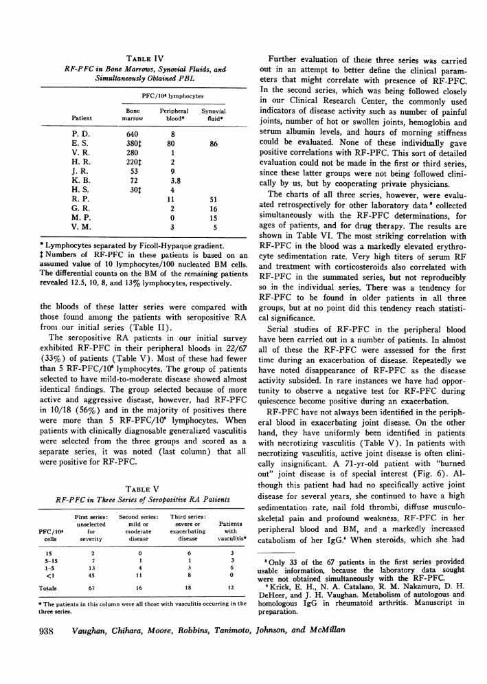
TABLE IVRF-PFC in Bone Marrows, Synovial Fluids, and
Simultaneously Obtained PBL
PFC/106 lymphocytes
Bone Peripheral SynovialPatient marrow blood* fluid*
P. D. 640 8E. S. 380t 80 86V. R. 280 1H. R. 220$ 2J. R. 53 9K. B. 72 3.8H. S. 30t 4R. P. 11 51G. R. 2 16M. P. 0 15V.M. 3 5
* Lymphocytes separated by Ficoll-Hypaque gradient.$ Numbers of RF-PFC in these patients is based on anassumed value of 10 Iymphocytes/100 nucleated BM cells.The differential counts on the BM of the remaining patientsrevealed 12.5, 10, 8, and 13% lymphocytes, respectively.
the bloods of these latter series were compared withthose found among the patients with seropositive RAfrom our initial series (Table II).The seropositive RA patients in our initial survey
exhibited RF-PFC in their peripheral bloods in 22/67(33%) of patients (Table V). Most of these had fewerthan 5 RF-PFC/106 lymphocytes. The group of patientsselected to have mild-to-moderate disease showed almostidentical findings. The group selected because of moreactive and aggressive disease, however, had RF-PFCin 10/18 (56%) and in the majority of positives therewere more than 5 RF-PFC/10' lymphocytes. Whenpatients with clinically diagnosable generalized vasculitiswere selected from the three groups and scored as aseparate series, it was noted (last column) that allwere positive for RF-PFC.
TABLE VRF-PFC in Three Series of Seropositive RA Patients
First series: Second series: Third series:unselected mild or severe or Patients
PFC/106 for moderate exacerbating withcells severity disease disease vasculitis*
15 2 0 6 35-15 7 1 1 31-5 13 4 3 6<1 45 11 8 0
Totals 67 16 18 12
* The patients in this column were all those with vasculitis occurring in thethree series.
Further evaluation of these three series was carriedout in an attempt to better define the clinical param-eters that might correlate with presence of RF-PFC.In the second series, which was being followed closelyin our Clinical Research Center, the commonly usedindicators of disease activity such as number of painfuljoints, number of hot or swollen joints, hemoglobin andserum albumin levels, and hours of morning stiffnesscould be evaluated. None of these individually gavepositive correlations with RF-PFC. This sort of detailedevaluation could not be made in the first or third series,since these latter groups were not being followed clini-cally by us, but by cooperating private physicians.The charts of all three series, however, were evalu-
ated retrospectively for other laboratory data collectedsimultaneously with the RF-PFC determinations, forages of patients, and for drug therapy. The results areshown in Table VI. The most striking correlation withRF-PFC in the blood was a markedly elevated erythro-cyte sedimentation rate. Very high titers of serum RFand treatment with corticosteroids also correlated withRF-PFC in the summated series, but not reproduciblyso in the individual series. There was a tendency forRF-PFC to be found in older patients in all threegroups, but at no point did this tendency reach statisti-cal significance.
Serial studies of RF-PFC in the peripheral bloodhave been carried out in a number of patients. In almostall of these the RF-PFC were assessed for the firsttime during an exacerbation of disease. Repeatedly wehave noted disappearance of RF-PFC as the diseaseactivity subsided. In rare instances we have had oppor-tunity to observe a negative test for RF-PFC duringquiescence become positive during an exacerbation.RF-PFC have not always been identified in the periph-
eral blood in exacerbating joint disease. On the otherhand, they have uniformly been identified in patientswith necrotizing vasculitis (Table V). In patients withnecrotizing vasculitis, active joint disease is often clini-cally insignificant. A 71-yr-old patient with "burnedout" joint disease is of special interest (Fig. 6). Al-though this patient had had no specifically active jointdisease for several years, she continued to have a highsedimentation rate, nail fold thrombi, diffuse musculo-skeletal pain and profound weakness, RF-PFC in herperipheral blood and BM, and a markedly increasedcatabolism of her IgG.' When steroids, which she had
"Only 33 of the 67 patients in the first series providedusable information, because the laboratory data soughtwere not obtained simultaneously with the RF-PFC.
'Krick, E. H., N. A. Catalano, R. M. Nakamura, D. H.DeHeer, and J. H. Vaughan. Metabolism of autologous andhomologous IgG in rheumatoid arthritis. Manuscript inpreparation.
938 Vaughan, Chihara, Moore, Robbins, Tanimoto, Johnson, and McMillan

TABLE VIClinical Characteristics of Patients Positive and Negative for RF-PFC Detectable in PBL
Series 1* Series 2 Series 3 Totals: series 1-3
Pos. Neg. Pos. Neg. Pos. Neg. Pos. Neg.(20) (13) (5) (1 1) (10) (8) (35) (32)
% % % % P value§Latex > 1,280 62 25 60 27 50 67t 57 31 0.06
> 640 76 63 80 45 90 83$ 80 60 NSESR > 60 33 13 20 0 80 33t 43 13 <0.02
> 40 71 56 20 27 100 50 71 41 <0.05Age, yr > 50 67 56 60 54 70 63 66 56 NSSteroid Rx 65 55 0 0 80 38 60 31 <0.05
Abbreviation: ESR, erythrocyte sedimentation rate.Numbers in parentheses represent the number of patients studied.* On review of the records of 67 patients in this original survey, only 33 had had determinations ofRF-PFC, latex, and ESR on the same day, so only the data from the 33 patients are displayed here.t These figures are derived from only six of the eight patients who were RF-PFC negative in this series,since two patients did not have RF-PFC, latex, and ESR done on the same day.§ P values for Pos.-Neg. differences in the totalled series are derived from fourfold contingency tableswith Yates' correction (35).
received for many years, were briefly decreased duringthe early observation period in our Clinical ResearchCenter, she developed diffuse erythema marginata, es-pecially over the back. Serial BM and peripheral bloodevaluations were carried out (Fig. 6). Three featuresare evident: (a) the BM regularly exhibited moreRF-PFC/10' lymphocytes than did the peripheral blood;
5120
F128020-
Vn 15
151FIGURE 660S00 600 460 366i360 cg e-
Cytuls E
:1.22~~~~~~~~~~~~~~~~~~
toid vasculitis. The numbers of RF-PFC/106 nucleated BMcells are illustrated in the stippled bars. RF-PFC in PBLare indicated in the open bars. LFT is shown in the solidline. Absolute lymphocyte count is shown in the dashedline. The patient had been under continuous prednisonetherapy for 10 yr. The changes accompanying institutionof immunosuppressive therapy with cytoxan and chloram-bucil can be seen.
(b) two peaks of RF-PFC occurred in the peripheralblood which did not match changes in BM numbers;and (c) there was a lessening and disappearance of RF-PFC in both tissues during treatment with the immuno-suppressive agents cyclophosphamide and chlorambucil.Types of extra-articular rheumatoid disease other
than necrotizing vasulitis may also be correlated withRF-PFC in the peripheral blood. We have recently ob-served a patient with "burned out" arthritis, but con-tinued active formation of subcutaneous nodules, Felty'ssyndrome, a severe bilateral sensory polyneuropathy, avery high sedimentation rate, and a markedly increasedcatabolic decay of her own IgG.' Numerous RF-PFCpersistently have been present in her blood.
DISCUSSION
In the RF-PFC assay reported herein, direct hemolysisof sensitized SRC was produced in agar. This is as-sumed to be due to cells producing complement-fixingIgM-RF (8, 22). Reduced and alkylated IgG hemolysinwas used as the SRC sensitizer, since reduced and alky-lated hemolysin has no ability by itself to fix comple-ment to the SRC, but remains reactive with RF (9,23). The plaque assay system is specific for RF. It isinhibited by as little as 2-10 Ag of human or rabbit IgGin the agar, less so by bovine IgG, and not at all by10% FBS.
It was of some interest that the order of the abilitiesof the various species of IgG to inhibit RF-PFC wasdifferent from that by which they inhibited the RF inthe blood serums (SCAT). Human IgG was most ef-ficient in the RF-PFC, while tabbit IgG was most ef-ficient in the SCAT. This difference brings up the possi-
Rheumatoid Factor-Producing Cells Detected by Direct Hemolytic Assay 939
bility that the RF that we have traditionally studied inthe peripheral blood serum is not properly representa-tive of RF as it is made, but rather it is the residual ofthe original RF after complexing with interstitial andcirculating IgG, precipitation, phagocytosis, or otherfate in the tissues. This is obviously an important areafor further study.We have previously speculated that polyclonal IgMI-
RF may be composed of a spectrum of monoclonal RFmolecules, the majority of which are noncomplementfixing, a minority being complement fixing (9). Sincenoncomplenment-fixing IgMI-RF would not be detectedin our direct hemolytic plaque assay system, we haveused a rabbit anti-IgM serum in an attempt to bringout such postulated nonhemolytic RF in an indirectassay. This attempt has been unsuccessful. We used forthis attempt, however, an IgG anti-IgM, and the IgGof the antiserum was in sufficient concentration to causeby itself some inhibition of the system such as thatillustrated in Fig. 1. An IgM anti-IgM would avoidthe problem, and we plan to investigate this questionagaini with such an antiserum. An IgM anti-IgG willalso be developed in an attempt to detect IgG RF-PFC.
Demonstration of RF-PFC was optinmal when thecells were kept in full tissue culture media for the maxi-mal possible time. The need for continued active proteinsynthesis and an intact microtubular or membrane struc-ture for optimal RF-PFC production have been indicatedby the inhibition studies with cycloheximide and vin-blastine.The clinical findings in these studies are: (a) RF-PFC
may be found in the peripheral bloods of patients inacute exacerbations, or with persistent aggressive dis-ease such as necrotizing vasculitis; (b) the BM is agood source of RF-PFC, sometimes exhibiting themwhen the peripheral blood is free of them; and (c) thesynovial fluid may also exhibit RF-PFC in acute flaresof synovitis, at which time it too may have more PFC/106 lymphocytes than the peripheral blood.The BM has long been known to be a source of
lymphocytes capable of repopulating thymic, splenic,and nodal organs (24). It is also an organ capable ofconsiderable antibody (25) and gamma globulin (26)production. We have previously reported in systemiclupus erythematosus anti-DNA PFC in high numbers inthe BM of patients with acute exacerbations (27) ofthat disease.The higher numbers of RF-PFC in joint fluids than
in simultaneously obtained peripheral bloods may meanlocal RF-PFC production in the joints; but it also maymean local entrapment of RF-PFC from the circulatingblood. In a study of autoimmune thyroiditis in the rab-bit, Clinton and Weigle (28) have shown accumulationof PFC making autoantibody to thyroglobin in the in-
flamed gland. They suggested that locally producedthyroglobulin could act as an affinity absorbent for anti-thyroglobulin-producing PFC. Later studies by Clagettet al. (29) emphasized that such local accumulation ofPFC could also provide a means of delivering largeamiiounts of autoantibody into the immediate vicinityof the gland with resultant local immune precipitationwith thyroglobulin. Whether the synovial tissues, whichare known to be capable of IgG production (8), actsimilarly as an affinity absorbent for RF-PFC andprecipitant for their RF is a possibility that needs fur-ther study.The intermittent appearance of RF-PFC in the periph-
eral blood in RA has a parallel in experimental aninmalsundergoing active immunization. PFC to various im-munizing antigens appear in the peripheral bloods ofanimals only for the brief periods during which thereis active proliferation of new antibody-producing cellsby the lymiiphoid tissues (30-34). This was noted in miceat about 5 and 13 days after primary immunization withaggregated human IgG by Romball and Weigle (34).Serum antibody titers did not correspond with thenumbers of PFC in the peripheral blood, but ratherlagged after the PFC. Sometimes the serum antibodylevel continued to rise after the peripheral blood PFCdisappeared, indicating continuing antibody productionby cells in the fixed lymphoid tissues.We have observed a number of patients with par-
ticularly active and aggressive disease in whom theperipheral blood was persistently positive for RF-PFC,but with change from positive to negative duringtherapy. Except for one case, all showed improvementof clinical symptoms on therapy. We have also seenpatients spontaneously lose their peripheral blood RF-PFC while spontaneously improving clinically. Fall inthe RF-PFC often occurred either with no change in theRF titer of the serum, or with a diminution that laggedconsiderably after the fall in RF-PFC. Thus, we con-
firm the well-recognized failure of the titer of serum RFto correlate well with changes in disease activity. RF-PFC in the peripheral blood, however, shows a closercorrelation with disease activity.We note that the intermittency with which RF-PFC
are seen in the blood of RA patients may reflect an in-termittency in an immunizing process in their disease.WXTe note also that the qualitative differences of the RFdetected in the RF-PFC assay from the RF detected inthe SCAT may indicate selective removal of portions ofRF from the body fluids, and this asks for attention to
the possible role of this portion of the RF in the patho-genesis of RA.
ACKNOWLEDGMENTSThe invaluable assistance of Miss Jean Rose and Mrs. MaryWu in carrying out the experiments, and Mrs. Karen
940 Vaughan, Chihara, Moore, Robbins, Tanimoto, Johnson, and McMillan

Hazard and Mrs. Anna Milne in assembling the manuscript,is greatly appreciated. The criticism and suggestions ofDoctors William Weigle and Richard Dutton at variousphases of this study and in reading the manuscript are alsogratefully acknowledged.
This work was supported by research grants AM 16994,AM 05693, AM 07144, and RR 00833 from the NationalInstitutes of Health.
REFERENCES1. Mellors, R. C., R. Heimer, J. Corcos, and L. Korngold.
1959. Cellular origin of rheumatoid factor. J. Exp. Mcd.110: 875-886 and Plates 92-96.
2. Mellors, R. C., A. Nowoslawski, L. Korngold, and B.L. Sengson. 1961. Rheumatoid factor and the patho-genesis of rheumatoid arthritis. J. Exp. Med. 113: 475-484 and Plates 49-54.
3. McCormick, J. N. 1963. An immunofluorescence studyof rheumatoid factor. Ann. Rheurm. Dis. 22: 1-10.
4. Bonomo, L., A. Tursi, and U. Gillardi. 1968. Distributionof the anti-gamma globulin factors in the synovial mem-brane and other tissues in various diseases. Ann. Rheuzn.Dis. 27: 122-129.
5. Milde, E-J., and 0. Tonder. 1968. Demonstration ofrheumatoid factor in tissue by mixed agglutination withtissue sections. Arthritis Rheum. 11: 537-545.
6. Bach, J-F., F. Delrieu, and F. Delbarre. 1970. Therheumatoid rosette. A diagnostic test unifying seroposi-tive and seronegative rheumatoid arthritis. Ain. J. Med.49: 213-222.
7. Durance, R. A., A. Micheli, and F. H. Fallet. 1974.Rosette formation in rheumatoid and non-rheumatoidstates. Anin. Rheum. Dis. 33: 216-220 .
8. Smiley, J. D., C. Sachs, M. Ziff. 1968. In vitro synthe-sis of immunoglobulin by rheumatoid synovial mem-brane. J. Clin. Invest. 47: 624-632.
9. Tanimoto, K., N. R. Cooper, J. S. Johnson, and J. H.Vaughan. 1975. Complement fixation by rheumatoid fac-tor. J. Clin. Invest. 55: 437-445.
10. Jerne, N. K., A. A. Nordin, and C. Henry. 1963. Theagar plaque technique for recognizing antibody producingcells. In Cell Bound Antibodies. B. Amos and H. Kop-rowski, editors. Wistar Institute Press, Philadelphia.109.
11. A Committee of the American Rheumatism Association.1958. Revision of diagnostic criteria of rheumatoidarthritis. Arthritis Rheum. 2: 16-20.
12. Hess, E. V. 1974. A standard database for rheumaticdiseases. Arthritis Rheum. 17: 327-336.
13. Boyum, A. 1968. Separation of leukocytes from blood andbone marrow. Scand. J. Clii;. Lab. Invest. 21 (Suppl. 97):77-89.
14. Cassen, B., J. Hitt, and E. F. Hays. 1958. The efficientseparation of lymphocytes from human blood. J. Lab.Clin. Med. 52: 778-783.
15. Levine, S. 1956. Magnetic techniques for in vitro iso-lation of leukocytes. Science (Wash. D. C.). 123: 185-186.
16. Heller, G., A. S. Jacobson, and M. H. Kolodny. 1949.A modification of the hemagglutination test for rheuma-toid arthritis. Proc. Soc. Exp. Biol. Med. 72: 316-323.
17. Butler, V. P., Jr., and J. H. Vaughan. 1965. The reactionof rheumatoid factor with animal gamma-globulins:Quantitative considerations. Immunology. 8: 144-159.
18. Butler, V. P., Jr., and J. H. Vaughan. 1964. Hemag-glutination by rheumatoid factor of cells coated with
animal gamma globulins. Proc. Soc. Exp. Biol. Med.116: 585-593.
19. Waller, M. V., and J. H. Vaughan. 1956. Use of anti-Rhsera for demonstrating agglutination activating factorin rheumatoid arthritis. Proc. Soc. Exp. Biol. Med. 92:198-200.
20. Bensch, K. G., and S. E. Malawista. 1969. Microtubularcrystals in mammalian cells. J. Cell. Biol. 40: 95-107.
21. Mishell, R. I., and R. W. Dutton. 1967. Immunizationof dissociated spleen cell cultures from normal mice. J.Exp. Med. 126: 423-442.
22. Schmid, F. R., and M. J. Rocha. 1968. Complement fix-ation by IgM antibody (rheumatoid factor) : Dependenceon antigen concentration. J. Lab. Clii;. Med. 72: 1014-1015. (Abstr.)
23. Zvaifler, N. J., and P. Schur. 1968. Reactions of aggre-gated mercaptoethanol treated gamma globulin withrheumatoid factor-Precipitation and complement fixa-tion studies. Arthritis Rheumni. 11: 523-536.
24. Micklem, H. S., C. E. Ford, E. P. Evans, and J. Gray.1966. Interrelations of myeloid and lymphoid cells:Studies with chromosome-marked cells transfused intolethally irradiated mice. Proc. R. Soc. Loitd B. Biol. Sci.165: 78-102.
25. Benner, R., F. Meima, G. M. van der Meulen, and W. B.van Muiswinkel. 1974. Antibody formation in mousebone marrow. I. Evidence for the development of plaque-forming cells in situ. Imuninology. 26: 247-255.
26. McMillan, R., R. L. Longmire, R. Yelenosky, J. E.Lang, V. Heath, and C. G. Craddock. 1972. Immuno-globulin synthesis by human lymphoid tissues: Normalbone marrow as a major site of IgG production. J.Imi7tz&tol. 109: 1386-1394.
27. Bell, D. A., C. Clark, S. E. Blomgren, and J. H.Vaughan. 1973. Anti DNA antibody production by lym-phoid cells of NZB/W mice and human systemic lupuserythematosus (SLE). Cli,;. Im111nu1nol. Immu111tlnopathol.1: 293-303.
28. Clinton, B. A., and W. 0. Weigle. 1972. Cellular eventsduring the induction of experimental thyroiditis in therabbit. J. Exp. Mcd. 136: 1605-1615.
29. Clagett, J. A., C. B. Wilson, and W. 0. Weigle. 1974.Interstitial immune complex thyroiditis in mice: Therole of autoantibody to thyroglobulin. J. Exp. Med.140: 1439-1456.
30. Friedman, H. 1964. Distribution of antibody plaqueforming cells in various tissues of several strains ofmice injected with sheep erythrocytes. Proc. Soc. Exp.Biol. Med. 117: 526-530.
31. Hiramoto, R. N., N. M. Hamlin, and H. L. Harris.1968. The detection of antibody-containing cells in theblood stream during the IgM cellular antibody response.J. Immunil 7tol. 100: 637-640.
32. Roseman, J. M., L. D. Leserman, F. W. Fitch, and D. A.Rowley. 1969. Do antibody-forming cells circulate inthe blood? J. Immnunol. 102: 1002-1007.
33. Luzzati, A. L., I. Lefkovits, and B. Pernis. 1973. Anti-body response by rabbit peripheral blood lymphocytesin microcultures. Etur. J. Iiiunilzteol. 3: 632-635.
34. Romball, C. G., and W. 0. Weigle. 1973. A cyclicalappearance of antibody-producing cells after a single in-jection of serum protein antigen. J. Exp. Aled. 138:1426-1442.
35. Mainland, D. 1964. Comparison of samples of frequencydata. In Elementary MIedical Statistics. W. B. SaundersCo., Philadelphia. 2nd edition. XII: 224.
Rheumatoid Factor-Producing Cells Detected by Direct Hemolytic Assay 941