rheumatic heart disease - vydehi institute of … ug teaching slides 2015‐16 mitral stenosis •...
TRANSCRIPT
RHEUMATIC HEART DISEASE
IAP UG Teaching slides 2015‐16
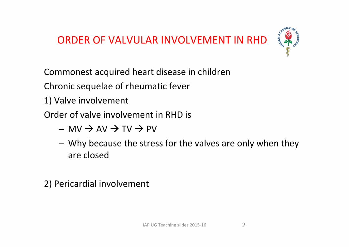
ORDER OF VALVULAR INVOLVEMENT IN RHD
Commonest acquired heart disease in childrenChronic sequelae of rheumatic fever1) Valve involvementOrder of valve involvement in RHD is
– MV AV TV PV– Why because the stress for the valves are only when they are closed
2) Pericardial involvement
2
IAP UG Teaching slides 2015‐16
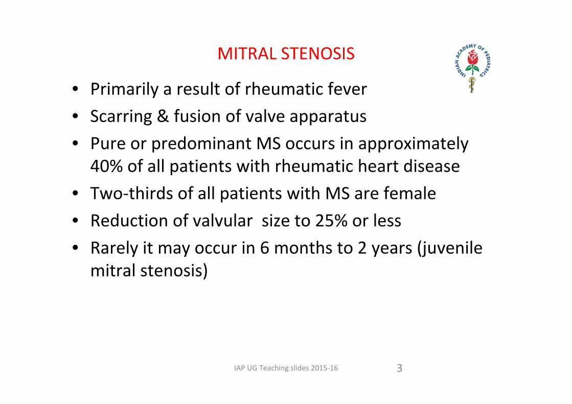
MITRAL STENOSIS
• Primarily a result of rheumatic fever• Scarring & fusion of valve apparatus• Pure or predominant MS occurs in approximately 40% of all patients with rheumatic heart disease
• Two‐thirds of all patients with MS are female• Reduction of valvular size to 25% or less• Rarely it may occur in 6 months to 2 years (juvenile mitral stenosis)
3

IAP UG Teaching slides 2015‐16
MITRAL STENOSIS:PATHOPHYSIOLOGY
• Normal valve area: 4‐6 cm2
• Mild mitral stenosis: – MVA 1.5‐2.5 cm2
– Minimal symptoms• Mod mitral stenosis
– MVA 1.0‐1.5 cm2 usually does not produce symptoms at rest
• Severe mitral stenosis– MVA < 1.0 cm2
4

IAP UG Teaching slides 2015‐16
RHEUMATIC MS
5

IAP UG Teaching slides 2015‐16
PATHOLOGY
• Fibrosis of the mitral ring with contracture of valve leaflets, chordae and papillary muscles and commissural fusion
6

IAP UG Teaching slides 2015‐16
MITRAL STENOSIS : PATHOPHYSIOLOGY
Right Heart Failure:Hepatic Congestion
JVDTricuspid Regurgitation
RA Enlargement
Pulmonary HTNPulmonary Congestion
LA EnlargementAtrial Fib
LA Thrombi LA Pressure
RV Pressure OverloadRVH
RV FailureLV Filling
7

IAP UG Teaching slides 2015‐16
MITRAL STENOSIS : SYMPTOMS
8
• Atrial fibrillation• Systemic embolism• Worsened by conditions that cardiac output.– Exertion, fever, anemia, tachycardia, Atrial fibrillations, pregnancy, thyrotoxicosis
• Hemoptysis
• Palpitations• Cough• Left sided failure
– Orthopnea– PND
• Pulmonary infection• Right sided failure
– Hepatic Congestion– Edema
IAP UG Teaching slides 2015‐16
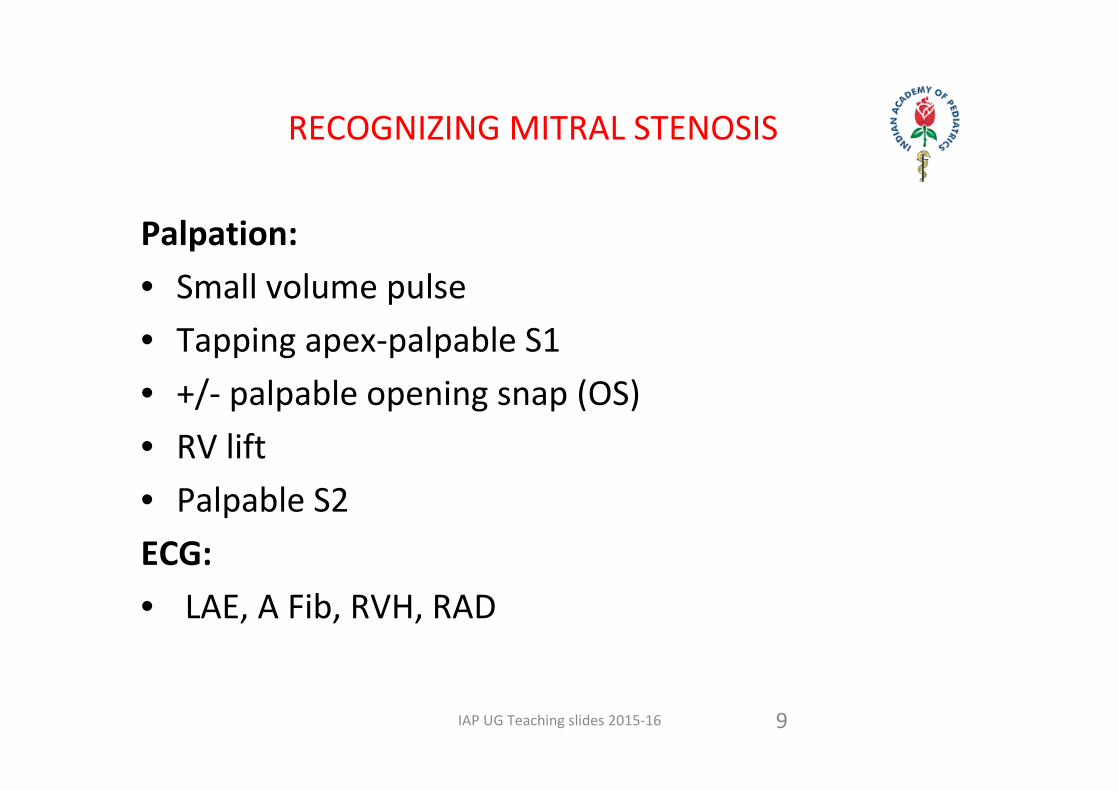
RECOGNIZING MITRAL STENOSIS
9
Palpation:• Small volume pulse• Tapping apex‐palpable S1• +/‐ palpable opening snap (OS)• RV lift• Palpable S2ECG:• LAE, A Fib, RVH, RAD

IAP UG Teaching slides 2015‐16
Auscultation: Loud S1‐ as loud as S2 in aortic area
A2 to OS interval inversely proportional to severity Diastolic rumble: length proportional to severity In severe MS with low flow‐ S1,
OS & rumble may be inaudible
10
IAP UG Teaching slides 2015‐16
PHYSICAL EXAMINATION
11
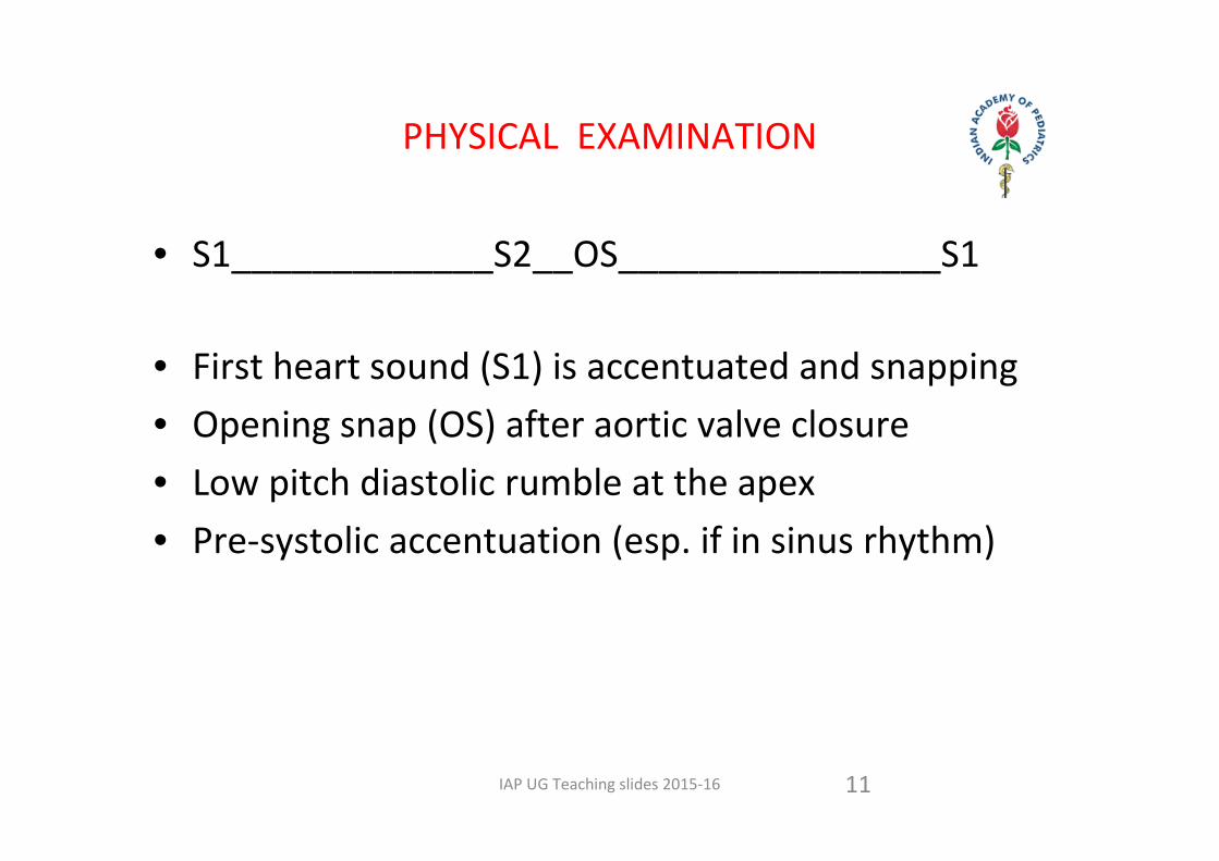
• S1_____________S2__OS________________S1
• First heart sound (S1) is accentuated and snapping• Opening snap (OS) after aortic valve closure• Low pitch diastolic rumble at the apex• Pre‐systolic accentuation (esp. if in sinus rhythm)

IAP UG Teaching slides 2015‐16
MITRAL STENOSIS:NATURAL HISTORY
• Progressive, life long disease, • Usually slow & stable in the early years.• Progressive acceleration in the later years• 20‐40 year latency from rheumatic fever to symptom onset.
• Additional 10 years before disabling symptoms
12

IAP UG Teaching slides 2015‐16
INVESTIGATION
ECG :‐• LAE• RVH• Premature contractions • Atrial flutter and/or fibrillation
– freq. in pts with mod‐severe MS for several years
– A fib develops in 30% to 40% of pts with symptoms
• X ray chest• Echocardiogram
13
IAP UG Teaching slides 2015‐16
MITRAL STENOSIS:THERAPY
• Medical– Diuretics for LHF/RHF– Digitalis/Beta blockers/CCB: Rate control in A Fib– Anticoagulation: In A Fib– Endocarditis prophylaxis
• Balloon valvuloplasty– Effective long term improvement
14
IAP UG Teaching slides 2015‐16
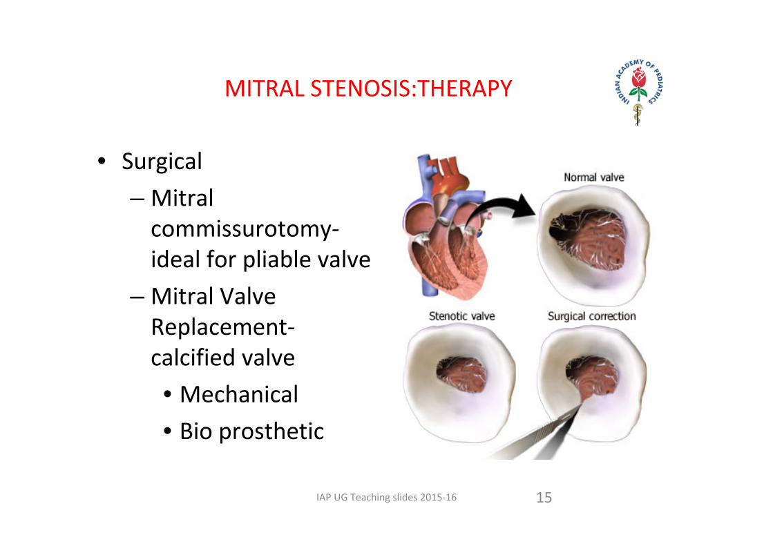
MITRAL STENOSIS:THERAPY
15
• Surgical– Mitral commissurotomy‐ ideal for pliable valve
– Mitral Valve Replacement‐ calcified valve• Mechanical• Bio prosthetic

IAP UG Teaching slides 2015‐16
MITRAL REGURGITATION
16

IAP UG Teaching slides 2015‐16
PATHOLOGY
• Loss of valvular structure and shortening of chordae tendinae
17

IAP UG Teaching slides 2015‐16
MR PATHOPHYSIOLOGY
• Chronic LV volume overload compensatory LVE initially maintaining cardiac output
• Decompensation (increased LV wall tension) CHF
• LVE annulus dilation increased MR
• Backflow LAE, Afib, Pulmonary HTN
18

IAP UG Teaching slides 2015‐16
MR SYMPTOMS
19
• Similar to MS• Dyspnea, Orthopnea, PND
• Fatigue• Pulmonary HTN, right sided failure
• Hemoptysis• Systemic embolization in A Fib
IAP UG Teaching slides 2015‐16
RECOGNIZING CHRONIC MR
20
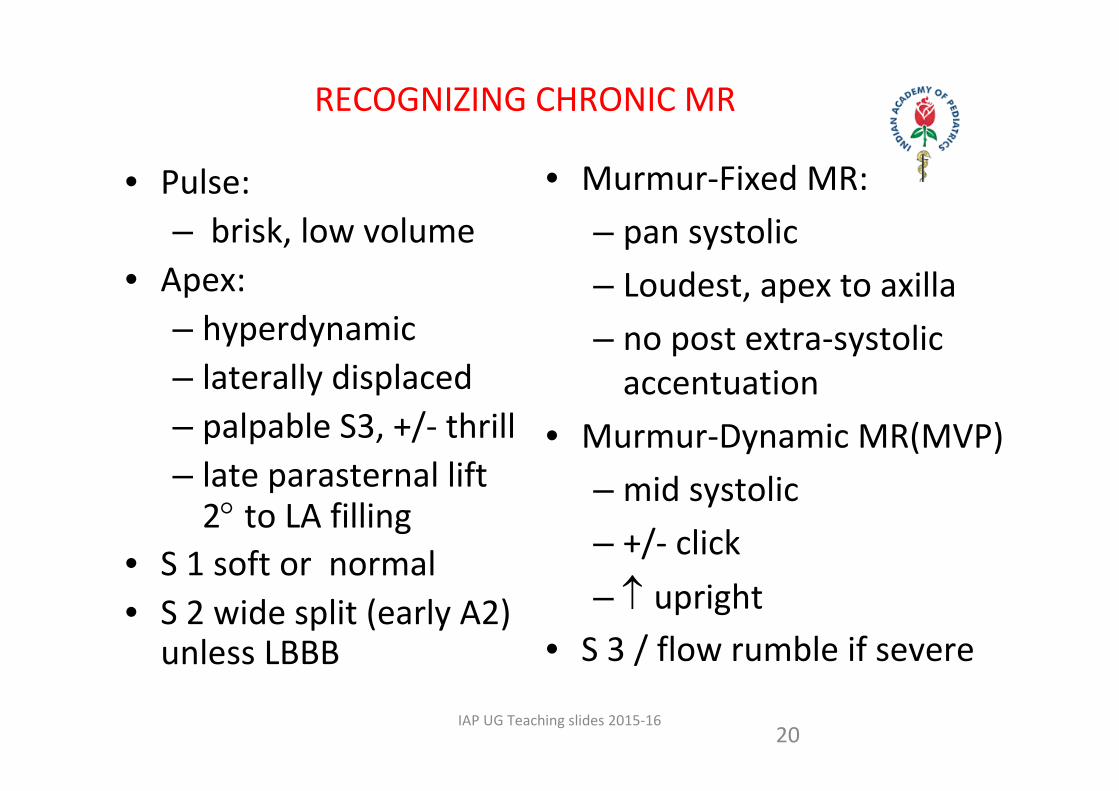
• Pulse:– brisk, low volume
• Apex:– hyperdynamic– laterally displaced– palpable S3, +/‐ thrill– late parasternal lift 2 to LA filling
• S 1 soft or normal• S 2 wide split (early A2) unless LBBB
• Murmur‐Fixed MR:– pan systolic– Loudest, apex to axilla– no post extra‐systolic accentuation
• Murmur‐Dynamic MR(MVP)– mid systolic– +/‐ click– upright
• S 3 / flow rumble if severe

IAP UG Teaching slides 2015‐16
RECOGNIZING ACUTE SEVERE MR
21
• Acute severe dyspnea, CHF & hypotension
• LV size normal• LV may/may not be hyper dynamic
• Loud S1• Systolic murmur may/may not be pan‐systolic
• Inflow/rumble• S3 present‐may be only abnormality
• RV lift• Chordal or papillary muscle rupture/tear– Infarction with papillary muscle ischemia or tear
– Infectious endocarditis with leaflet perforation or disruption or chordal tear
– Flail MV segment
IAP UG Teaching slides 2015‐16
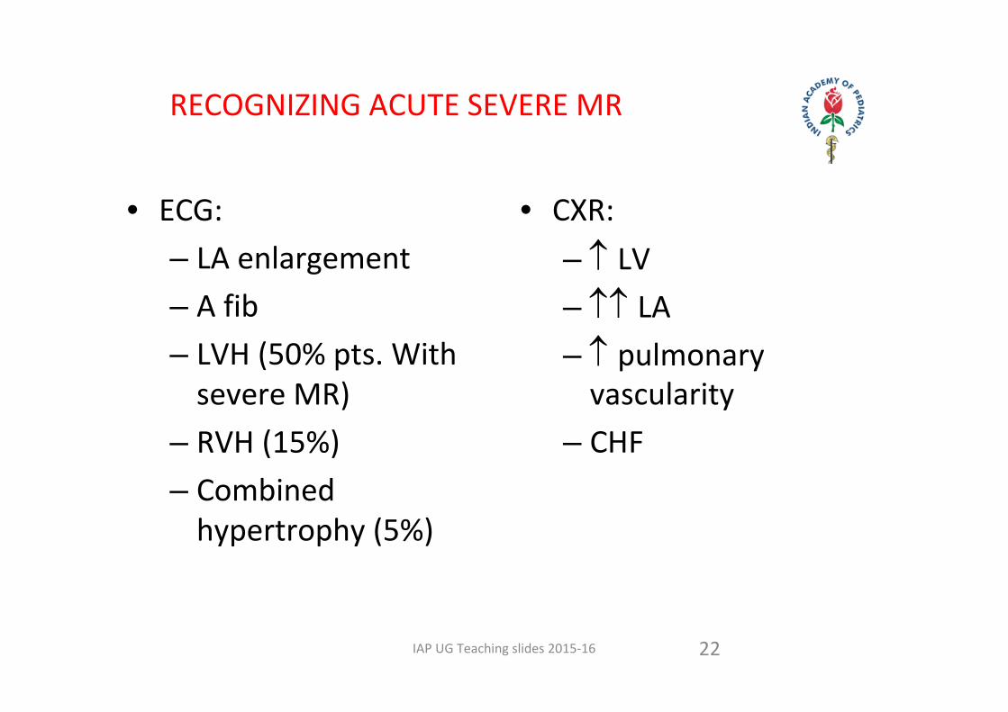
RECOGNIZING ACUTE SEVERE MR
22
• ECG:– LA enlargement– A fib– LVH (50% pts. With severe MR)
– RVH (15%)– Combined hypertrophy (5%)
• CXR:– LV– LA– pulmonary vascularity
– CHF

IAP UG Teaching slides 2015‐16
MR ECHOCARDIOGRAM
23

IAP UG Teaching slides 2015‐16
MR STAGES
LV size and function defined by echo• Stage 1‐compensated:
– End‐diastolic dimension less 63mm, ESD less 42mm
– EF more than 60• Stage 2‐transitional
– EDD 65‐68mm, ESD 44‐45mm, EF 53‐57• Stage 3‐decompensated
– EDD more than 70mm, ESD more than 45mm, EF less than 50
24
IAP UG Teaching slides 2015‐16
MITRAL VALVE SURGERY
• Only effective treatment is valve repair/replacement• Optimal timing determined:
– Presence/absence of symptoms– Functional state of ventricle– Feasibility of valve repair– Presence of A fib/PHTN– Preference/expectations of patient
25
IAP UG Teaching slides 2015‐16
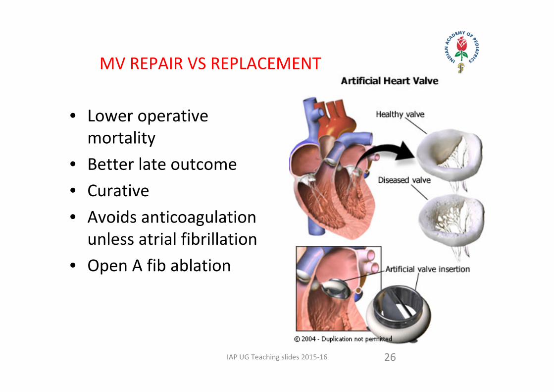
MV REPAIR VS REPLACEMENT
26
• Lower operative mortality
• Better late outcome• Curative• Avoids anticoagulation unless atrial fibrillation
• Open A fib ablation

IAP UG Teaching slides 2015‐16
CONTD:
27
• Valve replacement:– Mortality 2‐7%– Anti‐coagulation– Decreased LVEF
• Tissue prosthetic valve degeneration
• Mechanical prosthetic valve dysfunction/ thrombosis
• Valve repair– Mortality 2‐3%– No anticoagulation (unless Afib)
– Preservation of LVEF• Valve repair always preferable– Feasible in 70‐90% of patients
IAP UG Teaching slides 2015‐16
PATHOPHYSIOLOGY
• Sclerosis of aortic valve and retraction of cusps• Left ventricular enlargement • Mild cases are asymptomatic
28

IAP UG Teaching slides 2015‐16
SYMPTOMS
• Orthopnea• Wide pulse pressure • Exertional angina • Heaving apex • Blowing early diastolic murmur in aortic area‐ best heard in expiration
• Austin Flint murmur – apical presystolic murmur due to large flow across mitral valve
29

IAP UG Teaching slides 2015‐16
INVESTIGATIONS
• X‐ ray• ECG• Echocardiogram• Cardiac catheterization
30

IAP UG Teaching slides 2015‐16
TREATMENT
• Rheumatic prophylaxis• Decongestive‐ vasodilators, ACE inhibitors (digoxin increases the regurgitation)
• Infective endocarditis prophylaxis • Valve replacement if left ventricular function is progressively reducing
31

IAP UG Teaching slides 2015‐16
AORTIC REGURGITATION
32
IAP UG Teaching slides 2015‐16
SYMPTOMS
• Orthopnea• Wide pulse pressure • Exertional angina • Heaving apex • Blowing early diastolic murmur in aortic area‐ best heard in expiration
• Austin Flint murmur – apical presystolic murmur due to large flow across mitral valve
33
IAP UG Teaching slides 2015‐16
INVESTIGATIONS
• X‐ ray• ECG• Echocardiogram• Cardiac catheterization
34

IAP UG Teaching slides 2015‐16
TREATMENT
• Rheumatic prophylaxis• Decongestive‐ vasodilators, ACE inhibitors (digoxin increases the regurgitation)
• Infective endocarditis prophylaxis • Valve replacement if left ventricular function is progressively reducing
35

IAP UG Teaching slides 2015‐16
VALVULOPLASTY
36

IAP UG Teaching slides 2015‐16
PERICARDITIS
37

IAP UG Teaching slides 2015‐16
PATHOPHYSIOLOGY
• Noninflammatory involvement of pericardium• Accumulation of fluid in the cavity more than 10 to 15 ml
38

IAP UG Teaching slides 2015‐16
PERICARDITIS
39

IAP UG Teaching slides 2015‐16
CLINICAL FEATURES
• Precordial pain• Cough• Dyspnea • Pericardial rub
40
IAP UG Teaching slides 2015‐16
INVESTIGATION
• X ray chest – water bottle appearance of heart• ECG – low voltage of QRS complexes, elevation of ST segment and T wave inversion
• Echocardiography• Pericardial tap
41

IAP UG Teaching slides 2015‐16
TREATMENT
• Bed rest• Rheumatic prophylaxis• Steroids• Aspiration of pericardial effusion
42

IAP UG Teaching slides 2015‐16
THANK YOU
43