rhelax - tulip group rf.pdf · corresponding author's e-mail : [email protected] published by...
TRANSCRIPT

Performance Evaluations
RHELAX -RF ®
Latex agglutination slide test for Rheumatoid factors

INDEX
S. No. Name of the Publication Pg Nos
1. Indian Res. J. Genet. & Biotech. 6(3) : (2014)
International Journal of Leprosy Volume 73, Number 2 93-99
Tropical Journal of Medical Research, Vol 18 • Issue 1 • Jan-Jun 2015 5-9
International Journal of Pharmacy and Biological Sciences, Volume 3, Issue3, 180-193 Jul-Sep 2013
Journal,2012, Dec 9 ( Suppl.1) 1-7
479-482 2.
3. 4.
5. Dental Research
Performance Evaluations
RHELAX -RF ®
Latex agglutination slide test for Rheumatoid factors

Performance Evaluations
AS A REFERENCE PRODUCT
RHELAX -RF ®
Latex agglutination slide test for Rheumatoid factors

HIV status and re-activity to RA, ASO, CRP, Syphilis and Facs countamong leprosy patients
Manoj Kumar1 and Tahziba Hussain2
1Central JALMA Institute for leprosy and Other Mycobacterial Disease (ICMR) Tajganj, Agra
2 SRO, HIV/AIDS unit, Central JALMA Institute for leprosy and Other Mycobacterial Disease (ICMR) Tajganj, Agra
(Received : May, 2014 : Revised : June, 2014; Accepted : July 2014)
Abstract
Twenty five leprosy patient’s blood serum compare to control healthy person’s blood samples were
tested for HIV status and re-activity torheumatoid arthritis, ASO, CRP, VDRL and facs count among
leprosy.None of the sample was found to be positive for antibodies to HIV ½. Out of twenty five
leprosy samples, three were reactive for Rheumatoid Arthritis factor and seven were positive for
antibodies to syphilis as revealed by the test. Further two were reactive for C-reactive protein and
one were having titre of Anti-Streptolysin-O. Screening for HIV, Rheumatoid Arthritis and Anti-
Streptolysin-O, C-reactive protein and Syphilis (VDRL) were done in order to know the prevalence
levels of these infections, as biological markers of risk. Thus, screening the leprosy patients for
these would go a long way in early detection of these co-infections. Early treatment, if initiated,
would help in further deterioration of the condition of these patients.
Key words : HIV, leprosy, rheumatoid arthritis, ASO,
CRP, Syphilis.
Inroduction
India has the largest number of known cases of
leprosy and happens to incidentally be endemic for
HIV as well. According to Ridley and Jopling (1966)
studies done in North and North-Eastern India did not
ûnd any association of HIV infection with leprosy
patients. A few studies from South Indian states
showed a higher prevalence of HIV infection among
leprosy patients, but these studies alone do not
provide any indication of its association with leprosy
(Jayasheela et al., 1994). Leprosy caused by
Mycobacterium lepraehas an unusually long
incubation period, and infection with HIV leads to a
profound drop in CD4+ T-lymphocyte count and
function and compromises the cell-mediated immune
response, as well (Miller, 1991; Saha et al., 1993).
Earlier studies carried out in this center suggested
that per thousand (5/4025:0.124%) of the leprosy
patients harbored HIV infection. Follow-up of these
patients at an interval of six months, revealed that none
of them downgraded into a severe form of leprosy nor
developed ARC or AIDS (Hussain et al., 2000).
Although this study indicated that leprosy is not a risk
factor for developing HIV-1 infection, the HIV
surveillance studies on this population was continued
with a view to assess the risk and ûnd out the trend
in an area where both the infections are prevalent.
Some studies shows that the histological features of
leprosy also appear to be preserved in HIVinfected
patients (Moran et al., 1995; Pereira et al.,2004).
One of the commonly observed complaints
among leprosy patients was pain in the joints. Many
studies have proven that microbial agents might
Indian Res. J. Genet. & Biotech. 6(3) : 479-482 (2014)
Corresponding author's e-mail : [email protected] by Indian Society of Genetics, Biotechnology Research and Development, 5, E Biotech Bhawan, Nikhil Estate, MugaliaRoad, Shastripuram, Sikandra, Agra 282007Onlince management by www.isgbrd.co.in

480 Evaluation of Moringa oleifera Seed Extracts for Antibacterial Activity....... [Vol. 6 No.3]
trigger the autoimmune phenomenon and induce
rheumatoid arthritis (Albert et al., 1980; Cossermelli-
Messina et al., 1997; Gibson et al., 1994). In order
to ûnd out if arthritis is present in the HIV-leprosy
co-infected patients, the sera from these cases were
tested for Rheumatoid arthritis (RA) factor. Many risk
behaviors as well as the routes of transmission for
HIV infection are identical to those for other sexually
transmitted diseases (STDs). For this reason, the
leprosy sera samples were tested for Rheumatoid
Arthritis andVDRL simultaneously with HIV.
Materials and methods
Leprosy patients, across the spectrum, i.e.,
tuberculoid (TT), borderline-tuberculoid (BT), mid-
borderline (BB), borderlinelepromatous (BL),
lepromatous (LL) and neuritic (N) types, classiûed,
according to Ridley-Jopling criteria (Ponnighaus et
al., 1991), attending the Unit-I of the Outpatient’s
Department (OPD) of the Central JALMA Institute for
Leprosy and other Mycobacterial Diseases
(CJILOMD) were included in the study. The leprosy
cases in the study were neither newly admitted nor
untreated patients, although a few were newly
detected cases. For bacteriological determination, the
six skin sites used were the two ear lobes and four
representative active skin sites, i.e., hand (right arm
and left arm), elbow (right and left), back, forehead,
and the site of the lesion. In our OPD, four skin sites
are routinely used for determination of the
bacteriological index (B.I.). The inclusion criteria
were: adult leprosy patients between the age group
of 16 to 48 yrs. Children and old patients were
excluded from the study as it was assumed they were
not likely to be sexually active.
Blood samples were collected asceptically from
twenty five leprosy patients and twenty five nornal
healthyby ante-cubital venipuncture after obtaining
pre-informed consent. Samples left at room
temperature for 3-4 hours or kept overnight at 4ºC
for serum separation. Then serum is separated and
collected in eppendrof tubes and labeled properly.
The sera samples collected after stored at -20ºC until
the assays were performed. ELISA was done using
Genedia HIV-1/2 EIA kit (Greencross, Korea). Those
found positive were conûrmed by rapid (HIV capillus
latex aggregation assay, Trinity Biotech PLC, Ireland)
and Western blot assays (WesternBlot, BIO-RAD,
NEWLAVBLOT), Nippon Bio-Rad Laboratories,
Japan. After post-test counselling, a report was
handed over to those found HIV-positive and patient
was referred to clinicians for further care and
management. To ûnd out any other co-infections, the
samples were further tested by HBsAg kit, (Immuno-
chromatography test ERBA Hepline, Transasia Bio-
Medicals Ltd., Mumbai, India) and VDRL and
Rheumatoid Arthritis kits (Carbogen and Rhelax, RF
of Tulip Diagnostics (P) Ltd., Bambolim, Goa, India).
Result and Discussion
None of the sample was found to be positive for
antibodies to HIV ½ (Fig. 1). Out of twenty five leprosy
samples, three were reactive for Rheumatoid Arthritis
factor and seven were positive for antibodies to
syphilis as revealed by the test. Further two were
reactive for C-reactive protein and one were having
titre of Anti-Streptolysine-O.
C-reactive protein and Anti-Streptolysin-O rise
in acute phase of infections. In the absence of
definite diagnosis, much before, appearance of the
symptoms of the disease, assessing the levels of
reactivity of the C-reactive protein and Anti-
Streptolysin-O might be a useful diagnostic tool.
CD4+/CD8+ count declines in leprosy patients,
the CD4+ cells counts were found to be were also
low i.e. below 500, CD8+ cells count 299 and the
CD4+/CD8+ ratio was 1.38.CD4+/CD8+ count
declinesin HIV infection, the CD4+ cells counts were
found to be were also low i.e. below 100, CD8+ cells
count high i.e. up to 500and the CD4+/CD8+ ratio
was 0.17. CD4+/CD8+ count declines in in normal
healthy control, the CD4+ cells counts were found to
be were also high i.e. upto 800, CD8+ cells count
high i.e. up to 400and the CD4+/CD8+ ratio was 2.02.
Screening for HIV, Rheumatoid Arthritis and Anti-
Streptolysin-O, C-reactive protein and Syphilis
(VDRL) were done in order to know the prevalence
levels of these infections, as biological markers of
HIV status and re-activity to RA, ASO, CRP, Syphilis and Facs count ....

Aug., 2014 Santosh Kumar Singh 481
risk. Thus, screening the leprosy patients for these
would go a long way in early detection of these co-
infections. Early treatment, if initiated, would help in
further deterioration of the condition of these patients.
Table 1. HIV status and FACS counts of leprosy patients, HIV positive and normal healthy controls.
Samples HIV Status FACS Count
Elisa Capillus Latex CD4+ CD8+ CD3+ CD4+/CD8+ ratio
agglutination
Leprosy patients -ve -ve 315 229 562 1.38
HIV Positive patients +ve +ve 91 536 624 0.17
Normal healthy controls -ve -ve 857 422 1429 2.02
In normal healthy individuals CD4+ cell counts is
higher than CD8+ cells. In HIV infective patients CD4+
cells counts is lower than CD8+ cells. In leprosy
patients both CD4+ and CD8+ cells counts is decline
than normal value.
Reference
1. Moran C. A., Nelson A. M. and Tuur S. M.
(1995) Leprosy in five human immunodeficiency
virus-infected patients.Mod Pathology, 8: 662–64.
2. Pereira G. A., Stefani M. M. and
AraujoFilho J. A. (2004) Human
immunodeficiency virus type 1 (HIV-1) and
Mycobacterium lepraecoinfection:HIV-1
subtypes and clinical, immunologic,
andhisto-pathologic profiles in a Brazilian
cohort. American Journal of Trop Med
Hyg,71: 679–84.
3. Albert D. A., Weisman M. H. and Kaplan
R. (1980)The rheumatic manifestations of
leprosy (Hansen’s disease).Medicine
(Baltimore).59 (6): 442-448.
4. Bednarsh H. and Eklund K. (2003)
Management of occupational exposure to
Hepatitis B, Hepatitis C, and human
immunodeûciency virus.Compend.Contin.
Educ. Dent. 2002.23: 561-566.
5. Cossermelli-Messina W. and Cossermelli
W. (1997)Possible mechanisms of chronic
leprosy-related arthritis. Rev. Paul. Med.
115(2): 1406-1409.
6. Gibson T., Ahsan Q. and Hussain K. (1994)
Arthritis of leprosy.British Journal of
Rheumatology.33(10): 963-966.
7. Hussain T., Kulshreshtha K., Ghei S. K.,
Manoj Kumar & Tahziba Hussain

482 Evaluation of Moringa oleifera Seed Extracts for Antibacterial Activity....... [Vol. 6 No.3]
Natarajan M., Katoch K. and Sengupta U.
(2000) HIV sero-prevalence in leprosy
patients.International Journal of Leprosy and
Other Mycobacterial Disease. 68: 67-69.
8. Jayasheela M., Sharma R. N., Sekar B.
and Thyagarajan S. P. (1994) HIV infection
amongst leprosy patients in South
India.International Journal of Leprosy and
Other Mycobacterial Disease.66: 429-433.
9. Miller R. A. (1991) Leprosy and AIDS: a
review of the literature and speculations on
the impact of CD4+ lymphocyte depletion on
immunity to Mycobacterium leprae.
International Journal of Leprosy and Other
Mycobacterial Disease.59: 639-644.
10. Ridley D.S., Jopling W. H., (1966)
Classiûcation of leprosy according to
immunity: a ûve group system. International
Journal of Leprosy and Other Mycobacterial
Disease. 34: 255-267.
11. Saha K., Chattopadhya D., Dash K.,
Saha, Uma, Tyagi, Pradip K., Gupta
Madan M., Parashri, Sharma A. and
Amar K. (1993) STDs in leprosy patients in
North and Northeastern India.A futile search
for HIV antibody.International Journal of
Leprosy and Other Mycobacterial Disease.
58(4): 660-665. ***
HIV status and re-activity to RA, ASO, CRP, Syphilis and Facs count ....

1 Received for publication on 21 September 2004. Accepted for publication on 13 February 2005.2 T. Hussain, Senior Research Officer; S. Sinha, Senior Research Fellow (CSIR); K. K. Kulshreshtha, Senior
lab. Technician; K. Katoch, Deputy Director (Senior Grade); V. S. Yadav, Statistical Officer; U. Sengupta,Scientist-Emeritus, and V. M. Katoch, Director Central JALMA Institute for Leprosy and other MycobacterialDisease, Tanjganj, Agra, India.
Reprint requests to: Tahziba Hussain, Senior Research Officer, HIV/AIDS UNIT and Clinical Division,Central JALMA Institute for Leprosy and other Mycobacterial Diseases (Indian Council of Medical Research),Tajganj, Agra - 282001. INDIA. E-mail: [email protected]
INTERNATIONAL JOURNAL OF LEPROSY Volume 73, Number 2Printed in the U.S.A.
(ISSN 0148-916X)
INTERNATIONAL JOURNAL OF LEPROSY
and Other Mycobacterial Diseases
VOLUME 73, NUMBER 2 JUNE 2005
Seroprevalence of HIV Infection among Leprosy
Patients in Agra, India: Trends and Perspective1
Tahziba Hussain, Shikha Sinha, K. K. Kulshreshtha, Kiran Katoch, V. S. Yadav, U. Sengupta, and V. M. Katoch2
ABSTRACTThis study compares the results of HIV seroprevalence, which was carried out in two
phases, i.e., 1989 to 1993 and 1999 to 2004. Although the number of leprosy patientsscreened for HIV infection in the second phase is less (2125) as compared to those screenedduring the first phase (4025), a rise in HIV infection from 0.12% to 0.37% is certainly dis-turbing since this area appears to be endemic for both the infections. During the study pe-riod, the Out Patient department attendance of a few types of leprosy patients like borderlineand borderline lepromatous have risen, whereas others like borderline tuberculoid and polartuberculoid have declined in the second phase as compared to that of the first phase. Thetrend over a decade suggests that HIV infection is low among the leprosy patients whencompared with other risk groups. Follow-up of these patients at an interval of six months,revealed that none of them downgraded into a severe form of leprosy nor developed ARC orAIDS. In this study, it appears that neither infection precipitated the other. The occurrenceof downgradation as well as reversal reactions and neuritis (both chronic and acute) was notobserved among the leprosy patients. None of them developed erythema nodosum leprosumreactions. Similarly, the HIV-positive leprosy cases did not develop either AIDS relatedcomplex (ARC) or full blown case of AIDS.
RESUMECette étude compare les résultats de séroprévalence du VIH, obtenus en 2 phases dis-
tinctes : de 1989 à 1993 et de 1999 à 2004. Bien que le nombre de patients testés pour l’in-fection par le VIH soit moindre dans la seconde phase (2125) que dans la première (4025),une augmentation de prévalence de 0.12% à 0.37% est préoccupante puisque la régionétudiée est endémique pour les 2 infections. Pendant la durée de cette étude, si la secondephase est comparée à la première, la présentation de patients au service de Consultations Ex-ternes a augmenté pour quelques types de patients lépreux comme les patients borderline etborderline lépromateux et diminué pour les patients borderline tuberculoïdes et tubercu-
93

94 International Journal of Leprosy 2005
loïdes polaires. La tendance dégagée sur une décennie suggère que l’infection par le VIH estfaible chez les patients lépreux, comparés à d’autres groupes à risque. Le suivi tous les 6mois de ces patients indique qu’aucun d’entre eux n’a rétrogradé en une forme sévère de lalèpre ou n’a développé le complexe associé au SIDA (ARC) ou le SIDA. Dans cette étude,il apparaît qu’aucune de ces infections ne précipite l’autre. Il ne fut pas observé de déplace-ment vers le bas le long du spectre immuno-pathologique ou de réactions inverses ou denévrites (à la fois chroniques ou aiguës) parmi les patients hanséniens. Aucun n’a développéde réaction de type érythème noueux lépreux. Concomitamment, les cas de lèpre aussi posi-tifs au VIH n’ont développé ni de syndrome ARC ni de SIDA terminal.
RESUMENEste estudio compara los resultados de una encuesta sobre la prevalencia del VIH en pa-
cientes con lepra, realizada en dos fases, la primera de 1989 a 1993 y la segunda de 1999 a2004. Aunque el número de pacientes investigados para VIH fue mayor en la primera fase(4025) que en la segunda (2125), se notó un incremento en la infección por VIH de 0.12% a0.37%. Esto es preocupante porque sugiere que esta área es endémica para las dos enfer-medades. En la segunda fase del estudio, se observó un incremento en el número de pa-cientes BL/LL que acudieron al Instituto y una disminución en el número de los pacientesBT/TT. Los resultados globales indican que la infección por VIH es baja entre los pacientescon lepra en comparación con la infección en otros grupos de riesgo. El examen de estos pa-cientes a los 6 meses de seguimiento reveló que ninguno de ellos “se degradó” a una formamás severa de la lepra, ni desarrolló los signos del complejo asociado al SIDA, ni la enfer-medad en si. Además, ninguna de las enfermedades precipitó a la otra. Ninguno de los pa-cientes desarrolló reacciones reversas (neuritis agudas y crónicas), ni eritema nodos leproso(ENL).
India has the largest number of knowncases of leprosy and happens to incidentallybe endemic for HIV as well. Some of theearlier studies done in North and North-Eastern India did not find any association ofHIV infection with leprosy patients (24). Afew studies from South Indian statesshowed a higher prevalence of HIV infec-tion among leprosy patients, but these stud-ies alone do not provide any indication ofits association with leprosy (12). Leprosycaused by Mycobacterium leprae has an un-usually long incubation period, and infec-tion with HIV leads to a profound drop inCD4+ T-lymphocyte count and functionand compromises the cell-mediated im-mune response, as well (19, 25). Earlier stud-ies carried out in this center suggested that1 per thousand (5/4025:0.124%) of the lep-rosy patients harbored HIV infection.Follow-up of these patients at an interval ofsix months, revealed that none of themdowngraded into a severe form of leprosynor developed ARC or AIDS (10). Althoughthis study indicated that leprosy is not a riskfactor for developing HIV-1 infection, theHIV surveillance studies on this populationwas continued with a view to assess the riskand find out the trend in an area where both
the infections are prevalent. This studycompares the results of HIV seropreva-lence, which was carried out in two phases;first, from April, 1989 to March, 1993 whenHIV infection was being detected in Indiain different risk group populations to assessthe risk among leprosy patients, and thenfrom September, 1999 to March, 2004. Thisis the first report of a decade of HIV screen-ing of leprosy patients in this region of thecountry and the longest follow-up of HIV-leprosy co-infected cases.
One of the commonly observed com-plaints among leprosy patients was pain inthe joints. Many studies have proven thatmicrobial agents might trigger the autoim-mune phenomenon and induce rheumatoidarthritis (1, 5, 8). In order to find out if arthri-tis is present in the HIV-leprosy co-infectedpatients, the sera from these cases weretested for Rheumatoid arthritis (RA) factor.Many risk behaviors as well as the routes oftransmission for HIV, Hepatitis B virus(HBV) and Hepatitis C virus (HCV) in-fection are identical to those for other sexu-ally transmitted diseases (STDs) (3). Forthis reason, the leprosy sera samples weretested for HBsAg and VDRL simultane-ously with HIV.

73, 2 Hussain et al.: HIV Infection Among Leprosy Patients 95
MATERIALS AND METHODSLeprosy patients, across the spectrum, i.e.,
tuberculoid (TT), borderline-tuberculoid(BT), mid-borderline (BB), borderline-lepromatous (BL), lepromatous (LL) andneuritic (N) types, classified, according toRidley-Jopling criteria (23), attending theUnit-I of the Outpatient’s Department(OPD) of the Central JALMA Institute forLeprosy and other Mycobacterial Diseases(CJILOMD) were included in the study.The leprosy cases in the study were neithernewly admitted nor untreated patients, al-though a few were newly detected cases.For bacteriological determination, the sixskin sites used were the two ear lobes andfour representative active skin sites, i.e.,hand (right arm and left arm), elbow (rightand left), back, forehead, and the site of thelesion. In our OPD, four skin sites are rou-tinely used for determination of the bacteri-ological index (B.I.). The inclusion criteriawere: adult leprosy patients between theage group of 16 to 48 yrs. Children and oldpatients were excluded from the study as itwas assumed they were not likely to be sex-ually active. In order to ensure that the pa-tients were not screened over and overagain, their OPD cards were marked, “HIV-Screened.” This helped in excluding the re-peat testing of the patients. Blood was col-lected asceptically from leprosy patients byante-cubital venipuncture after obtainingpre-informed consent. The sera samplescollected after centrifugation at 2500 gwere stored at –20°C until the assays wereperformed. ELISA was done using GenediaHIV-1/2 EIA kit (Greencross, Korea).Those found positive were confirmed byrapid (HIV capillus latex aggregation assay,Trinity Biotech PLC, Ireland) and Westernblot assays (WesternBlot, BIO-RAD,NEWLAVBLOT), Nippon Bio-Rad Labo-ratories, Japan. After post-test counselling,a report was handed over to those foundHIV-positive and patient was referred toclinicians for further care and management.To find out any other co-infections, thesamples were further tested by HBsAg kit,(Immuno-chromatography test ERBA Hep-line, Transasia Bio-Medicals Ltd., Mumbai,India) and VDRL and Rheumatoid Arthritiskits (Carbogen and Rhelax, RF of Tulip Di-agnostics (P) Ltd., Bambolim, Goa, India).
RESULTSThe prevalence of HIV-1 infection in lep-
rosy patients was observed in two phases.In phase one, 4025 patients [30 indetermi-nate (I), 141 polar tuberculoid (TT), 1888boderline tuberculoid (BT), 409 borderline(BB), 600 borderline lepromatous (BL),751 polar lepromatous (LL), 200 N] werescreened between 1989 and 1993, out ofwhich only 8 were ELISA positive and 5were Western Blot reactive. Subsequently,in the second phase from 1999 to 2004,2125 patients (21 I, 19 TT, 646 BT, 332 BB,610 BL, 324 LL, 173 N) were screened, outof which 8 were ELISA positive and 5 wereWestern Blot reactive (Table 1). The varia-tion in the results of the two tests correlatedwell with the titre of HIV-1/2 antibodies inthe sera samples. The strongly positivesamples having a high absorbance value,ranging between 1.5 and 2.0, measured interms of O.D. at 450 nm in an ELISAreader had an excellent pattern of reactivityin Western Blot. The samples with weak ormoderate positivity in ELISA, with an O.D.ranging between 0.5 and 0.7, did not reactwith Western Blot. A rise in HIV infectionfrom 0.124% to 0.376% was observed. Twosamples were reactive to HIV-2 by WesternBlot. Among all the HIV-positive leprosypatients, there were no other co-infectionslike Hepatitis B, Syphilis and RA. Out ofthe 8 HIV-leprosy co-infected patients, 2each were BT and BL types, 3 were BB and1 was LL type of leprosy.
The predominant clinical features werehypo-pigmented lesions, clawing of fingersand toes, pain, and hand muscle atrophy.Whereas 4 patients had deformity in hands,only one of them reported acute pain. Allthe patients completed a full course of stan-dard anti-leprosy multi-drug therapy, re-sponded satisfactorily, and were later clini-cally and bacteriologically negative. Theinitial bacterial index, prior to treatment,which ranged between 2+ and 3+ becamenegative on completion of the treatment.Two of the 8 HIV-leprosy co-infected pa-tients (BL, LL) became bacteriologicallynegative after 6 months and another 2 (BT,BL) became negative after 24 months oftreatment (Table 2). We have observed thatfollowing treatment, B.I. became negativeeven in BL and LL cases. The HIV-positive
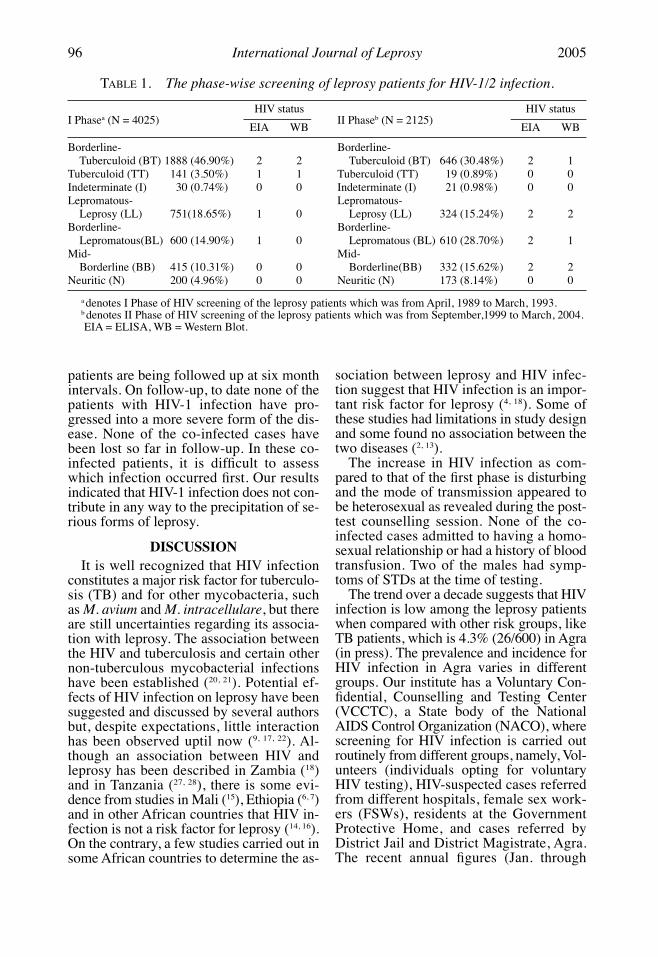
TABLE 1. The phase-wise screening of leprosy patients for HIV-1/2 infection.
I Phasea (N = 4025)HIV status HIV status
EIA WB EIA WB
Borderline- Borderline-Tuberculoid (BT) 1888 (46.90%) 2 2 Tuberculoid (BT) 646 (30.48%) 2 1
Tuberculoid (TT) 141 (3.50%) 1 1 Tuberculoid (TT) 19 (0.89%) 0 0Indeterminate (I) 30 (0.74%) 0 0 Indeterminate (I) 21 (0.98%) 0 0Lepromatous- Lepromatous-
Leprosy (LL) 751(18.65%) 1 0 Leprosy (LL) 324 (15.24%) 2 2Borderline- Borderline-
Lepromatous(BL) 600 (14.90%) 1 0 Lepromatous (BL) 610 (28.70%) 2 1Mid- Mid-
Borderline (BB) 415 (10.31%) 0 0 Borderline(BB) 332 (15.62%) 2 2Neuritic (N) 200 (4.96%) 0 0 Neuritic (N) 173 (8.14%) 0 0
a denotes I Phase of HIV screening of the leprosy patients which was from April, 1989 to March, 1993.b denotes II Phase of HIV screening of the leprosy patients which was from September,1999 to March, 2004.EIA = ELISA, WB = Western Blot.
96 International Journal of Leprosy 2005
patients are being followed up at six monthintervals. On follow-up, to date none of thepatients with HIV-1 infection have pro-gressed into a more severe form of the dis-ease. None of the co-infected cases havebeen lost so far in follow-up. In these co-infected patients, it is difficult to assesswhich infection occurred first. Our resultsindicated that HIV-1 infection does not con-tribute in any way to the precipitation of se-rious forms of leprosy.
DISCUSSIONIt is well recognized that HIV infection
constitutes a major risk factor for tuberculo-sis (TB) and for other mycobacteria, suchas M. avium and M. intracellulare, but thereare still uncertainties regarding its associa-tion with leprosy. The association betweenthe HIV and tuberculosis and certain othernon-tuberculous mycobacterial infectionshave been established (20, 21). Potential ef-fects of HIV infection on leprosy have beensuggested and discussed by several authorsbut, despite expectations, little interactionhas been observed uptil now (9, 17, 22). Al-though an association between HIV andleprosy has been described in Zambia (18)and in Tanzania (27, 28), there is some evi-dence from studies in Mali (15), Ethiopia (6, 7)and in other African countries that HIV in-fection is not a risk factor for leprosy (14, 16).On the contrary, a few studies carried out insome African countries to determine the as-
sociation between leprosy and HIV infec-tion suggest that HIV infection is an impor-tant risk factor for leprosy (4, 18). Some ofthese studies had limitations in study designand some found no association between thetwo diseases (2, 13).
The increase in HIV infection as com-pared to that of the first phase is disturbingand the mode of transmission appeared tobe heterosexual as revealed during the post-test counselling session. None of the co-infected cases admitted to having a homo-sexual relationship or had a history of bloodtransfusion. Two of the males had symp-toms of STDs at the time of testing.
The trend over a decade suggests that HIVinfection is low among the leprosy patientswhen compared with other risk groups, likeTB patients, which is 4.3% (26/600) in Agra(in press). The prevalence and incidence forHIV infection in Agra varies in differentgroups. Our institute has a Voluntary Con-fidential, Counselling and Testing Center(VCCTC), a State body of the NationalAIDS Control Organization (NACO), wherescreening for HIV infection is carried outroutinely from different groups, namely, Vol-unteers (individuals opting for voluntaryHIV testing), HIV-suspected cases referredfrom different hospitals, female sex work-ers (FSWs), residents at the GovernmentProtective Home, and cases referred byDistrict Jail and District Magistrate, Agra.The recent annual figures (Jan. through
II Phaseb (N = 2125)

73, 2 Hussain et al.: HIV Infection Among Leprosy Patients 97
Dec., 2004) revealed that the local preva-lence and incidence of HIV-positivity in thearea is, 40.31% (156/387) among Volun-teers and 43.39% (46/106) among the Re-ferred cases (communicated).
In the second phase as compared to thatof the first phase, the OPD attendance of afew types of leprosy patients has risen dur-ing the study phase, whereas others havedeclined. A striking feature which hasemerged during the second phase of thestudy is that there is an increase in the at-tendance of BB and BL types of leprosy pa-tients, whereas there is a decrease in the BTand TT types of leprosy patients as depictedin Table 1. This could be one of the reasonsfor the higher HIV-positivity observedamong the BB and BL cases. Another onecould be attributed to the better control dueto multi-drug therapy (M.D.T.) and de-creased transmission of M. leprae, with newcases dominated by a long period of incuba-tion, in the lepromatous leprosy cases. Al-though the number of leprosy patientsscreened for HIV infection in the secondphase is less as compared to those screenedduring the first phase, a rise in HIV infec-tion is disturbing since this area appears tobe endemic for both the infections.
Expansion of the HIV epidemic couldhave a significant effect on the epidemiol-ogy of leprosy. In this study, it appears thatneither of the infections precipitated theother. The incidence of downgradation, aswell as reversal reactions and neuritis (bothchronic and acute), was not observed amongthe leprosy patients. None of them devel-
oped Erythema Nodosum Leprosum (ENL)reactions. The total cases of HIV-positiveleprosy patients were only thirteen in boththe phases (5 in phase I, and 8 in phase II),which have been followed up very carefullyand with special care. We have also ob-served that reversal reactions and ENL didnot occur among any of the HIV-leprosy co-infected cases. If the number of cases weremore, then probably one might have notedsome reversal or ENL reactions. To resolvethe issue, a larger study, with longer follow-up is required. Clinical manifestations oflepromatous leprosy cases might be im-munologically mediated and these featurescould be abrogated by HIV infection.
Similarly, the HIV-positive leprosy casesdid not develop either AIDS related complex(ARC) or full blown case of AIDS. None ofthe co-infected cases have been lost so far inthe follow-up. This is the first report of adecade of HIV screening of leprosy patientsin this region of the country and the longestfollow-up of the largest number of HIV-leprosy co-infected cases. Other studies havereported follow-up of very less number ofthe co-infected cases (11, 26). The underlyingmechanism by virtue of which the severityof both the diseases is lowered is not known.The infectious agents and host defencesseem to have co-evolved to reach balancedstates where virus and host survive. WhileHIV has not quite yet reached an optimalbalance, tuberculosis (TB), leprosy, HBV,HCV in humans or lymphocytic chorio-meningits virus (LCMV) in mice have suc-cessfully established persistence (29).
TABLE 2. Clinical presentations and bacteriological index among the HIV-leprosy co-infected patients.
Clinical findings
Skin Lesions Nerves Pain Deformity
1 BL >5 5 Pain Nil Smear 3+ (Negative after 24 months)2 BL >5 4 Nil Nil Smear 2+ (Negative after 6 months)3 BB >5 1 Nil Nil Negative4 BT 1 Nil Nil Nil Negative5 LL >5 4 Nil Hand Smear 3+ (Negative after 6 months)6 BB 1 4 Nil Hand Negative7 BB >5 6 Nil Nil Negative8 BT >5 4 Nil Hand Smear 3+ (Negative after 24 months)

98 International Journal of Leprosy 2005
Although the present study does notshow any association between HIV and lep-rosy, future study is warranted to find outthe reasons for cross-protection, if any, atthe genetic and molecular level.
Acknowledgement. This study was supported byfunds from the Indian Council of Medical Research,New Delhi. Shikha Sinha is a recipient of Senior Re-search Fellowship of the Council of Scientific and In-dustrial Research (CSIR). The authors thank Mr. K. L.Verma, Mr. M. M. Alam, Mr. Sushil Prasad, Mr. P. N.Sharma, and Mr. M. S. Tomar of the HIV/AIDS Unit andthe entire staff of OPD for their assistance in the study.
REFERENCES1. ALBERT, D. A., WEISMAN, M. H., and KAPLAN, R.
The rheumatic manifestations of leprosy (Han-sen’s disease). Medicine (Baltimore). 59(6) (1980)442–448.
3. ANDRADE,V. L., MOREIRA, ALVES T., REGAZZI,AVELLEIRA, J. C., and BAYONA, M. Prevalence ofHIV-1 in leprosy patients in Rio de Janeiro, Brazil.Acta Leprol. 10(3) (1997) 159–163.
4. BEDNARSH, H., and EKLUND, K. Management ofoccupational exposure to Hepatitis B, Hepatitis C,and human immunodeficiency virus. Compend.Contin. Educ. Dent. 2002.23 (2003) 561–566.
5. BORGDORFF, M. W., VANDEN, BROEK, J., CHUM, H.J., KLOKKE, A. H., GROF, P., BARONGO, L. R., andNEWELL, J. N. HIV-1 infection as a risk factor forleprosy; a case control study in Tanzania. Int. J.Lepr. Other Mycobact. Dis. 61 (1993) 556–562.
6. COSSERMELLI-MESSINA, W., and COSSERMELLI, W.Possible mechanisms of chronic leprosy-relatedarthritis. Rev. Paul. Med. 115(2) (1997) 1406–1409.
7. FROMMEL, D., TEKLE-HAIMANOT, R., VERDIER, M.,NEGESSE, Y., BULTO, T., and DENIS, F. HIV infec-tion and leprosy : a four- year survey in Ethiopia.Lancet. 344 (1994) 165–166.
8. GEBRE, S., SAUNDERSON, P., MESSELE, T., andBYASS, P. The effect of HIV status on the clinicalpicture of leprosy: a prospective study in Ethiopia.Lepr. Rev. 71 (2000) 338–343.
9. GIBSON. T., AHSAN, Q., and HUSSAIN, K. Arthritis ofleprosy. Br. J. Rheumatol. 33(10) (1994) 963–966.
10. GORMUS, B. J. HIV-1 infection and leprosy. Int. J.Lepr. Other Mycobact. Dis. 62 (1994) 610–613.
11. HUSSAIN, T., KULSHRESHTHA, K., GHEI, S. K.,NATARAJAN, M, KATOCH, K., and SENGUPTA, U.HIV seroprevalence in leprosy patients. Int. J.Lepr. Other Mycobact. Dis. 68 (2000) 67–69.
12. JACOB, M., GEORGE, S., PULIMOOD, S., andNATHAN, N. Short-term follow up of patients withmultibacillary leprosy and HIV infection. Int. J.Lepr. Other Mycobact. Dis. 64(4) (1996)392–395.
13. JAYASHEELA, M., SHARMA, R. N., SEKAR, B., andTHYAGARAJAN, S. P. HIV infection amongst lep-
rosy patients in South India. Ind. J. Lepr. OtherMycobact. Dis. 66 (1994) 429–433.
14. KAWUMA, H. J., BWIRE, R., and ADATU-ENGWAU,F. Leprosy and infection with the human immuno-deficiency virus in Uganda; a case-control study.Int. J. Lepr. Other Mycobact. Dis. 62(4) (1994)521–526.
15. LEONARD, G., SANGARE, A., VERDIER, M., SASSOU-GUESSEAU, E., PETIT,G., MILAN, J., M’BOUP, S.,REY, JEAN-LOUP, DUMAS, JEAN-LUC, HUGON, J.,GAPORO, I. N., and DENIS, F. Prevalence of HIVinfection among patients with leprosy in Africancountries and Yemen. J. Acquir. Immune Def. Syn.3(11) (1990) 1109–1113.
16. LIENHARDT, C., KAMATE, B., JAMET, P., TOUNKARA,A., FAYE, O. C., SOW, S. O., and BOBIN, P. Effectof HIV infection on leprosy: a three-year survey inBamako, Mali. Int. J. Lepr. Other Mycobact. Dis.64(4) (1996) 383–391.
17. LUCAS, S. B., FINE, P. E. M., STERNE, J. A., PON-NIGHAUS, J. M., TURNER, A. C., DE COCK, K. M.,and ZUCKERMAN, M. Infection with human im-munodeficiency virus type 1 among leprosy pa-tients in Zaire. J. Infect. Dis. 171 (1995) 502–504.
18. MACHADO, P., DAVID, Y., PEDROSO, C., BRITES, C.,BARRAL, A., and BARRAL-NETTO, M. Leprosy andHIV infection in Bahia, Brazil. Int. J. Lepr. OtherMycobact. Dis. 66(2) (1998) 227–229.
19. MEERAN, K. Prevalence of HIV infection amongpatients with leprosy and tuberculosis in ruralZambia. Brit. Med. J. 298 (1989) 364–365.
20. MILLER, R. A. Leprosy and AIDS: a review of theliterature and speculations on the impact of CD4+lymphocyte depletion on immunity to Mycobacte-rium leprae. Int. J. Lepr. Other Mycobact. Dis. 59(1991) 639–644.
21. NUNN, P. P., and MCADAM, R. P. W. J. Mycobac-terial infections and AIDS. Br. Med. Bull. 44(1988) 801–803.
22. OREGE, P. A., FINE, P. E. M., LUCAS, S.B., OBURA,M., OKELO, C., OKUKU, P., and WERE, M. A casecontrol study on human immunodeficiency virus-1 (HIV-1) infection as a risk factor for tuberculo-sis and leprosy in Western Kenya. Tubercle. Lung.Dis. 74 (1993) 377–381.
23. PONNIGHAUS, J. M., MWANJASI, L. J., FINE, P. E.,SHAW, M. A., TURNER, A. C., OXBORROW, S. N., LU-CAS, S. B., JENKINS, P. A., STERNE, J. A., and BLISS,L. Is HIV infection a risk factor for leprosy? Int. J.Lepr. Other Mycobact. Dis. 59 (1991) 221–228.
24. RIDLEY, D. S., and JOPLING, W. H. Classificationof leprosy according to immunity: a five groupsystem. Int. J. Lepr. Other Mycobact. Dis. 34(1966) 255–267.
25. SAHA, K., CHATTOPADHYA, D., DASH KALPANA,SAHA, UMA, TYAGI, PRADIP, K., GUPTA, MADAN,M., PARASHRI, ADITYA, SHARMA, and AMAR, K.STDs in leprosy patients in North and NortheasternIndia. A futile search for HIV antibody. Int. J. Lepr.Other Mycobact. Dis. 58(4) (1993) 660–665.

73, 2 Hussain et al.: HIV Infection Among Leprosy Patients 99
26. SAMPAIO, ELIZABETH P., CANESHI, JAQUELINE R. T.,NERY, JOSE A. C., DUPRE, NADIA C., PEREIRA,GER-ALDO M. B., VIEIRA, LEILA M. M., MOREIRA, AN-DRE L., KAPLAN, GILLA, and SARNO, EUZENIR N.Cellular immune response to Mycobacterium lep-rae infection in Human Immunodeficiency Virus-infected individuals. Infect. Immunity. 63(5)(1995) 1848–1854.
27. SAYAL, S. K., DAS, A. L., and GUPTA, C. M. Con-current leprosy and HIV infection: a report ofthree cases. Ind. J. Lepr. Other Mycobact. Dis.69(3) (1997) 261–265.
28. VANDENBROEK, J., CHUM, H. J., SWAI, R., andO’BRIEN, R. J. Association between leprosy andHIV infection in Tanzania. Int. J. Lepr. Other My-cobact. Dis. 65(2) (1997) 203–210.
29. VANDENBROEK, J., MFINANGA, S., MOSHIRO, C.,O’BRIEN, R. J., and MUGOMELA, A. Survival ofHIV-positive and HIV-negative leprosy patients inMwanza, Tanzania. Int. J. Lepr. Other Mycobact.Dis. 66(1) (1998) 53–56.
30. ZINKERNAGEL, R. M. Immunity, Immunopathol-ogy and vaccine against HIV? Vaccine 20 (2002)19113–19117.

Tropical Journal of Medical Research | Vol 18 • Issue 1 • Jan-Jun 2015 5
Diagnostic utility of anti‑CCP antibodies and rheumatoid factor as inflammatory biomarkers in comparison with C‑reactive protein and TNF‑α in rheumatoid arthritis
Bineeta Kashyap, Urvashi Tiwari1, Arun Garg2, Iqbal R. Kaur1
Department of Microbiology, Maulana Azad Medical College, 1Departments of Microbiology and 2Medicine, University College of Medical Sciences, Guru Tegh Bahadur Hospital, New Delhi, India
AbstractAims and Objectives: Rheumatoid arthritis is a systemic inflammatory disease whose diagnosis is primarily based on clinical manifestations because of lack of suitable diagnostic tests. As substantial joint damage already occurs by the time patient presents clinically, a validated biomarker for the diagnosis is urgently required. Materials and Methods: Sera from a total of 68 clinically suspected rheumatoid arthritis patients and 68 age-and sex-matched controls were tested for rheumatoid factor (RF), anti-cyclic citrullinated peptide (anti-CCP) antibodies, C-reactive protein (CRP), and tumor necrosis factor-alpha (TNF-α). Results: Anti-CCP and CRP were found to be positive in all patients with positive RF; however, TNF-α was present in only two of them. As regards anti-CCP antibodies, out of the 10 samples that showed positive results, RF, CRP, and TNF-α were also present in 4, 5, and 4 cases, respectively. Conclusion: The recognition of utility of such markers is essential to gain insight into the activity of this disease, which is important for early treatment that may limit functional disability consequent to the disease.
Keywords: Anti-cyclic citrullinated peptide antibodies, C-reactive protein, rheumatoid arthritis, rheumatoid factor, tumor necrosis factor-alpha
IntroductionRheumatoid arthritis(RA) is a severe, progressive, comorbid systemic inflammatory disease of unknown etiology. Since timely intervention with new and effective treatments can alter the course of the disease, reduce functional impairment, and lengthen life, better biomarkers for diagnosis and prognosis are needed to identify these patients at an early stage in order to fine‑tune therapeutic options to the individual patient.[1]
Rheumatoid factor(RF), an antibody specific for the Fc portion of human IgG, has been historically considered a marker for RA and was one of the diagnostic criteria for RA that was established by the American College of
Access this article onlineQuick Response Code:
Website:www.tjmrjournal.org
DOI: 10.4103/1119-0388.152534
Rheumatology (ACR).[2] The sensitivity and specificity of RF for the diagnosis of RA has been reported in the range of 50‑80% and 70‑80%, respectively.[3,4] The specificity of the RF test is known to be relatively poor and is often questioned. With around 5% false positivity in the general population, RF is found in many patients with other diseases of infectious or autoimmune origin. Consequently, a search for better diagnostic markers, especially those with improved specificity for RA, ensued. Though due to a low sensitivity and moderate specificity RF has a little diagnostic utility, it has retained its place in practice because of its prognostic capacity and lack of an alternative test.
Citrullination (deimination) of proteins is a chemical reaction which occurs when inflammatory cells release enzymes in local tissues. Citrullination of synovial antigens, especially fibrin, during synovial inflammation probably allows the induction of anti‑cyclic citrullinated
Address for correspondence: Dr. Bineeta Kashyap, Flat No. C‑402, Vimal CGHS Ltd, Plot‑3, Sector‑12, Dwarka, New Delhi ‑ 100 078, India. E‑mail: [email protected]
ORiginAl ARticle
[Downloaded free from http://www.tjmrjournal.org on Thursday, June 25, 2015, IP: 117.204.140.193]

Kashyap, et al.: Anti‑CCP as inflammatory marker in rheumatoid arthritis
Tropical Journal of Medical Research | Vol 18 • Issue 1 • Jan-Jun 20156
and the acute phase response acts as a biomarker of pro‑inflammatory cytokine production. The objective of the study was to compare the diagnostic utility of the two most widely used serological markers of RA, i.e, anti‑CCP antibodies and RF, and correlate their potential as inflammatory biomarkers with important markers of disease activity like CRP and TNF‑α.
Materials and MethodsThis case–control study was conducted in the immunology section of the Department of Microbiology, University College of Medical Sciences and Guru Tegh Bahadur Hospital, Delhi. Sixty‑eight patients with RA as per ACR criteria were enrolled in the study. At inclusion, the patients had symptom duration of at least 6 weeks, but less than 6 months, and were not receiving any glucocorticoid or immunosuppressant drug. Each enrolled patient gave written consent prior to being included in the study. Healthy hospital personnel (n = 68) without any history of inflammatory diseases served as controls. Serum samples were obtained from both patients and controls, and were aliquoted and stored at 80°C until assayed.
Detection of RF, anti‑CCP antibodies, CRP, and TNF‑α was done with the help of commercially available kits following the manufacturers’ instructions. RF was detected by RHELAX‑RF (Tulip Diagnostic, Goa, India) which is a latex agglutination slide test for the detection of RFs of the IgM class with a sensitivity of 10 IU/ml. Anti‑CCP antibodies were analyzed with a commercial enzyme‑linked immunosorbent assay (ELISA) IMTEC‑CCP‑Antibodies (IMTEC Human, Weisbaden, Germany) which is a test system for measuring IgG class autoantibodies against cyclic citrullinated peptides in human serum or plasma. The interpretation of the results was possible by correlating the absorbance of the reference control and the samples. CRP was assayed using RHELAX‑CRP (Tulip Diagnostic, India) which is a slide test for detection of CRP based on the principle of latex agglutination with a sensitivity of 0.6 mg/dl. TNF‑α was assayed by a commercial solid‑phase sandwich ELISA (Diaclone, Besancon Cedex, France) and the sensitivity minimum detectable dose was found to be 8 pg/ml.
ResultsIn our study, 50% of patients were between 21 and 40 years of age and 20% were between 41 and 50 years of age. The extremes of age, i.e, less than 20 years and more than 50 years, constituted 30% of the enrolled patients. Fifty‑one out of 68 patients, i.e, 75% of patients, enrolled
peptide (anti‑CCP) antibody in RA patients through an antigen‑conducted activation of B cells. Capacity to form antibodies to citrullinated peptides and not citrullination of peptides per se seems to be unique to RA; the exact significance of antibodies to these peptides, however, remains uncertain. Greater sensitivity and specificity than IgM RF and probable predictability of erosive disease in RA or the eventual development of undifferentiated arthritis into RA makes anti‑CCP antibodies potentially important surrogate markers for the diagnosis and prognosis in RA.[5] The progressive evolution of assays for anti‑citrullinated peptide antibody (ACPA) detection has led to a high level of diagnostic accuracy with a specificity of 95‑97% and a sensitivity of 67‑80%.[6,7] The new 2010 RA Classification Criteria, updated to diagnose RA in an earlier phase, include detection of ACPA as a key item for diagnosing the disease.[8] Anti‑CCP guided aggressive treatment at an early stage or correlation of anti‑CCP levels with various therapeutic interventions are, however, important areas for research.
Another potential marker for increased risk of RA and disease activity may be C‑reactive protein (CRP), since CRP is a sensitive marker of systemic inflammation and is elevated in patients with RA.[9] The acute‑phase reactants like CRP are a class of serum proteins whose concentration in the blood increases after various stimuli such as trauma or inflammation. The magnitude of the acute‑phase protein response is roughly proportional to the severity of the stimulus and, therefore, measurements of these proteins can be used to monitor the progress of an inflammatory disorder. Though CRP is a part of ACR core data set for measuring disease activity in RA, the quantitative usefulness has been evaluated in many studies with no clear consensus.
Tumor necrosis factor‑alpha (TNF‑α), yet another marker of disease activity, is one of the key cytokine molecules that causes inflammation in RA and plays a dominant role in rheumatoid synovitis.[10] TNF‑α is now recognized as a mediator of a wide variety of effector functions which are recognized components of the RA disease spectrum, including endothelial cell activation and chemokine amplification leading to leukocyte accumulation; osteoclast and chondrocyte activation promoting articular destruction; nociceptor sensitization; impaired cognitive function and metabolic syndrome.[11] All these have led to the potential role of TNF‑α inhibitors to induce a rapid and sustained attenuation of disease activity in patients with RA.
The best characterized predictors for rapid progression are the number of swollen joints and the levels of acute phase reactants, as swollen joints indicate synovitis
[Downloaded free from http://www.tjmrjournal.org on Thursday, June 25, 2015, IP: 117.204.140.193]

Kashyap, et al.: Anti‑CCP as inflammatory marker in rheumatoid arthritis
Tropical Journal of Medical Research | Vol 18 • Issue 1 • Jan-Jun 2015 7
in the study were females. Male:Female ratio in our study was 1:3 [Figure 1]. The frequency distribution of clinical wards from where the patients were admitted is depicted in Figure 2. Out of a total of 68 patients, 29 patients were from orthopedics ward and 23 from medicine ward. Four patients were from neurology, three from dermatology, two from surgery, and one patient was from pediatrics ward.
Out of 68 patients who were clinically suspected cases of RA, only 4 patients had positive RF while 10 patients had positive anti‑CCP. CRP was positive in 9 cases and TNF‑α was positive in 14 of the total cases. None of the controls were positive for any of the markers except anti‑CCP antibodies which were present in two females who were 22 and 25 years old, respectively [Figure 3].
Table 1 shows the comparative evaluation of RF and anti‑CCP antibodies in correlation with CRP and TNF‑α. Anti‑CCP and CRP were found to be positive in all patients with positive RF; however, TNF‑α was present in only two of them. As regards anti‑CCP antibodies, out of the 10 samples that showed positive results, RF, CRP, and TNF‑α were also present in 4, 5, and 4 cases, respectively. All four markers were simultaneously present in only two cases, whereas combinations of anti‑CCP/RF/CRP, anti‑CCP/RF/TNF‑α, anti‑CCP/CRP/TNF‑α, and RF/CRP/TNF‑α were present in four, two, three, and two cases, respectively.
DiscussionFor decades, the diagnosis of RA has been primarily based on clinical manifestations due to lack of reliable alternative tests. Approximately one‑third of the RA patients do not fulfill the ACR classification criteria, which makes the diagnosis of this disease difficult in the early stages.[12] Adding to the problem is the fact that substantial irreversible joint damage occurs within the first 2 years by the time the diagnosis can be confirmed by radiological
or laboratory parameters.[13,14] Hence, optimization of timely and aggressive disease‑modifying anti‑rheumatic drug (DMARD) treatment demands prompt and accurate diagnosis and prognostic information. Though RF test has been widely used routinely in the diagnosis of RA, the enhanced specificity and early prediction of joint
Figure 1: Age and sex distribution of the patients enrolled in the study (N = 68)
Figure 2: Ward-wise distribution of cases (N = 68)
Table 1: Correlation of rheumatoid factor, anti‑CCP antibodies, C‑reactive protein, and TNF‑α among the cases (N=68)Inflammatory markers
Rheumatoid factor
Anti‑CCP antibodies
Positive (n=4)
Negative (n=64)
Positive (n=10)
Negative (n=58)
TNF‑αPositive (n=14) 2 12 4 10Negative (n=54) 2 52 6 48
CRPPositive (n=9) 4 5 5 4Negative (n=59) 0 59 5 54
CRP=C‑reactive protein, TNF‑α=Tumor necrosis factor‑alpha
Figure 3: Comparative positivity of inflammatory markers among the cases (N = 68)
[Downloaded free from http://www.tjmrjournal.org on Thursday, June 25, 2015, IP: 117.204.140.193]

Kashyap, et al.: Anti‑CCP as inflammatory marker in rheumatoid arthritis
Tropical Journal of Medical Research | Vol 18 • Issue 1 • Jan-Jun 20158
damage have made the assay for anti‑CCP antibodies an attractive option. To better correlate these two markers for the diagnosis of RA and also to evaluate their role in inflammation, this study was planned.
RA, the most common inflammatory arthritis affecting roughly 0.5‑1% of the general population worldwide with a male to female ratio of 1:2.5, may appear at any age, but it is most commonly seen among those aged from 40 to 70 years.[15] In our study, 36.76% of the clinically suspected RA patients belonged to this age group; the maximum (70.59%) belonged to 21‑50 years age group with an overall male to female ratio of 1:3.
A recent study reports RF positivity in 90% and 40% of anti‑CCP positive and negative patients, respectively, compared to a positivity of 40% and 0% in anti‑CCP positive and negative patients, respectively, in our study.[14] The same study also found a small but significant correlation between RF and anti‑CCP, though no significant correlation was found between anti‑CCP and CRP as a marker of disease activity.
Another study reported that 50% of the patients were positive in both tests with 78% of the RF‑positive and 40% of the RF‑negative RA patients being anti‑CCP antibody positive, unlike our study where 100% and 9% of RF‑positive and ‑negative patients, respectively, were positive for anti‑CCP antibodies.[3] This study confirms that the diagnostic sensitivity of anti‑CCP antibodies in patients with recent‑onset RA is the same as that of agglutinating RF and that seropositivity for the two tests correlates significantly. A study reported that 41.4% of patients were positive for RF and 41.7% of patients positive for anti‑CCP at baseline, in contrast to 5.9% and 14.7% anti‑CCP and RF positivity, respectively, in our study.[16] Kroot et al., identified 66% positive RA serum samples with CCP‑ELISA, which was only 14.6% in our case.[17] In another study,[18] the results from the seronegative RA patients demonstrated high prevalence of anti‑CCP positivity (60%) in the RF‑negative RA patients, which was higher than previously published results where prevalence was reported to be between 20%[19] and 43%,[17] 3and in our study was as low as 8.8%. Such a variation in the positivity is not clear though generation of ELISA used, population dynamics, or geographic location could be responsible for this. Also, a study on the prediction of disease course of RA by anti‑CCP reports that the proportion of anti‑CCP antibody positive patients increases with the number of ACR criteria fulfilled.[3] In another study, both RF and anti‑CCP antibody tests were reported to be positive in 40.4% and negative in 28.1% of cases, as compared to our study where they were together
positive in 5.9% and negative in 85.3% of RA patients.[20]
Patients with RA show considerable variability in disease activity that can be difficult to predict at the onset of disease. The characterization of acute phase reactants’ responses in RA is essential to gain insight into the activity of this disease and to assess the degree of inflammation. A study on the association between acute phase reactant response and the disease activity score concluded that CRP was elevated in RA patients as compared to controls, with a significant correlation observed with the disease activity score.[21] In our study, CRP was positive in 9 out of 68 RA patients and it correlated with RF in 4, anti‑CCP in 5, and TNF‑α in 4 cases. A previous study observed that CRP was significantly higher in the anti‑CCP positive patients than in the anti‑CCP negative group and the differences in disease activity measures between IgM RF or IgA RF‑positive and‑negative patients showed the same tendency as with anti‑CCP.[3] In our study, CRP was positive in 5/10 (50%) anti‑CCP positive and 4/58 (6.9%) anti‑CCP negative patients and in 4/4 (100%) RF‑positive and 0/64 (0%) RF‑negative patients. One study, however, did not show a significant and convincing trend, contrary to other studies, regarding the use of CRP in RA patients.[22] Soluble TNF receptors are found in high concentrations in the synovial fluid and serum of patients with RA.[23] In our study, TNF‑α was positive in 14 out of the 68 suspected cases of RA and the positivity correlated with RF, anti‑CCP, and CRP in 2, 4, and 4 cases, respectively. In another study on 242 RA patients, anti‑CCP antibodies positively correlated with higher erythrocyte sedimentation rate (ESR), CRP, swollen joint count, and worse physician global assessment ratings. If other possible causes of alteration in these surrogate inflammatory markers values are closely monitored before interpretation, the diagnostic utility of this measure should further improve.
Though RA is a disease defined by well‑accepted criteria, the clinical presentation and molecular pathogenesis of this disease are varied and complex due to which prioritizing diagnostic tests or predicting treatment responsiveness is often not so easy. Our study addresses the important issue of the status of serological markers present in RA, which may predict the development of disease or prognosticate the damage that has occurred. The recognition of utility of such markers is important, as early detection of the disease will allow for early treatment, which may limit functional disability consequent to the disease.
References1. deVries‑Bouwstra JK,DijkmansBA,Breedveld FC.Biologics
in early rheumatoid arthritis. RheumDis Clin North Am2005;31:745‑62.
[Downloaded free from http://www.tjmrjournal.org on Thursday, June 25, 2015, IP: 117.204.140.193]

Kashyap, et al.: Anti‑CCP as inflammatory marker in rheumatoid arthritis
Tropical Journal of Medical Research | Vol 18 • Issue 1 • Jan-Jun 2015 9
2. BanalF,DougadosM,CombescureC,GossecL.Sensitivityandspecificity of theAmericanCollegeof Rheumatology 1987criteriaforthediagnosisofrheumatoidarthritisaccordingtodiseaseduration:Asystemicliteraturereviewandmeta‑analysis.AnnRheumDis2009;68:1184‑91.
3. KastbomA, Strandberg G, Lindroos A, Skogh T. Anti‑CCPantibodytestpredictsthediseasecourseduring3yearsinearlyrheumatoidarthritis(theSwedishTIRAproject).AnnRheumDis2004;63:1085‑9.
4. BasS,PernegerTV,SeitzM,TiercyJM,Roux‑LombardP,GuernePA.Diagnostictestsforrheumatoidarthritis:Comparisonofanti‑cycliccitrullinatedpeptideantibodies,anti‑keratinantibodiesandIgMrheumatoidfactors.Rheumatology(Oxford)2002;41:809‑14.
5. LeeW,WeismanMH. The predictive power of anti‑cycliccitrullinatedpeptideantibodies:Window intounderstandinggene/environment/immunity interactions. J Rheumatol2006;33;1216‑8.
6. Nishimura K, SugiyamaD, Kogata Y, Tsuji G, Nakazawa T,Kawano S, et al.Meta‑analysis:Diagnosticaccuracyofanti‑cycliccitrullinated peptide antibody and rheumatoid factor forrheumatoidarthritis.AnnInternMed2007;146:797‑808.
7. BizzaroN,TonuttiE,TozzoliR,VillaltaD.Analyticalanddiagnosticcharacteristicsof112nd‑ and 3rd‑generationimmunoenzymaticmethodsforthedetectionofantibodiestocitrullinatedproteins.ClinChem2007;53:1527‑33.
8. AletahaD,NeogiT,SilmanAJ,FunovitsJ,FelsonDT,BinghamCO3rd, et al. 2010 rheumatoid arthritis classification criteria:An American College of Rheumatology/European LeagueagainstRheumatismcollaborative initiative.ArthritisRheum2010;62:2569‑81.
9. OtternessIG.ThevalueofC‑reactiveproteinmeasurementinrheumatoidarthritis.SeminArthritisRheum1994;24:91‑104.
10. ChoyEH,PanayiGS.Cytokinepathwaysandjointinflammationinrheumatoidarthritis.NEnglJMed2001;344:907‑16.
11. BrennanFM,McInnesIB.Evidencethatcytokinesplayaroleinrheumatoidarthritis.JClinInvest2008;118:3537‑45.
12. VallbrachtI,HelmkeK.Additionaldiagnosticandclinicalvalueofanti‑cyclic citrullinatedpeptideantibodies comparedwithrheumatoidfactorisotypesinrheumatoidarthritis.AutoimmunRev2005;4:389‑94.
13. vanderHeijdeDM. Joint erosions andpatientswith earlyrheumatoidarthritis.BrJRheumatol1995;34Suppl2:74‑8.
14. SerdaroğluM, Cakirbay H, DeğerO, Cengiz S, Kul S. Theassociation of anti‑CCP antibodieswith disease activity inrheumatoidarthritis.RheumatolInt2008;28:965‑70.
15. Lee DM, Weinblatt ME. Rheumatoid arthritis. Lancet2001;358:903‑11.
16. BizzaroN, Bartoloni E,MorozziG,Manganelli S, Riccieri V,SabatiniP,et al.;TheForumInterdisciplinareperlaRicercanelleMalattieAutoimmuni(FIRMAGroup).Anti‑cycliccitrullinatedpeptideantibodytiterpredictstime to rheumatoidarthritisonsetinpatientswithundifferentiatedarthritis:Resultsfroma2‑yearprospectivestudy.ArthritisResTher2013;15:R16.
17. Kroot EJ, de Jong BA, van LeeuwenMA, Swinkels H, vandenHoogen FH, van’t HofM,et al. The prognostic valueof anti‑cyclic citrullinated peptide antibody in patientswith recent‑onset rheumatoid arthritis. Arthritis Rheum 2000;43:1831‑5.
18. QuinnMA, Gough AK, GreenMJ, Devlin J, Hensor EM,Greenstein A, et al.Anti‑CCPantibodiesmeasuredatdiseaseonsethelpidentifyseronegativerheumatoidarthritisandpredictradiological and functionaloutcome.Rheumatology (Oxford)2006;45:478‑80.
19. SchellekensGA,VisserH, de JongBA, vandenHoogenFH,Hazes JM,Breedveld FC,et al. Thediagnosticpropertiesofrheumatoidarthritisantibodiesrecognizingacycliccitrullinatedpeptide.ArthritisRheum2000;43:155‑63.
20. delValdelAmoN,IbanezBoschR,FitoMantecaC,GutierrezPoloR,LozaCortinaE.Anti‑cycliccitrullinatedpeptideantibodyinrheumatoidarthritis:Relationwithdiseaseaggressiveness.ClinExpRheumatol2006;24:281‑6.
21. YildirimK,KaratayS,MelikogluMA,GureserG,UgurM,SenelK.Associationsbetweenacutephasereactantlevelsanddiseaseactivityscore(DAS28)inpatientswithrheumatoidarthritis.AnnClinLabSci2004;34:423‑6.
22. KeenanRT,SwearingenCJ,YaziciY.ErythrocytesedimentationrateandC‑reactiveprotein levelsarepoorly correlatedwithclinicalmeasuresofdiseaseactivity in rheumatoidarthritis,systemiclupuserythematosusandosteoarthritispatients.ClinExpRheumatol2008;26:814‑9.
23. PallintiV,GanesanN,RajasekharG.Roleof tumornecrosisfactor‑alphainrheumatoidarthritis:Areview.IntJRheumDis2007;10:270‑4.
How to cite this article: Kashyap B, Tiwari U, Garg A, Kaur IR. Diagnostic utility of anti-CCP antibodies and rheumatoid factor as inflammatory biomarkers in comparison with C-reactive protein and TNF-α in rheumatoid arthritis. Trop J Med Res 2015;18:5-9.Source of Support: Nil, Conflict of Interest: None declared.
[Downloaded free from http://www.tjmrjournal.org on Thursday, June 25, 2015, IP: 117.204.140.193]

Available Online through
www.ijpbs.com (or) www.ijpbsonline.com IJPBS |Volume 3| Issue 3 |JUL-SEP|2013|180-193
Research Article
Biological Sciences
International Journal of Pharmacy and Biological Sciences (e-ISSN: 2230-7605)
M.GURAVAIAH* et al Int J Pharm Bio Sci www.ijpbs.com or www.ijpbsonline.com
Pag
e18
0
IMMUNOLOGICAL AND SERIOLOGICAL PROFILE OF ARTHRITIS 1M.Guravaiah, 2M.Krishna kumara & 3S.S.V.Ramana
1Department of Biotechnology, Jagarlamudy Kuppuswamy Choudary College, Guntur-530 003, A.P
2Department of Home Science, Sri Padmavathi Mahila Vishva Vidyalam, Tirupati. A.P 3Department of Orthopedics,Guntur Medical College, Guntur-530 003, A.P.
*Corresponding Author Email: [email protected]
ABSTRACT Arthritis is one of the common diseases. in this present study twenty patients with symptoms of arthritis were
included is as control another twenty patients without arthritis are included. The serum samples of these
patients were collected and left for RF(or) RA which detects the immunoglobulin of the class IgM, IgA and IgE and
careful prognostic marker of RA ie IgM is detected. Different reagents like RHELAX RF reagent, positive control,
and negative control are used. Mean while interpretation of rest results were observed. An enhanced immuno
turbiclimetric test i.e CRP test was conducted (c-reactive protein Turbilaten), which represents a useful laboratory
test for diction of acute infection as well as for monitoring inflammatory processes also in acute rheumatic and
gashroin testinal diseases. To detect the presence of antibodies in blood that are sensitive to temperature changes
cold agglutinins test is performed, finally serum sample was tested for cryoprecipitation to detect immune
complex. Different types of joints and their classification is included in this present study. And types of movements
at synovial joints, classification of Arthritis like osteoarthritis, rheumatoid arthritis, neuropathic arthropathy,
metabolic arthritis etc where studied, out of twenty arthritis patients rheumatoid factor was detected in ten
samples, c-reactive protein detected in nine samples, cold agglutination detected in one sample, and immuno
compleres were detected in two samples of sera both rheumatoid factor and c-reactive proteins were detected. In
two RF, CRPS, Ic. In 4at synorial fluid Rheumatoid factor and c-reactive proteins were detected. Our study includes
that most of arthritis observed in females and mainly Rheumatoid arthritis. Serologically c-reactive protein
detected in most of the patients.
KEY WORDS Arthritis Synovial fluid Joints, Rheumatoid Arthritis, Immunological, serological.
INTRODUCTION
Free and active movements of various parts of the
body like limbs and head are due to formation of
joints between different bones at their terminal ends.
Stability to these joints is provided by Muscle and
ligaments (Rahu et al., 2003).Inner side of joints is
maintained smooth by synovial membrane .synovial
fluid lubricates the surface and prevents friction
during movement.
The rigid nature and mode of growth of skeletal tissue
requires that the skeleton consists of multiple
osseous elements, each joined to its neighbours by a
variety of structural arrangements. All such unions
are grouped as arthroses (synonyms: articulations,
junctrurae (classical); joints, articulations, junctions
(Anglicized). (Allander et al., 1974). Arthorses are
concerned with differential growth, transmission of
forces (tensile, compressive, shear and torsion) and
movement (from consolidation and complete rigidity
at one extreme, through to relatively free but
controlled movement at the other). Which of these
attributes predominates varies with site and age,

Available Online through
www.ijpbs.com (or) www.ijpbsonline.com IJPBS |Volume 3| Issue 3 |JUL-SEPT|2013|180-193
International Journal of Pharmacy and Biological Sciences (e-ISSN: 2230-7605)
M.GURAVAIAH* et al Int J Pharm Bio Sci www.ijpbs.com or www.ijpbsonline.com
Pag
e18
1
often changing markedly with the latter. The scientific
study of the fuctional topography and temporal
variation of arthroses is Arthrology.
Arthroses are classified in a number of ways, with
different criteria and degrees of quantitative
accuracy being adopted by different groups of
workers, Hence, sources limited to a single
classification should be considered with respect to
the intended audience, varying from a simplified
introductory grouping, through a more detailed
vocational grouping, to greater mensural
information-used by specialist kinesiologists. All
these approaches to classification are given here,
initially as a synopsis of principal heading and terms;
in later pages and (where indicated) elsewhere, these
are defined and described in greater detail. Where
the morphology is mixed, or changes radically with
time, and in some exceptional situations (e.g. the
costal cartilages and larynx), additional classificatory
groups have been included. Although unusual, this
confers a more complete logic to the frameworks
employed (Roy et al., 2004).
Joint classifications
Joints are classified structurally, based of their
anatomical characteristics, and functionally, based on
the type of movement they permit.
The structural classification of joints is based on two
criteria: (1) the presence or absence of a space
between the articulating bones, called a Synovial
cavity, and (2) the type of connective tissue that binds
the bones together, structurally, joints are classified
as one of the following types:
Fibrous joints: The bones are held tighter by fibrous
connective tissue that is rich in collagen fibers: they
lack a Synovial cavity.
Cartilaginous joints The bones are held tighter by
cartilage, they lack a Synovial cavity.
Synovial joints: The bones forming the joints have a
Synovial cavity and are united by the dense irregular
connective tissue of an articular capsule, and often by
necessary ligaments.
The functional classification of joints relates to the
degree of moment they permit. Functionally, joints
are classified as one of the following types
Synarthrosis Sin’-ar THRO-sis-= together): An
immovable joint. The plural is synarthroses.
Amphirthrosis (am’-fe-ar THRO-sis = movable joint); A
freely movable joint. The plural is diarthroses.
Diarthroses: (di – ar. THRO – sis = movable joint) A
freely movable joint. Plural id diarthruses. Ass
diarthroses are Synovial joints. They have a variety of
shapes and permit several different types of
movements (Uppal et al., 2003).
Types of Fibrous joints:
Sutures
A suture (SOO-chur; suture- =seam) is a fibrous joint
composed of a thin layer of dense fibrous connective
tissue that unites only bones of the skull.
Some sutures, although present during childhood, are
replaced by bone in the adult. Such a suture is called a
synostrisis.
Syndesmoses
A slyndesmosis (sin’ – dez- OM-sis; syndesmo - =band
or ligament) is a fibrous joint in which, compared to a
suture, there is a greater distance between the
articulating bones and more fibrous connective tissue.
Types of Cartilaginous Joints:-
Synchondroses:
A synchondrosis (sin’-kon-DRO-sis chondro - =
cartilage) is a cartiliaginous joint in which the
connecting material is hyaline cartilage.
Symphyses
A symphysis (SIM – fi-sis = growing together) is a
cartilaginous joint in which the ends of the
articulating bones are covered with hyaline cartilage,
but a broad, flat disc of fibrocartilage connects the
bones. All symphyses occur in the midline of the
body .

Available Online through
www.ijpbs.com (or) www.ijpbsonline.com IJPBS |Volume 3| Issue 3 |JUL-SEPT|2013|180-193
International Journal of Pharmacy and Biological Sciences (e-ISSN: 2230-7605)
M.GURAVAIAH* et al Int J Pharm Bio Sci www.ijpbs.com or www.ijpbsonline.com
Pag
e18
2
This type of joint is also found at the intervertebral
joints between the bodies of vertebrae.
Types of Synovial joints:-
Synovial joints (si-NO-ve-al) have certain
characteristics that stinguish them form other joints.
The unique characteristic of Synovial joint is the
presence of a space called a synovial joint) cavity
between the articulating bones (Laham et al., 1982).
Synovial cavity allows a joint to be freely movable;
hence, all Synovial joints are classified functionally as
diathroses. The bones at a synovial joint are covered
by articular cartilage, which is hyaline cartilage. The
cartilage voers the articulating surface of the bones
with a smooth, slippery surface but does not bind
them together. Anrticular cartilage reduces friction
between bones in the joint during movement and
helps to absorb shock (Tanaka et al., 2004).
Figure 1: Structure of a typical synovial joint.
Articular Capsule
A sleeve like articular capsule surrounds a synovial
joint, encloses the synovial cavity, and unites the
articulating bones. The articular capsule is composed
of two layers, an outer fibrous capsule and an inner
synovial membrane.
Synovial Fluid
The synovial membrane secrets synovial fluid (ov- =
egg), which forms a thin film over the surfaces within
the articular capsule. This viscous, clear or pale
yellow fluid was named for its similarity in
appearance and consistency to uncooked egg white
(albumin). Synovial fluid consists of hyaluronic acid,
secret by fibroblast –like cells in the Synovial
membrance, and interstitial fluid filtered from blood
plasma.
Synovial fluid also contains phagocytic cells that
remove microbes and the debris that results from
normal wear and tear in the joint (Singer et al., 1974).
Other Types of Synovial Joints:
1. Planar Joints: - The articulating surfaces of bones in
a planar joint are flat or slightly curved planar joints
primarily permit side-to-side and back-and forth
gliding movements.
2.Hinge Joints :In a hinge joints, the convex surface of
one bone fits into the concave surface of another
bone As the name imlies, hinge joints produce an
angular, opening-and –closing motion like that of a
hinged door. Hinge joint is monaxial because they
typically allow motion around a single axis.

Available Online through
www.ijpbs.com (or) www.ijpbsonline.com IJPBS |Volume 3| Issue 3 |JUL-SEPT|2013|180-193
International Journal of Pharmacy and Biological Sciences (e-ISSN: 2230-7605)
M.GURAVAIAH* et al Int J Pharm Bio Sci www.ijpbs.com or www.ijpbsonline.com
Pag
e18
3
3. Pivot Joints: In pivot joints, the rounded or pointed
surface of one bone articulates with a ring formed
partly by another bone and partly by a ligament. A
pivot joint is monaxial because it allows rotation
around its on longitudinal axis only.
4. Condyloid Joints: In a condyloid joint (KON-di-loyd;
condyl-=knuckle) or ellip soidal joint, the convex oval-
shaped projection of one bone fits into the oval-
shaped depression of another bone. A condyloid joint
is biaxial because the movement it permits is around
two axes.
5. Saddle Joints: In a saddle joints, the articular
surface of one bone is saddle shaped, and the
articular surface of the other bone fits into the
“saddle” as sitting rider would sit. A saddle joint is a
modified condylod joint in which the movement is
somewhat freer. Saddle joints are biaxial, producing
side-to-side and up-and-down movements.
6. Ball-and-Scoket Joints: A ball-and-socket joint
consists of the ball-like surface of one bone fitting
into a cuplike depression of another bone. Such
joints are multiaxial (polyaxial) because they permit
movement around three axes plus all directions in
between.
Arthritis:
Classification of Arthritis
Inflammation of a joint is known as arthritis; clinically
arthritis may be classified as:
Osteoarthritis (degenerative)
Primary
Secondary
Rheumatoid arthritis
Seropositive
Rheumatoid arthritis
Juvenile Rheumatoid arthritis
Scronegative
Ankylosing spondylitis
Reiter’s disease
Psoriatic arthritis
Enteropathic arthritis
Neuropathic arthropathy
Metabolic arthritis
Gout
Pseudogout
Alkaptonuric arthritis
Arthritis in systemic disease –
Haemophilia
Others:
Villonodular synovitis
Synovial chondromatosis.
1. Degenerative Osteoarthritis (Osteo-arthrosis):
Osteoarthritis is a degenerative disease of the joints.
It is a pure degenerative pathology without any
inflammatory afflication of the joint. Inflammation of
the synovium may occur late in disease and is
certainly not the cause of it. The term osteoarthritis is
therefore a misnomer. The appropriate term is
osteoarthritis. In this condition the joints are
constantly subjected to cyclical loading of forces
which result in constant stresses acting at the joint
surfaces. Osteoarthritis is of two types; (i) Primary,
and (ii) secondary.
1. Primary osteoarthritis: - This is due to the wear
and tear occurring in the joints with aging. The exact
cause is not known, however, obesity, hormonal and
genetic factors have been blamed to predispose this
condition. It commonly affects the weight bearing
joints like hip and keen. However, it is also in spine,
carpometacarpal join of the thumb and distal inter
phalangeal joints of the fingers.

Available Online through
www.ijpbs.com (or) www.ijpbsonline.com IJPBS |Volume 3| Issue 3 |JUL-SEPT|2013|180-193
International Journal of Pharmacy and Biological Sciences (e-ISSN: 2230-7605)
M.GURAVAIAH* et al Int J Pharm Bio Sci www.ijpbs.com or www.ijpbsonline.com
Pag
e18
4
2. Secondary osteoarthritis: - This is due to the wear
and tear occurring in an abnormal joint. The
abnormality may be due to:
a) Incongruous joint surfaces as in intra-articular
fractures.
b) Abnormally oriented joint surfaces as in juxta-
articular fractures, of in congenital
maldevelopment.
c) c.Previous disease or infection destroying the
articular cartilage.
d) Deformity of one of the constituent bone as in
osteohondrosis, genu valgum or varum.
e) A vascular necrosisi of the femoral head (Gerad
et al., 1992).
Movement of joints restricted in the following
condition
Trauma may cause tear of an alignment, fracture of
the bones involved is joints, and diglocation of joints.
With advancing of age the Synovial fluid gets depleted
and joints surface becomes dry. Movement of such a
joint will be painful and restricted.
Joints also become swolles and painful whenever
there is inflammation involving bones are Synovial
membranes or both. Inflammation of joint is called
Arthritis. It is due to infection by micro organisms it is
called petic arthritis.
Arthritis canal so occur due to immunological
reactions. One of such is rheumatoid Arthritis which
is the commonest especially among middle aged
females. Another non infective type of arthritis is
ankylosing spondylitis.
In about 50% of patients with rheumatoid arthritis,
denatured globulin called Rheumatoid factor is
detected in the blood.
In any inflammatory conditions including Arthritis a
protein called C - reactive protein can be
demonstrated in blood samples of some patients.
Another evidence of immunological mechanism in
Arthritis cases can be detection of immune complexes
in the blood and Synovial fluid of the patients (Cecil et
al., 1932).
2. Rheumatoid Arthritis: Rheumatoid arthritis is a
systemic disease. It involves system/organs other
than the joints. However, only the orthopaedic
aspects of this disease will be discussed here.
1. Seropositive Rheumatioid Arthritis: Seropositive
rheumatoid arthritis is a systemic inflammatory
disease. It is an autoimmune disease mainly affecting
the connective tissue. Hence the greatest effect is
seen in the parts with more of connective
interstitium.
2. Rheumatiod Arthritis:Rheumatoid arthritis is a
chronic inflammatory systemic disease of young or
middle-aged adults, characterized, by destructive and
proliferative changes in Synovial membrane,
periarticula structure, skeletal muscle, and perineural
sheaths, skeletal muscle, and perineural sheaths.
Eventually, joints are dwstroyed, ankylosed, and
deformed (Heller et al., 1954).
3 Juvenile Rheumatoid Arthritis (JRA):
Juvenile rheumatoid affects adolescents. It is
commonly seen among females. It can manifest in
three forms:
1. Systemic onset (Still’s disease): High fever,
rashes, lymphadenopathy, splenomegaly,
carditis and arthritis.
2. Pauciarticular onset: It involves than 4 joints in
the body. It is the most common presentation. It
is seen in 40-50% of the cases.
3. Polyarticular onset: This is seen in 30-40% of
patients. It involves four or more joints.
4. Neuropathic arthropathy: Neuropathic
osteoarthropathy can be defined as bone and
joint changes that occur secondary to loss of
sensation and that accompany a variety of
disorders. Charcot first described the
relationship between loss of sensation and
arthropathy in 1868.
The radiographic changes include destruction of
articular surfaces, opaque subchondral bones, joint
debris, deformity, and dislocation. Neuropathic
arthropathy poses a special problem in imaging when
it is associated with a soft tissue infection.

Available Online through
www.ijpbs.com (or) www.ijpbsonline.com IJPBS |Volume 3| Issue 3 |JUL-SEPT|2013|180-193
International Journal of Pharmacy and Biological Sciences (e-ISSN: 2230-7605)
M.GURAVAIAH* et al Int J Pharm Bio Sci www.ijpbs.com or www.ijpbsonline.com
Pag
e18
5
Pathophysiology: The pathophysiology of
neuropathic arthropathy is debatable. The general
consensus is that the loss of proprioception and deep
sensation leads of recurrent trauma, which ultimately
leads to progressive destruction, degeneration, and
disorganization of the joint, Another theory
postulates that neurally mediated vascular reflex
results in hyperemia, which can cause osteoclastic
bone resorption.
Causes of neuropathic arthropathy include the
following:
Diabetes
Use of steroids
Alcoholism
Trauma
Infection
Amyloidosis
Leprosy
Connective disorders, such as rheumatoid arthritis
and scleroderma
Thalidomide embropathy(congenital arthropathy in
offspring of exposed mothers)
Paraneoplastic sensory neuropathy
4. Metabolic arthritis:-
Metabolic arthritis is mainly 3 types
1. Gout:
Gout is a metabolic disorder characterized by
abnormally high levels of the byproduct, uric acid, in
the blood and tissues, in gout, crystals of uric acid are
deposited in the joints, where they cause gouty
arthritis. They also may be deposited in the kidneys,
where they can cause kidney stones. In some
patients, the high levels of uric acid are triggered by a
diet rich in chemicals called purines, which are found
in anchovies, nuts and organ foods such as liver,
kidney and sweetbreads. In other patients, the body’s
own production of uric acid is simply too high
regardless of their diet. This also may occur in certain
inherited genetic metabolic disorders, leukemia and
cytotoxic treatment for cancer. Lastly, gout also can
happen when the kidney’s excretion of uric acid is too
low. This occurs in some forms of kidney disease, in
starvation and with alcohol intake. For some patients,
it is a combination of these factors that leads to
excess uric acid in the body and subsequent gout
(Buschmann et al., 2004.)
Some of the major risk factors for gout include
obesity or sudden weight gain; a purine-rich diet;
alcohol use, especially binge drinking, high blood
pressure, especially if treated with diuretic drugs such
as hydrochlorothiazide, a family history of gout;
trauma or major surgery; and certain types of cancer
or cancer treatments. About 90 percent of patients
with gout are men older than 40. Gout is quite rare in
younger women and typically occurs in women many
years after menopause.
2. Pseudogout:
Pseudogout (pronounced soo-doe-gowt) is a type of
arthritis that is caused by the build of calcium
(pronounced cal-see-um) in the body.
The calcium forms crystals that deposit in the joints
between bones. This causes swelling and pain in the
area. This is called inflammation.
The calcium deposits and inflammation can cause
parts of the joints to get weak and break down.
Cartilage is the tough elastic material that covers and
protects the ends of bones. Which pseudo gout bits of
cartilage may break off and cause more pain and
swelling in the joint.
Over time the cartilage may wear away entirely, and
the Bones rub together.
Pseudogout results form a buildup of calcium crystals
(calcium pyrophosphate dehydrates) in a joint. The
joint reacts to the calcium crystals by becoming
inflamed. The calcium deposits and chronic
inflammation can cause parts of the joint structure to
weaken and break down. Cartilage, the tough elastic

Available Online through
www.ijpbs.com (or) www.ijpbsonline.com IJPBS |Volume 3| Issue 3 |JUL-SEPT|2013|180-193
International Journal of Pharmacy and Biological Sciences (e-ISSN: 2230-7605)
M.GURAVAIAH* et al Int J Pharm Bio Sci www.ijpbs.com or www.ijpbsonline.com
Pag
e18
6
material that cushions the ends of the bones, can
begin to creak and get holes in it, Bits of cartilage may
break off into the joint space and irritate soft tissues,
such as muscles, and cause problems with movement.
Much of the pain of pseudogout is a result of muscles
and the other tissues that help joints move (such as
tendons and ligaments) being forced to work in ways
for which they were not designed, as a result of
damage to the cartilage. Cartilage itself does not have
nerve cells, and therefore cannot sense pain, but the
muscles, tendons, ligaments and bones do. After
many years of cartilage erosion, bones may actually
rub together. This grinding of bone against bone adds
further to the pain. Bones can also thicken and form
growths, called spurs or osteophytes, which rub
together (Gladman et al., 2004).
The word ‘pseudogout’ actually means ‘fake’ or
‘imitation gout,’ like the disease gout, pseudogout can
come on as sudden, recurrent attacks of pain and
swelling in a single joint. Gout is also caused by the
build-up of crystals within a joint. However, gout is
caused by the build-up of uric acid crystals, rather
than the calcium crystals. Gout usually attacks the big
toe, while pseudogout most often attacks the knee.
3. Alkaptonuric Arthritis:
Alkaptonuria is a congenital disorder of amino acid
metabolism affecting the joints. The breakdown of
the amino acid tyrosine does not go beyond the stage
of homogentic acid due to the absence of its oxidizing
enzyme. Hence homogentistic acid appears in the
urine. This condition is rare.
Clinically, the first phase of the condition is simple
alkaptonuria ; the child passes urine which on
exposure to air turns black. In the second phase,
there is a deposit of ‘ocher’ coloured deposirs in the
bone, cartilages, tendon sheaths and in the pinna of
the ear. The cartilage of the pinna of the ear. The
cartilage of the pinna of the ear appears darkly
pigmented. This stage is called ‘ochronosis’. The
sclera show characteristic slate coloured slit like
patches on either side of the cornea. In the stage of
alkaptomuric arthritic occurring in the 4th or 5th
decade, the patient develops generalised pain and
stiffening of the spine, peripheral joints like hip and
knee.
Movement of joints gets restricted in the following
condition:-
Trauma may cause tear of alignment, muscle, fracture
of the bones involved in joints and dislocation of
joints with advancing of age the synovial fluid gets
depleted and joints surface the comes dry.
Movement of such a joint will be painful and
restricted.
Joints also become swollen and painful whenever
there is inflammation involving bones or synovial
membranes or both. Inflammation of joint is called
Arthritis if it is due to infection by micro organisms it
is called septic arthritis.
Arthritis can also occur due to immunological
reactions. One of such is Rheumatoid Arthritis which
is the commonest especially among middle aged
females. Another non infective type of arthritis is
ankylosing spondylitis (Chun-Jen et al., 1978).
In about 50% of patients with rheumatoid arthritis, a
denatured globulin called rheumatoid factor is
detected in the blood.
In any inflammatory conditions including Arthritis a
protein called C-Reactive protein can be
demonstrated in blood samples of some patients.
Another evidence of immunological mechanism in
Arthritis cases can be detection of immune complexes
in the blood and synovial fluid of the patients.
MATERIALS AND METHODS
Immunological and serological profile of arthritis
Perform of patient sheet
Name: Age: Sex:
Present Symptoms with duration:
Any H/O Injury
Clinical Diagnosis:
Available Online through
www.ijpbs.com (or) www.ijpbsonline.com IJPBS |Volume 3| Issue 3 |JUL-SEPT|2013|180-193
International Journal of Pharmacy and Biological Sciences (e-ISSN: 2230-7605)
M.GURAVAIAH* et al Int J Pharm Bio Sci www.ijpbs.com or www.ijpbsonline.com
Pag
e18
7
Tests Performed
Blood Synovial fluid
1. RA Test 1.RA Test
2. CRP Test 2.CRP Test
3. Cold agglutination 3.Cryprecipition
4. Cryprecipition
Reports of the test:
Interpretation in Diagnosis:
Patients attending Orthopedic department of N.R.I.
General Hospital formed the subjects for the present
statement.
Only patients giving the History justify of Arthritis
were selected after clinical examination only non
infective cases of arthritis for including the statement.
Blood and synovial fluid samples were collected from
this patients.
The blood was allowed to clot and serum were
separated and stored in the incubator. Synovial fluid
was physical examined to detect the colts’ poor net
nature and stored in the incubator.
TEST FOR RF (or) RA
SLIDE TEST FOR RHEUMATOID FACTORS
Sometimes autoantibodies are produced by the
human body against self antigens. The precise role
that this aberrant immunity plays in the pathogenesis
of certain rheumatic diseases in unknown. However
the presence of these auto antibodies serve as
credible marker of the disease.
In rheumatoid arthritis, diagnostically useful auto
antibodies termed as “Rheumatoid factors” (RF) can
be detected which are immunoglobulin of the class
IgM, IgG, IgA and IgE. Practically, IgM class RF with
specificity to human IgG (Fc) is the most useful
prognostic marker of RA . The clinical significance of
RF determinations consists in differentiation between
rheumatoid arthritis, in which RF of modified IgM
class have been demonstrated in the serum of
approximately 80% of the cases examined and
rheumatic fever, in which RF are almost always
absent. The agglutination test is most frequently
used because of its greater sensitivity and simplicity.
RHELAX RF is a latex agglutination slide test for
detection of rheumatoid factors of the IgM class
(Caporali et al., 2004)
REAGENT
1. RHELAX RF reagent: A uniform suspension of
polystyrene latex particles coated with suitably
modified Fc fraction of IgG. The reagent is
standardized to detect = 10 IU/ml of RF or more.
2. Positive control, reactive with the RHELAX RF
reagent.
3. Negative control, non-reactive with the RHELAX RF
reagent. Each batch of reagents undergoes rigorous
quality at various stages of manufacture for its
specificity, sensitivity and performance.
REAGENT STRORAGE AND STABILITY
1. Store the reagents at 2-8 C DO NOT FREEZE.
2. The shelf life of the reagent is as per the expiry
date mentioned on the reagent vial label.
PRINCIPLE
RHELAX RF side rest for detection of rheumatoid
factors is based on the principle of agglutination. The
test specimen is mixed with RHELAX RF latex reagent
and allowed to react. It RF is present within
detectable levels then a visible agglutination is
observed. If RF is absent below detectable levels then
no agglutination is abserved.
NOTE
1. In vitro diagnostic reagent for laboratory and
professional use only. Not for medicinal use.
2. All the reagents derived from human source
have been tested for HBsAg and Anti-HIV
antibodies and are found to be non-reactive.
However handle the material as if infections.

Available Online through
www.ijpbs.com (or) www.ijpbsonline.com IJPBS |Volume 3| Issue 3 |JUL-SEPT|2013|180-193
International Journal of Pharmacy and Biological Sciences (e-ISSN: 2230-7605)
M.GURAVAIAH* et al Int J Pharm Bio Sci www.ijpbs.com or www.ijpbsonline.com
Pag
e18
8
3. Reagent contains 0.1% Sodium acide as
preservative. Avoid contact with skin and
mucosa. On disposal flush with large
quantities of water.
4. The reagent can be damaged due to
microbial contamination or on exposure to
extreme temperatures. It is recommended
that the performance of the reagent be
verified with positive and negative controls
provided with the kit.
5. Shake the RHELAX RF latex reagent well
before use to disperse the latex particles
uniformly and improve test readability.
6. Only a clean and dry glass slide must be
used. Clean the slide with distilled water and
wipe dry.
7. Accessories provided with the kit only must
be used for optimum results.
SPECIMEN COLLECTION AND PREPARATION
No special preparation of the patient is required prior
to specimen collection by approved techniques. Only
serum should be used for testing. Should a delay in
testing occur, store the sample at 2-8 C. Samples can
be stored for upto a week. Do not use hemolysed
serum.
MATERIAL PROVIDED WITH THE KIT Reagent
Rhelax RF latex reagent, Positive control, Negative
control.
Accessories
Glass slide with six reaction circles. Sample
dispensing pipettes. Mixing sticks, Rubber teat.
ADDITIONAL MATERIAL REQUIRED
Stopwatch, Test tubes, A high intensity direct light
source, Isotonic saline.
TEST PROCEDURE
Bring reagent and samples to room temperature
before use.
Qualitative Method
1. Pipette one drop of serum onto the glass
slide using the disposable pipette provided
with the kit.
2. Add one drop of RHELAX RF latex reagent to
the drop of serum on the slide. Do not let
the dropper tip touch the liquid on the slide.
3. Using a mixing stick, mix the serum and the
RHELAX RF latex reagent uniformly over the
entire circle.
4. Immediately start a stopwatch. Rock the
slide gently, back and forth, observing for
agglutination macroscopically at two
minutes.
Semi Quantitative Method
1. Using isotonic saline prepare serial dilutions
of the serum sample positive in the
qualitative method 1:2, 1:4, 1:8, 1:16, 1:32,
1:64 and so on.
2. Pipette each dilution of the serum sample
onto separate reaction circles.
3. Add one drop of RHELAX RF latex reagent to
each drop of the diluted serum sample on
the slide. Do not let the dropper tip touch
the liquid on the slide.
4. Using a mixing stick, mix the sample and the
latex reagent uniformly over the entire circle.
5. Immediately start a stop watch. Rock the
slide gently, back and forth, observing for
agglutination macroscopically at two
minutes.
INTERPRETATION OF TEST RESULTS
Qualitative Method
Agglutination is a positive test result and indicates the
presence of rheumatoid factors in the test specimen.
No agglutination is a negative test result and indicates
the absence of rheumatoid factors in the test
specimen.
Semi Quantitative Method
Agglutination in the highest serum dilution
corresponds the approximate amount of rheumatoid
factors in IU/ml present in the test specimen.

Available Online through
www.ijpbs.com (or) www.ijpbsonline.com IJPBS |Volume 3| Issue 3 |JUL-SEPT|2013|180-193
International Journal of Pharmacy and Biological Sciences (e-ISSN: 2230-7605)
M.GURAVAIAH* et al Int J Pharm Bio Sci www.ijpbs.com or www.ijpbsonline.com
Pag
e18
9
To calculate the RF in IU/ml, use the following
formula:
RF (IU/ml) = S .D
Where S = Sensitivity of the reagent i.e. 10IU/ml.
D = Highest dilution of serum showing
agglutination.
TEST FOR CRP
C-REACTIVE PROTEIN (CRP)Turbilatex
Method
Particle enhanced immunoturbidimetric test
Clinical Significance
CRP is an acute phase protein present in normal
serum, which increases significantly after most forms
of tissue injuries, bacterial/viral infections,
inflammation and malignant neoplasia (Aubin et al.,
2004).
It is elevated up to 500mg/l in acute inflammatory
processes associated with bacterial infections, post-
operative conditions or tissue damage within 6 hours
reaching a peak 48 hours. The measurement of CRP
represents a useful laboratory test for detection of
acute rheumatic and gastrointestinal diseases. CRP
testing shows various advantages in comparison to
the erythrocyte sedimentation rate (ESR) and the
leukocyte count. In fact, it is more sensitive, the
increase occurs earlier and its levels return to the
reference range more rapidly after healing. In recent
studies it has been CRP concentrations and the risk
for developing coronary heart disease (CHD) (Beaton
et al., 2004).
Principle
The assay is based on photometric measurement of
antigen-antibody-reaction. In this kit, CRP present in
patient’s sample is reacted against anti-CRP coated
micro latex and the values are measured photo
metrically.
Reagents
Reagent 1: Diluent
Tri Buffer 20 mmol/L,
Sodium Azide 0.95g/LpH 8.2
Reagent 2: Latex Reagent
Suspension of latex particles coated.
Which anti human CRP.
Spdium azide. 95g/L
Reagent 3: CRP Calibrator
Lyophilized Human serum.
CRP concentration on label.
Calibration
The assay is calibrated to the Reference Material CRM
470/RPPHS. Other commercial calibrators are not
recommended.
Reagent Preparation & Stability
Working Reagent: Gently mix the contents of the
Latex Reagent vial. Prepare working reagent as
follows (See Note).
1 ml Latex Reagent + 9 ml Diluent .
This working reagent is stable for 30 days at 2 – 8 C.
NOTE: It is recommended that each Laboratory
prepares a weekly requirement of Latex Working
Reagent based on its workload. The Reagent may be
prepared on a 1: 10 ratio, or by taking lower multiples
of the above mentioned volume.
CRP Calibrator: Reconstitute the contents of the vial
with 1.0 ml of Distilled Water. Wait for 10 minutes
before use.
Stable for 30 days at 2 – 8 C or 3 months at – 30 C.

Available Online through
www.ijpbs.com (or) www.ijpbsonline.com IJPBS |Volume 3| Issue 3 |JUL-SEPT|2013|180-193
International Journal of Pharmacy and Biological Sciences (e-ISSN: 2230-7605)
M.GURAVAIAH* et al Int J Pharm Bio Sci www.ijpbs.com or www.ijpbsonline.com
Pag
e19
0
Stability: All kit components are stable up to expiry
dates printed on them. Do not freeze reagents.
Present of visible particulate matter indicates
deterioration of reagents.
DO NOT FREEZE REAGENTS.
Samples
Fresh Serum is preferred, though samples stored for
8 days at 2 – 8 C or 3 months at – 20 C may also be
used.
Centrifuge samples showing visible particles.
Hemolysed or lipemic samples should not be used.
Equipments Required
Photometer,
Liquid dispensing systems
General laboratory equipments.
Procedure
Assay protocol
Wavelength : 540 nm (530 – 550nm)
Blank : Distilled Water
Temperature : 37C
Cuvette path Length : 1cm
Pipetting system
Dispense Calibrator samples
Working Reagent 500uL 500ul.
Calibrator 3uL -
Sample - 3uL
Blank Instrument with Distilled water. Pipette W.R
and Calibralor/Sample as per the pipetting system.
Measure Absorbance immediately (A1) and exactly
after 2 minutes (A2) of sample addition.
Auto Analyzers may be suitably programmed in Fixed
time mode with a nominal delay of 10 sec and a read
interval 0 120 sec. Specific procedure guidance is
available from the company.
Calculation
(A2-A1)Sample Calibratorconcentration
(A2-A1) Calibrator
Reference Values
Adults : Up to 6mg/L
New borns up to 3 weeks : < 4.1 mg/L
Infants 7 Children : 2.8 mg/L

Available Online through
www.ijpbs.com (or) www.ijpbsonline.com IJPBS |Volume 3| Issue 3 |JUL-SEPT|2013|180-193
International Journal of Pharmacy and Biological Sciences (e-ISSN: 2230-7605)
M.GURAVAIAH* et al Int J Pharm Bio Sci www.ijpbs.com or www.ijpbsonline.com
Pag
e19
1
Performance Characteristics
Linearity:
Up to 100mg/L, under the described assay conditions.
The linearity may be increased by decreasing sample
volume, though this will compromise sensitivity of the
test. When values exceed this range the samples
should be diluted 1 + 1 with NaCI solution (9g/I) and
the result should be multiplied by 2. For better
linearity, sample volume can be reduced to 3 ul.
Detection Limit: Values less than 2 mg/L yielded non-
reproducible results.
Prozone Effect: No prozone effect was detected up
to 800 mg/L.
Interferences
Bilirubin up to 20mg/dl; Rheumatiod Factors up to
300 IU/ml; Hemoglobin up to 5g/L & Lipids up to
20g/L do interfere.
Literature
Lars – Olof Hanson et al. Current opinion in infect.
Diseases 1997 10 196-201.
Chetana Vaishnavi: Immunology & Infections Diseases
1996 139 – 144.
Kan pulki et al J clin Lab Invest 1986 46 606 – 607.
Precision (n = 20)
Intra- assay
Precision
Mean
(mg/L)
SD (mg/L) CV [%]
Sample1
Sample2
Sample3
6.6
20.4
88.5
0.3
0.6
3.1
4.7
0.3
3.5
Intra- assay
Precision(daily calibration)
Mean
(mg/L)
SD (mg/L) CV [%]
Sample1
Sample2
Sample3
7.3
22.1
95.0
0.4
0.6
1.2
5.9
2.6
1.3
3. Cold Agglutination:-
The cold agglutinins test is performed to detect the
presence of antibodies in blood that are sensitive to
temperature changes. Antibodies are proteins
produced by the immune system in response to
specific disease agents; autoantibodies are antibodies
that the body produces against one of its own
substances. Cold agglutinins are autoantibodies that
cause red blood cells to clump, but only when the
blood is cooled below the normal body temperature
of 98.6 F (37 0C). The clumping is most pronounced at
temperatures below 78 F (25.6 0C).
The cold agglutinins test is used to confirm the
diagnosis of certain diseases that stimulate the body
to produce cold agglutinins. The disease most
commonly diagnosed by this test is mycoplasmal
pneumonia, but mononucleosis, mumps, measles,
scarlet fever, some parasitic infections, cirrhosis of
the liver, and some types of hemolytic anemia can
also cause the formation of cold agglutinins.
Hemolytic anemia is conditions in which the blood is
low in oxygen because the red blood cells are
breaking down at a faster rate than their normal life
expectancy of 120 days. In addition to these illnesses,
some people have a benign condition called chronic
cold agglutinin disease, in which exposure to cold
causes temporary clumping of red blood cells and
consequent numbness in ears, fingers, and toes.

Available Online through
www.ijpbs.com (or) www.ijpbsonline.com IJPBS |Volume 3| Issue 3 |JUL-SEPT|2013|180-193
International Journal of Pharmacy and Biological Sciences (e-ISSN: 2230-7605)
M.GURAVAIAH* et al Int J Pharm Bio Sci www.ijpbs.com or www.ijpbsonline.com
Pag
e19
2
Clinical Diagnosis:
Antibodies that cause clumping of red blood cells
when the blood temperature falls below normal body
temperature (98.6 F/37 C) (Mickelson et al., 2000.)
4. Cryoprecipitation:-
Done to detect immune complexes 1ml of Serum was
diluted and suspension was over night in refrigerator.
It was observed for precipitation (Leonard et al.,
1997).
RESULT
In the present study twenty patients with symptoms
of arthritis were included. As control another twenty
patients without arthritis were included. Serum
samples from all the patients and controls were
tested for Rheumatoid factor. C - reactive protein,
cold agglutination and immune complexes. From ten
patients with arthritis synovial fluid was tested for
Rheumatoid arthritis and C - reactive protein.
Out of twenty arthritis patients Rheumatoid factor
was detected in ten samples. C - reactive protein
detected in nine samples, cold agglutination defected
in one sample, and immuno complexes were detected
in two samples of sera both Rheumatoid factor and C-
Reactive proteins were detected. In two RF, CRPS, IC.
In four samples of synovial fluid Rheumatoid factor
and C - reactive protein were detected. In the same
positive for Rheumatoid factor and C - reactive
protein.
Out of the total positive serum samples, eight were
female patients and two were male patients. In the
two positive male patients both serum and synovial
fluid Rheumatoid factor and C - reactive protein were
detected.
Out of eight positive female patients, in tow both
serum and synovial fluid were positive for
Rheumatoid factor and C - reactive protein. Immune
complexes were detected in two female patients in
whom serum and synovial fluid were positive for
Rheumatoid factor and C - reactive protein.
DISCUSSION
Among non septic arthritis cases the common cause is
autoimmune mechanism resulting in Rheumatoid
arthritis, ankylosing spondylitis. Even though
Rheumatoid factor detection is one of the important
tests indicative of autoimmune mechanism, its
positivity in only 405 of cases limits its usefulness.
Detection of immune complexes can be made by
many sensitive methods such as PEG 6ooo
prescription method, CI fixation and
cryoprecipitation. A combination of these tests
would yield more positive results compared to cryo
precipitation. CRP is doubtful value as it can be
detected in any inflammation. Its positivity in control
group may be due to other underlying inflammatory
condition.
All the twenty serum samples from control group
were negative for Rheumatoid factor. In two female
controls C - reactive protein was detected.
SUMMARY
I took blood & synovial fluid samples from men &
women.
First I took blood & following tests were done; 1) R.A,
2) C.R.P, 3) cold agglutination 4) cryo precipitation.
Secondly I took synovial fluid & the following tests
were done. 1) R.A, 2) C.R.P, 3)cryo precipitation.
Finally I observed some members were suffering from
arthritis.
REFERENCES
Allander, E., Bjornsson, J. 1974. Normal Range of
Joint Movements in Shoulder, Hip,Wrist and Thumb
with Special Reference to Side: a Comparison
between Two populations, Department of Social
Medicine. 2(3): 434-41.
Aubin,J. et.al 2004. Functional Genomics of Cartilage:
Molecular Approaches to identify novel diagnostic
and therapeutic targets, Laboratory manual 6th
edition 300- 400. Pearson Education, Singapore.

Available Online through
www.ijpbs.com (or) www.ijpbsonline.com IJPBS |Volume 3| Issue 3 |JUL-SEPT|2013|180-193
International Journal of Pharmacy and Biological Sciences (e-ISSN: 2230-7605)
M.GURAVAIAH* et al Int J Pharm Bio Sci www.ijpbs.com or www.ijpbsonline.com
Pag
e19
3
Beaton, M. Gignac, D.,Lacaille, S., Solway .2004.
Disability while at work; A comparison of different
measures in persons with arthritis, joints journal 7:
210 – 260.
Buschmann, J.,Dicky, S.,Shirazi,A., White, L., Wilkins
,J .2004. Risk factors that predict the progression of
osteoarthritis after knee injury: A pilot project,
immunology journal. 16:1723-1733
Caporali, R.2004. Arthritis in beta-thalassemic trait
clinical, serologic and immunogenetic profile, Joint
Bone Spine. 71:17-20.
Cecil, R., L. Nichlols, E.E. 1932, Am. Journal of Med.
Sci.8: 182:83.
Gerad, J. , Tortora,M.1992. Principl’s of Anatomy and
Physiology. Clinical Laboratory Diagnostics, Edited by
Lothar Thomas, M.D., 1st Ed., 1998. TH-Books
Verlagsgesellschaft mbH, Frnakfurt, Germany. P-67-68
Chun-Jen,C.1978. MyD88-dependent IL-1 receptor
signaling is essential for gouty inflammation
stimulated by monosodium urate crystals, J. Clin.
Invest. 116:2262-2271.
Gladman,W.,Maksymowych,P.Rahman M., Stone ,N.
2004. Biology and Outcomes in Spondyloarthriitis; a
transdisciplinary prospective multicentre study,
journal microbiology.3: 234 – 250.
Heller, G., Jacobson, S.,Koloday, M.,
Kammerer,M.H.1954. Journal of Immunology .7: 62-
66.
Laham, AL. 1982. Experimental and numerical
determination of stress concentration factors of fillet
welded rhs k-joints with gap, J. Clin. Invest. 176:
5262-5271.
Leonard, H.,Sigal .K.1997. LYME DISEASE: A Review
of Aspects of Its Immunology and
Immunopathogenesis, annualreview, immunology
.25:61-63.
Mickelson,EM. et.al 2000.The presence of patient
alloantibodies against donor antigens demonstrated
by appositive crossmatch is a strong risk factor for
graft rejection. Hum Immunol. 61: 92-100.
Rahu, L.March.2003.Pediatric Generalized Joint
Hypermobility with and without Musculoskeletal
Complaints: A Localized or Systemic Disorder .P-248-
254.
Roy, H., Lidtke, DPM. ,Cped,J., George, BS.2004.
Anatomy, Biomechanics, and Surgical Approach to
Synovial Folds within the Joints of the Foot. 94: 6 519-
527 .
Singer J., Rheum Dis, M.1974. Dissease of Arthritis
.Journal of American Medical. 24: 762-63
Tanaka ,SM., Sun HB., Yokota , H. June. 2004.
Bone formation induced by a novel form of
mechanical loading on joint tissue, Biol Sci Space.
18:41-42.
Uppal, S., Mutairi, M., Malaviya, A.2003. Clinical
and immunological profile of primary Sjogren’s
syndrome in Kuwait; First report from Middle East,
volume 6: 16-20.
*Corresponding Author: M.GURAVAIAH Department of P.G Biotechnology. Jagarlamudy Kuppuswamy Choudary College, Guntur-530 003, A.P. INDIA. Email:- [email protected]

7/7/2015 A radiographic evaluation of temporomandibular and hand (Metacarpophalangeal) / wrist joints of patients with adult rheumatoid arthritis
http://www.ncbi.nlm.nih.gov/pmc/articles/PMC3692197/ 1/7
Go to:
Go to:
Dent Res J (Isfahan). 2012 Dec; 9(Suppl 1): S32–S38. PMCID: PMC3692197
A radiographic evaluation of temporomandibular and hand (Metacarpophalangeal) /wrist joints of patients with adult rheumatoid arthritisSeema Kurup, Harshkant Gharote, and Renju Jose
Department of Oral Medicine and Radiology, Amrita School of Dentistry, Kochi, IndiaDepartment of Oral Medicine and Radiology, People's College of Dental Science and research Centre, Bhopal, IndiaAddress for correspondence: Dr. Seema Kurup, Department of Oral Medicine and Radiology, Amrita School of Dentistry, AIMS campus, Ponekkara,Kochi, Kerala, India. Email: [email protected]
Received 2012 Mar; Accepted 2012 Jul.
Copyright : © Dental Research Journal
This is an openaccess article distributed under the terms of the Creative Commons AttributionNoncommercialShare Alike 3.0 Unported, which permitsunrestricted use, distribution, and reproduction in any medium, provided the original work is properly cited.
Abstract
Background:
A review of literature revealed that, although the involvement of temporomandibular joint (TMJ) in rheumatoidarthritis (RA) patients is not uncommon, variation in presentation persist. Comparative studies of bony changes inthe right and left TMJ with the right and left peripheral hand (MetacarpophalangealMCP)/wrist joints have notbeen done, to the best of our knowledge.
Materials and Methods:
In this crosssectional study, the temporomandibular and hand (MCP) and wrist joints of fifteen rheumatoid arthritispatients were evaluated with questionnaires, clinical and lab assessment and radiographically using conventionalradiographs and computed tomography. Students ttest was applied for the statistical analysis of the data obtainedand a P value of 0.05 was considered as statistically significant.
Results:
Comparisons between the right TMJ with right MCP/wrist joint and left TMJ with left MCP/wrist joint did notreveal statistically significant results. Radiographically, flattening and erosions were the common manifestations.MCP joints were more affected than the wrist, but whenever the wrist was involved, it was more likely to bebilaterally affected.
Conclusions:
Although the TMJ showed osseous changes of a higher grade than the hand (MCP) and wrist jointsradiographically, it was observed that patients were more aware of the peripheral joint discomfort. There were nosignificant differences between TMJ and peripheral joints on both right and left sides.
Keywords: Diagnostic imaging, hand joints, rheumatoid arthritis, temporomandibular joint
INTRODUCTION
The temporomandibular joints (TMJ) or the craniomandibular joints are one of the important joints in the bodywhich may be subjected to disease processes like degeneration and inflammation.
The commonest of the inflammatory joint diseases is Rheumatoid Arthritis (RA) which is a chronic multisystemdisease. The disease is characterized by a pathologic immune response that affects the synovial cells, cartilage andbone resulting in joint destruction and permanent disability.
1 2 1
12

7/7/2015 A radiographic evaluation of temporomandibular and hand (Metacarpophalangeal) / wrist joints of patients with adult rheumatoid arthritis
http://www.ncbi.nlm.nih.gov/pmc/articles/PMC3692197/ 2/7
Go to:
It is not specific to any particular race or region, the prevalence being 12% of the population world wide. Femalesare three times more prone for affliction than males. Although, it can occur at any age, the peak incidence is duringthe fourth and sixth decades of life. RA often affects the peripheral small joints like the fingers and toes, eventuallyinvolving other joints like knee and shoulder.[1,2,3,4] Studies of the incidence of TMJ symptoms in RA haveshown wide range of figures of involvement varying from 286%.[2,3,4,5]
The position and relative anatomy of TMJ and its surrounding structures requires a skillful and accurate techniqueof imaging for a detailed study. The first reported formal study of TMJ in RA was done by Cadenat and Blanc(1958) in fifteen patients, which revealed radiographic changes in the TMJ, as well as other joints. The frequencyof radiologic findings in the TMJ has been reported to be between 19% and 84% in patients with RA.[6] Also,studies thus far have revealed an involvement of TMJ in RA with many patients having significant changesradiographically, but less clinical signs and symptoms when compared to the peripheral joints.
With this in mind, a study was undertaken to evaluate radiographically the extent of TMJ involvement as comparedto hand (MCP)/wrist joints in RA patients by using conventional and modern imaging techniques. Also, anassessment of the TMJ's and peripheral joints on the right and left sides was done, to ascertain, if certain joints weremore prone to disease processes, based on functional inequalities.
MATERIALS AND METHODS
This study was approved by the ethics committee of SDM College of Dental Sciences, Karnataka, India. Fifteenpatients in the age group of 2060 years (11 females and 4 males) were selected for this study, out of a referral poolof patients from the Oral Medicine, Rheumatology and Orthopaedics clinics. Informed consent was obtained fromall the patients.
Inclusion criteria
Patients with a diagnosis of RA according to the American Rheumatism Association's Revised Criteria forRheumatoid Arthritis.[7] Only patients over the age of 18 were included.
Exclusion criteria
Patients with joint involvement other than the TMJ/hand (MCP) and wrist joint were not included. Patients withmyofascial pain, TMJ ankylosis, headache, patients with known history of previous trauma, cervical spondylosisand pregnant females were not included.
Detailed case history as per the questionnaire was taken. As the patients were known RA patients, the primaryfocus was to ascertain the onset, duration, site, type of symptoms like pain, limited/altered movement of the handsand jaw etc. Visual Analog scale was used. Patients were also questioned on psychosocial history and any priortreatments undertaken. Clinical assessment of TMJ for function and range (deviation/limitation) of movement, painin the joint proper or while biting, audible or palpable clicking of joints were recorded. Examination of the mouthwas carried out at the same time as the clinical examination of the joint. Any occlusal discrepancies were noted.Examination of hand/wrist joints for soft tissue swelling, pain, limitation of movement, tenderness to palpation,difficulty in performing simple functions like writing or picking up objects and deformities was done. In all cases,only abnormalities present at the time of examination were recorded.
Laboratory tests involving erythrocyte sedimentation rate (ESRwestergren's method) and RA test using the RhelaxRf slide test kit (Tulip, India) was used in this study for the determination of rheumatoid factor.
Radiographic evaluation of the TMJ was carried out using the conventional projections, transcranial and OPG andadvanced computed tomography (CT). Posteroanterior view (PA) of the hand and wrist joints was also taken.
Transcranial views of right and left TM joints of all patients were taken using Siemens Vertix 100 extraoralradiographic machine with a setting of 7072 mAs and 7482 KVp on an average.
Orthopantomographs were taken using the Villa rotograph machine with a machine setting of 7075 KVp and 60mAs.
Ten patients were subjected to CT scans using the Toshiba 2D CT scanner model 300 S and with a machine

7/7/2015 A radiographic evaluation of temporomandibular and hand (Metacarpophalangeal) / wrist joints of patients with adult rheumatoid arthritis
http://www.ncbi.nlm.nih.gov/pmc/articles/PMC3692197/ 3/7
Go to:
setting of 150 KVp and 250 mAs on an average. Slices of 2 mm thickness were taken in both the coronal and axialsections. The remaining five patients were not willing for CT examinations.
PA views of the hand and wrist were made with the Siemens Vertix 100 extra oral radiographic machine with asetting of 55 KVp and 68 mAs on an average.
Detailed analysis of the hand and wrist radiographs was performed based on Larsen's gradings[8] [Table 1]. For theTMJ, the gradings were restricted to the condyle proper, as the glenoid fossa could not be clearly visualized in allthe radiographs. Since the anatomy of the TMJ is different from that of the hand and foot joints, a modification ofthe grading system proposed by Larsen et al.[8] was made for the TMJ [Tables 2 and 3].[9] These radiographswere assessed individually by a medical and oral radiologist. During the process of deliberation the conclusionarrived at by the examiners were noted.
Table 1Radiographic interpretation (gradings) of hand and wrist joints from PAview
Table 2Modified grading system for evaluation of TMJ radiographs[9]
Table 3Radiographic interpretation (gradings) of TMJ from transcranial, OPG andCT
Student's ttest was applied for the statistical analysis of the data obtained.
RESULTS
The study was carried out in the proposed manner and the data was statistically analyzed.
Of the 15 patients taken randomly, 11 (73.3%) were females and 4 (26.6%) were males. Ten patients (67%) wereseropositive for rheumatoid factor. Values higher than 20 mm for ESR have been taken as abnormal i.e. 13 patients(87%), thereby showed abnormal values.
Comparison of the TMJ with the hand (MCP) and wrist on the right and left side did not reveal any statisticallysignificant results. The results were not significant when the individual joints were compared on the right and leftsides [Tables 4 and 5].
Table 4Comparison of right TMJ (Transcranial and OPG) with right hand (MCP)and wrist and comparison of left TMJ (Transcranial and OPG) with lefthand (MCP) and wrist
Table 5Comparison between right and left sides of hand and wrist joints and TMJ
It was however noticed that hand (MCP) joints were more frequently affected when compared to wrist joints andwhen the wrist was involved, it was bilaterally involved [Figure 1].
Figure 1No of patients with unilateral/bilateral/normal joint involvement of the handMCP and Wrist and TMJ as visualised from Transcranial (trans), OPG andCT

7/7/2015 A radiographic evaluation of temporomandibular and hand (Metacarpophalangeal) / wrist joints of patients with adult rheumatoid arthritis
http://www.ncbi.nlm.nih.gov/pmc/articles/PMC3692197/ 4/7
Go to:
Comparison of the mean gradings of the TMJ and MCP and wrist joints on the right and left side indicated a highergrade on left side [Figure 2].
Figure 2Bar graph showing the radiographic mean gradings across TMJ's andperipheral joints
DISCUSSION
Rheumatoid arthritis is a chronic destructive inflammatory disease process known to commonly affect theperipheral joints. This results in joint effusion, swelling, pain, limitation in movement and deformities. The TMJlike the peripheral hand and wrist joints can be a site for chronic synovial inflammation. TMJ involvement in RAresults in clinical symptoms like pain, swelling, crepitation, stiffness and limitation in jaw movement.[10]
Radiography is a frequently used diagnostic aid to detect and assess disease severity and response to treatment. Theradiographic findings ascribed to RA in the TMJ are erosion and flattening of the condylar head, limitation ofmovement, marginal proliferations and sclerosis.
In this study, the temporomandibular joints of 15 patients (30 joints) with definite RA were compared with theirhand (MCP) and wrist joints to assess the degree of involvement in each case using the transcranial radiograph,OPG and CT scans. Of the 15 patients, 11 (73%) were females and 4 (23%) were males, in the ratio of 2.7:1. Thisincidence rate indicates that RA affects women approximately three times more than men and is consistent with thewell documented findings of other authors.[11,12,13,14]
The mean age of incidence of rheumatoid arthritis in this study is 34.5 years. This finding is comparable with thatof the age of patients (35) in the study conducted by Mc Carthy[11] but not in agreement with that of 55 years ofOgus[12] and 56.4 years of Syrjanen.[3] This disparity could be because of the difference in sample size, the higherage group and also the racial differences in other studies.
It was observed that although all the patients presented with complaints of either swelling, morning stiffness orlimitation of movement of the MCP and wrist joints, the TMJs were symptomatic in five patients only (33%). TMJsymptoms were less noticed by the patient who has more stiffness and pain with visible inflammatory signs in otherjoints. Also, patients often attributed TMJ symptoms to tooth related problems. In this study, other symptoms ofTMJ involvement like restriction in mouth opening was not observed which is consistent with previous reports.[5,12]
Most studies indicate that TMJ lesions found radiographically vary in frequency from 50% to 80% in RA.[15] Inthis study, conventional methods revealed that 13 RA patients (87%) had radiographic evidence of TMJabnormalities (from slight to marked) in one or both TM joints, which is in consonance with the previous studies.[12,16] However, bilateral presentation was observed in 9 (60%) and 10 (67%) patients with the transcranial andOPG, respectively. Symmetric involvement of both right and left joints have been reported in previous studies aswell.[12,13]
CT scans were performed to assess whether any additional radiographic changes could be observed in the TMJ andto confirm the accuracy of the findings obtained with transcranial and OPG. It was found that all the patientsexhibited radiographic abnormalities, with 9 (90%) patients showing bilateral presentation.
The present study shows that flattening [Figure 3] and bone erosion of the condyle was the most commonradiographic manifestations. Of the 15 patients i.e. 30 joints evaluated, transcranial view revealed involvement of 9TM joints (30%) as erosions and 16 joints (44%) as flattening. This was followed by osteophytes/marginalproliferations seen in 3 joints (10%) and subcortical cyst in 1 joint (3%). Sclerosis and ankylosis could not bevisualized from this technique.
Figure 3OPG showing flattening of the temporomandibular joint on the left side withnormal right TMJ

7/7/2015 A radiographic evaluation of temporomandibular and hand (Metacarpophalangeal) / wrist joints of patients with adult rheumatoid arthritis
http://www.ncbi.nlm.nih.gov/pmc/articles/PMC3692197/ 5/7
Go to:
In OPG, 9 joints (30%) revealed bone erosions, flattening in 14 joints (46%), osteophytes in 2 joints (6%) and cystin 1 joint (3%). No other radiographic abnormalities could be detected. The low frequency of erosions in thepresent study is comparable with the finding of less involvement of 24% of the joints in the study by Syrjanen,1985.
CT scans of all the ten patients showed either mild or moderate involvement of the temporomandibular joint. Thehigher incidence of positive findings like erosions and joint space narrowing observed in this study is comparablewith other studies [Figures 4 and 5].[17,18]
Figure 4Coronal CT showing erosive changes and decreased joint space of right andleft TMJ's
Figure 5Axial CT shows subchondral cyst in the left TMJ
The analysis of the data revealed possibility of a relationship between TMJ abnormalities and the duration andseverity of disease. It was observed that in long standing cases, there was a corresponding increase in degree ofstructural changes as evidenced in radiographs. In this study, 4 of the 5 patients with TMJ symptoms had durationof disease exceeding 3 years. This may be attributed to the fact that as the disease progresses the chronicinflammatory process results in increased joint destruction. The most common radiographic findings are erosion andflattening of the head of the mandible and articular fossa and reduction of the joint spaces, usually noticed 5 to 10years after the onset of symptoms.[19,20]
The hand (MCP) and wrist joints of all the patients exhibited radiographic abnormalities on both right and left sides[Figure 6]. Interestingly, it was noted that MCP joints were more affected than the wrist, but whenever wrist wasinvolved, it was more likely to be bilaterally affected and showed a greater grade on radiographs. The mostcommon findings were erosions, flattening and decreased joint space. However, one patient presented withankylosis of the wrist joints on the left side. This patient aged 60 years had suffered from the disease for more than10 years.
Figure 6PA view of the hands shows erosive changes in the MCP and wrist joints ofboth hands
Upon evaluation of the data obtained from this study, it was found that the TMJ and the hand (MCP) and wristjoints of patients with rheumatoid arthritis exhibited similar radiographic abnormalities on both right and left sides.This finding is consistent with the findings of Akerman et al., in 1991[21] where they found no significantdifferences between the changes in TMJ and hand (MCP) and wrist joints.
Functional inequalities, if any, owing to the use of the right side over the left in the peripheral joints does not seemto have any significance with regard to the extent of disease progression. In the TMJ, functional movements likechewing on the right or left side more than the other also does not cause a corresponding increase in disease activityas evidenced by radiographic findings.
CONCLUSION
The results of the present study showed similar changes in the TMJ and MCP and wrist joints of patients with RA.Flattening and erosions were the common manifestations seen on radiographs. Though, 87% of patients haddefinite radiographic changes in the TMJ, only 33% of them had clinical symptoms such as pain on biting followed

7/7/2015 A radiographic evaluation of temporomandibular and hand (Metacarpophalangeal) / wrist joints of patients with adult rheumatoid arthritis
http://www.ncbi.nlm.nih.gov/pmc/articles/PMC3692197/ 6/7
Go to:
Go to:
by tenderness to palpation.
Few studies have compared radiographically the bony changes in right and left TMJ's with the right and left hand(MCP) and wrist joints in Rheumatoid Arthritis patients. Although, statistically insignificant results and sample sizelimits this study, it is worthwhile to note that most RA patients exhibit similar TMJ bony changes along withperipheral joints.
FootnotesSource of Support: Nil
Conflict of Interest: None declared
REFERENCES
1. Carlsson GE, Kopp S, Oberg T. Temporomandibular Joint Function and Dysfunction. Copenhagen:Munksgaard; 1979. Arthritis and Allied Diseases of the Temporomandibular joint; pp. 269–320.
2. Ericson S, Lundberg M. Alterations in the temporomandibular joint at various stages of rheumatoid arthritis.Acta Rheumatol Scand. 1967;13:257–74. [PubMed]
3. Syrjanen SM. The temporomandibular joint in rheumatoid arthritis. Acta Radiol Diagn (Stockh) 1985;26:235–43. [PubMed]
4. Tegelberg A, Kopp S. Subjective symptoms from the stomatognathic system in individuals with rheumatoidarthritis and osteoarthrosis. Swed Dent J. 1987;11:11–22. [PubMed]
5. Franks AS. Temporomandibular joint in adult rheumatoid arthritis. A comparative evaluation of 100 cases. AnnRheum Dis. 1969;28:139–45. [PMC free article] [PubMed]
6. Goupille P, Fouquet B, Cotty P, Goga D, Mateu J, Valat JP. The temporomandibular joint in rheumatoidarthritis. Correlations between clinical and computed tomography features. J Rheumatol. 1990;17:1285–91.[PubMed]
7. Arnett FC, Edworthy SM, Bloch DA, McShane DJ, Fries JF, Cooper NS, et al. The American RheumatismAssociation 1987 revised criteria for the classification of rheumatoid arthritis. Arthritis Rheum. 1988;31:315–24.[PubMed]
8. Larsen A, Dale K, Eek M. Radiographic evaluation of rheumatoid arthritis and related conditions by standardreference films. Acta Radiol Diagn (Stockh) 1977;18:481–91. [PubMed]
9. Rohlin M, Petersson A. Rheumatoid arthritis of the temporomandibular joint: Radiologic evaluation based onstandard reference films. Oral Surg Oral Med Oral Pathol. 1989;67:594–99. [PubMed]
10. BessaNogueira RV, Vasconcelos BC, Duarte AP, Bezerra TP, Góes PS. Targeted assessment of thetemporomandibular joint in patients with rheumatoid arthritis. J Oral Maxillofac Surg. 2008;66:1804–11. [PubMed]
11. Mc Carthy. 3rd ed. Philadelphia: W.B. Saunders; 1972. A textbook of medical problems in dentistry.
12. Ogus H. Rheumatoid arthritis of the temporomandibular joint. Br J Oral Surg. 1975;12:275–84. [PubMed]
13. Zide MF, Carlton DM, Kent JN. Rheumatoid disease and related arthropathies. Oral Surg Oral Med OralPathol. 1986;61:119–25. [PubMed]
14. Klasser GD, Balasubramaniam R, Epstein J. Topical reviewconnective tissue diseases: Orofacialmanifestations including pain. J Orofac Pain. 2007;2:171–84. [PubMed]
15. Larheim TA, Tveito L, Dale K, Ruud AF. Temporomandibular joint abnormalities in rheumatoid arthritis:Comparison of different radiographic methods. Acta Radiol Diagn (Stockh) 1981;22:703–7. [PubMed]
16. Chalmers IM, Blair GS. Rheumatoid arthritis of the temporomandibular joint. A clinical and radiological studyusing circular tomography. Q J Med. 1973;42:369–86. [PubMed]
17. Goupille P, Fouquet B, Cotty P, Goga D, Valat JP. Direct coronal computed tomography of the

7/7/2015 A radiographic evaluation of temporomandibular and hand (Metacarpophalangeal) / wrist joints of patients with adult rheumatoid arthritis
http://www.ncbi.nlm.nih.gov/pmc/articles/PMC3692197/ 7/7
temporomandibular joint in patients with rheumatoid arthritis. Br J Radiol. 1992;65:955–60. [PubMed]
18. Ardic F, Gokharman D, Atsu S, Guner S, Yilmaz M, Yorgancioglu R. The comprehensive evaluation oftemporomandibular disorders seen in rheumatoid arthritis. Aust Dent J. 2006;51:23–8. [PubMed]
19. Yoshida A, Higuchi Y, Kondo M, Tabata O, Ohishi M. Range of motion of the temporomandibular joint inrheumatoid arthritis: Relationship to the severity of disease. Cranio. 1998;16:162–7. [PubMed]
20. Celiker R, GokceKutsal Y, Eryilmaz M. Temporomandibular joint involvement in rheumatoid arthritis:Relationship with disease activity. Scand J Rheumatol. 1995;24:22–5. [PubMed]
21. Akerman S, Jonsson K, Kopp S, Petersson A, Rohlin M. Radiologic changes in temporomandibular, hand andfoot joints of patients with rheumatoid arthritis. Oral Surg Oral Med Oral Pathol. 1991;72:245–50. [PubMed]
Articles from Dental Research Journal are provided here courtesy of Medknow Publications
