review recent progress of sers nanoprobe for ph detecting
TRANSCRIPT

Review
Recent Progress of SERS Nanoprobe for pH Detecting and its
Application in Biological Imaging
Lei Zhang1,*, Qianqian Zhao1, Zhitao Jiang1, Jingjing Shen1, Weibing Wu2, Xingfen Liu1, Quli Fan1 and Wei
Huang1,3
1 Institute of Advanced Materials (IAM), Nanjing University of Posts and Telecommunications (NUPT)
9 Wenyuan Road, Nanjing 210023, China; [email protected] 2 Jiangsu Provincial Key Lab of Pulp & Paper Science & Technology, Nanjing Forestry University, 159
Longpan Road, Nanjing 210023, China; [email protected] 3 Frontiers Science Center for Flexible Electronics (FSCFE), Shaanxi Institute of Flexible Electronics (SIFE),
Shaanxi Institute of Biomedical Materials and Engineering (SIBME), Northwestern Polytechnical University
(NPU), 127 West Youyi Road, Xi'an 710072, China; [email protected]
* Correspondence: [email protected]
Abstract: pH value almost affects the function of cells and organisms in all aspects, so in
biology, biochemical and many other research fields, it is necessary to apply simple, intu-
itive, sensitive, stable detection of pH and base characteristics inside and outside the cell.
Therefore, many research groups have explored the design and application of pH probes
based on surface enhanced Raman scattering(SERS). In this review article, we discussed
the basic theoretical background of explaining the working mechanism of pH SERS sen-
sors, and also briefly described the significance of cell pH measurement, and simply clas-
sified and summarized the factors that affected the performance of pH SERS probes. Some
applications of pH probes based on surface enhanced Raman scattering (SERS) in intra-
cellular and extracellular pH imaging and the combination of other analytical detection
techniques are described. And finally, the development prospect of this field is pro-
spected.
Keywords: pH; SERS; probe; detection; imaging
1. Introduction
It is well known that many life activities in cells are accompanied by changes in pH,
so local detection of pH in cells is important to understand the mechanism of their activi-
ties. Because mitochondrial respiration and fermentation metabolism produce large
amounts of CO2 and lactic acid, respectively. All tissues produce acid, including tumors.
Compared with other ions, the detection and quantification of hydrogen ions in living
cells are of more concern in the field of biosensors at present, because they play a crucial
role in the physiological and pathological processes of single cells and organisms, and the
change of their concentrations directly affects the physiological functions of normal living
organisms [1]. This information is essential for a better understanding of a wide range of
physiological and metabolic processes as well as some biotechnological applications. Sur-
face enhanced Raman scattering (SERS) has been widely used in many fields due to its
Preprints (www.preprints.org) | NOT PEER-REVIEWED | Posted: 1 July 2021 doi:10.20944/preprints202107.0031.v1
© 2021 by the author(s). Distributed under a Creative Commons CC BY license.

unique advantages such as high sensitivity, strong specificity, good reuse ability and op-
tical stability, especially in biological detection and biological imaging in recent years [2-
4]. Compared with other pH detection imaging technologies, the pH probe based on SERS
mainly has the following advantages: (1) There is no damage to the detected biological
samples; (2) The fluorescence of biological system and the Raman signal of water can be
ignored; (3) There will be no quenching or photobleaching. Therefore, the construction of
pH SERS probe with good performance is of great significance for the study of physiolog-
ical activities and related biochemical reactions in cells, which also provides a powerful
platform for the pathological study of many diseases.
In this article, the latest progress in the design, development and application of cell
pH probes based on surface enhanced Raman scattering (SERS) spectroscopy in detecting
various biological systems is reviewed from the aspects of pH Raman signal molecules
and sensor substrates. The motivation of measuring cell pH and the mechanism of SERS
pH detection was also briefly introduced. In principle, the pH measurement based on
SERS mainly relies on the measurement of the corresponding spectral changes of the pH
sensor, such as the change of peak height and peak position, etc. Unlike to the direct de-
termination of many other compounds in cells, the determination of pH in cells is usually
based on the measurement and analysis of the relationship between the various types of
spectral characteristic peaks of the signal molecules. The most important aspect is to select
appropriate pH Raman signal molecules, which will respond to the pH and base condi-
tions of the environment and provide the relevant SERS spectrum. Therefore, it is neces-
sary to make a systematic summary and classification of Raman signal molecules required
to construct pH SERS probes. Secondly, the structure and assembly design of pH SERS
probes is also an important factor affecting the sensitivity and reliability of the sensor, so
it is necessary to review its design strategy. Finally, some recent hot research reports on
SERS probes for pH imaging in and out of living cells were also summarized. We hope
that this review will encourage more researchers to join the research field and provide
guidance for new research teams to conduct research in this field.
2. The significance of pH SRES probe in the study of physiological activities of cells
2.1. Surface enhanced Raman scattering (SERS)
Raman scattering is a very weak phenomenon, but it has been found that the surface
enhancement Raman scattering (SERS) effect of organic molecules adsorbed on the surface
of plasma nanoparticles (gold, silver, etc.) can increase the original very weak Raman sig-
nal by up to 14 orders of magnitude [5], which makes it possible to construct chemical
sensors based on the inherent chemical specificity and very low detection limit of Raman
spectroscopy. It was first discovered in the mid-1970s by Fleischmann [6] and his col-
leagues when they measured the Raman scattering of pyridine on a coarse silver electrode,
and they attributed this enhancement to the surface area effect with an enhancement fac-
tor of 105-106 [7]. Subsequently, Moskovits [8] proposed that the SERS strength was related
to the enhanced field excited by local surface plasma in the nanostructured metal. Surface
enhanced Raman scattering spectroscopy is a nondestructive and non-destructive photon
scattering technique that provides molecular specific information about the vibrational
energy levels of molecules. It is a unique vibrational mode based on the target analyte to
provide specific chemical information, and has been widely used in a variety of biomedi-
cal detection and imaging [9]. Electromagnetic enhancement mechanism and chemical en-
hancement mechanism are the two main mechanisms of SERS enhancement. The former
is mainly related to the resonance excitation of the surface body of metal nanoparticles,
while the latter involves the photoinduced charge transfer (CT) transition between sub-
strate Fermi level and molecular energy level [10]. Among them, the improvement of Ra-
man signal in SERS effect mainly comes from the electric field intensity when the plasma
nanostructure is irradiated by laser, that is, physical enhancement [11].
Preprints (www.preprints.org) | NOT PEER-REVIEWED | Posted: 1 July 2021 doi:10.20944/preprints202107.0031.v1

2.2 Significance of cell pH value in the study of cell physiological activities
Cell pH differences mainly comes from the H+ and biological chemical water ioniza-
tion. The concentration of H+ can affect most aspects of cell function [12], such as cell
metabolism [13] (the change of the enzyme active site shape, even make its degeneration
inactivation), cytoskeleton elements[14], (actin and tubulin, etc.) crosslinking, aggrega-
tion, proliferation and migration, the ability of muscle cells to generate tension [15] and
the working efficiency of ion selective channels [16]. The pH value of body fluids, organs,
different cells and tissues in the organism is usually strictly regulated by acid-base home-
ostasis, such as lysosome, endoplasmic reticulum (ER), Golgi apparatus, lipid droplets
and other organelles. The regulation and stability of intracellular pH value has a signifi-
cant impact on a series of physiological activities of cells. In addition, the change of pH
value can also change the charge properties of the substrate, so that the substrate can nei-
ther bind to the active site nor catalyze [17]. Talley and colleagues were the first to achieve
in-situ pH values in cells using a 4-mercaptobenzoic acid functionalized silver nano-
spheres pH probe [5], recognizing that acid-base reaction molecules within cells are highly
dependent on external stimuli [18]. Changes in pH value can also be caused by some cel-
lular or tissue lesions, such as epilepsy, Parkinson's disease, and Alzheimer's disease [19].
Similarly, the pH of cells is not uniform, and different organelles within the same cell have
different pH values. For example, the pH value of mitochondria is about 8.0. The pH value
of cytoplasm is basically maintained at about 7.2, and the acidity of lysosome is relatively
high, about 4.5-5.0. The cell needs to stabilize these values through a series of biochemical
reactions and physiological activities. Therefore, it is of great significance to realize the
real-time monitoring of the pH of cell tissue for understanding the physiological activities
of organisms, and even to study the working mechanism of some diseased cells and tis-
sues.
3. PH SERS probe molecules
In recent years, surface enhanced Raman scattering scattering (SERS) has shown a
very high application prospect in pH detection of microenvironment, and it is also of
great significance to understand various biological processes and multiphase chemical
reactions. We know that the reason why pH SERS probes could have a certain response
to the acidity and alkalescence of the environment was mainly because the pH probe
molecules had a certain pH response. To put it simply, pH probe molecules are mainly a
class of substances that are very sensitive to changes in the pH of the surrounding envi-
ronment [20]. They are functional groups with a series of protonation and deprotonation
reactions. When the pH of the external environment changes, their Raman scattering
spectra will change accordingly with it. At present, the sensitive molecules mainly used
to construct pH SERS probes are some carboxyl, amino and pyridine rings containing
three kinds of functional groups with pH response. Among them, the three most repre-
sentative signal molecules are 4-mercaptobenzoic acid (4-MBA) [21-23], 4-mercapto-
pyridine (4-Mpy)[24-26], 4-aminobenzene mercaptan (4-ATP)[27, 28], etc. pH sensitive
molecules are usually bound together by the formation of sulfhydryl groups and amino
groups with precious metals. For these mainstream pH-sensitive molecules, Huang et al.
[29] systematically evaluated the pH response range, anti-co-existing ions, accuracy and
stability of the pH SERS probes constructed by them. Among them, they demonstrated
that 4-MBA exhibited good sensing performance in neutral and alkaline regions, while
the pH response range of 4-Mpy and 4-ATP tended to be acidic and neutral. It is worth
mentioning that the pH probe constructed using 2-mercaptobenzoic acid (2-ABT) [30]
had poor performance. Therefore, this paper mainly discusses the action mechanism and
application research of the above three common pH-sensitive molecules with good per-
formance in detail, and will not repeat too much about 2-ABT.
3.1. 4-mercaptobenzoic acid (4-MBA)
Preprints (www.preprints.org) | NOT PEER-REVIEWED | Posted: 1 July 2021 doi:10.20944/preprints202107.0031.v1

Among the three kinds of mainstream pH probe molecules, 4-mercaptobenzoic acid
was often widely used as pH Raman report molecules by various research groups to con-
struct pH SERS probes [35, 36]. The main reasons why it can be used as a Raman report
molecule for the construction of pH Raman probe are its simple structure, wide pH re-
sponse range, high photochemical stability, mercaptan mediated metal surface affinity
and its ability to form a single molecular layer on the metal surface through molecular
self-assembly. As shown in Figure 1a, the reason why 4-MBA could be used for the con-
struction of pH SERS probe was mainly because its -COO- tensile vibration peak located
around 1420 cm-1 increased with the increase of pH value. When pH was greater than ~6.5,
the increase of de-proton carboxyl group led to a larger increase, and sometimes the peak
value would also be redshifted with the increase of pH value. This suggests that –COO—
may be involved in the formation of hydrogen bonds. On the contrary, the strength of the
symmetric tensile vibration peak at -C=O at 1690cm-1 will decrease with the decrease of
pH [37]. This opposite trend also makes 4-MBA widely used in the field of PH SERS
probes. Park et al [33]coated 4-mercurybenzoic acid (4-MBA) functionalized gold nano-
rods on microneedle nanorods made from a commercial polymer binder and found that
the pH probe had a good linear relationship between Raman signal and pH value in the
pH range of 5~9. It can also be used for in situ pH detection of human skin (Figure. 1d).
These experiments show that 4-mercaptobenzoic acid is indeed a very potential pH-sen-
sitive molecule, and the research on its application in the construction of pH SERS probes
has been very mature and extensive.
3.2 4-mercaptopyridine (4-Mpy)
In addition to the 4-mercaptobenzoic acid (4-MBA) mentioned above, 4-mercapto-
pyridine (4-Mpy) is also one of the pH reporter molecules often used by research groups
in this field to construct pH SERS probes. The main mechanism of its pH response is that
the N in the pyridine ring contains a lone pair of electrons, which can bind to H+ in the
environment under acidic conditions (Figure. 1b), so it can respond to the pH value in the
environment to a certain extent. Bai et al. [32]combined SERS and etching technology to
develop a SERS PH nanosensor with 4-mercaptopyridine as probe molecule, and coated
its surface with bovine serum protein (BSA) to improve the stability and biocompatibility
of nanoparticles. Shen et al. [38] developed and designed a gold nanorod (AuNRs) func-
tionalized by a pH-responsive molecule (4-mercaptopyridine, Mpy) and a peptide that
can specifically deliver AuNRs to a target subcellular organelle to determine the pH value
of a specific organelle.
3.3 4-aminobenzene mercaptan (4-ATP)
The pH-dependent surface enhanced Raman scattering (SERS) of 4-aminobenzene
mercaptan (4-ATP) on metal nanoparticles has been proved by a large number of studies,
but its mechanism is still not very clear. We believe that with the decrease of pH value,
the amino group in 4-aminobenzene mercaptan tends to be protonated, and conversely, it
tends to form a pair double bond (Figure 1c). This will make the corresponding Raman
scattering spectrum will change accordingly so as to achieve the purpose of pH detection.
Ji et al. [10] analyzed the pH-dependent behavior of 4-ATP in a static buffer solution with
pH from 3.0 to 2.0, and studied the change sequence of different vibration intensity by
using two-dimensional correlation SERS spectrum. They suggest that the amino protona-
tion of 4-ATP in an acidic medium leads to the rearrangement of the electron clouds in the
benzene ring, thereby altering the vibration of the ring skeleton. Zong et al. [17] reported
a pH SERS probe treated with 4-ATP functionalized hydrochloric acid (HCl) (Figure. 1f),
which realized the detection of intracellular pH value. Yang et al. [39] also used 4-ATP to
modify a new type of composite nanofiber for the construction of pH SERS probe. Chen's
research group[40] reported a 4-ATP functionalized multi-walled nanotubes coated with
Au@Ag nanospheres as pH SERS probes, which had a wider pH response range (3.0-14.0),
good stability and biocompatibility.
Preprints (www.preprints.org) | NOT PEER-REVIEWED | Posted: 1 July 2021 doi:10.20944/preprints202107.0031.v1

Figure 1. (a) Representative SERS spectrum of 4-MBA-AuNPs exposed to solutions at
different pH values [31] (b) Normalized SERS spectrum and pH standard curve of 4-
Mpy functionalized pH nanosensors and changes of 4-Mpy molecular structure [32] (c)
Schematic diagram of 4-ATP-GNRs structure in acidic and alkaline solutions [17] (d) 4-
MBA modified plasma microneedle array pH response mechanism diagram [33] (e)
Schematic diagram of 4-MPY modified nanospheres array cell pH probe [34] (f) Flow-
chart of 4-ATP-GNR nano-pH sensor preparation [17]
3.4 Other novel pH probe molecules
In addition to the above several commonly used pH probe molecules, many interest-
ing new pH probe molecules have been studied and discussed by many research groups
in recent years. For example, Lawson [41] reported a novel pH-sensitive disulfide reporter
molecule. The difference between 2, 5-dimercapto benzoic acid and 4-mercapto benzoic
acid is that the benzene ring of this molecule contains two sulfhydryl groups, which
makes it easy to join two metal nanoparticles together to form a dimer. The probe mole-
cule acts as a pH-sensitive molecule and also acts as a dimer bridging molecule. In recent
years, Paulo et al. [42] calculated the characterized vibrational assignment of the spectrum
of thionicotinamide (TNA), thioisonicotinamide (iTNA) and 5-(4-pyridyl)-1,3,4-oxadia-
zole-2-thiol (Hpyt) on gold surface and characterize the self-assembled monolayers
(SAMs). Although structurally similar, they exhibit different chemical behaviors in the
same acid-base environment. At pH=6, the self-assembled monolayer of Hpyt on gold
surface is not protonated, while TNA and iTNA are partially protonated and completely
protonated, respectively. In the process of developing other excellent pH reporting mole-
cules, Kong et al. [43] designed and utilized a new type of pH composite reporting mole-
cule to develop a pH sensing technology based on SERS. Interestingly, they used arene
chromium tricarbonyl linked aminothiophenol (Cr(CO)3-ATP) to design novel pH-sensi-
tive molecules. The mechanism of pH-sensing is mainly the protonation and deprotona-
tion of the pH-responsive amino group in ATP, which causes the internal electronic
changes at the coupling point between the aromatic ring and Cr(CO)3. This is reflected in
the Raman peak movement of the -CO tensile vibration ~1820cm-1, which breaks the tra-
ditional method of constructing the probe by analyzing the signal strength of the tradi-
tional pH Raman probe.
4. pH SERS probe
Surface enhanced Raman scattering (SERS) shows a very high prospect in biomedical
detection and biological imaging. The unique optical properties and chemical stability of
noble metal nanoparticles make them ideal probes for studying biological systems. For
example, silver nanoparticles are used as substrates due to their strong local surface plas-
monic resonance (LSPR) properties. Compared with the former, gold nanoparticles have
better chemical stability and lower biological toxicity [45]. However, the SERS perfor-
mance of metal nanoparticles depended on the size, shape and structure of the nanopar-
ticles besides the influence of the material type. In this study, Zhang et al. [44] studied 18
Preprints (www.preprints.org) | NOT PEER-REVIEWED | Posted: 1 July 2021 doi:10.20944/preprints202107.0031.v1
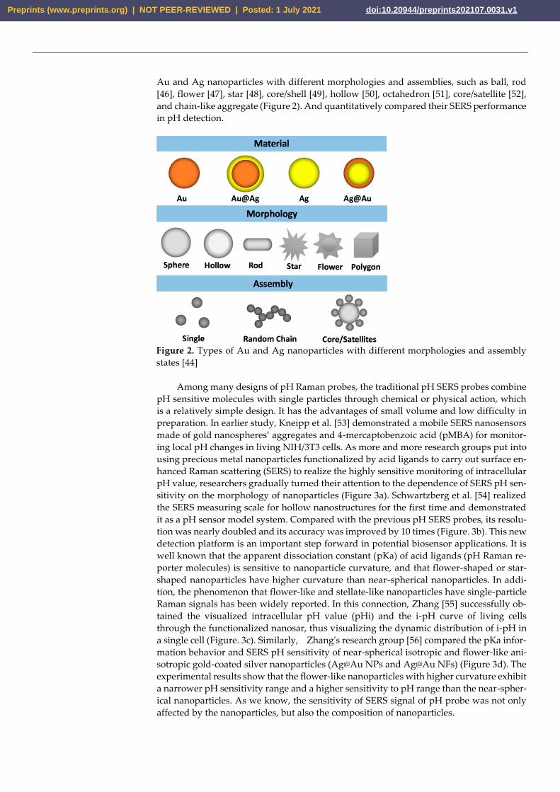
Au and Ag nanoparticles with different morphologies and assemblies, such as ball, rod
[46], flower [47], star [48], core/shell [49], hollow [50], octahedron [51], core/satellite [52],
and chain-like aggregate (Figure 2). And quantitatively compared their SERS performance
in pH detection.
Figure 2. Types of Au and Ag nanoparticles with different morphologies and assembly
states [44]
Among many designs of pH Raman probes, the traditional pH SERS probes combine
pH sensitive molecules with single particles through chemical or physical action, which
is a relatively simple design. It has the advantages of small volume and low difficulty in
preparation. In earlier study, Kneipp et al. [53] demonstrated a mobile SERS nanosensors
made of gold nanospheres’ aggregates and 4-mercaptobenzoic acid (pMBA) for monitor-
ing local pH changes in living NIH/3T3 cells. As more and more research groups put into
using precious metal nanoparticles functionalized by acid ligands to carry out surface en-
hanced Raman scattering (SERS) to realize the highly sensitive monitoring of intracellular
pH value, researchers gradually turned their attention to the dependence of SERS pH sen-
sitivity on the morphology of nanoparticles (Figure 3a). Schwartzberg et al. [54] realized
the SERS measuring scale for hollow nanostructures for the first time and demonstrated
it as a pH sensor model system. Compared with the previous pH SERS probes, its resolu-
tion was nearly doubled and its accuracy was improved by 10 times (Figure. 3b). This new
detection platform is an important step forward in potential biosensor applications. It is
well known that the apparent dissociation constant (pKa) of acid ligands (pH Raman re-
porter molecules) is sensitive to nanoparticle curvature, and that flower-shaped or star-
shaped nanoparticles have higher curvature than near-spherical nanoparticles. In addi-
tion, the phenomenon that flower-like and stellate-like nanoparticles have single-particle
Raman signals has been widely reported. In this connection, Zhang [55] successfully ob-
tained the visualized intracellular pH value (pHi) and the i-pH curve of living cells
through the functionalized nanosar, thus visualizing the dynamic distribution of i-pH in
a single cell (Figure. 3c). Similarly, Zhang's research group [56] compared the pKa infor-
mation behavior and SERS pH sensitivity of near-spherical isotropic and flower-like ani-
sotropic gold-coated silver nanoparticles (Ag@Au NPs and Ag@Au NFs) (Figure 3d). The
experimental results show that the flower-like nanoparticles with higher curvature exhibit
a narrower pH sensitivity range and a higher sensitivity to pH range than the near-spher-
ical nanoparticles. As we know, the sensitivity of SERS signal of pH probe was not only
affected by the nanoparticles, but also the composition of nanoparticles.
Preprints (www.preprints.org) | NOT PEER-REVIEWED | Posted: 1 July 2021 doi:10.20944/preprints202107.0031.v1

Traditional single particle pH sensors based on surface enhanced Raman scattering
(SERS) often rely on the physical enhancement effect generated by the aggregation of na-
noparticles, but the aggregation degree of nanoparticles is difficult to be accurately con-
trolled, which leads to poor repeatability of SERS signals of this kind of pH sensors. There-
fore, many research groups designed a series of array platforms for the construction of pH
SERS probes with good repeatability and stability on this basis. Dzięcielewski et al. [57]
based on chemical etching of GaN crystals developed a substrate similar to " nanowhisk-
ers " array for the construction of pH SERS probe(Figure. 3e). However, the array structure
has an obvious disadvantage. Compared with the single particle pH SERS probe, its size
is often much larger, which makes it an obstacle to enter the cell to detect pH. Similarly,
Zhao's group [58] successfully developed a pH sensor using carbon nanotubes modified
with bimetallic nanoparticles (Figure. 3f). A carbon nanotube can continuously measure
pH values in its vicinity over a range of 5.6 to 8.2 pH units. As we all know, the size of the
dimer is often much smaller than that of the array structure, so on this basis, Pallaoro et
al. [59] used hexanediamine (HMD) to connect two silver nanospheres functionalized by
pH-sensitive molecule 4-mercaptobenzoic acid together. The addition of the protective
agent polyvinylpyrrolidone (PVP) could well prevent the further polymerization of nano-
particles, which made the SERS probe of dimer pH successfully prepared (Figure 3i). Chen
et al. [60] systematically studied the formation and evolution of dynamic surface en-
hanced Raman scattering (SERS) hot spot, and on this basis, they prepared nano gold sup-
ported pH responsive poly (2-vinylpyridine) (AuNPs/P2VP) composite microgel through
in situ chemical reduction method (Figure. 3g). What is interesting about the pH response
mechanism of such particles is that when the pH value of the external environment was
from 2.0 to 5.0, the nanometer gap between AuNPs could be easily adjusted, creating a
huge SERS hot spot for electromagnetic enhancement.
Traditional pH SERS probes tend to generate different degrees of aggregation in high
ionic strength medium and complex biological system, and the probe molecules on it may
also interact with biological molecules. As a result, SERS detection results were not relia-
ble enough and biocompatibility was low, which made it difficult for traditional pH SERS
probes to be used for intracellular pH detection. In order to overcome this interference,
the strategy often used by researchers is to modify a layer of bovine serum protein (BSA)
or other substances on the surface of the designed pH SERS probes to increase the bio-
compatibility and stability of the pH probes, so that the pH SERS probes can be used for
cell pH monitoring accurately and sensitively. Based on this, Zheng's research group [61]
also prepared a 4-mercaptopyridine functionalized gold nanospheres modified with bo-
vine serum protein (BSA) as pH SERS probe (Figure. 3i), which was used to monitor the
pH distribution of living cells. It was found that the nanoparticles modified with bovine
serum protein (BSA) had high sensitivity to pH value, good biocompatibility and stability.
Bi et al. [62] prepared a gold nanoarray platform with good SERS sensitivity by adopting
the efficient evaporation self-assembly method, and on this basis developed a pH sensor
based on SERS with high sensitivity bovine serum protein (BSA) coated 4-mercapto-
pyridine (4-Mpy) connected gold nanorod array (Figure. 3j). It was successfully used to
detect pH in the blood of C57BL/6 mice, providing a favorable platform for the develop-
ment of a sensitive bioon chip platform designed for the point of care device. In a different
way, Wang [63] encapsulated the constructed silver nano pH SERS probe in the silicon
layer (Figure. 3k), whose small hole could block the large biomolecules outside the sensor
without preventing H+ from entering into it and interacting with 4-mercaptobenzoic acid
on the silver surface. The stability, pH sensitivity and reliability of the probe are main-
tained. Bi et al. [64] developed a simple, sensitive and stable Prussian blue (PB) cage pH
response SERS probe (Figure. 3l), which was used to quantify the dynamic change of pH
(pHi) in the live fine. Compared with other pH SERS probes, the subnanometer porous
PB shells in the structure could allow the free diffusion reaction of H+ or OH- while block-
ing the contact between proteins, DNA and other biological sulfhydryl compounds in the
cell. In addition, the C-N triple bond group of nitrile in PB shell can be used as an internal
Preprints (www.preprints.org) | NOT PEER-REVIEWED | Posted: 1 July 2021 doi:10.20944/preprints202107.0031.v1

standard without background interference to accurately analyze the distribution of probes
in the whole cell.
Figure 3. Some examples of traditional single particles pH SERS probes (top panel),
assembled pH SERS probe (middle panel) and encapsulated pH SERS probe (bottom
panel). (a) Schematic diagram of pMBA modified gold nanospheres pH SERS
probe [53]. (b) TEM of hollow gold nanospheres [54]. (c) Schematic diagram of prepar-
ing gold nanostar pH SERS probe [55]. (d) Schematic diagram of preparing
Ag@Au nanoflowers pH SERS probe [56]. (e) TEM of Au-sputtered GaN nanowhisk-
ers array [57]. (f) Schematic diagram of Ag@Au bimetallic nanoparticles modified carbon
nanotubes [58]. (g) Schematic diagram of silver nanospheres dimer [59]. (h) Schematic di-
agram of gold nanospheres supported poly (2-vinylpyridine) composite microgel [60]. (i)
Schematic diagram of bovine serum protein (BSA) modified 4-Mpy functionalized gold
nanorods [61]. (j) Schematic diagram of serum protein (BSA) coated 4-MPY connected
gold nanorod array [62]. (k) Schematic diagram of silicon encapsulated silver nanoparti-
cles with stable pH response [63]. (l) Schematic diagram of Prussian blue (PB) cage gold-
coated nanorods with stable pH response [64].
5. pH SERS probe was used for cell pH value monitoring and to pH imaging
The pH near the cell surface is called extracellular pH (pHe). Extracellular pH (pHe)
and intracellular pH (pHi) are not only the important regulatory factors affecting a variety
of physiological activities of cells (such as cell proliferation, differentiation and apoptosis,
ion transport, endocytosis, muscle contraction, nucleic acid formation and metabolite re-
lease). It is also one of the important parameters for the study of related pathological pro-
cesses, so the acids and bases inside and outside the cell play an important role in chemical
reactions and biological processes [66, 67]. Abnormal pHe values are associated with var-
ious pathological states, such as tumor, iron-deficiency stroke, infection, and inflamma-
tion [68]. Similarly, small changes in pHi can lead to major changes in metabolism that
can lead to disease. It is worth noting that extracellular pH (pHe) is often used as an im-
portant marker of cancer. This is mainly due to the Warburg effect and the action of car-
bonic anhydrase on the cell surface, tumor cells will produce a large amount of extracel-
lular acid, which leads to a slightly lower pHe (6.2-6.9) of tumor cells than pHi (7.2-7.4)
[65, 69] (Figure 4). In addition to cancer, there are other diseases that are closely related to
pHe and pHi abnormalities in cells, such as quite a number of neurological diseases [70].
Due to the abnormal cell pH (pHe and pHi) in these diseases, the phenomenon of abnor-
mal cell pH has attracted extensive attention in clinical medicine and researchers. There-
fore, accurate and sensitive in monitoring and intuitive imaging characterization of pHe
Preprints (www.preprints.org) | NOT PEER-REVIEWED | Posted: 1 July 2021 doi:10.20944/preprints202107.0031.v1

and pHi are very worthy of expectation for the pathological research and the development
of new treatment methods of these diseases.
Figure 4. Schematic of reversal changes in extracellular and intracellular pH values of
cancer cells compared with normal cells [65]
The perception of pH value of living cells is of great significance for understanding
various physiological and pathological processes[72, 73]. As an ultrasensitive and nonde-
structive spectroscopic technology, surface enhanced Raman spectroscopy (SERS) has
been widely used in the monitoring and imaging studies of cell pH value [74, 75]. How-
ever, there are still three major problems to be overcome when applying pH SERS probe
to cell pH value detection and imaging: (1) In the cellular system, the selected peak used
to detect the pH response of the overall buffer solution is still reliable; (2)Because the ag-
gregation trend of nanoparticles after being absorbed by cells would increase [76], which
would affect the reliability of pH SERS probe monitoring results;(3) Whether a variety of
biomolecules existing in the living cell system can replace or interact with Raman probes
or probe molecules, and whether pH SERS probe can remain intact, will lead to changes
in the stability and reliability of probes [61]. Sun et al. [37] immobilized 4-MBA on Au
Quasi-3D Plasma Nanostructure Array (Q3D-PNA) through the chemical action of Au-S
bond, and constructed a pH probe based on surface enhanced Raman spectroscopy
(SERS), which has high sensitivity and repeatability. It was successfully applied to draw
pHe map of living cells (Figure. 5a). Xu et al. [34] used 4-mercolbenzoic acid (4-MBA)
functionalized gold nanoparticle array as substrate for pH imaging of three different types
of cells (Figure. 5b). Guo et al. [77] developed a nano-microtube with size less than 200nm
and SERS activity for detecting intracellular pH value (pHi) of a single eukaryotic cell. Li
et al. [71] prepared a simple plasma Raman probe (SMB) using nano stars as a Raman
enhanced substrate, 4-Mpy as a Raman reporter molecule, and bovine serum albumin
(BSA) as a protective molecule. Subsequently, SERS imaging technology was used to mon-
itor the changes of lysosomal pH in the process of autophagy and apoptosis. According
to SERS imaging, the pH value of each pixel was given, so that the pHi distribution map
could be drawn (Figure. 5c).
Preprints (www.preprints.org) | NOT PEER-REVIEWED | Posted: 1 July 2021 doi:10.20944/preprints202107.0031.v1

Figure 5. (a) Optical images and pHe SERS map of NIH/3T3 and HepG2 cells after cul-
ture on 4-MBA modified Q3D-PNA SERS substrates [37] (b) Bright field microscope im-
age(upper panel) and real-time in situ pHe images of a living cell (lower panel) [34] (c)
In situ SERS imaging and schematic of pHi during autophagy and apoptosis of lyso-
somes [71]
6. pH SERS probe combined with other technologies
In numerous pH imaging fluorescence, because its cost is relatively low and good
security in vitro research, it is widely used in cell pH monitoring imaging[78, 79]. How-
ever, in cell pH detection and imaging, fluorescence spectrum technology has defects such
as easy quenching, photobleaching, large background interference and slow imaging,
while SERS spectrum technology did not have these problems. Based on this, Yang et al.
[80] constructed a nano-probe with dual signals of fluorescence and surface enhanced Ra-
man scattering (SERS) in recent years to sense the pH value in cells (Figure. 6a). In this
study, they designed a double-signal core-shell nanoprobe with fluorescence and SERS by
co-functionalization of SiO2-coated gold nanorods by using fluorescein isothiocyanate
(FITC) and p-mercaptobenzoic acid (pMBA). Then through the study and analysis of the
fluorescence and SERS spectra of the nano-probes at different pH values, the double-sig-
nal response to the intracellular pH value could be realized. Different from the former,
Yue et al. [81] prepared a new generation of fluorescence-SERS dual-mode pH sensors by
embed functionalized nanoparticles into stimulus-responding hydrogels. Such sensors
were equipped with synergistic enhancement of the properties of each component: im-
proving the mechanical strength of hydrogels and reducing nanoparticle aggregation
(Figure. 6b). The complementary advantages of fluorescence and SERS in response to pH
made it successfully applied to study the changes of pH value in microenvironment
caused by external stimulation of different cells. Yue's research group [82] also success-
fully developed a nanosensor based on fluorescence imaging and SERS spectrum in recent
two years to monitor the dynamic changes of pH in mitochondrial microenvironment of
two kinds of cancer cells (HepG2 and MCF-7 cells) and one kind of normal cells (LO2
cells) during the process of PDT (Figure. 6d). This study also provides a reference for eval-
uating the side effects of PDT.
In addition to enhancing the reliability of pH sensors by combining with fluorescence
spectroscopy technology, simple, convenient, fast and sensitive electrochemistry [85] has
also gradually entered the field of view of researchers. Pan's research group [83] designed
a surface enhanced Raman scattering (SERS) active microneedle to detect redox potential
and pH value at rat joints. The design of composite SERS probe was based on the mi-
croneedles used in traditional Chinese medicine acupuncture and moxibustion. The
grooves formed by chemical etching on the microneedles contain two kinds of molecules,
namely redox sensitive and pH sensitive, respectively, and then enter the muscle at the
minimum cost of damage, making the muscle undergo a redox state and pH dynamic
Preprints (www.preprints.org) | NOT PEER-REVIEWED | Posted: 1 July 2021 doi:10.20944/preprints202107.0031.v1

evolution for 5 minutes (Figure. 6c). This has advanced the pathological study of diseases
such as arthritis, and will also be a multifunctional analytical tool to advance biomedical
research. Many research groups have made many attempts and efforts to combine pH
detection with biological diagnosis and treatment. Jung et al. [84] designed a nano-probe
with both pH response and photothermal therapy(Figure. 6e). Multi-function pH probe
in the golden ball surface is designed to develop a positive charge and negative charge.
Under the action of weak acid, they are induced to gather together rapidly by electrostatic
interaction, while the emergence of coupled plasma model causes their absorption to
transfer to the near-infrared region, where a direct selective absorption transfer to depth
is carried out, through tissue and heat treatment (Figure. 6d).
Figure 6. Some examples of PH SERS probe combined with other technologies.
(a) AuNRs@SiO2@PMA-FITC nanoprobe preparation and its mechanism in
cells [80]. (b)Schematic diagram of pH-induced swell and shrink of single-phase hydrogel
microparticles encapsulating AuNPs-(4MBA)-BSA (upper), single-phase hydrogel micro-
particles encapsulating carbon dots (middle), and Janus hydrogel microparticles encapsu-
lating AuNPs-(4MBA)-BSA and carbon dots respectively in duals semispheres (lower)
[81]. (c) Schematic diagram of multiplexing SERS active microneedles for simultaneous
measurement of redox potential and pH value [83]. (d) Procedure of the selective deter-
minations of microenvironmental changes in mitochondria during PDT progress [83]. (e)
Schematic diagram of the working mechanism of 4-MBA-functionalized smart nanoparti-
cles in cancer cells [84].
7. Conclusion
High sensitivity of SERS, excellent multiplexing ability of a single excitation source,
low background and minimal photobleaching of SERS nanotags lead to its application in
cell pH detection imaging technology becoming more and more widely. At present, the
main problems of developing pH SERS probes that can be used for cell pH detection and
imaging focus on ensuring that they can maintain good detection ability and avoid the
interference of microenvironment, which requires researchers to design and construct
well-defined, uniform and stable metallic nanostructures and assemblies. In addition, the
synthesis and discovery of pH-sensitive Raman reporter molecules with highly stable and
repeatable signals could be an important step forward in this field. In recent years, the pH
SERS probes reported tended to have a wide pH response range, so the subsequent pH
SERS probes should develop towards a narrower response range and higher sensitivity,
so that they could be better applied in biological systems. In addition, it is also very im-
portant to further develop highly selective and non-toxic probe structures. We believe that
Preprints (www.preprints.org) | NOT PEER-REVIEWED | Posted: 1 July 2021 doi:10.20944/preprints202107.0031.v1

the additional complementary information obtained by combining with other different
independent methods can improve the reliability of probe signals, or become a multi-
functional intelligent probe so that pH SERS probes can be better applied in the biomedi-
cal field.
By summarizing and classifying the latest and most representative research reports
on pH SERS probes, this paper mainly reviewed the design and assembly strategies of
several major types of pH probes and their structures in this field, as well as the latest
progress in pH imaging in and out of cells. At present, the purpose of the research groups
to study pH SERS probe is mainly to make the signal stable and reliable, and to make the
imaging fast and large. In addition, in the construction of probes for cell pH monitoring
and SERS imaging, the design of protective matrix and the enhancement of biocompati-
bility of nanometer tags also need the efforts of future researchers.
References
1. Pezzulo, A.A.; Tang, X.;X.; Hoegger, M.J; Abou Alaiwa, M.H.; Ramachandran, S. ; Moninger, T.O.; Karp, P.H.;
Wohlford-Lenane, C.L.; Haagsman, H.P.; Martin van Eijk; et al. Reduced airway surface pH impairs bacterial
killing in the porcine cystic fibrosis lung. Nature 2012, 487, 109-113.
2. Malek, K.; Jaworska, A.; Krala, P.; Kachamakova-Trojanowska, N.; Baranska, M. Imaging of macrophages by
surface enhanced Raman spectroscopy (SERS). Bio. Spectrosc. Imaging 2013, 2, 349-357.
3. Zhang, W.; Jiang, L.; Piper, J.A.; Y., Wang. SERS nanotags and their applications in biosensing and bioimaging.
J. Anal. Test. 2018, 2, 26-44.
4. Ma, D.; Zheng, J.; Tang,P.; Xu, W.; Qing, Z.; Yang, S.; Li, J.; Yang R. Quantitative monitoring of hypoxia-Induced
intracellular acidification in lung tumor cells and tissues using activatable surface-enhanced Raman scattering
nanoprobes. Anal. Chem. 2016, 88, 11852-11859.
5. Talley, C.E.; Jusinski, L.; Hollars, C.W.; Lane, S.M.; Huser, T. Intracellular pH sensors based on surface-enhanced
Raman scattering. Anal. Chem. 2004, 7064-7068.
6. Fleischmann, M.; Hendra, P.J.; McQuillan, A.J. Raman spectra of pyridine adsorbed at a silver electrode. Chem.
Phys. Lett. 1974, 26, 163-166.
7. Kwart, H.; George, T.J. Anomalously intense Raman spectra of pyridine at a silver electrode. J. Am. Chem. Soc.
1977, 99, 5215–5217.
8. Moskovits, M. Surface roughness and the enhanced intensity of Raman scattering by molecules adsorbed on
metals. J. Chem. Phys. 1978, 69, 4159-4161.
9. Qiu, C.; Cheng, Z.; Lv,C.; Wang, R.; Yu, F. Development of bioorthogonal SERS imaging probe in biological
and biomedical applications. Chin. Chem. Lett. 2021.(accepted)
10. Ji, W.; Spegazzini, N.; Kitahama, Y.; Chen, Y.; Zhao, B.; Ozaki, Y. pH-response mechanism of
p-aminobenzenethiol on Ag nanoparticles revealed by two-dimensional correlation surface enhanced Raman
scattering spectroscopy. J. Phys. Chem. Lett. 2012, 3, 3204-3209.
11. Langer, J.; Jimenez de Aberasturi, D.; Aizpurua, J.; Alvarez-Puebla, R.A.; Auguie, B.; Baumberg, J.J.; Bazan, G.C.;
Bell, S. E.J.; Boisen, A.; Brolo, A.G.; et al. Present and future of surface-enhanced Raman scattering. ACS nano
2020, 14, 28-117.
12. Swietach, P. What is pH regulation, and why do cancer cells need it? Cancer Metastasis Rev. 2019, 38, 5-15.
13. Tatapudy, S.; Aloisio, F.; Barber, D.; Nystul, T. Cell fate decisions: emerging roles for metabolic signals and cell
morphology. EMBO Rep. 2017, 18, 2105-2118.
14. Schotthofer, S.K.; Bohrmann, J. Bioelectrical and cytoskeletal patterns correlate with altered axial polarity in the
follicular epithelium of the drosophila mutant gurken. BMC Dev. Biol. 2020, 20, 5.
15. Miller, S.; Ptok, M.; Jungheim, M. Influence of acid swallows on the dynamics of the upper esophageal sphincter.
Preprints (www.preprints.org) | NOT PEER-REVIEWED | Posted: 1 July 2021 doi:10.20944/preprints202107.0031.v1

Dysphagia 2021, 36, 443-455.
16. Park, J.; Tabet, A.; Moon, J.; Chiang, P.H.; Koehler, F.; Sahasrabudhe, A.; Anikeeva, P. Remotely controlled
proton generation for neuromodulation. Nano Lett. 2020, 20, 6535-6541.
17. Zong, S.; Wang, Z.; Yang, J.; Cui, Y. Intracellular pH sensing using p-aminothiophenol functionalized gold
nanorods with low cytotoxicity. Anal. Chem. 2011, 83, 4178-4183.
18. Piotrowski, Piotr; Bukowska, Jolanta. Ion permeation through silica coating of silver nanoparticles
functionalized with 2-mercaptoethanesulfonate anions: SiO2-encapsulated SERS probes for metal cations. J.
Phys. Chem. C 2016, 120, 12092-12099.
19. Fang, B.; Wang, D.; Huang, M.; Yu, G.; Li, H. Hypothesis on the relationship between the change in intracellular
pH and incidence of sporadic Alzheimer's disease or vascular dementia. Int. J. Neurosci. 2010, 120, 591-595.
20. Wang, Y.; Yan, B.; Chen, L. SERS tags: novel optical nanoprobes for bioanalysis. Chem. Rev. 2012, 113, 1391-1428.
21. Xie, M.; Li3, F.; Gu, P.; Wang, F.; Qu, Z.; Li, J.; Wang, L.; Zuo, X.; Zhang, X.; Shen, J. Gold nanoflower-based
surface-enhanced Raman probes for pH mapping of tumor cell microenviroment. Cell Proliferation 2019, 52,
e12618.
22. Potara, M.; Nagy-Simon, T.; Craciun, A.M.; Suarasan, S.; Licarete, E.; Imre-Lucaci, F.; Astilean, S. Carboplatin-
loaded, Raman-encoded, chitosan-coated Silver nanotriangles as multimodal traceable nanotherapeutic
delivery systems and pH reporters inside human ovarian cancer cells. ACS Appl. Mater. Interfaces 2017, 9, 32565-
32576.
23. Johnson, R.P.; Richardson, J.A.; Brown, T.; Bartlett, P.N. Real-time surface-enhanced Raman spectroscopy
monitoring of surface pH during electrochemical melting of double-stranded DNA. Langmuir 2012, 28, 5464-
5470.
24. Scott, B.L; Carron, K.T. Dynamic Raman scattering studies of coated gold nanoparticles: 4-mercaptopyridine,
4-mercaptophenol, and benzenethiol. J. Phys. Chem. C 2016, 120, 20905-20913.
25. Chao, Y.; Zhou, Q.; Li, Y.; Yan, Y.; Wu, Y.; Zheng, J. Potential dependent surface-enhanced Raman scattering of
4-mercaptopyridine on electrochemically roughened silver electrodes. J. Phys. Chem. C 2007, 111, 16990-16995.
26. Zhang, H.; He, H.X.; Wang, J.; Liu, Z.F. Atomic force microscopy evidence of citrate displacement by 4-
mercaptopyridine on gold in aqueous solution. Langmuir 2000, 16, 4554-4557.
27. Pienpinijtham, P.; Vantasin, S.; Kitahama, Y.; Ekgasit, S.; Ozaki, Y. Nanoscale pH profile at a solution/solid
interface by chemically modified tip-enhanced Raman scattering. J. Phys. Chem. C 2016, 120, 14663-14668.
28. Kim, K.; Kim, K.L. ; Shin, D. ; Choi, J.Y. ; Shin, K.S. Surface-enhanced Raman scattering of 4-aminobenzenethiol
on Ag and Au: pH dependence of b2-type bands. J. Phys. Chem. C 2012, 116, 4774-4779.
29. Huang, Y.; Liu, W.; Wang , D.; Gong, Z.; Fan, M. Evaluation of the intrinsic pH sensing performance of surface-
enhanced Raman scattering pH probes. Microchem. J. 2020, 154,
30. Ma, C.; Harris, J.M. Surface-enhanced Raman spectroscopy investigation of the potential-dependent acid-base
chemistry of silver-immobilized 2-mercaptobenzoic acid. Langmuir 2011, 27, 3527-3533.
31. Capocefalo, A.; Mammucari, D.; Brasili, F.; Fasolato, C.; Bordi, F.; Postorino, P.; Domenici, F. Exploring the
potentiality of a SERS-active pH nano-biosensor. Front Chem. 2019, 7, 413.
32. Bai, L.; Wang, X.; Zhang, K.; Tan, X.; Zhang, Y.; Xie, W. Etchable SERS nanosensor for accurate pH and hydrogen
peroxide sensing in living cells. Chem. Commun. 2019, 55, 12996-12999.
33. Park, J.E.; Yonet-Tanyeri, N.; Ende, E.V. ; Henry, A.I.; Perez White, B.E.; Mrksich, M. ; Van Duyne, R. P. Plasmonic
microneedle arrays for in situ sensing with surface enhanced Raman spectroscopy (SERS). Nano Lett. 2019, 19,
6862-6868.
34. Xu, M.; Ma, X.; Wei, T.; Lu, Z.X.; Ren, B. In situ imaging of live-cell extracellular pH during cell apoptosis with
Preprints (www.preprints.org) | NOT PEER-REVIEWED | Posted: 1 July 2021 doi:10.20944/preprints202107.0031.v1

surface-enhanced Raman spectroscopy. Anal. Chem. 2018, 90, 13922-13928.
35. Zhao, X.; Campbell, S.; Wallace, G.Q.; Claing, A.; Bazuin, G.; Masson, J. Branched Au nanoparticles on
nanofibers for surface-enhanced Raman scattering sensing of intracellular pH and extracellular pH gradients.
ACS Sens. 2020, 5, 2155−2167.
36. Scarpitti, B.T.; Morrison, A.M.; Buyanova, M.; Schultz, Z.D. Comparison of 4-mercaptobenzoic acid surface-
enhanced Raman spectroscopy-based methods for pH determination in cells. Appl. Spectrosc. 2020, 74, 1423-1432
37. Sun, F.; Zhang, P.; Bai, T.; Galvan, D.D.; Hung, H.C.; Zhou, N.; Jiang, S.; Yu, Q. . Functionalized plasmonic
nanostructure arrays for direct and accurate mapping extracellular pH of living cells in complex media using
SERS. Biosens. Bioelectron. 2015, 73, 202-207.
38. Shen, Y.; Liang, L.; Zhang, S.; Huang, D.; Zhang, J.; Xu, S.; Liang, C.; Xu, W. Organelle-targeting surface-
enhanced Raman scattering (SERS) nanosensors for subcellular pH sensing. Nanoscale 2018, 10, 1622-1630.
39. Yang, T.; Ma, J.; Zhen, S.J.; Huang, C.Z. Electrostatic assemblies of well-dispersed AgNPs on the surface of
electrospun nanofibers as highly active SERS substrates for wide-range pH sensing. ACS Appl. Mater. Interfaces
2016, 8, 14802-14811.
40. Chen, P.; Wang, Z.; Zong, S.; Chen, H.; Zhu, D.; Zhong, Y.; Cui, Y. A wide range optical pH sensor for living
cells using Au@Ag nanoparticles functionalized carbon nanotubes based on SERS signals. Anal. Bioanal. Chem.
2014, 406, 6337-6346.
41. Lawson, L.; Huser, T. Synthesis and characterization of a disulfide reporter molecule for enhancing pH
measurements based on surface-enhanced Raman scattering. Anal. Chem. 2012, 3574-3580.
42. Tercio de F. Paulo, † Ro mulo A. Ando, † Izaura C. N. Diogenes, and Marcia L. A. Temperini. Understanding the
equilibria of thio compounds adsorbed on gold by surface-enhanced Raman scattering and density functional
theory calculations. J. Phys. Chem. C 2013, 117, 6275-6283.
43. Kong, K.V.; Dinish, U.S.; On Lau, W.K.; Olivo, M. Sensitive SERS-pH sensing in biological media using metal
carbonyl functionalized planar substrates. Biosens. Bioelectron. 2014, 54, 135-140.
44. Zhang, Z.; Bando, K.; Mochizuki, K.; Taguchi, A.; Fujita, K.; Kawata, S. Quantitative evaluation of surface-
enhanced Raman scattering nanoparticles for intracellular pH sensing at a single particle level. Anal. Chem. 2019,
91, 3254-3262.
45. Gao, C.; Hu, Y.; Wang, M.; Chi, M.; Yin, Y. Fully alloyed Ag/Au nanospheres: combining the plasmonic property
of Ag with the stability of Au. J. Am. Chem. Soc. 2014, 136, 7474-7479.
46. Khlebtsov, B.N.; Khanadeev, V.A.; Burov, A.M.; Le Ru, E.C.; Khlebtsov, N.G. Reexamination of surface-
enhanced Raman scattering from gold nanorods as a function of aspect ratio and shape. J. Phys. Chem. C 2020,
124, 10647-10658.
47. Xie, J.; Zhang, Q.; Lee, J.Y.; Wang, D.L.C. The synthesis of SERS-active gold nanoflower tags for in vivo
applications. ACS nano 2008, 2, 2473-2480.
48. Jimenez de Aberasturi, D.; Serrano-Montes, A.B.; Langer, J.; Henriksen-Lacey, M.; Parak, W.J.; Liz-Marzán, L M.
Surface enhanced Raman scattering encoded gold nanostars for multiplexed cell discrimination. Chem. Mater.
2016, 28, 6779-6790.
49. Zhao, J.; Zhang, K.; Li, Y.; Ji, J.; Liu, B. High-resolution and universal visualization of latent fingerprints based
on aptamer-functionalized core-shell nanoparticles with embedded SERS reporters. ACS Appl. Mater. Interfaces
2016, 8, 14389-14395.
50. Sanles-Sobrido, M.; Exner, W.; Rodríguez-Lorenzo, L.R.; Rodríguez-González, B.; Correa-Duarte, M.A.;
Álvarez-Puebla, R.A.; Liz-Marzán, L.M. Design of SERS-encoded, submicron, hollow particles through
confined growth of encapsulated metal nanoparticles. J. Am. Chem. Soc. 2009, 131, 2699-2705.
Preprints (www.preprints.org) | NOT PEER-REVIEWED | Posted: 1 July 2021 doi:10.20944/preprints202107.0031.v1

51. Chang, C.C.; Wu, H.L.; Kuo, C..H.; Huang, M.H. Hydrothermal synthesis of monodispersed octahedral gold
nanocrystals with five different size ranges and their self-assembled structures. Chem. Mater. 2008, 20, 7570-7574.
52. Rong, Z.; Xiao, R.; Wang, C.; Wang, D.; Wang, S. Plasmonic Ag core-satellite nanostructures with a tunable silica-
spaced nanogap for surface-enhanced Raman scattering. Langmuir 2015, 31, 8129-8137.
53. Kneipp, J.; Kneipp, H.; Wittig,B.; Kneipp, K. Following the dynamics of pH in endosomes of live cells with SERS
nanosensors. J. Phys. Chem. C 2010, 114, 7421–7426.
54. Schwartzberg, A.M.; Oshiro, T.Y.; Zhang, J.Z.; Huser, T.; Talley, C.E. Improving nanoprobes using surface-
enhanced Raman scattering from 30-nm hollow gold particles. Anal. Chem. 2006, 4732-4736.
55. Zhang, Y.; Aberasturi, D.J.; Henriksen-Lacey, M.; Langer, J.; Liz-Marzán, L.M. Live-cell surface-enhanced
Raman spectroscopy imaging of intracellular pH: from two dimensions to three dimensions. ACS Sens. 2020, 5,
3194-3206.
56. Zhang, Q.; Wen, H.; Watanabe, K.; Kotani, I.; Ricci, M.; Fortuni, B.; Dao, A.T.N.; Masuhara, A.; Hirai, K.; Kasai,
H. Low-cytotoxic gold-coated silver nanoflowers for intracellular pH sensing. ACS Appl. Nano Mater. 2020, 3,
7643-7650.
57. Dzięcielewski, I. ; Krajczewski, J.; Dzwolak, W. pH-responsive mixed-thiol-modified surface of roughened GaN:
A wettability and SERS study. Appl. Surf. Sci. 2020, 502,
58. Zhao, L.; Shingaya, Y.; Tomimoto, H.; Huang, Q.; Nakayama, T. Functionalized carbon nanotubes for pH
sensors based on SERS. J. Mater. Chem. C 2008, 18, 4759-4761.
59. Pallaoro, A.; Braun, G.B.; Reich, N.O.; Moskovits, M. Mapping local pH in live cells using encapsulated
fluorescent SERS nanotags. Small 2010, 5, 618-622.
60. Chen, H.; You, T.; Jiang, L.; Gaoa, L.; Yin, P. Creating dynamic SERS hotspots on the surface of pH-responsive
microgels for direct detection of crystal violet in solution. RSC Adv. 2017, 7, 32743-32748.
61. Zheng, X.S.; Hu, P.; Cui, Y.; Zong, C.; Feng, J.M.; Wang, X.; Ren, B. BSA-coated nanoparticles for improved
SERS-based intracellular pH sensing. Anal. Chem. 2014, 86, 12250-12257.
62. Bi, L.; Wang, Y.; Yang, Y.; Li, Y.; Mo, S.; Zheng, Q.; Chen, L. Highly sensitive and reproducible SERS sensor for
biological pH detection based on a uniform gold nanorod array platform. ACS Appl. Mater. Interfaces 2018, 10,
15381-15387.
63. Wang, F.; Widejko, R.G.; Yang, Z.; Nguyen, K.T.; Chen, H.; Fernando, L.P.; Christensen, K.A.; Anker, J.N.
Surface-enhanced Raman scattering detection of pH with silica-encapsulated 4-mercaptobenzoic acid-
functionalized silver nanoparticles. Anal. Chem. 2012, 84, 8013-8019.
64. Bi, Y.; Di, H.; Zeng, E.; Li, Q.; Li, W.; Yang, J.; Liu, D. Reliable quantification of pH variation in live cells using
prussian blue-caged surface-enhanced Raman scattering probes. Anal. Chem. 2020, 92, 9574-9582.
65. Damaghi, M.; Wojtkowiak, J. W.; Gillies, R. J. pH sensing and regulation in cancer. Front Physiol. 2013, 4, 1-10.
66. Lardner, A. The effects of extracellular pH on immune function. J. Leukocyte Biol. 2001, 69, 522-530.
67. Jaworska, A.; Malek, K.; Kudelski, A. Intracellular pH-Advantages and pitfalls of surface-enhanced Raman
scattering and fluorescence microscopy -A review. Spectrochim. Acta Part A 2021, 251, 119410.
68. Liu, L.; Dou, C.X.; Liu, J.W.; Wang, X.N.; Ying, Z.M.; Jiang, J.H. Cell surface-anchored DNA nanomachine for
dynamically tunable sensing and imaging of extracellular pH. Anal. Chem. 2018, 90, 11198-11202.
69. Hashim, A.I.; Zhang, X.; Wojtkowiak, J.W.; Martinez, G.V.; Gillies, R.J. Imaging pH and metastasis. NMR Biomed.
2011, 24, 582-591.
70. Munteanu, R.E.; Stanica, L.; Gheorghiu, M.; Gaspar, S. Measurement of the extracellular pH of adherently
growing mammalian cells with high spatial resolution using a voltammetric pH microsensor. Anal. Chem. 2018,
90, 6899-6905.
Preprints (www.preprints.org) | NOT PEER-REVIEWED | Posted: 1 July 2021 doi:10.20944/preprints202107.0031.v1

71. Li, S.S.; Zhang, M.; Wang, J.H.; Yang, F.; Kang, B.; Xu, J.J.; Chen, H.Y. Monitoring the changes of pH in lysosomes
during autophagy and apoptosis by plasmon enhanced Raman imaging. Anal. Chem. 2019, 91, 8398-8405.
72. Lee, Y.J.; Kang, H.C.; Hu, J.; Nichols, J.W.; Jeon, Y.S.; Bae, Y.H. pH-sensitive polymeric micelle-based pH probe
for detecting and imaging acidic biological environments. Biomacromolecules 2012, 13, 2945-2951.
73. Lin, M.T.; Beal, M.F. Mitochondrial dysfunction and oxidative stress in neurodegenerative diseases. Nature 2006,
443, 787-795.
74. Dey, S.; Trau, M.; Koo, K. M. Surface-enhanced Raman spectroscopy for cancer immunotherapy applications:
opportunities, challenges, and current progress in nanomaterial strategies. Nanomaterials 2020, 10, 1145-1160.
75. Zhang, Y.; Mi, X.; Tan, X.; Xiang, R. Recent progress on liquid biopsy analysis using surface-enhanced Raman
spectroscopy. Theranostics 2019, 9, 491-525.
76. Kneipp, J.; Kneipp, H.; McLaughlin, M.; Brown, D.; Kneipp, K. In vivo molecular probing of cellular
compartments with gold nanoparticles and nanoaggregates. Nano Lett. 2006, 6, 2225-2231.
77. Guo, J.; Sesena Rubfiaro, A.; Lai, Y.; Moscoso, J.; Chen, F.; Liu, Y.; Wang, X.; He, J. Dynamic single-cell
intracellular pH sensing using a SERS-active nanopipette. Analyst 2020, 145, 4852-4859.
78. Dong, B.; Song, X.; Wang, C.; Kong, X.; Tang, Y.; Lin, W. Dual site-controlled and lysosome-targeted
intramolecular charge transfer-photoinduced electron transfer-fluorescence resonance energy transfer
fluorescent probe for monitoring pH changes in living cells. Anal. Chem. 2016, 88, 4085-4091.
79. Yu, F.; Jing, X.; Lin, W. Single-/dual-responsive pH fluorescent probes based on the hybridization of
unconventional fluorescence and fluorophore for imaging lysosomal pH changes in HeLa cells. Anal. Chem.
2019, 91, 15213-15219.
80. Yanga, G.; Zhanga, Q. ; Lianga, Y.; Liub, H.; Qua, L.; Li, H. Fluorescence-SERS dual-signal probes for pH sensing
in live cells. Colloids Surf. A 2019, 562, 289-295.
81. Yue, S.; Sun, X.; Wang, N.; Wang, Y.; Wang, Y.; Xu, Z.; Chen, M.; Wang, J. SERS-fluorescence dual-mode pH-
sensing method based on Janus microparticles. ACS Appl. Mater. Interfaces 2017, 9, 39699-39707.
82. Yue, J.; Shen, Y.; Liang, L.; Cong, L.; Xu, W.; Shi, W.; Chongyang Liang, C.; Xu, S. Revealing mitochondrial
microenvironmental evolution triggered by photodynamic therapy. Anal. Chem. 2020, 92, 6081-6087.
83. Pan, C.; Li, X.; Sun, J.; Li, Z.; Zhang, L.; Qian, W.; Wang, P.; Dong J. A multiplexed SERS-active microneedle for
simultaneous redox potential and pH measurements in rat joints. ACS Appl. Bio Mater. 2019, 2, 2102-2108.
84. Jung, S.; Nam, J.; Hwang, S.; Park, J.; Hur, J.; Im, K.; Park, N.; Kim, S. Theragnostic pH-sensitive gold
nanoparticles for the selective surface enhanced Raman scattering and photothermal cancer therapy. Anal. Chem.
2013, 85, 7674-7681.
85. Wang, H.; Zhang, X.; Wang, S.; Ma, H.; Shen, Y.; Wang, X. A multifunctional electrochemical sensor for the
simultaneous detection of ascorbic acid, dopamine, uric acid, and nitrite. J. AOAC. Int. 2021, 104, 860-866.
Preprints (www.preprints.org) | NOT PEER-REVIEWED | Posted: 1 July 2021 doi:10.20944/preprints202107.0031.v1