review of literature - inflibnetshodhganga.inflibnet.ac.in/bitstream/10603/33688/9/09...the term...
TRANSCRIPT

REVIEW OF LITERATURE
Abdurahiman.T “A study on the efficacy of homoeopathic treatment in hyperlipoproteinemia” Thesis. Department of Life Sciences, University of Calicut , 2005

Chapter - 2
REVIEW OF LITERATURE
2.1 Lipids
Lipids are ubiquitous in the body tissues and have an important role in
virtually all aspects of life - serving as hormones or hormone precursors, aiding in
digestion, providing energy storage and metabolic fuels, acting as functional and
structural components in cell membranes, and forming insulation to allow nerve
conduction or to prevent heat loss
The term lipid applies to a class of compounds that are soluble in organic
solvents and nearly insoluble in water. Chemically, lipids are either compound
that yields fatty acids on hydrolysis or complex alcohols that can combine with
fatty acids to form esters. Some lipids are more complex, containing non lipid
groups such as sialic, phosphoryl, amino or sulphate groups. presence of
these groups gives them the property (amphipathic) of having an affinity for both
water and organic solvents, which is important in the formation of membranes.
2.2 Fatty acids
Fatty acids are one of the simpler forms of lipids. They are generally
indicated by the chemical formula R- COOH, where "R" stands for an alkyl chain.
Fatty acid chain length varies and is commonly classified according to the number
of carbon atom present. They may be short chain, medium chain or long chain
fatty acids. Those of importance in human nutrition and metabolism are of long
chain class containing an even number of carbon atoms.

Fatty acids are further classified according to their degree of saturation.
Saturated ktty acids have no double bonds between carbon atoms,
monounsaturated fatty acids contain one double bond, and polyunsaturated fatty
acids contain more than one double bond. The double bonds in polyunsaturated
fatty acid of both animal and plant origin are usually three carbons apart. The fatty
acids commonly found in human tissue are Lauric, Myristic, Palmitic, Palmitoleic,
Stearic, Oleic, Linleic, Linoleinic, Arachidic and Arachidonic acids.
Most fats in the human body are derived from the diet, which on average
contains up to 40% fat, 90% of which are triglyceride. In addition, human can
synthesise most fatty acids, including saturated, monounsaturated, and some
polyunsaturated fats. However, some fatty acids cannot be synthesised. As these
are important for growth and development, they are called essential fatty acids.
They are Linoleic acid and linoleinic acids.
Much of the fatty acids in plasma exists as either esters with cholesterol or
glycerol or is transported as fatty acid-albumin complex or fatty acid - prealbumin
Fatty acids have four major physiologic roles:
1. They are the building blocks of phospholipids and Glycolipids.
2. Many proteins are modified by the covalent attachment of fatty acid,
which targets them to membrane locations.
3. Fatty acids are fuel molecules. They are stored as triacylglycerols, which
are charged esters of glycerol.
4. Fatty acid derivatives serve as hormones and intracellular messengers.

2.3 Triacylglycerols
Triacylglycerols are high concentrated stores of metabolic energy because
they are reduced and anhydrous. They are very non polar, and so they are stored
nearly anhydrous form, where as proteins and carbohydrates are much more
polar and hence more highly hydrated. In mammals the major sites of
accumulation of triglycerides is the c~toplasm of adipose cells. Droplets of
triacylglycerol coalesce to form a large globule, which may occupy most of the
cell volume. Adipose cells are specialized for the synthesis and storage of the
triacylglycerols and for their mobilization into fuel molecules that are transported
to other tissues by the blood.
In human nutrition, triglycerides are the most prevalent glycerol esters
encountered. They constitute 95% of tissue storage fat and are the predominant
form of glycerol ester found in plasma. The fatty acids residues found in mono, di,
or triglycerides vary considerably and usually include combinations of the long
chain fatty acids.
Triglyceride from plants (e.g. Corn, sunflower seed, and safflower oils)
tends to have large amounts of C18: 2 or Linoleic residues and are liquid at room
temperature. Triglycerides from animals, especially ruminants, tend to have C12:O
fatty acid residues (saturated fats) and are solid at room temperature. Some plant
triglycerides, such as, coconut oil are highly saturated.

2.4 Cholesterol
The word cholesterol is derived from Greek: chole = bile; ster = solid and 01
= alcohol.
Cholesterol is an alicyclic compound whose structure includes:
a). the perhydrocyclopentanophenanthrene ring,
b). a single hydroxyl group at C- 3,
c). an unsaturated center between C5 and C6
d). an eight - membered branched hydrocarbon chain attached to the D
ring at position 17, and
e). a methyl group (designated C-19) attached at position 10 and another
methyl group (designated C- 18) attached at position 13.
In terms of physical properties, cholesterol is a lipid with very low
solubility in water; at 25' C, the limit of solubility is approximately 0.2 mgI100m1,
or 4.7p.M. The very high solubility of cholesterol in blood is due to the presence
of proteins called plasma lipoproteins that have ability to bind and thereby
solubilise large amount of cholesterol.
Actually, only about 30% of the total circulating cholesterol occurs in free
form as such; approximately 70% of the cholesterol in plasma lipoproteins exists
in the form of the cholesterol esters wliere some long chain fatty acid, usually
Linoleic acid, is attached by an ester bond to the OH group on C-3 of the A ring
The presence of the long - chain fatty acid residue ,enhances the hydrophobicity
of cholesterol'

Cholesterol can be obtained from the diet or it can be synthesized de novo.
More than 80% of the cholesterol in the body is synthesised by liver and less than
20% comes from food. [*l An adult on low cholesterol diet typically synthesise
about 800 mg of cholesterol per day. Foods that are derived from the animal
products contain cholesterol. The foods, which are particularly rich in cholesterol,
include eggs, diary products such as butter, cheese, and cream, and most meat.
Some of the cholesterol that is contained in these animal products is in the form of
cholesteryl esters. Therefore, the ordinary diet contains a mixture of cholesterol
and cholesteryl esters. Humans can readily absorb the cholesterol contained in
the diet. Most people in western societies eat between 400 and 800 mg /day of
cholesterol and absorb from 300 - 400 mg / day. When the dietary intake is
relatively small, absorption is efficient. However, when the dietary intake exceeds
approximately 500 mg /day, cholesterol absorption become somewhat less
efficient.
Although de novo synthesis of cholesterol occurs virtually in all cells, this
capacity is greatest in liver, intestine, adrenal cortex, and reproductive tissues,
including ovaries, testes, and placenta


2. Stage two is the condensation of six molecules of isopentenyl
pyrophosphate to form Squalene.
3. In stage three, Squalene cyclises in an astounding reaction and
tetracyclic product is subsequently converted to cholesterol.
The rate of cholesterol formation is highly responsive to the cellular level
of cholesterol. This feed back regulation is mediated primarily by changes in the
amount and activity of HMG CoA reductase.
1) The rate of synthesis of Reductase mRNA is controlled by the sterol
regulatory element binding protein (SREBP).
2) The rate of translation of Reductase mRNA is inhibited by non-sterol
metabolites from mevalonate as well as by dietary cholesterol.
3) The degradation of the Reductase is stringently controlled. The
enzyme is bipartite-its cytosolic domain carries out catalysis and its
membrane domain sense signals that lead to degradation.
4) Phosphorylation decreases the activity of reductase. This enzyme is
switched off by an AMP -activated protein kinase.
All four regulatory mechanisms are modulated by receptors that sense the
presence of cholesterol in the blood.
5). Circadian rhythm: Cholesterol synthesis also varies at different times
during the day. This effect occurs predominantly in liver.. Synthesis
reaches a peak about 6 hour after dark and passes through a minimum
about 6 hours after re-exposure to light. The activity of hepatic HMG
CoA reductase exhibits an identical diurnal variation - highest at
midnight and lowest at noon. Therefore the circadian variation in
cholesterol synthesis is secondary to changes in the activity of HMG
CoA reductase.

Cholesterol homoeostasis is crucial for the optimal performance of
multiple biochemical pathways and it is maintained by a delicate equilibrium
between the cholesterol derived from dietary intake and de novo synthesis on one
side and its utilization on other side. The free cholesterol pool in liver is
maintained by the input of cholesterol from:
a). Peripheral tissues to liver via high density lipoprotein
b). Low density lipoprotein pathway
c). Non receptor mediated pathway
d). De novo synthesis of cholesterol
e). Absorption from gut, and
f). Re-absorption from entero-hepatic circulation and utilization through:
a). Synthesis of bile salt and bile
b). Conversion to cholesterol ester.
C). Synthesis of various lipoproteins
d). Synthesis of various steroid hormones, and
e). Synthesis of vitamin D.
The energy available in fatty acids needs to be distributed through out the
body from the site of fatty acid absorption, bio.synthesis or storage to the
hnctioning tissues that consume them. This transport is closely integrated with
transport of other lipids, especially cholesterol.

Human body uses three types of substances as vehicles to transport lipid based
cncrgy :
1. Chylomicrons and other plasma lipoproteins in which triglycerides are
carried in protein-coated droplets, the latter also containing other lipids.
2. Fatty acids bound to serum
3. The so-called "ketone bodies"
These three vehicles are used in varying proportions to carry the energy in
the blood stream via three routes:
1. Transport of dietary fatty acids as chylomicrons through out the body
from the intestine after absorption.
2. Transport of lipid based energy processed by or synthesised in the
liver and distributed either to adipose tissue for storage or other tissues
for utilisation. In this case they used "ketone bodies" and plasma
lipoproteins other than chylomicrons.
3. The transport of energy released from storage in adipose tissue to the
rest of the body in the form of fatty acids that are bound to serum
albumin.
2.5 Lipoproteins
Lipids synthesised in the liver and the intestines have to be transported to the
various tissues to accomplish their metabolic functions. Because of their

insolubility, they are transported in the plasma in macromolecular complexes
called Lipoprotei~ts.
Lipoproteins are spherical particles with non-polar lipids (Triglycerides
and cholesterol esters) in their core and more polar lipids (phospholipids and free
cholesterol) oriented near surface. They also contain one or more specific
proteins, called apolipoproteins that are located on their surfaces. The protein
components of these macromolecular aggregates have two roles- they solubilise
hydrophobic lipids and contain cell targeting signals. The association of the core
lipids with phospholipids and protein coat in non-covalent, occurring primarily
through hydrogen bonding and vander Waals forces. The binding of lipid to
protein is loose enough to allow the ready exchange of lipids among the plasma
lipoproteins and between cell membrane and lipoprotein, yet strong enough to
allow the various classes and sub classes of lipoprotein to be isolated by a variety
of analytical techniques.
There are 5 major classes of lipoproteins - chylomicrons; very low density
lipoprotein, intermediate density lipoprotein, low density lipoprotein and high
density lipoproteins.

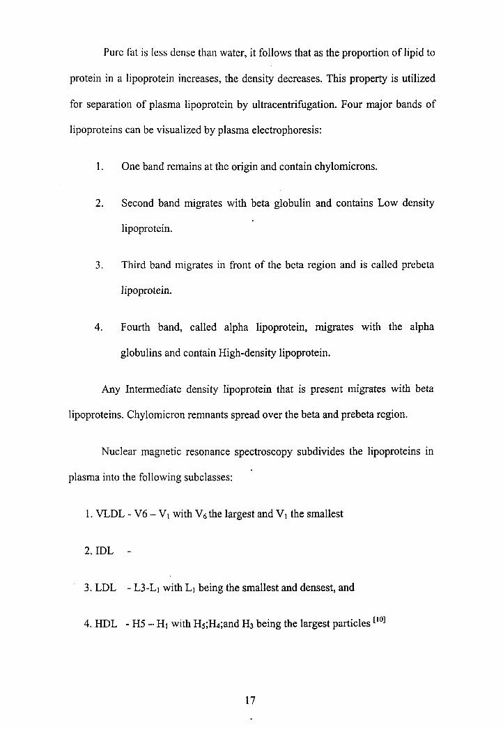
Pure fat is less dense than water, it follows that as the proportion of lipid to
protein in a lipoprotein increases, the density decreases. This property is utilized
for separation of plasma lipoprotein by ultracentrihgation. Four major bands of
lipoproteins can be visualized by plasma electrophoresis:
1. One band remains at the origin and contain chylomicrons.
2. Second band migrates with beta globulin and contains Low density
lipoprotein.
3. Third band migrates in front of the beta region and is called prebeta
lipoprotein.
4. Fourth band, called alpha lipoprotein, migrates with the alpha
globulins and contain High-density lipoprotein.
Any Intermediate density lipoprotein that is present migrates with beta
lipoproteins. Chylomicron remnants spread over the beta and prebeta region.
Nuclear magnetic resonance spectroscopy subdivides the lipoproteins in
plasma into the following subclasses:
1. VLDL - V6 - V1 with V6 the largest and V, the smallest
3. LDL - L3-L1 with L1 being the smallest and densest, and
4. HDL - H5 - HI with Hs;H4;and H3 being the largest particles [lo1

2.6 Apolipoprotein
The protein moiety of the lipoprotein is known as apolipoprotein,
constituting nearly 70% of some HDL and as little as 1% of chylomicrons. Some
apolipoproteins are integral and cannot be removed, whereas others are free to
transfer to other lipoproteins. The apolipoproteins have 3 fknctions:
1. Provide the structural element to lipoprotein particles and these are
important in maintaining stability,
2. Act as ligands for specific receptors, and
3. Act as activators or inhibitors of specific enzymes involved in lipoprotein
metabolism.
Individual apolipoproteins may display one, two or all of these functions.
Nomenclature of apolipoproteins has been made on the basis of electrophoretic
mobility of lipoproteins. - Apo - A being the apolipoprotein derived from HDL
(alpha lipoprotein) and Apo- B being derived from LDL (beta lipoprotein). With
the discovery of other apo- lipoproteins, succeeding letters of the alphabet have
been allocated in turn. ['l1
Each class of lipoprotein has a variety of apolipoproteins in differing
proportions, with the exception of LDL, which contain only Apo-B100.
Apolipoprotein AI and AI1 constitute about 90% of total HDL protein. The ratio
of Apo lipoprotein AI to AI1 in HDL is about 3:l. In addition to being an
important structural component of HDI,, Apolipoprotein AI is a CO factor for
Lecithin Cholesterol Acyl Transferase (LCAT), the enzyme responsible for

forming cholesterol esters in plasma. Some evidence suggests that Apolipoprotein
AI may inhibit LCAT and activate hepatic triglyceride lipase.
Apolipoprotein A-I is one of the first apolipoprotein to be identified
and characterised. The sequence of Apolipoprotein A-I was determined by Brewer
et al., and followed by cloning and characterisation of its c-DNA and genomic
DNA. The gene encoding apolipoprotein A-I is a member of the apolipoprotein
multigene super family, which includes genes encoding exchangeable
apolipoproteins (apolipoprotein A-I; A-11; CS and E).
Apo A-I can form discoidal as well as spherical complexes, depending on
the conditions of association. In vitro constituted discoidal Lipoprotein- A-I
complexes usually have a pre P - HDL migration on agarose gel electrophoresis
and change to an a- mobility following incubation with plasma factors (d>1.21
g/mL) or LCAT alone. Mutations in N-terminal domain have been identified and
these mutations are often associated with amyloidosis. Though several mutations
in the central domain of apolipoprotein A-I were described, only few of them have
been associated with clear defects in lipid binding properties. Only very few
mutations has been described in the C-terminal domain of apolipoprotein A-I.
Funke et al., reported cornea1 opacity and reduced plasma LCAT activity in one
case. Han et al., identified a new mutant associated with low HDL cholesterol
level, possibly because of an increased turnover of the mutant protein, which may
be due to a reduced lipid binding affinity. [ l2]

Apo A-I1 is also syntlicsised in liver and is structural component of
HDL. Its role in vivo is unclear; although in vitro it has been demonstrated to
activate hepatic triglyceride lipase.
Apolipoprotein A-IV is a component of newly secreted Chylomicrons;
VLDL; LDL and HDL. Majority of this protein in plasma is not associated with
lipoproteins, but exists in the free form. It has suggested that Apolipoprotein A-IV
is necessary for maximal activation of lipoprotein lipase by Apolipoprotein CII,
shown to activate LCAT in vitro and plays a role in the transport of cholesterol
from peripheral tissues to liver.
Apolipoprotein -B exists in two forms: Apo B-100 and Apo B-48. The
two proteins are known to be translocation product of single structural gene.
Apolipoprotein B-100, a single polypeptide of over 4500 amino acids, is the full
length translation product of ApoB gene. One gene located on chromosome 2
codes for both apo B-48 and ApoB-100. In the small intestine the mRNA derived
from this gene is specifically modified at codon 2153, the replacement of uracil
residue by cytosine changing into a stop codon. Apo B48 does not contain the
LDL receptor binding domain present in Apo B100 and as a result, it is not
recognised by the LDL receptor. ["l
In human, ApoB-100 is made in the liver and secreted to the plasma as a
part of VLDL. Apo B-100 is the major Apolipoprotein of the LDL, the end
product of the VLDL catabolism. Each VLDL particle contains one molecule of
Apo B- 100. In the fasting state, most of the ApoB in plasma is ApoB- 100. Unlike

other apolipoptrotein, ApoB-100 can not move from one lipoprotein to another
and VLDL ApoB- 100 remains with Lipoprotein as it is catabolised to LDL.
Apo B - 48 contains 2152 amino acids and is identical to the amino acid
terminal portion of Apo B-100, i.e.: amino terminal 48% of apo B-100 forms
apo~-48.[' '] Apo B48 results from the post transcriptional modification of
internal Apo B- 100 m-RNA, in which a single base substitution produces a stop
codon corresponding to the residue 2153 of apo B100. Apo B-48 is made in the
intestine and is the major Apo - B component of chy~omicrons. Both Apo B-100
and Apo B-48 play important roles in the secretion of. the VLDL and
chylomicrons respectively. ['l Apo B-100 contains several hydrophobic areas that
probably serve as strong lipid binding domains. It also has several domains that
could serve as binding sites for heparin like molecules and form the basis for
some of the cell surface interactions of the Apo-B containing lipoproteins. In
addition, it contains a LDL receptor-binding domain (amino acid 3400), which
allows the specific uptake of LDL by the cellular receptor. [l2]
There are three different Apolipoprotein C, which are synthesised in
liver. In plasma they transfer between the Triglyceride rich lipoproteins
(Chylomicrons, VLDL, and their remnants) and HDL.
Apo C-I is the smallest of C apolipoproteins forms a minor component of
VLDL, IDL and HDL and has been reported to activate LCAT in vitro.
Apo C-I1 occurs in Chylomicrons and VLDL, in which it functions as an
activator of lipoprotein lipase and hence plays an important role in the metabolism

of triglyceride rich lipoprotein. (Chylornicrons and VLDL). It is also found in IDL
and HDL.
Apo C-111 forms major structural component of VLDL, but is also present
in Chylomicrons, IDL and HDL ["] Because of the difference in sialic acid
content, apo C-I11 exists in at least three polymorphic forms. It appears to have
two functions. It inhibits Lipoprotein lipase and it also inhibits hepatic uptake of
chylomicrons and VLDL receptor particles possibly by preventing interaction of
ApoE on these remnant particles with hepatic receptors.
Apo D is a minor component of HDL, VLDL, and IDL. Its fbnction is
unknown. [l 'l.
Apo E is the plasma lipoprotein that is formed primarily in Chylomicrons,
VLDL, HDL and, chylomicron and VLDL remnants. Apo E is synthesised and
secreted by a variety of tissues, but primarily by hepatocytes. Normal
concentration of apoE is in the range of 30-70 pg/mL' The major physiological
role of apoE is to mediate the interaction of lipoproteins with lipoprotein
receptors, including LDL receptor and chylomicron receptors. Because it serves as
a ligand for these receptors, apo-E plays a central role in determining the
metabolic fate of plasma lipoproteins and therefore of cholesterol.
Apo E gene locus has multiple alleles that give rise to protein
polymorphism that can be detected by isoelctric focusing. Uterman demonstrated
this for the first time and Breslow fbrther demonstrated two types of
polymorphism:

1. A gcnctically dctcrmined polymorphism, and
2. Polymorphism caused by post translational glycosylation.
Three major isoforms of apoE termed as apoE2, apoE3 and apo E4, that
results from three alleles ~ 2 ~ ~ 3 and ~4 respectively. These alleles occur with a
frequency of 8%, 77% and 15% respectively. Six common phenotypes, three
homozygous (E 212; E 313; E414) and three heterozygous (E 413; E312; and E412)
result from the expression of any two major three alleles.
The molecular basis for the genetically determined polyrnorphism is single
amino acid differences at two sites in apo-E protein. Because apo-E 3 is the most
frequently occurring of these three, it is considered as wild type, and apo-E 2 and
apo-E 4 are considered as variants. Both apo E2 and apoE4 differ from E3 by one
amino acid substitution (residue 158 in apo E2 and residue 112 in apoE4) that
accounts for the charge differences among three isoforms.
The three major apoE alleles and their resultant protein product have a
significant impact on normal variation of plasma lipid and lipoprotein parameters.
Compared with 63 allele, the ~2 allele is associated with lower levels of plasma
cholesterol, LDL- cholesterol and LDL apoB and with slightly higher levels of
plasma triglycerides and significantly higher levels of Apo- E. In contrast, ~4
allele has just opposite effect on all these parameters.
ApoE molecule is organised as two distinct structural domains, that also
have different functions. - an amino terminal domain and a caboxy terminal
domain. Apo-E3 and apo E4 binds normally to LDL receptor, but apo E2 has less

than 2% of normal binding ability. Substitution of Cystine for Arginine at residue
158 may bc responsible for the binding defect of apo-E2 and hence important in
the development type I11 hyperliporoteinemia. Carboxy terminal domain of apoE
represents major lipid binding region. Apo-E4 has a greater preference for
association with triglyceride rich lipoproteins, compared with apo-E2 and apo-E4,
both of which prefer HDL. [l3]
Apolipoprotein (a) is a high molecular weight glycoprotein which
exhibits remarkable size heterogeneity with phenotypes ranging in size from
280000 - 700000 Daltons. It reveals striking similarities with plasminogen. It is
postulated that Apo(a) gene originated via gene duplication of the plasminogen
gene The apo(a) gene is located in close proximity on chromosome 6.[14].
Apolipoprotein J is a glycoprotein present in HDL*. It is synthesised by
foam cells in atheroma plaques, but not by intestinal cells. Human apoJ gene has
been mapped to chromosome number 8. Apo-J inhibits macrophage mediated cell
damage. [ls1
2.7 Transport of Lipoprotein
Most of the cholesterol in plasma is transported as three major lipoprotein
classes - VLDL cholesterol, LDL, cholesterol and HDL cholesterol. The total
cholesterol is the sum of all the cholesterol carried by these three lipoproteins. [lo1.
The pathway of lipoprotein metabolism can be divided into exogenous and
endogenous pathways depending on whether they carry lipids from intestinal or
hepatic origin.

Exoge~lous pathway: Chylomicrons are synthesised in the intestinal mucosa.
Since the triacylglycerol are water insoluble, whereas the digestive enzymes are
water soluble, triacylglycerol digestion takes place at lipid - water interfaces. The
rate of triacylglycerol digestion therefore depends on the surface area of the
interface, a quantity that is greatly increased by the churning of peristaltic
movements of the intestine combined with the e~nulsifying action of the bile
acids. Pancreatic lipase catalyses the hydrolysis of the triacylglycerol at their 1
and 3 positions to form 1, 2-diacylglycerol and 2-acylglycerols. The enzymatic
activity of pancreatic lipase greatly increases when it contacts the lipid - water
interface, a phenomena known as interfacial activation.
The mucosal cells in the small intestine absorb the products of digestion
and the lipid contained in the bile. A mixture of fatty acids and 2-
monoglycerides enters through the villi and triglycerides are synthesised from the
fatty acids and monoglycerides in smooth endoplasmic reticulum . Peter 0
Kwiterovich reports that enterocytes also absorb free cholesterol from the gut. [lo1.
Meera Penumetcha, Nadya Khan Merchant, and Sampath Parthasarathy suggest
that oxidized fatty acids, by enhancing the solubilisation of lumina1 cholesterol
increases the uptake of cholesterol .[l6]
After re-esterification, cholesteryl ester and triglycerides are incorporated
into the core of the chylomicron particles. Enterocytes synthesise apo B48, apo A-
I; Apo A-11, and Apo A-IV, which together with phospholipids, forms surface
layer of chylomicron particles. Apo B-48 is essential for the chylomicron
secretion. There is only one apo B-48 molecule per chylomicron particle and it

remains with the particle through out the life, until it is taken up by the liver as
chylomicron remnant. Chylomicron is released from the intestinal cell by fusion of
secretary vacuole with the cell membrane. Chylomicrons passes into the spaces
between the intestinal cells, eventually making the way into the lymphatic system
(lacteals) draining the intestine. The newly secreted chylomicrons pass intestinal
lymph and gain access to the vascular system via th~racic duct. From the time of
secretion they undergo constant modifications, gaining apo-CII, apo-C111 apo-E,
phospholipids, and cholesterol from HDL. Acquisition of apoCII allows
chylomicrons to interact with Lipoprotein lipase, which is sited in the endothelial
surface of the blood vessels of the peripheral tissues, especially adipose tissues
and muscles. [' ' l
Human LPL is a glycoprotein, mainly synthesised in adipocytes. LPL is
anchored on the cell surface with a prteoglycan chain.. LPL plays an important
role in regulation of plasma triglyceride concentration by hydrolysing
triglycerides in chylomicron and V L D L ' ~ ~ the first step in their metabolism. In
this reaction, LPL require apoCII as an essential factor and produces chylomicron
remnants and VLDL remnants, thereby releasing free fatty acids, which are either
used for energy or re-esterified for endogenous triglyceride storage in adipocytes.
. Apo C-I11 acts as an inhibitor of LPL. The resulting lipoprotein remnants
appears to be additionally processed by the hepatic triglyceride lipase. Small
amount of lipoprotein lipase and hepatic triglyceride lipase [HTGL] are detected
in the plasma as inactive forms, whereas intravenous injection of certain amount
of heparin ( 10-100 idkg of body weight) release significant amount of both
lipases into circulation, as active form. Plasma obtained after injection of the

heparin (post heparin plasma) is used as a clinical sample for measuring LPL or
hepatic triglyceride lipase. Analysis of LPL in post heparin plasma is usually
performed as an important diagnostic measure in order to elucidate the underlying
aetiology of the impaired clearance of chylomicrons and VLDL in patients with
hypertriglyceridemia, which is thought to elevate the risk of Coronary artery
disease. [''I
LPL acts extracellularly to hydrolyse triglycerides within chylomicron
core, the fatty acid thus released can be either utilized as an energy source or re-
esterified and stored in adipose tissue as triglycerides. As hydrolysis proceeds, the
chylomicron core reduces in size and excess surface components, free cholesterol,
phospholipids, apo C-I1 and apo C-I11 are transferred back to HDL. Apo C-I11 may
exert a modulating effect as LPL catalyses chylomicron hydrolysis. Both apo C-I1
and LPL are necessary for normal chylomicron catabolism. Continuing loss of
apoC-I1 result in either a change in its orientation or its mass is reduced to a
critical level such that interaction with LPL occurs no more. Thus chylomicron
remnant particles are generated which have a relatively high content of
Cholesteryl ester and apo-E. These remnant particles are taken up by liver by one
or more, yet unidentified receptor dependant pathways. A specific receptor has
been postulated, Apo-E appears to be thd ligand for this receptor mediated uptake
mechanism. Remnant particles accumulate in the plasma of the subjects who are
homozygous for the apo E2 isoform or who lack apo E. The cholesteryl ester
delivered to the liver by the remnant particle may be utilized for the synthesis of
bile acids or membrane or may be secreted in VLDL, while apo B-48 undergo
degradation.[' 'l

Endogenous pathway: The hepatocytes are the originator and often also the
acceptor of particles involved in the endogenous pathway. Hepatocyte has the
ability to synthesise triglycerides from carbohydrates and fatty acids. In addition,
when dietary cholesterol acquired from the receptor mediated uptake of
chylomicron remnants is insufficient, hepatocytes also synthesise their own
cholesterol by increasing the activity of HMG CoA reductase. ApoB-100 is
synthesised in the ribosomes of the rough endoplasmic reticulum, which is the
main site of triglyceride synthesis. Lipoprotein passes through Golgi apparatus,
where carbohydrate residues are added to lipoprotein. The endogenously made
triglycerides and cholesterol are packaged in secretory vesicles in Golgi
apparatus, transported by exocytosis into the extra cellular space and introduced
into the circulation through he fenestrae of the hepatic sinusoidal epithelium in the
form of nascent VLDL. This triglyceride rich particle contains apo B100, apoE,
and small amount of apoC at its surface. Additional cholesterol and
apolipoproteins are transferred after secretion from HDL .The size of the VLDL
particles secreted by the liver varies according to the hepatic availability of
triglycerides. In situations where there is an excess (obesity, Diabetes, alcohol
excess) large VLDL particles are secreted. Large VLDL particles are also secreted
in familial hypertriglyceridemia, where as in familial combined hyperlipidemias
and hypobetalipoproteinemia the rate of VLDL secretion is increased but a
relative scarcity of triglycerides ensures that the individual VLDL particles are
smaller.["l Apo C-I1 present in the surface of the VLDL activates LPL on
endothelial cells, which leads to hydrolysis of VLDL triglycerides and results in
the release of free fatty acids. Rate of hydrolysis of VLDL triglyceride is

significantly lower than that of chylomicron triglycerides. The average of
residence time of VLDL triglyceride is 15-16 minutes, compared with the 5-10
minute of chylomicron triglyceride. This difference may be attributed to the fact
that VLDL are smaller particles and bind to fewer LDL molecules than
chylomicrons.
During the hydrolysis of VLDL triglycerides, the apo-C are transferred
back to HDL, VLDL particles are thus converted to VLDL remnants, some of
which are taken up by liver and the rest, converted to smaller, denser particles
called IDL (Intermediate density lipoprotein)
Binding of the triglyceride rich lipoprotein (produced from chylomicrons
and VLDL) to liver is probably mediated by heparin sulphate proteoglycans and 1
or LPL as well as lipoprotein receptors (LDL receptor; LDL receptor related
protein {LW), or VLDL receptor). VLDL receptor has been described as a new
member of the LDL receptor super gene family that specifically binds VLDL in
vitro via apo-E and LPL.
L W is also called as alpha 2-macroglobulin receptor. L W is a multi-
ligand receptor whose ligands include ApoE containing lipoproteins,
chylomicrons, LPL, protease / inhibitor complexes and toxins. An additional
ligand, called receptor associated protein, is an intracellular protein that appears to
function as a chaperone for LW.
Junghan Song et al.. reported significant inter racial distribution of
remnant receptor polymorphism and a significant association with Lipoprotein-
(a), suggesting that Lp(a) metabolism in part ,mediated by the uptake through

remnant re~e~tor.~" ' . Large IDL particles, which also have several molecules of
apo-E, bind to hepatic remnant receptor and are removed by hepatocytes. Surface
materials from IDL, including some phospholipids, free cholesterol and
apolipoproteins are transferred to HDL or form HDL de novo in circulation.
Lipoprotein remnant take up appears to be mediated by receptors specific for apo-
E. Both LDL receptor and a remnant receptor specific for apo-E receptor take part
in remnant uptake. One candidate for remnant receptor is LRP. The net result of
coupled lipolysis and cholesteryl ester exchange reaction is replacement of much
of the triglyceride core of the original VLDL with cholesteryl esters. IDL
undergoes a further hydrolysis in which most of the remaining triglycerides are
removed and all apolipoproteins, except apoB-100, are transferred to other
lipoproteins. Further hydrolysis of the IDL by hepatic triglyceride lipase (HTGL)
results in the formation of LDL. HTGL has a dual role of acting as a ligand for
lipoprotein and hydrolysis of triglycerides and phospholipids. In subjects who
lack apo-E or are homozygous for 212 isoform, IDL accumulate in addition to
chylomicrons. IDL also accumulates in subjects deficient in HTGL.
LDL is the major cholesterol carrying lipoprotein in the plasma and
usually accounts for 70% or more of the total plasma cholesterol. Virtually, the
only protein contained in the LDL particle is a simple molecule of apoBlOO and
this act as a ligand for the LDL receptor. LDL receptors are present on
hepatocytes as well as peripheral tissues. Approximately 50% of plasma LDL
uptake by the LDL receptor mediated mechanism is hepatic. The major
determinant of plasma LDL concentration is the number of hct ional LDL
receptor. In subjects, who lack functional receptors (homozygous familial

hypercholesterolemia) the LDL level is markedly elevated and LDL removal from
blood is entirely dependant on non - receptor mediated me~hanism.["~
If LDL is oxidised, it can enter the macrophage through the scavenger
receptors, on the surface of m a c r ~ ~ h a ~ i : . ~ ' ~ ]
2.8 LDL Receptor
LDL receptor is a glycoprotein present on the surface of most cells that
mediates the uptake and degradation of LDL. It is responsible for the 80% of the
clearance of LDL from circulation, the bulk of which occurs in liver, and its
activity has a major influence upon the plasma cholesterol concentration. LDL
receptor is discovered by Golstein and Brown in 1974.
LDL receptor has to carry out several quite distinct functions and it shall
have different structural features on protein that are required for each of these
functions. Thus the protein must contain:
l . Sufficient information to ensure that it is correctly glycosylated and
then transported to and inserted in the cell membrane after its
synthesis in endoplasmic reticulum.
2. Specific extra-cellular high affinity binding sites for its ligands apo E
and ApoB and there may be additional structural information to
facilitate interaction between receptors.

3. As endocytosis of ligand and receptors occurs by movement of
receptors into clathrin-coated pits, the receptor must contain some
recognition signal for a protein in the coated pits.
4. Structural feature required for recycling while ensuring that the ligand
is delivered to appropriate site for degradation
5. Regulatory sites, as the activity of receptor are regulated by the flux of
free cholesterol.
The LDL receptor protein consists of 5 structural region or domain: First domain
of the LDL receptor encompasses the N-terminal 292 amino acid residues of the
protein and comprises seven copies of a Cystine rich, negatively charged 40
amino acids in length that shares strong homology, particularly in the spacing of
the Cystine residues, with a 40 amino acid sequence found in the terminal
complement component C9. This domain contain ligand binding site.

................................................................................... I
I
l
I
I
I
I
I
l
I
1. Ligand binding
I 292 amino I
l
I
I
l
l
l
l
3. 0-linked sugars 58 amino acids
l
l
I
I
I 4. Mnmhrclnn.
22 amino acids
5. Cytoplasmic 50 amino acids
I L----------------------------------------------------------------------------------
Figure 3: Structure of LDL Receptor
Second domain comprises approximately 400 amino acids with
remarkable homology to the precursor of epidermal growth factor (EGF),
including another three Cystine rich repeats known as growth factor like receptor.
Even the introns - exon arrangement is identical in these two genes.
The third domain consists of 88 amino acid enriched in serine and
threonine residues. The majority of the 18 hydroxylated amino acids are
glycosylated, confirming that this part of the receptor is extra-cellular. These
sugars are added CO-translationally and are modified as the precursor protein
undergoes maturation by passage of Golgi apparatus to the cell surface. This

. change in carbohydrate residue is responsible for the apparently anomalous
increment in apparent molecular weight that occurs as protein matures.
Fourth domain of the protein is a 22 amino acid stretch of hydrophobic
residues bordered by charged residues that is believed to anchor the receptor in the
cell surface by spanning the membrane.
Fifth domain is a COOH terminal cytoplasmic tail of 50 amino acid
residues.
Once the LDL receptor protein has been transported to the cell surface, its
insertion into the membrane appears to depend solely on the presence of an intact
membrane-spanning domain.
Depending on the ability of the receptors to bind LDL and facilitate
internalization and degeneration, and on the characteristic behavior of newly
synthesised LDL receptor protein, four classes of mutation was described.
Class I mutations are defined as those in which no immuno-precepitable
LDL receptor protein can be detected. This group is more correctly subdivided
into:
a. Mutations, which result in no detectable mRNA for the receptor.
b. Those mutations in which mRNA is produced, but no protein.
These mutations invariably produce a receptor negative phenotype.
Class I1 mutations are those in which the newly synthesised precursor
form of the receptor does not undergo the normal process of maturation and
translocation into the cell surface. If the defect in processing is absolute, class I1
mutants also produce a receptor negative phenotype, but if at least some of the
mutant protein reaches the cell surface and functions to some extent, then the
phenotype will be receptor deficient.
Class I11 mutation are those in which an apparently normal protein is
synthesised, but transported to the cell surface but fails to bind the ligand
normally.
Class IV mutations are a group in which the receptor protein is synthesised
and appears normally on the cell surface where it is fully capable of binding
ligand. However the mutant receptors fail to cluster in the coated pits and are not
internalised.
Changes in the amount of the LDL receptors in the cell are associated with
corresponding changes in the rate receptor synthesis. The mRNA for the receptor
is most abundant in tissues known to express high LDL receptor activity and its
concentration has been shown to be increased by cholesterol depletion, in wide
varieties of cultured cells. LDL receptor synthesis and mRNA content in cultured
cells are decreased by free cholesterol or cholesterol delivered in LDL or other
lipoproteins. The most potent inhibitor are oxysterols, either present as impurity
or formed from cholesterol in the cell that is the metabolically active agent.
Liver expresses significant numbers of receptor and is responsible for the
majority of the receptor-mediated clearance of the LDL from circulation. In cells

such as fibroblasts, regulation of the expression of LDL receptor is part of
mechanism for maintaining the cholesterol homoeostasis and providing
cholesterol for cell growth division. In hepatic G2 cells inhibition of cholesterol
esterification greatly enhances the suppression of LDL receptor and HMG CoA
reductase activity by LDL.
Synthesis of receptor and mRNA content has been shown to be increased
by the stimulators of steroid hormone production such as ACTH and chorionic
gonadotrophin in their target cells. Oestrogen treatment can produce ten fold
increases in LDL receptor protein in rat liver. Hypothyroidism leads to reduction
in hepatic receptor content of the rat liver, thyroxin can stimulate LDL uptake in
isolated rat hepatocytes. LDL uptake by fibroblasts is decreased by epinephrine
and increased by insulin. A similar effect is seen by insulin in hepatic G2 cells [lg1.
As LDL receptor is able to bind apolipoprotein ligands, apoBlOO and
apoE, it is sometimes referred as B-100 or E receptor.
Uptake of LDL, via LDL receptor is mediated through apo B100. IDL
binds to LDL receptor via apoE and not via apoB100. The subclass of HDL
containing apoE can also bind to LDL receptor. Lipoproteins that contain multiple
copies of apoE bind to LDL receptor with much greater affinity than does LDL.
[ I 11.
Once the LDL receptor reaches the surface of the cell, they cluster in
specialized thickened and indented regions of the plasma membrane called coated
pits. Coated pits are distinguished by the presence of a polygonal matri? of
proteins that coats their endoplasmic surfaces. These proteins include the major

structural protein, called clathrin, and a complex of .clathrin associated protein.
The presence of a defined short sequence of amino acids in the cytoplasmic
domain of LDL receptor serves as a signal to direct its clustering into coated pits.
The LDL receptors cluster even in the absence of their ligands, LDL. The LDL 1
LDL receptor complex initially enter the cell because of the invagination and
pinching off of the cell surface of the coated pits. The vesicle formed by this
process are called coated endocytic vesicle. The coat of proteins dissociates from
the coated vesicles and the vesicles form the structure called endosomes. Proton
pumps acidifj the endosomal lumen. Under the mildly acidic conditions in the
endosome, LDL dissociates from its receptor. This key step permits the spatial
separation of the LDL and its receptor. The receptor can then be transported back
to the cell surface, where it again clusters in the coated pits and can bind and
internalize more LDL. This recycling of the LDL receptor permits efficient
internalization of large amounts of LDL by use of relatively few LDL receptors,
LDL itself is transferred into another intracellular organelle, the lysosome. Within
the low pH milieu of the lysosome, an array of acid hydrolases degrades the LDL
particles into its component parts - proteins are digested to amino acids, and
cholesteryl esters in the core of LDL particles are converted into free cholesterol
and fatty acids. Finally the cholesterol is transported out of the lysosome and
enters the pool of metabolically active cholesterol in the cell, where it can be used
for many purpose.['g1 The released free cholesterol is available for further
metabolic transformation as well as to regulate, probably via the formation of the
oxysterols, the transcription and 1 or translation of HMG CO A reductase and LDL
receptor genes. The cholesterol may be re-esterified by Acyl Coenzyrne A:

Cholesteryl Acyl Transferase (ACAT) and stored, or may be utilized for bile acid,
steroid or membrane synthesis depending upon tissue and cellular requirement.["].
Hepatic catabolism of the LDL via non- LDL receptor pathway are also
suggested. LDL receptor independent pathway was estimated to account for 42%
of LDL cholesterol clearance in human under normal physiological conditions.
Such activity could be mediated by the lipolysis stimulated receptor, a receptor
that binds and degrades LDL; however the receptor is detectable only in the
presence of free fatty acids. Freshly arriired free cholesterol or derivative thereof
modulates the activity of enzyme that ensures cell cholesterol homoeostasis-
HMG CoA reductase, acyl CoA: cholesterol acyl transferase and cholesterol 7
alpha hydrolase (CYPY). When cellular free cholesterol reaches a threshold,
ACAT is activated and conjugate excess free cholesterol with long chain fatty
acid to cholesterol ester. The synthesis of bile acids is catalysed by the rate
limiting enzyme CYPY, that is expressed only in the liver and which expression is
under control of diurnal cycle, hormone regulation and entero-hepatic circulation.
High levels of bile acids returning to liver via the entero-hepatic circulation were
shown to suppress CYPY activity. Many studies have shown that cholesterol
induces the activity of CYPY, whereas some provided the data that dietary
cholesterol does not have a stimulatory effect on CYPY.
When cholesterol uptake occurs without a parallel uptake of
apolipoproteins, cholesterol ester selective uptake is suggested. Marie Claude

Charest et al., reports that LDL - cholesterol ester selective uptake pathway
plays an important physiological role in Hep G2 cell cholesterol homoeostasis. 1201
High Density Lipoprotein [HDL] Pathway: Nascent HDL particles are
produced by the liver and intestine, They composed of phospholipids, mainly apo
A-I and apo A-11. Nascent HDL is disc shaped, but undergoes rapid
transformation which involves esterification of cholesterol by the action of
Lecithin cholesterol acyl transferase [LCAT].~"] The transfer of cholesterol from
the extra hepatic cell across the cell membrane to the nascent HDL particle in the
plasma during reverse cholesterol transport occurs by virtue of Adenosine-
triphosphate - binding cassette protein or ABC 1 transporter protein. This is one of
a family of proteins that mediate the transport of molecules across cell membrane.
Unesterified cholesterol is removed from peripheral cells, through the transfer of
cholesterol across the cell membrane by ABCl transporters, is picked up by
nascent HDL particle. A free fatty acid from lecithin is transferred to unesterified
cholesterol in nascent HDL by action of LCAT and Apo A-l and spherical HDL
is formed. [lo1. Cholesteryl esters thus formed increase the volume of HDL core, so
that HDL particle within the peripheral circulation is spherical rather than discoid.
Both nascent disc shaped HDL and young spherical, relatively cholesterol poor
HDL particle falls within the denser part of the HDL density range (d= 1.125 -
1.21 gmld L) and are referred to as HDL3. Formation of cholesterol esters
increases the capacity of the surface of the HDL particles for the free cholesterol
which is acquired from the cell membrane possibly after interaction with a
specific HDL receptor. As the volume of core increases it allows other

apolipoproteins ( C-11, C-I11 & E ) and phospholipids to be accommodated in the
surface layer; these are acquired by transfer from the triglyceride rich
lipoproteins- chylomicrons and VLDL- and results in larger, lipid enriched
particles of lower density ( d= 1.063 - 1.125 ), referred as HDL2. HDL2 particles
can act as donors of cholesteryl esters to chylomicrons and VLDL receptors. This
transfer is effected by the action of cholesteryl ester transfer protein (CETP). ['l1.
Yunchi Fusigawa et al., reports that, cholesteryl ester fatty acid composition of
lipoproteins varied widely among diet groups, with the more polyunsaturated
cholesteryl ester of poly group being associated with a higher rate of cholesteryl
ester transfer to endogenous acceptor apo- B containing lipoproteins. CETP
contains binding sites for cholesteryl ester and triglycerides and probably acts by a
carrier mediated mechanism. CETP mediates catabolism of HDL cholesteryl
esters, with secondary decrease in HDL size and protein content. CETP plays a
central role in reverse cholesterol transport, i.e. centripetal movement of
cholesterol from periphery back to liver. CETP gene expression is up regulated in
response to increased response to increased dietary cholesterol or endogenous
hypercholesterolemia. Although CETP reduced HDL levels, its role in reverse
cholesterol transport suggests a dominant anti atherogenic action in vivo. P21
The cholesterol ester in the mature HDL particle may be selectively taken
up by the liver through interaction of HDL with HDL receptor, also called as SR-
B1 receptor, [lo1 (Scavenger receptor class B type 1). SR - B1 is a fatty acylated
glycoprotein that mediate selective lipid uptake from HDL to cells. About
50% of cholesterol ester in mature HDL will be delivered to liver through the
HDL - receptor. Other 50% are transferred by CETP from HDL to apo- B

containing lipoproteins - VLDL, IDL and LDL. HDL receptor interacts with HDL
on the surface of steroidogenic tissues, such as Liver, Adrenal and ovary. The
cholesterol esters in the core of the HDL are selectively taken up by these cells
and, HDL (depleted in cholesteryl ester) is released into plasma to recycle and
pick up more cholesterol. After hydrolysis of cholesteryl esters, the liberated
cholesterol can be used to form steroid hormone or bile acid synthesis. [lo1
TISSUE n
INTESTINE - LIVER
SR -B 1
CM & VLDL
Chung et al., has showed that endogenous CETP and LCAT in plasma
lipoprotein from individuals, together with lipoprotein lipase in plasma lipoprotein
lipase, resulted in an alteration in LDL density and this effect become more
pronounced as the triglyceride content of the plasma is increased, resulting in
production of small dense LDL.

Arnbrosch et al., has proved that increasing CETP activity was associated
with decreasing LDL particle size. Variation in CETP gene determines CETP
activity. As CETP promotes transfer of cholesteryl ester from HDL to triglyceride
rich lipoproteins in exchange of triglycerides, CETP is considered as a candidate
for determining LDL size heterogeneity. Guerin et al., found that capacity of LDL
particle to receive cholesteryl ester from HDL was highly correlated to LDL-
triglyceride content. Chapman et al., suggested that the reason for small, dense
LDL particle in hyperlipidemic state is due tohigh production of VLDL, which
results in an increase in triglyceride pool, thus re-directing Cholesteryl ester from
HDL to VLDL and the preferential transfer of Cholesteryl ester from HDL to
VLDL rather than LDL. The LDL particle then became triglyceride enriched, and

the subsequent action of hepatic lipase on the trigIyceride rich LDL particle
converting them to small, dense particle. [241 The cholesteryl ester in the core of
HDL may be exchanged by CETP for triglycerides in VLDL producing
triglyceride enriched, but cholesteryl ester depleted HDL. Such HDL appears to
be catabolised more rapidly by kidney. Thus, HDL level is reduced. 1'01
A number of life style factors as well as various pharmaceutical agents
affect serum HDL cholesterol levels. Cigarette smoking, obesity, sedentary life
style as well as beta blockers and androgenic steroids are all associated with lower
HDL cholesterol concentration.[251
Increased visceral fat, substitution of carbohydrates for saturated fat in
diet, reduces the HDL cholesterol levels while moderate alcohol consumption
increases HDL cholesterol.[261
There are four potential mechanisms by which HDL cholesterol may be
cardio protective:
l. Reverse cholesterol transport,
2. Inhibition of LDL oxidation,
3. Reduction in the adhesion proteins such as vascular cell adhesion
molecules E selectin, and
4. Increased fibrinolysis [lo1
Lp(a): Lp(a) was first described in 1963 by Kare ~ e r ~ h . [ ' ~ ] It was considered to
be an autosomal dominant trait and represents quantitative rather than qualitative
marker, the concentration of which can vary enormously between different
individual. It has been recognized as an independent risk factor for coronary

artery disease and Lp(a) level above 30 mg/ d L form a threshold above which the
risk of premature CAD increases rapidly. [271. Stable adult level of Lp(a) is
reached early in infancy and its atherogenesity is ten times more than LDL
cholesterol.[281
Lp(a) is a spherical particle of 250 Angstroms diameter that floats in a
density range of 1.05- 1.18lml. The lipid composition of Lp(a) closely resembles
that of LDL. The protein moiety consist primarily of two distinct proteins, namely
the apoB-100 and a unique carbohydrate rich protein - apo(a). These two proteins
are linked together by one or more disulphide bonds within the lipoproteins.
Apo(a) can be separated from Lp(a) by reduction of disulphide bonds linking it
to apoB-100 and the residual lipoprotein particle is similar to LDL in many of its
physico-chemical and immunochemical properties. Further more this particle has
similar affinity for LDL receptor in cultured fibroblasts while un-reduced Lp(a)
has much reduced affinity and capacity for binding and degradation via this
receptor. The modification effected by attachment of apo(a) to apoB- 100 via
disulphide bond confers distinct physico irnmuno-chemical characteristics upon
Lp(a) can not be measured by nuclear magnetic resonance, so an imrnuno-
chemical assay must be used.
Lp(a) competes with plasminogen binding sites on the endothelial cell
surface through its apo(a) component, decreasing the.conversion of plasminogen
to plasmin. Consequently, fibrinolysis decreases, thereby promoting thrombosis.
Lp(a) can be also oxidized in the vascular wall. Lp(a) can also promote

thrombosis by stimulating plasminogen activator inhibitor 1 synthesis.['01. Apart
from above effects it is postulated that Lp(a) can promote smooth muscle cell
growth, and endothelial damage. [281
The Liver appear to be the obvious source of Lp (a). This is supported
from the fact that individuals with chronic hepatic disease such as cirrhosis have
low levels of Lp(a) and that apoB-100, is predomiriantly of hepatic origin. The
concentration of Lp(a) is minimally influenced by metabolic, endocrine, and
anthropometric variables. Though Lp (a j levels are largely determined by a major
autosomal genetic locus (presumably apo (a) gene itself) a number of other gene
loci andtor environmental factors may be influential in determining the Lp(a)
levels.[271
Lipoprotein X [ LpX] : This lipoprotein does not occur in normal people, but is
seen in the plasma of subjects with cholestasis and also in persons with familial
lecithin cholesterol acyl transferase deficiency. It is composed of phospholipids,
free cholesterol and protein; the major portion being albumin, but small amounts
of apo-C and D are also present. Unlike other lipoproteins it migrates to the
cathode on agar gel ele~tro~horesis~"~.. LpX has a lamellar structure and on
electron microscopy appears as rouleaux of stacked disc like vesicles. LpX
comprises of 6% protein, of which half or more is albumin enclosed within the
vesicles. Its presence in blood is largely due to the reflux of biliary phospholipids
into circulation, which attract cholesterol out of the cell membranes. LpX is
catabolised by the reticuloendothelial system including kupffer cell. It may
interfere with hepatic uptake of chylomicron remnants. [291

The major pathways of lipoprotein metabolism are tightly interrelated and
interdependent.
DIETARY FAT
EXTRA
RONS CHYLOMICRON REMNANTS 2 L I V E R \ 1
HEPATIC TISSUES
/ IDL. I 1

2.9 Hyperlipoproteinemias
Mutation in transporter molecules or receptors can lead to an accumulation of
cholesterol and breakdown in the normal process of reverse cholesterol transport
and cholesterol metabolism. The most common abnormalities lead to
accumulation of excessive lipids in plasma. These diseases are collectively known
as the hyperlipidemias, or more properly, the hyperlipoproteinemias.
Four types of lipoprotein abnormalities are frequently found in the population:
1. Increased low-density lipoprotein cholesterol level
2. Decreased high-density lipoprotein level, usually accompanied by
increased triglyceride or VLDL levels.
3. Increased concentration of chylomicron remnants or intermediate
density lipoprotein, and
4. Presence of increased concentration of abnormal lipoprotein called
LP(a).
1. Increased LDL cholesterol levels:
The genetic disorder may involve LDL receptor or apoB. Increased
mutation in LDL receptor gene has characterised to produce high levels of LDL
cholesterol in Familial hypercholesterolemia. The genetic disorder may involve
LDL receptor or apoB. They have a frequency 1 in 500 in general population and
heterozygosity for a mutant allele approximately doubles the LDL cholesterol
levels. ApoB gene abnormalities can be of 2 types. In familial defective apoB-

100, a mutation causing amino acid 3500 to change from arginin to glutamine
resulting in LDL particles that fail to bind receptors and accumulate in the plasma.
In some population the frequency of the mutation is as high as 1 in 500.
Heterozygosity for one mutant allele increases the LDL cholesterol levels by 50%.
Second type of apoB gene mutation causes the disorder hypobetalipoproteinemia.
Many different mutations have been described with an aggregate frequency of 1 in
1000. The common feature of these mutations is that they all fail to allow
translation of full length apoB polypeptide, that may act either to decrease VLDL
production or to increase LDL catabolism. In either case, affected heterozygote
has about 50-70% reduction of LDL cholesterol levels. [301
An increase in LDL often occurs secondary to overproduction of VLDL,
resulting in expression of 2 related disorders, familial combined hyperlipidemia
and familial hyperapobetalipoproteinemia. Polygenic hypercholesterolemia
results from the small contribution of a number of genes and is usually associated
with moderately elevated total cholesterol and LDL cholesterol levels. ['01
2. Decreased HDL cholesterol levels and increased triglyceride levels:
Relatively rare defects in apo A-I gene can affect HDL cholesterol levels.
So also, rare genetic defect in LPL, apo C-I1 genes and over expression of ApoC-
111 gene can result in hypertriglyceridemia. There is an inverse relation between
HDL and triglyceride levels.
There are at least two mechanisms known to link triglyceride and HDL
metabolism:

a). In the hydrolysis of VLDL triglycerides by LPL excess, surface
components including apo-C are transfened to HDL. This can result
in increase in HDL cholesterol level.
b). VLDL triglyceride can exchange with cholesteryl ester from HDL in
presence of CETP causing decreased HDL cholesterol levels.
3. Increased Chylomicron remnants and IDL cholesterol levels:
Defects in apoE allele, results in defective clearance and subsequent
accumulation of chylomicron remnants and IDL
4. Increased LP (a) levels:
Lp (a) may be atherogenic by interfering with both LDL and plasminogen
metabolism r301
It is estimated that over 60% of variability in serum lipids are genetically
determined. Most of them being due to iolygenic influences. Interaction between
the later and the environmental factors is probably the commonest cause of
hyperlipidemia in the general population. [)l1
Hyperlipidemia frequently CO-exists with other diseases. The CO-existent
disease may occur as:
a. Complication of existing hyperlipidemia - as in case of Coronary
artery disease (CAD) in familial hypercholesterolemia or acute
pancreatitis in hypertriglyceridemia.

b. Anothcr primary disease affecting the lipoprotein metabolism - can
give rise to secondary hyperlipidemia.
c. Hyperlipidemia may be associated with another disorder, when neither
occurs as a complication of the other, such as hypertriglyceridemia and
gout [291
World health organisation's classification of lipoprotein phenotypes
provides a useful means of indicating which lipoprotein are present in excess in
individual patients but has a major limitation that it does not differentiate between
primary and secondary forms of hyperlipidemia. This differentiation depends on
the demonstration of presence or absence of an underlying causes and the result of
family studies. 13'].
Table 1: WHO classification of hyperlipidemias:
Primary hyperlipidemias characterised by severe hypertrigIyceridemia
predisposes to acute pancreatitis, where as those disorders characterized by
WHO type
I
IIa
IIb
111
IV
v
Major lipid
Abnormalities
?Triglycerides
T~holesterol
?cholesterol
T~riglycerides
Tcholesterol
TTiglycerides
TTriglycerides
T~riglycerides
Minor lipid
Abnormalities
holest sterol
h hole sterol
T Cholesterol
Electrophoretic change
(compared to normal)
Staining at origin
?p band
?p and pre p band
Broad p band
? pre p band
? pre P band & staining at origin
Lipoprotein abnormality
Chylomicron present
TLDL
TLD &TVLDL
IDL present in detectable amount
TVLDL
VLDL CMs
11 11

hypercholesterolemia, apart from hyperalphalipoproteinemia, are associated with
increased risk of premature vascular diseases.
Frequently, primary hyperlipidemia is polygenically determined and
therefore rather ill defined, but several monogenic or dominantly inherited
disorders have been described, as well as some, which are recessively inherited.
The chief clinical consequence of type I and V phenotype is acute pancreatitis,
while phenotype 11,111 and IV are associated with peripheral vascular diseases.13'l.
Extra-vascular manifestations of hyperlipidemias are:
1. Xanthelasma (periorbital) - seen in any form of hypercholesterolemia;
may occur in subjects with normal lipid levels; common in
hyperlipidemias due to cholestasis
2. Arcus Juvenilis- Arcus senilis can occur with increasing age in
subjects with normal lipid levels. If present in a subject of less than 45
years, probably denotes presence of significant underlying
hyperlipidemia
4. Tendon xanthoma- Typical of autosomal dominant familial
hypercholesterolemia. Also found in the very rare conditions of P -
sitosterolemia and cerebrotendinous xanthomatosis
5. Tuberous & tuberoeruptive xanthomas- seen in Remnant
hyperlipoproteinemia and in homozygous familial
hypercholesterolemia

6. Planar xanthomas- seen in Remnant hyperlipidemia
7. Eruptive xanthoma - seen in Chylomicronemia [l']
Type I hyperliporoteinemia: This is an autosomal dominant condition and this
phenotype includes:
1. LPL deficiency l familial hyperchylomicronemia,
2. Apolipoprotein C-I1 deficiency / C-I1 anapolipoproteinemia I ApoC-I1
deficiency
3. Familial chylomicronemia due to circulating inhibitor of LPL
Familial I~yperchylomicronemia:
Holt et al., reported first the familial occurrence of this syndrome. This
group is characterized by massive hyperchylomicronemia, when the patient is on
normal diet and disappears completely in a few days on fat fiee diet. On a normal
diet alpha and beta lipoproteins are low. A defect in the removal of chylomicron
(fat induced) and of other triglyceride rich (carbohydrate induced) is present.
Reduced Post heparin lipase activity is npted. Clinically manifested by attacks of
abdominal pain, hepatosplenomegaly, eruptive xanthoma and lactascence of
plasma. Heterozygotes may show slight hyperl'ipemia and reduced post heparin
lipase activity. Deficiency of LPL is the basic defect in type I
hyperliporoteinemia. This can also occur in deficiency of activator of LPL - apo
C11 -which is called as fat induced hypertriglyceridemia.

Although heterozygotes do not usually displays the gross phenotypic
fcaturcs such as chylomicronemia, xanthomata or episodes of abdominal pain,
they have only half normal LPL activity'which might not suffice to keep plasma
triglyceride concentration within the normal limit when stress is placed on the
plasma lipid transport system. Responds readily to antioxidant therapy.
Apo C-ZI deficiency:
Apo C-I1 is a necessary CO-factor for the activation of LPL, the enzyme
that hydrolyses triglycerides in plasma and transfer the fatty acid to tissues.
Beckenridge et al., (1978) reported the first case of complete deficiency of apo C-
11. Clinically and bio-chemically this disorder closely resembles LPL deficiency
or type I hyperlipoproteinemia and is referred as hyperliporoteinemia type IB.
Xanthoma and hepatosplenomegaly are less common than LPL deficiency. Apo
C11 deficiency is inherited as an autosomal recessive trait. Heterozygotes have no
abnormality of plasma lipid and lipoproteins in spite of reduced plasma apo CII.
Familial chylomicronemia due to circulating inhibitor of LPL:
Brunzell et al., described type I hyperliproteinemia with very low levels of
post heparin lipase activity and circulating inhibitor of LPL, with much higher
levels of LPL in adipose tissues and normal or increased levels of ApoC-11. There
appeared to have an inhibitor to LPL activity, which have been present in post
heparin of normals. The inhibitor was non- dialyzable, heat stable and sensitive to
repeated freezing and thawing; it appeared to be present in non- lipoprotein
fraction of the plasma. Dietary fat restriction reduced triglyceride levels and
prevented recent attacks of pancreatitis.

Type I1 hyperlipoproteinemia:
Type I1 hyperlipoproteinemia is classified to type IIa and IIb depending on
the absence or presence of hypertriglyceridemia respectively.
Familial hypercholesterolemia: FHILDL receptor disorder:
Familial hypercholesterolemia is an autosomal disorder characterized by
elevation of serum cholesterol bound to LDL. Mutation in LDL receptor gene on
chromosome 19 causes this disorder.
Heterozygotes develop tendinous xanthoma, cornea1 arcus, and CAD, the
last usually becomes evident in the fourth or fifth decade. Homozygous develop
these features at an accelerated rate in addition to planar xanthoma, which may be
evident at birth in the web between first two digits. Biochemical changes are
increase in serum cholesterol and LDL cholesterol (in mg/d L) of 250- 450 and
200-400 in heterozygous, greater than 500 and 400 in affected homozygous, 150-
250 and 75 -175 in unaffected hornozygous. Individuals with CAD may have
significantly higher mean Lp(a). Defects with LDL receptor affect proper
internalisation resulting in withdrawal of regulation of cholesterol biosynthesis by
HMG CoA reductase, and clearance of LDL cholesterol. The frequency of
homogygotes in population is 1 in million and heterozygotes not less than 1 in
500[~'][~~]. Diagnosis of heterozygous familial hypercholesterolemia at birth is best
achieved by measuring LDL cholesterol in cord blood.
Eric J Sijbrands et al., reports that mortality in familial
hypercholesterolemia increased after 1915 reached its maximum between 1935

and 1964 and rcduccd thcrcaftcr. The variation in mortality points to strong
interaction with environmental factors. [3j1
Hyper beta lipoproteinemia / Hyper low density lipoproteinemia / familial
hypercholesterolemic xanthomatosis:
Characterised by increased LDL - apoB of more than 120 mg/d L in
plasma despite a normal concentration of LDL cho le~ te ro l .~~ '~~ On a normal diet,
the blood shows an increase in beta lipoprotein, resulting in increased serum
cholesterol, while the phospholipids and triglycerides remaining constant. Clinical
features are xanthoma tuberosum and tendinosum, cornea1 arcus, and
atheromatosis. This phenotype is the commonest type of hyperlipoproteinemia.
Individuals who are not affected by the LDL receptor defect, but who demonstrate
a type I1 phenotype will likely and eventually be shown to have an abnormality
that interferes with regulation of activity bf HMG CoA. Most of the biochemical
variants can be considered as dominant traits, though their prevalence makes
appearance of homozygotes or genetic compounds relatively frequent. The latter
individuals generally are more severely affected than heterozygotes and their
presence in families may suggest autosomal recessive inheritance.
Familial Defective Apo B100:
Characterised by a single base substitution in two codon for Arginine 3500
in the apoB gene. This gives rise to a form of apo B100 which impairs the ability
of LDL to bind to the LDL receptor. Affected individual have a moderate
hypercholesterolemia with a raised LDL.

Polygenic Hypercholesterolemia:
Summated effect of many different genes and environmental factors
determine the distribution of cholesterol levels in the population. Clustering in of
several genes which tends to induce moderate elevation of the plasma cholesterol
should theoretically result in polygenic hypercholesterolemia.
Familial Combined Hyperlipidemia :
This condition was first described by Goldstein et al., Over all 50%
relatives of affected subjects were hyperlipidemic - one third
hypercholesterolemic (type IIa), one third hypertriglyceridemic (type IV or V)
and one third with both abnormalities (type IIb). This disease is familial and
relatively common occurring in up to 0.5% of general population. This is
characterised by increased synthesis of the apo B-100, with high rate of turn over
of both VLDL and LDL apoB. VLDL triglyceride synthesis is increased but to a
lesser extent than in familial hypertriglyceridemia, where as VLDL apoB
synthesis increased to a more marked extent than in the later disorder. Conversion
of IDL is increased, LDL cholesterol : apoB ratio is decreased and HDL
cholesterol concentration tends to be low. No distinctive clinical features. -
Diagnosis depends on demonstrating multiple phenotypes within the family. In
practice individuals with type b phenotype, who does not have tendon xanthoma
are often presumed to have familial hypercholesterolemia. [311.

Type I11 Hyperlipoproteinemia / Dysbetalipoproteinemia:
This condition is autosomal recessive with pseudo-dominance due to high
gene frequency. In normal individual, chylomicron remnants and VLDL
remnants are rapidly removed from the circulation by the receptor mediated
endocytosis in liver. In type I11 hyperlipoproteinemia, increased plasma
cholesterol and triglycerides are the consequence of impaired clearance of the
chylomicron remnants and VLDL remnants because of a defect in apoE.
Accumulation of the remnants can result in xanthomatosis and premature
peripheral vascular diseases. Type I11 hyperlipoproteinemia can be either due to
primary heritable defects in lipoprotein metabolism or to other conditions such as
hypothyroidism, systemic lupus erythamatosis or diabetic acidosis. Most patients
are homozygous for E2 isoform. Only rarely this disorder takes place in
homozygous phenotypes. E2 isoform shows defective binding or the remnants to
hepatic lipoprotein receptors and delayed clearance from the plasma. Additional
genetic and I or environmental factors must be required for the disorder, because
only 1-4 % of E2 l E2 homozygotes develop familial dysbetalipoproteinema. As
this disorder involves exogenous cholest~rol transport system, the degree of
hypercholesterolemia is sensitive to the level of cholesterol in the diet. Even on
normal diet, the patient may show increased plasma cholesterol and the presence
of an abnormal lipoprotein called beta -VLDL. VLDL in general is increased,
while LDL is reduced. Carbohydrates induce or exacerbate the hyperlipidemia,
resulting in marked variability in plasma levels of lipoproteins. Often tuberous
and planar and sometimes tendon xanthomas occur as well as precocious
atherosclerosis and abnormal glucose tolerance. D21. Clinical features include
57

cornea1 arcus, xanthoma, tuberoeruptive and palmar striae. Both serum
triglycerides and cholesterol are increased. On ultracentrifugation the density
4 .006 fraction contains cholesterol rich remnants with beta mobility on
lipoprotein electrophoresis. LDL cholesterol is reduced. Management involves
remedying of obvious precipitating factors like hypothyroidism, diabetes mellitus
and iatrogenic influences. 13'] The abnormal pattern of apoE by isoelctric focusing,
specifically, the absence of apoE3 is the most characteristic feature of type I11
hyperlipoproteinemia. ApoE isolated from patients suffering from type I11
hyperlipoproteinemia had a decreased fractional catabolic rate. A variety of
factors modulates or exacerbates type I11 hyperlipoproteinemia. In women it
occurs mostly after menopause and such patients are sensitive to oestrogen
therapy. Hypothyroidism exacerbates type I11 hyperlipoproteinemia and thyroid
hormone is known to enhance receptor mediated lipoprotein metabolism. Obesity,
diabetes mellitus and age are associated with increased hepatic synthesis of VLDL
and 1 or cholesterol. [321.
Type IV Hyperlipoproteinemia/Carbohydrate Induced Hyperlipoproteinemia:
An autosomal dominant condition, in which plasma triglycerides are
persistently increased, while plasma cholesterol are usually within normal limit.
Precocious atherosclerosis, abnormal glucose tolerance and atheroeruptive
xanthoma may occur. This disorder is heterogenous and phenotype is strongly
influenced by the environmental factor, particularly carbohydrate and ethanol
consumption. This disorder is caused by uremia, hypopitutarism, contraceptive
steroids and glycogen storage disease. 13*]. Affected subjects have larger than
normal VLDL particles, with increased triglyceride: apoB-100 ratio and

accompanied by a decrease in HDL - cholesterol. VLDL triglyceride synthesis is
increased than VLDL apoB synthesis. Fractional catabolic rate of both VLDL
components is reduced. Free fatty acid flux into triglyceride is increased,
especially when they are placed on high carbohydrate intake and the resultant
increase in VLDL synthesis is accompanied by a decrease in the proportion of
VLDL converted to LDL This maintains plasma LDL cholesterol levels within the
normal range. Glucose intolerance and Hyperuricemia are common
accompaniments. Management involves adherence to a modified fat diet designed
to achieve ideal body weight, avoidance of sugars and alcohol, and
encouragement of physical activity. 1311
Type V Hyperlipoproteinemia:
Autosomal dominant disorder characterised by increased amount of
chylomicron and VLDL, and decreased LDL and HDL. Many conditions can
cause this phenotype including Insulin dependant diabetes mellitus, contraceptive
steroids, and alcoholism and glycogen storage disease. 13'] Liability to develop
acute pancreatitis and subjects with this type may develop xanthoma, glucose
intolerance, Hyperuricemia, and peripheral neuropathy. VLDL apoB synthesis is
increased and more marked decrease in fractional catabolic rate. 1311.
Secondary Hyperlipidemia:
Secondary hyperlipidemia is common and occurs frequently in disorders
such as obesity, alcoholism, diabetes mellitus, hypothyroidism, liver and renal
diseases, and as a side effect of drug therapy, particularly for hypertension.
Secondary hyperlipidemia is important because:

1. The primary disease presenting as hyperlipidemia may be an important
diagnosis on its own right.
2. Secondary hyperlipidemia mziy be a cause of morbidity, e.g.: CAD in
diabetes and renal diseases.
3. Disordered lipoprotein metabolism may accelerate the progress of
primary disease as has been suggested in renal and liver diseases.
The impact of secondary hyperlipidemia depends on the milieu in which
they occur and will be more extreme in people already genetically and
nutritionally predisposed to hyperlipoproteinemia. Secondary
hyperlipoproteinemias are not only associated with increased levels of circulating
lipoproteins, but also with changes in their composition, altering both chemical
and physical properties. Even in the absknce of elevated lipid levels, qualitative
changes in lipoprotein may render them more atherogenic.
The causes of secondary hyperlipoproteinemia are given below:
Endocrine
Nutritional
Renal diseases
Diabetes mellitus
Thyroid diseases.
Pituitary diseases
Pregnancy
Obesity
Alcohol
Anorexia nervosa
Nephrotic syndrome.
Chronic renal failure

Hepatic diseases Cholestasis
Hepatocellular disorders.
Cholelithiasis
Hepatoma
Porphyria
Immunoglobulin excess Myeloma
~acroglobulinemia
Systemic lupus erythamatosis
Hyperuricemia
Drugs
Miscellaneous
Beta adrenoreceptor blockers.
Thiazide diuretics
Steroid hormones.
Microsomal enzyme inducing agents
Retinoic acid derivatives
Stress
Intestinal malabsorption
Glycogen storage disease.
Lipidodystrophy
Idiopathic hypercalceamia of infants
Hypervitaminosis D
Osteogenesis imperfecta.
Sphingolipodystrophies
Progeria
Werners syndrome
Cholesteryl ester storage disease.
Carnitine acyl transferase deficiency
Tangier disease
Familial LCAT deficiency

The major effects of secondary hyperlipidemia on serum lipoprotein levels
are given below:
Table 2: Effects of Secondary Hyperlipidemia on Serum Lipoprotein
Levels
Diabetes Mellitus:
Cause
NIDDM
IDDM
Hypothyroidism
Pregnancy
Obesity
Alcohol
Nephrotic syndrome
Chronic renal failure
Cholestasis
Hepatocellular disease
Hyperuricemia
Diabetes is not only a disorder of carbohydrate metabolism, but also of
lipid and protein metabolism. Two major complication of diabetes, atherosclerosis
and keto acidosis are disorders of lipid ketabolism. The dominant hyperlipidemia
in diabetes is hypertriglyceridemia. The enzyme LPL is activated by insulin.
Insulin deficiency and 1 or insulin resistance associated with uncontrolled diabetes
VLDL
Increased
Increased
Increased
Increased
Increased
Increased
Increased
Increased
Increased IDL
Increased
LDL
Increased
Normal / reduced
Increased
Increased
Normal / increased
Normal / increased
Increased
-
LpX increased
-
-
HDL
Reduced
Normal / reduced
Reduced
Increased
Reduced
Increased
Normal / reduced
Reduced
Reduced
Reduced
Reduced

may lead to elevation of triglyceride levels. In patients under reasonable glycemic
control, hypertriglyceridemia is produced by overproduction of VLDL. Insulin
deficiency or resistance produces increased lipolysis and the consequent
overproduction of non esterified fatty acids results in the enhanced synthesis of
the VLDL-triglyceride by liver. Increased secretion of VLDL from liver is
facilitated by decreased insulin secretion and / or insulin resistance which will
P O 1 decrease the direct inhibitory effect of insulin on secretion of VLDL. .
Compositional changes in lipoproteins in diabetes includes glycosylation of LDL
making it susceptible to oxidation, smaller and denser LDL and LDL has more
affinity to arterial wall. The disordered lipoprotein metabolism associated with
diabetes mellitus are:
1. Glycosylated VLDL is catabolised more slowly than normal
VLDL.
ii. Glycosylated HDL may be less able to mobilize cholesterol out of
the cells.
iii. Perhaps, most importantly, glycosylated LDL itself is more
atherogenic than native LDL and is more easily oxidated, there by
producing more atherogenic form of LDL.
Type 2 diabetes mellitus is associated with increased triglycerides, reduced
HDL cholesterol, preponderance of small, dense LDL, increased Lp (a) and
increased apoB fraction. The major causes of morbidity and mortality are related
to the long term vascular complication in type 2 diabetes (NIDDM) - increased
risk of large vessel diseases such as coronary artery disease, stroke, and

peripheral vascular diseases. In type 1 diabetes (IDDM) the major cause of
morbidity and mortality is small vessel diseases (microangiopathy), which
includes retinopathy and nephropathy. [341.
Two abnormalities characterises the lipoprotein metabolism in NIDDM.
Fasting and post prandial concentration of triglyceride rich lipoprotein, especially
the VLDL are higher and HDL are lower than the non diabetic people. Insulin
resistant state impairs the normal suppression of fatty acid release from adipose
tissue in the postprandial state. Consequently, the flux of free fatty acids to liver
increases and over production of VLDL from these substrates is effected.
In non - diabetic person insulin maintains the balance between triglyceride
rich lipoprotein derived from intestine and liver. In NIDDM this regulation fails
and inappropriate production of VLDL results.
Hydrolysis of core triglyceride by hepatic lipase produces small, dense,
HDL particles that have high fractional catabolic rate.
Small and dense LDL particles predominate.
Lipoprotein lipase activity is reduced.
Hypertriglyceridemia contribute to low HDL concentration via:
a. Reduced LPL activity and impaired lipolysis results in reduction of
surface remnants available for incorporation into HDL particle.
b. Increased amount of triglyceride rich lipoprotein and their prolonged
residence time in circulation increases the exchange of cholesteryl

ester from HDL to triglyceride rich lipoprotein and triglyceride to
HDL, mediated by CETP. Thus HDL becomes enriched by triglyceride
and triglyceride enriched HDL have faster catabolic rate that tends to
reduction in number of circulating HDL particles.
HDL particles are smaller due to higher activity of hepatic lipase. Small
P61 dense HDL2 predominate at the expense of larger, cholesteryl enriched HDL 3. .
Reaven and others have developed the concept of metabolic syndrome -
also called as Syndrome -X, pleurimetabolic syndrome, Reavens syndrome, P41 -
based on the insulin resistance that includes hypertriglyceridemia, decreased
HDL, hypertension and Hyperuricemia, [361 raised concentration of glucose and
high insulin levels in plasma[371.
Kay Tea Khaw et al., reports that glycosylated hemoglobin (Hb Alc) was
continuously related to subsequent all causes, cardiovascular and ischeamic heart
disease (IHD) mortality through the whole population distribution, with lowest
rate in those with HbAlc concentration below 5%. An increase of 1% in HbAlc
was associated with 28% increase in the risk of death independent of age, blood
pressure, body mass index, and cigarette smoking.
Patients with type 2 diabetes have a two fold to three fold increased
incidence of disease related atheroma and those who presents in their 40S and 50S
have a two fold increased total mortality. R.C Turner et al., reported an increased
CAD risk of 1 1% for each increment of 1% HbAlc.

Proteinuria in diabetes nephropathy may indicate a generalized increase in
vascular permeability and thus macromolecules such as LDL may enter the
arterial sub intima at increased rate.
Thyroid Diseases:
Hypothyroidism is characterised by low HDL cholesterol and less
frequently increased triglyceride levels. There is decreased receptor mediated
LDL catabolism and triglyceride catabolism and LPL activity may be also
reduced. Sub clinical hypothyroidism does slight effect on LDL. There is a
tendency for decreased LDL and HDL level in hyperthyroidism.
Renal Diseases:
Proteinuria in patients with relatively normal creatinine clearance produces
hypercholesterolemia due to an increase in LDL. Severity of hypercholesterolemia
is often proportional to reduction in serum albumin. Hypertriglyceridemia is
presented in patients with chronic renal failure and is due to deficiency of LPL
activity. Hypercholesterolemia is usually not associated with increased levels of
LDL from VLDL and intravenous infusion of albumin reduces the LDL level.
This suggests a direct hepatic secretion i f LDL without a precursor VLDL and
this may be important. Dr.C.Short reports that patients with Nephrotic syndrome
have a high serum concentration of Lp(a). Serum HDL and apoA-l levels are
either normal or decreased. Even when total HDL is normal, HDL2 fraction is
increased. Synthesis of apoA- l is increased, and the increased catabolism might
contain normal or low circulating level. There is increased loss through kidney,
related to selectively and extent of glomerular leak.

In chronic renal failure without proteinuria serum triglyceride are raised
both in VLDL and LDL and remnant particles persist in circulation. This may be
due to decreased activity of LPL and hepatic lipase. Hemodialysis exacerbates
hypertriglyceridemia due to depletion of LPL from frequent use of heparin and
apo C11 fiom circulation. Chronic ambulatory dialysis leads to absorption of
considerable amount of glucose producing obesity and hypertriglyceridernia.
Serum HDL is reduced. Pre beta HDL is increased and this may be accumulated
due to decreased LCAT activity. ["l
Renal diseases were observed in which there was an abnormal
accumulation of lipids - lipoprotein thrombi - in glomerular capillary membrane.
This has been described as lipoprotein glomerulopathy. This disorder is
characterised by proteinuria, normal LCAT activity, type I11 hyperlipoproteinemia
like lipid profile and significantly higher levels of plasma apoE.
Obesity:
Obesity will exacerbate any primary hyperlipoproteinemia. Its dominant
effect is to produce hypertriglyceridemia, particularly type I11
hyperlipoproteinemia, but in susceptible individuals hypercholesterolemia due to
increased LDL will be exacerbated. Android obesity is more likely to provoke
hypertriglyceridemia, than gynoid obesity assessed by determining waist: hip
ratio. The cause of hypertriglyceridernia is increased VLDL production, resulting
fiom increased release of non-esterified fatty acids from adipose tissue. Increased
VLDL secretion is often matched by an enhanced catabolism due to increased
LPL activity, so that hypertriglyceridemia does not invariably ensue.

Hypertriglyceridemia is more likely if there is pre-existing defect in triglyceride
metabolism. There is an increased synthesis of cholesterol and serum HDL
cholesterol tends to be decreased. Though triglycerides and cholesterol decreases
during weight reduction, even after substantial weight reduction, HDL is not
increased. [301.
A Gerald Shaper reports that "the body mass index (BMI) associated with
a lowest incidence of CAD, stroke, and diabetes is not known. The risk of cardio
vascular mortality, heart attack, and diabetes increased progressively from a BM1
of less than 20, the lowest risk was in the range of 20-24, the levels of wide
range of cardiovascular risk factor increased progressively fkom an index of less
than 20 and a healthy BM1 in middle aged men seems to be around 22". [401.
Abdominal obesity measured by the waist: hip ratio is associated with insulin
resistance. High waist and fasting triglyceride measurements is a marker for
metabolic syndrome. Waist circumference associated with hypuinsulinemia and
high apoB and hypertriglyceridemia is associated with dense LDL cholesterol
particles. Hence the abdominal obesity has taken as a marker for the production of
CAD and type 2 diabetes. [4'1..~shton et al., reports that as women BM1 increased
fiom less than 20 to more than 30, the blood pressure, total cholesterol, LDL
cholesterol, apoB, fasting triglycerides, fasting blood sugar, levels are increased
and HDL cholesterol - apoA-l IeveIs are decreased. A recent study in Chinese
peasants also shown that Blood pressure, total cholesterol, LDL, triglycerides
and blood glucose levels are increased significantly as BM1 increased fiom less
than 17 to 24 and HDL cholesterol decreased. [421. Obesity, defined as BM1 of

more than 27, is accompanied by increased risk for coronary heart disease in men
and women. ['l.
Thomas D Rea et al., reports that excess adiposity as measured by BM1
was associated with an increased risk of recurrent coronary artery events
following acute myocardial infarction, especially among those who was obese.
[431. Visceral obesity also has been shown to be associated with increased risk of
cardio vascular diseases. Desirable waist: hip ratio (WEIR) in men is less than 0.9
where as for middle aged and elderly women it is less than 0.8 ['l Jassim A1
Suwardi et al., reports that obesity is associated with structural as well as
hctional abnormalities of early coronary artery atherosclerosis. The mechanism
by which obesity contributes to the of atherosclerosis is independent
of traditional risk factors. Intra vascular coronary ultrasound study demonstrates
that obesity is independently associated with coronary atherosclerosis in-patient
with angiographically normal or mild coronary artery disease.
Alcohol:
The dominant effect of alcohol is to produce hypertriglyceridemia by
increasing hepatic and triglyceride synthesis. Here is an increased hepatic VLDL
secretion. Fatty liver ensues, if this fails to keep pace with production of
triglycerides. Type IV hyperlipoproteinemia is usually produced, but individuals
with constitutional tendency to delayed triglyceride catabolism, type V
hyperlipoproteinemia may occur. Ethanol inhibition of oxidation of substrates
tends to divert non esterified fatty acids away from oxidative pathways into
triglyceride synthesis. Triglyceride synthesis further accelerate increased release
of non esterified fatty acids, particularly when ethanol is taken during fasting or

by food induccd htty acidcmia., whcn alcohol is taken during a meal. Serum LDL
cholesterol tends to be low in chronic alcoholics and HDL cholesterol tends to be
raised unless liver disease develops. 12']. Eric B Rimrn et al., reports that alcohol
intake of approximately 30 gm per day, taken as beer 1 wine 1 spirits, increased
concentration of HDL cholesterol by approximately 3.99 gmfd L and apo AI by
almost 8.22 gmld L, along with moderate increase of about 5.69gm/dL of
triglycerides. Alcohol induced changes in lipid and haemostatic factors reduce the
risk of coronary artery disease by 27%. 14']. M.S.Van der Gaag reported that
plasma cholesterol esterification was increased by 10.8% after alcohol. HDL
lipids changed after alcohol consumption - HDL total cholesterol, HDL
cholesteryl ester, HDL free cholesterol, HDL phospholipids and apo A1 are all
increased. [461.
Drugs: A large number of drugs affect the serum lipoprotein concentration.
Table 3: Effect of Drugs on Lipoprotein Levels
HDL
Reduced
Increased
Reduced
Reduced
Increased
Increased
LDL
Increased
- or decrease in postmenopausal
women Increased
Increased
Increased
- or decreased in post menopausal
women
DRUG
Beta adreno receptor blockers without intrinsic syrnpathomimetic activity
Thiazides
Oestrogens
Progesterons
Androgens
Glucocorticoids
Hepatic microsomal enzyme inducing agents- phenytoin, phenobarbitone, rifampicin, griesofluvin
Retinoic acid derivatives. eg: Etretinate
VLDL
Increased
Increased
Increased
Decreased
- or increased
- 1 may be un- sustained increase
Increased

Decrease in triglyceride rich lipoprotein by beta adrenoreceptor blockers is due to
its direct effect in reducing LPL activity. Oestorgen raises the triglyceride level by
increased hepatic VLDL production.
Liver Diseases:
Obstructive jaundice without severe Hepatocellular dysfunction is
associated with hypercholesterolemia and sometimes a moderate
hypertriglyceridemia. This disorder is usually associated with LpX.
Moderate hypertriglyceridemia often accompanies hepatocellular diseases.
This is due to triglyceride rich lipoprotein with density between VLDL and LDL
range, and having beta mobility forming broad beta band on electrophoresis. HDL
also has beta mobility. The accumulation of small HDL and decrease in
cholesteryl ester is secondary to LCAT deficiency. Presence of lipoprotein
intermediate between VLDL and LDL is due to hepatic lipase deficiency and
other defects to the remnant removal mechanism.
Hyperuricemia and Gout:
Hypertriglyceridemia and Hyperuricemia are not causally related since
lowering of uric acid with allopurinol does not affect triglyceride levels and
conversely, with the two exemptions of nicotinic acid and fenofibrate, lipid
lowering therapy does not alter the serum urate concentration. ["l.

2.10 National Cholesterol Education Program (NCEP)
NCEP Suggest two major strategies for preventing coronary artery disease
by lowering blood cholesterol level:
1. Clinical or patient based approach - seeks to identification of high risk
individual, who will benefit from intensive intervention effort.
2. Population or public health approach. - Attempt to lower the blood
cholesterol levels in the population by effecting changes in dietary
habits and physical activity.
A large body of epidemiologic evidence supports a direct relationship
between the level of serum LDL cholesterol (or total cholesterol) and rates of
CAD. High blood cholesterol is a powerful risk factor for CAD. In many animal
species, both spontaneous and diet induced hypercholesterolemia cause a form of
atherosclerosis. This lesion regress when the serum cholesterol level is lowered by
diet or drugs, suggesting that atherosclerosis may be reversible under certain
circumstances. A large number of trials have shown that cholesterol and LDL
lowering significantly reduced the incidence of CAD in primary prevention
(lipid research clinic, coronary primary prevention trials, Helsinki heart study) as
well as recurred CAD events and in secondary prevention (coronary drug trial).
Angiographic studies have further shown that, intensive cholesterol lowering
often to LDL cholesterol of 100 mgld L or below retards the rate of progression or
regress the atherosclerosis.
Clinical trails of relatively short term duration indicate that a 2% reduction
in CAD rates results from each 1% reduction in serum cholesterol. And the
reduction achievable with long term cholesterol lowering may be, perhaps, 3%
for each 1 % reduction in cholesterol. The demonstration that cholesterol lowering

prevents CAD is the cornerstone of both the public health and clinical approach to
the controlling of high blood cholesterol. Lower cholesterol level is responsible
for the fewer incidences of CAD in Japan, as well as seventh day Adventists
group in United States. [l1. In the projected leading causes of morbidity and
mortality in 2020 Car J Pepine reports that ischemic heart disease will rank first
world wise - being first in developed countries and third in developing
countries.i471
A review of a large number of prospective cohort studies reported that the
lowest total mortality apparently occurred in men having total cholesterol level
below 200 mg/dL specifically in the range of 160-199 mg/d L. The relation
between cholesterol levels and mortality appeared J-shaped. Higher total mortality
occurred with high level of cholesterol, but a relatively higher mortality was also
noted in men with very low total cholesterol levels. The lowest category of
cholesterol analysed was that below 160mg/d L.
CAD is the leading cause of death for both men and women in United
States, and accounts for 500,000 deaths per year. The total burden costs United
States from $50 to $100 billion per year, including $20 to $40 billion for direct
medical care costs. These amounts do not take into account of psychological
[l1 costs. .
Eastern stroke and coronary heart disease collaborative research group
reports a positive association of cholesterol with non -hemorrhagic stroke and
negative association with hemorrhagic stroke in eastern Asia.
Studies have shown that CAD prevalence in Indian urban population
increased from 3.5% in 1960' to 9.5% in 1990'. In rural areas it increased from 2%

in 197090 4% at present. Several studies have clearly shown that CAD is a
significant problem in ~ndia.[~']. Cardiovascular disease in 1998 accounted for
30.2% of all cause mortality in India compared to 25.5% in 1990. CAD mortality
in India accounted for 16.9% CAD death worldwide. CAD prevalence shows
that the problem is increasing in India, more in urban than in rural areas. Gopinath
et al., reported that a mean cholesterol in normal subjects of 199mg/d L and 169
mgld L in urban and rural subjects. Rajeev Gupta reports a mean of 191 k 53 mg/d
L in a cohort of 210 adult men of higher class in Jaipur. The increase in total
cholesterol levels in urban India is in contrast to the falling mean population
cholesterol in USA. [*I. Aleyamma Joseph, V.Ramankutty and C.R.Soman reports
the alarming situation of mean levels of lipids in a community study of an urban
settlement in Thiruvananthapuram. Mean value of serum lipids of this study was
given below:
Table 4: Mean Value of Serum Lipids in a study of an urban community
settlement in Thiruvananthapuram
Indians around the globe have the highest rate of morbidity and mortality
from CAD, despite the fact that nearly half of them are life long vegetarians.
Difference
0
-7.2
0.6
7.8
34.5 l61
Lipid
Total cholesterol
HDL cholesterol
LDL cholesterol
VLDL cholesterol
Triglyceride
Women
223.7* 45.8
5 7 2 13.8
145.7k42.8
20.6* 12
103.2*59.5
All
223.7 k 45.3
54.1 k 13.2
145.93~41
23.9k16.2
1 17.8i78.2
Men
223.7 rt 44.9
5M11.2
146.3438.7
28.4* 19.8
137.7k9.5

Incidence of CAD rates are same among vegetarians and non - vegetarians,
Hindus and Muslims, and in men and women. More over, prematurity is an
important feature of CAD among Indians. The increasing incidence of CAD in
India is due to combination of genetic predisposition (elevated levels of Lp(a))
and environmental factors or life style changes. The mean cholesterol level in
kerala (219 mg/d L) is substantially higher than the rest of India-(New Delhi -190
mg/d L). Kerala has almost the double the rate of adults with high cholesterol
(more than 240mgld L ) compared to US (Kerala 32% and US 18%). The HDL
cholesterol level in Indians is 5-10 mg /d L lower than US. ['l.
Executive summary of the adult treatment panel I11 (ATP 111) ["l of
National cholesterol eradication program have classified the LDL cholesterol,
HDL cholesterol total cholesterol and triglyceride levels as follows:
LDL cholesterol: Below 100 mg/d L - Optimal
l00 - 129 mg/d L - Near optimal / above optimal
130 - 159 mgl d L - Border line high
More than 190 mgld L -Very high
HDL Cholesterol: Less than 40 mg/d L - Low
More than 60 mg/d L - High

Total Cholesterol: Less than 200mg/dL -Desirable
200- 239 mg/d L -Border line high
More than 240 mg/dL -High
Triglyceride: Less than 150 mg/d L -Normal
150 -199 mg/d L -Border line high
200- 499 mgld L - High
More than 500 mg/d L -Very high
ATP I11 identifies the sum of LDL + VLDL cholesterol (termed as non -
HDL cholesterol) as a secondary target'of therapy in persons with triglyceride
level of 200 mg/d L or more. The goal for non - HDL cholesterol in person with
high serum triglycerides can be set at 30 mg/d L higher than that of LDL
cholesterol (130 mg/d L.), i.e. 160mg/d L, on the premise that a VLDL cholesterol
level of 30mgld L or less is normal.
ATP I11 recommends following LDL Cholesterol and non HDL
cholesterol for three risk categories:
Table 5: Risk Categories (ATP 111)
Risk Category LDL goal (mg/d L) Non-HDL cholesterol (mg/d L)
CHD & CHD equivalents Less than 100 Less than 130 (10 year risk for CAD more than 20%)
Multiple (2+) risk factors Less than 130 Less than 160 (10 year risk for CAD 20% or less)
0- 1 Risk factors Less than 160 Less than 190

ATP 111 does not specify a goal for HDL raising, Although clinical trial
results suggests that HDL will reduce risk, the evidence is insufficient to specify a
goal of therapy. Further more, currently available drugs do not robustly raise HDL
cholesterol. Never the less a low HDL should receive clinical attention and
management according to the following sequence. In all persons with low HDL
cholesterol, the primary target of therapy is LDL cholesterol and after the LDL
goal is achieved, emphasis should be given to weight reduction and increased
physical activity. (When metabolic syndrome is present). When low HDL is
associated with high triglycerides (200-499 mg/d L) the second priority goes to
achieving non - HDL cholesterol. Currently available drugs that affect lipoprotein
metabolism and their major characteristics as given in ATP I11 is given below:
Table 6: Effects of Currently Available Drugs on Lipoprotein Levels
Clinical trial results
Reduced major coronary events, CAD deaths, need for comnary procedures. stroke, and total mortality
Reduced major coronary events and CAD deaths
Reduced major coronary events, and possibly total mortality
Reduced major coronay events
1511
Contra indications
Absolute: Active l chronic liver disease Relative: Concomitant use of some drugs
Absolute: Dysbetalipoprotei ncmia TG- > 400 mg I d L Relative: TG >200 mg/d L
Absolute: Chronic liver disease. Severe Gout. Relative: Diabetes. Hyperuricemia Peptic ulcer disease
Absolute: Severe renal disease Svere hepatic disease
Side effects
M Y O P ~ ~ ~ Y increased liver enzymes
Gastrointestinal distress. Constipation Decreased absorption of other drugs
Flushing Hyperglycemia Hyperuricemia Upper G1 distress Hepatotoxicity
Dyspepsia Gallstones M Y O P ~ ~ ~ Y Unexplained non- CAD deaths
Drug class Agents and daily doses
HMG CoA reductase inhibitors (statins)
Bile acid sequestrants
Nicotinic acid
Fibrlc acids
Lipid I Lipoprotein Effects
LDL $ 18-55% HDL t 5-15% TG $ 7-30%
LDL $ 15-30% HDL t 3-5% TG No change or
increase
LDL $5-25% HDL t15-35% TG d20-50%
LDL $5-20% HDL ?10-20% TG d20-50%

The changes in lipoprotein in the major studies - Scandinavian simvastatin
survival study (49 , Cholesterol and recruitment events (CARE), Long term
intervention with pravastatin in ischemic heart disease (LIPID), West of Scotland
coronary prevention study (WOSCOPS), Air force1Texas coronary atherosclerosis
prevention study (AFCAPS / TexCAPS ) are given below:
Landmark trial 4s CARE LIPID WOSCOPS AFCAPSI TexCAPS
Number of subjects 4,444 4,159 9,014 6,595 6,605
Mode of therapy Simva Prava Prava Prava Lova statin Statin statin statin statin
Duration of study (years) 5.4 5 6.1 5 5.2
Baseline mean total cholesterol 270 209 218 272 22 1 (mgfd L)
Total cholesterol reduction (%) 25 20 19 20 18
LDL reduction (%) 3 5 28 27 26 25
HDL increase (9'0) 8 5 4 5 6
Triglyceride reduction (%) 10 14 13 12 15

Thc changcs produccd by the currently available drugs are given below:
Examples
Stntiris Nicutiriic acid Fibrates Bile acid
Sequestrants
Atorvastatin Pravastatin Simvastatin
Gemfibrozil Colestipol Cholestyramine
LDL cholesterol & 20-25% 10-25% 10- 15%( may in 15-30% Patients with TG
HDL cholesterol & 520% 10-35% 5- 15% 3 4 %
Triglycerides 4 530% 20-50% 20-50% NONE [5 l]
It was reported by Ian P Hargreaves that statin lowers the concentration of
endogenous coenzymeQio. A major function of Qlo is to serve as essential electron
camer in the mitochondrial respiratory chain, in which it plays an essential part in
oxidative phosphorylation with concomitant production of ATP. A reduction in
this enzyme produces severe deficits in initochondrial energy metabolism, which
in some instances presents as myopathy with exercise intolerance, and recurrent
episodes of rhabdomyolysis and myoglobulinuria. Gemfibrozil was also found to
decrease coenzyme QIO. r521.
2.11 Review of Homoeopathy:
As a system of medicine Homoeopathy is based on the doctrine of the law
of similars - "similia similibus curentur". Any agent capable of producing
sickness in the healthy individual is capable of curing it in diseased individual, but

on the condition resembling its effects under experiment. Homoeopathy considers
symptom complex or syndrome or disease as the result of reaction of defense
mcchanisni (vitality) of thc body to countcr specific and non- specific causes. So
also, its removal takes place as the resultant effect of counter action of defense
mechanism to the particular drug. The actions of the drugs are studied by noting
the changes produced in the spirit -psyche - physical levels during the proving on
healthy individual. More over homoeopathic medicines are prepared by the
process of dilution and succession - called as potentisation. The sources of
homoeopathic medicines include plants, animals, minerals, secretions and
excretions from healthy organs, product's of diseases and various energies (like
magnet, electricity etc.). By the process of potentisation the toxicity of the
medicines is made null or negligible depending upon the potency. Homoeopathic
medicines are given singly and in minimum doses.
Homoeopathy considers human existence in the three dimensional
constitution:
1. Psychosomatic constitution - the actual emotional and physical
constitution.
2. Developmental constitution - the phases through which the person passes
to reach the actual psychosomatic constitution.
3. Environmental constitution - The changes effected in relation to the
en\ ironmental factors of the individual.

On this background each individual have a unique constitutional entity. This
unique feature of the individual is attributed to the "miasmatic" influence of the
individual. The term miasm is the general terminology applied to the cause of
diseases before the germ theory of disease was established. Accordingly, three
basic miasms are defined - psora (the deficiency miasm or hypo active miasm),
sycosis (the proliferative or hyperactive or depositive miasm) and syphilis (the
degenerative or destructive miasm). Combination of these miasm in different
proportions predisposes the individual to different disease process and
pathological changes. The development of hyperlipoproteinemia is predominantly
due to sycotic miasm on a psoric background. The psoric influence produces the
"deficiency of either the synthesis orland activity of enzymes 1 hormones I
receptors" involved in lipoprotein metabolism, while sycotic miasm induces
"hyper-action" that will increase the production of the lipoproteins leading to
hyperlipoproteinemia. The influence of sycotic miasm manifests its "depositive"
action by deposition of cholesterol particles on the intima of vessels leading to the
development of atherosclerosis. A well developed thrombus can interfere with
blood supply to end organ leading to development of infarction 1 gangrene I
necrosis. This effect is produced under the influence of the "syphilitic miasm". As
the existence of the human being is based on the psychosomatic, developmental
and environmental constitution, the characteristics of which are detennined by the
miasmatic influence, the study of disease and drug action is also based on this
background.
While proving the drugs the nature of miasm and its expression in
psychosomatic, developmental and environmental constitution are noted as the

characteristic along with other common features and same methodology is
accepted in the study of disease also. The medicine is selected on the background
of similarity of the above features.
Homoeopathic method of treatment is found to be effective in non-
communicable, chronic, systemic and metabolic disorders apart from acute
diseases. The drug indicated in different clinical condition under various
circumstances is mentioned in different books on materia medica and repertories.
This includes some of the clinical condition that can produce secondary
hyperlipoproteinemia like Glycosuria, hypertension, thyroid enlargement (Goiter),
inflammation of kidney, albuminuria, casts in urine, Gout, jaundice, obesity and
other clinical conditions that have been produced as a complication of
hyperlipoproteinemia like peripheral vascular disease ( Gangrene), claudication,
angina pectoris, and cerebrovascular accidents (apoplexy).
Some of the commonly used homoeopathic medicines for the above
conditions are given below:[541
Arteriosclerosis: Adrenalinum, Ammonium iodatum, Ammonium vanadinicum,
Amyl nitrosum, Antimonium arsenicosum, Argenturn nitricurn, Arnica,
Arsenicum album, Aurum metallicum, Aurum bromatum, Aurum iodatum,
Aurum mur natronatrum, Baryta carbonicum, Baryta iodatum, Batryta
muriaticum, Bellis perrennis, Benzoic acid, Cactus grandifolis, Calculus renalis,
Calcarea carbonica Hahnemanni, Calcarea arsenicosa, Calcarea flourica naturalis,
Carduus marianus, Chininum sulph, Chloralum hydraturn, Conium maculatum,
Crataegus oxycantha et monogyna,Cuprum metallicum, Ergotinum, Flouricum

acidum, Formica rufa, Forrnicum acidum, Fucus vesiculosis, Glonoinum, Hedra
helix, Hypericum perforatum, Iodium purum, Kali iodatum, Kali Salicylicum,
Kreosotum, Lachesis mutus, Lithium carbonicum, Magnesia flourata, Manganum
aceticurn, Naja tripudians, Natrum iodatum, Nitric acid, Phosphorus, Plumbum
metallicum, Plumbum iodatum, Polygonum aviculare, Radium bromatum,
Rauwolfia serpentine, Secale cornutum, Silicea, Solidago viraurea, Strontium
carbonicum, Strontium iodatum, Strophantus, Sumbucus moschatus,
Tabacum,Thalaspi bursa pastoris, Thyroidinum, Vanadium metallicum,Viscum
album, Zincum phosphoricum.
Atheroma: Aurum muriaticum; Belladonna; Bromium;Calcarea carbonica
Hahnemanni, Calcarea fluoratum, Capsicum,Graphites naturalis, Kali iodatum,
Lachesis mutus, Lactic acid, Lycopodium clavatum, Phosphorus, Plumbum
metallicum, Silicea, Sulphur.
Stroke, Apoplexy: Aconitum napellus, Agaricus muscarius, Alcoholus,
Anacardium orientale, Antimonium crudum, Antimonium tartaricum, Arnica
Montana, Arsenicum album, Arsenic sulphuratum flavum, Asarum europaeum,
Asterias rubens, Aurum metallicum, Baptisia tinctora, Baryta carbonicum,
Bellaonna, Bromium,Bryonia alba, Bufo rana, Cactus grandiflorus., Cadmium
bromatum, Cadmium sulphuratum, Calcarea carba Hahnemanni, Camphors,
Carboneum hydrogenisatum, Carboneum sulphuratum, Carbo vegetables,
Causticum Hahnemanni, Chenopodium anthelminthicum, China oficianalis,
Chininum arsenicosum, Chloralum hydratum, Cocculus indica, Coffea cruda,
Conium maculatum, Crocus sativus,Crotzrlus horridus, Cuprum metallicum,

Cuprum aceticum, Digitalis purpurea, Erigeron canadensis, Ferrum metallicum,
Flouricum acidum, Formica rufa, Gastein aqua, Gelsemium sempervirens,
Glonoinum, Gaurea trichillodes, Helleborus niger, Hepar sulphuris calcareum,
Hydrocyanic acidum, Hyoscyamus niger, Ignatia amara, , Iodium, Ipecacuanha,
Juniperus virginiana, Kali bromatum, Kali muriaticum, Kreosotum, Lachesis
mutus, Laurocerasus, Lithium bromatum, Loleum temulentum, Lycopodium
clavatum, Mercurius solubilis, Millefolium, Morphinum aceticum, Natrum
muriaticum, Natrum nitricum, Natrum nitrosum, Nux moschata, Nux
vomica,Oenanthe crocata, Oleander, Opium, Oxallicum acidum, Phosphoricum
acidum, Plumbum metallicum, Pulsatilla Nigricans, Ranunculus glacialis, Rhus
toxicodendron, Sabadilla officianalis, Sambucus nigra, Sarasaparilla officianalis,
Secale cornutum, Sepia, Silicea, Sinapis nigra, SolanurnArrebenta, Stramrnonium,
Strontium carbonicum, Sulphur, Tabacum, Thuja occidentalis, Veratrum album,
Veratrum viride, Viola odorata, Vipera berus.
Adipose tissue increased: Agaricus muscarius, Ambra grisea, Ammonium
bromatum, Ammonium muriaticum, Anagallis arvensis, Antimonium tartaricum,
Arnica montana, Arsenicum album, Asafoetida, Aurum metallicum, Baryta
carbonicum, Belladonna, Borax veneta, Bryonia alba , Calcarea carba
Hahnemanni, Calcarea arsenicosa, Camphor, Cantharis vesicatoria, Capsicum
annum, Chamomilla romana, China officianalis, Clematis erecta, Coccus cacti ,
Colocynthis, Conium maculatum, Crocus sativa, Cuprum metallicum, Digitalis
purpurea, Euphorbium officianarum, Fermm metallicum, Graphites naturalis,
Guaicum officianale, Helleborus niger, Hyoscyamus niger, Iodium, Ipecacuanha,
Kali carbonicum, Lac vaccinum defloratum, Lachesis mutus, Laurocerasus,

Lycopodium clavatum, Magnesia carbonica, Mercurius, Muriaticum acidum,
Natrum carbonicum, Nux moschata, opium, Phytolacca decandra, Platinum
metallicum, Plumbum metallicum, Pulsatilla nigricans, Rheum Palmaturn,
Sabadilla officianalis, Sarasparilla officianalis, Senega, Sepia, Silicea, Spigelia
anthelmia, Spongia tosta, Strammonium, Sulphur, Thuja occidentalis, Veratrum
album, Viola odorata.
Gouty constitution: Apis mellifica, Asparagus officianalis, Benzoicum acidum,
Calcarea carba Hahnemanni, Calcarea phosphoricum, Capsicum annum,
Chamomilla romana,Colchicum autemnale, Crotalus horridus, Guaicum
officianale, Ledum palustre, Lithium carbonicum, Lycopodium clavatum,
Magnesia carbonica, Menianthus trifoliata, Sabina, Urtica urens.
Hypertension: Adonis vernalis. Adrenalinum, Agaricus muscarius, Arnyl
nitrosum, Aranea diadema, Argentum nitricum, Arsenicum album, Asarum
europaeum, Asterias rubens, Aurum metallicum, Aurum iodatum, Aurum
muriaticum, Awvm muriaticum natro natrum, Bayta carb, Barium muriaticum,
Calcarea renalis, Calcarea carbonicum Hahnemanni, Calcarea flouraturn, Calcarea
phosphoricum, Causticum Hahnemanni, Chininum sulphuricum, Coffea cruda,
Conium maculatum, Cortisonum, Crataegus oxycantha et monogyna, Cuprum
metallicum, Cuprum aceticum, Digitalis purpures, Flouricum acidum, Glonoinum,
Gratiola officialis, Ignatia amara, Iodum, Iris versicolor, Kalium arsenicosum,
Kalium muriaticum, Lahesis mutus, Latrodectus mactans, Lycopodium clavatum,
Lycopus virginicus, Magnesium carbonicum, Naja tripudiens, Natrum
muriaticum, Nitric acid, Nux vomica, Phosphoricum acidum, Phosphorus,

Picricum acidum, I'ilutariuni postcrium,Plumbum metallicum, Psorinum,
Pulsatilla nigricans, Radium bromatum, Reserpinum, Rauwolfia serpentina, Rhus
toxicodendron, Sanguinaria canadensis, Scopolia carniolica,Secale cornutum,
Sepia, Silicea, Squilla maritima, Strontium carbonicum, Stropahntus hispidus,
Sulphur, Sumbulus moschatus, Tabacum, Thlaspi bursa pastoris, Thuja
occidentalis, Valarianum, Vanadium, Veratrum album, Veratrum viride, Viscum
album.
Jaundice: Aconitum napellus, Aesculus hippocastanwn, Agaricus muscarius,
Agnus castus, Aloe socotrina, Alumina, Ambra grisea, Ammonium muriaticum,
Antimonium crudum, Antimonium tartaricum, Argentum nitricum, Arnica
montana, Arsznicum album, Arsenicum iodatum, Arsenic sulph flavatum,
Asafoetida, Astacus fluvitalis, Aurum metalicum, Aurum mur natronatrum,
Belladonna, Berberis vulgaris, Bryonia alba, Bufo rana, Calcarea carbonicum
Hahnemanni, Calcarea phosphoricum, Calcarea sulphuratum, Calendula
officianalis, Cannabis sativa, Cantharis, Carboneum sulph, Carbo vegetabilis,
Carduus marianus, Cascarilla, Causticum Hahnemanni, Oenanthe crocata,
Cedron, Chamomilla romana, Chelidonium majus, Chenopodium
anthelminthicum, China officianalis, Chininum Arsenicosum, Chionanthus
verginica, Cholesterinum, Cina, Coca, Coccus cacti, Coniwn maculatum, Cornus
circibata, Cornus florida, Crocus sativis, Crotalus horridus, Cuprum met,
Digitalis purpurea, Dolichos pruriens, Dulcamera, Elatcrium officinarum,
Euphorbium officinarum, Eupatorium perfoliatum, Ferrum metallicum, Ferrum
arsenicosum, Ferrum iodatum, Gelsemium sempewirens, Graphites naturalis,
Helleborus niger, Hepar sulphuris calcareum, Hydrastis canadensis, Ignatia arnara,

Iodium, Iris versicolor, Juglens cinerea, Kali arsenicosum, kali bichromicum,
kali phosphoricum, Lachesis mutus, Laurocerasus, Leptandra virginica,
Lycopodium clavatum, Magnesium muriaticum, Manganum aceticum,
Medorrhinum, Mercurius, Mercurius corrosives, Mercurius dulcis, Mercurius
sulph, Myrica cerifera, Natrum arsenicosum, Natrum carbonicum, Natrum
muriaticum, Natrum phosphoricum, Natrum sulphuricum, Nitric acid, Nux
vomica, Oleander, Opium, ostrya virginica, Petroleum, Phosphoric acid,
Phosphorus, Picric acid, Plumbum metallicum, Podophyllum peltaturn, Ptelia
trifoliata, Pulsatilla nigricans, Rananculus bulbosus, Rheum palmaturn, Rhus
toxicodendron, Rumex crispus, Ruta graveolens, Sabadilla officianalis,
Sanguinaria Canadensis, Secale cornutum, Sepia; Silicea, Spigelia anthelmia,
Stillingia silvatica, Sulphur, Sulphuric acid, Tabacum, Taraxacum officianale,
Tarentula hispanica, Thuja occidentalis, Thyroidinum, Veratrum album, Vipera
berus, Yucca filamentosa..
Urine - Albuminous - Proteinuria - chronic: Atropinum purum aut
sulphuricum, Cedron, Glonoinum, Petroleum, Plumbum metallicum.
Urine - Granular casts : Cantharis, Carbolicum acidum, Carcinosinum Burnett,
Coccus cacti, Mercurius corrosivus, Natnun hypochlorosum, Petroleum,
Phosphorus, Plumbum metallicum.
Urine - Casts - Hyaline: Carbolicum acidum, Brachyglottis
repens,Medorrhinum, Petroleum, Phosphorus, Plumbum metallicum.
Urine - Sugar: Aceticum acidum, Adrenalinum, Alfalfa, Allium sativum,
Alumina, Ammonium carb, Antimonium tartaricum, Argentum metallicum,

Argentum nitricum, Aristolochia milhomens, Arnica Montana, Arsenicum album,
Arsenicum bromatum, Arsenicum iodatum, Aurum metallicum, Baryta carbonica,
Belladonna, Benzoicum acidum, Acid boricum, Bovista lycoperdon, Bryonia
album, Calcarea carbonicum, Calcarea phosphoricum, Camphor, Capsicum
annum, Carbolicum acidum, Oenanthe crocata, Chamomilla romana, Chelidonium
majus, Chimaphilla umbellata, China offianalis, Chininum arsenicosum,
Chionanthus verginica, Coca, Codeinum purum aut phosphoricum, Coffea cruda,
Colchicum auturnnale, Conium maculatum, Convallaria majalis, Crotalus
horridus, Cuprum metallicum, Cuprum arsenicosum, Curare, Elaps corallinus,
Eupatorium perfoliatum, Fe1 tauri, Fermm iodatum, Ferrum metallicum,
Flouricum acidum, Glonoinum, Glycerinum, Grindelia robusta, Helleborus niger,
Helonias diocia, Hepar sulphuris calcareum, Iodum, Iris versicolor, Kalium
arsenicosum, Kalium bromatum, Kalium chlorosum, Kalium nitricum, Kalium
phosphoricum, Kreasotum, Lac defloratum, Lac caninum, Lecithinum, Lithium
carbonicum, Lycopodium clavatum, Lycopus virginicus, Lyssinum, Magnesium
sulphurica, Medorrhinum, Moschus, Morphinum, Muriaticum acidum, Murex
purpureus, Natrum muriaticum, Natrum phosphoricum, Natrum sulphuricum,
Nitric acid, Nux vomica, Opium, Petroleum, Phosphoricum acidum, PCaseolus
nanus,Phosphorus, Picricum acidum, Plumbum metallicum, Podophyllum
peltatum, Ratanhia peruviana, Rhus aromaticus, Salicylicum acidum,Secale
cornutum. Sepia, Silicea, Squilla maritima, Sulphur, Sulphuricum acidum,
Taraxacum officianale, Tarentula hispanica, Terebinthiniae oleum, Thuja
occidentalis, Thyroidinum, Tuberculinum bovinum Kent, Uranium nitricum,
Urea, Vanadium, Zincum metallicum, Zizia aurea.

Hon~oeopathic medicines are prepared in three different methods of potentisation
- decimal, centesimal and 50 millesimal scale. Of these centesimal scale
medicines are most widely used. These medicines are available in 30, 200, 1000
(lm), 10m, 50m and cm potencies. The potency of medicine is selected on the
basis of the susceptibility of patient, which is modified by seat of disease, nature
and intensity of the disease, stage and duration of disease, and the previous
methods of treatment. The nature of different medicinal substances, the corporeal
constitution of the patient, and the magnitude of the disease guide the repetition of
medicine.
Though the drugs for various causative and modifj4ng factors of above
clinical conditions are explained in detail, no biochemical correlations of them are
yet mentioned. It is to be particularly noted that the drugs for cholesterol
metabolism disorders were not yet been known to be mentioned in the
Homoeopathic literature. At the same time, many of the patients with above
clinical conditions were found to be responding well to the Homoeopathic method
of treatment. On this background this study was conducted.