review of literature -...
TRANSCRIPT

Review of Literature
25
2. REVIEW OF LITERATURE
2.1 Diabetes
Diabetes mellitus is a group of metabolic disorder characterized by elevated
blood glucose levels (hyperglycemia) resulting from defects in insulin secretion,
insulin action or both. Insulin is a hormone manufactured by the beta cells of the
pancreas, which is required to utilize glucose from digested food as an energy source.
According to Salim, (2005) the insulin deficiency leads to chronic hyperglycaemia
with disturbances of carbohydrate, fat and protein metabolism. Oral hypoglycaemic
agents sulphonylureas, biguanides, alpha glucosidase inhibitors, meglitinide
analogues and thiazolidenediones are used to correct the metabolic disorder, such as
insulin resistance and inadequate insulin secretion. Zimmet et al., (2001) and Clifford
and Caroline (1989) recorded more than 400 traditional plant treatments for diabetes
mellitus, but only a small number of these have received scientific and medical
evaluation to assess their efficacy and their hypoglycemic action and has been
confirmed in animal models and non-insulin-dependent diabetic patients. The present
study is aimed at assessing the effect of herbal compounds isolated from
Pithecellobium dulce against diabetes mellitus. The literature pertaining to the study
has been reviewed extensively.
2.2 Prevalence of diabetes
By the year 2010, it was estimated that more than 200 million people in
worldwide had diabetes mellitus and 300 million will subsequently have the disease
by 2025 (WHO, 2011) Li Chan et al., (2009) observed that among the 10 countries
with the largest number of people predicted to have diabetes mellitus in 2030, five are

26
in Asia, they are China, India, Pakistan, Indonesia and Bangladesh. Lei et al., (2012)
reveiwed that over the past three decades, the number of people with diabetes mellitus
have more than doubled globally, making it one of the most important public health
challenges to all nations. The number of people with diabetes who are > 20 years of
age in all countries of the world during the years 1995, 2000 and 2025 were calculated
by Hilary et al., (1998) and found the prevalence of diabetes in adults worldwide was
estimated to be 4.0% in 1995, 5.4% in 2000. About the year 2025, > 7.5% of people
with diabetes will reside in developing countries.The countries with the largest
number of people with diabetes are, and will be in the year 2025, India, China, and
U.S.A.
Sara et al., (2004), Vashitha et al., (2012) and Manouk, (2013) attributed the
prevalence of type 2 diabetes mellitus in rural population was low compared to the
urban population. However females were highly prone to diabetes mellitus than males
above the age group of 65. Ramachandran,(1993) observed the prevalence of type 1
diabetes in children as 0.26/1000 in an urban area in south India and showed it was
quite comparable to the prevalence of 0.27/1000 reported from Algeria which was
higher than that reported from many other Asian countries namely Japan (0.06/1000)
and China (0.09/1000). Christos et al.,(1996) has extensively measured the
prevalence of diabetes mellitus in a Greek primary health care district and estimated
prevalence appears to be similar to the prevalence rates reported in other areas of
rural Greece. Mohan et al., (2007) and Sher et al., (2010) while studying diabetes
complications observed prevalence of premature coronary artery disease is much
higher in Indians and diabetes mellitus was significantly more common in obese
coronary artery disease patients as compared to non-obese patients.

27
2.3. Diagnosis
The nature of diagnosis pattern in diabetes have been detailed by Ariful et
al.,(2009) ;Tushar et al., (2008). They stated that hypoglycemic effects was diagnosed
using oral glucose tolerance test (OGTT). Stumvoll et al., (2000) while studying 104
diabetic patients found the insulin release and insulin resistance were the apparent
cause as determined using Oral Glucose Tolerance Test (OGTT). Edelman et
al.,(2004) found that norm glycoslated haemoglobin have a low incidence of diabetes
and may not require rescreening in 3 years and the patients with high normal HbA1c
may require follow up sooner than 3 years and suggested that HbA1c may be a useful
test for periodic diabetes screening. Comprehensive studies on HbA1c, estimates a
borderline (5.6-6.4%) and high (> or = 6.5%) levels of HbA1c and was found to
strongly predict future drug treatment for diabetes mellitus (Shimazaki et al., 2007).
Kumar et al., (2012) studied that the inhibitors of carbohydrate hydrolyzing
enzymes (such as α-glucosidase and α-amylase) have been useful as oral drugs for the
control of hyperglycemia, especially in patients with type II diabetes mellitus. Samir
(2007) while studying insulin mechanism identified that the free fatty acids are known
to play a key role in maintaining the insulin level. Vijayakumar (2012) observed that
hyperglycemia alters function through a variety of mechanisms including polyol
pathway activation, increased formation of advanced glycation end products,
diacylglycerol activation of protein kinase C and increased glucose shunting in the
hexosamine pathway. The same mechanisms may be operative in the brain and induce
the changes in cognitive function that have been detected in patients with diabetes.
Patel, (2012) reviewed the profiles of plants with hypoglycemic properties reported
in the literature from 2009 to 2011 and showed the use of these plants may delay the
28
development of diabetic complications and can correct the metabolic abnormalities
through variety of mechanisms .
2.4 Plants and diabetes treatment
Remya, (2012) made a base line study for the use of ayurvedic medicines and
herbal drug preparations used in the treatment of diabetes. According to Mentreddy et
al., (2005) and Syed and Neethu, (2013) about 800 plant species have possess
antidiabetic properties. Several plant species have been used for prevention of
diabetes by the Native Americans, Chinese, South Americans and Asian Indians.
Among these species, Allium cepa, Allium sativum, Aloe vera, Coccinia indica, Caesa
pinia bonducella, Eugenia jambolana, Ficus bengaensis, Gymnema sylvestre,
Momordica charantia, Mucuna prurins,Ocimum sanctum syn. tenuitorum,
Pterocarpus marsupium, Swertia chirayita, Syzigium cumini, Tinospora cordifolia,
and Trigonella foenum-graecum are considered the more effective and more
extensively studied in relation to diabetes and its complications. Plant species adapted
to North America, such as prickly pear (Opuntia robusta), Rosmarinus officinalis,
Ocimum gratissimum, and noni (Morinda citrifolia) have also been evaluated for their
hypoglycemic properties using laboratory animal models in western countries.
Kavishankar et al., (2011) reviewed 136 traditional plants which are used
throughout the world for the therapy of diabetes mellitus. Amal et al., (2009) showed
most dominant antidiabetic plant bearing family was Fabaceae followed by Poaceae
and Liliaceae. However majority of plants members of Caesalpiniaceae family are
seen to be more effective on diabetes. Chavre et al., (2010) showed that 39 common
herbal plants are used for diabetes treatment in India. Baby and Jini, (2011) and

29
Elavarasi et al., (2013) reviewed the Indian herbal plants used in the treatment of
diabetes and has given information about scientific name, common name, family and
the parts of the plant used to treat diabetes in the Indian system of medicines.
Ediriweera and Sooriya,(2009) observed about 126 plants belonging to 51 families
are used to treat diabetes in Srilanka. Jose et al., (2005) reviewed 178 antidiabetic
plants from South ,Central and North America. Rajendran and Manian, (2011)
reviewed a total of 16 species belonging 16 genera and 13 families and identified for
the treatment of diabetes in Eastern Ghats. It was also found that Azardirachta,
Psidium, guajava (amrud), allivum sativum (garlic) are herbal plants for treating
diabetes mellitus (Bhudwani et al., 2010).
Nidal, (2007) observed 56% of diabetic patients in the West Bank of Palestine
using conventional medical treatment with herbal therapy, uses the commonest herbs
Fenugreek, Nigella seeds, Aloe vera, sage, garlic and onion. In many other plants also
antidiabetic activity was observed. According to ayurvedic system of medicine Vikas
and Vijay, (2010) reported Ficus bengalensis Linn (Banyan tree) was well known to
be useful in diabetes. The recent discoveries of anti-diabetic compounds according to
their chemical structures and mechanisms of action, were summarized by Hsin et al.,
(2012). Edmond , (2000) studied the effects of a native herbal tea in patients with
type 2 diabetes, the data shows beneficial effects in patients with poor glycemic
control, possibly by lowering post-prandial glucose levels. The presence of phenolic
compounds, flavonoids, terpenoids, coumarins and other constituents are responsible
for reduction in blood glucose levels and antidiabetic activity of medicinal plants
(Rao et al., 2010).

30
2.5 Pithecellobium dulce
Pithecellobium dulce (Roxb) Benth. is a common tropical tree and small to
medium sized growing up to 18 m in height. It belongs to the family Leguminosae:
sub family –Mimosoidae. The review of literature encompasses information on
folklore claims, pharmacognosy, phytochemistry and biological activities of
Pithecellobium dulce and its related species. It was found that very fragmentary
information was available for Pithecellobium dulce as compared with its related
species. Pithecellobium species have wide range of medicinal properties. The
objective of the study is also to identify the active principles by bioactivity guided
fractionation.
The bark extracts of Pithecellobium avaremotem is used for treating the
uterine and blood disorders (Hartwell, 1971). However Cheryllans et al., (2001)
showed that Pithecellobium unguis-cati leaves decoction was used by the the tribals
of Trinidad of South Caribbean for its antiparasitic effect. Ali et al., (2006),
Ruzilawati et al., (2012) and Wong et al., (2007) studied on Pithecellobium jiringa
and found that in Chinese and Malaysian traditional medicine, it was used for treating
diabetes, however it was consumed by the local population in Malaysia for acute renal
failure and it was observed that all extracts of Pithecellobium jiringa showed the
antimicrobial and antifungal activities against the test organisms. Ibrahim et al.,
(2012) investigated the gastroprotective mechanisms of Pithecellobium jiringa
ethanol extract against ethanol induced gastric mucosal ulcers and this finding was
also confirmed by histology of gastric mucosa which showed severe damage to the
gastric mucosa with edema and leucocyte infiltration of the submucosal layer. The

31
ulcer protective effect of this plant may possibly be due to its preservation of gastric
wall mucus along with the reduction of oxidative stress.
The forthcoming literature survey retrieves the significant folklore properties
of Pithecellobium dulce. According to Duke and Wain, (1981) Pithecellobium dulce
was known for its folk remedy for convulsions, dysentery, dyspepsia, ear ache,
leprosy, peptic ulcers, sores, toothache and venereal disease and the usage of plant as
abortifacient, anodyne, astringent, larvicidal were also observed. Sugumaran, (2006)
also showed the uses of Pithecellobium dulce widely as a plaster to allay pain even
from venereal sores and convulsions and also revealed that the leaves have been
utilized to cure indigestion and also induce abortion. Insulin like principle had been
reported in the leaves. The saline extract of the seeds showed a haemolytic
agglutinating reaction with human blood. The bark of the root is utilized as a remedy
for dysentery. Fall et al., (1998) found that the leaves were widely used as medicine
for intestinal disorders.
Pharmacognostical survey of Pithecellobium species pertained to the study of
many authors. Raiha et al., (2006) made the ultra structural studies on the root
nodules of Pithecellobium dulce and found that rhizobial infection on root surface
started with curling of root hair. The curled and straight root hairs were observed. The
internal structure of a mature nodule showed an epidermis, cortex, vascular region and
bacteriod region. Vascular bundles were amphicribal. A distinct periderm consisting
of sclereid tissue could be observed in the cortex outside the vascular tissue. The
bacteroid region contained infected and uninfected cells intermingled with each other.
Infected cells of developing nodules as well as mature nodules were vacuolated.

32
Infection threads were also observed in the bacteriod zone. The rhizobia were released
from the infection thread in to the host cytoplasm from rhizobial droplets. Rhizobia
were also in the intercellular spaces between infected cells. Starch grains were
observed in the interstitial cells. Furthur Day et al., (2010) recognized two
homogeneous groups of species in P. plesiosorum complex. One of them is identified
based on the type of anastomosis of the veins. Polypodium conterminans, originally
considered in the group of Pithecellobium dulce with free venation.
2.6 Phytochemical studies on Pithecellobium species
Several authors have studied on the leaves of Pithecellobium dulce and
characterized several compounds. Zapesochnaya et al., (1980) isolated three
compounds from the dried leaves of Pithecellobium dulce. They were identified as
kaempferol, afzelin and quercetin -3-O-alpha-L-rhamanopyranoside.
O
OOH
OH
OH
OH
O
CH3
OH
OH
OH
O
OH
OH
O
OH
OH
O
Kaempferol Quercetin -3-O-alpha-L-rhamnopyranoside

33
Kaempferol 3 –o-alpha-L-rhamnose
Lee et al., (1992) extensively studied the leaves of Pithecellobium lobatum
which had led to the isolation and characterization of five flavan-3-ol derivatives
including new flavan-3-ol gallates, gallocatechin 3’ and 4’-o-gallates and
gallocatechin 7, 3’ and 7’ 4 – di-O-gallates (racemic) which occur as equilibrium
mixtures. Examination of the pods offered three proanthocyanidins (procyanidins B-3,
B-4 and Prodelphinidin B-1) together with flavan-3-ol. According to the isolation of
Nigam et al., (1970) the alcoholic extract of the leaves of Pithecellobium dulce ,
afforded different fractions from solvent segregation and chromatography yield
octacosanol, b - D- glucoside of a- spinasterol, a- spinasterol and kaempferol-3-
rhamnoside. Adinarayana and Ramachandraiah, (1985) reported dulcitol from acetone
extract of leaves of Pithecellobium dulce.
From the edible part of Pithecellobium dulce number of phytoconstituents
are isolated. Nigam et al., (1968) reported that the sweet pulpy mesocarp of
the Pithecellobium dulce legumes yielded hexacosanol and a sterolglucoside-A, (m.p.
282–286°), aglycone, (m.p136–138°). The sugar (66.5%; mostly glucose) and the
amino acids, L-proline, L-leucine, L-valine asparagines and also the alcoholic extract

34
of the seed powder yielded pure lecithin (0.7%), saponin (m.p. 175–181°) sapogenin
provisionally named pithogenin, C28H44O4 (m.p. 207–208°). The aril of
Pithecellobium dulce gave the following value moisture,77.9; protein, 0.7%;
fat,0.6%; fibre,1.2%; carbohydrates,19.9% and mineral matter, 0.7%; calcium,13.0
mg; phosphorus 54.0 mg; iron, 1.4mg ; thiamine, 222 mg; riboflavin, 59 mg; nicotinic
acid, 0.36 mg and ascorbic acid, 120 mg/100gm.The essential aminoacids found in the
aril were: valine 143; lysine 178; phenylalanine, 41 and tryptophan 26 mg./100g. As
calcium pectate, pectin occurs as 0.96% of the sugars (mostly glucose) analysis of the
aril.
According to Nigam and Mitra, (1970) the leaves contains 29.0% crude
protein, crude fibre 17.5%, ash 5.6 % as well as some trace amount of 1.14% Ca and
0.35% phosphorus are also present. Analysis of the fatty oil gave the following
values: specific gravity, 0.9044; saponification No.185.3; iodine No. 80.7; acid
value,1.2; thiocyanogen value, 56.0 and unsaponification matter, 0.6%. The work
done on Pithecellobium dulce flowers by Nigam and Mitra, (1968) reported the
isolation and identification of α spinasterol, β-D-glucoside of α-spinasterol,
hexacosanol and hexacosane.
Considerable literature is available on the isolation of compounds from seeds
Nigam et al., (1997) characterized seven new saponins named Pithedulosides isolated
from the seeds of Pithecellobium dulce. The structure has been established through
spectral analysis as echinocystic acid and oleanolic acid saponins.
Compound -I : 3-O-α-L-arabinopyranosyl – (1-6) – β-D-glucopyranoside,
echinocystic acid and oleanolic acid.
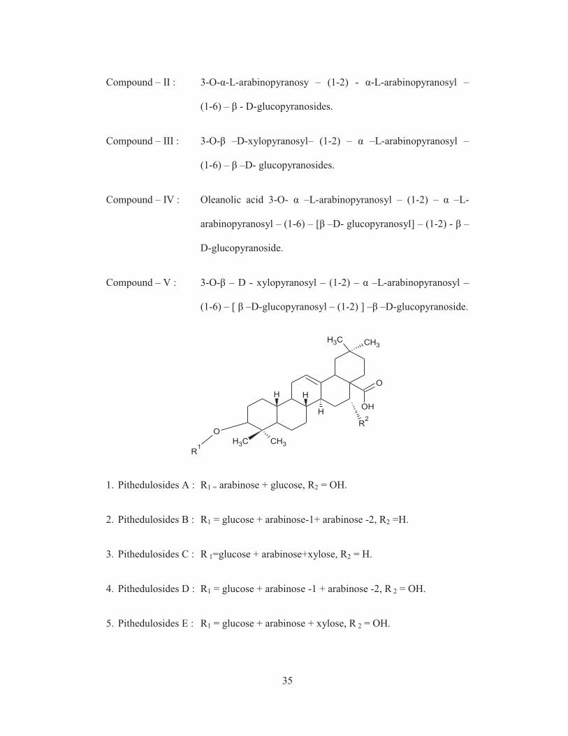
35
Compound – II : 3-O-α-L-arabinopyranosy – (1-2) - α-L-arabinopyranosyl –
(1-6) – β - D-glucopyranosides.
Compound – III : 3-O-β –D-xylopyranosyl– (1-2) – α –L-arabinopyranosyl –
(1-6) – β –D- glucopyranosides.
Compound – IV : Oleanolic acid 3-O- α –L-arabinopyranosyl – (1-2) – α –L-
arabinopyranosyl – (1-6) – [β –D- glucopyranosyl] – (1-2) - β –
D-glucopyranoside.
Compound – V : 3-O-β – D - xylopyranosyl – (1-2) – α –L-arabinopyranosyl –
(1-6) – [ β –D-glucopyranosyl – (1-2) ] –β –D-glucopyranoside.
CH3 CH3
O
OHH
CH3 CH3
H H
O
R1
R2
1. Pithedulosides A : R1 = arabinose + glucose, R2 = OH.
2. Pithedulosides B : R1 = glucose + arabinose-1+ arabinose -2, R2 =H.
3. Pithedulosides C : R 1=glucose + arabinose+xylose, R2 = H.
4. Pithedulosides D : R1 = glucose + arabinose -1 + arabinose -2, R 2 = OH.
5. Pithedulosides E : R1 = glucose + arabinose + xylose, R 2 = OH.

36
6. Pithedulosides F : R1 = glucose -1+glucose -2+arabinose -1+arabinose –2, R 2=H .
7. Pithedulosides G : R 1 = glucose -1 + glucose -2 + arabinose +xylose, R 2 = H.
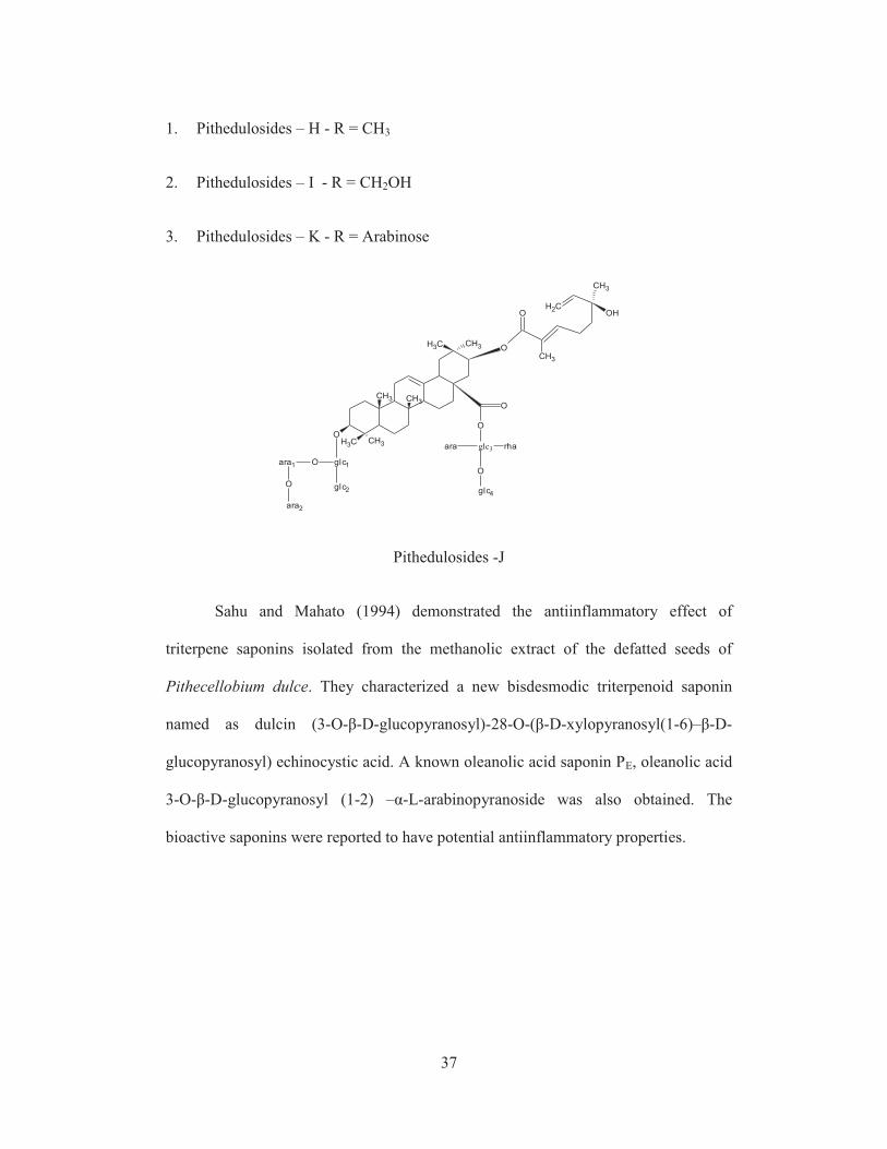
However Yoshikawa et al., (1997) identified four new oleanane type of
triterpene glycosides namely, Pithedulosides H-K which were isolated from the seeds
of Pithecellobium dulce.The structures have been characterized by extensive Nuclear
Magentic Resonance experiments and chemical methods. Compounds 1-3 comprised
of acacic acid as the aglycon and either monoterpene carboxylic acid and its xyloside
or monoterpene carboxylic acid as the acyl moiety at C-21. The oligosaccharide
moieties were linked to C-3 and C- 28 were determined as α –L- arabinopyranosyl-
(1-2) – α –L-arabinopyranosyl – (1-6) – [ β –D-glucopyranosyl and α –L- arabino
furanosyl – (1-4) –[ β –D-glucopyranosyl –(1-3) ] – α –L-rhamnopyranosyl – (1-2) –
β – D - glucopyranosyl ester respectively. Compound 4 was established as an
echinocystic acid – 3-O-glycoside having the same sequences as 1-3. Another known
compound was also identified associated with the new compounds which is
designated as echinocystic acid -3-O-β –D-xylopyranosyl – (1-2) – α –L-
arabinopyranosyl – (1-6) – [ β –D-glucopyranosyl] – (1-2) –β –D-glucopyranoside.
CH3 CH3
O
O
CH3
CH2
CH3
O
O
O
H O
OH H
H OH
O
O
CH2
OH
MTA2
R
O
O
glc3
O
rhaara
Oglc4
CH3CH3
O
glc1
glc2
ara1
ara2
37
1. Pithedulosides – H - R = CH3
2. Pithedulosides – I - R = CH2OH
3. Pithedulosides – K - R = Arabinose
O
CH3 CH3
CH3CH3
CH3
O
O
CH3
CH2OH
CH3
O
O
glc3 rhaara
O
glc4
CH3
glc1
glc2
Oara1
O
ara2
Pithedulosides -J
Sahu and Mahato (1994) demonstrated the antiinflammatory effect of
triterpene saponins isolated from the methanolic extract of the defatted seeds of
Pithecellobium dulce. They characterized a new bisdesmodic triterpenoid saponin
named as dulcin (3-O-β-D-glucopyranosyl)-28-O-(β-D-xylopyranosyl(1-6)–β-D-
glucopyranosyl) echinocystic acid. A known oleanolic acid saponin PE, oleanolic acid
3-O-β-D-glucopyranosyl (1-2) –α-L-arabinopyranoside was also obtained. The
bioactive saponins were reported to have potential antiinflammatory properties.

38
Sahu et al., (1999) characterized a novel acylated triterpenoid saponin,
designated Pithecelloside from the methanolic extract of defatted, air-dried seeds of
Pithecellobium dulce. The compound had been elucidated by interpreting the spectral
datas and identified as 3-O- [ α –L-arabinopyranosyl- (1-2) – α –L-arabinopyranosyl –
(1-6) –β –D- glucopyranosyl] – 21 β – O – [ (2’E) – 6’- hydroxyl – 2’6’- dimethyl
octa – 2’7’ –dienoyl] acacic acid.
CH3CH3
CH3
O
CH3
O
OHOH
CH3CH3
O
O
CH3
CH2
CH3
OH
O
OH
OH
O
OH
OH
OH
H
OOH
OHO
H
O
OH
H
Pithecelloside
O
O
O
O
O
O
O
CH3CH3
CH3CH3
O
O
O
R1
OR
1
O
R1
O
R1O
R1
O
R1
O R1
O
R1
O
R1
OR
1
O
R1
O
R1
O
R
39
Further Niranjan et al., (1994) characterized echinocystic acid bisdesmoside a
new bisdesmodic triterpenoid saponin, from the seeds of Pithecellobium dulce.
Kallappa and Hosamani, (1995) characterized vernolic acid (12,13-epoxy-octadec-cis-
9-enoic acid, 10.0%), malvalic acid [7-(2-octacyclopropen-1-yl) heptanoic acid,
3.2%] and sterculic acid [8-(2-octacyclopropen-1-yl) octanoic acid, 2.0%] from
Pithecellobium dulce, Benth (syn.Inga dulcis, Willd) seed oil, belonging to the
Leguminosae plant family, The other normal fatty acids are palmitic (12.1%), stearic
(4.2%), behenic (10.6%), oleic (34.1%), and linoleic (23.8%).
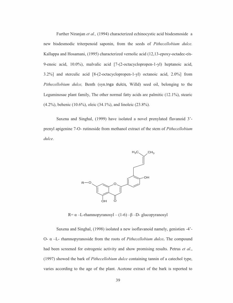
Saxena and Singhal, (1999) have isolated a novel prenylated flavanoid 3’-
prenyl apigenine 7-O- rutinoside from methanol extract of the stem of Pithecellobium
dulce.
O
O
OH
CH3 CH3
OH
OR
R= α –L-rhamnopyranosyl – (1-6) –β –D- glucopyranosyl
Saxena and Singhal, (1998) isolated a new isoflavanoid namely, genistien -4’-
O- α –L- rhamnopyranoside from the roots of Pithecellobium dulce. The compound
had been screened for estrogenic activity and show promising results. Petrus et al.,
(1997) showed the bark of Pithecellobium dulce containing tannin of a catechol type,
varies according to the age of the plant. Acetone extract of the bark is reported to
40
consist mostly of 3,4,7,3’,4’- pentahydroxy flavan a compound which combines the
properties of both leucoanthocyanidin and phlobotannin. The antitumour compound,
b-sitostero and campesterol, stigmasterol and a-spinasterol were reported in the heart
wood of this plant.
2.7 Biological activity of isolated compounds from pithecellobium dulce
The fungistatic activity and antimycobacterial study has been worked out by
many authors. The fungistatic activity of Pithecellobium dulce on mycelial growth of
Alternaria sp., Botrytis cinerea, Colletotrichum gloeosporioides, Fusarium
oxysporum, Penicillium digitatum, Pestalotiopsis sp. and Rhizopus stolonifer was
done and the seeds had the highest fungistatic activity against the fungi tested
(Barrera and Bautista, 2002). Mukesh kumar et al., (2013) evaluated the
antimicrobial activity of leaf of Pithecellobium dulce against twenty pathogenic
microorganisms and assessed the antimicrobial activity of leaf extracts against five
Gram-positive (Bacillus subtilis, Enterococcus faecalis, Micrococcus luteus,
Staphylococcus aureus and Staphylococcus epidermidis), seven Gram-negative
(Aeromonas hydrophila, Alcaligenes faecalis, Enterobacter aerogenes, Escherichia
coli, Klebsiella pneumoniae, Pseudomonas aeruginosa and Salmonella typhimurium)
bacteria and eight fungi (Aspergillus flavus, Aspergillus niger, Aspergillus oryzae,
Aspergillus terreus, Alternaria alternata, Alternaria brasicola, Alternaria solani and
Alternaria vitis). Sukantha et al., (2011) also found the antibacterial potential of the
fruit peel of Pithecellobium dulce.
The leaves of Pithecellobium dulce Benth. showed activity by
BACTEC460TBR radiospirometric system and found at the concentration of
41
20mg/ml leaf extracts shows highest activity when comparable with standard drugs
like, streptomycin, isoniazid, rifampicin, ethambutol and pyrazinamide
(Shanmugakumaran et al., 2005 and 2006). However Bautista et al., (2003) and Ali
et al., (2001) worked on the methanolic and hexane extracts of Pithecellobium dulce
seeds and proved methanolic extract of Pithecellobium dulce showed higher efficacy
against bacteria and fungi than hexane extract.According to Barrera et al., (2003) The
fractions and compounds from seed powder were tested against various postharvest
fungi. The most sensitive strain was Fusarium oxysporum and the less sensitive strain
was Penicillium digitatum.
Megala and Geetha, (2010) studied the gastroprotective effect of
hydroalcoholic fruit extract of Pithecellobium dulce on some important antioxidant
enzymes such as superoxide dismutase , catalase, glutathione reductase , glutathione
peroxidase and myeloperoxidase. The antioxidant activity of anthocyanin extracted
from Pithecellobium dulce fruit pericarp and fruit peel have been detailed by
Ponmozhi et al., (2011), Sukantha et al., (2011), Arul and Muthukumaran, (2011);
Bhargavakrishna et al.,(1970) reported the analgesic and anti-inflammatory effects of
methanol extract and saponins from Pithecellobium dulce. Govindarajan et al., (2012
and 2013) studied the larvicidal and ovicidal potential of the crude hexane, benzene,
chloroform, ethyl acetate and methanol solvent extracts from the medicinal plant
Pithecellobium dulce against the mosquito vector, Anopheles stephensi and Aedes
aegypti (Diptera: Culicidae) and found seed extracts have low potency compared to
leaf extracts. The study involves antioxidant enzymes and the adulticidal efficacy of
seed extracts of Pithecellobium dulce against Culex quinquefasciatus for controlling
filariasis vector mosquitoes was also determined .
42
Sundarrajan et al., (2010) found the anti-hyperlipidemic activity of aqueous
extract of leaves of Pithecellobium dulce against triton induced hyperlipidemia.
According to Mule et al., (2011) the anticonvulsant activity testing by
pentylenetetrazole method showed that aqueous and ethanolic extract of
Pithecellobium dulce could not decrease the convulsions as well as mortality in mice.
However the aqeous fruit extract of Pithecellobium dulce protects murine liver against
CCl4-induced oxidative impairments probably by its antioxidative property (Prasenjit
et al., 2010).
Delgado et al., (2004) reported for the first time the isolation and
characterization of a protease inhibitor from the seeds of Pithecellobium dulce. The
N-terminal sequence of PDTI has the highest similarity with the seed inhibitor of
Acacia confuse. However Rao et al., (2011) evaluated chemical composition and
sensory quality of white and pink aril powders of Pithecellobium dulce in two
packaging systems during storage for six months. The protein contents were 12.4 and
15.0% in white and pink aril powders. The titrable acidity of white and pink aril
powders were 2.4 and 4.8%.Ca and Fe contents in white aril powder samples were 60
and 12 mg/100 g where as in pink aril powder 62 and 16 mg/100 g. The anthocyanin
content in pink powder decreased from 50.5 to 11.2 and 14.1 mg/100 g in samples
packed in polyethylene and metalized polyester polyethylene laminated pouches.
According to Sugumaran et al., (2008) the locomotor activity of aqueous and
alcoholic extracts of leaves of Pithecellobium dulce Benth was determined and CNS
depressant activity of leaf extracts of Pithecellobium dulce was evaluated using
actophotometer in albino mice.
43
Pithayanukul et al., (2005) studied on the aqueous extract of
Pithecellobium dulce for their inhibitory activities against Naja kaouthia venom by
invitro neutralization method. The antivenom activity study of the extract could
completely inhibit the lethality of the venom at 4 LD50 concentration and the venom
necrotizing activity at the minimum necrotizing dose also inhibited up to 90% of the
acetylcholinesterase activity of Naja kaouthia venom at much lower tannin
concentration. The ED50 of plant tannins inhibiting Naja kaouthia venom activities
due to condensed tannins and their content in the extracts. The spermicidal effect of
saponins in Pithecellobium dulce was also reported by Misra et al., (1979).Sugumaran
et al., (2006) examined the macroscopical characters of the leaves, leaf constants,
physico chemical constants, extractive values with different solvents, micro chemical
test, flouresence characters of liquid extracts and leaf powder of Pithecellobium dulce.
Khanzda et al., (2013) measured some essential and toxic elements As,Cu, Cd, Fe, K,
Mg, Na, Pb & Zn in different concentrations Zn & K being the highest (26.89 mg/kg)
and Pb (0.19mg/kg) and As(17.6μg/kg) were the lowest in concentrations justifying
its medicinal applications.
The antidiabetic effect of Pithecellobium dulce has been revealed by many
authors both by invivo and invitro methods. Pradeepa et al., (2013) evaluated the
antidiabetic potential of Pithecellobium dulce fruits in STZ - induced experimental
diabetes in rats. Oral administration of Pithecellobium dulce fruit extract (300 mg/kg
b.w. /day) to diabetic rats for 30 days significantly reduced the levels of blood
glucose. Similar observation was found by Sugumaran et al., (2009) Pithecellobium
dulce leaves using streptozotocin induced diabetic model in rats. The aqueous extract

44
showed significant activity (P<0.01) than the alcoholic extract at the tested dose level
which was comparable to glibenclamide, a standard antidiabetic drug.
The invivo antidiabetic effect was shown by many studies. Dnyaneshwar and
Archana, (2013) reported the inhibitory action of methanolic extract and acetone
extract of Pithecellobium dulce seeds, bark and leaves. Seeds showed inhibitory
action on α- amylase and α-glucosidase enzymes and compounds responsible for
these activities are characterized. The enzyme inhibitory activity of Pithecellobium
dulce methanolic extract may be endorsed to the presence of oleanolic acid
triterpenoid. The bark and leaves of Pithecellobium dulce were evaluated for α-
amylase and α-glucosidases inhibition in vitro, compared with acarbose. Acetone
extracts of bark and leaves showed more sucrase inhibition than maltase while
methanol extract of bark and leaves showed more sucrase inhibition than maltase.
2.8 In-vitro study
Non-enzymatic glycosylation of haemoglobin assay of Pithecellobium dulce
Invitro study was done outside the living body. The antioxidant ability of
enzymatic and non enzymatic process decreases the active oxygen species responsible
for many diseases including diabetes. The rise in glucose level in the blood leads to
the binding of haemoglobin and glucose with the formation of the reactive species.
(Clifford et al., 1989). Apart from reducing blood glucose level, the glucose level
was controlled by inhibition of α-glucosidase activity in the intestine. According to
Kirchner et al., (1996) inhibition of α-glucosidase activity in the intestine was
achieved by using luteolin, kaempferol, chrysin and galangin to study the effect of
absorption and metabolism of carbohydrates. Megha et al., (2013) worked on dried
45
(crude) petroleum ether and hexane extracts of stem bark of Bauhinia purpurea. In-
vitro anti-diabetic activity reports showed significant level of activity, the highest
concentration of extract was effective as an anti-diabetic agent. Similar invitro
antidiabetic activity of phytochemical bioactive compounds of the methanolic extract
of Psidium guajava leaves was observed by Manikandan et al., (2013).
Andichettiyar and Tapan, (2013) and Ali et al., (2013) reported the in vitro α-
amylase and α-glucosidase enzymes inhibitory activity in ethanol and chloroform
extracts of Polyalthia longifolia (Sonner). The leaves and fractions of the dried fruit
pericarp of Phaleria macrocarpa potently inhibiting carbohydrate hydrolysing
enzyme. Further study showed the inhibitory action of methanolic extract of
Pithecellobium dulce seeds on α- amylase and α-glucosidase enzymes. The IC50
values of methanolic extract of Pithecellobium dulce against pancreatic α-amylase
was found to be 16.75±1.81 mg/ml.
Faiyaz and Asna, (2009) compared the hypoglycemic effect of Ficus
racemosa stem bark (Moraceae) and the glucose diffusion activity of wheat bran and
and acarbose using in vitro technique. Ficus racemosa bark exhibited significantly
higher (P ≤ 0.01) glucose-binding capacity than wheat bran and acarbose
consequently showed significantly higher (P ≤ 0.01) retardation of glucose diffusion
compared to wheat bran and acarbose. Ashok Kumar et al., (2013) revealed in vitro
antidiabetic screening of Nisamalaki Churna by α-amylase inhibition using starch
iodine method.
Stalin et al., (2013) examined the crude n-hexane, ethanol, methanol and
aqueous leaf extracts of cardiospermum and the invitro inhibitory effect of glucose
46
utilization. Palaniswamy and Sellapa, (2012) compared the inhibitory activity of the
aqueous extracts of four seaweeds Gracilaria edulis ,Sargassum polycystum, Ulva
lactuca, Gracilaria corticata collected from Gulf of Mannar coastal waters and tested
for α - amylase and α –glucosidase inihibition and found Gracilaria corticata with
significant inhibitory activity against α - amylase and α - glucosidase enzymes.
2.9 Toxicity test in mice
Animal models are indispensable tools in biomedical research. They have
been used to find out the effectiveness and the toxicities of various medicines and
chemicals. Investigation of acute toxicity is the first step in toxicological analysis of
herbal drugs .The present study was undertaken to determine the acute oral toxicity
and safety parameters of compounds isolated from the plant parts. Ogbonnia et al.,
(2013) and Arthur et al., (2011) shown a detailed study on acute and subchronic
toxicity in animals in polyherbal formulation and aqueous extract of A. muricata
leaves using standard procedures and the LD50 value indicated the drug as being safe.
At the highest dose, the formulation exhibited deleterious effect on the hepatic tissue.
The outcome of acute oral toxicity of ethanolic extract of M. pruriens on Swiss albino
mice was reported by Parekar and Somkumar , (2011) found that no mortality or
evidence of adverse effect have been observed in Swiss albino mice following acute
oral administration of ethanolic extract at the dose of 2000 mg /kg .
According to Protus et al., (2012) Sub-acute (aqueous and ethanol extract)
and sub-chronic oral toxicity (aqueous only) were carried out on the aqueous and
ethanol leaf extracts of Carica papaya (CP) in Wistar rats. Control groups received
water and corn oil respectively.The result suggests that aqueous extract showed lesser
47
toxicity than the ethanol extract. Paramakrishnan et al., (2012) and Lalitha et al.,
(2012) reported the acute oral toxicity test on the various extracts of aerial parts of
parviflorab and Eichhornia crassipes and the animals were orally administered a
single dose of 100, 250, 500,750, 1000, 2000 mg/kg body weight. Signs of toxicity
and mortality were noted after 1, 4 and 24h of administration of the extract for 14
days. The highest dose administered (2000mg/kg body weight) did not produce
mortality or changes in general behaviour of the test animals.Further Subramanion et
al., (2011) investigated the acute oral toxicity of C. fistula seeds extract in mice and
found that oral administration of crude extract at the highest dose of 5000 mg/kg
resulted in no mortalities or evidence of adverse effects, implying that C. fistula is
nontoxic and the extract can be utilized for pharmaceutical formulations. Acute and
subacute toxicity of ethanol (95% v/v) extract of aerial parts of Cansjera rheedii J.
Gmelin (Opiliaceae) and Tephrosia purpurea, the limit test dose of 2000 mg/kg were
used for Cansjera rheedii J. Gmelin (Opiliaceae) and 400mg/kg for T. purpurea. No
significant variation (p < 0.05) in the body and organ weights between the control and
the treated group was observed after 28 days of treatment.
2.10 Invivo antidiabetic study
Animal model is important to assess the biological activity of compounds
found in plants. Mouse is the most cited animal model, 99% of mouse genes have
human counterparts. Mouse models of many human diseases have also been
developed to advance the studies of disease pathogenesis, and to evaluate the
effectiveness of various candidate drugs.
48
Considerable literature is available on hypoglycemic effect of various plant
parts using mice model. Comprehensive studies on different plant extracts, isolated
plant compounds have been made by various authors. The hypoglycemic effect on
alloxan induced diabetic mice were studied on various plants (Meshram et al.,2013);
hydro alcoholic extract of stem bark of Bauhinia purpurea (Mishra, 2013); aqueous
extract of Anethum graveolens seeds (Abu et al., 2010); ethanolic extracts of
Scoparia dulcis and ethyl acetate extract of Peporemia pellucide (Hasib et al., 2012).
Other important work on alloxan induced diabetic mice include those of
Gunjan and Krishnendua, (2013); in ethanolic extract of Astraeus hygrometricus
(Pers.) Morg. (Astraeceae); Ihechilure, (2010); crude extract of I .trichantha leaves;
Franscisco et al., (2006); in Hexane, dichloromethane, methanol and aqueous extracts
of the Plantago Major seed; in T. belerica and crude methanolic extract of the
Potentilla fulgens L roots (Sabu and Kuttan, 2009).
Syiem et al., (2002) studied the effect of the methanolic extract of Albizia
odoratissima Benth. bark and showed the reduction of blood glucose level with
various other factors like serum cholesterol, triglycerides,serum glutamic pyruvic
transaminase, alkaline phosphatase and decreases level of total proteins in alloxan
induced diabetic mice. Dinesh Kumar et al., (2011) while studying the effect of
crude methanolic extract of the Potentilla fulgens L roots for its hypoglycemic effect
found that it depends upon the dosage level and time factor. Njagi et al., (2012)
observed that aqueous stem bark extract of Ficus sycomorus safely lowered blood
glucose level to levels below insulin, the model drug in a dose-dependent manner.

49
Hypoglycemic effect was also analyzed in streptozotocin induced diabetic
mice on the methanolic extract of Diplocyclos Palmatus (Jeyanarayan et al., 2012).
Many studies have been conducted on streptozotocin diabetic induced rats model by
several workers (Palaniswamy et al., 2011); Ellagic acid ; (Sayed et al., 2011);
aqueous extracts of Mangifera indica and Psidium guajava ; (RajKumar et al., 2011) ;
methanolic extract of Hibiscus cannabinus.
Various authors showed the hypoglycemic effect on streptozotocin induced
diabetic rats at different dosages Nilufer et al., (2006) observed the acute and
subacute antidiabetic activities of the ethanolic extract of Vitis vinifera L. leaves at
250 mg/kg dose was found to possess a high antidiabetic and antioxidant activity.
Oral administration of the methanol extract of E.jambolana (EJ) (150mg/kg bw) for
60 days to streptozotocin (STZ) (60mg/kgbw) induced male diabetic wistar rats was
able to significantly (p<0.05) decrease the blood glucose concentration (Jasmine and
Daisy, 2011). The potential antidiabetic ability of Gymnema sylvestre was identified
by Vijanand, (2012) the dose of 200 and 400mg/kg decreases the glucose level
significantly.
Papiya et al., (2008) made a detailed study on hypoglycemic effect of an
isolated compound (Ficanone) from Ficus arnottiana bark in normal and
streptozotocin diabetic rats. When administered to diabetic rats, Ficanone (50 mg/kg,
p.o.) caused a significant (p<0.01) reduction in glucose level. Mi-Jang et al., (2011)
determined the hypoglycemic effect of Chenopodium ambrosioides in mice fed with
high-fat diet for two weeks before induction of diabetes mellitus by injection of
streptozotocin. Animals treated with crude extract (100, 200 and 300 mg/kg) showed

50
significant (p<0.05) hypoglycemic effect in comparison to control. Rupali et al.,
(2011) tested the anti hyperglycaemic and antidiabetic potential of leaf extracts of
Albizzia lebbeck (Benth), Psidium guajava (Linn), and Trigonella foenum-graecum
(Linn) against alloxan and streptozotocin induced diabetic models of mice and
showed a positive trend in regulating blood glucose levels in Swiss albino mice by
using Rosiglitazone as a standard drug. Aicha et al., (2013) showed the effect of the
aerial crude aqueous extract part of Anacyclus valentinus L. on diabetes induced
Wistar rats by streptozotocin and endowed significant antidiabetic activity.
Rekha et al., (2010) justified the use of a combination of aqueous extracts of
pulp of Syzygium cumini and bark of Cinnamon zeylanicum for the remedial effects
against streptozotocin induced diabetic state .However. Rajesh et al., (2005) revealed
the antidiabetic activity of the water extract of Annona squamosa (custard apple) in
diabetic animals. Hot-water extract of the leaves of A. squamosa was prepared by
boiling fresh and air dried leaves (25°C for 5 days) with water (20 ml/g) for 2 h and
was tried by oral route in alloxan (80 mg/kg bw)-induced diabetic rabbits and
streptozotocin (STZ) (50 mg/kg bw)-induced diabetic rats and reveals that
A. squamosa has both hypoglycaemic and antidiabetic activity. Oyedepo et al.,
(2013) confirmed that the oral administration of aqueous leaf extract of Moringa
oleifera may reduce the plasma lipid imbalances associated with diabetes mellitus.
Further Kavishankar et al., (2010) found the antidiabetic effects of aqueous fruit
extract of Morinda citrifolia and Cocinia Indica in alloxan induced rats for a period
of 30 days. Syamsudin, (2010) conducted antidiabetic test on active fractions of
methanol extract from Leucaena leucocephala (lmk) De Wit seeds using alloxan-
induced rats. Manikandan et al., (2009) observed hyperglycemic nature of
51
Isolated 1, 2 di-substituted idopyranose from Vitex negundo against streptozotocin-
induced diabetes.
Nancy et al., (2011) evaluated antidiabetic property of Sonneratia alba Sm,
when subjected to anti-diabetic bioassay using standard Glucometer, The blood sugar
level was reduced by 19.2% during the first 6 hours and reduced further to 66.9%
after 12 hours. Momoh et al., (2011) analyzed significant hypoglycaemic effect
(p<0.05) of Costus afer in adult male wistar strain albino rats and the effect was
comparable with the standard drug glibenclamide .
2.11 Compound isolation and identification of compounds using spectroscopic
technique
Number of bioactive compounds were isolated from different plant parts by
chromatographic technique and the structure of the compounds were identified using
spectroscopic method. Shah et al., (2013) isolated the antidiabetic compounds from
the kernel of mango using column and thin layer chromatographic techniques and
compounds are identified using 1H-NMR spectroscopy. Fang et al., (2005) has
widely shown the isolation and identification of sesquiterpene lactones was carried
out in in three steps: multiple extractions, fractionation using column chromatography
and the separated compounds was determined on the basis of 1HNMR and
13CNMR.
Shalabia, (2011) examined the plant Atriplex halimus and isolated eleven free amino
acids and revealed the presence of three alkaloids at Wadi-Sudr habitat and two
alkaloids at Wadi-Hof habitat.
Isolation of bioactive compound from the leaves of Excoecaria agallocha was
done using thin layer chromatography and column chromatographic techniques
52
(Patra et al., 2012) Crotepoxide was isolated from ethyl acetate mixture of Croton
macrostachyus berries using chromatographic technique (Habtamu et al., 2012).
Getahun et al., (2012) have worked using the same column chromatography and
spectroscopic methods (IR and NMR) for the isolation and identification of betulinic
acid and benzoic acid from the root extract of Helinus mystachinus. Using column
chromatography Christinah et al., (2012) isolated the bioactive from G. kola seeds.
Ali et al., (2013) showed that chromatographic techniques were used to isolate
phenolic compounds from plants.
Abdul et al., (2010) included liquid layer chromatography, column
chromatography and preparative thin layer chromatography for the isolation of the
compounds dillenetin, betulinic acid, β – sitosterol and stigmasterol from the crude
leaf extracts of Dillinea indica Linn. Surendar et al., (2011), Veena and Ritu (2013)
separated the saponins from Moringa oleifera using thin layer chromatography
(TLC) on mobile phase of Chloroform: methanol: H2O (7:3:1) on silica gel glass
plates and high performance liquid chromatography (HPLC) is used to isolate
compound from benzene extract and further showed characterization of isolated
saponin was done using eletrospray ionization mass spectrometry ESI-MS, IR
,1HNMR and
13C-NMR. San et al., (2011) showed that thin layer chromatography,
ultraviolet (UV) and Infra-red (FTIR) spectroscopic method were used in the
isolation and identification of chemical constituents of compounds from ethanolic
extract of leaves of C. inerme Gaertn. Mary et al., (2012) and Rajkumar et al., (2012)
isolated the bioactive compound using silica gel column chromatography from
Aspergillus sp and O. indicum stem bark.
53
The present review give knowledge about diabetes, and its various herbal
treatment. The investigation on Pithecellobium dulce give knowledge about
Pithecellobium species with reference to its pharmacological potentials and
biochemical spectrum. The present study was chosen with the following aim and
scope
· Extract preparation of leaf ,fruit and fruit peel of Pithecellobium dulce using
various solvents using soxhlet extractor.
· Determination of various physiochemical parameters.
· Determination of inorganic minerals in plant parts.
· The various extracts are subjected to preliminary phytochemical screening for
the presence of alkaloids, flavones, terpenes, steroids, tannins, quinines and
glycoside.
· Estimating the amount of protein, carbohydrate, total lipids, total alkaloids,
total flavanoid, total phenol using quantitative phytochemical analysis.
· Phytochemical isolation of compounds present in leaves ,fruit and fruit peel
using column chromatography .
· To isolate kaempferol and afzelin from methanolic extracts of leaves using
column chromatography .
· To isolate quercitrin from methanolic extract of fruit using column
chromatography.
· To isolate two new compounds from the methanol extract of fruit peel using
column chromatography.

54
· To identify the isolated compounds using UV, IR, 1HNMR,
13CNMR and
mass spectroscopy.
· To detect the invitro hypoglycemic activity of isolated compounds from leaf,
fruit and fruit peel of Pithecellobium dulce using non enzymatic glycosylation
of haemoglobin assay and the enzymatic study alpha-amylase inhibition
assay .
· To detect the invivo antidiadetic effect of isolated compounds from leaf, fruit
and fruit peel of Pithecellobium dulce using alloxan induced diabetic Swiss
albino mice .
· To compare the the antidiabetic hypoglycemic effect of different isolated
compounds from the leaf, fruit and fruit peel.
· To determine the angiogenisis activity of the newly isolated compound from
fruit peel, using CAM assay.