review glucose^induced alterations in nerve - diabetes care
TRANSCRIPT

Review
Glucose^induced Alterations inNerve Metabolism: Current Perspective on
the Pathogenesis of DiabeticNeuropathy and Future Directions for
Research and TherapyDOUGLAS A. GREENE, M.D., SARAH LATTIMER, B.S., JAN ULBRECHT, M.D., AND PATRICIA CARROLL, M.D.
Recent animal and in vitro studies have identified several interrelated metabolic abnormalities in diabeticnerve that are attributable to elevated ambient glucose concentrations. In combination, these metabolicchanges may induce a variety of biochemical and biophysical alterations in peripheral nerve that arehighly relevant to the pathogenesis of diabetic neuropathy. This article reviews the current status ofseveral of these metabolic defects and describes ways in which their interaction could lead to patho-genetically important changes in nerve metabolism, function, and structure. Areas of related futureresearch are also discussed, DIABETES CARE 1985; 8:290-99.
Peripheral neuropathy is probably the most common1
and certainly one of the most troubling chroniccomplications of diabetes. As with other major di-abetic complications, its pathogenesis remains en-
igmatic and its therapy controversial. Although the trueincidence of diabetic neuropathy has never been rigorouslyestablished in a population-based study, the prevalence ofneuropathy appears to correlate with duration and severity ofhyperglycemia in both insulin-dependent and non-insulin-dependent diabetes mellitus.2 This observation suggests thatelevated blood glucose fluctuations, or other metabolic con-sequences of insulin deficiency in the diabetic state, contrib-ute to the pathogenesis of diabetic neuropathy,3 a contentionfurther supported by the improvement in nerve conductionparameters following metabolic intervention in diabetic hu-man beings4"10 and animals.11"13 Thus, a pathogenetic rolefor the metabolic alterations of insulin deficiency is now gen-erally accepted for diabetic neuropathy, but the extent andimportance of that role remain subjects of great controversy.An array of interrelated metabolic alterations in diabetic nervethat are consequences of elevated ambient glucose concen-tration have recently emerged (Table 1). Several of themappear to interact in a self-reinforcing or synergistic fashion,and may impact on important metabolic and functional proc-
esses in peripheral nerve; thus they have potentially importantroles in the pathogenesis of diabetic neuropathy and providepotential sites for new and specific therapeutic intervention.This article reviews several of these metabolic defects, de-scribes how they could contribute to the pathogenesis of di-abetic neuropathy, and addresses potential areas for thera-peutic intervention and future research.
GLUCOSE AND ENERGY METABOLISM IN NORMALAND DIABETIC NERVE
Consideration of potential mechanism(s) by which hypergly-cemia and/or insulin deficiency could alter nerve functionrequires a brief discussion of general nerve carbohydrate andenergy metabolism. Peripheral nerve, like brain, is seques-tered behind a specialized and relatively impermeable diffu-sion barrier, the "blood-nerve barrier," comprised of peri-neurial epithelial and nonfenestrated endoneurial endothelialcells. The permeability characteristics of this barrier, whichresemble those of the blood-brain barrier, severely limit thetype of substrate available to peripheral nerve by excludingsuch important fuels as free fatty acids.1415 In the brain, whichis also largely inaccessible to circulating free fatty acids, energymetabolism is nearly completely dependent on glucose as a
290 DIABETES CARE, VOL. 8 NO. 3, MAY-JUNE 1985
Dow
nloaded from http://diabetesjournals.org/care/article-pdf/8/3/290/497220/8-3-290.pdf by guest on 19 M
arch 2022
GLUCOSE-INDUCED ALTERATIONS IN NERVE METABOLISM/D. A. GREENE AND ASSOCIATES
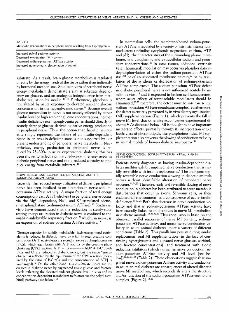
TABLE IMetabolic abnormalities in peripheral nerve resulting from hyperglycemia
Increased polyol pathway activityDecreased m^o-inositol (MI) contentDecreased sodium-potassium ATPase activityIncreased nonenzymatic glycosylation of protein
substrate. As a result, brain glucose metabolism is regulateddirectly by the energy needs of the tissue rather than indirectlyby hormonal mechanisms. Studies in vitro of peripheral nerveenergy metabolism demonstrate a similar substrate depend-ency on glucose, and an analogous independence from met-abolic regulation by insulin.16"18 Furthermore, glycolysis isnot altered by acute exposure to elevated ambient glucoseconcentration in the hyperglycemic range.18 Because overallglucose metabolism in nerve is not acutely affected by eitherinsulin level or high ambient glucose concentrations, neitherinsulin deficiency nor hyperglycemia per se should directly oracutely derange glucose-derived oxidative energy productionin peripheral nerve. Thus, the notion that diabetic neurop-athy simply represents the failure of an insulin-dependenttissue in an insulin-deficient state is not supported by ourpresent understanding of peripheral nerve metabolism. Nev-ertheless, energy production in peripheral nerve is re-duced by 25-30% in acute experimental diabetes; this hasbeen shown to reflect a primary reduction in energy needs indiabetic peripheral nerve and not a reduced capacity to pro-duce energy from metabolic substrate.18*
NERVE ENERGY AND myo-INOSITOL METABOLISM, AND THESODIUM-POTASSIUM ATPase
Recently, the reduced energy utilization of diabetic peripheralnerve has been localized to an alteration in nerve sodium-potassium ATPase activity. A major fraction of total energyconsumption (i.e., ATP hydrolysis) in peripheral nerve occursvia the Mg2+-dependent, Na+- and K+-stimulated adeno-sinetriphosphatase (sodium-potassium ATPase).19 Studies invitro have demonstrated that the reduction in steady-stateresting energy utilization in diabetic nerve is confined to theouabatn-inhibitable respiratory fraction,20 which, in nerve, isan expression of sodium-potassium ATPase activity."
'Storage capacity for rapidly mobilizable, high-energy-bond equiv-alents is reduced in diabetic nerve by a fall in total creatine con-centration (ATP equivalents are stored in nerve as phosphocreatine[P-Cr], which equilibrates with ATP and Cr by the creatine phos-phokinase [CPK] reaction: ATP + Cr <- •> ADP + P-Cr; bothP-Cr and Cr are reduced in diabetic nerve, but the tissue "energycharge" as reflected by the equilibrium of the CPK reaction [meas-ured by the ratio of P-Cr:Cr and the concentration of ATP] isunchanged).18 On the other hand, tissue substrate stores are in-creased in diabetic nerve by augmented tissue glucose and fructoselevels reflecting the elevated ambient glucose level in vivo and itsconcentration-dependent metabolism to fructose via the polyol (sor-bitol) pathway (see below).18
In mammalian cells, the membrane-bound sodium-potas-sium ATPase is regulated by a variety of intrinsic intracellularmodulators (including cytoplasmic magnesium, calcium, ATP,and pH), the characteristics of the surrounding plasma mem-brane, and cytoplasmic and extracellular sodium and potas-sium concentrations.21 In some tissues, additional extrinsic(e.g., hormonal) modulation may occur via phosphorylation-dephosphorylation of either the sodium-potassium ATPaseitself21 or of an associated membrane protein,22 or by regu-lation of the synthesis or degradation of sodium-potassiumATPase complexes.21 The sodium-potassium ATPase defectin diabetic peripheral nerve is not influenced acutely by in-sulin in vitro,18 and is expressed in broken cell homogenates,where acute effects of water-soluble modulators should beeliminated;2023 therefore, the defect must be intrinsic to thesodium-potassium ATPase-membrane complex. Furthermore,the defect is entirely prevented by in vivo dietary myo-inositol(MI) supplementation (Figure 1), which prevents the fall innerve MI level that otherwise accompanies experimental di-abetes.20 As discussed below, MI is thought to have importantmembrane effects, primarily through its incorporation into alabile class of phospholipids, the phosphoinositides. MI sup-plementation also prevents the slowing of conduction velocityin animal models of human diabetic neuropathy.12
NERVE CONDUCTION, SODIUM-POTASSIUM ATPase, AND MIIN DIABETES
Patients newly diagnosed as having insulin-dependent dia-betes mellitus exhibit impaired nerve conduction that is rap-idly reversible with insulin replacement.4 The analogous rap-idly reversible nerve conduction slowing in diabetic animalsoccurs without identifiable alteration of peripheral nervestructure.11'2425 Therefore, early and reversible slowing of nerveconduction in diabetes has been attributed to acute metabolicdisturbances that occur in axons, Schwann cells, or theirendoneurial environment3 as a consequence of acute insulindeficiency.111226 Both this decrease in nerve conduction ve-locity and that in sodium-potassium ATPase activity havebeen causally linked to an alteration in nerve MI metabolismin diabetic animals.12>20'27'28 This conclusion is based on theobserved parallel responses of nerve MI content, sodium-potassium ATPase activity, and motor nerve conduction ve-locity in acute animal diabetes under a variety of differentconditions (Table 2). This parallelism persists during insulinreplacement, oral MI supplementation (in the face of con-tinuing hyperglycemia and elevated nerve glucose, sorbitol,and fructose concentrations), and treatment with aldosereductase inhibitors (which normalize nerve conduction, so-dium-potassium ATPase activity and MI level [see be-low]) 12i2Oi27~30 (Table 2). These observations suggest that im-paired nerve sodium-potassium ATPase activity and conductionin acute animal diabetes are consequences of altered diabeticnerve MI metabolism, which secondarily alters the structureand/or function of the sodium-potassium ATPase-membranecomplex (Figure 2).13'20
DIABETES CARE, VOL. 8 NO. 3, MAY-JUNE 1985 291
Dow
nloaded from http://diabetesjournals.org/care/article-pdf/8/3/290/497220/8-3-290.pdf by guest on 19 M
arch 2022

GLUCOSE-INDUCED ALTERATIONS IN NERVE METABOLISM/D. A. GREENE AND ASSOCIATES
TABLE 2Parallel responses of rat nerve myo-inositol (MI) content, sodium-potassium ATPase activity, and motor nerve conduction velocity (MNCV) to hyperglycemiaand metabolic intervention
NondiabeticDiabeticDiabetic +
insulinDiabetic +
MIDiabetic +
sorbinil
Plasmaglucose(mg/dl)
178 ± 7566 ± 30
75 ± 18
600 ± 32
623 ± 25
PlasmaMI
(M-M)
35 ± 235 ± 3
30 ± 2
245 ± 27
30 ± 3
Nerve MI(mmol/kg)
3.0 ± 0.22.2 ± 0 . 1
3.0 ± 0.2
3.9 ± 0.2
3.1 ± 0 . 1
MNCV(m/s)
62 ± 151 ± 1
63 ± 1
61 ± 1
—
Nerve sodium-potassium ATPase
(mmol/kg/h)
97 ± 555 ± 3
120 ± V
88 ± 4
96 ± 5
Diabetes leads to, and insulin therapy prevents, a reduction in nerve MI content, MNCV, and sodium-potassium ATPase activity. These changes are alsoprevented by treatment with aldose reductase inhibitors (e.g., sorbinil), which inhibit the increase in polyol pathway activation in diabetic nerve and,hence, in accumulation of nerve sorbitol and fructose. Dietary MI supplementation is however sufficient to normalize nerve MI, MNCV, and sodium-potassium ATPase in face of persistent hyperglycemia and elevated nerve glucose, sorbitol, and fructose. Hence, changes in diabetic nerve MI metabolismappear central to pathogenesis of the defects in MNCV and nerve sodium-potassium ATPase.'Data compiled from the streptozocin-diabetic rat'
2l82C69 and older insulin-replaced spontaneously diabetic BB-Wistar rats" whose controls have highernerve sodium-potassium ATPase activity (117 + 10).
THE REGULATION OF INTRACELLULAR MI
Peripheral nerve normally maintains a tissue-to-plasmaMI concentration gradient of 90-100-fold; this gra-dient is reduced in both human31 and animal121318'32
diabetes (Figure 1). A specific, high-affinity, so-dium-dependent, carrier-mediated MI transport system prob-ably contributes to the establishment and/or maintenance ofthis concentration gradient in nerve.27 Hyperglycemic con-centrations of glucose competitively inhibit MI uptake viathis transport system,27 providing a potential mechanismby which diabetes could reduce nerve MI content (Figure2). The low-transport-capacity, high-concentration-gradientcharacteristics of this transport system suggest that factorsthat influence passive efflux of MI might also greatly influencethe MI concentration gradient.
NERVE Ml TRANSPORT AND THE SODIUM-POTASSIUM ATPase
The peripheral nerve MI transport system is driven by thesodium gradient generated by the sodium-potassium AT-Pase.27 Therefore, it might be anticipated that the reducedsodium-potassium ATPase activity of diabetic peripheral nervewould secondarily impair sodium-dependent MI uptake. Re-cent experiments support this view, since sodium-dependentMI uptake remains reduced in diabetic peripheral nerve invitro even after tissue glucose concentrations are normal-ized.33 Furthermore, experimental conditions that improvesodium-potassium ATPase activity also correct sodium-de-pendent MI uptake by diabetic peripheral nerve in vitro,33
suggesting that it is the reduced diabetic nerve sodium-po-tassium ATPase activity that further compromises endoneu-rial sodium-dependent MI uptake. These relationships thusset the stage for a self-re info re ing cyclic metabolic derange-
ment involving MI and the sodium-potassium ATPase in di-abetic peripheral nerve (Figure 2).
RELATIONSHIP OF THE POLYOL (SORBITOL) PATHWAY TO NERVE MlAND SODIUM-POTASSIUM ATPase IN DIABETIC NERVE
The polyol pathway enzymes aldose reductase and sorbitoldehydrogenase are characteristically found in tissues in whichglucose entry is neither modulated by insulin nor rate-limitingfor glycolysis; hence, glucose levels in these tissues reflectthat of their milieu.34 Since both polyol pathway enzymes arehighly substrate dependent, the sequential reduction of glu-cose to sorbitol and oxidation to fructose are primarily reg-ulated by the intracellular glucose concentration, and are thusaccelerated by hyperglycemia (Figure 3).34 Sorbitol, whichdiffuses poorly across cell membranes, accumulates intracel-lularly when polyol pathway flux is high.34
CH2OHO
HO
myo-inositol D-glucose
FIG. I. Structure of myo-inositol. Ml is a cyclic hexahydruxy hexanolfound in high concentrations in most mammalian tissues, including nerve.It is rapidly and reversibly incorporated into a labile class of phospholipids,the phosphoinositides, which are thought to have important membranefunctions. It is synthesized from glucose-6-P; its three-dimensional struc-ture is similar to that of glucose, which inhibits Ml uptake in nerve.
292 DIABETES CARE, VOL. 8 NO. 3, MAY-JUNE 1985
Dow
nloaded from http://diabetesjournals.org/care/article-pdf/8/3/290/497220/8-3-290.pdf by guest on 19 M
arch 2022

GLUCOSE-INDUCED ALTERATIONS IN NERVE METABOLISM/0. A. GREENE AND ASSOCIATES
F/G. 2. Postulated relationship between hyper-glycemia, polyol pathway, myo-inositol, sodium-potassium ATPase, and nerve conduction in dia-betes. Glucose at hyperglycemic concentrationsreduces nerve Ml by competitively inhibiting itsuptake. Increased polyol pathway activity, also aconsequence of hyperglycemia, further contributesto reduced nerve Ml by as yet undefined mecha-nism(s). The reduction in nerve Ml leads to adecrease in sodium-potassium ATPase activity,possibly through an alteration in phosphoinositidemetabolism. Since Ml uptake in nerve is sodiumdependent, a self-reinforcing metabolic cycle in-volving Ml is postulated. Acute reduction in nerveconduction velocity in diabetes is ascribed in partto impaired sodium-potassium ATPase activity. Afurther consequence of impaired sodium-potassiumATPase activity is a reduction in sodium-dependentamino acid uptake; interference with other sodium-linked processes is likely but as yet unproven. De-fects resulting from reduced nerve Ml levels otherthan those mediated by changes in sodium-potas-sium ATPase await further investigation.
i Motor NerveConduction
Velocity
Na+-Dependent V^Amino Acid 4 i Na
Uptake f
? | OtherNa+-Dependent
Processes
Hyperglycemia11
CompetitiveInhibition
1i Na+-Dependent
J1 Ml Uptake
+/K+ ATPaseActivity
^ ^ ^ AlteredPhosphoinositide
Metabolism
\? Other Effects
t Polyol PathwayActivity
I Tissue -4-^Ml
y
Osmotic swelling, secondary to intracellular sorbitol ac-cumulation, has been invoked as a cause of both functionaland morphologic alterations in diabetic nerve.3 This "osmotichypothesis" is derived primarily from studies of experimentalsugar cataracts in rats where experimental diabetes or glucose,galactose, or xylose intoxication causes millimolar intracel-lular accumulations of polyol pathway intermediates.34 Thisaccumulation results in a marked increase in tissue watercontent, and the rapid development of lenticular opacities.34
Lenticular hyperhydration is attributed directly to the osmoticeffects of accumulated polyol pathway intermediates.34
Direct extrapolation of the osmotic hypothesis from lensto diabetic peripheral nerve is problematic for several reasons.First, diabetic peripheral nerve accumulates only micromolarrather than millimolar concentrations of sorbitol, which areunlikely to be osmotically significant unless highly localizedanatomically.3 Second, as discussed earlier, MI administra-tion, which corrects the characteristically reduced tissue MIcontent without influencing nerve sorbitol concentration,normalizes diabetic nerve function.122728 Finally, the tem-poral association in lens between polyol pathway-induced de-fects and MI metabolism suggests that the tissue swellingusually ascribed directly to polyol pathway product accumu-lation may in fact be mediated indirectly by polyol-inducedchanges in MI metabolism.35
Peripheral nerve glucose, sorbitol, and fructose levels areraised in both human31 and experimental diabetes3'28'2936 as aresult of the activation of nerve polyol pathway by hypergly-cemia. Specific aldose reductase inhibitors improve nervefunction in diabetes,9'37 although the acute effects in humanbeings are small. However, since polyol osmotic effects areunlikely to be important in peripheral nerve dysfunction,other consequences of increased polyol pathway must be in-voked. Aldose reductase inhibitors such as sorbinil completely
prevent the fall in peripheral nerve MI content2829'36 andsodium-potassium ATPase activity in diabetic animals (Table2).38 Thus, increased peripheral nerve polyol pathway activityappears to contribute to the derangement in diabetic nerveMI metabolism, thereby affecting nerve function indirectly(Figure 2). (Alternatively, aldose reductase inhibitors operateon peripheral nerve MI metabolism via a mechanism otherthan aldose reductase inhibition.) The action of aldose re-ductase inhibitors in diabetic nerve is therefore attributableat least in part to their effects on the Mi-related sodium-potassium ATPase defect discussed above. The biochemicalmechanism linking polyol pathway activity with nerve MImetabolism is not yet understood. Furthermore, the relativeimportance of the two mechanisms that lead to reduced MIlevels in diabetic nerve (competitive inhibition of sodium-dependent MI uptake by glucose, and increased polyol path-way activity [Figure 21) is uncertain. However, the two mostcommonly cited, pathophysiologically relevant biochemicalabnormalities in diabetic peripheral nerve, altered MI andsorbitol metabolism, are metabolically and perhaps function-ally closely linked.
NADPH NADP+ NAD+ NADH
GLUCOSEaldose
reductase
SORBITOL FRUCTOSEsorbitol
dehydrogenase
FIG. 3. Polyol pathway. The enzymes aldose reductase and sorbitoldehydrogenase are found in many tissues but in health-significant pro-duction of fructose is seen only in the prostate. Since the reaction issubstrate-concentration dependent, diabetes leads to an increase in polyolpathway activity by virtue of increased tissue glucose concentration. Thiseffect has now been demonstrated in the lens, peripheral nerve, renalglomerulus, and retina.
DIABETES CARE, VOL. 8 NO. 3, MAY-JUNE 1985 293
Dow
nloaded from http://diabetesjournals.org/care/article-pdf/8/3/290/497220/8-3-290.pdf by guest on 19 M
arch 2022

GLUCOSE-INDUCED ALTERATIONS IN NERVE METABOLISM/D. A. GREENE AND ASSOCIATES
NONENZYMAT1C GLYCOSYLATION OF PERIPHERAL NERVE PROTEINS
Nonenzymatic glycosylation occurs in a variety of tissue pro-teins as a result of increased ambient glucose concentration39"45
and in at least some instances protein function is secondarilyaltered.46 This process has been implicated in diabetic catar-actogenesis.41 Peripheral nerve protein glycosylation is in-creased by acute experimental diabetes.45 Glycosylated nerveproteins(s) have not been exhaustively identified, and theirpossible role in the functional and/or structural alterationsin diabetic peripheral nerve has not yet been critically ex-amined. For instance, possible nonenzymatic protein glyco-sylation of the sodium-potassium ATPase molecule has neverbeen excluded. Of significant interest is the recent discoverythat brain tubulin may be nonenzymatically glycosylated invitro and in vivo, with dramatic alteration in its self-assemblyand solubility characteristics.47 Such glycosylation in periph-eral nerve tubulin might well have important effects on bothfast and slow axonal transport, and on the physical charac-teristics of the axon cylinder. Furthermore, recent evidencesuggests that peripheral nerve myelin basic protein is alsoglycosylated in diabetes, and that further derivatization ofglycosylated myelin basic protein renders it immunogenic;48
this process may relate to chronic alterations in myelin me-tabolism or turnover in diabetic neuropathy, which is thoughtto have a demyelinating component.3
ISCHEMIA, SODIUM-POTASSIUM ATPase,AND DIABETIC NEUROPATHY
The possible role of vascular disease in the patho-genesis of diabetic neuropathy has been a contro-versial issue for many years.3 Numerous patho-anatomic studies demonstrate coexistence of
neuropathic and macrovascular disease in long-standing di-abetes.3 The observation that severe occlusive disease of largeblood vessels does not alone produce peripheral neuropathyled to the rejection of a macrovascular ischemic pathogeneticmodel of neuropathy.49'50 While macrovascular etiology hasbeen discounted, disease of medium and small vessels are stillinvoked as contributory factors. Recent three-dimensionalcomputer reconstructions of proximal peripheral nerve trunksfrom diabetic autopsy specimens reveal distinct spatial het-erogeneity of nerve fiber loss.51 This spatial pattern is con-sistent either with a generalized ischemic phenomenon with-out discrete vascular occlusion or with metabolic heterogeneitywithin nerve fascicles analogous to that in the liver lobule.Recent studies in heart, liver, and brain suggest that ischemiamay reduce membrane phosphoinositide by activating en-dogenous phospholipases, thereby presumably reducing mem-brane sodium-potassium ATPase. An analogous process innerve would provide an attractive link between the vascularand metabolic hypotheses mentioned previously.52
UNIFYNG METABOLIC HYPOTHESIS
Hyperglycemia alters nerve metabolism in several ways. Bothincreased polyol pathway activity and competitive inhibitionof sodium-dependent MI uptake by glucose appear to reduce
nerve MI content, alter nerve phosphoinositide metabolism,and impair sodium-potassium ATPase activity, further reduc-ing sodium-gradient-dependent MI uptake and thereby ini-tiating a self-reinforcing cycle (Figure 2). Ischemia mightfurther lower nerve phosphoinositide by activation of endog-enous Upases, while paradoxically raising water-soluble in-ositol metabolites. The resulting impairment of the electro-genie sodium-potassium ATPase secondarily and acutely reducesnerve conduction velocity,53 while probably also inhibitingother sodium-gradient-dependent processes such as aminoacid uptake and intracellular water homeostasis. Other mem-brane defects may also occur as a consequence of alteredinositol phospholipid metabolism, or as a consequence ofnonenzymatic glyco.sylation possibly involving other mem-brane-bound proteins such as the voltage-dependent sodiumchannel or other membrane cationic ATPases. These abnor-malities may become self-reinforcing cyclic defects, with po-tentially widespread pathophysiologic implications, leadingto impaired nerve conduction, axonal transport, intermediarymetabolism, and water balance, and possibly contributing tolater structural defects in diabetic peripheral nerve as well.
RELATIONSHIP OF GLUCOSE-INDUCED ALTERATIONS IN NERVEMETABOLISM TO DIABETIC NEUROPATHY
Once diabetic neuropathy becomes clinically evident, struc-tural and functional abnormalities within the peripheral nerv-ous system are presumably widespread and longstanding; thus,clinical neuropathy represents a late stage in a chronic un-derlying process whose course may be determined by multipleindependent genetic, environmental, and metabolic factors.54
Hence, it is unlikely that a one-to-one relationship existsbetween discrete alterations in nerve metabolism resultingfrom hyperglycemia and the development of clinical neurop-athy. On the other hand, the metabolic defects reviewed thusfar provide a plausible basis by which hyperglycemia couldinduce a series of metabolic, functional, and structuralalterations in peripheral nerve that might significantly con-tribute to the development of clinically overt diabetic neu-ropathy.
GLUCOSE-INDUCED ALTERATION IN NERVE METABOLISM: CURRENTAND FUTURE QUESTIONS FOR RESEARCH
A conditioning role for the intraneural metabolic alterationsinduced by hyperglycemia in the development of clinical neu-ropathy has enormous therapeutic potential. Its exploitationrequires critical analysis of both the metabolic defects them-selves and their functional and biochemical implications. Forinstance, a rational approach to reversal or prevention ofneuropathy with state-of-the-art "glucose control" (which rarelyif ever completely normalizes fuel homeostasis in diabeticsubjects) requires detailed quantitative assessment of each linkin the metabolic scheme and elucidation of its threshold fordeleterious functional and/or structural effects in diabetic pe-ripheral nerve. Furthermore, metabolic alterations attributedto hyperglycemia may interact with other metabolic defectsin diabetic nerve, e.g., ganglioside metabolism (see below).
294 DIABETES CARE, VOL. 8 NO. 3, MAY-JUNE 1985
Dow
nloaded from http://diabetesjournals.org/care/article-pdf/8/3/290/497220/8-3-290.pdf by guest on 19 M
arch 2022

GLUCOSE-INDUCED ALTERATIONS IN NERVE METABOLISM/D. A. GREENE AND ASSOCIATES
Many of these relevant questions, currently under investi-gation in various laboratories, are briefly reviewed below.
OII
H2C-O-C-RT
METABOLIC RELATIONSHIPS
The role of inositol phospholipids in the Mi-related sodium-potas-sium ATPase abnormality in diabetic nerve. MI is ubiquitous inboth animal and plant cells at millimolar intracellular con-centrations. In regions of the brain, MI levels approach 50mM, a concentration exceeding that of any other water-soluble organic solute.55 Because of its distinctly widespreaddistribution and generally high intracellular concentration,the presumably important role of MI in intermediary metab-olism has been a subject of much speculation. MI is a substratefor the synthesis of membrane phosphatidylinositol (PI) andthe polyphosphoinositides phosphatidylinositol-phosphate (PI-P) and phosphatidylinositol-bisphosphate (PI-P2) (Figure 4);whether MI has other roles in mammalian intermediary me-tabolism is not known.
The effect of altered nerve MI metabolism on the sodium-potassium ATPase is thought to be mediated by secondarychanges in nerve phosphoinositide metabolism.20'54 Since PIhas been invoked as a possible endogenous regulator of renalmicrosomal sodium-potassium ATPase,5657 the effect of MIon nerve sodium-potassium ATPase may be mediated directlyby membrane-bound PI (Figure 5A). Alternatively, mem-brane PI, PI-P, and PI-P2 may exert indirect regulatory effectson nerve sodium-potassium ATPase. When metabolized, PI,PI-P, and PI-P2 release their glycerol backbone as diacylglyc-erol, a potent regulator of a membrane-associated, Ca2 + - andphospholipid-dependent protein kinase (protein kinase C, orPK-C), which may regulate membrane-bound enzymes suchas sodium-potassium ATPase through a phosphorylation-de-phosphorylation mechanism58 (Figure 5B). PI-P2 turnover alsoreleases inositol-l,4,5-trisphosphate (IP3), a rapidly metab-olized, water-soluble intermediate that may have importanteffects on the mobilization of intracellular Ca2 + .59 These pos-tulated mechanisms involving alterations in the metabo-lism of Mi-containing phospholipids in diabetic peripheralnerve are supported by the observations that nerve phos-phoinositides are reduced in human31 and animal diabetes,32
and that free MI levels may limit nerve phosphoinositideturnover.60 Clear definition of the operative mechanism(s)linking altered MI metabolism to the sodium-potassium AT-Pase defect may provide a future locus for metabolic inter-vention in the treatment and/or prevention of diabetic neu-ropathy.
Relationship of hyperglycemia-associated metabolic defects toother possible metabolic abnormalities in diabetic peripheralnerve. Gangliosides (sialic acid containing glycosphingo-lipids) constitute only a minor fraction of membrane phos-pholipids but are extremely important in a variety of spec-ialized functions, especially in neurons where they are thoughtto play an important role in neurotransmitter receptor func-tion. Brain ganglioside fractions stimulate axon formation61
and other manifestations of differentiation in neuroblastomacells grown in monolayer,62 i63 a finding that has led to their
O
R2-C-O-CHO
H2C-0-P-0OPO3HC
OPO3HC
FIG. 4- Structure of the phosphoinositides. Structure of phosphatidyli-nositol-bisphoshate (Pl-P2) is shown. R, and R2 represent fatty acids linkedto a glycerol backbone. In Pl-P2 the inositol moiety is phosphorylated inpositions 1, 4, and 5; inphosphotidylinositol-phosphate (PI-P) in positions1 and 4; and in phosphotidylinositol (PI) in position 1 only. Inositol-1,4,5-trisphosphate is a product of Pl-P2 hydrolysis. (Adapted from Schucht,
} . , Werner, N. D., andLodhi, S.: Interaction of aminocyclitol antibioticswith polyphosphoinositides in mammalian tissues and artificial membranes.In Cyclitols and Phosphoinositides. Wells, W. W., and Eisenberg, F.,]r., Eds. Mew York, Academic Press, Inc., 1978:153-65.)
therapeutic application in experimental and human diabeticneuropathy.64 Intraperitoneal injection of 1-10 mg/kg/day ofa brain cortex ganglioside mixture for 30 days in db/db micesignificantly improved motor nerve conduction velocity(MNCV) and prevented morphometric abnormalities in pe-ripheral nerve fibers at age 180 days.64 Since nanomolar con-centrations of ganglioside have been shown to specificallystimulate sodium-potassium ATPase activity in brain micro-somes, ganglioside treatment may ameliorate functional andstructural abnormalities in diabetic subjects via an MI-so-dium-potassium ATPase mechanism.65 By this construct, nor-mal Mi-sodium-potassium ATPase cycle activity would bemaintained in diabetic nerve by the ability of ganglioside tocounteract the depressant effect of diabetes on the sodium-potassium ATPase activity. Whether nerve ganglioside me-tabolism is altered by diabetes is not known, but such a defectmight further influence the sodium-potassium ATPase im-pairment ascribed to hyperglycemia.
Other possible metabolic consequences of reduced nerve sodium-potassium ATPase. Reductions in diabetic nerve action po-tential generation13 and sodium-dependent MI uptake13 re-flect in part the sodium-potassium ATPase defect. Therefore,other sodium-gradient-dependent processes may also be im-paired in diabetic nerve. With the exception of Ca2 + , H + ,and K+ ions, all known examples of net extrusion or accu-mulation of any substance against a concentration gradientacross animal cell plasma membranes involves the couplingof that movement to the electrochemical gradient of sodiumcreated by the sodium-potassium ATPase.66 Therefore, a de-fect in diabetic peripheral nerve sodium-potassium ATPaseactivity could lead to multiple secondary biochemical andphysiologic abnormalities affecting any and all substrates andmetabolites actively transported across the cell membrane.(For instance, reduced nerve P-Cr and Cr levels are ascribed
DIABETES CARE, VOL. 8 NO. 3, MAY-JUNE 1985 295
Dow
nloaded from http://diabetesjournals.org/care/article-pdf/8/3/290/497220/8-3-290.pdf by guest on 19 M
arch 2022

GLUCOSE-INDUCED ALTERATIONS IN NERVE METABOLISM/D. A. GREENE AND ASSOCIATES
Outside
GLUCOSE
FIG. 5. (A) Postulated interaction of Ml with thesodium-potassium ATPase via membrane-boundphosphoinositides (PI). Ml is seen being incorpo-rated into PI in close proximity to the sodium-potassium ATPase, thereby modulating its activitythrough compositional changes in the surroundingmembrane. Reduced intracellular Ml leads to areduction in sodium-potassium ATPase activity,and a subsequent reduction in the sodium gradientand hence in sodium-dependent Ml uptake.
to a decrease in the sodium-dependent uptake of Cr by dia-betic peripheral nerve.18) Resultant long-term metabolic andelectrolyte imbalances might well culminate in structural al-terations in peripheral nerve that contribute to clinical dia-betic neuropathy. However, this relationship may not be en-tirely straightforward. We have recently developed evidencethat the reduction in sodium-potassium ATPase activity indiabetic peripheral nerve limits the capacity of the nerve totake up amino acids by sodium-dependent transport (Greeneet al., unpublished data). On the other hand, intensive ex-ogenous insulinization reaching primarily the systemic (ratherthan portal) circulation of patients with diabetes may resultin abnormally low plasma amino acid concentrations. Hence,intensive insulin therapy in the presence of a pre-existingdefect in nerve sodium-potassium ATPase activity might tran-siently enhance rather than reverse a defect in nerve aminoacid metabolism.
Link between polyol [sorbitol] metabolism and the Mi-related,sodium-potassium ATPase defect in diabetic nerve. Increased po-lyol pathway activity and the effects of aldose reductase in-hibitors on peripheral nerve are probably mediated primarilyvia the Mi-related sodium-potassium ATPase defect describedearlier. Elucidation of the link between sorbitol metabolismand MI or the sodium-potassium ATPase is therefore essentialto comprehending the action of aldose reductase inhibitors.Aldose reductase inhibition would be expected to alter the
intracellular concentration of several polyol pathway inter-mediates including sorbitol, fructose, and cofactors of thereaction NADP+, NADPH, NAD+, and NADH (Figure 3).There are several possible links between these intermediatesand MI and sodium-potassium ATPase metabolism.
MI is synthesized from glucose-6-P in peripheral nerve, butthe contribution made by endogenous synthesis to peripheralnerve MI content is unknown. The rate-limiting enzyme forMI synthesis, L-m^o-inositol-l-P synthase, is an NAD + -re-quiring enzyme. Synthase activity in rat sciatic nerve is un-affected by diabetes,67 but sorbitol oxidation to fructose re-duces NAD+, thereby diminishing the concentration of oneof the cofactors necessary for MI synthesis. Hence, endoge-nous MI synthesis may be reduced in diabetic peripheral nerveas a consequence of increased polyol pathway activity.
A second hypothesis linking polyol pathway to MI and thesodium-potassium ATPase is derived from studies in the lens.Increased polyol pathway activity should be expected to alterthe NADP+/NADPH ratio since the reduction of glucose tosorbitol requires the oxidation of NADPH to NADP + . TheNADP +/NADPH redox potential is linked to that of theglutathione couple by the glutathione reductase reaction:GSSG + NADPH + H+ •> 2GSH + NADP+.
glutarhione reducrase
Sorbinil prevents a 60% decrease in the concentration ofGSH in the lens of the alloxan-diabetic rat.68 GSH levels are
irmrnm?FIG. 5.(B) Postulated interaction of Ml with thesodium-potassium ATPase via protein kinase C.Membrane-bound phosphoinositides are hydrolyzedto Ml phosphates and diacylglycerol. The latteractivates protein kinase C by binding to a specificreceptor, which phosphorylates and thereby acti-vates sodium-potassium ATPase activity. Reducedintracellular Ml leads to a reduction in sodium-potassium ATPase activity through this protein ki-nase C mechanism, and a subsequent reduction inthe sodium gradient and hence in sodium-dependentMl uptake.
296 DIABETES CARE, VOL. 8 NO. 3, MAY-JUNE 1985
Dow
nloaded from http://diabetesjournals.org/care/article-pdf/8/3/290/497220/8-3-290.pdf by guest on 19 M
arch 2022

GLUCOSE-INDUCED ALTERATIONS IN NERVE METABOL1SM/D. A. GREENE AND ASSOCIATES
particularly relevant to the MI-sodium-potassium-ATPase cycle:GSH oxidation in lens has been associated with a secondaryinhibition of lens sodium-potassium ATPase activity attrib-uted to a direct action of GSH in the inhibition of the for-mation of necessary disulfide bridges in the sodium-potassiumATPase enzyme.69 A similar phenomenon has been noted inkidney cortex slices, where oxidation of GSH decreases so-dium-dependent carbohydrate and amino acid uptake.70 Re-duced concentrations of GSH may thus impair the functionof membrane transport proteins, at least two of which, thesodium-potassium ATPase and the sodium-dependent MItransporter, participate in the MI-sodium-potassium-ATPasecycle. This hypothesis is particularly attractive from a ther-apeutic point of view because a variety of agents can influenceintracellular glutathione concentrations.
FUNCTIONAL AND STRUCTURAL RELATIONSHIPS
Studies in the spontaneously diabetic BB rat have linkedthe sodium-potassium ATPase to abnormalities instructure and function of the node of Ranvier. (Thesestudies, conducted in collaboration with Dr. A. A.
F. Sima of Winnipeg, Canada, are reviewed in detail else-where [Greene et al., manuscript in preparation].) The so-dium-potassium ATPase is thought to be localized in the nodaland paranodal region of large myelinated axons.71 Early an-atomic and functional alterations in the BB diabetic rat con-sist of paranodal swelling, and decreased axolemmal sodiumpotential, and permeability which may be related to the so-dium-potassium ATPase defect, as well as a decrease in MNCV(Sima and Brismar, manuscript in preparation). These earlychanges are followed by disruption of axo-glial junctionalcomplexes and early paranodal demyelination (Sima et al.,manuscript in preparation). Coexisting malorientation of ax-oplasmic tubular elements has been speculatively ascribed tononenzymatic protein glycosylation (Sima et al., manuscriptin preparation). Later, axonal degeneration may reflect im-paired protein metabolism due in part to reduced uptake ofamino acids and other substrates concentrated by sodium-dependent mechanisms. Although many of these relation-ships remain speculative, they provide a hypothetic frame-work of metabolic-functional-anatomic interrelationships thatcan potentially account for multiple abnormalities in diabeticperipheral nerve.
SUMMARY
An array of interacting metabolic abnormalities in diabeticperipheral nerve that are attributable to hyperglycemia mayprovide a comprehensible basis for metabolically mediatedalterations in peripheral nerve function and structure in di-abetes. Primary among them is a sequence of defects involvingincreased polyol pathway activity, reduced MI uptake, re-duced tissue MI, and impaired sodium-potassium ATPase ac-tivity. Although in themselves unlikely to explain the fullrange of clinical variability of diabetic neuropathy, these met-abolic alterations may provide a basis for a conditioning in-
fluence of hyperglycemia on the development of diabetic neu-ropathy. To the extent that they do so, they become logicaltargets for present and future preventative and therapeuticstrategies and are the rationale for much of the current re-search effort directed at diabetic neuropathy.
ACKNOWLEDGMENTS: This work was supported in part by USPHSResearch Grant R01-Am29892, Juvenile Diabetes Founda-tion Research Grant 82R231, and the Harry Soffer MemorialResearch Fund.
From the Diabetes Research Laboratories, Department of Med-icine, University of Pittsburgh School of Medicine, Pittsburgh,Pennsylvania 15261.
Address reprint requests to Douglas A. Greene, M.D., at theabove address.
REFERENCES1 Ellenberg, M: Diabetic neuropathy. In Diabetes Mellitus, Prin-
ciples and Practice. Ellenberg, M., and Rifkin, H., Eds. New York,Medical Examination Publishing Co., 1983:777-802.
2 Pirart, J.: Diabetes mellitus and its degenerative complications:a prospective study of 4,400 patients observed between 1947 and1973. Diabetes Care 1978; 1:168-88 and 252-63.
3 Clements, R. S.: Diabetic neuropathy: new concepts of its etiol-ogy. Diabetes 1979; 28:604-11.
4 Gregerson, G.: Variations in motor nerve conduction velocityproduced by acute changes in the metabolic state in diabetic pa-tients. Diabetologia 1968; 4:273-77.
5 Fraser, D. M., Campbell, I. W., Ewing, D. J., Murray, A.,Neilson, M. J. J., and Clarke, B. F.: Peripheral and autonomic nervefunction in newly diagnosed diabetes mellitus. Diabetes 1977; 26:546—50.
6 Eng, G. D., Nellington, H., and August, G. P.: Nerve con-duction velocity determination in juvenile diabetics. Mod. Probl.Pediatr. 1975; 12:213.
7 Pietri, A., Ehle, A. L., and Raskin, P.: Changes in nerve con-duction velocity after six weeks of glucose regulation with portableinsulin infusion pumps. Diabetes 1980; 29:668-71.
8 Fagius, J., and Jameson, S.: Effects of aldose reductase inhibitortreatment in diabetic polyneuropathy: a clinical and neurophysio-logical study. J. Neurol. Neurosurg. Psychiatry 1981; 44:991-1001.
9 Judzewitsch, R. G., Jaspan, J. B., Polonsky, K. S., Weinberg,C R., Halter, J. B.( Halar, E., Pfeifer, M. A., Vukadinovic, C ,Bernstein, L., Scheider, M., Liang, K.-Y., Gabbay, K. H., Rub-enstein, A. H., and Porte, D., Jr.: Aldose reductase inhibitionimproves nerve conduction velocity in diabetic patients. N. Engl.J. Med. 1983; 308:119-25.
10 Graf, R. J., Halter, J. B., Pfeifer, M. A., Halar, E., Brozovich,F., and Porte, D.: Glycemic control and nerve conduction abnor-malities in non-insulin-dependent diabetic subjects. Ann. Intern.Med. 1979; 90:298-303.
" Jakobsen, J.: Early and preventable changes of peripheral nervestructure and function in insulin deficient diabetic rats. J. Neurol.Neurosurg. Psychiatry 1979; 42:509-18.
12 Greene, D. A., Dejesus, P. V., and Winegrad, A. I.: Effectsof insulin and dietary myoinositol on impaired peripheral motornerve conduction velocity in acute streptozocin diabetes. J. Clin.Invest. 1975;55:1326-36.
13 Greene, D. A., Yagihashi, S., Lattimer, S. A., and Sima,
DIABETES CARE, VOL. 8 NO. 3, MAY-JUNE 1985 297
Dow
nloaded from http://diabetesjournals.org/care/article-pdf/8/3/290/497220/8-3-290.pdf by guest on 19 M
arch 2022

GLUCOSE-INDUCED ALTERATIONS IN NERVE METABOLISM/D. A. GREENE AND ASSOCIATES
A. A. F.: Nerve Na + -K+-ATPase, conduction and myoinositol inthe insulin deficient BB rat. Am. J. Physiol. 1984; 247:E534-39.
14 Bradbury, M. W. B., and Crowder, J.: Compartments and bar-riers in the sciatic nerve of rabbits. Brain Res. 1976; 103:515-26.
15 Olsson, Y.: Vascular permeability in the peripheral nervoussystem. In Peripheral Neuropathy. Dyck, P. J., Thomas, P. K., andLambert, E. M., Eds. Philadelphia, Saunders, 1975:190-200.
16 Greene, D. A., Winegrad, A. I., Carpentier, J. L., Brown,M. J., Fukuma, M., and Orci, L.: Rabbit sciatic nerve fascicle andendoneurial preparations for in vitro studies of peripheral nerveglucose metabolism. J. Neurochem. 1979; 33:1007-18.
17 Greene, D. A., and Winegrad, A. I.: In vitro studies of thesubstrates for energy production and the effects of insulin on glucoseutilization in the neural compartments of peripheral nerve. Diabetes1979; 28:878-87.
18 Greene, D. A., and Winegrad, A. I.: Effects of acute experi-mental diabetes on composite energy metabolism in peripheral nerveaxons and schwann cells. Diabetes 1981; 30:967-74.
19 Ritchie, J. M.: The oxygen consumption of mammalian non-myelinated nerve fibers at rest and during activity. J. Physiol. (Lond.)1967; 188:309-29.
20 Greene, D. A., and Lattimer, S. A.: Impaired rat sciatic nervesodium-potassium adenosine triphosphatase in acute streptozocindiabetes and its correction by dietary myo-inositol supplementation.J. Clin. Invest. 1983; 72:1058-63.
21 Trachtenberg, M. C., Packey, D. J., and Sweeney, T.: In vivofunctioning of the Na+ + K + -activated ATPase. In Current Topicsin Cellular Regulation. Horecher, B. L., and Studtman, E. R., Eds.New York, Academic Press, Inc., 1981:159-217.
22 Lingham, R. B., and Sen, A. K.: Regulation of rat brain(Na+ + K+)-ATPase activity by cAMR- Biochim. Biophys. Acta1982; 688:475-85.
23 Das, P. K., Bray, G. M., Aguayo, A. J., and Rasminsky, M.:Diminished ouabain sensitive, sodium potassium ATPase activity insciatic nerves of rats with streptozotocin induced diabetes. Exp.Neurol. 1976; 53:285-88.
24 Sharma, A. K., and Thomas, P. K.: Peripheral nerve structureand function in experimental diabetes. J. Neurol. Sci. 1974; 23:1-15.
25 Brown, M. J., Sumner, A. J., Greene, D. A., Diamond,S. M., and Asbury, A. K.: Distal neuropathy in experimental di-abetes mellitus. Ann. Neurol. 1980; 8:118-78.
26 Sima, A. A. F.: Peripheral neuropathy in the spontaneouslydiabetic BB Wistar rat. Acta. Neuropathol. 1980; 51:223.
27 Greene, D. A., Lewis, R. A., Lattimer, S. A., and Brown,M. J.: Selective effects of nryo-inositol administration on sciatic andtibial motor nerve conduction parameters in the streptozocin dia-betic rat. Diabetes 1982; 31:573-78.
28 Mayer, J. H., and Tomlinson, D. R.: The influence of aldosereductase inhibition and nerve myoinositol on axonal transport andnerve conduction velocity in rats with experimental diabetes. J.Physiol. (Lond.) 1983; 340:25-26.
29 Finegold, D., Lattimer, S., Nolle, S., Bernstein, M., and Greene,D. A.: Polyol pathway activity and nvyo-inositol metabolism. Dia-betes 1983; 32:988-92.
30 Greene, D. A.: Metabolic abnormalities in diabetic peripheralnerve: relation to impaired function. Metabolism 1983; 32:118-23.
31 Mayhew, J. A., Gillon, K. R. W., and Hawthorne, J. N.: Freeand lipid inositol, sorbitol and sugars obtained post mortem fromdiabetic and control subjects. Diabetologia 1983; 24:13-15.
12 Palmano, K. P., Whiting, P. H., and Hawthorne, J. N.: Freeand lipid myo-inositol in tissues from rats with acute and less severestreptozotocin induced diabetes. Biochem. J. 1977; 167:229-35.
53 Greene, D. A., and Lattimer, S. A.: Impaired energy utilizationand Na-K-ATPase in diabetic peripheral nerve. Am. J. Physiol.1984; 246:E311-18.
34 Gabbay, K. H.: The sorbitol pathway and the complications ofdiabetes. N. Engl. J. Med. 1973; 288:831-36.
35 Winegrad, A. I., Clements, R. S., Jr., and Morrison, A. D.:Insulin independent pathways of carbohydrate metabolism. In Hand-book of Physiology-Endocrinology I, Vol. 1. Steiner, D. F., andFreinkel, N., Eds. Baltimore, Williams and Wilkins Co., 1972:457-71.
36 Gillon, K. R. W., and Hawthorne, J. N.: Sorbitol, inositol andnerve conduction in diabetes. Life Sci. 1983; 32:1943-47.
37 Fagius, J., and Jameson, S.: Effects of aldose reductase inhibitortreatment in diabetic polyneuropathy—a clinical and neurophy-siologic study. J. Neurol. Neurosurg. Psychiatry 1981; 44:991-1001.
38 Greene, D. A., and Lattimer, S. A.: Action of sorbinil indiabetic peripheral nerve. Relationship of polyol (sorbitol) pathwayinhibition to a nryo-inositol-mediated defect in sodium-potassiumATPase activity. Diabetes 1984; 33:712-16.
39 Day, J. F., Thorpe, S. R., and Baynes, J. W.: Non-enzymaticallyglycosylated albumin. In vitro preparation and isolation from normalhuman serum. J. Biol. Chem. 1975; 254:595-97.
40 S h a p i r o , R . , M c M a n e s , M . J . , Za lu t , S h a p i r o , R . , M c M a n e s ,M. J., Zalut, C , and Bunn, H. E: Site of non-enzymatic gluco-sylation of human hemoglobin A. J. Biol. Chem. 1980; 255:3120-27.
41 Rosenberg, H., Modral, J. B., Hassing, J. M., et al.: Glyco-sylated collagen. Biochem. Biophys. Res. Commun. 1979; 91:498-501.
42 Pande, A., Garner, W. H., and Spector, A.: Glycosylation ofhuman lens protein and cataractogenesis. Biochem. Biophys. Res.Commun. 1979; 89:1260-66.
45 Miller, J. A., Gravallese, E., and Bunn, H. B.: Non-enzymaticglucosylation of erythrocyte membrane proteins. J. Clin. Invest.1980; 65:896-901.
44 Schnider, S. L., and Kohn, R. R.: Glucosylation of humancollagen in aging and diabetes mellitus. J. Clin. Invest. 1980; 66:1179-81.
45 Vlassara, H., Brownlee, M.( and Cerami, A.: Non-enzymaticglycosylation of peripheral nerve protein in diabetes mellitus. Proc.Natl. Acad. Sci. USA 1981; 78:5190-92.
46 Bunn, H. E, Gabbay, K. H., and Gallop, P. M.: The glyco-sylation of hemoglobin: relevance to diabetes mellitus. Science 1978;200:21-27.
47 Williams, S. K., Howarth, N. L., Devenny, J. J., and Bitensky,M. W.: Structural and functional consequence of increased tubulinglycosylation in diabetes mellitus. Proc. Natl. Acad. Sci. USA 1982;79:6546-50.
48 Vlassara, H., Brownlee, M., and Grainer, A.: Recognition ofadvanced protein glycosylation endproducts stimulates degradationby human monocytes. Diabetes 1984; 33 (Suppl. 1):42A.
49 Chopra, J. S., and Hurwitz, L. J.: A comparative study ofperipheral nerve conduction in diabetic and non-diabetic chronicocclusive peripheral vascular disease. Brain 1969; 92:83-96.
50 Chopra, J. S., Hurwitz, L. J., and Montgomery, D. A. D.: Thepathogenesis of sural nerve changes in diabetes mellitus. Brain 1969;92:391-418.
51 Sugimura, K., and Dyck, P. J.: Multifocal fiber loss in proximalsciatic nerve in symmetrical distal diabetic neuropathy. J. Neurol.Sci. 1982; 53:501-509.
52 Takahashi, K., and Kako, K. L.: Ischemia induced changes insarcolemmal Na+K+-ATPase, K + -PNPPase, sialic acid and phos-
298 DIABETES CARE, VOL. 8 NO. 3, MAY-JUNE 1985
Dow
nloaded from http://diabetesjournals.org/care/article-pdf/8/3/290/497220/8-3-290.pdf by guest on 19 M
arch 2022

GLUCOSE-INDUCED ALTERATIONS IN NERVE METABOLISM/D. A. GREENE AND ASSOCIATES
pholipid in the dog and the effects of N.soldipene and chlorprom-azine treatment. Biochem. Med. 1984; 31:271-86.
" Brismar, T., and Sima, A. A. F.: Changes in nodal functionin nerve fibers of the spontaneously diabetic BB-wistar rat. Potentialclamp analysis. Acta Physiol. Scand. 1981; 113:499-506.
54 Winegrad A. I., Simmons, D. A., and Martin, D. B.: Hasone diabetic complication been explained? (Editorial) N. Engl. J.Med. 1983; 308:152-54.
" Sherman, W. R., Packman, P. M., Laird, M. H., and Boshane,R. L.: Measurement of myoinositol in single cells and defined areasof the nervous system by selected ion monitoring. Anal. Biochem.1977; 78:119-31.
56 Roelofsen, B.: The (non)specificity in the lipid requirement ofthe calcium and (sodium plus potassium) transporting adenosinetriphosphatases. Life Sci. 1981; 29:2235-47.
" Mandersloot, J. B., Roelofson, B., and DeGier, J.: Phospha-tidylinositol as the endogenous activator of the (Na+ + K + ) AT-Pase in microsomes of rabbit kidney. Biochim. Biophys. Acta 1972;508:478-85.
58 Spector, M., O'Neal, S., and Racker, E.: Phosphorylation ofthe beta subunit of Na + K+ATPase in Ehrlich ascites tumor by amembrane bound protein kinase. J. Biol. Chem. 1983; 255:8370—73.
59 Streb, H., Irvine, R. F., Berridge, M. J., and Schulz, I.: Releaseof Ca++ from a non-mitochondrial intracellular store in pancreaticacinar cells by inositol 1,4,5 triphosphate. Nature 1983; 306:67-69.
60 S i m m o n s , D . A . , W i n e g r a d , A . I . , a n d M a r t i n , D . B . : S ig -nificance of tissue myoinositol concentrations in metabolic regula-tion in nerve. Science 1982; 217:848-57.
61 Facci, L., Leon, A., Toffano, G., Sonnino, S., Ghidoni, R.,and Tettamanti, G.: Promotion of neuritogenesis in mouse neuro-
blastoma cells by exogenous gangliosides: relationship between theeffect and the cell association of ganglioside GM|. J. Neurochem.1984; 42:299-305.
62 Morgan, J. I., and Seifert, W.: Growth factors and gangliosides:a posssible new perspective in neuronal growth control. J. Supramol.Struct. 1979; 10:111-24.
65 Leon, A., Facci, L., Benvegnu, D., and Toffano, G.: Mor-phological and biochemical effects of gangliosides in neurohlastomacells. Dev. Neurosci. 1982; 5:108-14-
64 Norido, F., Canella, R., and Gorio, A.: Ganglioside treatmentof neuropathy in diabetic mice. Muscle Nerve 1982; 5:107-10.
65 Leon, A., Facci, L., Toffano, G., Sonnino, S., and Tettamanti,G.: Activation of (Na + , K+)-ATPase by nanomolar concentrationsof GM, ganglioside. J. Neurochem. 1981; 37:350-57.
66 Kyte, J.: Molecular considerations relevant to the mechanismof active transport. Nature (London) 1981; 16:201-204-
67 Whiting, P. H., Palmano, K. P., and Hawthorne, J. N.: En-zymes of myo-inositol and inositol lipid metabolism in rats withstreptozotocin-induced diabetes. Biochem. J. 1979; 179:549-53.
68 Gonzalez, A.-M., Sochor, M., and McLean, P.: The effect ofan aldose reductase inhibitor (Sorbinil) on the level of metabolitesin lenses of diabetic rats. Biochem. J. 1983; 210:775-81.
69 Epstein, D. L., and Kinoshita, J. H.: The effect of diamide onlens glutathione and lens membrane function. Invest. Ophthalmol.Vis. Sci. 1970; 9:629-38.
70 Pillion, K. J., and Leibach, F. H.: The effect of diamide andglutathione on the uptake of alpha-methyl-D-glucoside by slices ofrat kidney cortex. Biochim. Biophys. Acta 1975; 382:246—52.
71 Vorbrodt, A. W., Lossinsky, A. S., and Wisniewski, H. M.:Cytochemical localization of ouabain-sensitive, K + -dependent, p-nitro-phenylphosphatase (transport ATPase) in the mouse centraland peripheral nervous systems. Brain Res. 1982; 243:225-34.
DIABETES CARE, VOL. 8 NO. 3, MAY-JUNE 1985 299
Dow
nloaded from http://diabetesjournals.org/care/article-pdf/8/3/290/497220/8-3-290.pdf by guest on 19 M
arch 2022