review genome packaging in influenza a viruseprints.gla.ac.uk/117553/1/117553.pdfdownloaded from by...
TRANSCRIPT

Downloaded from www.microbiologyresearch.org by
IP: 130.209.115.106
On: Wed, 16 Mar 2016 16:19:57
Review Genome packaging in influenza A virus
Edward C. Hutchinson,13 Johann C. von Kirchbach,2 Julia R. Gog2
and Paul Digard1
Correspondence
Paul Digard
1Division of Virology, Department of Pathology, University of Cambridge, Tennis Court Road,Cambridge CB2 1QP, UK
2DAMTP, Centre for Mathematical Sciences, University of Cambridge, Wilberforce Road, CambridgeCB3 0WA, UK
The negative-sense RNA genome of influenza A virus is composed of eight segments, which
encode 12 proteins between them. At the final stage of viral assembly, these genomic virion
(v)RNAs are incorporated into the virion as it buds from the apical plasma membrane of the cell.
Genome segmentation confers evolutionary advantages on the virus, but also poses a problem
during virion assembly as at least one copy of each of the eight segments is required to produce a
fully infectious virus particle. Historically, arguments have been presented in favour of a specific
packaging mechanism that ensures incorporation of a full genome complement, as well as for an
alternative model in which segments are chosen at random but packaged in sufficient numbers to
ensure that a reasonable proportion of virions are viable. The question has seen a resurgence of
interest in recent years leading to a consensus that the vast majority of virions contain no more
than eight segments and that a specific mechanism does indeed function to select one copy of
each vRNA. This review summarizes work leading to this conclusion. In addition, we describe
recent progress in identifying the specific packaging signals and discuss likely mechanisms by
which these RNA elements might operate.
Introduction
Influenza A virus is the prototype of the familyOrthomyxoviridae and, like all members of this family,the negative-sense RNA that comprises its genome isdivided into separate segments (Cheung & Poon, 2007;Neumann et al., 2004). These virion (v)RNA segmentsshare a common organization; a long central codingregion (in antisense), sometimes encoding more than onepolypeptide, flanked by relatively short untranslatedregions (UTRs) and at the termini, sequences conservedbetween segments that show partial complementarity (Fig.1a). The vRNA segments are separately encapsidated intoribonucleoprotein (RNP) structures by viral polypeptides:stochiometric amounts of a single-strand RNA-bindingnucleoprotein (NP; at one monomer per 24 nt) and asingle copy of a heterotrimeric RNA-dependent RNApolymerase composed of the PB1, PB2 and PA polypep-tides (Fig. 1b–d) (Neumann et al., 2004; Portela & Digard,2002). The RNPs act as independent units for thepurposes of vRNA synthesis, which occurs in the nucleiof infected cells (Neumann et al., 2004). Replicated vRNAsare exported (as RNPs) from the nucleus via the cellularCRM1 pathway (Boulo et al., 2007; Neumann et al., 2004),
and at the final stage of viral assembly, are incorporatedinto the virion as it buds from the apical plasmamembrane of the cell (Fig. 2) (Cheung & Poon, 2007).The process of virion assembly is not well understood butis thought to involve a series of protein–protein interac-tions between the cytoplasmic tails of the viral integralmembrane proteins, the matrix protein and the RNPs(Schmitt & Lamb, 2005).
Genome segmentation confers evolutionary advantages oninfluenza viruses, but also poses a problem in virionassembly. The eight segments encode 12 identified poly-peptides, only two of which are non-essential outside of thelaboratory setting (Chen et al., 2001; Wise et al., 2009). Asthese two non-essential proteins are encoded by segment 2,along with the essential PB1 polymerase subunit, it followsthat at least one copy of each of the eight vRNAs must bepackaged for a single virion to be able to initiate aproductive infection. Until recently, the process by whichthis was achieved was poorly understood, but a clearerpicture has begun to emerge of a mechanism forspecifically packaging a full genome, mediated by cis-actingpackaging signals in the vRNAs. This review aims tosummarize the thought processes and experimentalevidence leading up to the currently accepted model forinfluenza A genome packaging and to highlight the mainquestions remaining.
3Present address: Sir William Dunn School of Pathology, University ofOxford, South Parks Road, Oxford OX1 3RE, UK.
Journal of General Virology (2010), 91, 313–328 DOI 10.1099/vir.0.017608-0
017608 G 2010 SGM Printed in Great Britain 313

Downloaded from www.microbiologyresearch.org by
IP: 130.209.115.106
On: Wed, 16 Mar 2016 16:19:57
Genome segmentation: a mixed blessing
The majority of known virus families have monopartitegenomes, and although segmentation has probably arisenindependently on various occasions – suggesting that thereis no substantial barrier to its evolution – it is much morecommon in RNA viruses (Ball, 2007). That segmentationevolved at all suggests that it can confer an evolutionaryadvantage, but it also results in problems for a virus,notably the increased complexity of genome packaging.
Genome segmentation could in theory provide variousadvantages. Chief among these for RNA viruses is probablythe increased control it allows over gene expression(Belshaw et al., 2008). However, other evolutionaryconsequences may follow. For influenza viruses, segmenta-tion can be considered as providing a rudimentary sexualmechanism, taking a broad definition of sex as an exchangeof genetic material (Chao et al., 1997). A nascent virionassembles its genome from a pool of segments. Some ofthese will be miscopied, and if the cell has been infected bymore than one virus, some may be derived from different
parents. The evolutionary advantages of the resultingreassortment are twofold. Firstly, the ability to reconstitutean error-free genome from a selection of miscopied andcorrectly copied segments – by an assembly checkpoint orby multiplicity reactivation – may provide a way to escapefrom Muller’s ratchet (the irreversible accumulation ofdeleterious mutations in an asexually reproducing genome;Muller, 1964; Belshaw et al., 2008; Chao et al., 1997;Pressing & Reanney, 1984). The pressure of deleteriousmutations is likely to be acute in influenza viruses becausetheir RNA genomes are reproduced with relatively lowfidelity and are prone to damage (Nobusawa & Sato, 2006;Suarez et al., 1992) while seasonal genetic bottlenecksperiodically reduce the effective population size of the virus(Gog et al., 2003; Rambaut et al., 2008), increasing theimportance of genetic drift (Moya et al., 2004). The abilityof segmentation to restore a functional influenza A genome
Fig. 1. Structure of influenza virus vRNA and RNPs. (a) Schematicdiagram of a typical vRNA segment. A large ORF (open box) isflanked by short UTRs (lines) containing terminal promotersequences (green boxes) that form the polymerase binding siteand are essentially identical in all segments. Specific packagingsignals (red wedges) overlap the UTRs and terminal codingregions and are apparently discontinuous. (b) EM image of an RNPnegatively stained with uranyl acetate. Approximate magnification�300 000. Adapted from the paper by Jennings et al. (1983) withpermission from Elsevier. (c, d) Possible cartoon models of RNPstructure. NP and the polymerase are represented as blue andyellow spheres respectively, while vRNA is represented as a linecoloured according to (a). Adapted from the paper by Portela &Digard (2002) with permission of the Society for GeneralMicrobiology.
Fig. 2. Cartoon models of influenza virus particle assembly andgenome packaging. The eight individual segments (differentiatedby colour) are replicated independently in the nucleus before beingexported to the cytoplasm and migrating to the apical plasmamembrane (PM). There, they interact with other viral structuralproteins and new virus particles form by budding. (a) The randommodel for genome packaging proposes that more than eight RNPsare incorporated in a segment non-specific manner such that areasonable proportion of virions contain at least one copy of eachsegment. (b) The specific model proposes that unique segment-specific packaging signals operate to form a defined array of eightRNPs containing one copy of each segment. (c) A negativelystained EM section through an A/PuertoRico/8/34 (H1N1)influenza virion showing the distinctive 7+1 array of RNPs(appearing as dots in transverse section). Adapted, with permis-sion from the American Society for Microbiology, from the paper byHutchinson et al. (2008).
E. C. Hutchinson and others
314 Journal of General Virology 91

Downloaded from www.microbiologyresearch.org by
IP: 130.209.115.106
On: Wed, 16 Mar 2016 16:19:57
after damage or mutation can be observed experimentallyby complementation (Sugiura et al., 1972) and multiplicityreactivation (Barry, 1961). A second notable evolutionaryadvantage provided by a sexual mechanism such assegmentation is the acquisition of beneficial alleles. Thisis well-documented in influenza A virus through thephenomenon of antigenic shift, in which segments ofhuman and animal virus genomes reassort to produceantigenically novel strains with the potential to causepandemics and to establish themselves in new hostpopulations (Webster et al., 1992). Such reassortmentevents triggered the 1957 and 1968 pandemic outbreaks aswell as playing a major role in the evolution of the currentswine-derived H1N1 pandemic virus (Garten et al., 2009;Smith et al., 2009).
Genome segmentation does, however, come at the cost ofincreasing the complexity of virion assembly, by necessit-ating the transmission of every segment for an infection tosucceed. For those segmented viruses that initiate infectionat high multiplicities, for example through verticalinfection or propagation by insect vectors, correct genomepackaging is not critical. An infected cell is likely to receivemultiple copies of each segment due to co-infection, andinfection is even possible with a ‘multicompartment’strategy where each segment is separately encapsidated(Pressing & Reanney, 1984). This is particularly true ofplant viruses, in which the majority of segmented virusesemploy multicompartment genome packaging (Lazarowitz,2007). Conversely, the transmission of influenza virus byaerosol and/or fomite spread (or, for waterfowl, by contactwith highly diluted virus in water) presumably means thatmany influenza infections are initiated at a low m.o.i.Consequently, it is necessary for the virus to adopt a‘monocompartment’ strategy, with at least one copy ofeach individual segment in a single virion (Pressing &Reanney, 1984). Segmentation therefore poses a packagingproblem – as influenza viruses cannot guarantee consis-tently high m.o.i. values, virus particles that fail toincorporate all eight genome segments will not replicate.
Genome packaging: random or segment-specific?
There are two models for the packaging of a segmented,monocompartment genome. In a random model, amechanism exists to distinguish pieces of the viral genomefrom cellular RNA and non-genomic viral RNAs andincorporate them into virions, but has no way ofdistinguishing between different segments (Fig. 2a). Underthis scheme, a fully infectious virion would acquire acomplete genome purely through chance, with the prob-ability of success being increased by packaging moresegments than the minimum required for a completegenome (Compans et al., 1970; Hirst, 1962). Infectiousbursal disease virus, with a two segment double-stranded(ds)RNA genome, may provide an example of such astrategy, as distinct populations of virions with increasingnumbers of segments and specific infectivities can be
isolated (Luque et al., 2009). Conversely, in a specificpackaging model, a mechanism ensures that one copy ofeach different segment is specifically selected during viralassembly (Fig. 2b) (Kingsbury, 1970). The dsRNA bacterio-phage W6 is a good example of this strategy as a specificpackaging mechanism has been demonstrated for its threesegments and the particle : p.f.u. ratio of the virus is close to1 (Mindich, 2004). These models represent the extreme casesof segment-specificity – absolutely no specificity in theformer case and unfailingly rigorous selection in the latter.The extent to which packaging of the influenza A genome isin fact segment-specific has been debated, and evidence forvarying degrees of segment-specificity has been presented.
Initial studies appeared to favour segment-specific pack-aging. Along with the realization that the influenza Agenome was segmented, it was shown that genetic markersin influenza viruses fell into distinct complementationgroups (Duesberg, 1968; Pons & Hirst, 1968). It was notedthat although the genes of co-infecting viruses readilycomplemented each other, even when the parent virusesdid not have plaque-forming ability in their own right(Barry, 1961; Hirst, 1973; Hirst & Pons, 1973), the progenyvirions seldom carried more than one allele at a specificlocus (Laver & Downie, 1976; Lubeck et al., 1979;Nakajima & Sugiura, 1977; Varich et al., 2008). Plausibleinstances of partial heterozygotes were rare (Laver &Downie, 1976; Nakajima & Sugiura, 1977); when one suchvirus was examined it was found to be unstable whenpassaged (Scholtissek et al., 1978). It was inferred from thisthat influenza A virus normally has a haploid genome inwhich each segment is present only once (Laver & Downie,1976; Nakajima & Sugiura, 1977). With only eightsegments present, the probability of assembling a completegenome by random incorporation is very low; simplecombinatorial calculations give a value of 8!/88 or 1/416(Fig. 3) (Enami et al., 1991; Nakajima & Sugiura, 1977).
Fig. 3. A simple mathematical model of random genomepackaging. The probabilities of obtaining one copy of eachsegment in a single virus particle are plotted for random selectionwith increasing numbers of RNPs per virion [using standardprobability theory (Enami et al., 1991)].
Influenza A genome packaging
http://vir.sgmjournals.org 315

Downloaded from www.microbiologyresearch.org by
IP: 130.209.115.106
On: Wed, 16 Mar 2016 16:19:57
This value is incompatible with the approximately 1/10–1/100 proportion of infectious influenza virions estimatedfrom particle : p.f.u. measurements (Donald & Isaacs, 1954;Hutchinson et al., 2008; Kingsbury, 1970; Nakajima &Sugiura, 1977). Consequently, it was concluded that theremust be some form of segment specificity in genomepackaging (Laver & Downie, 1976; Nakajima & Sugiura,1977). Further support was lent to the segment-specificpackaging model by the observation that segments werepresent at equimolar levels in virions (Hatada et al., 1989;McGeoch et al., 1976) even when their ratios in infectedcells differed (Bergmann & Muster, 1995; Smith & Hay,1982), suggesting that some form of selection process wasoperating.
Opposition to the segment-specific packaging modeldeveloped with the ability to introduce artificial segmentsinto influenza viruses through reverse genetics (Luytjeset al., 1989). Synthetic vRNAs were produced in which viralopen reading frames (ORFs) were replaced by reportergenes, retaining only the terminal UTRs of the originalsegment. As discussed above (Fig. 1a), the UTRs of eachsegment consist of 12–13 nt of highly conserved sequence,forming a panhandle structure required for polymerasebinding (Neumann et al., 2004), as well as a variableamount (typically around 25 nt) of segment-specificsequence. Studies showed that the UTRs of segments 4, 5or 8 were sufficient, in the presence of a helper virus, todirect replication of the segment, to package it into virionsand, in some experiments, to maintain it for severalpassages, indicating that the UTRs contained the minimaldeterminants of segment packaging (Luytjes et al., 1989;Neumann et al., 2004; Neumann & Hobom, 1995;Tchatalbachev et al., 2001). This approach was then usedto produce a virus with nine distinct segments (Enami etal., 1991). Segment 8 was duplicated such that one copycontained only the NS1 gene, while the other encoded bothNS2/NEP and a temperature-sensitive NS1 protein. Whenintroduced into a virus, the short segment 9 complementedthe temperature-sensitive NS1 allele in the full-lengthsegment 8. At the non-permissive temperature, it wasshown that nine-segment viruses, including both the full-length mutant segment 8 and the NS1-only construct,could be propagated clonally with single hit infectivitykinetics. The artificial nine-segment virus showed reducedinfectivity at the non-permissive temperature and this wasdiscussed with respect to segment packaging. Using arandom packaging model based on standard probabilitymethods, the observed proportion of infectious particleswas consistent with the incorporation of around 10–11segments per particle (Enami et al., 1991). However,maintenance of this nine-segment virus required strongselection, without which the synthetic segment was rapidlylost (Enami et al., 1991), consistent with the unstablenature of a naturally occurring partial heterozygote(Scholtissek et al., 1978). More recently, it was shown thatthe UTRs of segments 5 (encoding NP), 6 (NA) and 8 werealso able to promote the packaging of reporter genes, and
that, consistent with the random packaging model,constructs containing only the UTRs did not compete forpackaging in a segment-specific fashion (Bancroft &Parslow, 2002). By using two-colour reporter genes flankedby UTRs from the same segment, it was estimated that 3–5 % of virions were functionally diploid. This was proposedto be consistent with the packaging of 9–11 segments pervirion (Bancroft & Parslow, 2002).
Thus, the conclusions from the first applications ofmolecular biology to influenza genetics conflicted withthe interpretation of classical viral genetics about themechanism of genome packaging. However, furtherevidence in favour of specific packaging was alreadyavailable, from the characterization of defective-interfering(DI) RNAs. These are truncated forms of genome segmentswhich retain their ability to replicate and be packaged andwhich, due to their smaller size, out-compete full-lengthsegments. Viruses containing DI RNAs arise in influenza Aduring high-multiplicity passage (von Magnus, 1954),particularly in certain genetic backgrounds (Nakajima etal., 1979; Odagiri & Tobita, 1990; Ueda et al., 1980).Sequencing of DI RNAs showed that they were derivedfrom genomic segments by internal deletion, and retainedthe terminal regions of the segment (Davis et al., 1980;Davis & Nayak, 1979; Duhaut & Dimmock, 1998, 2000;Duhaut & McCauley, 1996; Hughes et al., 2000; Jennings etal., 1983; Moss & Brownlee, 1981; Nakajima et al., 1979;Nayak & Sivasubramanian, 1983; Nayak et al., 1982; Noble& Dimmock, 1995). As it became possible to determine thesegment from which a DI RNA was derived, it becameapparent that in many cases, their presence correlated withreduced amounts of the parent segment in virus particles(Akkina et al., 1984; Nakajima et al., 1979; Odagiri &Tobita, 1990; Ueda et al., 1980). Furthermore, thisinterference was shown to act at the level of packaging(rather than solely at the point of synthesis in cells) andalso to affect the incorporation of the homologous segmentin mixed infections of DI-containing and non-defectivewild-type virus stocks (Duhaut & Dimmock, 2002; Duhaut& McCauley, 1996; Odagiri & Tashiro, 1997; Odagiri et al.,1994). This segment-specific competition implied thatpackaging involved selection for distinctive features sharedby the segment and its DI RNA. The terminal regionsretained in DI RNAs included portions of the codingregion as well as the UTRs (Davis et al., 1980; Duhaut &Dimmock, 1998, 2000; Duhaut & McCauley, 1996; Hugheset al., 2000; Jennings et al., 1983; Liu & Air, 1993; Nayak &Sivasubramanian, 1983; Nayak et al., 1982; Noble &Dimmock, 1995) (see also Fig. 4). Although this could beexplained by constraints on the minimum length of a DIRNA or the mechanism by which they were generated, it isplausible that the coding regions were part of a specificpackaging signal. Consistent with this, when reversegenetics was used to systematically vary the length ofsegment 1-derived DI RNAs, it was found that portions ofthe 59 coding region increased DI stability during passage(Duhaut & Dimmock, 2000) and were necessary for
E. C. Hutchinson and others
316 Journal of General Virology 91

Downloaded from www.microbiologyresearch.org by
IP: 130.209.115.106
On: Wed, 16 Mar 2016 16:19:57
interference with a wild-type virus (Duhaut & Dimmock,2002). Although not widely accepted at the time, thesestudies can be viewed as the first to demonstrate a cis-acting RNA packaging signal in influenza A virus.
A further study that used reverse genetics to follow upclassical virology provided the evidence that has led towidespread acceptance of the specific-packaging model(Fujii et al., 2003). Work in the 1990s showed that NA-
Fig. 4. Location of specific packaging signals in the influenza A virus genome. The eight segments are shown to approximatescale, with nucleotide coordinates (cRNA sense) shown beneath. (a) ORFs are shown in black (or for the overlapping PB1-F2gene, as cross hatching), with untranslated regions in white. Introns in spliced genes are indicated by angled lines. (b) DI RNAs.For segments 1–3, blue histograms (with a bin size of 10 nt) show the boundary positions of the major internal deletions of anumber of sequenced DI RNAs (Duhaut & Dimmock, 1998, 2000; Duhaut & McCauley, 1996; Jennings et al., 1983; Nayak &Sivasubramanian, 1983; Nayak et al., 1982; Noble & Dimmock, 1995). For segments 4–8, where fewer sequences are available,blue bars show the shortest known terminal sequences retained by DI RNAs (Hughes et al., 2000; Jennings et al., 1983). Nosequence information is available for segment 7. (c) Packaging of reporter genes. The shortest sequences shown to causeefficient packaging of reporter genes are indicated by green bars (Fujii et al., 2003; Fujii et al., 2005; Liang et al., 2005; Marsh et al.,2007; Ozawa et al., 2007, 2009). The region indicated for segment 7 promotes packaging relatively poorly, and may not containthe complete packaging signal (Muramoto et al., 2006; Ozawa et al., 2009). (d) Sequence conservation. A measure of normalizedcodon variation averaged over a window of 11 positions is shown as a black line, with lower values indicating greater conservation(Gog et al., 2007). Pink shading indicates areas where the probability of the averaged value of codon variation arising throughchance (given the observed codon distributions per site in the segment) is less than 5 %. (e) Point mutagenesis. Red lines indicateregions where clusters of mutations or small deletions have substantially reduced segment packaging (Fujii et al., 2005; Gog et al.,2007; Hutchinson et al., 2008, 2009; Liang et al., 2008; Marsh et al., 2007, 2008).
Influenza A genome packaging
http://vir.sgmjournals.org 317

Downloaded from www.microbiologyresearch.org by
IP: 130.209.115.106
On: Wed, 16 Mar 2016 16:19:57
deficient influenza viruses could be selected for in tissueculture using exogenously supplied bacterial neuramini-dase and antibodies against the viral NA (Liu & Air, 1993;Yang et al., 1997). These viruses all retained internallydeleted versions of segment 6, structurally akin to DIRNAs, strongly suggesting a positive selection pressure toretain the terminal regions of the vRNA (Yang et al., 1997).At the time, it could not be determined whether thisreflected a need for the truncated vRNAs and/or the shortNA peptides they encoded. However, Fujii et al. (2003)utilized reverse genetics to examine this question andfound that efficient virus particle formation required thepresence of eight different vRNA segments, independent ofthe presence or absence of their encoded peptide(s).Furthermore, inclusion of the terminal coding regions ofsegment 6 in addition to the UTRs promoted packaging ofa reporter gene far more efficiently than the UTRs alone(Fujii et al., 2003). Similar studies have now been reportedfor all eight segments (summarized in Fig. 4c), and in everycase, the terminal coding regions, comprising sequencesunique for each segment, have been found to promotemore efficient packaging than the UTRs alone (Dos SantosAfonso et al., 2005; Fujii et al., 2003; Fujii et al., 2005; Goget al., 2007; Liang et al., 2005, 2008; Marsh et al., 2007;Muramoto et al., 2006; Ozawa et al., 2007, 2009; Watanabeet al., 2003). The observation that virus formation isinefficient in the absence of the terminal regions of all eightvRNAs has also been corroborated and extended byexamining the effects of removing other segments (deWit et al., 2006; Fujii et al., 2009; Gao et al., 2008; Gao &Palese, 2009; Marsh et al., 2007).
Taken as a whole, data gained from analysing reassortantviruses, DI RNAs, and most tellingly, the fact that reversegenetics can be used to map unique sequences in all eightsegments which promote incorporation of the vRNA intovirus particles, provides compelling evidence in favour of asegment-specific packaging mechanism and against apurely random-packaging model. Further supportingevidence comes from electron microscopy (EM) of virusparticles. The random-packaging model requires incorp-oration of more than eight segments per virion to becompatible with measured values for particle : p.f.u. ratios.Most versions of this model have suggested fairlyconservative levels of overpackaging: typically 10–12segments, the minimum required to produce between 10and 20 % of particles with a complete genome (Fig. 3)(Bancroft & Parslow, 2002; Enami et al., 1991; Lamb &Choppin, 1983; Luo et al., 1992). Although this isminimally compatible with estimates for influenza A virusparticle : p.f.u. ratios, these conservative models therebyattribute the high proportion of apparently non-infectiousinfluenza virions solely to defective packaging of thegenome. Non-segmented negative-sense viruses, such asNewcastle disease virus and mumps virus, have similarparticle : infectivity ratios to influenza viruses (Isaacs &Donald, 1955; Kingsbury, 1970), so these proposals requireevery other stage of influenza virus assembly and infection
to be disproportionately robust in comparison. To take justone example arguing against this, it has been estimated thatin one particular system, around two-thirds of influenzavirions were intrinsically defective for membrane fusion(Lakadamyali et al., 2003). Variability in the cell popu-lation is also likely to contribute to lowering the apparentinfectivity of a virus preparation (Snijder et al., 2009). Intheory, the virus could package even higher numbers ofsegments, thereby increasing the chance of obtaining a fullgenome and allowing a greater proportion of non-infectious particles to be attributed to other defects.However, the mathematics of random incorporation ofeight segments dictate that to obtain around 50 % viablegenomes, 20 segments must be packaged, and that as manyas 38 are required to produce a 95 % success rate (Fig. 3).Not only are such levels of overpackaging incompatiblewith observed viral genetics, as discussed above, but alsoroutine polyploidy to even a limited degree is inconsistentwith EM imaging of virus particles. Early studies thatexamined negatively stained virus sections revealed elec-tron-dense interior structures, sometimes with apparentorganization suggesting bundles of aligned RNPs(Apostolov & Flewett, 1969; Bachi et al., 1969; Birch-Andersen & Paucker, 1959; Compans & Dimmock, 1969;Morgan et al., 1956) that in cross section could be seen tobe organized into a ‘7+1’ array of seven particlessurrounding a central member (Oxford & Hockley,1987). A recent analysis confirmed that the interior densityof virions did indeed represent RNPs and moreover,showed that many particles from a variety of influenzavirus strains contained eight RNPs organized into thischaracteristic 7+1 array (Noda et al., 2006). An example ofthis virion morphology is shown in Fig. 2(c) and arepresentation is shown in Fig. 5. Application of morespecialized EM imaging techniques to influenza viruses hasprovided data that are, at least in part, consistent with thisarrangement; although not all particles were seen tocontain organized bundles of RNPs, no evidence forroutine incorporation of more than eight RNPs has beenpresented (Harris et al., 2006; Yamaguchi et al., 2008).
Thus, overall, a large body of data contradict the purelyrandom-packaging model, leaving the specific model as theoption that better fits observation. The favoured currentmodel is therefore that the predominant route of viralassembly involves the packaging of eight RNPs includingone copy of each of the segments.
Defining segment-specific packaging signals
Both the random and segment-specific packaging modelsrequire that segments possess packaging signals. In the caseof the random model, all that is required is a means ofdistinguishing viral genomic RNA from non-genomic andcellular RNA, whereas the specific model requires this plusa means of distinguishing individual vRNA segments. Forinfluenza A virus, it appears that the two processes areachieved through adjacent but largely separate cis-acting
E. C. Hutchinson and others
318 Journal of General Virology 91
Downloaded from www.microbiologyresearch.org by
IP: 130.209.115.106
On: Wed, 16 Mar 2016 16:19:57
RNA elements present in all vRNAs. Genomic RNA isdistinguished from non-genomic by the presence of theconserved terminal promoter sequences located at the 59
and 39 end of all segments (Fig. 1, depicted in green). Thesesequences are partially base-paired in RNPs to form apanhandle or corkscrew structure (Fig. 1c, d) although the59-arm contains the primary sequence-specific motifrecognized by the viral RNA polymerase (Neumann et al.,2004). Without a functionally intact terminal panhandlestructure, an RNA will not be packaged into an RNP andthus will not be a substrate for packaging into virions,irrespective of the presence or absence of a specificpackaging signal (Luytjes et al., 1989; Neumann &Hobom, 1995). The partial complementarity of the 59-and 39-termini means that a similar but not identicalpanhandle structure is present in the plus-sense cRNAreplicative intermediates from which progeny vRNAs aretranscribed (Neumann et al., 2004). In the context of asynthetic vRNA lacking an influenza gene and therefore a
specific packaging signal, the unpaired A10 residue in the59-arm of vRNA is apparently crucial for differentiatingvRNA from cRNA, operating to prevent packaging of thelatter by not supporting its nuclear export (Tchatalbachevet al., 2001).
As discussed above, segment-specific packaging signals arefound in unique regions adjacent to the panhandle of eachsegment, including the UTRs and coding regions.Sequences contributing to these specific packaging signalshave been identified by various means: (i) the structure ofDI RNAs, (ii) the flanking sequences required to efficientlypackage reporter genes, (iii) sequence conservation and(iv) the effect of point mutations on packaging.
Naturally occurring DI RNAs are predominantly derivedfrom the three largest segments (Davis et al., 1980; Davis &Nayak, 1979; Duhaut & Dimmock, 1998; Jennings et al.,1983). Examples from every segment have been identified,but as yet, no sequence data have been reported for asegment 7 DI RNA (Davis et al., 1980; Davis & Nayak,1979; Duhaut & Dimmock, 1998, 2000; Duhaut &McCauley, 1996; Hughes et al., 2000; Jennings et al.,1983; Liu & Air, 1993; Nayak & Sivasubramanian, 1983;Nayak et al., 1982; Noble & Dimmock, 1995). The majorityof DI RNAs studied have a single major internal deletion(Jennings et al., 1983; Nayak et al., 1982; Winter et al.,1981) and retain the terminal UTRs of their parentsegment along with a variable amount of coding sequence(Davis et al., 1980; Davis & Nayak, 1979; Moss & Brownlee,1981; Nakajima et al., 1979). The mechanism of DI RNAgeneration is not known, but it is likely that the size andlocation of deletions is determined by several factors,including the juxtaposition of non-contiguous sequencesbrought about by the coiled path of the vRNA in the RNP(Jennings et al., 1983). In addition, for a DI RNA to beselected for and reach detectable levels it must out-competeother segments (most importantly its parent segment)during replication and at the point of genome packaging. Itcan therefore be assumed that a DI RNA will containsignals sufficient for both processes, while being sufficientlysmaller than the parent segment to have a significantreplicative advantage. In general, this seems to equate toretaining between 100 and 300 nt of sequence from eachend of the segment, but wide variation is evident (Duhaut& Dimmock, 1998, 2000; Jennings et al., 1983; Nayak &Sivasubramanian, 1983; Nayak et al., 1982; Noble &Dimmock, 1995). Noting the regions incorporated in DIRNAs thus provides a first approximation of the regionsrequired for segment packaging (Fig. 4b).
Further evidence for the location of specific packagingsignals comes from applying reverse genetics to the DIprinciple. Artificial segments, usually carrying an easilyassayable reporter gene replacing the majority of theinfluenza ORF, can readily be expressed in cells andpotentially packaged into a superinfecting influenza A virus(Luytjes et al., 1989; Neumann et al., 1994). Infection of asecond set of cells then allows a quantitative measure of the
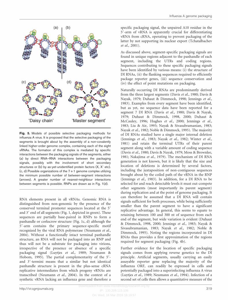
Fig. 5. Models of possible selective packaging methods forinfluenza A virus. It is proposed that the selective packaging of thesegments is brought about by the assembly of a non-covalentlylinked higher-order genome complex, containing each of the eightvRNAs. The formation of this complex is mediated by specificinteractions between the packaging signals of the segments, either(a) by direct RNA–RNA interactions between the packagingsignals, possibly with the involvement of short secondarystructures or (b) by as-yet-unidentified protein factors (X, X9 etc).(c, d) Possible organizations of the 7+1 genome complex utilizingthe minimum possible number of between-segment interactions(arrows). A greater number of nearest-neighbour interactionsbetween segments is possible. RNPs are drawn as in Fig. 1(d).
Influenza A genome packaging
http://vir.sgmjournals.org 319

Downloaded from www.microbiologyresearch.org by
IP: 130.209.115.106
On: Wed, 16 Mar 2016 16:19:57
efficiency with which the segment is acquired by virions. Byvarying the amount of flanking sequence, regions ofsegments promoting efficient packaging have been mapped(Dos Santos Afonso et al., 2005; Duhaut & Dimmock,2000; Enami et al., 1991; Fujii et al., 2003; Fujii et al., 2005;Liang et al., 2005; Luytjes et al., 1989; Marsh et al., 2007;Muramoto et al., 2006; Ozawa et al., 2007, 2009; Watanabeet al., 2003). There are certain caveats for this approach.Depending on the experimental setup, detection ofreporter gene expression in the second set of cells maydepend on the ability of the infecting virus to replicate itsgenome. This presupposes the presence of the foursegments encoding the viral polymerase and NP(Bancroft & Parslow, 2002) and, as discussed below,disruption of the packaging of one segment can affect theincorporation of others. It is also important to control foradventitious effects of the mutations on synthesis and/orstability of the mutant vRNA molecule or its mRNA.Although the core promoter sequences for vRNA synthesisand encapsidation reside in the conserved terminal regionsof the segments (Neumann et al., 2004), instances havebeen noted where alteration of more distal sequenceswithin the unique regions of a segment have affected itsaccumulation and/or transcription (Bergmann & Muster,1996; Hutchinson et al., 2008; Zheng et al., 1996). Also,depending on the experimental design, the reporterconstruct may or may not be competing for packagingwith the genomic segment from which it was derived, whilerequirements for packaging may differ for single-cycle andmulti-cycle growth. Factors such as these (as well as the useof different virus strains) may explain differences inoutcomes when different laboratories have attempted tomap signals on the same segment (e.g. Liang et al., 2005;Marsh et al., 2007; Muramoto et al., 2006; Watanabe et al.,2003). It is also not known to what extent the length of apackagable vRNA is limited by the maximum total size ofthe segment, or by the stoichiometry of nucleotides to NPmonomers in the RNP, though neither has been shown yetto have an obvious effect on packaging (Dos Santos Afonsoet al., 2005; Marsh et al., 2007).
Caveats notwithstanding, this approach has been usedsuccessfully to map packaging elements in all eightsegments (Dos Santos Afonso et al., 2005; Fujii et al.,2003; Fujii et al., 2005; Liang et al., 2005; Marsh et al., 2007;Muramoto et al., 2006; Ozawa et al., 2007, 2009; Watanabeet al., 2003). In all cases, segments were found to have abipartite specific packaging signal that involved extendedsequences at both ends of the vRNA (Fig. 4c), in broadagreement with the inferences drawn from the study of DIRNA structure. For most segments, the regions defined ascontaining the minimal specific packaging signals bydeliberate deletion mutagenesis tend to be slightly smallerthan those contained in the smallest reported DI RNA (Fig.4b, c). To some extent, this apparent difference may bemore artificial than real. In most cases, incrementaldeletion of flanking sequences reduced the packagingefficiency of the reporter vRNA gradually rather than
revealing a sharp cut-off point between functional andnon-functional. Thus, as well as making the definition of aminimal ‘efficient’ packaging signal somewhat arbitrary,this also suggests that complete packaging signals arecomposed of numerous contributing parts rather than asingle discrete sequence element. Although both ends of thesegment contribute to the packaging signal in all segments,the 59 end was shown to be more important for segments1–3 (Liang et al., 2005; Muramoto et al., 2006), whereas the39 end contributed more for segments 6 and 8 (Fujii et al.,2003; Fujii et al., 2005).
Examination of sequence conservation has been furtherused to define the location of specific packaging signals. Ascis-acting RNA sequences, they will be subject to functionalselection and should therefore be conserved. Sincepackaging signals overlap with coding regions, it is difficultto tease out whether conservation of a particular nucleotidesequence arose from selection for protein and/or for RNAfunction. The large numbers of influenza sequencesavailable makes it possible to address this by consideringthe levels of synonymous variation within the codingregions (Gog et al., 2007; Liang et al., 2008; Marsh et al.,2008). These analyses identified codons with very low levelsof synonymous variation at positions where variationshould have been possible, consistent with functionalconservation of the primary RNA sequence beyond thatrequired for its coding capacity. A large-scale analysis thatconsidered all eight segments of the genome (Gog et al.,2007) found that these conserved codons tended to clusterin the terminal regions of each segment, in the same areasimplicated by experimental or DI RNA structure ascontaining packaging signals (Fig. 4d, pink shading).Although the conserved codons formed clusters, thesewere not necessarily totally contiguous and the degree ofconservation tended to decrease with increasing distancefrom the termini (Gog et al., 2007), consistent with the‘fuzzy’ boundaries of packaging elements defined bydeletion mapping. Other statistically significant clustersof low variability codons in regions outside of the terminalpackaging signals were also identified, some attributable tofeatures such as overlapping ORFs or splice sites, others inareas with no known constraint (Fig. 4d; Gog et al., 2007).
Further information on the identity of the packagingsignals comes from point mutagenesis of regions thoughtto be part of the signal. A major advantage of this approachis that it can be applied to otherwise wild-type virus (Fujiiet al., 2005; Hutchinson et al., 2008, 2009; Liang et al.,2008; Marsh et al., 2007, 2008; Ozawa et al., 2009) as well asto constructs containing reporter genes (Fujii et al., 2005;Fujii et al., 2009; Gog et al., 2007; Liang et al., 2008). For allsegments, point mutagenesis has identified nucleotides,individually or in small clusters, whose alteration causessignificant reductions in packaging of the altered segmentas well as (where examined) reductions in the titre ofreplicated virus (Fujii et al., 2005; Fujii et al., 2009; Gog etal., 2007; Hutchinson et al., 2008, 2009; Liang et al., 2008;Marsh et al., 2007, 2008) (Fig. 4e). The finding that
E. C. Hutchinson and others
320 Journal of General Virology 91

Downloaded from www.microbiologyresearch.org by
IP: 130.209.115.106
On: Wed, 16 Mar 2016 16:19:57
alteration of a small number of nucleotides (even as few asone) can have a profound effect on segment incorporationis perhaps surprising, given the apparently large size of thesignals. However, in many cases, other mutations withinareas predicted to be part of the packaging signal had noapparent effect on its function (Fujii et al., 2005; Gog et al.,2007; Hutchinson et al., 2008, 2009; Liang et al., 2008;Marsh et al., 2007, 2008). This may, in part, reflect thediscontinuous nature of the signals, as suggested byanalysis of sequence conservation (Gog et al., 2007).However, in some instances, mutation of conservednucleotides had no phenotypic effect and indeed, studieshave found that a variety of randomly selected sequenceswithin the 39-end of the segment 8 or the 59-end of thesegment 7 packaging signals supported substantial levels ofsegment packaging despite having little homology to thewild-type sequence other than perhaps a tendency to beingU-rich (Fujii et al., 2009; Ozawa et al., 2009). Large or smalldeletions with the region of the signal, however, do notseem to be well tolerated, leading to the suggestion thatlength is as important as sequence (Liang et al., 2008;Ozawa et al., 2009). This apparent functional plasticity ofthe packaging signals is puzzling and awaits explanation.
Thus, taking all the available threads of evidence together,the conclusion is that all segments contain a genericbipartite packaging signal contained within their 59- and39-terminal 12–13 nt that (through its operation as thepromoter for the viral RNA polymerase) serves to identifythem as vRNAs and substrates for encapsidation into RNPs(Fig. 1a, green boxes). In addition, each of the eightsegments also contains specific packaging signals adjacentto these conserved promoter sequences (at 59- and 39-ends)that span the UTRs and extend some distance into thecoding regions. Within these sequences, the functionallysignificant elements are probably discontinuous and, ratherthan ending at a discrete boundary, tend to wane inimportance with increasing distance from the termini(Fig. 1a, red dashes). This organization of packaging signalsbears comparison with the dsRNA cystoviruses such asbacteriophage W6, where the three segments contain aconserved 18 nt element at their 59-ends followed byunique pac signals of around 200 nt each (Mindich, 2004).In contrast with influenza A virus, the conserved andunique signals of W6 are separated by 50 nt, are locatedonly at the 59-end of the segment and the pac sequences arecontained entirely within the segment UTRs. The discon-tinuity of the influenza packaging signals can perhaps beexplained by encapsidation of the vRNAs into RNPs, eventhough the vRNAs are still partially accessible to RNasesand other modifying agents. A variety of approachesestimate a stoichiometry of around 24 nt per unit of NPprotein in the RNP (Portela & Digard, 2002), which, giventhe physical sizes of the components, implies a degree oflooping of the RNA (Jennings et al., 1983). Taken togetherwith the existence of a likely RNA-binding cleft in the NPstructure (Ng et al., 2008; Ye et al., 2006), it is thereforeunlikely that all regions of the encapsidated vRNA
molecule will be equally accessible. It is also worthwhileconsidering the ‘tertiary’ structure of the RNP. EM imagingstudies show that the rod-like structure of the RNP is infact helical, often opening out (when examined inphysiological salt conditions) into a terminal loop(Fig. 1b). Cartoon models of this often place the viralpolymerase at the opposite end of the RNP (Fig. 1c).However, this organization is uncertain as negative staintechniques have not permitted unambiguous visualizationof the viral polymerase on authentic RNPs. Although animmuno-gold labelling study showed that the polymerasewas located at one end of the RNP, terminal loops in thebackbone of the RNP were not seen in this case (Murti etal., 1988). Indeed, one study presenting a detailed form ofthe RNP model considered it more likely that thepolymerase was located at the open end (Fig. 1d), basedon a possible mechanism for the generation of DI RNAs(Jennings et al., 1983). In light of the extended and perhapsflexible nature of the packaging signals, this latterorganization is plausible. Thus, although the position ofthe packaging signals in the primary nucleotide sequence isincreasingly clearly delineated, their position in the 3Dstructure of the RNP remains ambiguous. Ongoing workcharacterizing the structure of intact RNPs (e.g. Coloma etal., 2009) may well rectify this gap in the future.
Mechanisms of segment selection
Although there is now compelling evidence for segment-specific packaging in influenza A virus, the mechanism ofthis process remains unclear. The best characterizedmechanism of specific packaging in a segmented virus isthat of the bacteriophage W6. Here, three segments of plus-strand RNA (later converted into dsRNA) are sequentiallyrecruited into a preformed capsid through a series ofconformational changes driven by interactions with thesegment packaging signals that expose and then masksequence/structure-specific RNA binding sites while simul-taneously expanding the capsid to allow incorporation ofmore RNA (Huiskonen et al., 2006; Mindich, 2004). Thismodel seems unlikely to be applicable to influenza virus forseveral reasons. During influenza infections, preformedcapsids are not seen in the cytoplasm, with the virusinstead assembling at the point of budding through theplasma membrane (Cheung & Poon, 2007). In addition,the pleiomorphy of influenza virus particles seemsintrinsically ill-suited to a mechanism founded on orderedconformational changes of the capsid that regulate specificRNA-binding sites in the virion interior. Indeed, it haseven been proposed that the viral glycoproteins alone candrive particle assembly and that not all influenza virionseven contain a matrix protein layer (Chen et al., 2007;Harris et al., 2006). Instead, as EM analyses suggest thatbudding influenza A virions incorporate a parallel 7+1array of eight RNPs (Harris et al., 2006; Noda et al., 2006;Oxford & Hockley, 1987; Yamaguchi et al., 2008), it hasbeen plausibly hypothesized that this represents a specificmulti-segmental ‘genome complex’ containing one copy of
Influenza A genome packaging
http://vir.sgmjournals.org 321

Downloaded from www.microbiologyresearch.org by
IP: 130.209.115.106
On: Wed, 16 Mar 2016 16:19:57
each of the eight vRNAs whose assembly confers specificityon packaging (Heggeness et al., 1982; Noda et al., 2006).
The way in which this putative viral genome complexassembles remains a matter of speculation (as does theexistence of the complex itself) but it presumably wouldinvolve recognition of the various specific packagingsignals present on the eight vRNAs. Theoretically, thismight involve the action of sequence-specific RNA-bindingproteins (Fig. 5b), of either viral or cellular origin.However, the formation of a specific complex of eightRNPs requires a minimum of seven inter-RNP interactions(Fig. 5c, d) and no evidence has been found so far for evena single protein able to recognize an influenza virus specificpackaging signal, let alone the 14 or more separate RNAelements required to bring together eight RNPs. This is notto say that such a mechanism is impossible; although notstrictly analogous, the structures of ribosomal subunitsillustrate how megadalton ribonucleoprotein assembliescan be constructed through multiple individual proteinsbinding to non-contiguous RNA elements that areprimarily recognized as structures rather than as nucleotidesequences (Brodersen et al., 2002; Klein et al., 2004). Somecellular proteins have been shown to be incorporated intoreleased influenza virus particles (Shaw et al., 2008); furtherlow abundance components (perhaps only present at onecopy per virion) could still remain to be discovered. Amore parsimonious (but not necessarily exclusive) hypo-thesis is that the RNA signals themselves function as therecognition surfaces via direct RNA–RNA interactions (Fig.5a) (Fujii et al., 2003; Kingsbury & Webster, 1969). Thishypothesis has viral precedents in the formation andpackaging of homodimers of the retroviral genome(Greatorex, 2004), as well as (in a heterodimeric context)during RNA synthesis in red clover necrotic mosaic virus(Sit et al., 1998). It is also generally consistent with thestructural diversity of RNA, even when in the form of anRNP (Brodersen et al., 2002; Holbrook, 2005; Klein et al.,2004). However, attempts to identify plausible interactingmotifs by in silico analysis of vRNA sequences have so farbeen unsuccessful, as might be expected given the complexway in which vRNA coils around NP to form the RNP, thediscontinuous nature of the signals and that the hypothe-sized RNA–RNA interactions need not be limited toWatson–Crick base-pairs. Thus at present, our under-standing of the mechanism by which specific genomepackaging in influenza A virus is achieved is at the stage oftesting plausible hypotheses. Nevertheless, some priorevidence, albeit equivocal, has a bearing on these proposals.
A key component of the currently favoured hypothesis isthe formation of a specific genome complex of the eightRNPs. Although EM imaging indicates that some virionscontain a 7+1 array of RNPs, this is not proof of acomplex with a specific composition or even that the RNPsare actually associated; the apparent order could potentiallyresult from the geometry of packing eight rods into aspherical particle and/or the mechanism of the buddingprocess itself. Images showing the 7+1 array appear to be
less common when released particles are examined, incomparison to sections across budding virions (Booy et al.,1985; Harris et al., 2006; Noda et al., 2006; Oxford &Hockley, 1987; Yamaguchi et al., 2008; Yazaki et al., 1984).This may well be because released particles are free torotate, reducing the chance of seeing RNPs in cross section,whereas cell-associated virions maintain the directionalityof budding. However, it could also indicate that the RNPsare not bound together in the particle and redistribute oncefreed from an initial constraint imposed by the buddingprocess; a hypothesis supported by a tomographic study inwhich ordered arrays of RNPs were found more frequentlyin elongated virions than in larger spherical particles(Harris et al., 2006). As a counter-argument against generaldisorder in virion contents, magnetic birefringence analysisof released virions indicated that the interior componentswere probably ordered to some degree (Torbet, 1983).Similarly, EM studies have also provided striking images oflong helical structures released from partially disruptedvirions, proposed to be RNP (Almeida & Brand, 1975;Murti et al., 1980). These helical structures were often toolong to represent individual RNPs and the authorsspeculated they might represent non-covalently linkedcomplexes formed as part of the genomic packagingprocess. However, again in counter-argument, a sub-sequent study disputed this, proposing instead that thestructure were helices of M1 (Ruigrok et al., 1989).Furthermore, sedimentation analysis of material releasedfrom purified virus particles has so far failed to provideevidence for the existence of a packaging complex, withRNPs instead migrating at the approximate positionsexpected for their individual molecular masses (Companset al., 1972; Duesberg, 1969; Krug, 1971; Pons et al., 1969).It should be noted, however, that these early experimentswere all performed in buffer conditions that would notnecessarily maintain structured RNA–RNA interactions.Therefore, overall, microscopic and biochemical evidencefor the existence of a specific influenza A genome complexis ambivalent; further work in this area is required.
A further key prediction of the current hypothesis for howspecific packaging operates is that there is a specific suite ofinter-segment interactions. Within a 7+1 array of rod-likestructures all containing their packaging signals at one end,not every RNP is likely to interact with all others; instead,various specific linkage schemes can be envisaged. In a‘daisy chain’ network, most segments would interact withtwo others while the end ones only interact with one(Fig. 5c). Alternatively, a ‘master’ segment (perhaps in thecentre of the array) that interacts with many or all othersegments can be envisaged (Fig. 5d). More complex‘hybrid’ schemes with additional interactions betweenadjacent RNPs are also plausible, but in any case, themodel predicts that deleterious mutations in the packagingsignal of a given segment have the possibility of affectingthe incorporation of adjacent, interacting segments. Initialevidence consistent with this hypothesis came fromstudying the incorporation of reporter vRNAs with
E. C. Hutchinson and others
322 Journal of General Virology 91

Downloaded from www.microbiologyresearch.org by
IP: 130.209.115.106
On: Wed, 16 Mar 2016 16:19:57
flanking sequences from the three largest segments. In asystematic examination of all eight segments, the presenceof the coding regions from segment 1 (and in particular the59-end) was found to be especially important not only forpackaging of the recombinant vRNA but also to theefficiency with which infectious virus-like particles (VLPs)were formed (Muramoto et al., 2006). Detection of theVLPs relied on their incorporating either segment 4 (HA)or 5 (NP) so this effect could have arisen from knock-oneffects on the incorporation of other segments and/or theactual budding process itself. Subsequent work hasconfirmed that both mechanisms potentially play a part.Several studies have since examined the phenotype ofpackaging mutants in the context of viable virus (ratherthan via artificial reporter constructs) and directlyexamined the relative amounts of segments incorporatedper virion. Consistent with the previous VLP study byMuramoto et al. (2006), mutations to segment 1 werefound to reduce packaging of not only itself but also allother segments (Marsh et al., 2008). Similarly, mutations inthe segment 7 packaging signals reduced incorporation ofeach of the eight segments by an approximately equalextent, such that the majority of virions contained anincomplete genome (Hutchinson et al., 2008). Lesions insegment 7 also reduced virus budding (Hutchinson et al.,2008), consistent with the prior data inferred fromexamining VLPs and viruses with less than eight segments(de Wit et al., 2006; Fujii et al., 2005; Fujii et al., 2009; Gaoet al., 2008; Marsh et al., 2007; Ozawa et al., 2009). Morespecific secondary effects on segment incorporation aftermutation of a single packaging signal have also been seen.Mutations to segment 4 that reduced its packaging alsoreduced incorporation of other segments: notably 2, butalso (to lesser extents) 3, 5 and 6 (Marsh et al., 2007).Similarly, mutations in the segment 3 packaging signalaffected incorporation of segments 1 and 5, in someinstances to the point where they showed a greaterpackaging deficit than the mutated segment 3 (Marshet al., 2008). Conversely, a packaging signal mutation insegment 5 reduced incorporation of segment 3(Hutchinson et al., 2009).
Thus, taken as a whole, evidence for trans-acting effects ofpackaging mutations on other segments are consistent withan important prediction of the ‘genome complex’ hypo-thesis; that packaging of individual segments is notindependent but instead is interlinked. It is tempting totry and deduce likely patterns of inter-segment interactionfrom the available data. For instance, comparison ofexperiments suggests a reciprocal packaging interactionbetween segments 3 and 5 (Hutchinson et al., 2009; Marshet al., 2008). However, other data suggest complexitiesarguing against such straightforward interpretations. Forinstance, a single nucleotide mutation to codon 745 of PB2in the 59-end of segment 1 vRNA reduced the relativepackaging of the segment by nearly tenfold and alsodecreased incorporation of segments 2 and 5 by overfivefold. However, mutation of the adjacent codon 744
only significantly affected packaging of segment 1 itself(Marsh et al., 2008), suggesting complex position-depend-ent effects of packaging signal mutations that mayconfound simple interpretation. Thus, while furtherexperimentation of this sort may elucidate a proposedweb of inter-segment interactions, other approaches areneeded. At present, it is probably safe to conclude that thedata do not support the hypothesis of a simple ‘daisy chain’to assemble the genome complex (Fig. 5c), but insteadsuggest more complex interaction patterns. Segments 1 and7 seem to be potential candidates for ‘master segments’(Fig. 5d), based on the pleiotropic effects mutation of theirpackaging signals have on virus budding and incorporationof other vRNAs (Hutchinson et al., 2008; Marsh et al.,2008; Muramoto et al., 2006; Ozawa et al., 2009).
Packaging signals and influenza virus evolution
Genome segmentation clearly plays a major role ininfluenza virus evolution, both within a single host speciesand in facilitating jumps in host range (Dugan et al., 2008;Garten et al., 2009; Hatchette et al., 2004; Kuiken et al.,2006; Nelson & Holmes, 2007; Smith et al., 2009). Asdiscussed above, although the strategy of a dividedgenomic structure confers fitness benefits, it comes at thedirect cost of increasing the complexity of virus assembly.It is clear that the virus has evolved a solution to thisproblem, although the exact mechanism by whichspecificity in packaging is achieved is still uncertain. Doesthe nature of the solution impose constraints on virusevolution? In one sense the answer is an unequivocal ‘yes’;the footprints of conservation left by the specific packagingsignals on the viral terminal coding regions are clear to see(Gog et al., 2007; Liang et al., 2008; Marsh et al., 2008).This may be exploitable for intervention strategies. Forinstance, it has been suggested that packaging signalsare good targets for oligonucleotide-based inhibition(Giannecchini et al., 2009). In a similar vein, the M2ectodomain has been proposed as a candidate immunogenfor a ‘universal’ influenza vaccine, for which antigenicescape mutants will be less likely to develop because theM2e coding region is either congruent with or overlapsthat of M1 (Saelens, 2008). We speculate that the fact thatthe first nine codons of the M2e coding region also overlapthe segment 7 packaging signal (Gog et al., 2007;Hutchinson et al., 2008; Ozawa et al., 2009) will furtherconstrain the development of escape mutants. Betterunderstanding of influenza virus genome packaging hasalso facilitated attempts to use the virus as a gene deliveryvector (Gao et al., 2008; Shinya et al., 2004) as well asproviding an ingenious approach to improving engineeredvirus biosafety through reducing the probability ofsuccessful reassortment with ‘wild’ viruses (Gao & Palese,2009).
We also hypothesize that the evolutionary solution thevirus has found to the packaging problem has broaderimplications for its biology. The fact that the same stretches
Influenza A genome packaging
http://vir.sgmjournals.org 323

Downloaded from www.microbiologyresearch.org by
IP: 130.209.115.106
On: Wed, 16 Mar 2016 16:19:57
of RNA perform multiple functions in addition to acting aspackaging signals [coding for proteins, as well as cis-actingfunctions such as promoter or splice signals (Gog et al.,2007; Hutchinson et al., 2008)] creates potentially conflict-ing requirements that reduce the chances of finding anoptimal solution to the problems of genome packaging.Although the evidence for a specific packaging method isoverwhelming, the mechanism is clearly not perfect. Theexistence of nine segment viruses (Enami et al., 1991; Laver& Downie, 1976; Nakajima & Sugiura, 1977; Scholtissek etal., 1978) and the low levels of ‘background’ packaging ofreporter vRNAs lacking specific packaging signals seen byseveral laboratories and for all segments (Bancroft &Parslow, 2002; Dos Santos Afonso et al., 2005; Fujii et al.,2003; Luytjes et al., 1989; Muramoto et al., 2006; Neumannet al., 1994; Tchatalbachev et al., 2001) suggest that there isa measurable degree of imprecision in the mechanism. Weare also presented with the apparent paradox, that in thelaboratory, small mutations (even a single nucleotide) cansignificantly disrupt packaging of a particular segment tothe point where the virus replicates noticeably more poorly(e.g. Gog et al., 2007; Hutchinson et al., 2008; Marsh et al.,2008) and yet in other circumstances, selection of efficientpackaging signals from a pool of randomized sequencesreveals no clear consensus sequence (Fujii et al., 2009;Ozawa et al., 2009). Similarly, reassortment in nature seemsreadily capable of bringing together segments fromdiverged genetic backgrounds that could, on occasion,include changes to packaging signals (Dugan et al., 2008;Ghedin et al., 2009; Hatchette et al., 2004). Thisdiscrepancy lacks a molecular explanation at present, butit provides further evidence that the virus tolerates a degreeof imprecision in its packaging mechanism.
Why then, has packaging in influenza A virus not evolved ahigher degree of precision? As already discussed, theoverlap of packaging signals with open reading framesand cis-acting RNA functions may hinder the evolution ofa ‘perfect’ genome packaging solution of the sort achievedby the bacteriophage W6 (Mindich, 2004), as indeed mayconstraints from other aspects of virus biology such asvirion morphology or the mechanism of budding. However(to end on a speculative note), we wonder if an imperfectpacking strategy is actually beneficial to virus fitness, byproviding flexibility for reassortment. Studies of naturalisolates suggest that widespread and continuous reassort-ment events drive influenza virus evolution (Dugan et al.,2008; Ghedin et al., 2009; Hatchette et al., 2004), and inthis respect the ability to package evolutionarily divergedsegments would carry benefits. As well as increasing theselective advantage that the rudimentary sexual process ofreassortment provides to the virus, this can perhaps beviewed in terms of selection acting on individual segments.Participation of a ‘selfish segment’ in a selective packagingmechanism would be balanced between the need to becorrectly packaged in the context of its current genome andretaining sufficient flexibility to be able to occasionally‘jump ship’ through facile reassortment with an evolutio-
narily diverged genome. At the level of both the virus andits segments, a mechanism of selective genome packagingthat is neither entirely random nor unfailingly rigorousmay have proven to be the most successful evolutionarystrategy for influenza A virus.
Acknowledgements
We thank Dr Helen Wise for comment and discussion. Research in
the authors’ laboratories is supported by the MRC, Wellcome Trust
and BBSRC. E. C. H. was supported by a studentship from the
Wellcome Trust. J. C. v. K. is supported by a studentship from the
MRC. J. R. G. is a Royal Society University Research Fellow.
References
Akkina, R. K., Chambers, T. M. & Nayak, D. P. (1984). Mechanism of
interference by defective-interfering particles of influenza virus:
differential reduction of intracellular synthesis of specific polymerase
proteins. Virus Res 1, 687–702.
Almeida, J. D. & Brand, C. M. (1975). A morphological study of the
internal component of influenza virus. J Gen Virol 27, 313–318.
Apostolov, K. & Flewett, T. H. (1969). Further observations on the
structure of influenza viruses A and C. J Gen Virol 4, 365–370.
Bachi, T., Gerhard, W., Lindenmann, J. & Muhlethaler, K. (1969).Morphogenesis of influenza A virus in Ehrlich ascites tumor cells as
revealed by thin-sectioning and freeze-etching. J Virol 4, 769–776.
Ball, L. A. (2007). Virus replication strategies. In Fields Virology, 5th
edn, pp. 119–140. Edited by D. M. Knipe & P. M. Howley.
Philadelphia, PA: Lippincott Williams & Wilkins.
Bancroft, C. T. & Parslow, T. G. (2002). Evidence for segment-
nonspecific packaging of the influenza A virus genome. J Virol 76,
7133–7139.
Barry, R. D. (1961). The multiplication of influenza virus. II.
Multiplicity reactivation of ultraviolet irradiated virus. Virology 14,
398–405.
Belshaw, R., Gardner, A., Rambaut, A. & Pybus, O. G. (2008). Pacing
a small cage: mutation and RNA viruses. Trends Ecol Evol 23, 188–
193.
Bergmann, M. & Muster, T. (1995). The relative amount of an
influenza A virus segment present in the viral particle is not affected
by a reduction in replication of that segment. J Gen Virol 76, 3211–
3215.
Bergmann, M. & Muster, T. (1996). Mutations in the nonconserved
noncoding sequences of the influenza A virus segments affect viral
vRNA formation. Virus Res 44, 23–31.
Birch-Andersen, A. & Paucker, K. (1959). Studies on the structure of
influenza virus. II. Ultrathin sections of infectious and noninfectious
particles. Virology 8, 21–40.
Booy, F. P., Ruigrok, R. W. & van Bruggen, E. F. (1985). Electron
microscopy of influenza virus. A comparison of negatively stained
and ice-embedded particles. J Mol Biol 184, 667–676.
Boulo, S., Akarsu, H., Ruigrok, R. W. & Baudin, F. (2007). Nuclear
traffic of influenza virus proteins and ribonucleoprotein complexes.
Virus Res 124, 12–21.
Brodersen, D. E., Clemons, W. M., Jr, Carter, A. P., Wimberly, B. T. &Ramakrishnan, V. (2002). Crystal structure of the 30 S ribosomal
subunit from Thermus thermophilus: structure of the proteins and
their interactions with 16 S RNA. J Mol Biol 316, 725–768.
E. C. Hutchinson and others
324 Journal of General Virology 91

Downloaded from www.microbiologyresearch.org by
IP: 130.209.115.106
On: Wed, 16 Mar 2016 16:19:57
Chao, L., Tran, T. T. & Tran, T. T. (1997). The advantage of sex in theRNA virus W6. Genetics 147, 953–959.
Chen, W., Calvo, P. A., Malide, D., Gibbs, J., Schubert, U., Bacik, I.,Basta, S., O’Neill, R., Schickli, J. & other authors (2001). A novelinfluenza A virus mitochondrial protein that induces cell death. NatMed 7, 1306–1312.
Chen, B. J., Leser, G. P., Morita, E. & Lamb, R. A. (2007). Influenzavirus hemagglutinin and neuraminidase, but not the matrix protein,are required for assembly and budding of plasmid-derived virus-likeparticles. J Virol 81, 7111–7123.
Cheung, T. K. & Poon, L. L. (2007). Biology of influenza a virus. AnnN Y Acad Sci 1102, 1–25.
Coloma, R., Valpuesta, J. M., Arranz, R., Carrascosa, J. L., Ortin, J. &Martin-Benito, J. (2009). The structure of a biologically activeinfluenza virus ribonucleoprotein complex. PLoS Pathog 5, e1000491.
Compans, R. W. & Dimmock, N. J. (1969). An electron microscopicstudy of single-cycle infection of chick embryo fibroblasts byinfluenza virus. Virology 39, 499–515.
Compans, R. W., Dimmock, N. J. & Meier-Ewart, H. (1970). In TheBiology of Large RNA Viruses, pp. 87–108. Edited by R. D. Barry &B. W. J. Mahy. New York: Academic Press.
Compans, R. W., Content, J. & Duesberg, P. H. (1972). Structure ofthe ribonucleoprotein of influenza virus. J Virol 10, 795–800.
Davis, A. R. & Nayak, D. P. (1979). Sequence relationships amongdefective interfering influenza viral RNAs. Proc Natl Acad Sci U S A76, 3092–3096.
Davis, A. R., Hiti, A. L. & Nayak, D. P. (1980). Influenza defectiveinterfering viral RNA is formed by internal deletion of genomic RNA.Proc Natl Acad Sci U S A 77, 215–219.
de Wit, E., Spronken, M. I., Rimmelzwaan, G. F., Osterhaus, A. D. &Fouchier, R. A. (2006). Evidence for specific packaging of theinfluenza A virus genome from conditionally defective virus particleslacking a polymerase gene. Vaccine 24, 6647–6650.
Donald, H. B. & Isaacs, A. (1954). Counts of influenza virus particles.J Gen Microbiol 10, 457–464.
Dos Santos Afonso, E., Escriou, N., Leclercq, I., van der Werf, S. &Naffakh, N. (2005). The generation of recombinant influenza Aviruses expressing a PB2 fusion protein requires the conservation of apackaging signal overlapping the coding and noncoding regions at the59 end of the PB2 segment. Virology 341, 34–46.
Duesberg, P. H. (1968). The RNA of influenza virus. Proc Natl AcadSci U S A 59, 930–937.
Duesberg, P. H. (1969). Distinct subunits of the ribonucleoprotein ofinfluenza virus. J Mol Biol 42, 485–499.
Dugan, V. G., Chen, R., Spiro, D. J., Sengamalay, N., Zaborsky, J.,Ghedin, E., Nolting, J., Swayne, D. E., Runstadler, J. A. & otherauthors (2008). The evolutionary genetics and emergence of avianinfluenza viruses in wild birds. PLoS Pathog 4, e1000076.
Duhaut, S. D. & Dimmock, N. J. (1998). Heterologous protection ofmice from a lethal human H1N1 influenza A virus infection by H3N8equine defective interfering virus: comparison of defective RNAsequences isolated from the DI inoculum and mouse lung. Virology248, 241–253.
Duhaut, S. & Dimmock, N. J. (2000). Approximately 150 nucleotidesfrom the 59 end of an influenza A segment 1 defective virion RNA areneeded for genome stability during passage of defective virus ininfected cells. Virology 275, 278–285.
Duhaut, S. D. & Dimmock, N. J. (2002). Defective segment 1 RNAsthat interfere with production of infectious influenza A virus requireat least 150 nucleotides of 59 sequence: evidence from a plasmid-driven system. J Gen Virol 83, 403–411.
Duhaut, S. D. & McCauley, J. W. (1996). Defective RNAs inhibit the
assembly of influenza virus genome segments in a segment-specificmanner. Virology 216, 326–337.
Enami, M., Sharma, G., Benham, C. & Palese, P. (1991). An influenzavirus containing nine different RNA segments. Virology 185, 291–298.
Fujii, Y., Goto, H., Watanabe, T., Yoshida, T. & Kawaoka, Y. (2003).Selective incorporation of influenza virus RNA segments into virions.Proc Natl Acad Sci U S A 100, 2002–2007.
Fujii, K., Fujii, Y., Noda, T., Muramoto, Y., Watanabe, T., Takada, A.,Goto, H., Horimoto, T. & Kawaoka, Y. (2005). Importance of both the
coding and the segment-specific noncoding regions of the influenza A
virus NS segment for its efficient incorporation into virions. J Virol79, 3766–3774.
Fujii, K., Ozawa, M., Iwatsuki-Horimoto, K., Horimoto, T. &Kawaoka, Y. (2009). The incorporation of influenza A virus genomesegments does not absolutely require wild-type sequences. J Gen Virol
90, 1734–1740.
Gao, Q. & Palese, P. (2009). Rewiring the RNAs of influenza virus to
prevent reassortment. Proc Natl Acad Sci U S A 106, 15891–15896.
Gao, Q., Brydon, E. W. & Palese, P. (2008). A seven-segmentedinfluenza A virus expressing the influenza C virus glycoprotein HEF.
J Virol 82, 6419–6426.
Garten, R. J., Davis, C. T., Russell, C. A., Shu, B., Lindstrom, S., Balish, A.,Sessions, W. M., Xu, X., Skepner, E. & other authors (2009). Antigenic
and genetic characteristics of swine-origin 2009 A(H1N1) influenza
viruses circulating in humans. Science 325, 197–201.
Ghedin, E., Fitch, A., Boyne, A., Griesemer, S., DePasse, J., Bera, J.,Zhang, X., Halpin, R. A., Smit, M. & other authors (2009). Mixedinfection and the genesis of influenza virus diversity. J Virol 83, 8832–
8841.
Giannecchini, S., Clausi, V., Nosi, D. & Azzi, A. (2009).Oligonucleotides derived from the packaging signal at the 59 end of
the viral PB2 segment specifically inhibit influenza virus in vitro. Arch
Virol 154, 821–832.
Gog, J. R., Rimmelzwaan, G. F., Osterhaus, A. D. & Grenfell, B. T.(2003). Population dynamics of rapid fixation in cytotoxic Tlymphocyte escape mutants of influenza A. Proc Natl Acad Sci
U S A 100, 11143–11147.
Gog, J. R., Afonso Edos, S., Dalton, R. M., Leclercq, I., Tiley, L.,Elton, D., von Kirchbach, J. C., Naffakh, N., Escriou, N. & Digard, P.(2007). Codon conservation in the influenza A virus genome defines
RNA packaging signals. Nucleic Acids Res 35, 1897–1907.
Greatorex, J. (2004). The retroviral RNA dimer linkage: different
structures may reflect different roles. Retrovirology 1, 22.
Harris, A., Cardone, G., Winkler, D. C., Heymann, J. B., Brecher, M.,White, J. M. & Steven, A. C. (2006). Influenza virus pleiomorphy
characterized by cryoelectron tomography. Proc Natl Acad Sci U S A103, 19123–19127.
Hatada, E., Hasegawa, M., Mukaigawa, J., Shimizu, K. & Fukuda, R.(1989). Control of influenza virus gene expression: quantitative
analysis of each viral RNA species in infected cells. J Biochem 105,
537–546.
Hatchette, T. F., Walker, D., Johnson, C., Baker, A., Pryor, S. P. &Webster, R. G. (2004). Influenza A viruses in feral Canadian ducks:
extensive reassortment in nature. J Gen Virol 85, 2327–2337.
Heggeness, M. H., Smith, P. R., Ulmanen, I., Krug, R. M. & Choppin,P. W. (1982). Studies on the helical nucleocapsid of influenza virus.Virology 118, 466–470.
Hirst, G. K. (1962). Genetic recombination with Newcastle disease
virus, polioviruses, and influenza. Cold Spring Harb Symp Quant Biol27, 303–309.
Influenza A genome packaging
http://vir.sgmjournals.org 325

Downloaded from www.microbiologyresearch.org by
IP: 130.209.115.106
On: Wed, 16 Mar 2016 16:19:57
Hirst, G. K. (1973). Mechanism of influenza recombination. I. Factorsinfluencing recombination rates between temperature-sensitivemutants of strain WSN and the classification of mutants intocomplementation–recombination groups. Virology 55, 81–93.
Hirst, G. K. & Pons, M. W. (1973). Mechanism of influenzarecombination. II. Virus aggregation and its effect on plaqueformation by so-called noninfective virus. Virology 56, 620–631.
Holbrook, S. R. (2005). RNA structure: the long and the short of it.Curr Opin Struct Biol 15, 302–308.
Hughes, M. T., Matrosovich, M., Rodgers, M. E., McGregor, M. &Kawaoka, Y. (2000). Influenza A viruses lacking sialidase activity canundergo multiple cycles of replication in cell culture, eggs, or mice.J Virol 74, 5206–5212.
Huiskonen, J. T., de Haas, F., Bubeck, D., Bamford, D. H., Fuller,S. D. & Butcher, S. J. (2006). Structure of the bacteriophage W6nucleocapsid suggests a mechanism for sequential RNA packaging.Structure 14, 1039–1048.
Hutchinson, E. C., Curran, M. D., Read, E. K., Gog, J. R. & Digard, P.(2008). Mutational analysis of cis-acting RNA signals in segment 7 ofinfluenza A virus. J Virol 82, 11869–11879.
Hutchinson, E. C., Wise, H. M., Kudryavtseva, K., Curran, M. D. &Digard, P. (2009). Characterisation of influenza A viruses withmutations in segment 5 packaging signals. Vaccine 27, 6270–6275.
Isaacs, A. & Donald, H. B. (1955). Particle counts of haemagglutinat-ing viruses. J Gen Microbiol 12, 241–247.
Jennings, P. A., Finch, J. T., Winter, G. & Robertson, J. S. (1983). Doesthe higher order structure of the influenza virus ribonucleoproteinguide sequence rearrangements in influenza viral RNA? Cell 34, 619–627.
Kingsbury, D. W. (1970). Replication and functions of myxovirusribonucleic acids. Prog Med Virol 12, 49–77.
Kingsbury, D. W. & Webster, R. G. (1969). Some properties ofinfluenza virus nucleocapsids. J Virol 4, 219–225.
Klein, D. J., Moore, P. B. & Steitz, T. A. (2004). The roles of ribosomalproteins in the structure assembly, and evolution of the largeribosomal subunit. J Mol Biol 340, 141–177.
Krug, R. M. (1971). Influenza viral RNPs newly synthesized during thelatent period of viral growth in MDCK cells. Virology 44, 125–136.
Kuiken, T., Holmes, E. C., McCauley, J., Rimmelzwaan, G. F.,Williams, C. S. & Grenfell, B. T. (2006). Host species barriers toinfluenza virus infections. Science 312, 394–397.
Lakadamyali, M., Rust, M. J., Babcock, H. P. & Zhuang, X. (2003).Visualizing infection of individual influenza viruses. Proc Natl AcadSci U S A 100, 9280–9285.
Lamb, R. A. & Choppin, P. W. (1983). The gene structure andreplication of influenza virus. Annu Rev Biochem 52, 467–506.
Laver, W. G. & Downie, J. C. (1976). Influenza virus recombination. I.Matrix protein markers and segregation during mixed infections.Virology 70, 105–117.
Lazarowitz, S. D. (2007). Plant viruses. In Fields Virology, 5th edn, pp.641–705. Edited by D. M. Knipe & P. M. Howley. Philadelphia, PA:Lippincott Williams & Wilkins.
Liang, Y., Hong, Y. & Parslow, T. G. (2005). cis-Acting packagingsignals in the influenza virus PB1, PB2, and PA genomic RNAsegments. J Virol 79, 10348–10355.
Liang, Y., Huang, T., Ly, H. & Parslow, T. G. (2008). Mutationalanalyses of packaging signals in influenza virus PA, PB1, and PB2genomic RNA segments. J Virol 82, 229–236.
Liu, C. & Air, G. M. (1993). Selection and characterization of aneuraminidase-minus mutant of influenza virus and its rescue bycloned neuraminidase genes. Virology 194, 403–407.
Lubeck, M. D., Palese, P. & Schulman, J. L. (1979). Nonrandomassociation of parental genes in influenza A virus recombinants.Virology 95, 269–274.
Luo, G., Bergmann, M., Garcia-Sastre, A. & Palese, P. (1992).Mechanism of attenuation of a chimeric influenza A/B transfectantvirus. J Virol 66, 4679–4685.
Luque, D., Rivas, G., Alfonso, C., Carrascosa, J. L., Rodriguez, J. F. &Caston, J. R. (2009). Infectious bursal disease virus is an icosahedralpolyploid dsRNA virus. Proc Natl Acad Sci U S A 106, 2148–2152.
Luytjes, W., Krystal, M., Enami, M., Parvin, J. D. & Palese, P. (1989).Amplification, expression, and packaging of foreign gene by influenzavirus. Cell 59, 1107–1113.
Marsh, G. A., Hatami, R. & Palese, P. (2007). Specific residues of theinfluenza A virus hemagglutinin viral RNA are important for efficientpackaging into budding virions. J Virol 81, 9727–9736.
Marsh, G. A., Rabadan, R., Levine, A. J. & Palese, P. (2008). Highlyconserved regions of influenza A virus polymerase gene segments arecritical for efficient viral RNA packaging. J Virol 82, 2295–2304.
McGeoch, D., Fellner, P. & Newton, C. (1976). Influenza virusgenome consists of eight distinct RNA species. Proc Natl Acad SciU S A 73, 3045–3049.
Mindich, L. (2004). Packaging, replication and recombination of thesegmented genome of bacteriophage W6 and its relatives. Virus Res101, 83–92.
Morgan, C., Rose, H. M. & Moore, D. H. (1956). Structure anddevelopment of viruses observed in the electron microscope. III.Influenza virus. J Exp Med 104, 171–182.
Moss, B. A. & Brownlee, G. G. (1981). Sequence of DNAcomplementary to a small RNA segment of influenza virus A/NT/60/68. Nucleic Acids Res 9, 1941–1947.
Moya, A., Holmes, E. C. & Gonzalez-Candelas, F. (2004). Thepopulation genetics and evolutionary epidemiology of RNA viruses.Nat Rev Microbiol 2, 279–288.
Muller, H. J. (1964). The relation of recombination to mutationaladvance. Mutat Res 106, 2–9.
Muramoto, Y., Takada, A., Fujii, K., Noda, T., Iwatsuki-Horimoto, K.,Watanabe, S., Horimoto, T., Kida, H. & Kawaoka, Y. (2006).Hierarchy among viral RNA (vRNA) segments in their role invRNA incorporation into influenza A virions. J Virol 80, 2318–2325.
Murti, K. G., Bean, W. J., Jr & Webster, R. G. (1980). Helicalribonucleoproteins of influenza virus: an electron microscopicanalysis. Virology 104, 224–229.
Murti, K. G., Webster, R. G. & Jones, I. M. (1988). Localization of RNApolymerases on influenza viral ribonucleoproteins by immunogoldlabeling. Virology 164, 562–566.
Nakajima, K. & Sugiura, A. (1977). Three-factor cross of influenzavirus. Virology 81, 486–489.
Nakajima, K., Ueda, M. & Sugiura, A. (1979). Origin of small RNA invon Magnus particles of influenza virus. J Virol 29, 1142–1148.
Nayak, D. P. & Sivasubramanian, N. (1983). The structure ofinfluenza virus defective interfering (DI) RNAs and their progenitorgenes. In Genetics of Influenza Viruses, pp. 255–279. Edited byP. Palese & D. W. Kingsbury. Wien: Springer.
Nayak, D. P., Sivasubramanian, N., Davis, A. R., Cortini, R. & Sung, J.(1982). Complete sequence analyses show that two defectiveinterfering influenza viral RNAs contain a single internal deletion ofa polymerase gene. Proc Natl Acad Sci U S A 79, 2216–2220.
Nelson, M. I. & Holmes, E. C. (2007). The evolution of epidemicinfluenza. Nat Rev Genet 8, 196–205.
Neumann, G. & Hobom, G. (1995). Mutational analysis of influenzavirus promoter elements in vivo. J Gen Virol 76, 1709–1717.
E. C. Hutchinson and others
326 Journal of General Virology 91

Downloaded from www.microbiologyresearch.org by
IP: 130.209.115.106
On: Wed, 16 Mar 2016 16:19:57
Neumann, G., Zobel, A. & Hobom, G. (1994). RNA polymerase
I-mediated expression of influenza viral RNA molecules. Virology 202,477–479.
Neumann, G., Brownlee, G. G., Fodor, E. & Kawaoka, Y. (2004).Orthomyxovirus replication, transcription, and polyadenylation. Curr
Top Microbiol Immunol 283, 121–143.
Ng, A. K., Zhang, H., Tan, K., Li, Z., Liu, J. H., Chan, P. K., Li, S. M.,Chan, W. Y., Au, S. W. & other authors (2008). Structure of the
influenza virus A H5N1 nucleoprotein: implications for RNA binding,
oligomerization, and vaccine design. FASEB J 22, 3638–3647.
Noble, S. & Dimmock, N. J. (1995). Characterization of putative
defective interfering (DI) A/WSN RNAs isolated from the lungs ofmice protected from an otherwise lethal respiratory infection with
influenza virus A/WSN (H1N1): a subset of the inoculum DI RNAs.
Virology 210, 9–19.
Nobusawa, E. & Sato, K. (2006). Comparison of the mutation rates of
human influenza A and B viruses. J Virol 80, 3675–3678.
Noda, T., Sagara, H., Yen, A., Takada, A., Kida, H., Cheng, R. H. &Kawaoka, Y. (2006). Architecture of ribonucleoprotein complexes in
influenza A virus particles. Nature 439, 490–492.
Odagiri, T. & Tashiro, M. (1997). Segment-specific noncoding
sequences of the influenza virus genome RNA are involved in the
specific competition between defective interfering RNA and itsprogenitor RNA segment at the virion assembly step. J Virol 71,
2138–2145.
Odagiri, T. & Tobita, K. (1990). Mutation in NS2, a nonstructuralprotein of influenza A virus, extragenically causes aberrant replication
and expression of the PA gene and leads to generation of defective
interfering particles. Proc Natl Acad Sci U S A 87, 5988–5992.
Odagiri, T., Tominaga, K., Tobita, K. & Ohta, S. (1994). An amino acid
change in the non-structural NS2 protein of an influenza A virusmutant is responsible for the generation of defective interfering (DI)
particles by amplifying DI RNAs and suppressing complementary
RNA synthesis. J Gen Virol 75, 43–53.
Oxford, J. S. & Hockley, D. J. (1987). Orthomyxoviridae. In
Perspectives in Medical Virology. Animal Virus Structure, pp. 213–
232. Edited by M. V. Nermut & A. C. Steven. New York: Elsevier.
Ozawa, M., Fujii, K., Muramoto, Y., Yamada, S., Yamayoshi, S.,Takada, A., Goto, H., Horimoto, T. & Kawaoka, Y. (2007).Contributions of two nuclear localization signals of influenza A virus
nucleoprotein to viral replication. J Virol 81, 30–41.
Ozawa, M., Maeda, J., Iwatsuki-Horimoto, K., Watanabe, S., Goto, H.,Horimoto, T. & Kawaoka, Y. (2009). Nucleotide sequence require-
ments at the 59 end of the influenza A virus M RNA segment for
efficient virus replication. J Virol 83, 3384–3388.
Pons, M. W. & Hirst, G. K. (1968). Polyacrylamide gel electrophoresis
of influenza virus RNA. Virology 34, 385–388.
Pons, M. W., Schulze, I. T., Hirst, G. K. & Hauser, R. (1969). Isolation
and characterization of the ribonucleoprotein of influenza virus.
Virology 39, 250–259.
Portela, A. & Digard, P. (2002). The influenza virus nucleoprotein: a
multifunctional RNA-binding protein pivotal to virus replication.J Gen Virol 83, 723–734.
Pressing, J. & Reanney, D. C. (1984). Divided genomes and intrinsic
noise. J Mol Evol 20, 135–146.
Rambaut, A., Pybus, O. G., Nelson, M. I., Viboud, C., Taubenberger,J. K. & Holmes, E. C. (2008). The genomic and epidemiologicaldynamics of human influenza A virus. Nature 453, 615–619.
Ruigrok, R. W., Calder, L. J. & Wharton, S. A. (1989). Electron
microscopy of the influenza virus submembranal structure. Virology173, 311–316.
Saelens, X. (2008). The influenza matrix protein 2 as a vaccine target.Future Virology 3, 167–178.
Schmitt, A. P. & Lamb, R. A. (2005). Influenza virus assembly andbudding at the viral budozone. Adv Virus Res 64, 383–416.
Scholtissek, C., Rohde, W., Harms, E., Rott, R., Orlich, M. & Boschek,C. B. (1978). A possible partial heterozygote of an influenza A virus.Virology 89, 506–516.
Shaw, M. L., Stone, K. L., Colangelo, C. M., Gulcicek, E. E. & Palese, P.(2008). Cellular proteins in influenza virus particles. PLoS Pathog 4,e1000085.
Shinya, K., Fujii, Y., Ito, H., Ito, T. & Kawaoka, Y. (2004).Characterization of a neuraminidase-deficient influenza A virus as apotential gene delivery vector and a live vaccine. J Virol 78, 3083–3088.
Sit, T. L., Vaewhongs, A. A. & Lommel, S. A. (1998). RNA-mediatedtrans-activation of transcription from a viral RNA. Science 281, 829–832.
Smith, G. L. & Hay, A. J. (1982). Replication of the influenza virusgenome. Virology 118, 96–108.
Smith, G. J., Vijaykrishna, D., Bahl, J., Lycett, S. J., Worobey, M.,Pybus, O. G., Ma, S. K., Cheung, C. L., Raghwani, J. & other authors(2009). Origins and evolutionary genomics of the 2009 swine-originH1N1 influenza A epidemic. Nature 459, 1122–1125.
Snijder, B., Sacher, R., Ramo, P., Damm, E. M., Liberali, P. &Pelkmans, L. (2009). Population context determines cell-to-cellvariability in endocytosis and virus infection. Nature 461, 520–523.
Suarez, P., Valcarcel, J. & Ortin, J. (1992). Heterogeneity of themutation rates of influenza A viruses: isolation of mutator mutants.J Virol 66, 2491–2494.
Sugiura, A., Tobita, K. & Kilbourne, E. D. (1972). Isolation andpreliminary characterization of temperature-sensitive mutants ofinfluenza virus. J Virol 10, 639–647.
Tchatalbachev, S., Flick, R. & Hobom, G. (2001). The packagingsignal of influenza viral RNA molecules. RNA 7, 979–989.
Torbet, J. (1983). Internal structural anisotropy of spherical virusesstudied with magnetic birefringence. EMBO J 2, 63–66.
Ueda, M., Nakajima, K. & Sugiura, A. (1980). Extra RNAs of vonMagnus particles of influenza virus cause reduction of particularpolymerase genes. J Virol 34, 1–8.
Varich, N. L., Gitelman, A. K., Shilov, A. A., Smirnov, Y. A. & Kaverin,N. V. (2008). Deviation from the random distribution pattern ofinfluenza A virus gene segments in reassortants produced under non-selective conditions. Arch Virol 153, 1149–1154.
von Magnus, P. (1954). Incomplete forms of influenza virus. AdvVirus Res 2, 59–79.
Watanabe, T., Watanabe, S., Noda, T., Fujii, Y. & Kawaoka, Y. (2003).Exploitation of nucleic acid packaging signals to generate a novelinfluenza virus-based vector stably expressing two foreign genes.J Virol 77, 10575–10583.
Webster, R. G., Bean, W. J., Gorman, O. T., Chambers, T. M. &Kawaoka, Y. (1992). Evolution and ecology of influenza A viruses.Microbiol Rev 56, 152–179.
Winter, G., Fields, S. & Ratti, G. (1981). The structure of twosubgenomic RNAs from human influenza virus A/PR/8/34. NucleicAcids Res 9, 6907–6915.
Wise, H. M., Foeglein, A., Sun, J., Dalton, R. M., Patel, S., Howard, W.,Anderson, E. C., Barclay, W. S. & Digard, P. (2009). A complicatedmessage: identification of a novel PB1-related protein translated frominfluenza A virus segment 2 mRNA. J Virol 83, 8021–8031.
Yamaguchi, M., Danev, R., Nishiyama, K., Sugawara, K. &Nagayama, K. (2008). Zernike phase contrast electron microscopyof ice-embedded influenza A virus. J Struct Biol 162, 271–276.
Influenza A genome packaging
http://vir.sgmjournals.org 327

Downloaded from www.microbiologyresearch.org by
IP: 130.209.115.106
On: Wed, 16 Mar 2016 16:19:57
Yang, P., Bansal, A., Liu, C. & Air, G. M. (1997). Hemagglutininspecificity and neuraminidase coding capacity of neuraminidase-deficient influenza viruses. Virology 229, 155–165.
Yazaki, K., Sano, T., Nerome, K. & Miura, K. (1984). Arrangement ofcoiled ribonucleoprotein in influenza A virus particles. J ElectronMicrosc (Tokyo) 33, 395–400.
Ye, Q., Krug, R. M. & Tao, Y. J. (2006). The mechanism by whichinfluenza A virus nucleoprotein forms oligomers and binds RNA.Nature 444, 1078–1082.
Zheng, H., Palese, P. & Garcia-Sastre, A. (1996). Nonconservednucleotides at the 39 and 59 ends of an influenza A virus RNA play animportant role in viral RNA replication. Virology 217, 242–251.
E. C. Hutchinson and others
328 Journal of General Virology 91