review: cancer morphogenesis: role of mitochondrial failure
TRANSCRIPT

Review:Cancer Morphogenesis: Role of Mitochondrial Failure
Egil FosslienDepartment of Pathology, College of Medicine, University of Illinois at Chicago, Chicago, Illinois
Abstract. Adenosine triphosphate (ATP) required for normal cell metabolism is mainly supplied by mitochondrial oxidative phosphorylation (OXPHOS), which is limited by available oxygen and modulated by cell signaling pathways. Primary or secondary OXPHOS failure shifts cell metabolism towards ATP generation by glycolysis (Warburg effect). The objective of this paper is to clarify the role of mitochondrial dysfunction in cancer morphogenesis and to elucidate how faulty morphogen gradient signaling and inflammatory mediators that regulate OXPHOS can cause cancer-induced morphogenesis. Developmental morphogenesis and cancer morphogenesis are regulated by morphogenetic fields. The importance of morphogenetic fields is illustrated by transplantation of metastatic melanoma cells into the chick-embryo; the tumor cells adapt morphologies that resemble normal cells and function normally in the host. A morphogen gradient is a simple form of morphogenetic field. Morphogens such as those of the transforming growth factor (TGF)-β family inhibit and stimulate basic cell proliferation at high and low concentrations respectively. Along a signaling gradient of declining TGF-β concentration, with increasing distance from the gradient source, cell proliferation is first gradually less inhibited, and then gradually stimulated, thus generating a concave curved structure. In 3D cell cultures, TGF-β concentration determines the diameter of the tubules it induces. TGF-β1 can modulate mitochondrial OXPHOS via adenine nucleotide translocase (ANT) or uncoupling protein (UCP) via COX-2 and prostaglandin (PG) E2. Thus, gradients of TGF-β can regulate the radius of curvature of tissues by modulating mitochondrial ATP generation. Derailment of morphogen control of mitochondrial ATP synthesis can lead to abnormal spatial variation in ATP supply, abnormal spatial distribution of cell proliferation, and cancer morphogenesis. Involvement of COX-2 in morphogen signaling is a mechanism whereby inflammation can promote carcinogenesis. Restoration of OXPHOS can reverse cancer morphogenesis and restore normal tissue morphology. Avoiding exposure to environmental mitochondrial toxins and toxic food ingredients should reduce the risk of cancer.
Keywords: cancer, mitochondrial failure, dual genome disease, mtDNA, nDNA, Krebs cycle, OXPHOS, morphogen gradient, morphogenesis, carcinogen, teratogen, mitochondrial toxins
Introduction
Morphogen signaling that determines develop-mental morphogenesis [1] is often abnormal in cancer morphogenesis, particularly signaling by transforming growth factor (TGF)-β and Wingless (Wnt) superfamilies [2,3]. Signaling defects may be due to faulty morphogen expression, faulty
expression of components in its signaling pathway, or defective response to the morphogen concen-tration gradient. The goal of this review is to examine experimental evidence and theories that can elucidate the mechanisms of morphogen signaling in cancer morphogenesis. It is therefore desirable to review morphogenetic field effect theories and experimental findings and to study in detail the effects of morphogenetic gradients on cell differentiation, tissue architecture, and morphogenesis.
Address correspondence to Egil Fosslien, M.D., 502 Fairview Avenue, Glen Ellyn, IL 60137, USA; tel 630 469 6824; e-mail [email protected].
0091-7370/08/0400-0307. $8.05. © 2008 by the Association of Clinical Scientists, Inc.
Available online at www.annclinlabsci.org
Annals of Clinical & Laboratory Science, vol. 38, no. 4, 2008 307
Of special interest are the signaling effects of morphogens such as TGF-β, a member of a large family of morphogens that has been shown to be extensively involved in developmental morpho-genesis and whose signaling is often abnormal in cancerous tissues. Like many other morphogens, TGF-β can inhibit and stimulate cell proliferation at high and low concentrations respectively [4-7], an effect referred to as bimodal, or alternatively, as biphasic, or hormetic [8]. A gradient of such a morphogen provides a basic form of a morphogenetic field. Experiments have demonstrated that a TGF-β concentration gradient can control cell differentiation along its path [9]. In addition, based upon laboratory studies of bimodal responses to this morphogen [5,6], it has been theorized that a concentration gradient of TGF-β can determine the radius of curvature of tubulular, spherical, and other curved biological structures [10,11]. In vitro tubulogenesis experi-ments seem to support this theory [12]. But how does the morphogen gradient control architecture? In view of the idea that the cell supply of ATP is the final controller of cell proliferation [13], the question to be answered becomes: Is there further evidence that–within its morphogenetic field–such a gradient can modulate the spatial mitochondrial production of ATP and thus determine cancer cell proliferation and tissue architecture? Germline mutations of various Krebs cycle and mitochondrial oxidative phosphorylation enzyme subunits demonstrate that primary mitochondrial energetics dysfunction can cause benign and malignant neoplasia [14,15] and induce the Warburg effect, the switch from ATP generation via OXPHOS to ATP generation via glycolysis, even in the presence of oxygen. This raises several questions: Does faulty morphogen signaling disrupt mitochondrial energetics and thus induce the Warburg effect? Can mitochondrial failure to properly respond to morphogenetic fields cause cancer morphogenesis? Can an abnormal morpho-gen gradient have a similar effect and cause tissue disorganization along its signaling field?
Morphogenesis
Developmental morphogenesis. Morphogens such as TGF-β, bone morphogenetic proteins (BMP),
activin, nodals, and the Wnt superfamily of morphogens coordinate developmental morpho-genesis. Their critical role is illustrated by TGF-β [16,17], which, signaling via fibroblast growth factor (FGF), controls the morphogenesis of frontal bones in mice; conditional inactivation of TGF-β or disruption of the TGF-β Type II receptor results in severe bone defects [18]. Secreted-TGF-β contains a latency-associated peptide (LAP), which renders the growth factor inactive (latent). It associates with latent TGF-binding proteins (LTBP). Removal of LAP activates TGF-β [19]. Many findings document an essential role of mitochondria in developmental morphogenesis and postnatal remodeling. Around the time of implantation, there is a significant shift from glycolytic to oxidative metabolism. Chemical agents that inhibit mitochondrial respiration may increase oxidative stress and cause limb and central nervous system malformations [20]. Many pesticides can inhibit OXPHOS, and neural tube defects, spina bifida, and anencephaly have been linked to pesticide exposure [21]. Moreover, mitochondrial dysfunction and reduced adenosine triphosphate (ATP) generation have been noted in hearts of patients with tetralogy of Fallot [22]. Several non-steroidal anti-inflammatory drugs (NSAIDs) alter mitochondrial function, and ingesting NSAID during pregnancy increases the risk of developmental malformations. A study of five common NSAIDs showed that indomethacin, which partially inhibits both isoforms of cyclooxygenase (COX-1 and COX-2), was the least teratogenic, whereas sulindac, which inhibits cytosolic phospholipase (cPLA2), was the most teratogenic [23]. Inhibition of cPLA2 by sulindac reduces conversion of membrane phospholipids to arachidonic acid, the substrate for COX-1 and COX-2, which convert arachidonic acid to prostaglandin (PG)-H2, the common precursor molecule for subsequent synthesis of prostanoids. Sulindac limitation of substrate supply for COX-1 and COX-2 may explain why sulindac was the most teratogenic NSAID in these studies. However, some of the same teratogens also inhibit or uncouple mitochondria. These findings demonstrate the importance of mitochondrial energy metabolism (energetics) and
Annals of Clinical & Laboratory Science, vol. 38, no. 4, 2008308

prostanoid metabolism in developmental morphogenesis. Furthermore, they raise questions about abnormal mitochondrial responses to morphogens in cancer morphogenesis. Excessive generation of ROS that damages DNA, RNA, proteins, and membranes is frequently blamed for such architectural alterations in neoplastic lesions.
Morphogenetic fields and morphogen gradients. At the beginning of the last century, there was great interest in finding rules of ordered form in embryology (Gestaltungsgesetze). Child published his pioneering paper on the topic in 1915 [24], followed 9 years later by a ground-breaking paper by Spemann and Mangold on transplantation experiments in Xenopus embryos. The authors found that tissue from a specific dorsal region of early newt gastrula induced formation of an additional, complete body axis when implanted on the ventral side of another embryo. Their experiments demonstrated the concept of an organizational field. The organizing entity in the dorsal region of the Xenopus embryo is now referred to as the Spemann organizer [25]. In 1931, Needham introduced the discipline of chemical embryology. It aimed at understanding the regulation of morphogenesis, the developmental differentiation and formation of structure, in chemical terms. The concept of morphogenetic gradients has a long history, apparently first described by Boveri in 1901. Such gradients can be considered special, limited forms of morphogenetic fields [26,27]. Child detected metabolic gradients in planarians, considered associations between gradients and genes, and suggested the differen-tiation of gene expression along morphogen gradients [28]. In 1953, Turing theorized how solutions containing two different chemicals such as ATP and cyclic adenosine monophosphate (cAMP) could develop simple structures, referred to as Child-Turing structures. Formation of a variety of self-organizing, Child-Turing structures has been experimentally verified. More recently, biochemical concepts of morphogenesis have become increasingly complemented by new insights from molecular biology; the emphasis is now on gene expression and transmission of genetic information as the basic mechanism of morphogenesis [29].
Genetic and epigenetic studies have revitalized the importance of morphogens and developmental morphogenetic fields. Recently, there has been increased interest in mechanism of preservation of tissue architecture of developed tissues. For example, it has been suggested, that morphostats–defined as “morphogen-like controller molecules” –help preserve normal adult tissue architecture [30,31]. TGF-β can modulate expression of the homeobox gene goosecoid (GSC), one of several important genes that have been identified in the Spemann organizer region. Goosecoid regulates developmental morphogenesis in many vertebrate species. The human goosecoid gene is located in chromosome region 14q32.1 [32]. Goosecoid mRNA induces the Spemann organizer effect when injected into the ventral side of Xenopus embryos. Retinoic acid, a morphogen and potent teratogen, down-regulates goosecoid expression in mouse embryos, and goosecoid-null mice develop embryopathies resembling those induced by retinoic acid [33]. Moreover, retinoic acid suppresses goosecoid transcription in vitro [34].
Cancer Morphogenesis
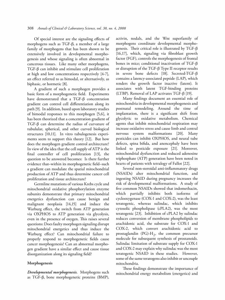
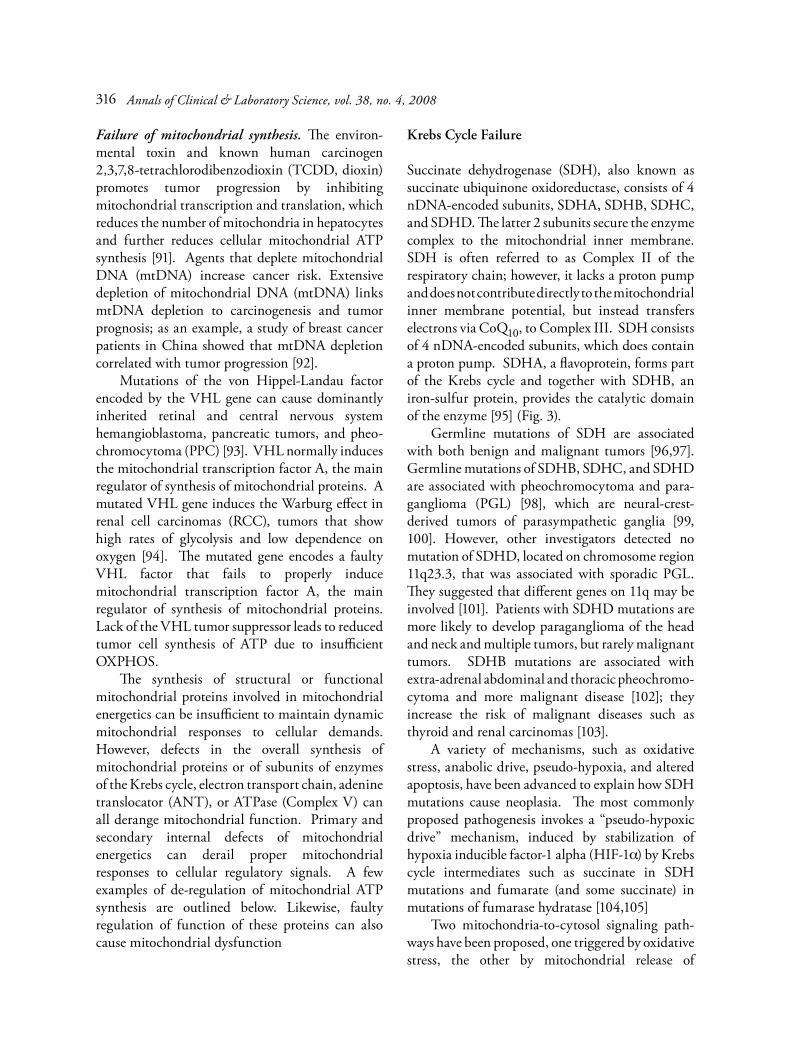
Whereas tissue organization during development morphogenesis is controlled by morphogenetic fields, neoplasia constitutes loss of control of tissue architecture due to aberrations in morphic fields [35]. Breakdown of the developmental morpho-genetic plan leading to cancer morphogenesis can be illustrated from observations of thyroid cancer compared to the tissue from which the tumor originated. As the tumor with time becomes less differentiated, follicles become increasingly smaller until no follicle is formed at all, at which point the cancer has become undifferentiated or anaplastic (Fig. 1). In this example, the radius of curvature of follicles became progressively smaller; however, in other cases, such as in tissue atrophy, with decreasing differentiation, the radius of curvature may increase as curvature control is lost due to age-related loss of response to morphogens or insufficient synthesis of morphogen. Field effects can reverse cancer morphogenesis. Signaling molecules and their receptors, interacting
Role of mitochondrial failure in cancer morphogenesis 309
with tumor gene expression, can regulate tumor phenotype. Metastatic melanoma cells transplanted into the chick-embryo neural tube can adapt cell morphologies resembling normal neural crest cells and migrate and function normally in host peripheral structures [36]. When cells derived from the core of a mouse teratocarcinoma were micro-injected into mice blastocycts of normal mice, complete reversibility of malignancy was observed. In the host, transplanted tumor cells participated in formation of adult, normally functioning mouse tissues including sperm, indicating a non-mutational basis for neoplastic conversion by developmental tissue disorganization rather than genetic aberration [37]. Highly aggressive breast cancer cells that harbor mutations of regulatory genes can be manipulated to exhibit a normal phenotype by correcting malfunctioning signaling pathways such as those involving epidermal growth factor, β-integrin, and E-cadherin [38]. Mammary tumor cells supplemented with integrin-blocking anti-bodies reverted to a normal cell phenotype, but upon removal of the antibody, the cells dedifferen-
tiated and re-formed disorganized colonies, reflecting a return to the tumor phenotype [39]. Nevertheless, minor tumor cell genetic differences vary in responses to their environment, such as their resistance to necrosis in the presence of hypoxia. Spheroids of highly tumorigenic, transfected cultured cell lines derived from rat embryonic fibroblasts show much higher tolerance to central hypoxia and necrosis than corresponding spontaneously transformed cell lines [40].
Warburg effect. Neoplastic lesions exhibit various degrees of altered mitochondrial morphology. These observations suggest that mitochondria are involved in the aberrant tissue morphology seen in human neoplasms. This notion is corroborated by recent findings that indicate that abnormal mitochondrial energetics plays a role in tumor initiation, progression, and regression [41]. During studies on tumor metabolism in the 1920’s Warburg [42] observed that cancer cells shift their metabolism from respiration to glycolysis. The rate of cancer cell glycolysis correlates inversely with lower mitochondrial bioenergetics [43].
Fig. 1. Evidence-based and hypothetical illustration of cancer morphogenesis due to progressive decrease of radius of curvature: (A) normal follicles showing secretion of latent bimodal growth factor (double arrow) and growth factor activator (dotted arrow) into its lumen. Active growth factor gradient (solid arrows) determines micro-architectural curvature. Declining radius of curvature due to increasing response to the morphogen and/or a steeper morphogen gradient causes progressive reduction in follicle size (B, C) and malignant growth (D), and finally leads to complete loss of curvature (anaplastic tumor) (E). By comparison, reduced latent growth factor growth or reduced growth factor activator secretion would cause dilatation of glandular structures such as can be seen in senile atrophy (not shown).
Annals of Clinical & Laboratory Science, vol. 38, no. 4, 2008310

Warburg detected increased lactate production in cancer tissues compared to corresponding normal tumor-free tissues. Warburg used the term “combustion” for mitochondrial oxidative phos-phorylation (OXPHOS). The Warburg effect, the change in cancer cells from combustion to glycolysis even in the presence of oxygen, has been confirmed by many investigators [44]. Warburg proposed several mechanisms to explain the effect named after him. He argued that faulty respiration is more common than impaired fermentation because respiration is more complicated as it requires many more enzymatic steps than glycolysis. Moreover, he argued, a shift from respiration to fermentation is facilitated by their both having the same catalyst (nicotinamide). Warburg linked oxygen metabolism to morphogenesis, considering it a sine qua non for cell differentiation and development of higher life forms. Respiration, but not fermentation, he maintained, creates and maintains cell differen-tiation, and a shift to fermentation causes dedifferentiation, which is cancer [45]. There are archezoans such as Giardia lamblia that lack mitochondria. However, the parasite harbors double membrane bounded, vestigial mitochondrial structures called mitosomes, which contain several mitochondrial markers and perform a limited number of mitochondrial functions such as protein import and iron-sulfur cluster biosynthesis [46]. The essential requirement for mitochondrial function in more complicated morphogenesis can be observed in mice, where dysfunctional oocyte mitochondria lower fertility and cause aberrant development [47]. Whereas supporters of the Warburg concept consider the shift in cellular energetics to be essential for cellular transformation, opponents of these concepts consider the Warburg effect not as the primary cause of cancer but as merely a manifestation of cancer [48]. Some investigators argue that the shift is just an adaptation to hypoxia in cancer tissues [49]. Yet others have suggested that enhanced glycolysis and increased OXPHOS with excessive ATP generation forms the basis for malignant tumor growth [50] or argue that the high rate of tumor glycolysis is necessary to support increased cell growth rather than to compensate for mitochondrial dysfunction [51].
Renewed interest in the Warburg effect in tumorigenesis has been stimulated by findings thar demonstrate that germline mutations of genes that encode Krebs cycle or OXPHOS enzyme subunits are associated with benign and malignant tumors. These findings clearly demonstrate that primary failure or dysfunction of mitochondrial energetics can cause benign and malignant tumors. The findings also suggest that agents such as environmental toxins and pesticides that cause secondary mitochondrial energetic failure or dysfunction may contribute to the development of benign and malignant tumors.
Bimodal morphogen gradients
Dose-response. Agents that induce low-dose stimulation and high-dose inhibition are referred to as bimodal, biphasic, or hormetic, and the response itself as hormesis. This type of morphogen dose-response is shared with many toxins and endogenous agents [8]. The term bimodal used in this paper corresponds broadly to the term biphasic or hormetic used by others. Virchow may have been the first to observe hormesis [52]. It has been proposed that exogenous and endogenous agents that signal via stimulatory or inhibitory pathways and receptor subtypes can form a generalized, hormetic, regulatory system [53]. However, some investigators have opined that bimodal or biphasic signaling that is receptor-mediated should not be viewed as hormesis; they limit the term hormesis to positive overcompensation mechanisms in response to exposure to a low dose of a harmful agent [54]. It seems that different mechanisms may be involved; however, the dose-response curves appear similar. Not only TGF-β, but other endogenous agents such a nitric oxide (NO) [55] and prostaglandins, can modulate mitochondrial ATP synthesis in a bimodal or biphasic manner. In rat cerebral cortex mitochondria, micromolar concentrations of a PGJ2 derivative, 15-deoxy-Δ12,14-J2 (15d-PGJ2), inhibits Complex I and markedly increases the rate of ROS formation at this site [56]. Prostaglandin PGE1 exhibits a bimodal effect on smooth muscle cells [57]. In cultured leukemia cells, PGJ2 and 15d-PGJ2 exhibit bimodal effects typical of bimodal morphogens, stimulating and inhibiting cell
Role of mitochondrial failure in cancer morphogenesis 311
proliferation at concentrations less than and more than 4 µM respectively; 30 µM led to apoptosis [58]. Statins, which inhibit the synthesis of co-enzyme Q10 (CoQ10), can exhibit bimodal effects on cultured endothelial cell proliferation and migration [59]. However, whether the bimodal effects of certain statins on angiogenesis in apoE- null mice are caused by modulation of CoQ10 synthesis and thereby OXPHOS is unclear [60]. Because a large number of tumor cell lines exhibit hormetic dose responses not only to morphogens, but also to a variety of exogenous agents [61], the extent to which such agents interfere with bimodal morphogen signaling is uncertain and in need of clarification. To permit fidelity of tissue curvature during a lifetime, separate mechanisms for regulation of mitochondrial ATP generation should exist in vivo: one for the overall, basic growth rate, and another, separate mechanism that permits morphogen gradient modulation of the basic growth rate. However, it seems likely that proper morphogen modulation of mitochondrial ATP production can only occur within a certain range of basic rates of cell proliferation, in which case significant abnormalities of basic growth rates could lead to cancer as well.
Radius of curvature. The concept of morphogen gradient control of tissue curvature originated in observations in my laboratory that showed that TGF-β modulates in a dose-dependent, bimodal manner the population doubling time of human cultured uterine smooth cells (SMC) and human uterine leiomyoma cell lines [5,6]. Modulation occurs as a percent of the basic doubling time of the particular cell line [62]. The observation and study of this bimodal modulation of SMC proliferation served as the basis for the development of the hypothesis that a TGF-β concentration gradient should be able to regulate curvature and thereby the morphology of tissue structures [10]. Along a TGF-β concentration gradient, as observed from the TGF-β source, inhibition of cellular proliferation is high at high concentration close to the source, and diminishes with increasing distance from the source. At one point along the gradient of decreasing concentration a point is reached beyond which the lower TGF-β increasingly
stimulates proliferation. At this particular point along the morphogen gradient, the forces of inhibitory and stimulatory morphogen signaling are equal. In this and in my previous papers [10,11,63], I refer to this point as the equidyne point and the morphogen concentration of the gradient at this point is referred to as the equidyne morphogen concentration. As seen from the TGF-β gradient source, the growth rate is first decreasingly inhibited up to the equidyne point, and then increasingly stimulated, thereby curving the tissue to form a concave curvature. The morphogen concentrations gradient determines the radius of the curvature. I refer to morphogens that induce such structures as concave morphogens. Molecules that stimulate at high concentration and inhibit at low concentration could be termed convex morphogens (Fig. 2). Such bimodal morphogens are very likely involved not only in formation of tubular structures but also in the formation of other curved tissue structures such as those found in cysts, eyes, and the femoral head. In breast cancer, ducts may develop glandular abluminal tissue in-folding; from a morphogen curvature theory point of view, a possible explanation would be that regionally lack of cell growth inhibition by the inhibitor morphogen signaling pathway would permit the tissue in between the morphogen source and the gradient equidyne point to proliferate, which would cause in-folding. Furthermore, interaction of more than one gradient of bimodal morphogens could conceivably produce quite complex structures; the equidyne point of one gradient could determine the path of a vascular channel, and a second bimodal morphogen gradient could determine the luminal diameter of the channel, a concept that might apply to vasculogenic mimicry, where channels are present but the lumen and walls are not appropriately developed.
Morphogen-induced tubulogenesis. Recent findings in the field of experimental tubulogenesis seem to corroborate the notion that bimodal morphogens can control curvature; these reports show that TGF-β concentration can control the radius of curvature in 3D tubulogenesis cultures in a dose-dependent manner [12].
Annals of Clinical & Laboratory Science, vol. 38, no. 4, 2008312

Altered architecture of cystic and tubular structures such as acini, follicles, ducts, and vessels is a common feature of cancer. Malignant melanomas can form de novo embryonic-type tubular, vascular channels also referred to as vasculogenic mimicry [64,65]. The pathogenesis of these peculiar, very narrow, vascular channels remains uncertain, although a few ideas are outlined above. However, the term “tubulogenesis” in this paper could apply to the morphogenesis of any hollow structure, such as spherical, tubulular, or domed structures. Many experiments corroborate involvement of bimodal morphogens in tubulo-genesis during development and in neoplasia. In an early model of tubulogenesis that employed 3-
dimensional (3D) gel cultures, capillary lumen formation by endothelial cells was stimulated and inhibited by TGF-β at concentrations ranging from 100 pg/ml to 1 ng/ml and 5-10 ng/ml, respectively [66]. More recently, 3D-gel culture experiments showed that TGF-β and other polypeptide growth factors such as the fibroblast growth factor family, glial cell-derived neurotrophic factor, and hepato-cyte growth factor/scatter factor (HGF/SF) can induce epithelial cells to form tubular structures. Importantly, the cysts and tubular structures induced by TGF-β exhibited luminal diameters that depended upon TGF-β concentration. Lumen diameter decreased with increasing morphogen concentration [12]. These results confirm that
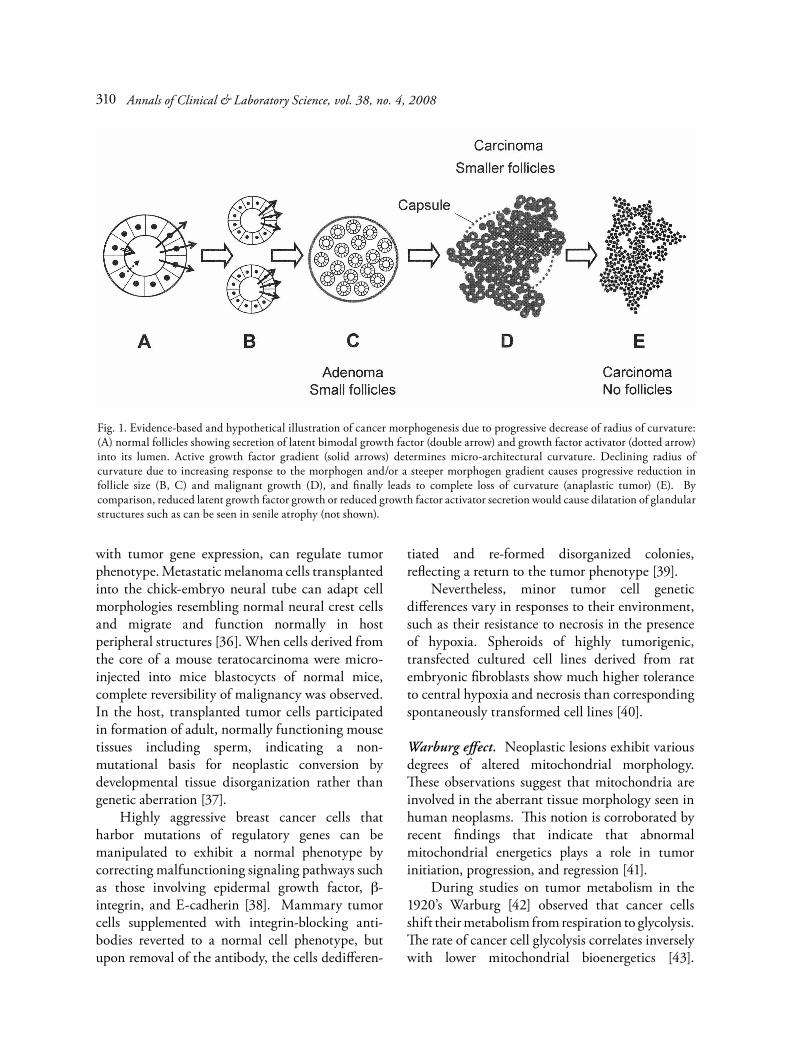
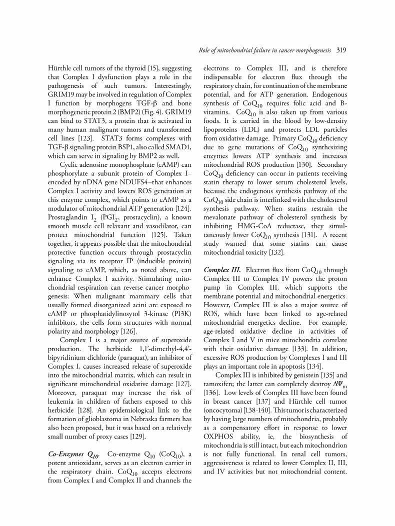
Fig. 2. Drawing illustrates the concept of bimodal morphogen control of curvature. By spatially modulating cell proliferation along its concentration diffusion/perfusion gradient, the morphogen controls tissue curvature as inhibition of cell proliferation gradually decreases (black areas) and stimulation gradually increases (hatched areas) with increasing distance from the morphogen source. Abbreviations: (C) Morphogen concentration at morphogen gradient source (GS); (D) Gradient distance; (INHIB) growth inhibition; (STIM) growth stimulation; (G) Growth; (DG) Growth differential; (MEC) Morphogen concentration at the point along the gradient where the morphogen inhibitory signal equals the morphogen stimulatory signal; (MEP) Morphogen Equidyne Point, point on the gradient where MEC is located; (MEL) line formed by intersection of MEPs in the tissue. Morphogen concentration at the source and the slope of the gradient determine the radius of curvature of concave tissue structures as viewed from the source.
Role of mitochondrial failure in cancer morphogenesis 313
TGF-β can modulate the radius of curvature of curved biological structures such as cysts and tubes, as has been proposed elsewhere [10]. In vitro, COX-1 regulates endothelial cell tubulogenesis [67]. COX-2 and prostanoids are involved in tubulogenesis by cultured rat gastric epithelial cells induced by transforming growth factor-alpha (TGF-a). The formation of tubular morphology was inhibited by COX-2 antisense oligonucleotide or COX-2 inhibition with NS-398 [68]. In vitro, PGE2 induced mouse endothelial cell migration and tubulogenesis via EP4 receptors, and PGE2 promoted in vivo angiogenesis in a sponge assay [69]. Tumor necrosis factor-alpha (TNF-a, cachectin) behaves as a bimodal morphogen during in vitro tubulogenesis. In vivo, when delivered by osmotic minipump or inserted in subcutaneous pellets in mice, TNF-a induces formation of tubular structures. Pump delivery of high concen-trations of TNF-a induces necrosis. Delivery via subcutaneous cannula of 35 ng/hr by osmotic mini-pump for one week induced capillary tube formation and a mass consisting of fibroblasts and collagen. Only a few isolated fibroblasts exhibited necrosis. By comparison, delivery of 170 ng/hr for up to 5 days led to massive necrosis that could be completely inhibited anti-TNF antibody [70]. Angiogenesis disks with central pellets containing TNF-a at 0.01-1 ng stimulated angiogenesis with a maximum at 0.1 ng; higher doses (1-5 ng) inhibited angiogenesis [71]. Polycystin-1 (PC-1) can induce renal epithelial cells to form cystic and tubular structures in vitro. The effect is inhibited by inhibition of PI3K. PC-1 promotes phosphorylation of Akt via PI3K, probably involving an intermediary molecule [72]. Another study showed that the hepatocyte growth factor (HGF) and its receptor Met augmented NF-kB DNA-binding and transcriptional activity, which stimulated formation of cystic and tubular structures by liver-derived cells. NF-kB inhibition abolished tubulogenesis. In this study, PI3K-signaling was not involved. The investigators noted that HGF-induced tubulogenesis and cell prolifer-ation appeared to be unrelated [73]. The observation that curvature control is independent of basic cell proliferation confirms theoretical predictions [10].
The ability to form tubes and cysts by different clones may vary considerably, even when clones are derived from the same tissue. For example, spontaneous formation of cysts and tubulogenic responses to hepatocyte growth factor (HGF) varies in different clones (A-D) derived from canine renal cells. Spontaneous responses in collagen gels were compared to formation after HGF supplementation: The A-clone formed cysts and then tubular structures, the B-clone made dense cell spheres and then tubular structures, and the D-clone made cysts but no tubules. In the first 2 clones, HGF-induced tubulogenesis was completely blocked by HGF-neutralizing antibody. Clone C required no HGF supplementation; it spontaneously formed tubules in the gel. The investigators suggested that cyst formation may represent a form of incomplete tubulogenesis [74]. Additional mechanisms may be involved in tubulogenesis in polycystic kidney disease (PKD). Luminal fluid from PKD cysts as well as exogenous cAMP and agonists (PGE1 or PGE2) induced fluid secretion by monolayers of cultured human kidney cortex cells, but among these agents, only PKD cyst luminal fluid induced cortex cells to proliferate and form cysts in collagen matrix gels. Epidermal growth factor (EGF) and TGF-β induced human kidney cortex (HKC) cells to proliferate and form cysts in matrix gels as well [75]. A more recent study isolated a cyst activating factor (CAP), a lipid factor in autosomal-dominant PKD and autosomal-recessive PKD cysts. CAP stimulated cAMP, cell proliferation, and anion secretion by cultured PKD cells [76]. By contrast, cAMP agonists failed to stimulate proliferation of HKC cells [77]
Morphogen modulation of mitochondrial energetics. The fact that germline mutations have demonstrated that mitochondrial primary energetics failure can cause cancer and that ATP is the final growth regulator led to consideration of concentration diffusion/perfusion gradients of bimodal morphogens as regulators of the radius of curvature of tissues via modulation of mitochondrial ATP generation [11]. Morphogen-responsive, specific regulatory elements present in several genes that encode mitochondrial proteins such as OXPHOS genes may enable morphogen gradients
Annals of Clinical & Laboratory Science, vol. 38, no. 4, 2008314

to modulate mitochondrial ATP synthesis. Such gene response may provide links to the role of morphogen control of mitochondrial energetics involved in cancer morphogenesis. For these reasons, a brief evidence-based and speculative discussion is included below, which looks at the interaction of morphogens with mitochondria, particularly as it applies to the mechanism of abnormal morphogen gradient signaling in cancer-induced formation of abnormal tubulular and cystic structures and in developmental and cancer morphogenesis.
Failure of cellular metabolic pathway regulation. Mitochondrial energetics failure can be due to reduction of overall synthesis of mitochondria, to failure of cell metabolic pathways, or to failure of parts of the mitochondrial energetics system such as the Krebs cycle or OXPHOS. The mitochondrion is a dual genome organelle; thus mitochondrial synthesis and energetics may be impaired by reduced supply of mtDNA- or nDNA-encoded mitochondrial proteins, or synthesis of one or more abnormal proteins from mutated genes [78]. Cancer cells may also show mitochondrial dysfunction in spite of increased transcription of many mito-chondrial and nuclear components of Krebs cycle and OXPHOS enzymes. The PI3K/protein kinase Akt (PI3K/Akt) and mammalian target of rapamycin (mTOR) pathways exert opposite effects on the contribution of ATP from glycolysis versus those from mitochondrial respiration [79]. Akt expression inhibits mito-chondrial respiration and activates cellular glyco-lytic metabolism, thus inducing a Warburg-type shift in cell energetics. Akt transfection alone can markedly increase the invasive potential of tumor cells. For example, cells from melanomas exhibiting radial growth can be converted by overexpression of Akt to melanomas that exhibit vertical growth and are highly invasive. Akt decreases Complex I activity, markedly increases ROS generation, and impairs mito-chondrial ATP generation. Moreover, Akt increases VEGF mRNA and protein levels and stimulates tumor-induced angiogenesis [80]. In addition, Akt1 signaling is involved in mice mammary tumor formation induced by an avian oncogene. Mice with disrupted Akt1 exhibited
reduced phosphorylation of tuberous sclerosis 2 (TSC2) in response to the oncogene, and tumor development and formation of tumor metastases was delayed [81]. NS-398, a selective COX-2 inhibitor, reduces growth of cultured human endometrial cancer cells in which mutated phosphatase tensin homolog (PTEN), a tumor suppressor and negative regulator of Akt/protein kinase B (Akt/PKB), fails to suppress Akt, an inducer of COX-2 expression. Elevated Akt phosphorylation levels increases COX-2 expression via NF-kB and increases tumor PGE2 levels. Blocking COX-2 catalytic activity induces cancer cell apoptosis [82]. In glioma models using PTEN mutant mice, the effect of loss of PTEN is similar to the effect of activated Akt [83]. Akt signaling can affect tumor morphology as well; manipulation of Akt signaling can alter tumor morphology from gliomatous to sarcomatous [84]. mTOR and the upstream and downstream components of its signaling pathways are often involved in cancer. mTOR is activated in tumors such as hamartoma, hemangioma, and neuro-blastoma [85,86]. In cultured MCF-7 breast cancer cells, mTOR inactivation induces autophagy [87]. In mice, expression of mTOR is essential for developmental morphogenesis; heterozygous disruption of mTOR results in developmental arrest at postcoitus day 5.5 [88]. Regulatory associated protein of mTOR (raptor) forms mTOR/raptor complexes that shift cell metabolism from glycolysis to mitochondrial respiration, thus reversing the Warburg effect [89]. In addition, mTOR regulates cellular translation rates and protein synthesis by controlling ribosome recruit-ment for the cellular translation machinery. mTOR function is affected by the cellular energy status. Glucose is a major upstream effector. Its signaling is influenced by ATP, and, importantly, mTOR itself functions as an ATP sensor [90]. Raptor up-regulates and tuberous sclerosis 1 (TSC1) and TSC2 inhibit mTOR activity, which correlates with oxygen consumption. Increased raptor/mTOR association increases mitochondrial function and retrograde signaling from mito-chondria can stabilize the mTOR/raptor complex. Knockdown of raptor decreases and knockdown of TSC2 increases ATP generation from respiration.
Role of mitochondrial failure in cancer morphogenesis 315
Failure of mitochondrial synthesis. The environ-mental toxin and known human carcinogen 2,3,7,8-tetrachlorodibenzodioxin (TCDD, dioxin) promotes tumor progression by inhibiting mitochondrial transcription and translation, which reduces the number of mitochondria in hepatocytes and further reduces cellular mitochondrial ATP synthesis [91]. Agents that deplete mitochondrial DNA (mtDNA) increase cancer risk. Extensive depletion of mitochondrial DNA (mtDNA) links mtDNA depletion to carcinogenesis and tumor prognosis; as an example, a study of breast cancer patients in China showed that mtDNA depletion correlated with tumor progression [92]. Mutations of the von Hippel-Landau factor encoded by the VHL gene can cause dominantly inherited retinal and central nervous system hemangioblastoma, pancreatic tumors, and pheo-chromocytoma (PPC) [93]. VHL normally induces the mitochondrial transcription factor A, the main regulator of synthesis of mitochondrial proteins. A mutated VHL gene induces the Warburg effect in renal cell carcinomas (RCC), tumors that show high rates of glycolysis and low dependence on oxygen [94]. The mutated gene encodes a faulty VHL factor that fails to properly induce mitochondrial transcription factor A, the main regulator of synthesis of mitochondrial proteins. Lack of the VHL tumor suppressor leads to reduced tumor cell synthesis of ATP due to insufficient OXPHOS. The synthesis of structural or functional mitochondrial proteins involved in mitochondrial energetics can be insufficient to maintain dynamic mitochondrial responses to cellular demands. However, defects in the overall synthesis of mitochondrial proteins or of subunits of enzymes of the Krebs cycle, electron transport chain, adenine translocator (ANT), or ATPase (Complex V) can all derange mitochondrial function. Primary and secondary internal defects of mitochondrial energetics can derail proper mitochondrial responses to cellular regulatory signals. A few examples of de-regulation of mitochondrial ATP synthesis are outlined below. Likewise, faulty regulation of function of these proteins can also cause mitochondrial dysfunction
Krebs Cycle Failure
Succinate dehydrogenase (SDH), also known as succinate ubiquinone oxidoreductase, consists of 4 nDNA-encoded subunits, SDHA, SDHB, SDHC, and SDHD. The latter 2 subunits secure the enzyme complex to the mitochondrial inner membrane. SDH is often referred to as Complex II of the respiratory chain; however, it lacks a proton pump and does not contribute directly to the mitochondrial inner membrane potential, but instead transfers electrons via CoQ10, to Complex III. SDH consists of 4 nDNA-encoded subunits, which does contain a proton pump. SDHA, a flavoprotein, forms part of the Krebs cycle and together with SDHB, an iron-sulfur protein, provides the catalytic domain of the enzyme [95] (Fig. 3). Germline mutations of SDH are associated with both benign and malignant tumors [96,97]. Germline mutations of SDHB, SDHC, and SDHD are associated with pheochromocytoma and para-ganglioma (PGL) [98], which are neural-crest-derived tumors of parasympathetic ganglia [99, 100]. However, other investigators detected no mutation of SDHD, located on chromosome region 11q23.3, that was associated with sporadic PGL. They suggested that different genes on 11q may be involved [101]. Patients with SDHD mutations are more likely to develop paraganglioma of the head and neck and multiple tumors, but rarely malignant tumors. SDHB mutations are associated with extra-adrenal abdominal and thoracic pheochromo-cytoma and more malignant disease [102]; they increase the risk of malignant diseases such as thyroid and renal carcinomas [103]. A variety of mechanisms, such as oxidative stress, anabolic drive, pseudo-hypoxia, and altered apoptosis, have been advanced to explain how SDH mutations cause neoplasia. The most commonly proposed pathogenesis invokes a “pseudo-hypoxic drive” mechanism, induced by stabilization of hypoxia inducible factor-1 alpha (HIF-1a) by Krebs cycle intermediates such as succinate in SDH mutations and fumarate (and some succinate) in mutations of fumarase hydratase [104,105] Two mitochondria-to-cytosol signaling path-ways have been proposed, one triggered by oxidative stress, the other by mitochondrial release of
Annals of Clinical & Laboratory Science, vol. 38, no. 4, 2008316

accumulated succinate. Either mechanism could explain how SDH dysfunction induces the high vascularity of such tumors. According to the first model, SDHC mutations elevate superoxide (O2
-.) and hydrogen peroxide (H2O2) production, which induces HIF-1 and features of cancer cells such as increased glucose use and development of genomic instability. These findings illustrate that SDHC promote an oxidative, stress-induced, mutation-prone phenotype [106]. In a second model, succinate that accumulates because of defective SDH function inhibits cytosolic HIF-a propylhydroxylase, which stabilizes and activates hypoxia-inducible factor (HIF)-1a [107,108]. Activation of HIF-1 invokes the Warburg effect and has been linked to malignant transformation and poor clinical outcome [109].
Gossypol, a component of cotton seed oil and an antifertility agent that reduces sperm mobility, inhibits mitochondrial energetics by inhibiting SDH [110]. In addition, gossypol stimulates TGF-β1 gene expression, which leads to inhibition of proliferation of cultured prostate cancer cells via the TGF-β inhibitory signaling pathway [111]. The inhibitory effect of gossypol can itself be inhibited by TGF-β antibody [112]. Alpha-ketoglutarate dehydrogenase carries electrons from the Krebs cycle to Complex I of the respiratory chain via a NADH/NAD+ shuttle. In mice, the tobacco carcinogen benzo[a]pyrene reduces enzyme activities of a-KGDH, SDH, and other Krebs cycle enzymes [113]. Others have shown that the pesticide acrolein [114] as well as
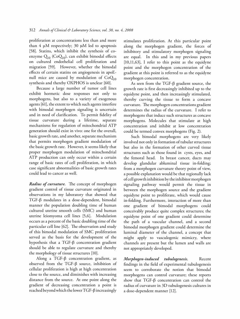
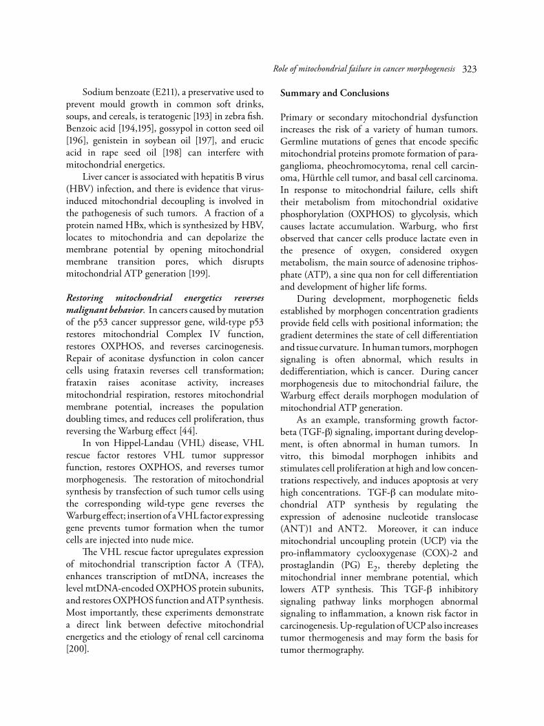
Fig. 3. Mitochondrial (mt) energy flux and neoplasia: Krebs cycle enzymes a-KGDH and SDH transfer electrons from food metabolites to the respiratory chain (electron transport chain, ETC), located in the mitochondrial inner membrane where electron-flux-driven proton-pumps of complexes I, III, and IV replenish the membrane potential (ΔΨm), which powers Complex V for synthesis of adenosine triphosphate (ATP). The potential also supports thermogenesis and import of nuclear-encoded proteins and synthesis of mtDNA-encoded enzyme subunits (not shown). Germline mutations of Complex I subunits inhibit transport of electrons by the NADH/NAD+ shuttle from a-KGDH via Complex I to CoQ10 and is associated with basal cell carcinoma and Hürthle cell carcinoma (oncocytoma). Defects of the transport of electrons via SDH (Complex II), caused by germline mutations of nuclear genes coding for its subunits A and B (in the Krebs cycle) and C and D (embedded in the mitochondrial inner membrane) to CoQ10 is associated with pheochromocytoma, paraganglioma, renal cell carcinoma, and thyroid carcinoma. Mutated fumarate hydratase (FH) has been detected in pheochromocytomas, paragangliomas, and leiomyomas. Prostate cancer is associated with reduced uptake of zinc and reduced inhibition of aconitase, which shift citrate to OXPHOS and reduce its release into seminal fluid. For further detail see text.
317

H2O2 [115] can inhibit a-KGDH and limit the supply of NADH to the respiratory chain.. Cancer is an age-related disease, and aging is associated with lower aconitase activity. In the house fly model of aging, old flies exhibited markedly lower aconitase activity compared to young flies [116]. In the human prostate, zinc normally inhibits mitochondrial aconitase activity, which channels high concentrations of citrate into the seminal fluid. By comparison, prostate carcinoma cells exhibit reduced zinc accumulation, which leads to increased citrate oxidation in the Krebs cycle. The increased oxidation of citrates may lead to increased respiratory chain electron flux and increased cancer-inducing ROS generation [117]. Sepsis lowers aconitase activity and can reduce mitochondrial ATP synthesis. Intra-peritoneal endotoxin injection of rats selectively reduces aconitase activity in heart mitochondria [118]. Germline mutations of fumarase hydratase
(FH) predispose to formation of papillary renal carcinomas and smooth muscle cell tumors such as leiomyomas of the uterus and the skin [14].
Respiratory Chain and Proton Pump Failure
Complex I. Abnormal subunits of NADH-ubiquinone oxidoreductase (Complex I) have been associated with human tumors. An investigation of gene expression in basal cells carcinomas found five genes involved in mitochondrial energetics that were consistently down-regulated compared to normal skin. One of these genes, NDUFA1, codes for the 7.5-kDa subunit of Complex I of the respiratory chain [119]. The subunit is essential for Complex I activity [120]. A tumor suppressor gene, GRIM19, encodes a protein that is vital for proper assembly and function of Complex I [121,122]. Somatic missense mutations of GRIM19 have been detected in
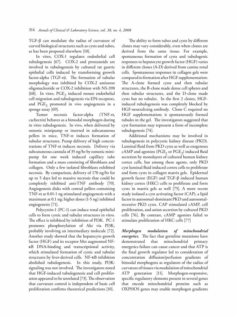
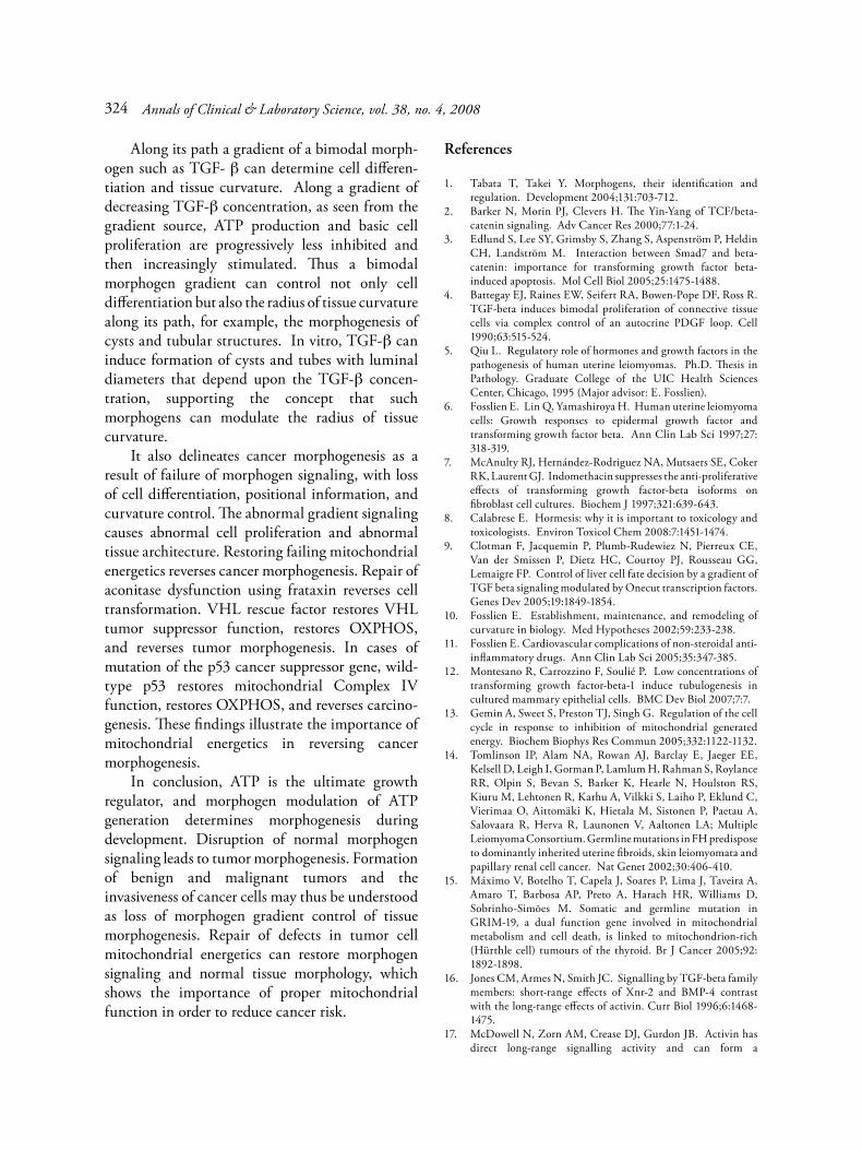
Fig. 4. The evidence-based and hypothetical diagram illustrates modulation of mitochondrial ATP synthesis by a bimodal morphogen, transforming growth-factor beta (TGF-β). The diagram divides mitochondrial energetics into respiration, which generates the inner membrane potential (ΔΨm), coupling of Ψmto adenosine triphosphate (ATP) synthesis by Complex V (F0F1-ATPase) and adenine nucleotide translocase (ANT), and loss of ΔΨm due to uncoupling or apoptosis. TGF-β can (1) regulate ANT1 and ANT2 in a bimodal manner, (2) regulate expression of GRIM19, a subunit of Complex I, (3) induce COX-2 and thereby synthesis of prostaglandin (PG) J2 that can regulate Complex I activity in a bimodal manner, (5) induce uncoupling protein (UCP) via PGE2 , (6) induce p53, which regulates Complex IV and thereby oxidative phosphorylation (OXPHOS), and (7) induce apoptosis via induction of Bax (not shown), which opens membrane pores and depletes ΔΨm. Dotted lines: electron flux; dashed lines: proton flux; AA: arachidonic acid; SCO2: cytochrome c oxidase 2. For further details see text.
Annals of Clinical & Laboratory Science, vol. 38, no. 4, 2008318
Hürthle cell tumors of the thyroid [15], suggesting that Complex I dysfunction plays a role in the pathogenesis of such tumors. Interestingly, GRIM19 may be involved in regulation of Complex I function by morphogens TGF-β and bone morphogenetic protein 2 (BMP2) (Fig. 4). GRIM19 can bind to STAT3, a protein that is activated in many human malignant tumors and transformed cell lines [123]. STAT3 forms complexes with TGF-β signaling protein BSP1, also called SMAD1, which can serve in signaling by BMP2 as well. Cyclic adenosine monophosphate (cAMP) can phosphorylate a subunit protein of Complex I– encoded by nDNA gene NDUFS4–that enhances Complex I activity and lowers ROS generation at this enzyme complex, which points to cAMP as a modulator of mitochondrial ATP generation [124]. Prostaglandin I2 (PGI2, prostacyclin), a known smooth muscle cell relaxant and vasodilator, can protect mitochondrial function [125]. Taken together, it appears possible that the mitochondrial protective function occurs through prostacyclin signaling via its receptor IP (inducible protein) signaling to cAMP, which, as noted above, can enhance Complex I activity. Stimulating mito-chondrial respiration can reverse cancer morpho-genesis: When malignant mammary cells that usually formed disorganized acini are exposed to cAMP or phosphatidylinosytol 3-kinase (PI3K) inhibitors, the cells form structures with normal polarity and morphology [126]. Complex I is a major source of superoxide production. The herbicide 1,1’-dimethyl-4,4’-bipyridinium dichloride (paraquat), an inhibitor of Complex I, causes increased release of superoxide into the mitochondrial matrix, which can result in significant mitochondrial oxidative damage [127]. Moreover, paraquat may increase the risk of leukemia in children of fathers exposed to this herbicide [128]. An epidemiological link to the formation of glioblastoma in Nebraska farmers has also been proposed, but it was based on a relatively small number of proxy cases [129].
Co-Enzymes Q10. Co-enzyme Q10 (CoQ10), a potent antioxidant, serves as an electron carrier in the respiratory chain. CoQ10 accepts electrons from Complex I and Complex II and channels the
electrons to Complex III, and is therefore indispensable for electron flux through the respiratory chain, for continuation of the membrane potential, and for ATP generation. Endogenous synthesis of CoQ10 requires folic acid and B-vitamins. CoQ10 is also taken up from various foods. It is carried in the blood by low-density lipoproteins (LDL) and protects LDL particles from oxidative damage. Primary CoQ10 deficiency due to gene mutations of CoQ10 synthesizing enzymes lowers ATP synthesis and increases mitochondrial ROS production [130]. Secondary CoQ10 deficiency can occur in patients receiving statin therapy to lower serum cholesterol levels, because the endogenous synthesis pathway of the CoQ10 side chain is interlinked with the cholesterol synthesis pathway. When statins restrain the mevalonate pathway of cholesterol synthesis by inhibiting HMG-CoA reductase, they simul-taneously lower CoQ10 synthesis [131]. A recent study warned that some statins can cause mitochondrial toxicity [132].
Complex III. Electron flux from CoQ10 through Complex III to Complex IV powers the proton pump in Complex III, which supports the membrane potential and mitochondrial energetics. However, Complex III is also a major source of ROS, which have been linked to age-related mitochondrial energetics decline. For example, age-related oxidative decline in activities of Complex I and V in mice mitochondria correlate with their oxidative damage [133]. In addition, excessive ROS production by Complexes I and III plays an important role in apoptosis [134]. Complex III is inhibited by genistein [135] and tamoxifen; the latter can completely destroy DΨm [136]. Low levels of Complex III have been found in breast cancer [137] and Hürthle cell tumor (oncocytoma) [138-140]. This tumor is characterized by having large numbers of mitochondria, probably as a compensatory effort in response to lower OXPHOS ability, ie, the biosynthesis of mitochondria is still intact, but each mitochondrion is not fully functional. In renal cell tumors, aggressiveness is related to lower Complex II, III, and IV activities but not mitochondrial content.
Role of mitochondrial failure in cancer morphogenesis 319

More aggressive tumors exhibit the greatest OXPHOS impairment [141].
Complex IV. A tumor suppressor gene, p53, which stimulates OXPHOS, is frequently mutated in human cancers. The mutated form induces the Warburg effect in mice and human cell lines; it fails to induce expression of cytochrome oxidase c oxidase 2 (SCO2), a copper-binding protein essential for Complex IV assembly and function. Transfection with wild-type p53 reverses the Warburg effect [142].
Coupling failure
Electron flux through the respiratory chain powers the proton pumps in Complexes I, III, and IV, to replenish the membrane potential as it is consumed by coupling to ATP synthesis by Complex V; or depleted by uncoupling proteins for heat production; or lost due to membrane leakage, for example due to insufficient membrane cholesterol. The electron flux must be constantly regulated to enable the pumps to maintain a membrane potential, typically about 160 mV. The transmembrane potential serves as a temporary energy buffer, conceptually similar to a capacitor that can store temporary energy in an electronic circuitry (for review see Fosslien [143]). The ATP-producing phosphorylation system includes Complex V (F0F1-ATPase) and adenine nucleotide translocases (ANT) that exchange ADP and ATP across mitochondrial membranes. The herbicide atrazine can inhibit sperm mitochondrial ATP synthesis by binding to and inhibiting Complex V. It has been suggested that similar triazine compounds may bind to Complex V and inhibit ATP synthesis as well [144]. Atrazine exposure can interfere with reproduction and development, and may be carcinogenic. The European Union (but not the US Environmental Protection Agency) has banned the use of atrazine [145]. Up-regulation of UCP by reactive oxygen species (ROS) can serve to counteract membrane hyperpolarization induced by hyperglycemia, reducing the excessive inner membrane potential, reducing excessive ROS formation, and protecting against ROS-induced cellular damage. Uncoupling
can also occur when mitochondrial membrane transition pores open completely, which can lead to complete depolarization of the membrane potential, mitochondrial loss of cytochrome c, apoptosis, and cell death (for review see Fosslien [143]).
Translocases. Human adenosine nucleotide trans-locase (ANT), located in the mitochondrial inner membrane, can regulate Complex V activity by controlling the exchange of ADP and ATP across the membrane. Some ANT promoter elements respond to basic growth regulators. There is experimental evidence that shows that other, separate, elements respond to morphogen signaling, and that morphogens can modulate mitochondrial ATP synthesis by modulating ANT. Mouse and human ANT1 and ANT2 show substantial homology [146]. The 3 main human isoforms are discussed in further detail below. ANT1 knockout mice show that ANT1 expression adapts to variable needs for ATP generation during embryogenesis and for post-natal energetic demands via OXBOX/REBOX regions of its promoter. REBOX has a ubiquitous tissue distribution and responds to thyroxin and NADH, whereas the OXBOX is only found in ANT1 in myogenic cells. The promoter for the β-subunit of F0F1-ATPase also contains OXBOX and REBOX elements, suggesting basic coordinated regulation of mitochondrial energy metabolism genes [147, 148]. The OXBOX/REBOX regions can respond to NADH and thyroxine [149]. TGF-β regulates ANT1 expression via TGF-β responsive elements present in the rodent ANT1 promoter [150]. Basic growth requirements can regulate ANT2 expression, increasing it when cells are stimulated by serum, epidermal growth factor (EGF), or platelet-derived growth factor (PDGF), and decreasing expression during experimental cell differentiation [151]. Thyroid hormone (T3) induces ANT2 expression in normal cells [152]. On the other hand, nuclear factor 1 (NF1) can suppress ANT2 expression by binding to two silencing sites of the ANT2 promoter [153]. ANT2 expression is increased under the glycolytic conditions in highly proliferative cells such as cancer cells that increase glycolysis to satisfy their demand for ATP.
Annals of Clinical & Laboratory Science, vol. 38, no. 4, 2008320

ANT2 is induced in tumor cells, Simian virus 40 (SV40) transformed cells, and rho null cells; the latter have mitochondria that lack mitochondrial DNA [154]. Several other transformed cell lines exhibit high rates of glycolysis and high levels of ANT2. In these cancer cells, ATP generated by glycolysis powers ANT2 and the β subunit of F0F1-ATPase. Reversal of the function of the latter pumps protons from the mitochondrial matrix to the intermembrane space. In this way, glycolysis supports the mitochondrial membrane potential. It has been suggested that this contributes to the aggressive growth of such cells [155]. This mechanism has also been observed in cases of defective oxidative phosphorylation due to mtDNA depletion, which causes a shift in metabolism from mitochondrial to glycolytic ATP synthesis [156]. ANT2 lacks a TGF-β-responsive element [150]. However, the ANT2 promoter contains specificity protein (Sp)-1-responsive elements, which are DNA sequence-specific transcription factor binding sites. As the TGF-β signaling cascade involves Smad proteins and Sp1, this arrangement indicates that TGF-β can indirectly regulate ANT2. This interpretation is supported by experimental evidence of bimodal regulation of ANT2 expression by Sp1 interacting with three regulatory elements of the ANT2 promoter [157]. Interacting in vitro with GC-box promoter elements, SP1 up-regulates and down-regulates ANT2 expression in a concentration-dependent manner. This type of positive and negative, bimodal modulation of gene expression is exceptional for an OXPHOS promoter [158]. Sp1 up-regulates ANT2 expression via two Sp1 activating promoter boxes (AB boxes), and down-regulates expression via binding to a repressor Cbox. Studies of mammalian cells and transfected Drosophila cells show that Sp1 binding to the Cbox is involved in repression of ANT2 expression. Thus Sp1 can induce or repress ANT2 expression [159]. The vital importance of morphogen regulation via Sp1 is illustrated by experiments using a Drosophila homolog of human Sp1 to restore Sp1 function. Sp1 is essential for morphogenesis of head segments at the blastoderm stage of development. Remarkably, human Sp1 introduced transgenetically restored formation of mandibular segments of
mutant flies that lack the homolog [160]. ANT3 probably serves as a component and regulator of the mitochondrial permeability transition pore; for instance, it is involved in apoptosis induced by tumor necrosis factor in human breast cancer cell lines [161].
Uncoupling. Uncoupling proteins UCP1, UCP2, and UCP3 are found in all mammals. Located in mitochondrial inner membranes, they return protons from the mitochondrial inter-membranous space into the matrix and liberate heat (thermo-genesis). Reactive oxygen species (ROS) up-regulate UCPs and reduce the inner membrane potential, which restrains excessive ROS formation; this feedback loop protects against ROS-induced cellular damage. Mutations of UCP2 are associated with neural tube defects; this demonstrates that alterations in mitochondrial energetics can interfere with developmental morphogenesis [162]. PGE2 uncouples or inhibits respiration of isolated rat mitochondria depending upon the calcium concentration in the incubation mixture [163]. In primary cultures of human muscle cells, omega-6 polyunsaturated fatty acid (PUFA) induces UCP-2 mRNA expression, via cyclooxy-genase, PGE2, PGI2, and cAMP. Indomethacin, a non-selective NSAID, inhibits the effects [164].
Arachidonic acid metabolism and cancer morpho-genesis. COX-2 is inducible by interleukin-1, NF-kB, oncogenes ras and scr, hypoxia, benzo{a}pyrene, epidermal growth factor (EGF), TGF-β, tumor necrosis factor (TNF)-a, and ultraviolet (UV) light. In mice, it supports ovulation, fertilization, blastocyst implantation, and decidualization [165]. UV exposure increases the risk of human skin cancer. In mice, genetic or UV-B light over-expression of COX-2 in skin promotes skin cancer. The primary prostaglandin in skin is PGE2, a downstream product of COX-2 [166]. NSAIDs that inhibit COX-2 lower the risk of cancer and reduce cancer burden [167,168], PGE2 is involved in cancer morphogenesis [169]. It has been linked to endometrial cancer [170] and cervical cancer. Cervical infection with human papilloma (HPV)-containing E6 and E7 elements is a strong risk factor for cervical cancer.
Role of mitochondrial failure in cancer morphogenesis 321

Both COX-2 and PGE2 are elevated in human cervical carcinoma cells. In vitro, human papilloma virus (HPV) E6 and E7 induce COX-2 expression via activator protein-1 (AP-1) and the cAMP regulatory element of the COX-2 promoter [171]. PGE2 inhibits apoptosis: incubation of human T-cell leukemia cells (Jurkat cells) with 5 nM PGE2 prior to camptothecin therapy reduces apoptosis. The effect is mediated by EP4R (a type of PGE2 receptor) and the PI3K/Akt signaling pathway [172]. In prostate cancer, EP1R expression is associated with cancer aggressiveness, but EP2R and EP4R are involved as well in the formation of the tumor [173]. In rats, terephthalic acid (benzene-1,4-dicarb-oxylic acid), used in the polyester industry, can increase cytosolic phospholipase-2 (cPLA2) and COX-2 expression, elevate PGE2 levels, and cause formation of bladder papilloma and transitional cell carcinoma (TCC) of the urinary bladder after 24 and 48 months of treatment, respectively [174]. This model of neoplastic development demonstrates the importance of duration of carcinogen exposure, and that tissue architecture is relatively resistant to neoplastic change. COX-2 can participate in the inhibitory effect of TGF-β. PGE2 can exhibit bimodal morphogen signaling, which indicates a link between chronic arachidonic acid metabolism, inflammation, bimodal regulation of growth, and carcinogenesis. PGE2 supplementation inhibits proliferation of cultured rabbit corneal endothelial cells. When such cell were incubated with inhibitory concen-trations of TGF-β2 without PGE2 supplementation, TGF-β2 at concentrations of 0.5 to 50 ng/ml enhanced cell secretion of PGE2 and inhibited cell proliferation in a concentration-dependent manner. Indomethacin reduced PGE2 secretion to very low levels and abolished TGF-β inhibition of cell proliferation, indicating that PGE2 is involved in the inhibitory signaling pathway of TGF-β2 [175]. As PGE2 induces UCP-2, the following probable mechanism emerges: TGF-β induces COX-2, which increases PGE2 synthesis; PGE2 then induces expression of UCP-2, which uncouples mito-chondria, lowers the mitochondrial inner membrane potential, reduces ATP synthesis, and limits cell proliferation.
Agents that lower mitochondrial energetics. Phthalates, plasticizers in food packaging, plastic bottles, cosmetics , floor tiles, and other everyday products can cause mitochondrial swelling [176], inhibit mitochondrial β-oxidation [177], alter inner membrane permeability [178], inhibit succinate dehydrogenase [179,180], inhibit OXPHOS at the level of cytochrome c reductase [181], inhibit adenine nucleotide translocase (AND) [182], and decrease the rate of ATP synthesis [183]. In rats, phthalates may behave as endocrine disruptors; it has been proposed that they may act in a dose-additive way, suggesting that cumulative exposure may be most important for human health risks [184]. Use of phthalates in baby care products, lotions, and shampoos has raised concerns because some investigators suggest that they can alter male reproductive development [185]. In mice, TCDD lowers CoQ10 levels, reduces the mitochondrial inner membrane potential, reduces the synthesis of F0F1-ATP synthase, and lowers production of ATP [186]. In 1997, TCDD was classified by the International Agency for Research on Cancer (IARC) as a human carcinogen, a decision that was supported by additional, later data [187]. A recent review of this topic by Hardell [188] emphasizes that early warning of carcinogenic risk of use of certain pesticides should be taken seriously to reduce cancer risk. The author voices concern about industry experts who make dismissive comments about such risks [188]. In addition to analyses of carcinogenic risk, it has been stressed that assessment of pesticide risk studies should include analyses of possible associations with congenital malformations in offspring of fathers who have been exposed to pesticides [189]. Exposure to the fungal mutagen aflatoxin–a contaminant of grains, grapes, peanuts, hazelnuts, and dried fruits–is associated with hepatocellular carcinoma. The mycotoxin binds to mitochondrial DNA and impairs mitochondrial transcription and translation [190]. Mitochondria isolated from livers of rats exposed to aflatoxin exhibit impaired respiration due to reduced activities of Complex II and IV and membrane damage [191] and mutations of β-catenin and tumor suppressor protein p53 genes [192].
Annals of Clinical & Laboratory Science, vol. 38, no. 4, 2008322
Sodium benzoate (E211), a preservative used to prevent mould growth in common soft drinks, soups, and cereals, is teratogenic [193] in zebra fish. Benzoic acid [194,195], gossypol in cotton seed oil [196], genistein in soybean oil [197], and erucic acid in rape seed oil [198] can interfere with mitochondrial energetics. Liver cancer is associated with hepatitis B virus (HBV) infection, and there is evidence that virus-induced mitochondrial decoupling is involved in the pathogenesis of such tumors. A fraction of a protein named HBx, which is synthesized by HBV, locates to mitochondria and can depolarize the membrane potential by opening mitochondrial membrane transition pores, which disrupts mitochondrial ATP generation [199].
Restoring mitochondrial energetics reverses malignant behavior. In cancers caused by mutation of the p53 cancer suppressor gene, wild-type p53 restores mitochondrial Complex IV function, restores OXPHOS, and reverses carcinogenesis. Repair of aconitase dysfunction in colon cancer cells using frataxin reverses cell transformation; frataxin raises aconitase activity, increases mitochondrial respiration, restores mitochondrial membrane potential, increases the population doubling times, and reduces cell proliferation, thus reversing the Warburg effect [44]. In von Hippel-Landau (VHL) disease, VHL rescue factor restores VHL tumor suppressor function, restores OXPHOS, and reverses tumor morphogenesis. The restoration of mitochondrial synthesis by transfection of such tumor cells using the corresponding wild-type gene reverses the Warburg effect; insertion of a VHL factor expressing gene prevents tumor formation when the tumor cells are injected into nude mice. The VHL rescue factor upregulates expression of mitochondrial transcription factor A (TFA), enhances transcription of mtDNA, increases the level mtDNA-encoded OXPHOS protein subunits, and restores OXPHOS function and ATP synthesis. Most importantly, these experiments demonstrate a direct link between defective mitochondrial energetics and the etiology of renal cell carcinoma [200].
Summary and Conclusions
Primary or secondary mitochondrial dysfunction increases the risk of a variety of human tumors. Germline mutations of genes that encode specific mitochondrial proteins promote formation of para-ganglioma, pheochromocytoma, renal cell carcin-oma, Hürthle cell tumor, and basal cell carcinoma. In response to mitochondrial failure, cells shift their metabolism from mitochondrial oxidative phosphorylation (OXPHOS) to glycolysis, which causes lactate accumulation. Warburg, who first observed that cancer cells produce lactate even in the presence of oxygen, considered oxygen metabolism, the main source of adenosine triphos-phate (ATP), a sine qua non for cell differentiation and development of higher life forms. During development, morphogenetic fields established by morphogen concentration gradients provide field cells with positional information; the gradient determines the state of cell differentiation and tissue curvature. In human tumors, morphogen signaling is often abnormal, which results in dedifferentiation, which is cancer. During cancer morphogenesis due to mitochondrial failure, the Warburg effect derails morphogen modulation of mitochondrial ATP generation. As an example, transforming growth factor-beta (TGF-β) signaling, important during develop-ment, is often abnormal in human tumors. In vitro, this bimodal morphogen inhibits and stimulates cell proliferation at high and low concen-trations respectively, and induces apoptosis at very high concentrations. TGF-β can modulate mito-chondrial ATP synthesis by regulating the expression of adenosine nucleotide translocase (ANT)1 and ANT2. Moreover, it can induce mitochondrial uncoupling protein (UCP) via the pro-inflammatory cyclooxygenase (COX)-2 and prostaglandin (PG) E2, thereby depleting the mitochondrial inner membrane potential, which lowers ATP synthesis. This TGF-β inhibitory signaling pathway links morphogen abnormal signaling to inflammation, a known risk factor in carcinogenesis. Up-regulation of UCP also increases tumor thermogenesis and may form the basis for tumor thermography.
Role of mitochondrial failure in cancer morphogenesis 323
Along its path a gradient of a bimodal morph-ogen such as TGF- β can determine cell differen-tiation and tissue curvature. Along a gradient of decreasing TGF-β concentration, as seen from the gradient source, ATP production and basic cell proliferation are progressively less inhibited and then increasingly stimulated. Thus a bimodal morphogen gradient can control not only cell differentiation but also the radius of tissue curvature along its path, for example, the morphogenesis of cysts and tubular structures. In vitro, TGF-β can induce formation of cysts and tubes with luminal diameters that depend upon the TGF-β concen-tration, supporting the concept that such morphogens can modulate the radius of tissue curvature. It also delineates cancer morphogenesis as a result of failure of morphogen signaling, with loss of cell differentiation, positional information, and curvature control. The abnormal gradient signaling causes abnormal cell proliferation and abnormal tissue architecture. Restoring failing mitochondrial energetics reverses cancer morphogenesis. Repair of aconitase dysfunction using frataxin reverses cell transformation. VHL rescue factor restores VHL tumor suppressor function, restores OXPHOS, and reverses tumor morphogenesis. In cases of mutation of the p53 cancer suppressor gene, wild-type p53 restores mitochondrial Complex IV function, restores OXPHOS, and reverses carcino-genesis. These findings illustrate the importance of mitochondrial energetics in reversing cancer morphogenesis. In conclusion, ATP is the ultimate growth regulator, and morphogen modulation of ATP generation determines morphogenesis during development. Disruption of normal morphogen signaling leads to tumor morphogenesis. Formation of benign and malignant tumors and the invasiveness of cancer cells may thus be understood as loss of morphogen gradient control of tissue morphogenesis. Repair of defects in tumor cell mitochondrial energetics can restore morphogen signaling and normal tissue morphology, which shows the importance of proper mitochondrial function in order to reduce cancer risk.
References
1. Tabata T, Takei Y. Morphogens, their identification and regulation. Development 2004;131:703-712.
2. Barker N, Morin PJ, Clevers H. The Yin-Yang of TCF/beta-catenin signaling. Adv Cancer Res 2000;77:1-24.
3. Edlund S, Lee SY, Grimsby S, Zhang S, Aspenström P, Heldin CH, Landström M. Interaction between Smad7 and beta-catenin: importance for transforming growth factor beta-induced apoptosis. Mol Cell Biol 2005;25:1475-1488.
4. Battegay EJ, Raines EW, Seifert RA, Bowen-Pope DF, Ross R. TGF-beta induces bimodal proliferation of connective tissue cells via complex control of an autocrine PDGF loop. Cell 1990;63:515-524.
5. Qiu L. Regulatory role of hormones and growth factors in the pathogenesis of human uterine leiomyomas. Ph.D. Thesis in Pathology. Graduate College of the UIC Health Sciences Center, Chicago, 1995 (Major advisor: E. Fosslien).
6. Fosslien E. Lin Q, Yamashiroya H. Human uterine leiomyoma cells: Growth responses to epidermal growth factor and transforming growth factor beta. Ann Clin Lab Sci 1997;27: 318-319.
7. McAnulty RJ, Hernández-Rodriguez NA, Mutsaers SE, Coker RK, Laurent GJ. Indomethacin suppresses the anti-proliferative effects of transforming growth factor-beta isoforms on fibroblast cell cultures. Biochem J 1997;321:639-643.
8. Calabrese E. Hormesis: why it is important to toxicology and toxicologists. Environ Toxicol Chem 2008:7:1451-1474.
9. Clotman F, Jacquemin P, Plumb-Rudewiez N, Pierreux CE, Van der Smissen P, Dietz HC, Courtoy PJ, Rousseau GG, Lemaigre FP. Control of liver cell fate decision by a gradient of TGF beta signaling modulated by Onecut transcription factors. Genes Dev 2005;19:1849-1854.
10. Fosslien E. Establishment, maintenance, and remodeling of curvature in biology. Med Hypotheses 2002;59:233-238.
11. Fosslien E. Cardiovascular complications of non-steroidal anti-inflammatory drugs. Ann Clin Lab Sci 2005;35:347-385.
12. Montesano R, Carrozzino F, Soulié P. Low concentrations of transforming growth factor-beta-1 induce tubulogenesis in cultured mammary epithelial cells. BMC Dev Biol 2007;7:7.
13. Gemin A, Sweet S, Preston TJ, Singh G. Regulation of the cell cycle in response to inhibition of mitochondrial generated energy. Biochem Biophys Res Commun 2005;332:1122-1132.
14. Tomlinson IP, Alam NA, Rowan AJ, Barclay E, Jaeger EE, Kelsell D, Leigh I, Gorman P, Lamlum H, Rahman S, Roylance RR, Olpin S, Bevan S, Barker K, Hearle N, Houlston RS, Kiuru M, Lehtonen R, Karhu A, Vilkki S, Laiho P, Eklund C, Vierimaa O, Aittomäki K, Hietala M, Sistonen P, Paetau A, Salovaara R, Herva R, Launonen V, Aaltonen LA; Multiple Leiomyoma Consortium. Germline mutations in FH predispose to dominantly inherited uterine fibroids, skin leiomyomata and papillary renal cell cancer. Nat Genet 2002;30:406-410.
15. Máximo V, Botelho T, Capela J, Soares P, Lima J, Taveira A, Amaro T, Barbosa AP, Preto A, Harach HR, Williams D, Sobrinho-Simões M. Somatic and germline mutation in GRIM-19, a dual function gene involved in mitochondrial metabolism and cell death, is linked to mitochondrion-rich (Hürthle cell) tumours of the thyroid. Br J Cancer 2005;92: 1892-1898.
16. Jones CM, Armes N, Smith JC. Signalling by TGF-beta family members: short-range effects of Xnr-2 and BMP-4 contrast with the long-range effects of activin. Curr Biol 1996;6:1468-1475.
17. McDowell N, Zorn AM, Crease DJ, Gurdon JB. Activin has direct long-range signalling activity and can form a
Annals of Clinical & Laboratory Science, vol. 38, no. 4, 2008324

concentration gradient by diffusion. Curr Biol 1997;7:671-681.
18. Sasaki T, Ito Y, Bringas P Jr, Chou S, Urata MM, Slavkin H, Chai Y. TGF-beta-mediated FGF signaling is crucial for regulating cranial neural crest cell proliferation during frontal bone development. Development 2006;133:371-381.
19. Oklü R, Hesketh R. The latent transforming growth factor beta binding protein (LTBP) family. Biochem J 2000;5;352 Pt 3:601-610.
20. Fantel AG, Person RE. Involvement of mitochondria and other free radical sources in normal and abnormal fetal development. Ann N Y Acad Sci 2002;959:424-433.
21. Rull RP, Ritz B, Shaw GM. Neural tube defects and maternal residential proximity to agricultural pesticide applications. Am J Epidemiol 2006;163:743-753.
22. Shinde SB, Save VC, Patil ND, Mishra KP, Tendolkar AG. Impairment of mitochondrial respiratory chain enzyme activities in tetralogy of Fallot. Clin Chim Acta 2007;377:138-143.
23. Montenegro MA, Palomino H. Induction of cleft palate in mice by inhibitors of prostaglandin synthesis. J Craniofac Genet Dev Biol 1990;10:83-94.
24. Child CM. A dynamic conception of the organic individual. PNAS USA 1915;1:164-172. Experiments by Hans Spemann and Hilde Mangold in 1924; see http://users.rcn.com/jkimball.ma.ultranet/BiologyPages/Spemann.html.
25. Meinhardt H. Models of biological pattern formation: from elementary steps to organization of embryonic axes. Curr Top Dev Biol 2008;81:1-63.
26. Tsikolia N. What is a role of the morphogen gradients in development? Riv Biol 2003;96:293-315.
27. Tsikolia N. The role and limits of a gradient based explanation of morphogenesis: a theoretical consideration. Int J Dev Biol 2006;50:333-340.
28. Child CM. Patterns and Problems of Development. University of Chicago Press, Chicago, 1941.
29. Fantini B. Embryology, ‘chemical geography’ of the cell and synthesis between morphology and chemistry (1930-1950). Hist Philos Life Sci 2000;22:353-380.
30. Potter JD. Morphostats: a missing concept in cancer biology. Cancer Epidemiol Biomarkers Prev 2001;10:161-170.
31. Potter JD. Morphogens, morphostats, microarchitecture and malignancy. Nat Rev Cancer 2007;7:464-474.
32. Ku M, Sokol SY, Wu J, Tussie-Luna MI, Roy AL, Hata A. Positive and negative regulation of the transforming growth factor beta/activin target gene goosecoid by the TFII-I family of transcription factors. Mol Cell Biol 2005;25:7144-7157.
33. Zhu CC, Yamada G, Blum M. Retinoic acid teratogenicity: the role of goosecoid and BMP-4. Cell Mol Biol (Noisy-le-grand) 1999;45:617-629.
34. Cho KW, Blumberg B, Steinbeisser H, De Robertis EM. Molecular nature of Spemann’s organizer: the role of the Xenopus homeobox gene goosecoid. Cell 1991;67:1111-1120.
35. Sonnenschein C, Soto AM. Somatic mutation theory of carcinogenesis: why it should be dropped and replaced. Mol Carcinog 2000;29:205-211.
36. Kulesa PM, Kasemeier-Kulesa JC, Teddy JM, Margaryan NV, Seftor EA, Seftor RE, Hendrix MJ. Reprogramming metastatic melanoma cells to assume a neural crest cell-like phenotype in an embryonic microenvironment. PNAS USA 2006;103:3752-3757.
37. Mintz B, Illmensee K. Normal genetically mosaic mice produced from malignant teratocarcinoma cells. PNAS USA 1975;72:3585-3589.
38. Wang F, Hansen RK, Radisky D, Yoneda T, Barcellos-Hoff MH, Petersen OW. Phenotypic reversion or death of cancer
cells by altering signaling pathways in three-dimensional contexts. J Natl Cancer Inst 2002;94:1494-1503.
39. Weaver VM, Petersen OW, Wang F, Larabell CA, Briand P, Damsky C, Bissell MJ. Reversion of the malignant phenotype of human breast cells in three-dimensional culture and in vivo by integrin blocking antibodies. J Cell Biol 1997;137:231-245.
40. Walenta S, Doetsch J, Mueller-Klieser W, Kunz-Schughart LA. Metabolic imaging in multicellular spheroids of oncogene-transfected fibroblasts. J Histochem Cytochem 2000;48:509-622.
41. van Waveren C, Sun Y, Cheung HS, Moraes CT. Oxidative phosphorylation dysfunction modulates expression of extracellular matrix–remodeling genes and invasion. Carcino-genesis 2006;27:409-418.
42. Warburg O. On the origin of cancer cells. Science 1956;123: 309-314.
43. López-Ríos F, Sánchez-Aragó M, García-García E, Ortega AD, Berrendero JR, Pozo-Rodríguez F, López-Encuentra A, Ballestín C, Cuezva JM. Loss of the mitochondrial bioenergetic capacity underlies the glucose avidity of carcinomas. Cancer Res 2007;67:9013-9017.
44. Schulz TJ, Thierbach R, Voigt A, Drewes G, Mietzner B, Steinberg P, Pfeiffer AF, Ristow M. Induction of oxidative metabolism by mitochondrial frataxin inhibits cancer growth: Otto Warburg revisited. J Biol Chem 2006;281:977-981.
45. Warburg O. On respiratory impairment in cancer cells. Science 1956;124:269-270.
46. Regoes A, Zourmpanou D, León-Avila G, van der Giezen M, Tovar J, Hehl AB. Protein import, replication, and inheritance of a vestigial mitochondrion. J Biol Chem 2005;280:30557-30563.
47. Nagai S, Mabuchi T, Hirata S, Shoda T, Kasai T, Yokota S, Shitara H, Yonekawa H, Hoshi K. Oocyte mitochondria: strategies to improve embryogenesis. Hum Cell 2004;17:195-201.
48. Garber K. Energy boost: the Warburg effect returns in a new theory of cancer. J Natl Cancer Inst 2004;96:1805-1806.
49. Semenza GL, Artemov D, Bedi A, Bhujwalla Z, Chiles K, Feldser D, Laughner E, Ravi R, Simons J, Taghavi P, Zhong H. ‘The metabolism of tumours’: 70 years later. Novartis Found Symp 2001;240:251-260.
50. Ray S, Ray M. Does excessive adenosine 5’-triphosphate formation in cells lead to malignancy? A hypothesis on cancer. Med Hypotheses 1997;48:473-476.
51. Bui T, Thompson CB. Cancer’s sweet tooth. Cancer Cell 2006;9:419-420.
52. Henschler D. The origin of hormesis: historical background and driving forces. Hum Exp Toxicol 2006;25:347-351.
53. Calabrese EJ, Baldwin LA. Agonist concentration gradients as a generalizable regulatory implementation strategy. Crit Rev Toxicol 2001;31:471-473.
54. van der Woude H, Alink GM, Rietjens IM. The definition of hormesis and its implications for in vitro to in vivo extrapolation and risk assessment. Crit Rev Toxicol 2005;35:603-607.
55. Calabrese EJ. Nitric oxide: biphasic dose responses. Crit Rev Toxicol 2001;31:489-501.
56. Martínez B, Pérez-Castillo A, Santos A. The mitochondrial respiratory complex I is a target for 15-deoxy-Δ12,14-prosta-glandin J2 action. J Lipid Res 2005;46:736-743.
57. Moreland RB, Kim N, Nehra A, Goldstein I, Traish A. Functional prostaglandin E (EP) receptors in human penile corpus cavernosum. Int J Impot Res 2003;15:362-368.
58. Emi M, Maeyama K. The biphasic effects of cyclopentenone prostaglandins, prostaglandin J2 and 15-deoxy-Δ12,14-prosta-glandin J2 on proliferation and apoptosis in rat basophilic
Role of mitochondrial failure in cancer morphogenesis 325
leukemia (RBL-2H3) cells. Biochem Pharmacol 2004;67:1259-1267.
59. Katsumoto M, Shingu T, Kuwashima R, Nakata A, Nomura S, Chayama K. Biphasic effect of HMG-CoA reductase inhibitor, pitavastatin, on vascular endothelial cells and angiogenesis. Circ J 2005;69:1547-1555.
60. Weis M, Heeschen C, Glassford AJ, Cooke JP. Statins have biphasic effects on angiogenesis. Circulation 2002;105:739-745.
61. Calabrese EJ. Cancer biology and hormesis: human tumor cell lines commonly display hormetic (biphasic) dose responses. Crit Rev Toxicol 2005;35:463-582.
62. Fosslien E. Mitochondrial medicine: molecular pathology of defective oxidative phosphorylation. Ann Clin Lab Sci 2000; 30:320-332.
63. Fosslien E. Cancer prevention: Role of preservation of mito-chondrial function. Ann Clin Lab Sci 2008;38:181.
64. Maniotis AJ, Folberg R, Hess A, Seftor EA, Gardner LM, Pe’er J, Trent JM, Meltzer PS, Hendrix MJ. Vascular channel formation by human melanoma cells in vivo and in vitro: vasculogenic mimicry. Am J Pathol 1999;155:739-752.
65. Hess AR, Margaryan NV, Seftor EA, Hendrix MJ. Deciphering the signaling events that promote melanoma tumor cell vascul-ogenic mimicry and their link to embryonic vasculogenesis: role of the Eph receptors. Dev Dyn 2007;236:3283-3296.
66. Pepper MS, Vassalli JD, Orci L, Montesano R. Biphasic effect of transforming growth factor-beta 1 on in vitro angiogenesis. Exp Cell Res 1993;204:356-363.
67. Tsujii M, Kawano S, Tsuji S, Sawaoka H, Hori M, DuBois RN. Cyclooxygenase regulates angiogenesis induced by colon cancer cells. Cell 1998;93:705-716.
68. Sawaoka H, Tsuji S, Tsujii M, Gunawan ES, Kawai N, Sasaki Y, Hori M, Kawano S. Involvement of cyclooxygenase-2 in proliferation and morphogenesis induced by transforming growth factor alpha in gastric epithelial cells. Prostaglandins Leukot Essent Fatty Acids 1999;61:315-322.
69. Rao R, Redha R, Macias-Perez I, Su Y, Hao C, Zent R, Breyer MD, Pozzi A. Prostaglandin E2-EP4 receptor promotes endothelial cell migration via ERK activation and angiogenesis in vivo. J Biol Chem 2007;282:16959-16968.
70. Piguet PF, Grau GE, Vassalli P. Subcutaneous perfusion of tumor necrosis factor induces local proliferation of fibroblasts, capillaries, and epidermal cells, or massive tissue necrosis. Am J Pathol 1990;136:103-110.
71. Fajardo LF, Kwan HH, Kowalski J, Prionas SD, Allison AC. Dual role of tumor necrosis factor-alpha in angiogenesis. Am J Pathol 1992;140:539-544.
72. Boca M, Distefano G, Qian F, Bhunia AK, Germino GG, Boletta A. Polycystin-1 induces resistance to apoptosis through the phosphatidylinositol 3-kinase/Akt signaling pathway. J Am Soc Nephrol 2006;17:637-647.
73. Müller M, Morotti A, Ponzetto C. Activation of NF-kappaB is essential for hepatocyte growth factor-mediated proliferation and tubulogenesis. Mol Cell Biol 2002;22:1060-1072.
74. Orellana SA, Neff CD, Sweeney WE, Avner ED. Novel Madin Darby canine kidney cell clones exhibit unique phenotypes in response to morphogens. In Vitro Cell Dev Biol Anim 1996;32: 329-339.
75. Ye M, Grant M, Sharma M, Elzinga L, Swan S, Torres VE, Grantham JJ. Cyst fluid from human autosomal dominant polycystic kidneys promotes cyst formation and expansion by renal epithelial cells in vitro. J Am Soc Nephrol 1992;3:984-994.
76. Belibi FA, Reif G, Wallace DP, Yamaguchi T, Olsen L, Li H, Helmkamp GM Jr, Grantham JJ. Cyclic AMP promotes growth and secretion in human polycystic kidney epithelial cells. Kidney Int 2004;66:964-973.
77. Yamaguchi T, Pelling JC, Ramaswamy NT, Eppler JW, Wallace DP, Nagao S, Rome LA, Sullivan LP, Grantham JJ. cAMP
stimulates the in vitro proliferation of renal cyst epithelial cells by activating the extracellular signal-regulated kinase pathway. Kidney Int 2000;57:1460-1471.
78. Fosslien E. Review: Mitochondrial medicin–cardiomyopathy caused by defective oxidative phosphorylation. Ann Clin Lab Sci 2003;33:371-395.
79. VanderWeele DJ, Zhou R, Rudin CM. Akt up-regulation increases resistance to microtubule-directed chemotherapeutic agents through mammalian target of rapamycin. Mol Cancer Ther 2004;3:1605-1613.
80. Govindarajan B, Sligh JE, Vincent BJ, Li M, Canter JA, Nickoloff BJ, Rodenburg RJ, Smeitink JA, Oberley L, Zhang Y, Slingerland J, Arnold RS, Lambeth JD, Cohen C, Hilenski L, Griendling K, Martínez-Diez M, Cuezva JM, Arbiser JL. Overexpression of Akt converts radial growth melanoma to vertical growth melanoma. J Clin Invest 2007;117:719-729.
81. Ju X, Katiyar S, Wang C, Liu M, Jiao X, Li S, Zhou J, Turner J, Lisanti MP, Russell RG, Mueller SC, Ojeifo J, Chen WS, Hay N, Pestell RG. Akt1 governs breast cancer progression in vivo. PNAS USA 2007;104:7438-7443.
82. St-Germain ME, Gagnon V, Parent S, Asselin E. Regulation of COX-2 protein expression by Akt in endometrial cancer cells is mediated through NF-kappaB/IkappaB pathway. Mol Cancer 2004;3:7.
83. Hu X, Pandolfi PP, Li Y, Koutcher JA, Rosenblum M, Holland EC. mTOR promotes survival and astrocytic characteristics induced by Pten/AKT signaling in glioblastoma. Neoplasia 2005;7:356-368.
84. Uhrbom L, Dai C, Celestino JC, Rosenblum MK, Fuller GN, Holland EC. Ink4a-Arf loss cooperates with KRas activation in astrocytes and neural progenitors to generate glioblastomas of various morphologies depending on activated Akt. Cancer Res 2002;62:5551-5558.
85. Petroulakis E, Mamane Y, Le Bacquer O, Shahbazian D, Sonenberg N. mTOR signaling: implications for cancer and anticancer therapy. Br J Cancer 2007;96Suppl:R11-15.
86. Brown RE, Tan D, Taylor JS, Miller M, Prichard JW, Kott MM. Morphoproteomic confirmation of constitutively activated mTOR, ERK, and NF-kappaB pathways in high risk neuroblastoma, with cell cycle and protein analyte correlates. Ann Clin Lab Sci 2007;37:141-147.
87. Paglin S, Lee NY, Nakar C, Fitzgerald M, Plotkin J, Deuel B, Hackett N, McMahill M, Sphicas E, Lampen N, Yahalom J. Rapamycin-sensitive pathway regulates mitochondrial membrane potential, autophagy, and survival in irradiated MCF-7 cells. Cancer Res 2005;65:11061-11070.
88. Gangloff YG, Mueller M, Dann SG, Svoboda P, Sticker M, Spetz JF, Um SH, Brown EJ, Cereghini S, Thomas G, Kozma SC. Disruption of the mouse mTOR gene leads to early post-implantation lethality and prohibits embryonic stem cell development. Mol Cell Biol 2004;24:9508-9516.
89. Schieke SM, Phillips D, McCoy JP Jr, Aponte AM, Shen RF, Balaban RS, Finke T. The mammalian target of rapamycin (mTOR) pathway regulates mitochondrial oxygen consumption and oxidative capacity. J Biol Chem 2006;281:27643-27652.
90. Dennis PB, Jaeschke A, Saitoh M, Fowler B, Kozma SC, Thomas G. Mammalian TOR: a homeostatic ATP sensor. Science 2001;294:1102-1105.
91. Biswas G, Srinivasan S, Anandatheerthavarada HK, Avadhani NG. Dioxin-mediated tumor progression through activation of mitochondria-to-nucleus stress signaling. PNAS USA 2008; 105:186-191.
92. Yu M, Zhou Y, Shi Y, Ning L, Yang Y, Wei X, Shang N, Hao X, Niu R. Reduced mitochondrial DNA copy number is correlated with tumor progression and prognsis in Chinese breast cancer patients. IUBMB Life 2007;59:450-457.
Annals of Clinical & Laboratory Science, vol. 38, no. 4, 2008326
93. Woodward ER, Maher ER. Von Hippel-Lindau disease and endocrine tumour susceptibility. Endocr Relat Cancer 2006;13: 415-425.
94. Rodríguez-Enríquez S, Moreno-Sánchez R. Intermediary metabolism of fast-growth tumor cells. Arch Med Res 1998;29: 1-12.
95. Bayley JP, Devilee P, Taschner PE. The SDH mutation database: an online resource for succinate dehydrogenase sequence variants involved in pheochromocytoma, paragan-glioma and mitochondrial complex II deficiency. BMC Med Genet 2005;6:39.
96. Brandon M, Baldi P, Wallace DC. Mitochondrial mutations in cancer. Oncogene 2006;25:4647-4662.
97. Deschauer M, Gizatullina Z, Schulze A, Pritsch M, Knöppel C, Knape M, Zierz S, Gellerich FN. Molecular and biochemical investigations in fumarase deficiency. Mol Genet Metab 2006; 88:146-152.
98. Favier J, Brière JJ, Strompf L, Amar L, Filali M, Jeunemaitre X, Rustin P, Gimenez-Roqueplo AP. Hereditary paraganglioma/pheochromocytoma and inherited succinate dehydrogenase deficiency. Horm Res 2005;63:171-179.
99. Niemann S, Müller U. Mutations in SDHC cause autosomal dominant paraganglioma, type 3. Nat Genet 2000;26:268-270.
100. Benn DE, Croxson MS, Tucker K, Bambach CP, Richardson AL, Delbridge L, Pullan PT, Hammond J, Marsh DJ, Robinson BG. Novel succinate dehydrogenase subunit B (SDHB) mutations in familial phaeochromocytomas and paragan-gliomas, but absence of somatic SDHB mutations in sporadic phaeochromocytomas. Oncogene 2003;22:1358-1364.
101. Braun S, Riemann K, Kupka S, Leistenschneider P, Sotlar K, Schmid H, Blin N. Active succinate dehydrogenase (SDH) and lack of SDHD mutations in sporadic paragangliomas. Anti-cancer Res 2005;25:2809-2814.
102. Benn DE, Gimenez-Roqueplo AP, Reilly JR, Bertherat J, Burgess J, Byth K, Croxson M, Dahia PL, Elston M, Gimm O, Henley D, Herman P, Murday V, Niccoli-Sire P, Pasieka JL, Rohmer V, Tucker K, Jeunemaitre X, Marsh DJ, Plouin PF, Robinson BG. Clinical presentation and penetrance of pheo-chromocytoma/paraganglioma syndromes. J Clin Endocrinol Metab 2006;91:827-836.
103. Neumann HP, Pawlu C, Peczkowska M, Bausch B, McWhinney SR, Muresan M, Buchta M, Franke G, Klisch J, Bley TA, Hoegerle S, Boedeker CC, Opocher G, Schipper J, Januszewicz A, Eng C. Distinct clinical features of paraganglioma syndromes associated with SDHB and SDHD gene mutations. JAMA 2004;292:943-951. Erratum: JAMA 2004;292:1686.
104. Pollard PJ, Wortham NC, Tomlinson IP. The TCA cycle and tumorigenesis: the examples of fumarate hydratase and succinate dehydrogenase. Ann Med 2003;35:632-639.
105. Pollard PJ, Brière JJ, Alam NA, Barwell J, Barclay E, Wortham NC, Hunt T, Mitchell M, Olpin S, Moat SJ, Hargreaves IP, Heales SJ, Chung YL, Griffiths JR, Dalgleish A, McGrath JA, Gleeson MJ, Hodgson SV, Poulsom R, Rustin P, Tomlinson IP. Accumulation of Krebs cycle intermediates and over-expression of HIF1alpha in tumours which result from germline FH and SDH mutations. Hum Mol Genet 2005;14:2231-2239.
106. Slane BG, Aykin-Burns N, Smith BJ, Kalen AL, Goswami PC, Domann FE, Spitz DR. Mutation of succinate dehydrogenase subunit C results in increased O2
.-, oxidative stress, and genomic instability. Cancer Res 2006;66:7615-7620.
107. Selak MA, Durán RV, Gottlieb E. Redox stress is not essential for the pseudo-hypoxic phenotype of succinate dehydrogenase deficient cells. Biochim Biophys Acta 2006;1757:567-572.
108. Selak MA, Armour SM, MacKenzie ED, Boulahbel H, Watson DG, Mansfield KD, Pan Y, Simon MC, Thompson CB, Gottlieb E. Succinate links TCA cycle dysfunction to
oncogenesis by inhibiting HIF-alpha prolyl hydroxylase. Cancer Cell 2005;7:77-85.
109. Lu H, Forbes RA, Verma A. Hypoxia-inducible factor 1 activ-ation by aerobic glycolysis implicates the Warburg effect in carcinogenesis. J Biol Chem 2002;277:23111-23115.
110. Jiang J, Ghosh PK, Kulp SK, Sugimoto Y, Liu S, Czekajewski J, Chang HL, Lin YC. Effects of gossypol on O2 consumption and CO2 production in human prostate cancer cells. Anticancer Res 2002;22:1491-1496.
111. Shidaifat F, Canatan H, Kulp SK, Sugimoto Y, Chang WY, Zhang Y, Brueggemeier RW, Somers WJ, Lin YC. Inhibition of human prostate cancer cells growth by gossypol is associated with stimulation of transforming growth factor-beta. Cancer Lett 1996;107:37-44.
112. Jiang J, Sugimoto Y, Liu S, Chang HL, Park KY, Kulp SK, Lin YC. The inhibitory effects of gossypol on human prostate cancer cells-PC3 are associated with transforming growth factor beta1 (TGFbeta1) signal transduction pathway. Anti-cancer Res 2004;24:91-100.
113. Senthilnathan P, Padmavathi R, Magesh V, Sakthisekaran D. Modulation of TCA cycle enzymes and electron transport chain systems in experimental lung cancer. Life Sci 2006;78: 1010-1014.
114. Pocernich CB, Butterfield DA. Acrolein inhibits NADH-linked mitochondrial enzyme activity: implications for Alzheimer’s disease. Neurotox Res 2003;5:515-520.
115. Tretter L, Adam-Vizi V. Inhibition of Krebs cycle enzymes by hydrogen peroxide: A key role of a-ketoglutarate dehydrogenase in limiting NADH production under oxidative stress. J Neurosci 2000;20:8972-8979.
116. Yarian CS, Sohal RS. In the aging housefly aconitase is the only citric acid cycle enzyme to decline significantly. J Bioenerg Biomembr 2005;37:91-96.
117. Dakubo GD, Parr RL, Costello LC, Franklin RB, Thayer RE. Altered metabolism and mitochondrial genome in prostate cancer. J Clin Pathol 2006;59:10-16.
118. Mason KE, Stofan DA. Endotoxin challenge reduces aconitase activity in myocardial tissue. Arch Biochem Biophys 2008;469: 151-156.
119. Mamelak AJ, Kowalski J, Murphy K, Yadava N, Zahurak M, Kouba DJ, Howell BG, Tzu J, Cummins DL, Liégeois NJ, Berg K, Sauder DN. Downregulation of NDUFA1 and other oxidative phosphorylation-related genes is a consistent feature of basal cell carcinoma. Exp Dermatol 2005;14:336-348.
120. Au HC, Seo BB, Matsuno-Yagi A, Yagi T, Scheffler IE. The NDUFA1 gene product (MWFE protein) is essential for activity of complex I in mammalian mitochondria. PNAS USA 1999;96:4354-4359.
121. Fearnley IM, Carroll J, Shannon RJ, Runswick MJ, Walker JE, Hirst J. GRIM-19, a cell death regulatory gene product, is a subunit of bovine mitochondrial NADH57 ubiquinone oxido-reductase (complex I). J Biol Chem 2001;276:38345-38348.
122. Huang G, Lu H, Hao A, Ng DC, Ponniah S, Guo K, Lufei C, Zeng Q, Cao X. GRIM-19, a cell death regulatory protein, is essential for assembly and function of mitochondrial complex I. Mol Cell Biol 2004;24:8447-8456.
123. Bromberg JF, Wrzeszczynska MH, Devgan G, Zhao Y, Pestell RG, Albanese C, Darnell JE Jr. Stat3 as an oncogene. Cell 1999;98:295-303.
124. Papa S. The NDUFS4 nuclear gene of complex I of mitochondria and the cAMP cascade. Biochim Biophys Acta 2002;1555:147-153.
125. Okabe K, Malchesky PS, Nose Y. Protective effect of prosta-glandin I2 on hepatic mitochondrial function of the preserved rat liver. Tohoku J Exp Med 1986;150:373-379.
126. Sandal T, Valyi-Nagy K, Spencer VA, Folberg R, Bissell MJ, Maniotis AJ. Epigenetic reversion of breast carcinoma pheno-type is accompanied by changes in DNA sequestration as
Role of mitochondrial failure in cancer morphogenesis 327
measured by AluI restriction enzyme. Am J Pathol 2007;170: 1739-1749.
127. Cochemé HM, Murphy MP. Complex I is the major site of mitochondrial superoxide production by paraquat. J Biol Chem 2008;283:1786-1798.
128. Monge P, Wesseling C, Guardado J, Lundberg I, Ahlbom A, Cantor KP, Weiderpass E, Partanen T. Parental occupational exposure to pesticides and the risk of childhood leukemia in Costa Rica. Scand J Work Environ Health 2007;33:293-303.
129. Lee WJ, Colt JS, Heineman EF, McComb R, Weisenburger DD, Lijinsky W, Ward MH. Agricultural pesticide use and risk of glioma in Nebraska, United States. Occup Environ Med 2005;62:786-792.
130. Quinzii CM, López LC, Von-Moltke J, Naini A, Krishna S, Schuelke M, Salviati L, Navas P, Dimauro S, Hirano M. Respiratory chain dysfunction and oxidative stress correlate with severity of primary CoQ10 deficiency. FASEB J 2008;22: 1874-1885.
131. Langsjoen PH, Langsjoen AM. The clinical use of HMG CoA-reductase inhibitors and the associated depletion of coenzyme Q10. A review of animal and human publications. Biofactors 2003;18:101-111.
132. Wagner BK, Kitami T, Gilbert TJ, Peck D, Ramanathan A, Schreiber SL, Golub TR, Mootha VK. Large-scale chemical dissection of mitochondrial function. Nat Biotechnol 2008; 26:343-351.
133. Choksi KB, Papaconstantinou J. Age-related alterations in oxidatively damaged proteins of mouse heart mitochondrial electron transport chain complexes. Free Radic Biol Med 2008; 44:1795-1805.
134. Orrenius S. Reactive oxygen species in mitochondria-mediated cell death. Drug Metab Rev 2007;39:443-455.
135. Salvi M, Brunati AM, Clari G, Toninello A. Interaction of genistein with the mitochondrial electron transport chain results in opening of the membrane transition pore. Biochim Biophys Acta 2002;1556:187-196.
136. Tuquet C, Dupont J, Mesneau A, Roussaux J. Effects of tamoxifen on the electron transport chain of isolated rat liver mitochondria. Cell Biol Toxicol 2000;16:207-219.
137. Putignani L, Raffa S, Pescosolido R, Aimati L, Signore F, Torrisi MR, Grammatico P. Alteration of expression levels of the oxidative phosphorylation system (OXPHOS) in breast cancer cell mitochondria. Breast Cancer Res Treat 2008; 110:439-452.
138. Bonora E, Porcelli AM, Gasparre G, Biondi A, Ghelli A, Carelli V, Baracca A, Tallini G, Martinuzzi A, Lenaz G, Rugolo M, Romeo G. Defective oxidative phosphorylation in thyroid oncocytic carcinoma is associated with pathogenic mito-chondrial DNA mutations affecting complexes I and III. Cancer Res 2006;66:6087-6096.
139. Savagner F, Franc B, Guyetant S, Rodien P, Reynier P, Malthiery Y. Defective mitochondrial ATP synthesis in oxyphilic thyroid tumors. J Clin Endocrinol Metab 2001;86: 4920-4925.
140. Stankov K, Biondi A, D’Aurelio M, Gasparre G, Falasca A, Romeo G, Lenaz G. Mitochondrial activities of a cell line derived from thyroid Hürthle cell tumors. Thyroid 2006;16:325-331.
141. Simonnet H, Alazard N, Pfeiffer K, Gallou C, Béroud C, Demont J, Bouvier R, Schägger H, Godinot C. Low mitochondrial respiratory chain content correlates with tumor aggressiveness in renal cell carcinoma. Carcinogenesis 2002;23: 759-768.
142. Matoba S, Kang JG, Patino WD, Wragg A, Boehm M, Gavrilova O, Hurley PJ, Bunz F, Hwang PM. p53 regulates mitochondrial respiration. Science 2006;312:1650-1653.
143. Fosslien E. Mitochondrial medicine–molecular pathology of defective oxidative phosphorylation. Ann Clin Lab Sci 2001;31: 25-67.
144. Hase Y, Tatsuno M, Nishi T, Kataoka K, Kabe Y, Yamaguchi Y, Ozawa N, Natori M, Handa H, Watanabe H. Atrazine binds to F1F0-ATP synthase and inhibits mitochondrial function in sperm. Biochem Biophys Res Commun 2008;366:66-72.
145. Sass JB, Colangelo A. European Union bans atrazine, while the United States negotiates continued use. Int J Occup Environ Health 2006;12:260-267.
146. Levy SE, Chen YS, Graham BH, Wallace DC. Expression and sequence analysis of the mouse adenine nucleotide translocase 1 and 2 genes. Gene 2000;254:57-66.
147. Li K, Hodge JA, Wallace DC. OXBOX, a positive trans-criptional element of the heart-skeletal muscle ADP/ATP translocator gene. J Biol Chem 1990;265:20585-20588.
148. Haraguchi Y, Chung AB, Neill S, Wallace DC. OXBOX and REBOX, overlapping promoter elements of the mitochondrial F0F1-ATP synthase beta subunit gene. OXBOX/REBOX in the ATPsyn beta promoter. J Biol Chem 1994;269:9330-9334.
149. Chung AB, Stepien G, Haraguchi Y, Li K, Wallace DC. Transcriptional control of nuclear genes for the mitochondrial muscle ADP/ATP translocator and the ATP synthase beta subunit. Multiple factors interact with the OXBOX/REBOX promoter sequences. J Biol Chem 1992;267:21154-21161.
150. Law AK, Gupta D, Levy S, Wallace DC, McKeon RJ, Buck CR. TGF-beta1 induction of the adenine nucleotide trans-locator 1 in astrocytes occurs through Smads and Sp1 trans-cription factors. BMC Neurosci 2004;5:1.
151. Battini R, Ferrari S, Kaczmarek L, Calabretta B, Chen ST, Baserga R. Molecular cloning of a cDNA for a human ADP/ATP carrier which is growth-regulated. J Biol Chem 1987;262: 4355-4359.
152. Li R, Luciakova K, Zaid A, Betina S, Fridell E, Nelson BD. Thyroid hormone activates transcription from the promoter regions of some human nuclear-encoded genes of the oxidative phosphorylation system. Mol Cell Endocrinol 1997;128:69-75.
153. Barath P, Poliakova D, Luciakova K, Nelson BD. Identification of NF1 as a silencer protein of the human adenine nucleotide translocase-2 gene. Eur J Biochem 2004;271:1781-1788.
154. Giraud S, Bonod-Bidaud C, Wesolowski-Louvel M, Stepien G. Expression of human ANT2 gene in highly proliferative cells: GRBOX, a new transcriptional element, is involved in the regulation of glycolytic ATP import into mitochondria. J Mol Biol 1998;281:409-418.
155. Chevrollier A, Loiseau D, Chabi B, Renier G, Douay O, Malthièry Y, Stepien G. ANT2 isoform required for cancer cell glycolysis. J Bioenerg Biomembr 2005;37:307-316.
156. Bonod-Bidaud C, Chevrollier A, Bourasseau I, Lachaux A, Mousson de Camaret B, Stepien G. Induction of ANT2 gene expression in liver of patients with mitochondrial DNA depletion. Mitochondrion 2001;1:217-224.
157. Li R, Hodny Z, Luciakova K, Barath P, Nelson BD. Sp1 activates and inhibits transcription from separate elements in the proximal promoter of the human adenine nucleotide translocase 2 (ANT2) gene. J Biol Chem 1996;271:18925-18930.
158. Zaid A, Li R, Luciakova K, Barath P, Nery S, Nelson BD. On the role of the general transcription factor Sp1 in the activation and epression of diverse mammalian oxidative phosphorylation genes. J Bioenerg Biomembr 1999;31:129-135.
159. Zaid A, Hodny Z, Li R, Nelson BD. Sp1 acts as a repressor of the human adenine nucleotide translocase-2 (ANT2) promoter. Eur J Biochem 2001;268:5497-5503.
Annals of Clinical & Laboratory Science, vol. 38, no. 4, 2008328

160. Wimmer EA, Jäckle H, Pfeifle C, Cohen SM. A Drosophila homologue of human Sp1 is a head-specific segmentation gene. Nature 1993;366:690-694.
161. Yang Z, Cheng W, Hong L, Chen W, Wang Y, Lin S, Han J, Zhou H, Gu J. Adenine nucleotide (ADP/ATP) translocase 3 participates in the tumor necrosis factor induced apoptosis of MCF-7 cells. Mol Biol Cell 2007;18:4681-4689.
162. Volcik KA, Shaw GM, Zhu H, Lammer EJ, Finnell RH. Risk factors for neural tube defects: associations between uncoupling protein 2 polymorphisms and spina bifida. Birth Defects Res A Clin Mol Teratol 2003;67:158-161.
163. Cherkasskaia MD, Matulis AA, Iasatis AA. Effect of prosta-glandin E2 on energy metabolism in isolated rat liver mito-chondria. Vopr Med Khim 1982;28:110-114.
164. Chevillotte E, Rieusset J, Roques M, Desage M, Vidal H. The regulation of uncoupling protein-2 gene expression by omega-6 polyunsaturated fatty acids in human skeletal muscle cells involves multiple pathways, including the nuclear receptor peroxisome proliferator-activated receptor beta. J Biol Chem 2001;276:10853-10860.
165. Diao HL, Zhu H, Ma H, Tan HN, Cong J, Su RW, Yang ZM. Rat ovulation, implantation and decidualization are severely compromised by COX-2 inhibitors. Front Biosci 2007;12:3333-3342.
166. Rundhaug JE, Mikulec C, Pavone A, Fischer SM. A role for cyclooxygenase-2 in ultraviolet light-induced skin carcino-genesis. Mol Carcinog 2007;46:692-698.
167. Fosslien E. Molecular pathology of cyclooxygenase-2 in neoplasia. Ann Clin Lab Sci 2000;30:3-21.
168. Fosslien E. Biochemistry of cyclooxygenase (COX)-2 inhibitors and molecular pathology of COX-2 in neoplasia. Crit Rev Clin Lab Sci 2000;37:431-502.
169. Cha YI, DuBois RN. NSAIDs and cancer prevention: targets downstream of COX-2. Annu Rev Med 2007;58:239-252.
170. Modugno F, Ness RB, Chen C, Weiss NS. Inflammation and endometrial cancer: a hypothesis. Cancer Epidemiol Biomarkers Prev 2005;14:2840-2847.
171. Subbaramaiah K, Dannenberg AJ. Cyclooxygenase-2 trans-cription is regulated by human papillomavirus 16 E6 and E7 oncoproteins: evidence of a corepressor/coactivator exchange. Cancer Res 2007;67:3976-3985.
172. George RJ, Sturmoski MA, Anant S, Houchen CW. EP4 mediates PGE2 dependent cell survival through the PI3 kinase/AKT pathway. Prostaglandins Other Lipid Mediat 2007;83:112-120.
173. Miyata Y, Kanda S, Maruta S, Matsuo T, Sakai H, Hayashi T, Kanetake H. Relationship between prostaglandin E2 receptors and clinicopathologic features in human prostate cancer tissue. Urology 2006;68:1360-1365.
174. Shi Y, Cui L, Dai G, Chen J, Pan H, Song L, Cheng S, Wang X. Elevated prostaglandin E2 level via cPLA2–COX-2–mPGES-1 pathway involved in bladder carcinogenesis induced by terephthalic acid-calculi in Wistar rats. Prostaglandins Leukot Essent Fatty Acids. 2006;74:309-315.
175. Chen KH, Hsu WM, Chiang CC, Li YS. Transforming growth factor-beta2 inhibition of corneal endothelial proliferation mediated by prostaglandin. Curr Eye Res 2003;26:363-370.
176. Ohyama T. Effects of phthalate esters on the latent ATPase and swelling of mitochondria. J Biochem 1977;82:9-15.
177. Grolier P, Elcombe CR. In vitro inhibition of carnitine acyltransferase activity in mitochondria from rat and mouse liver by a diethylhexylphthalate metabolite. Biochem Pharmacol 1993;45:827-832.
178. Melnick RL, Schiller CM. Mitochondrial toxicity of phthalate esters. Environ Health Perspect 1982;45:51-56.
179. Ohyama T. Effects of phthalate esters on the respiration of rat liver mitochondria. J Biochem 1976;79:153-158.
180. Melnick RL, Schiller CM. Effect of phthalate esters on energy coupling and succinate oxidation in rat liver mitochondria. Toxicology 1985;34:13-27.
181. Winberg LD, Badr MZ. Mechanism of phthalate-induced inhibition of hepatic mitochondrial beta-oxidation. Toxicol Lett 1995;76:63-69.
182. Bell FP. Effects of phthalate esters on lipid metabolism in various tissues, cells and organelles in mammals. Environ Health Perspect 1982;45:41-50.
183. Kora S, Sado M, Terada H. Effect of the plasticizer di-(2-ethylhexyl)phthalate on oxidative phosphorylation in rat liver mitochondria: modification of the function of the adenine nucleotide translocator. J Pharmacobiodyn 1988;11:773-778.
184. Wittassek M, Angerer J. Phthalates: metabolism and exposure. Int J Androl 2008;31:131-138.
185. Sathyanarayana S, Karr CJ, Lozano P, Brown E, Calafat AM, Liu F, Swan SH. Baby care products: possible sources of infant phthalate exposure. Pediatrics 2008;121:e260-e268.
186. Shertzer HG, Genter MB, Shen D, Nebert DW, Chen Y, Dalton TP. TCDD decreases ATP levels and increases reactive oxygen production through changes in mitochondrial F0F1-ATP synthase and ubiquinone. Toxicol Appl Pharmacol 2006; 217:363-374.
187. Steenland K, Bertazzi P, Baccarelli A, Kogevinas M. Dioxin revisited: developments since the 1997 IARC classification of dioxin as a human carcinogen. Environ Health Perspect 2004; 112:1265-1268.
188. Hardell L. Pesticides, soft-tissue sarcoma and non-Hodgkin lymphoma–historical aspects on the precautionary principle in cancer prevention. Acta Oncol 2008;47:347-354.
189. Phillips KP, Tanphaichitr N. Human exposure to endocrine disruptors and semen quality. J Toxicol Environ Health B Crit Rev 2008;11:188-220.
190. Niranjan BG, Bhat NK, Avadhani NG. Preferential attack of mitochondrial DNA by aflatoxin B1 during hepatocarcino-genesis. Science 1982;215:73-75.
191. Sajan MP, Satav JG, Bhattacharya RK. Activity of some respiratory enzymes and cytochrome contents in rat hepatic mitochondria following aflatoxin B1 administration. Toxicol Lett 1995;80:55-60. Erratum: Toxicol Lett 1996;84:125.
192. Su H, Zhao J, Xiong Y, Xu T, Zhou F, Yuan Y, Zhang Y, Zhuang SM. Large-scale analysis of the genetic and epigenetic alterations in hepatocellular carcinoma from Southeast China. Mutat Res 2008;641:27-35.
193. Tsay HJ, Wang YH, Chen WL, Huang MY, Chen YH. Treatment with sodium benzoate leads to malformation of zebrafish larvae. Neurotoxicol Teratol 2007;29:562-569.
194. Norman C, Howell KA, Millar AH, Whelan JM, Day DA. Salicylic acid is an uncoupler and inhibitor of mitochondrial electron transport. Plant Physiol 2004;134:492-501.
197. Yeh TC, Chiang PC, Li TK, Hsu JL, Lin CJ, Wang SW, Peng CY, Guh JH. Genistein induces apoptosis in human hepato-cellular carcinomas via interaction of endoplasmic reticulum stress and mitochondrial insult. Biochem Pharmacol 2007;73: 782-792.
198. Renner R, Innis SM, Clandinin MT. Effects of high and low erucic acid rapeseed oil on energy metabolism and mitochondrial functions of the chick. J Nutr 1979;109:376-387.
199. Chen J, Siddiqui A. Hepatitis B virus X protein stimulates the mitochondrial translocation of Raf-1 via oxidant stress. J Virol 2007;81:6757-6760.
200. Hervouet E, Demont J, Pecina P, Vojtísková A, Houstek J, Simonnet H, Godinot C. A new role for the von Hippel-Lindau tumor suppressor protein: stimulation of mitochondrial oxi-dative phosphorylation complex biogenesis. Carcinogenesis 2005;26:531-539.
Role of mitochondrial failure in cancer morphogenesis 329
US Postal Service Statement of Ownership, Management, and Circulation
1. Publication Title: Annals of Clinical and Laboratory Science2. Publication Number: 0091-73703. Filing Date: September 30, 20084. Issue Frequency: Quarterly5. Number of Issues Published Annually: 46. Annual Subscription Price (2008): Individuals: $150 (USA); $160 (Canada); $170 (Elsewhere); Hospitals, Libraries, Universities, Corporations: $180 (USA); $190 (Canada); $200 (Elsewhere); Large Institutions, Regional Consortia: #350 (USA); $360 (Canada); $370 (Elsewhere).7. Complete Mailing Address of Known Office of Publication: Association of Clinical Scientists, Inc., 25 Maxwell Place, Middlebury, VT 05753-4500, USA8. Complete Mailing Address of Headquarters or General Business Office of Publisher: Same as #79. Full Names and Complete Mailing Addresses of Publisher, Editor, and Managing Editor: Publisher: Association of Clinical Scientists, 25 Maxwell Place, Middlebury, VT 05753-4500 Editor: F. William Sunderman Jr., M.D., 25 Maxwell Place, Middlebury, VT 05753-4500 Managing Editor: F. William Sunderman Jr., M.D., 25 Maxwell Place, Middlebury, VT 0575310. Owner: Association of Clinical Scientists, Inc. (non-profit organization; same address as #7). Entered under the provisions of DMME212.2 (publication of institutions and societies).11. Known Bond Holders, Mortgagees, and Other Security Holders: none12. Tax Status: Has not changed during preceding 12 months13. Publication Title: Annals of Clinical and Laboratory Science14. Issue Date for Circulation Data Below: Autumn 2008 (vol 38, #4)
15. Extent and Nature of Circulation Average No. Copies No. Copies of Each Issue During Single Issue Nearest Preceding 12 Mo. to Filing Date a. Total Number of Copies 531 520 b1. Paid/Requested Outside-County Subscriptions 505 498 b2. Paid In-County Subscriptions none none b3. Sales Through Dealers and Carriers none none b4. Other Classes Mailed Through USPS none none c. Total Paid/Requested Circulation 505 498 d1. Free Distribution by Mail, Outside-County 12 11 d2. Free Distribution by Mail Inside-County none none d3. Free Distribution, Other Classes Mailed via USPS 10 8 e. Free Distribution Outside the Mail none none f. Total Free Distribution 22 19 g. Total Distribution 527 517 h. Copies Not Distributed 4 3 i. Total (Sum of 15g and 15h) 531 520 j. Percent Paid and/or Requested Circulation 99% 99%
16. Publication of Statement of Ownership: vol. 38, #4, p 330, Autumn 2008 (publication not required)17. Signature and Title of Editor, Publisher, Business Manager, or Owner: F. William Sunderman Jr., M.D., Editor Date: 30 September 2008
Annals of Clinical & Laboratory Science, vol. 38, no. 4, 330