review article the evolution of transcriptional …hahnlab/publications/wray_etal2003.pdfreview...
TRANSCRIPT

Review Article
The Evolution of Transcriptional Regulation in Eukaryotes
Gregory A. Wray, Matthew W. Hahn, Ehab Abouheif, James P. Balhoff, Margaret Pizer,Matthew V. Rockman, and Laura A. RomanoDepartment of Biology, Duke University
Gene expression is central to the genotype-phenotype relationship in all organisms, and it is an important component ofthe genetic basis for evolutionary change in diverse aspects of phenotype. However, the evolution of transcriptionalregulation remains understudied and poorly understood. Here we review the evolutionary dynamics of promoter, or cis-regulatory, sequences and the evolutionary mechanisms that shape them. Existing evidence indicates that populationsharbor extensive genetic variation in promoter sequences, that a substantial fraction of this variation has consequences forboth biochemical and organismal phenotype, and that some of this functional variation is sorted by selection. As withprotein-coding sequences, rates and patterns of promoter sequence evolution differ considerably among loci and amongclades for reasons that are not well understood. Studying the evolution of transcriptional regulation poses empirical andconceptual challenges beyond those typically encountered in analyses of coding sequence evolution: promoter or-ganization is much less regular than that of coding sequences, and sequences required for the transcription of each locusreside at multiple other loci in the genome. Because of the strong context-dependence of transcriptional regulation,sequence inspection alone provides limited information about promoter function. Understanding the functional con-sequences of sequence differences among promoters generally requires biochemical and in vivo functional assays.Despite these challenges, important insights have already been gained into the evolution of transcriptional regulation, andthe pace of discovery is accelerating.
1 Introduction
A gene embedded in random DNA is inert. In theabsence of sequence motifs and proteins capable of di-recting transcription, the protein it encodes will remain in-visible to selection. Every gene with a phenotypic impact isflanked by regulatory sequences that, in conjunction withthe expression and activity of proteins encoded elsewhere,regulate when expression occurs, at what level, under whatenvironmental conditions, and in which cells or tissues.Transcriptional regulatory sequences are as important forgene function as the coding sequences that determine thelinear array of amino acids in a protein.
Transcriptional regulation is also a crucial contribu-tor to evolutionary change in the genotype-phenotyperelationship. Understanding the dynamic link betweengenotype and phenotype remains a central challenge inevolutionary biology (Wright 1982; Raff 1996; Wilkins2002). Enormous advances have been made during the pastfew decades in understanding the dynamics of alleleswithin populations, the role of genes during development,and the evolution of phenotype. Although these studieshave progressed along nearly independent paths (for his-torical perspectives, see Raff [1996] and Wilkins [2002]),they have recently begun to intersect in fruitful and excitingways in studies of gene expression. This work is makingsubstantial contributions to the understanding of how thegenotype-phenotype relationship evolves.
The goal of this review is to bring transcriptionalregulation into the mainstream of molecular evolution.We are concerned here with promoters (cis-regulatory se-quences that influence transcription) and transcription
factors (proteins that interact with these sequences).Throughout, we emphasize three general points. First,changes in transcriptional regulation comprise a quantita-tively and qualitatively significant component of thegenetic basis for evolutionary change. Second, understand-ing how transcriptional regulation evolves requires a cleargrasp of how the relevant macromolecules interact andfunction in living cells. And third, studying the evolution oftranscriptional regulation poses unique and significantchallenges to both empirical and analytical approaches.These challenges are balanced, however, by extraordinaryopportunities to extend and deepen our understanding ofthe genetic basis for phenotypic evolution.
2 Why Promoter Evolution Matters
Several recent reviews have argued that changes intranscriptional regulation constitute a major component ofthe genetic basis for phenotypic evolution (Doebley andLukens 1998; Carroll 2000; Stern 2000; Tautz 2000;Theissen et al. 2000; Purugganan 2000; Wray and Lowe2000; Carroll, Grenier, and Weatherbee 2001; Davidson2001; Wilkins 2002). Although the authors reached similarconclusions, they provided limited evidence to support theclaim that mutations affecting transcriptional regulationhave important evolutionary consequences. In this sectionwe therefore review the theoretical arguments and em-pirical evidence that transcriptional regulation plays a per-vasive and important role in evolution.
2.1 Theoretical Arguments: Why Promoters Ought toContribute to Phenotypic Evolution
Before direct evidence was available, a few far-sightedbiologists argued on the basis of first principles that changesin gene expression should constitute an important part of thegenetic basis for phenotypic change (Jacob and Monod
Key words: binding site, enhancer, evolution of development,genotype-phenotype relationship, promoter, transcription factor.
E-mail: [email protected].
1377
Mol. Biol. Evol. 20(9):1377–1419. 2003DOI: 10.1093/molbev/msg140Molecular Biology and Evolution, Vol. 20, No. 9,� Society for Molecular Biology and Evolution 2003; all rights reserved.

1961; Wallace 1963; Zuckerkandl 1963; Britten andDavidson 1969, 1971; King and Wilson 1975; Wilson1975; Jacob 1977; Raff and Kaufman 1983). Theirarguments were based in part on the realization that thephenotypic impact of a gene is a function of two distinctcomponents: the biochemical activity of the protein itencodes and the specific conditions under which thatprotein is expressed and is therefore able to exert its activity.During subsequent decades, the field of molecular evolu-tion focused on the evolutionary implications of the firstcomponent of function, while developmental biologistswere more concerned with the functional implications of thesecond. The revival of ‘‘evo-devo’’ has focused attention ona more integrative view that encompasses both proteinfunction and gene expression (Raff 1996; Wilkins 2002).
Four additional considerations suggest that transcrip-tional regulation ought to be evolutionarily important. (1)Significant phenotypes. Many authors have commentedon the direct relationship between when or where a geneis expressed and the functionally significant phenotypesthat might result from changing these parameters (Raffand Kaufman 1983; Gerhart and Kirschner 1997; Carroll2000; Davidson 2001; Wilkins 2002). For instance, ear-lier expression of a hormone might result in acceleratedgrowth, whereas ectopic expression of a transcriptionfactor might result in a duplicated structure. Importantly,these phenotypic transformations can be independent ofchanges in protein sequences. Changes in precisely howtranscription is regulated can also have significantphenotypic consequences (Paigen 1989). For instance,synthesizing a digestive enzyme in response to feeding orresource availability might prove advantageous comparedwith continuous production (Jacob and Monod 1961).Such changes may form the basis of polyphenism andphenotypic plasticity (Schlichting and Pigliucci 1998;Gilbert 2001). (2) Coordinated pleiotropy. Because theproteins that regulate transcription interact with batteriesof functionally related genes, a mutation affecting thefunction or expression of a transcription factor canpotentially produce a coordinated phenotypic response(Raff and Kaufman 1983; Gerhart and Kirschner 1997;Carroll, Grenier, and Weatherbee, 2001; Wilkins 2002).Mutations in the expression of transcriptional regulatorsare therefore not simply more pleiotropic, they are morelikely to produce functionally integrated phenotypicconsequences. (3) The ‘‘Hox paradox.’’ The discoverythat many developmental regulatory genes and theirexpression profiles are phylogenetically widespread withinthe plant and animal kingdoms (Gerhart and Kirschner1997; Carroll et al. 2001) raises an obvious problem: Howdo orthologous regulatory proteins pattern anatomicallydisparate organisms? At least part of the answer seems tolie in evolutionary reorganization of gene networks, suchthat many interactions between these proteins and thecollection of genes that they regulate has changed sinceflies and mice last shared a common ancestor (Wray andLowe 2000; Davidson 2001; Wilkins 2002). (4) Evolv-ability. Promoters may be more ‘‘evolvable’’ than codingregions (Gerhart and Kirschner 1997; Stern 2000; Carroll,Grenier, and Weatherbee 2001; Wilkins 2002). Manypromoters are organized into functional modules, each of
which produces a discrete aspect of the overall expressionprofile (Arnone and Davidson 1997), confining pleiotropyand allowing selection to modify discrete aspects of theoverall expression profile independently. In addition, manypromoter alleles are likely to be codominant and thusimmediately visible to selection, increasing the efficiencywith which beneficial alleles are fixed and deleterious onesare eliminated.
2.2 Mutations in Transcriptional Regulation InfluencePhenotype
Transcriptional regulation is an integral componentof the way genotype is converted into phenotype. Manymutants that have emerged from genetic screens for de-velopmentally important genes involve defects in tran-scriptional regulation (Wilkins 1993, 2002; Gilbert 2000).The four-winged fly that results from certain mutations inUbx in Drosophila is perhaps the most famous: somemutations located in regulatory sequences affect thetranscription profile, and others locating in exons alterthe function of the protein in regulating the transcription ofother genes (Bender et al. 1983; Simon et al. 1990). Thephenotypic consequences of some Ubx promoter muta-tions are so distinct that they were originally thought torepresent separate genes (Lewis 1978).
Numerous studies have documented correlations be-tween gene expression and anatomy. (1) Induced mutations.The phenotypes of some induced mutations mimic naturaldifferences between species. Examples include homeoticmutations in Drosophila melanogaster that mimic segmentand appendage number and identity characteristic of otherinsects (Raff and Kaufman 1983; Carroll 1995), mutationsin Arabidopsis thaliana and Antirrhinum majus that mimicthe floral anatomy of other angiosperms (Lawton-Rauhet al. 2000), and mutations in Caenorhabditis elegans thatmimic the tail anatomy of other nematodes (Fitch 1997).Because most of these induced mutations generally do notreplicate the genetic basis for natural phenotypic differ-ences (Carroll 1995; Budd 1999), however, convincingevidence of the evolutionary significance of changes intranscriptional regulation must come from natural cases. (2)Comparisons of expression. In many cases, a gene requiredfor the development of a trait in one species showsa difference in expression in other species that correlateswith a difference in that trait (e.g., Burke et al. 1995;Brakefield et al. 1996; Dudareva et al. 1996; Sinha andKellogg 1996; Averof and Patel 1997; Stockhaus et al.1997; Abzhanov and Kaufman 2000; Kopp et al. 2000;Yamamoto and Jeffery 2000; Beldade, Brakefield, andLong 2002; Bharathan et al. 2002; Hariri et al. 2002). Acausal relationship is plausible but not proven in thesecases, because comparisons of gene expression cannot bythemselves demonstrate that a change in transcriptionalregulation is the genetic basis for a phenotypic difference.(3) Quantitative genetics. Anatomical changes that accom-panied the domestication of maize from teosinte are due inpart to changes within the inferred promoter region ofa single gene encoding the transcription factor teosinte-branched (Wang et al. 1999). Although this is a case ofartificial selection, it involved natural (rather than induced)
1378 Wray et al.

genetic variation. Some differences in bristle patternsamong Drosophila species are attributable to changes inpromoter sequences (Stern 1998; Skaer and Simpson2000; Sucena and Stern 2000). In other cases, geneticvariation in gene expression levels shows strong associ-ations with specific organismal phenotypes (Gerber,Fabre, and Planchon, 2000; Karp et al. 2000; Beldade,Brakefield, and Long 2002). Unfortunately, because of theconfounding effects of linkage disequilibrium, quantitativegenetics generally lacks the resolution to identify precisesequence differences that are responsible for particularphenotypes. When combined with experimental tests orcase associations, however, specific sequence variants canbe identified (Cooper 1999). Using this approach, morethan 160 segregating promoter variants that influencetranscription have been identified in humans (Cooper1999; Rockman and Wray 2002), and several have beenidentified in Drosophila melanogaster (e.g., Robin et al.2002).
2.3 Natural Populations Harbor Considerable FunctionalVariation in Gene Expression
Many examples of variation in gene expression areknown from natural populations. (1) Spatial extent ofexpression. In rainbow trout, an allele of PGM1 conferringexpression in the liver is associated with faster prehatchinggrowth (Allendorf, Knudsen, and Phelps 1982; Allendorf,Knudsen, and Leary 1983). The spatial expression ofamylase in the midgut varies within both Drosophilamelanogaster and D. pseudoobscura; the genetic basis inboth cases is trans and responds to artificial selection in D.pseudoobscura (Abraham and Doane 1978; Powell 1979;Powell and Licthenfels 1979). The spatial extent ofexpression of the transcription factor Distal-less withinthe wing of the butterfly Bicyclus anynana varies incorrelation with wing color pattern, and it also responds toartificial selection (Beldade, Brakefield, and Long 2002).(2) Level of expression. Intraspecific differences in ex-pression have been noted for GPDH in both larvae andadults of D. melanogaster (Laurie-Ahlberg and Bewley1983); b-glucuronidase in Mus domesticus (Pfister et al.1982; Bush and Paigen 1992); Cyp6g1, a cytochromeP450 family gene, in D. melanogaster (Daborn et al.2002); and prolactin in the teleost Oreochromis niloticus(Streelman and Kocher 2002). In all four cases, most or allof the polymorphisms described are in cis. Manyadditional examples are known from humans, wherenearly two-thirds of the known functional polymorphismsin cis-regulatory sequences have a greater than twofoldimpact on transcription rates (Rockman and Wray 2002).(3) Inducibility of expression. Inducibility of amylaseexpression in response to a starch diet varies within D.melanogaster and responds to artificial selection (Matsuoand Yamazaki 1984; Klarenberg, Sikkema, and Scharloo1987); expression of b-glucuronidase in response toandrogen varies within Mus domesticus (Bush and Paigen1992); and three different mobile element insertions intothe promoter of hsp70 reduce transcription in response tothermal stress in D. melanogaster populations (Lermanet al. 2003). Several other examples of variation in
inducibility are known from humans (Rockman and Wray2002). In the human and hsp70 cases, the genetic basis isknown to reside in cis.
Additional studies have estimated the extent ofheritable genetic variation in gene expression withinpopulations. (1) Protein-based surveys. Several studieshave measured levels of variation in gene expression from1- or 2-dimensional protein gels in a variety of organisms:Zea mays (Burstin et al. 1994; Damerval et al. 1994; deVienne et al. 2001), Pinus pinaster (Costa and Plomion1999), Glycine max (Gerber, Fabre, and Planchon 2000),Mus musculus (Klose et al. 2002), and Homo sapiens(Enard et al. 2002a). Studies with the first three organismsdocumented that protein abundance has a strong geneticcomponent, and all of these studies found that populationscontain considerable variation in expression level for mostof the proteins surveyed. In D. melanogaster, chromo-some substitution lines show substantial levels of variationin gene expression as measured by enzyme activities(Laurie-Ahlberg et al. 1980; Wilton et al. 1982; Clark1990). Although protein abundance and enzyme activityare indirect indices of transcription, these results sug-gest considerable genetic variation for gene expression ingeneral. (2) mRNA-based surveys.More direct estimates ofvariation in transcription come from microarray analysesthat survey thousands of loci. Studies in mice (Karp et al.2000; Schadt et al. 2003), humans (Schadt et al. 2003), theteleost Fundulus heteroclitus (Oleksiak, Churchill, andCrawford 2002), D. melanogaster (Jin et al. 2001; Rifkin,Kim, and White 2003), Zea mays (Schadt et al. 2003), andSaccharomyces cerevisiae (Cavalieri, Townsend, andHartl 2000; Brem et al. 2002), all indicate that geneticvariation in transcript abundance is pervasive withinpopulations. Much of this variation may be heritable.Schadt et al. (2003) found that 33% of the 23,574 locisurveyed from a cross of two inbred strains of miceshowed a genetic component for expression differenceswithin the liver, 29% of the 2,726 loci surveyed from 56humans belonging to four families showed a heritabledifference in expression within lymphoblasts, and 18,805genes consistently differed in transcription within ear leaftissue among progeny from a cross of two maize strains.What proportion of the genetic basis for this variationresides in the promoters of the genes showing transcrip-tional variation (cis) or in the sequences or expressionprofiles of their upstream regulators (trans) has beenexamined in a few cases. Quantitative trait loci (QTL)underlying variation in expression of at least 32% of 570variably expressed transcripts in yeast mapped in cis(Brem et al. 2002), whereas the comparable fraction ofgenes with cis-acting QTL in mouse liver is even higher(Schadt et al. 2003). Reverse transcriptase polymerasechain reaction (RT-PCR) offers more reliable quantitationthan microarrays, and it also provides a means of directlycomparing transcription rates among alleles. In a pre-liminary survey of 69 loci in four inbred lines of Musmusculus, Cowles et al. (2002) found quantitative andtissue-specific variation among alleles at 4 loci. Usinga similar approach, Yan et al. (2002) found evidence ofvariation in gene expression at 6 of 13 loci examined inhumans. Taken together, microarray and RT-PCR surveys
Evolution of Transcriptional Regulation 1379

of mRNA levels provide solid evidence of abundantgenetic variation in transcriptional regulation in diversespecies, and they suggest that much of this variationresides in cis regulatory sequences. (3) Detailed analysesof promoter function. The most extensive direct evidenceof functional variation in promoter sequences now avail-able comes from humans, where many specific poly-morphisms have been identified through direct functionalstudies (Cooper 1999). Although the human genome is notparticularly polymorphic, a typical individual is estimatedto be heterozygous for a functional promoter polymor-phism at ;40% of all loci (Rockman and Wray 2002).Comparable data do not yet exist for other species, but RT-PCR surveys (Cowles et al. 2002; Yan et al. 2002) providea rapid means of estimating heterozygosity that affectstranscription at many loci.
2.4 Natural Selection Operates on Allelic Variation inPromoters
Evidence for natural selection on eukaryotic promoteralleles comes from a variety of sources (also see section 4.7).(1) Human populations. Promoter polymorphisms atnumerous loci in humans have functional consequencesthat influence diverse aspects of physiology, behavior,anatomy, and life history (Cooper 1999; Rockman andWray2002). Some of these promoter alleles have likely fitnessconsequences (for examples, see next paragraph and section4.7). (2) Wild populations. A latitudinal cline of LDHpromoter allele frequencies in the teleost Fundulus hetero-clitus is probably maintained by temperature differences(Crawford, Segal, and Barnett 1999; Segal, Barnett, andCrawford 1999). Two other cases, mentioned earlier, areknown from D. melanogaster: promoter alleles segregatingat both Cyp6G1 and hsp70 appear to be under selection inwild populations (Daborn et al. 2002; Lerman et al. 2003).(3) Artificial selection and experimental evolution. Domes-tication of maize involved selection on the inferredregulatory region of the tb locus (Wang et al. 1999). Studieswith yeast point to regulation of transcription as a criticalcomponent of adaptive change. Adaptation of Saccharo-myces cerevisiae to glucose limitation was accompanied bytwofold or greater changes in the abundance of transcriptsfrom nearly 10% of all genes, consistently across replicates(Ferea et al. 1999). The evolution of drug resistance inexperimental populations of Candida albicans correlatedwith overexpression of the four known resistance genes(Cowen et al. 2000). (4) Sequence comparisons. Moreextensive, but less direct, evidence that natural selection actson promoters comes from cases of apparent evolutionaryconservation of cis-regulatory sequences among distantlyrelated species (for examples, see section 4.1). Consistentunderrepresentation of specific sequence motifs providesevidence for genome-wide selection to remove spurioustranscription initiation sequences in a broad diversity ofprokaryotes (Hahn, Stajich, and Wray 2003).
Several examples of natural selection operating ontranscriptional regulation involve pathogen-host interac-tions. For instance, some promoter alleles in Mycobacte-rium tuberculanum and hepatitis B alter transcription tothe pathogen’s benefit and may be under positive selection
(Buckwold et al. 1997; Rinder et al. 1998; Lee et al. 2000;Kajiya et al. 2001). The origin and subsequent fixation ofthese mutations in separate host individuals demonstratesthe ability of positive selection to operate in a predictableway on genetic variation within a promoter. Specificvariants within the human immunodeficiency virus (HIV)promoter, including gains of binding sites for host nuclearfactor kappa-B (NF-kB) and upstream stimulatory factor(USF), as well as functional modifications in the basalpromoter, cause differences in the level of viral transcrip-tion (Montano et al. 1997; Jeeninga et al. 2000). The Esubtype of HIV has significantly increased transcriptionrates and has gone to near fixation locally in southernAfrica; it is associated with increased levels of secondaryinfections and may be under positive selection to thepathogens’ advantage (Montano et al. 2000; Hunt,Johnson, and Tiemesse 2001). Conversely, human pop-ulations harbor promoter variants that influence suscepti-bility to pathogens or disease progression after infection.Because human generation times are much longer thanthose of pathogens, signatures of selection are moredifficult to detect. Nonetheless, promoter allelles at TNFa,IL-4, IL-10, FY, CCR5, and TGFb influence mortalityfrom a variety of viral, bacterial, and protoctistanpathogens and are likely to be under selection (Tourna-mille et al. 1995; Hamblin and Di Rienzo 2000; Shin et al.2000; Thurz 2001; Bamshad et al. 2002; Meyer et al.2002; Nakayama et al. 2002; Vidigal, Gemner, and Zein2002). Some promoter alleles confer protection from onepathogen while increasing susceptibility to another (e.g.,TNFa�380A: Meyer et al. 2002), raising the possibility ofbalanced polymorphisms.
2.5 Divergence in Promoter Function May Contribute toReproductive Isolation
Changes in transcriptional regulation may also beimportant in speciation. The Dobzhansky-Muller model ofspeciation requires interspecific differences at pairs ofinteracting loci (Dobzhansky 1936; Muller 1942). Becauseof the large number of highly specific interactions thatoccur between proteins and DNA within promoters, theseregions represent likely sites for postzygotic isolationresulting from multilocus epistasis (Johnson and Porter2000). Empirical support comes from genetic loci that areinvolved in reproductive isolation. Only four such loci havebeen identified definitively, and all have turned out toinvolve changes in transcriptional regulation: the codingsequence of the transcription factor Odysseus within thegenus Drosophila (Ting et al. 1998); promoter sequences ofXmk2 and CKDN2X within the teleost genus Xiphophorus(reviewed in Orr and Presgraves 2000); and a promoterpolymorphism in desaturase 2 of D. melanogaster that iscorrelated with intraspecific differences in mating behaviorand may be involved in premating isolation (Fang,Takahashi, and Wu 2002).
3 Transcriptional Regulation in Eukaryotes
The familiar regularities that characterize codingsequences, in particular the genetic code, are absent from
1380 Wray et al.

promoters. Understanding the functional consequences ofevolutionary differences in promoter sequences thereforerequires a clear knowledge of the mechanisms oftranscriptional regulation. In this section, we review thestructure and function of eukaryotic promoters. Theliterature on this topic is vast, and the emphasis here ison features directly pertinent to promoter evolution. Ourfocus is on the transcription of protein-coding loci, whichcomprise the majority of genes in eukaryotic genomes andabout which the most information is available. Transcrip-tional regulation in Eubacteria is distinct in many ways(Struhl 1999; Lewin 2000), whereas in Archaea it is notparticularly well understood (although the latter sharesmany features with eukaryotic regulation: Bell andJackson 1998; Weinzierl 1999). Neither prokaryotic groupis covered in this review. For more detailed reviews ofmechanisms of eukaryotic transcriptional regulation seeLatchman (1998), Weinzierl (1999); Carey and Smale(2000), Lee and Young (2000), Lewin (2000), Davidson(2001), Locker (2001), and White (2001).
3.1 Promoters and Gene Expression
Only some of the genes in a eukaryotic cell areexpressed at any given moment. The proportion andcomposition of transcribed genes changes considerablyduring the life cycle, among cell types, and in response tofluctuating physiological and environmental conditions(e.g., White et al. 1999; Iyer et al. 2001; Kayo et al. 2001;Mody et al. 2001; Arbeitman et al. 2002). Given thateukaryotic genomes contain on the order of 0.5 to 53 104
genes, regulating this differential gene expression requiresan exceptionally complex array of specific physical in-teractions among macromolecules.
3.1.1 Most Regulation of Gene Expression Occurs at theLevel of Transcription
Eukaryotes employ diverse mechanisms to regulategene expression, including chromatin condensation, DNAmethylation, transcriptional initiation, alternative splicingof RNA, mRNA stability, translational controls, severalforms of post-translational modification, intracellulartrafficking, and protein degradation (Lewin 2000; Albertset al. 2002). Of these broad categories, the most commonpoint of control is the rate of transcriptional initiation(Latchman 1998; Carey and Smale 2000; Lemon and Tjian2000; White 2001). For virtually every eukaryotic genewhere relevant information exists, transcriptional initiationappears to be the primary determinant, or one of the mostimportant determinants, of the overall gene expressionprofile.
3.1.2 Transcriptional Regulation Is Primarily Gene-Specific
To a first approximation, the transcription of eachgene in a eukaryotic genome is controlled independently.Operons (multi-locus transcripts regulated by a singlepromoter) are unusual in eukaryotes, a contrast with mostprokaryotes. (Eukaryotic exceptions include the protozoan
Trypanosoma brucei and the nematode Caenorhabditiselegans, where a substantial fraction of genes aretranscribed as polycistronic mRNAs: Blumenthal 1998).Even paralogs within gene families are typically regulatedindependently and often have quite different expressionprofiles (e.g., Ferris and Whitt 1979; Fang and Brandhorst1996; Christophides et al. 2000; Gu et al. 2002). Althougha regulatory region sometimes directly influences thetranscription of two loci (for examples, see section 3.3.7and fig. 2), such cases apparently are uncommon.Distributed transcriptional regulation allows selection tofine-tune the expression profile of each gene independently.
3.1.3 Gene Expression Profiles Are Complex
Most genes are differentially transcribed across thelife cycle, according to environmental conditions, in dif-ferent cell types and regions, and among sexes. Transcrip-tional regulation is a highly dynamic process: rates of RNAsynthesis can fluctuate by orders of magnitude, changeover time scales of minutes, and differ among adjacentcells. Most genes have spatially and temporally heteroge-neous expression profiles. Genes encoding regulatoryproteins possess some of the most complex expressionprofiles. In metazoans and metaphytes, most such genesare expressed in several distinct domains (Gerhart andKirschner 1997; Davidson 2001). For instance, thetranscription factor Pax-6 is expressed at different timesand at different levels in the telencephalon, hindbrain, andspinal cord of the central nervous system; in the lens,cornea, neural and pigmented retina, lacrimal gland, andconjunctiva of the eye; and in the pancreas (Kammandel etal. 1999). Where data are available, they link distinctphases of these complex expression profiles to distinctregulatory functions (Wray and Lowe 2000; Davidson2001; Wilkins 2002). Although the transcription profiles of‘‘housekeeping’’ genes are generally much simpler, mostare transcribed at different levels among cell types and areshut down in response to extreme environmental con-ditions such as heat shock.
3.1.4 Promoters Integrate Information and AlterTranscription Accordingly
At its most fundamental level, the function ofa promoter is to integrate information about the statusof the cell in which it resides, and to alter the rate oftranscriptional initiation of a single gene accordingly. Theinputs that a promoter integrates can take many forms. Thepromoters of genes expressed during early developmentintegrate spatial and temporal inputs to produce highlydynamic patterns of transcription in specific regions of theembryo (Davidson 2001; Wilkins 2002). The promoters ofgenes encoding housekeeping proteins are constitutivelyactive, but they can shut down in response to specificconditions, such as heat shock or starvation (Pirkkala,Nykanen, and Sistonen 2001). Other promoters are off bydefault, but they can be activated in response to specifichormonal, physiological, or environmental cues (Benecke,Gaudon, and Gronemeyer 2001; Shore and Sharrocks2001). These diverse inputs eventually reach promoters inthe form of transcription factors, proteins that bind in
Evolution of Transcriptional Regulation 1381

a sequence-specific manner to the DNA near a gene,altering rates of transcriptional initiation. The shiftingarray of active transcription factors within the nucleusdetermines whether a gene is transcribed or not and howmuch mRNA is produced from it.
3.2 Promoter Structure
The organization of promoters is much less regularthan that of coding sequences and lacks an equivalentof the genetic code or other sequence features that pro-vide a consistent relationship to function. This fact hasfar-reaching implications for studying the evolutionof promoter structure and function (see section 5).
3.2.1 Promoters Lack Universal Structural Features
No consistent sequence motifs exist for promoters ofprotein-coding genes. Two functional features are alwayspresent (fig. 1A), although they cannot always berecognized from sequence information alone. One isa basal promoter (or core promoter), the site upon whichthe enzymatic machinery of transcription assembles.Although necessary for transcription, the basal promoteris apparently not a common point of regulation, and itcannot by itself generate functionally significant levels ofmRNA (Kuras and Struhl 1999; Lee and Young 2000;Lemon and Tjian 2000). The other functional feature isa collection of diverse transcription factor binding sitesthat confer specificity of transcription. Proteins bound tothese sites produce a scalar response, the frequency with
which new transcripts are initiated (Latchman 1998;Davidson 2001; Locker 2001).
3.2.2 The Transcriptional Machinery Assembles on theBasal Promoter
Eukaryotic genes that encode proteins are transcribedby the RNA polymerase II holoenzyme complex, which iscomposed of 10 to 12 proteins (Orphanides, Lagrange, andReinberg 1998; Lee and Young 2000). This transcriptionalmachinery assembles on the basal promoter, a ;100-bpregion whose functions are to provide a docking site forthe transcription complex and to position the start oftranscription relative to coding sequences (Reinberg et al.1998; Lee and Young 2000; Pugh 2001). Basal promotersequences differ among genes. For many genes, the criticalbinding site is a TATA box, usually located about 25–30bp 59 of the transcription start site. However, many geneslack a TATA box and instead contain an initiator elementspanning the transcription start site. So-called null basalpromoters exist that contain neither a TATA box nor aninitiator element, and some basal promoters that containone or the other also contain additional protein bindingsites for general transcription factors (Carey and Smale2000; Ohler and Niemann 2001). A gene may have morethan one basal promoter, each of which initiates tran-scription at a distinct position (fig. 2J and K), and bothTATA and TATA-less basal promoters can be associatedwith alternate start sites of the same gene (Goodyer et al.2001). The functional consequences of differences in basalpromoter structure are not well understood, although geneswith TATA-less basal promoters may generally be
FIG. 1.—Promoter structure and function. (A) Organization of a generalized eukaryotic gene, showing the relative position of the transcription unit,basal promoter region (black box with bent arrow), and transcription factor binding sites (vertical bars). The position of transcription factor binding sitesdiffers enormously between loci; although they often reside within a few kb 59 of the start site of transcription (as shown here), many otherconfigurations are possible (fig. 2). (B) Idealized promoter in operation. Initiating transcription requires several dozen different proteins which interactwith each other in specific ways. These include the RNA polymerase II holoenzyme complex (;15 proteins); TATA-binding protein (TBP; 1 protein);TAFs (TBP-associated factors, also known as general transcription factors; ;8 proteins); transcription factors (precise composition and number bounddiffers among loci and varies in space and time and according to environmental conditions, but several to many any time transcription is active);transcription cofactors (again, precise composition and number will vary); and chromatin remodeling complexes (which can contain a dozen or moreproteins).
1382 Wray et al.

FIG. 2.—A bestiary of promoters. The known cis-regulatory regions of several genes from diverse eukaryotes are shown (partial promoters inpanels M and N). These locus maps are drawn to the same scale (upper right): black boxes¼precisely mapped regulatory regions; gray boxes¼ regionscontaining regulatory sequences that have not been mapped precisely (the actual extent of regulatory sequences is likely to be much smaller); whiteboxes¼ exons (UTRs and coding sequences); bent arrows¼ transcription start sites; numbers¼ distinct regulatory regions whose contribution to thetotal transcription profile has been defined experimentally; dashed lines indicate interactions between a module and more than one locus or a nonadjacentlocus. Note the wide range in the spatial extent and position of cis-regulatory sequences. The smallest promoters of polymerase II–transcribed genes arein the range of 200–300 bp (A, B, J); in exceptional cases regulatory modules may lie more than 200 kb from the start of transcription (Q). Transcriptionfactor binding sites generally reside in 59 flanking sequences (E–H), but may also lie in the 59 UTR (P: Scr module 1), introns (L: Otx modules 7–11),and 39 flanking sequences (Q: BMP5 modules 1–5). Nearly all promoters are compact in Saccharomyces cerevisiae (J), but promoter size differsenormously between loci in the sea urchin Strongylocentrotus purpuratus (compare A, E, L). Many promoters are highly modular, with differentregulatory regions producing discrete components of the transcription profile (D–F, I, K). Some modules regulate transcription at more than one timeand place during development (G, K: APETALA3 module 3 and eve modules 8–11). Conversely, some expression domains are regulated by more thanone module (I: eve modules 3–7, 10, and 11 are required to produce the seven embryonic stripes of eve transcription; K: modules 1 and 4 are requiredfor transcription of PAX6 in the retina). Genes expressed in similar patterns sometimes have rather different promoter organization (P: module 2 of ftzproduces seven embryonic stripes of transcription that are very similar to the ones produced by modules 3–7, 10, and 11 of eve). Although the cis-regulatory sequences of a given locus generally lie between it and the two flanking loci, in unusual cases there may be an intervening transcription unit.For instance, ftz lies between cis-regulatory sequences that interact only with Scr (P: module 4). In some cases, regulatory regions influencetranscription at more than one locus. These may be divergently or convergently transcribed tandem paralogs (M, N: Yp1/Yp2 and Dlx6/Dlx4,respectively) or even genealogically unrelated adjacent loci (J, M: GAL10/GAL1 and APOC3/APOA1). Loci, protein product, taxon, and references: (A)SpHE (metalloendoprotease) of the sea urchin Strongylocentrotus purpuratus (Wei et al. 1995); (B) DQA1 (histocompatibility protein) of Homosapiens (Petronzelli et al. 1995); (C) bMHC (myosin heavy chain) of Rattus rattus (Wright et al. 1999); (D) lbc3 (leghemoglobin) of Glycine max(Stougaard et al. 1987; (E) Endo16 (cell adhesion protein) of S. purpuratus (Yuh, Bolouri, and Davidson 1998, 2001); (F) forkhead (winged-helixtranscription factor) of the urochordate Ciona intestinalis (Di Gregorio, Corbo, and Levine 2001); (G) APETALA3 (MADS-box transcription factor) ofArabidopsis thaliana (Hill et al. 1998); (H) DOMADS1 (MADS-box transcription factor) of the orchid Dendrobium cv Madame Thong-In (Yu, Yang,and Goh 2002); (I) even-skipped (homeodomain transcription factor) of D. melanogaster (Sackerson, Fujioka, and Goto 1999); (J) GAL10 and GAL1(genealogically unrelated metabolic enzymes) of Saccharomyces cerevisiae (West, Yocum, and Ptashne 1984); (K) PAX6 (paired-box transcriptionfactor) of Mus musculus (Kammandel et al. 1999); (L) Otx (homeodomain transcription factor) of S. purpuratus (Yuh et al. 2002); (M) Yp1 and Yp2(paralogous yolk proteins) of D. melanogaster (Chung et al. 1996); (N) Dlx6 and Dlx4 (paralogous homeodomain transcription factors) of Danio rerio(Zerucha et al. 2000; the intron/exon structure of the loci is not known in detail; only shared regulatory elements are shown); (O) APOC3 and APOA1(genealogically unrelated lipid carrier proteins; only shared regulatory elements are shown) of H. sapiens (Li et al. 1995; Naganawa et al. 1997);(P) ftz and Scr (paralogous homeodomain transcription factors) of D. melanogaster (Calhoun, Stathopoulos, and Levine 2002); (Q) BMP5(signaling protein) of M. musculus (DiLeone, Russell, and Kingsley 1998; the position of exon 3 is not known precisely; splice patterns are omitted forsimplicity).
Evolution of Transcriptional Regulation 1383

transcribed constitutively at relatively low levels (Pugh2001). A key early step in transcriptional initiation isattachment of TATA-binding protein (TBP) to DNA(Jackson-Fisher et al. 1999; Kuras and Struhl 1999). Inpromoters lacking TATA boxes, proteins that associatewith other basal promoter motifs facilitate TBP associationwith DNA in a sequence-independent manner. Once TBPbinds, several TBP-associated factors (TAFs) guide theRNA polymerase II holoenzyme complex onto the DNA(fig. 1B). This step, which can be positively or negativelymodulated by transcription factors bound at other sites, isone of the most important points of transcriptionalregulation (Latchman 1998; Lee and Young 2000; Lemonand Tjian 2000).
3.2.3 The Start Site of Transcription Varies in BothSequence and Position
The start site of transcription, unlike the start site oftranslation, does not require a specific sequence motif andcannot be identified from sequence data. After the RNApolymerase II holoenzyme complex assembles onto DNA,a second contact is established ;30 bp downstream. Thissecond contact point is the start site of transcription. It isthus the physical size of the transcriptional machinery andthe particular composition of binding sites that facilitate itsbinding to the basal promoter and that determine wheretranscription begins (fig. 1B). Spacing between the startsites of transcription and translation differs considerablyamong genes, ranging from ;101 to 104 bp; the 59untranslated region (UTR) can also contain introns thatalter its length post-transcriptionally. The functional con-sequences of differences in 59 UTR length are not wellunderstood.
3.2.4 Basal Promoters Provide Limited TranscriptionalActivity and Specificity
By itself, a basal promoter initiates transcription ata very low rate, even when the local chromatin is suitablydecondensed (Jackson-Fisher et al. 1999; Kuras and Struhl1999; Lemon and Tjian 2000). Furthermore, most of theproteins that bind to basal promoter motifs are ubiqui-tously expressed and therefore provide little regulatoryspecificity (Carey and Smale 2000; Lee and Young 2000;Lemon and Tjian 2000). These proteins are known asgeneral transcription factors. A few tissue-specific iso-forms of these proteins are known, however, and mayexert some degree of transcriptional regulation (Holstegeet al. 1998; Smale et al. 1998). Additional mechanisms oftranscriptional regulation involving the basal promoter arediscussed later (see section 3.3.6).
3.2.5 Specificity of Transcription Is Controlled byProteins that Bind to Discrete, Idiosyncratic Sites
Producing functionally significant levels of mRNArequires the sequence-specific association of transcriptionfactors with DNA sequences outside the basal promoter(Weinzierl 1999; Carey and Smale 2000; Lemon and Tjian2000). The composition and organization of thesetranscription factor binding sites varies enormously amonggenes (fig. 2). The nucleotide sequences of these binding
sites determine which transcription factors are capable ofassociating with the promoter of a given gene. Whichtranscription factors actually do so depends on which ofthem is present in the nucleus in an active form and, inmany cases, on the presence of cofactors as well (Locker2001). The complement of active transcription factorswithin the nucleus differs during the course of devel-opment, in response to environmental conditions, acrossregions of the organism, and among cell types (Latchman1998; Davidson 2001). This changing array of transcrip-tion factors provides nearly all of the control over when,where, at what level, and under what circumstances a par-ticular gene is transcribed. Thus, the genetic basis for theexpression profile of each gene resides in part within itspromoter and in part within the many other segments ofthe genome that encode specific transcription factors thatbind to the promoter.
3.3 Transcription Factor Binding Sites
The composition and configuration of transcriptionfactor binding sites near a gene are major determinants ofits expression profile, and they therefore constitute animportant class of sequences that are potential targets ofnatural selection on gene expression.
3.3.1 Promoters Contain Numerous Transcription FactorBinding Sites
Identifying genuine binding sites is not straight-forward for a variety of reasons (see sections 3.3.3 and 5.2;Weinzierl 1999; Carey and Smale 2000). It is difficult tobe certain that all functional binding sites within a promoterhave been identified, and it is prudent to assume that somebinding sites remain uncharacterized even within well-studied promoters. Because of this uncertainty, the rangeand average number of binding sites found in a typicalpromoter is not known, much less any correlationsbetween these parameters and the nature of the geneproduct or mode of expression. Nonetheless, a perusal ofwell-characterized eukaryotic promoters suggests thatnumbers on the order of 10–50 binding sites for 5–15different transcription factors is not unusual (for examples,see Arnone and Davidson [1997] and Wilkins [2002]).
3.3.2 Transcription Factor Binding Sites Are DistributedSparsely and Unevenly
Binding sites typically comprise a minority of thenucleotides within a promoter region. This fraction rangesfrom 10% to 20% within relatively well-studied regulatoryregions (table 1, fig. 3). These regions are often inter-spersed with regions that contain no binding sites (fig. 2).Disjunct regulatory regions often produce discrete portionsof the total transcription profile (see section 3.5.4).Nucleotides that do not affect the specificity of transcrip-tion factor binding are generally assumed to be non-functional with respect to transcription. In some cases,however, these nucleotides may influence the localconformation of DNA, with direct consequences forprotein binding (e.g., Naylor and Clark 1990; Hizveret al. 2001; Rothenburg et al. 2001). Spacing between
1384 Wray et al.

binding sites varies enormously, from partial overlap totens of kilobases (figs. 2 and 3). Functional constraints onbinding site spacing are often related to protein inter-actions that take place during DNA binding (see section3.5.2).
3.3.3 Transcription Factor Binding Sites Are Short andImprecise
Because of the way transcription factors interact withDNA, several different criteria are used to define bindingsites. (1) Physical contact versus binding specificity. Thesegment of DNA protected from nuclease digestion bya transcription factor (its ‘‘footprint’’) is typically widerthan the nucleotides that confer binding specificity (itsbinding site). Most transcription factor binding sites span5–8 bp (table 2), whereas footprints are typically 10–20bp. (2) Single versus multiple sequences. Most bindingsites can tolerate at least one, and often more, specificnucleotide substitution without completely losing func-tionality (Latchman 1998; Courey 2001). This is evidentfrom comparing different binding sites known to bindthe same transcription factor and from in vitro assaysof protein-DNA binding (see section 3.4.4; for exampleswithin a single promoter, see fig. 3). The full range ofsequences (in practice, often poorly understood) that canbind a particular transcription factor with significantlyhigher specificity than random DNA under physiologicalconditions is often described by a position weight matrix,in which the probability that each position in the bindingsite will be represented by a particular nucleotide istabulated. When binding site matrices are factored in, thenumber of nucleotides required for specific protein bindingdrops to about 4–6 bp for a typical binding site (table 2).Although binding site matrices are generally composed ofrelated sequences, some transcription factors bind to ratherdifferent sequences in association with different bindingpartners (e.g., jun/jun, fos/jun, CRE-BP1/jun dimers:Latchman 1998, Fairall and Schwabe 2001). (3) Informaticversus functional consensus. The term consensus sequencerefers to the single ‘‘best’’ variant of the binding site matrixor to a degenerate sequence that captures most of thebinding site matrix (table 2). Two rather different criteriaare used to define consensus sequences: sequence compar-isons (most commonly, simply the average sequence of
multiple instances of binding sites for same protein) andbiochemical assays (the single variant with the highestaffinity for the protein in vitro).
3.3.4 Many Potential Binding Sites Are Nonfunctional
Given that there are many different transcriptionfactors with different binding matrices, and given thatbinding sites are short and imprecise, every kilobase ofgenomic DNA contains many dozens of potential tran-scription factor binding sites on the basis of randomsimilarity (Carroll, Grenier, and Weatherbee 2001; Stoneand Wray 2001). For a variety of reasons (fig. 4), many ofthese consensus matches don’t bind protein in vivo andhave no influence on transcription (Biggin and McGinnis1997; Weinzierl 1999; Li and Johnston 2001). Identifyingthe potential binding sites that actually bind proteinrequires biochemical and experimental tests (see sections5.2 and 5.3).
3.3.5 Variants Within a Binding Site Matrix Can DifferFunctionally
Althoughmost transcription factors can bind to severaldistinct sequences, they may do so with different kinetics(Czerny, Schaffner, and Busslinger 1993; Carey and Smale2000). Differences in binding affinities are particularlyimportant when two binding sites overlap physically or arelocated very near each other, because only one binding sitecan be occupied by protein at a time (fig. 3A: Otx, Z, andCG binding sites). In such cases, differences in proteinconcentrations and binding kinetics will determine whichbinding site is occupied most of the time. Differences inkinetics can also be important for binding sites not near eachother, because active promoters compete for a single pool oftranscription factors within each nucleus and there aretypically fewer transcription factors present than there arebinding sites in a genome.
3.3.6 Transcription Factor Binding Sites Occupy a WideRange of Positions Relative to the Transcription Unit
Although transcription factor binding sites sometimesoccupy a single, discrete region near the start site oftranscription (fig. 2A–E), in many cases they are dispersed
Table 1Density of Binding Site Nucleotides in Promoter Regions
Locus/Species Regiona Binding/NonbindingbProportionBinding Referencec
Endo16, Strongylocentrotus purpuratus Module A 33/130 0.22 1leghemoglobin, Glycine max 23/191 0.11 2Adh, Arabidopsis thaliana 60/440 0.12 3Adh, Drosophila melanogaster 345/1155 0.23 4even-skipped, Drosophila melanogaster Stripe 2 module 285/1430 0.10 5
a Few promoters have been analyzed in sufficient detail that nucleotides over their entire extent can be confidently assigned
to binding sites versus nonbinding sites (see section 5.2). For all the examples shown here, the promoter is larger (in some cases
much larger) than the region for which detailed information is available (fig. 2).b Nucleotides identified by the authors as involved in specific binding of transcription factors, as a fraction of all nucleo-
tides comprising the module or promoter region. Somewhat different criteria were used to identify binding sites in these stud-
ies, and tallies of binding site nucleotides are likely to be underestimates (see section 5.2).c References: (1) Yuh, Bolouri, and Davidson 2001; (2) Stougaard et al. 1987, Andersson et al. 1996; (3) Miyashita
2001; (4) Nurminsky et al. 1996; (5) Small, Blair, and Levine 1992.
Evolution of Transcriptional Regulation 1385

into several distinct clusters (fig. 2I, K–L, and P). Thephysical extent of cis-regulatory regions varies by nearlythree orders of magnitude, from a few hundred base pairsto .100 kb (fig. 2). An extreme example of physicaldispersion is a regulatory module of the Shh locus inhumans and mice that lies ;800 kb distant from the startsite of transcription (Lettice et al. 2002). The position oftranscription factor binding sites relative to the transcrip-tion unit also differs enormously among genes. They oftenlie within a few kb 59 of the basal promoter (fig. 2A–G),but they can occupy a wide range of other positions: . 30kb 59 of the basal promoter (e.g., Ubx in D. melanogaster:
Simon et al. 1990; Pax-6 in mouse: Kammandel et al.1999; APOB in humans: Nielsen et al. 1998); within the 59UTR (Scr in D. melanogaster: Calhoun, Stathopoulos, andLevine 2002); within introns (Otx in the sea urchinStrongylocentrotus purpuratus: Yuh et al. 2002; CCR5in humans: Bamshad et al. 2002); . 30 kb 39 of thetranscription unit (BMP5 in mouse: DiLeone, Russell, andKingsley 1998); and, in rare instances, even withina coding exon (keratin 18 in humans: Neznanov,Umezawa, and Oshima 1997; nonA in Drosophila:Sandrelli et al. 2001). This diversity of positions ispossible because DNA looping allows interaction between
FIG. 3.—Examples of binding site organization. Transcription factor binding sites within three cis-regulatory regions are shown to scale. Boxesindicate nucleotides that contribute to binding specificity (with the exception of Gt, where footprints are shown); transcription factor names or bindingsite motifs are shown above binding sites; tx initiation¼ start site of transcription; tl initiation¼ start site of translation; solid bars under inset maps oflocus organization indicate the approximate position of the sequence shown. Note that nucleotides contributing to protein binding comprise only a smallfraction of the total, even within those regions where binding sites are relatively dense (see also table 1). Multiply-represented binding sites are oftenpresent in both orientations and represent variations of the consensus (CG and GCF1 of Endo16; Kr and Bc of eve; CTCTT nodulin motif of lbc3).Spacing between binding sites provides clues to function: those centered ;10 bp apart may bind proteins that interact on the same side of the DNAhelix (Otx and CG of Endo16; upstream nodulin motifs of lbc3); those that overlap may operate as a switch, with one protein preventing the other frombinding under some conditions (Otx, Z, and CG of Endo16; Kr and Bc of eve); whereas those more than about 20 bp apart probably bind proteins thateither do not interact or do so through DNA bending or looping. (A) Module A and basal promoter of Endo16 from the sea urchin Strongylocentrotuspurpuratus (Yuh, Bolouri, and Davidson 1998). Module A contains 11 binding sites for five transcription factors, two of which interact with multiplebinding sites. (B) Stripe 2 module from eve of D. melanogaster (Small et al. 1992). This module contains 17 binding sites for four transcription factors,all of which interact with multiple binding sites. Note that boxed nucleotides for Gt are footprints rather than binding sites. (C) Proximal promoterregion of lbc3 of Glycine max (Stougaard et al. 1987). This region contains five binding sites for at least three different transcription factors. Multipleinstances of two different nodulin motifs are present; these binding sites are found upstream of several genes that are expressed in root nodules oflegumes.
1386 Wray et al.

proteins associated with DNA at distant binding sites (fig.1B) (see section 3.4.5). Binding sites may even lie on thefar side of an adjacent locus (fig. 2O). The position ofbinding sites for some transcription factors may befunctionally constrained. For instance, CCAAT bindingsites for the transcription factor CBP (CREB bindingprotein) are generally located 50–100 bp 59 of thetranscription start site, and those for Sp1 are often locatednear the basal promoter of many mammalian genes. Formost transcription factors, however, binding sites lack anyobvious spatial restriction relative to other features of thelocus. In general, the functional consequences of bindingsite position are poorly understood.
3.3.7 Specific Sequences Limit the Regulatory Influence ofBinding Sites
Because binding sites can interact with basalpromoters that are tens or even hundreds of kilobasesdistant, they are potentially able to influence transcriptionat more than one locus. At least three mechanisms spatiallyrestrict this influence. (1) Insulator sequences. Some, andperhaps many, promoters are bounded by insulatorsequences (also known as boundary elements) (Wolffe1994; Bell and Felsenfeld 1999; Dillon and Sabbattini2000). Mechanisms of insulator function are not wellunderstood but appear to involve chromatin modulation(Bell and Felsenfeld 1999). (2) Basal promoter selectivity.Some regulatory sequences interact preferentially withTATA or TATA-less basal promoters, even if a basalpromoter of the other kind is closer to them (Ohtsuki,Levine, and Cai 1998). (3) Selective tethering. Sequencesimmediately 59 of a basal promoter may help selectivelyrecruit transcription factor complexes bound at distantsites. For instance, an activator module (enhancer) locatedclose to the ftz locus in Drosophila associates only with themore distant basal promoter of Scr (fig. 2O) (Calhoun,Stathopoulos, and Levine 2002).
3.3.8 Some Binding Sites Affect Transcription at Morethan One Locus
Although most binding sites directly influence theexpression of just one gene, many exceptions are known.One manifestation is a ‘‘divergent promoter,’’ wherebinding sites regulate transcription of paralogous loci thatlie on opposite strands of DNA with their 59 ends centrallylocated (fig. 2M). Binding site ‘‘sharing,’’ or cross-regulation, of adjacent loci also occurs in other contexts:paralogs that are transcribed convergently (fig. 2N) or inparallel (e.g., beta-globin: Grosveld et al. 1993; Hoxcomplex: Ohtsuki, Levine, and Cai 1998; Kmita, Kondo,and Duboule 2000) and even among genealogically un-related loci that lie near each other (fig. 2J and O). Singlemutations in single binding sites may affect the transcriptionof more than one gene. In humans, for example, segregatingvariants are known that simultaneously influence transcrip-tion of the genes encoding beta-globin and gamma-globin(Metherall, Gillespie, and Forget 1988; Grosveld et al.1993), the insulin and IGF2 genes (Paquette et al. 1998),and the APOA1 and APOCIII genes (Li et al. 1995;Naganawa et al. 1997). In the last case (fig. 2O), nucleotidevariants have distinct effects on each locus: the rarehaplotype downregulates APOA1 in the colon but upregu-lates APOCIII in the liver. Cross-regulation may be thereason for the long-term physical linkage of genes in theHox complexes of animals (Lufkin 2001). The generalprevalence of cross-regulation remains uncertain (Bonifer2000). Even where cross-regulation is known to occur,however, the involved loci are sometimes each regulated byunique regulatory sequences as well as shared ones, pro-viding some degree of differential regulation.
3.4 Transcription Factors
The transcription of every gene is regulated bytranscription factors and cofactors that interact with itspromoter. The distant and dispersed regions of the genome
Table 2Size and Information Content of Transcription Factor Binding Sites
Transcription Factor Consensus Binding Sitea Information Contentb Referencec
C/EBP RTTGCGYAAY 17 bits 1Runt TGYGGTY 12 bits 2Krox-20 GCGGGGCG 16 bits 3Otx RGATTA 11 bits 4eve ATTA 8 bits 5Pax-5 RNNCANTGNNGCGKRACSR 23 bits 6AP1 CCWWWWWWGG 14 bits 7Myc-bHLH CACGTG 12 bits 8TATA-binding protein TATAWAW 12 bits 3MNF1 CCRCCC 11 bits 9Jun/Fos heterodimer TGAGTCA 14 bits 10Jun/CREB heterodimer TGACGTCA 16 bits 10PBC/Hox heterodimer TGATNNATTA 16 bits 11
a Consensus sequence, as reported by the authors of the reference in the right-hand column. R¼G/A, W¼A/T, Y¼ C/T,
K ¼ G/T, S¼ C/G, N ¼ A/C/G/T.b Each nonredundant nucleotide position contains two bits of information (i.e., can be represented by two binary states);
similarly, twofold redundant positions contain one bit.c (1) Osada et al. (1996); (2) Kramer et al. (1999); (3) Latchman (1998); (4) Klein and Li (1999); (5) Biggin and McGin-
nis (1997); (6) Czerny, Schaffner, and Busslinger (1993); (7) Riechmann, Wang, and Meyerowitz (1996); (8) Coller et al.
(2000); (9) Morishima (1998); (10) Benbrook and Jones (1990); (11) Lufkin (2001).
Evolution of Transcriptional Regulation 1387

that encode these proteins constitute a second importantclass of sequences that are potentially the target of naturalselection on the transcription profile of a particular gene.
3.4.1 Transcription Factors Belong to a Relatively SmallNumber of Gene Families
Most transcription factors belong to gene families(Latchman 1998; Locker 2001). The size of each tran-scription factor gene family differs considerably amonggenomes (table 3), but the reasons and functionalconsequences of these differences are not understood.Existing paralogs are the result of duplications thatoccurred across a wide range of times, from before thedivergence of eukaryotic kingdoms to much more recently(Duboule 1994; Bharathan et al. 1997; Dailey and Basilico2001; Stauber, Prell, and Schmidt-Ott 2002). There areapproximately 12 to 15 structurally distinct DNA-bindingdomains known from eukaryotic transcription factors(Harrison 1991; Fairall and Schwabe 2001). For intensivelystudied organisms, the known transcription factor familiesmay constitute a nearly complete list. Far less is knownabout the diversity and evolutionary history of transcriptioncofactors, proteins that bind to transcription factors but notto DNA (fig. 1B; see the following section and section3.4.5).
3.4.2 Transcription Factors Are Structurally andFunctionally Modular Proteins
Most transcription factors contain several distinctfunctional domains. These may include almost anycombination of the following. (1) DNA-binding domains.The amino acids that comprise the DNA binding regionmay be contiguous (e.g., homeodomain, MADS box) ordispersed within the primary sequence (e.g., Zn-fingers).Some transcription factors contain two distinct DNAbinding regions (e.g., many Pax family members containboth a homeodomain and a paired-box domain). (2)Protein-protein interaction domains. Transcription factorsengage in a variety of interactions with other proteins (seesection 3.4.5). Most transcription factors contain from oneto several such domains. Interaction domains, whichgenerally are more difficult to recognize from sequenceinspection than DNA binding domains, include leucinezippers and the pentapeptide motif of homeodomainproteins (Latchman 1998). (3) Domains that act asintracellular trafficking signals. Many transcription factorscontain a nuclear localization signal. In some cases, theactivity of a transcription factor may be modulated bycontrolling the ratio of cytoplasmic-to-nuclear localization(e.g., Exd: Abu-Shaar, Ryoo, and Mann 1999). (4) Aligand-binding domain. Some transcription factors, such asspecific steroid hormones, can bind ligands whichmodulate their activity. Most known cases belong to thenuclear receptor family (Benecke, Gaudon, and Grone-meyer 2001), but an unrelated Ca2þ-binding transcriptionfactor has recently been discovered (Carrion et al. 1999).
Many protein-DNA binding domains predate thedivergence of plants and animals (e.g., homeodomain:Bharathan et al. 1997), as do some protein-proteininteraction domains (Burglin 1997). The evolutionaryhistory of transcription factor gene families includes manyexamples of ‘‘domain shuffling’’ and loss of specificdomains. For instance, a paralog may retain a DNA-binding domain but lose a protein-protein interactiondomain responsible for transcriptional activation; theresulting protein will function as a repressor if it competesfor binding sites with a paralog that contains an activationdomain (e.g., Sp family: Suske 1999). Transcriptioncofactors, by definition, lack a DNA-binding domain, butthey typically contain domains that mediate a specificprotein-protein association with a transcription factor anddirectly or indirectly interact with effector complexes(either the transcriptional machinery or chromatin remod-eling complexes).
3.4.3 Transcription Factor Structure Determines DNABinding Specificity
The DNA binding domain of most transcriptionfactors is a short motif, most commonly an alpha helix butsometimes a beta-strand or a less organized loop, thatinserts into the major groove of double-stranded DNA(Choo and Klug 1997; Jones et al. 1999; Fairall andSchwabe 2001). A single amino acid substitution withinthe binding domain can alter binding specificity (Treismanet al. 1989; Mathias et al. 2001). DNA binding domainsare often highly conserved evolutionarily (Duboule 1994;
FIG. 4.—Context-dependence of transcriptional regulation. Thefunction of a transcription factor binding site is always context depen-dent to some extent. (A) The binding site for a protein that activatestranscription, for instance, will not function under several differentconditions: (B) when the transcription factor is absent; (C) when localchromatin is condensed, whether or not the transcription factor is present;(D) when an adjacent binding site is occupied, masking the binding site ofinterest; (E) when the transcription factor is present but in an inactiveform; or (F) when a different protein is present that has a higher affinityfor the binding site. (G) Many transcription factors interact with cofactorsto exert their influence on transcription. In such cases, additional situ-ations contribute to context dependence and the binding site will notfunction or will function differently: (H) when the cofactor is absent; (I)when a different cofactor is present that alters binding specificity; or(J) when a different cofactor is present that allows binding but alterssubsequent protein interactions.
1388 Wray et al.

Dailey and Basilico 2001), although functional poly-morphisms that lead to differences in binding kinetics areknown (e.g., Brickman et al. 2001). Sequence-specificprotein-DNA contacts rarely extend across more than 5 bp,and for some motifs, such as Zn-fingers, they extend only3 bp. The extent of this physical interaction is notsufficient to provide much sequence specificity, as a given5-bp sequence occurs on average every 1,024 bp. Threestructural features can increase DNA binding specificity(Latchman 1998; Fairall and Schwabe 2001): (1) multipleDNA binding domains can exist within a single transcrip-tion factor (e.g., most Pax family members contain bothpaired-box and homeodomain DNA binding domains,whereas all Zn-finger transcription factors contain multipleZn-fingers); (2) additional structural features can bindnearby nucleotides through minor groove contacts (e.g.,many homeodomain and GATA factors); and (3) bindingto DNA may require homodimerization or heterodimeri-zation (e.g., myc/mad/max, fos/jun, and most nuclearreceptor family members). All three structural featureseffectively increase the number of specific nucleotidesrequired for efficient binding and typically involve non-contiguous nucleotides within promoters (table 2).
3.4.4 Transcription Factors Bind to More than OneSequence, Although They Do So with DifferentAffinities
Transcription factors bind relatively tightly to double-stranded DNA (Kd is typically in the range of 10–9 to10–10), with a high degree of sequence specificity (Bigginand McGinnis 1997; Carey and Smale 2000). Because oftheir sequence specificity and binding kinetics, andbecause many potential target sites are present in a genome,eukaryotic transcription factors need to be present in copynumbers of ;5–20 3 103 per nucleus in order to bindefficiently (Droge and Muller-Hill 2001). Although theyassociate in a sequence-specific manner, most transcriptionfactors bind a range of motifs rather than a single one (seesection 3.3.3). The extent of this binding site matrix differsconsiderably among transcription factors (table 2). Bindingspecificity may be strongly influenced by cofactors. Forinstance, some Hox transcription factors interact withTALE family proteins, resulting in more efficient bindingor in binding to a narrower consensus (Knoepfler andKamps 1995; Berthelsen et al. 1998). Post-translationalmodifications, most commonly phosphorylation, can alsomodulate binding specificity. Several enzymes, including
the MAP and Janus kinases, fine-tune the phosphorylationstate of transcription factors, exerting a significant in-fluence on overall transcription patterns (Shore andSharrocks 2001). Paralogous transcription factors mayinteract with the same binding site (table 4), although theirbinding kinetics may differ. The consensus sequence formost transcription factors is not yet well defined, withmost consensus determinations based on sequence com-parisons rather than direct biochemical or functionalanalyses. Surprisingly little information exists aboutevolutionary changes in consensus sequences.
3.4.5 Transcription Factors Influence TranscriptionThrough Protein-Protein Interactions
All proteins that regulate transcription directly orindirectly influence the frequency with which the poly-merase II complex assembles onto the basal promoter. Thisinfluence is exerted through a wide variety of protein-protein interactions, the most common of which aresummarized in figure 1 and discussed below (Latchman1998; Courey 2001; Shore and Sharrocks 2001). Ingeneral, the protein-protein interaction domains of tran-scription factors are not as well characterized as theirDNA-binding domains. (1) A transcription factor bound toDNA can interact with components of the basal transcrip-tional machinery, facilitating or inhibiting its associationwith the basal promoter and resulting in an increase ordecrease in overall transcription rates. These interactionsare specific and take place through protein-proteininteraction domains (Triezenberg 1995; Torchia, Glass,and Rosenfeld 1998). Some transcription factors containactivation domains that associate directly with one of theTAFs (TBP-associated factors, also known as generaltranscription factors) to increase the frequency with whichthe RNA polymerase II complex initiates transcription(e.g., GAL4: Gill and Ptashne 1987), whereas otherscontain repression domains that have the opposite effect(e.g., engrailed: Jaynes and O’Farrell 1991). (2) Atranscription factor may interact with another transcrip-tion factor before or as it binds to DNA, in a variety offunctional contexts. Several transcription factors bindDNA only as homodimers or heterodimers (e.g., manynuclear receptor family members: Benecke, Gaudon, andGronemeyer 2001); others can bind DNA only when theyare not bound to a cofactor (e.g., myoD and Id: Benezraet al. 1990); and still others can bind DNA alone, buttheir specificity and/or association kinetics change when
Table 3Size of Selected Transcription Factor Families in Five Eukaryotes
Transcriptiona
Factor FamilySaccharomyces
cerevisiaeCaenorhabditis
elegansDrosophilamelanogaster
Homosapiens
Arabidopsisthaliana
Homeodomain 9 109 148 267 118Nuclear receptor 1 183 25 59 4Zn-finger 121 437 357 706 1,049Runt-domain 0 2 4 3 0Basic HLH 7 41 84 131 106Paired box 0 23 28 38 2Myb 15 17 18 32 243
a Tallies of number of genes in each family from Venter et al. (2001) and Lander et al. (2001).
Evolution of Transcriptional Regulation 1389

complexed with a cofactor (e.g., many homeodomainproteins: Lufkin 2001). Other transcription factors bind asheterodimers with a variety of partners, with distinct con-sequences for transcription (e.g., homeodomain proteins:Pinsonneault et al. 1997; max and partners myc/mad:Grandori et al. 2000). (3) A transcription factor boundto DNA may physically inhibit binding of a differenttranscription factor to a nearby site. For steric hindranceto work, the two binding sites must be near each other(usually on the same face of the DNA strand), and theaffinity of the blocking protein for its binding site or itsconcentration must exceed that of the blocked protein.Because steric hindrance involves nonspecific protein-protein interactions, in principle any transcription factorcan operate in this way. (4) A transcription factor bound toDNA may alter chromatin structure. Some transcriptionfactors maintain local chromatin in a decondensed state(e.g., trithorax: Mahmoudi and Verrijzer 2001), and otherscondense it (e.g., groucho: Chen and Courey 2000;polycomb: Jacobs and van Lohuizen 1999). Thesetranscription factors recruit multiprotein complexes suchas the SWI/SNF complex (Varga-Weisz 2001), enzymesthat acetylate, deacetylate, methylate, or demethylatehistones (Vogelauer et al. 2000; Richards and Elgin2002), or enzymes that methylate or demethylate DNA(Jones and Takai 2001). Chromatin remodeling is highlydynamic and is apparently regulated on spatial scales assmall as promoters, or even regions within a promoter(Kadosh and Struhl 1998; Wolffe 2001). Althoughchromatin condensation probably overrides most protein-DNA interactions by physically blocking access to bindingsites, some transcription factors can associate with DNA inpartially condensed chromatin (Narlikar, Fan, and King-ston 2002). (5) A transcription factor bound to DNA maystabilize the bending or looping of DNA. Some proteinsfacilitate local bending of DNA, allowing other boundproteins that are near each other but not in contact tointeract (e.g., Sox: Scaffidi and Bianchi 2001; Tcf/Lef-1:
Love et al. 1995; Sp1: Sjottem, Andersen, and Johansen1997). Other proteins stabilize DNA loops by forminghomodimers, facilitating interactions among transcriptionfactors bound at distant sites (e.g., GCF1: Zeller et al.1995; RIP60: Houchens et al. 2000). Some of these so-called architectural proteins may be necessary, rather thansufficient, to activate or repress transcription; others,however, play a direct role in modulating the frequencyof transcriptional initiation (e.g., Tcf/Lef-1: Fry andFarnham 1999). (6) Transcription cofactors that do notbind to DNA can mediate interactions between DNA-binding proteins and the transcriptional machinery orchromatin-remodeling enzymes. Some proteins influencetranscription by mediating specific interactions betweentranscription factors and effector proteins, primarilycofactors of the polymerase II complex or chromatin-remodeling complexes. Many such transcription cofactorshave been identified. Some seem to interact with a re-stricted set of transcription factors (e.g., OCA-B: Gstaigeret al. 1995; TIF-1: Glass, Rose, and Rosenfeld 1997), butothers interact with a variety of phylogenetically unrelatedtranscription factors (e.g., CBP/p300 interacts with CREB,myoD, Myb, Jun, Fos, nuclear receptor, and AP-1 familymembers: Shikama, Lyon, and La Thangue 1997, Wolffe2001). Promoter sequences contain no direct evidence ofcofactor interactions.
3.4.6 Many Transcription Factors Act Primarily asActivators or Repressors of Transcription
The presence of particular protein-protein interactiondomains dictates to a large extent what effect a giventranscription factor will have once it is bound to DNA (seesection 3.4.5). A variety of transcriptional activationdomains have been identified that mediate direct in-teraction with TBP or indirect interaction, by means ofa TAF (Triezenberg 1995). Some transcription factorscontain more than one activation domain (e.g., GAL4: Gilland Ptashne 1987; CREB: White 2001). Likewise, variousrepressor domains are known, although their mechanismsof operation are less well understood (Hanna-Rose andHansen 1996; Latchman 1998). ‘‘Domain-swapping’’experiments demonstrate that these domains alone aresufficient to turn a transcription factor from an activatorinto a repressor and vice-versa.
3.4.7 The Effect of Some Transcription Factors Is ContextDependent
The activity of many transcription factors depends onpost-translational covalent modifications, most commonlyphosphorylation (e.g., Oct-1: Segil, Roberts, and Heitz1991), acetylation (e.g., p53: Gu and Roeder 1997), andglycosylation (Sp1: Jackson and Tjian 1988). Thesemodifications often provide an important point of controlover transcription, and phosphorylation in particular isoften dynamically regulated (Roberts, Segil, and Heintz1991). The effect of a transcription factor may be stronglycontext dependent, even once it is bound to DNA anddespite the presence of an activation or repression domain(Biggin and McGinnis 1997; Yamamoto et al. 1998; Fry
Table 4Overlapping Binding Site Specificities
Binding SiteTranscription Factors
that Binda Referenceb
Paralogsc
ATTA Engrailed, even-skipped,fushi-tarazu
1
GGATTA Orthodenticle, goosecoid 2CACGTG Myc, Mad 3
Unrelated Proteinsd
CCATATTTGG SRE, YY1e 4
TCAATGT IRE-ABP, C/EBPe 5
GGGGCGTGGGCTG Sp1, Egre 4
a Even though these proteins all bind specifically to the sequences shown,
binding kinetics may differ. Proteins are probably recognizing a subset of
nucleotides in the longer target sites, based on their known specificities for other
sequences.b (1) Biggins and McGinnis (1997); (2) Angerer et al. (2001); (3) James and
Eisenman (2002); (4) Fry and Farnham (1999); (5) Buggs et al. (1998).c Proteins belonging to the same family; not necessarily the closest paralogs.d Proteins with no discernible genealogical relationship.e Overline is binding site of first protein listed, underline of second protein.
1390 Wray et al.
and Farnham 1999; see section 3.5.3). Some transcriptionfactors require other bound proteins to function; othersinteract synergistically, producing a much stronger effecton transcription in combination than alone (Sauer, Hansen,and Tjian 1995; Thanos and Maniatis 1995). Moredramatically, some transcription factors function as eitheractivators or repressors in different contexts. This canhappen in at least four ways: (1) activation and repressiondomains may be present in the same protein (e.g., Dorsal:Flores-Saaib, Jia, and Courey 2001); (2) a protein mayinteract with different partners which contain distinctinteraction domains (e.g., runt: Wheeler et al. 2000; manyhomeodomain proteins: Knoepfler and Kamps 1995 andPinsonneault et al. 1997; many nuclear receptor proteins:Benecke, Gaudon, and Gronemeyer 2001); (3) any DNA-binding protein can act as a repressor if it masks thebinding site of a transcriptional activator, an effect thatdoes not require a specialized repressor domain; and (4)because transcription factors can influence the expressionof other transcription factors, a transcriptional activatorcan repress other genes through the intermediate step ofactivating a repressor, or vice versa (e.g., RPD: Bernsteinet al. 2000).
3.4.8 Transcription Factors Are Not Intrinsically Limitedto Specific Developmental or Regulatory Roles
The aspects of organismal phenotype affected bya particular transcription factor are determined by thedownstream, or target, genes it regulates (and the genesaffected by their activity, and so forth). Unlike metabolicenzymes and structural proteins, whose biochemicalactivities determine phenotype, there is nothing abouta transcription factor that intrinsically links it to a particularaspect of phenotype. Some transcription factors havenames that imply dedicated functions (e.g., Eyeless ineye development, APETALA1 in floral patterning), butthese proteins have additional regulatory roles unrelated totheir eponymous ones. Many transcription factors haveextended evolutionary associations with particular de-velopmental processes, most famously Hox proteins withanteroposterior patterning in animals (Gerhart and Kirsch-ner 1997) and MADS-box proteins with floral patterning inplants (Lawton-Rauh et al. 2000). In principle, however,any transcription factor could bind to the promoter of anygene and regulate its expression, so long as an appropriatebinding site is present; conversely, a gene’s transcriptioncould, in principle, be regulated by any transcription factor.This lack of an obligate connection between a specifictranscription factor and a specific aspect of phenotype isevident on both developmental and evolutionary timescales: during development most transcription factors areexpressed in several temporally and spatially distinctphases, where they may regulate the expression of differentdownstream genes that influence completely unrelatedaspects of phenotype (Duboule and Wilkins 1998;Davidson 2001), and during the course of evolution thedownstream genes regulated by a transcription factor canchange dramatically (Keys et al. 1999; Davidson 2001;Wilkins 2002).
3.5 Promoter Function
The transcriptional output of a promoter is nota simple function of which binding sites are present. Therelative position, orientation, and nucleotide sequencesof these binding sites, as well as the expression profilesof their cognate transcription factors and cofactors, allinteract to produce the total transcription profile of a gene.These interactions are complex, nonlinear, and oftenstrongly context dependent. At least for the near term,we lack the ability to predict transcription profiles fromsequence inspection.
3.5.1 Transcription Is ‘‘Off ’’ by Default
Native chromatin is impervious to the RNA poly-merase II complex, and even a decondensed basalpromoter cannot efficiently direct transcription in theabsence of specific transcription factors (Carey and Smale2000; Courey 2001). Because transcription is ‘‘off’’ bydefault, all promoters contain binding sites for activators oftranscription but only some contain binding sites fornegative regulators (Arnone and Davidson 1997; Davidson2001). Although not ubiquitous, repression is common andcan be important for modulating the level of transcription,for restricting expression from inappropriate regions, andfor adjusting gene expression in response to extracellularsignals (Gray and Levine 1996; Shore and Sharrocks2001). Activating transcription requires decondensing thechromatin surrounding the basal promoter and aroundsome transcription factor binding sites, followed by DNAbinding by specific transcription factors capable ofrecruiting the RNA polymerase II complex onto the basalpromoter. In practice, activating transcription at a singlelocus requires dozens of specific interactions amongmacromolecules (Thanos and Maniatis 1995; Reinberget al. 1998; Wolffe 2001) (fig. 1B).
3.5.2 Binding Site Position and Orientation AreFunctionally Tied More Closely to Nearby BindingSites than to the Basal Promoter
Some protein-protein interactions depend on precisespacing and relative orientation of binding sites (e.g.,Hanes et al. 1994). In particular, steric hindrance and somecases of cooperative binding require binding sites to be inspecific positions relative to each other. These interactionsinvolve binding sites that typically lie no farther apart thanthe size of the proteins that they bind (in practice, up toa few tens of base pairs apart). Some interactions areprecisely phased to lie on the same side of nucleosomes(;40-bp multiples) or completely decondensed DNA(;10-bp multiples) (Lewin 2000; White 2001). Incontrast, many protein-protein interactions take placethrough DNA looping and are relatively insensitive toposition and orientation. Binding sites that lie more thana few tens of base pairs away from the basal promoter mustinteract with it via DNA bending or looping, and theyoften tolerate changes in position relative to the transcrip-tion unit (indeed, this was part of the original operationaldefinition of an ‘‘enhancer’’: Serfling, Jasin, and Schaffner1985; Atchison 1988). Binding sites that interact with
Evolution of Transcriptional Regulation 1391
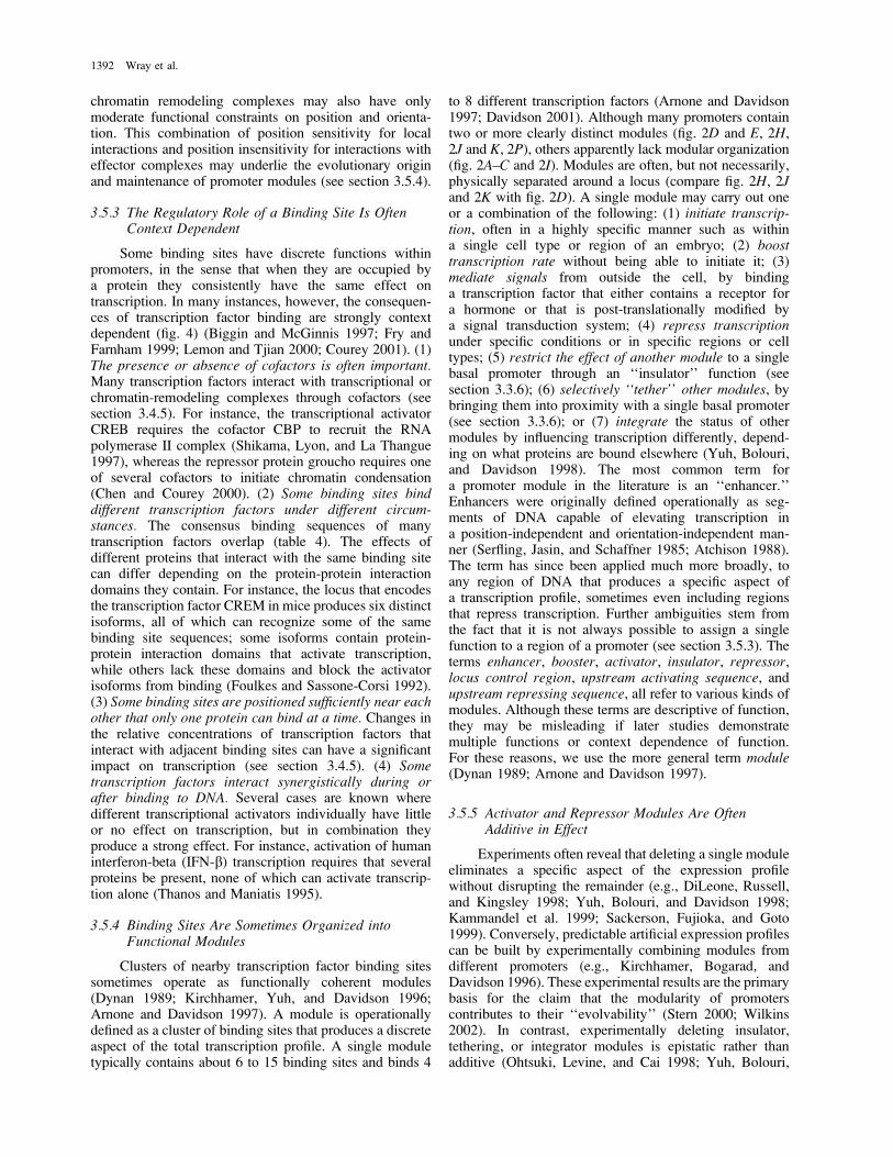
chromatin remodeling complexes may also have onlymoderate functional constraints on position and orienta-tion. This combination of position sensitivity for localinteractions and position insensitivity for interactions witheffector complexes may underlie the evolutionary originand maintenance of promoter modules (see section 3.5.4).
3.5.3 The Regulatory Role of a Binding Site Is OftenContext Dependent
Some binding sites have discrete functions withinpromoters, in the sense that when they are occupied bya protein they consistently have the same effect ontranscription. In many instances, however, the consequen-ces of transcription factor binding are strongly contextdependent (fig. 4) (Biggin and McGinnis 1997; Fry andFarnham 1999; Lemon and Tjian 2000; Courey 2001). (1)The presence or absence of cofactors is often important.Many transcription factors interact with transcriptional orchromatin-remodeling complexes through cofactors (seesection 3.4.5). For instance, the transcriptional activatorCREB requires the cofactor CBP to recruit the RNApolymerase II complex (Shikama, Lyon, and La Thangue1997), whereas the repressor protein groucho requires oneof several cofactors to initiate chromatin condensation(Chen and Courey 2000). (2) Some binding sites binddifferent transcription factors under different circum-stances. The consensus binding sequences of manytranscription factors overlap (table 4). The effects ofdifferent proteins that interact with the same binding sitecan differ depending on the protein-protein interactiondomains they contain. For instance, the locus that encodesthe transcription factor CREM in mice produces six distinctisoforms, all of which can recognize some of the samebinding site sequences; some isoforms contain protein-protein interaction domains that activate transcription,while others lack these domains and block the activatorisoforms from binding (Foulkes and Sassone-Corsi 1992).(3) Some binding sites are positioned sufficiently near eachother that only one protein can bind at a time. Changes inthe relative concentrations of transcription factors thatinteract with adjacent binding sites can have a significantimpact on transcription (see section 3.4.5). (4) Sometranscription factors interact synergistically during orafter binding to DNA. Several cases are known wheredifferent transcriptional activators individually have littleor no effect on transcription, but in combination theyproduce a strong effect. For instance, activation of humaninterferon-beta (IFN-b) transcription requires that severalproteins be present, none of which can activate transcrip-tion alone (Thanos and Maniatis 1995).
3.5.4 Binding Sites Are Sometimes Organized intoFunctional Modules
Clusters of nearby transcription factor binding sitessometimes operate as functionally coherent modules(Dynan 1989; Kirchhamer, Yuh, and Davidson 1996;Arnone and Davidson 1997). A module is operationallydefined as a cluster of binding sites that produces a discreteaspect of the total transcription profile. A single moduletypically contains about 6 to 15 binding sites and binds 4
to 8 different transcription factors (Arnone and Davidson1997; Davidson 2001). Although many promoters containtwo or more clearly distinct modules (fig. 2D and E, 2H,2J and K, 2P), others apparently lack modular organization(fig. 2A–C and 2I). Modules are often, but not necessarily,physically separated around a locus (compare fig. 2H, 2Jand 2K with fig. 2D). A single module may carry out oneor a combination of the following: (1) initiate transcrip-tion, often in a highly specific manner such as withina single cell type or region of an embryo; (2) boosttranscription rate without being able to initiate it; (3)mediate signals from outside the cell, by bindinga transcription factor that either contains a receptor fora hormone or that is post-translationally modified bya signal transduction system; (4) repress transcriptionunder specific conditions or in specific regions or celltypes; (5) restrict the effect of another module to a singlebasal promoter through an ‘‘insulator’’ function (seesection 3.3.6); (6) selectively ‘‘tether’’ other modules, bybringing them into proximity with a single basal promoter(see section 3.3.6); or (7) integrate the status of othermodules by influencing transcription differently, depend-ing on what proteins are bound elsewhere (Yuh, Bolouri,and Davidson 1998). The most common term fora promoter module in the literature is an ‘‘enhancer.’’Enhancers were originally defined operationally as seg-ments of DNA capable of elevating transcription ina position-independent and orientation-independent man-ner (Serfling, Jasin, and Schaffner 1985; Atchison 1988).The term has since been applied much more broadly, toany region of DNA that produces a specific aspect ofa transcription profile, sometimes even including regionsthat repress transcription. Further ambiguities stem fromthe fact that it is not always possible to assign a singlefunction to a region of a promoter (see section 3.5.3). Theterms enhancer, booster, activator, insulator, repressor,locus control region, upstream activating sequence, andupstream repressing sequence, all refer to various kinds ofmodules. Although these terms are descriptive of function,they may be misleading if later studies demonstratemultiple functions or context dependence of function.For these reasons, we use the more general term module(Dynan 1989; Arnone and Davidson 1997).
3.5.5 Activator and Repressor Modules Are OftenAdditive in Effect
Experiments often reveal that deleting a single moduleeliminates a specific aspect of the expression profilewithout disrupting the remainder (e.g., DiLeone, Russell,and Kingsley 1998; Yuh, Bolouri, and Davidson 1998;Kammandel et al. 1999; Sackerson, Fujioka, and Goto1999). Conversely, predictable artificial expression profilescan be built by experimentally combining modules fromdifferent promoters (e.g., Kirchhamer, Bogarad, andDavidson 1996). These experimental results are the primarybasis for the claim that the modularity of promoterscontributes to their ‘‘evolvability’’ (Stern 2000; Wilkins2002). In contrast, experimentally deleting insulator,tethering, or integrator modules is epistatic rather thanadditive (Ohtsuki, Levine, and Cai 1998; Yuh, Bolouri,
1392 Wray et al.

and Davidson 1998; Calhoun, Stathopoulos, and Levine2002).
3.5.6 Collaboration Among Modules Produces the TotalTranscription Profile
Two aspects of promoter function are reminiscent ofanalog logic circuits (Yuh, Stathopoulos, and Levine 1998;Davidson 2001; Yuh, Bolouri, and Davidson 2001). (1)Individual modules can function as Boolean (off/on) orscalar (quantitative) elements whose interactions havepredictable, additive effects on transcription. Multiplemodules are sometimes required to produce a single phaseof expression. For instance, the seven stripes of even-skipped transcription in the early embryo of Drosophila arecontrolled by six modules (Sackerson, Fujioka, and Goto1999; fig. 2H). Conversely, a single module may beinvolved in several different phases of expression. Forexample, module A in the Endo16 promoter of the seaurchin Strongylocentrotus purpuratus (fig. 2D) activatestranscription in the embryo, synergistically elevatestranscription in the larva, and is required for the functionof repressor modules (Yuh, Stathopoulos, and Levine1998; Yuh, Bolouri, and Davidson 2001). (2) Promotersintegrate multiple, diverse inputs and produce a single,scalar output: the rate of transcriptional initiation. Afamiliar analogy is a neuron, which receives input frommany sources but whose output is simply how often itfires. In many promoters, signal integration happens at thebasal promoter, through specific interactions betweenbound transcription factors and components of the RNApolymerase II enzyme complex (Latchman 1998; Lee andYoung 2000). In some promoters, however, a distinctmodule may integrate signals from other modules. Forinstance, module A of the Endo16 promoter relays thestatus of the other five modules to the basal promoter (fig.2D; Yuh, Stathopoulos, and Levine 1998; Yuh, Bolouri,and Davidson 2001).
3.6 Gene Networks
All genes are components of immensely complexnetworks of interacting loci. The binding of a transcriptionfactor to a promoter is one of several physical determinantsof gene network architecture. Understanding the organi-zation of gene networks (Lee et al. 2002; Milo et al. 2002)will be necessary for understanding how they evolve (vonDassow et al. 2000; Davidson 2001; Wagner 2001).
3.6.1 Most Transcription Factors Have NumerousDownstream Target Genes
A simple calculation demonstrates that most eukary-otic transcription factors must bind to the promoters ofmany downstream genes. Eukaryotic genomes contain onthe order of 0.5–5 3 104 genes, only a small fraction ofwhich encode transcription factors (table 3 providesa partial list for several species). Because the expressionof all genes requires that transcription factors bind to theirpromoters, and because most promoters contain bindingsites for at least five different transcription factors (andoften many more), transcription factors must on average
interact with the promoters of tens to hundreds of genes.These rough calculations agree with studies that have usedexperimental approaches to estimate the number of directdownstream targets of a specific transcription factor. InDrosophila melanogaster, Ubx isoform Ia alone regulatesan estimated 85–170 direct downstream targets (Mastick etal. 1995), whereas eve and ftz together appear to regulatethe majority of genes in the Drosophila genome (Liangand Biggin 1998). The number and identify of directdownstream targets has been assayed by in vivo binding formany transcription factors in Saccharomyces cerevisiae(Iyer et al. 2001; Lieb et al. 2001, 2002), and for sometranscription factors these analyses have been carried outon cells grown under more than one environmental con-dition (Ren et al. 2000). Even using conservative criteriafor recognizing interactions, these analyses indicate thatmost transcription factors directly regulate a few percent ofthe genes in the Saccharomyces genome. Genetic networksare therefore highly connected, with each node that isrepresented by a transcription factor linked to many othernodes. This high degree of connectivity may be responsiblein large part for the classical genetic phenomena ofepistasis, polygeny, and pleiotropy (Gibson 1996).
3.6.2 Transcription Is Often Modulated by FeedbackLoops
The expression profile of a gene is a system property,in that it is sensitive to changes in the expression andactivity of gene products encoded by many other loci.Thus, even if a mutation in a promoter region alterstranscription, the network of functionally interacting genesand gene products may modulate this effect (von Dassowet al. 2000). For instance, a mutation that doublestranscription rate may not result in twice as much proteinbeing produced if there is feedback from the cytoplasm tothe nucleus that is sensitive to protein level or to thefunctional consequences of protein activity (such asaccumulation of a particular metabolite). Feedback loopsare probably rather common components of gene networks(Lee et al. 2002; Milo et al. 2002) and may mask somefunctionally significant mutations in promoters.
3.6.3 A Significant Fraction of the Genome Is Involved inTranscriptional Regulation
Several large-scale interspecific sequence compari-sons have estimated that the number of conservedintergenic nucleotides is similar to the number ofconserved coding nucleotides (Shabalina and Kondrashov1999; Onyango et al. 2000; Bergman and Kreitman 2001;Frazer et al. 2001; Shabalina et al. 2001). This strikingresult suggests that the number of functional noncodingnucleotides is approximately equal to the number ofprotein-coding nucleotides, and that approximately half ofall functionally or phenotypically penetrant molecularevolution involves noncoding sequences. The hundreds oftranscription units encoding general and specific transcrip-tional factors, chromatin remodeling complexes, andtranscription cofactors add to the sequences involved intranscriptional regulation. A substantial fraction of a eu-
Evolution of Transcriptional Regulation 1393
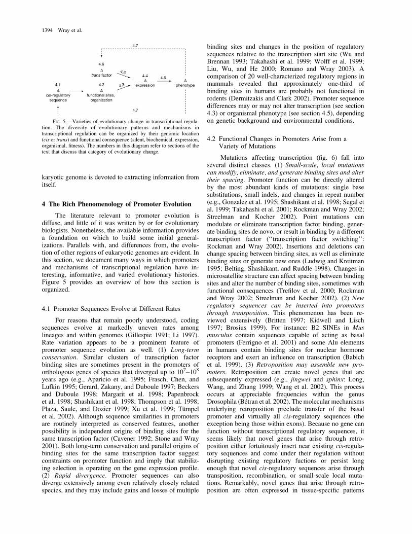
karyotic genome is devoted to extracting information fromitself.
4 The Rich Phenomenology of Promoter Evolution
The literature relevant to promoter evolution isdiffuse, and little of it was written by or for evolutionarybiologists. Nonetheless, the available information providesa foundation on which to build some initial general-izations. Parallels with, and differences from, the evolu-tion of other regions of eukaryotic genomes are evident. Inthis section, we document many ways in which promotersand mechanisms of transcriptional regulation have in-teresting, informative, and varied evolutionary histories.Figure 5 provides an overview of how this section isorganized.
4.1 Promoter Sequences Evolve at Different Rates
For reasons that remain poorly understood, codingsequences evolve at markedly uneven rates amonglineages and within genomes (Gillespie 1991; Li 1997).Rate variation appears to be a prominent feature ofpromoter sequence evolution as well. (1) Long-termconservation. Similar clusters of transcription factorbinding sites are sometimes present in the promoters oforthologous genes of species that diverged up to 107�108years ago (e.g., Aparicio et al. 1995; Frasch, Chen, andLufkin 1995; Gerard, Zakany, and Duboule 1997; Beckersand Duboule 1998; Margarit et al. 1998; Papenbrocket al. 1998; Shashikant et al. 1998; Thompson et al. 1998;Plaza, Saule, and Dozier 1999; Xu et al. 1999; Tumpelet al. 2002). Although sequence similarities in promotersare routinely interpreted as conserved features, anotherpossibility is independent origins of binding sites for thesame transcription factor (Cavener 1992; Stone and Wray2001). Both long-term conservation and parallel origins ofbinding sites for the same transcription factor suggestconstraints on promoter function and imply that stabiliz-ing selection is operating on the gene expression profile.(2) Rapid divergence. Promoter sequences can alsodiverge extensively among even relatively closely relatedspecies, and they may include gains and losses of multiple
binding sites and changes in the position of regulatorysequences relative to the transcription start site (Wu andBrennan 1993; Takahashi et al. 1999; Wolff et al. 1999;Liu, Wu, and He 2000; Romano and Wray 2003). Acomparison of 20 well-characterized regulatory regions inmammals revealed that approximately one-third ofbinding sites in humans are probably not functional inrodents (Dermitzakis and Clark 2002). Promoter sequencedifferences may or may not alter transcription (see section4.3) or organismal phenotype (see section 4.5), dependingon genetic background and environmental conditions.
4.2 Functional Changes in Promoters Arise from aVariety of Mutations
Mutations affecting transcription (fig. 6) fall intoseveral distinct classes. (1) Small-scale, local mutationscan modify, eliminate, and generate binding sites and altertheir spacing. Promoter function can be directly alteredby the most abundant kinds of mutations: single basesubstitutions, small indels, and changes in repeat number(e.g., Gonzalez et al. 1995; Shashikant et al. 1998; Segal etal. 1999; Takahashi et al. 2001; Rockman and Wray 2002;Streelman and Kocher 2002). Point mutations canmodulate or eliminate transcription factor binding, gener-ate binding sites de novo, or result in binding by a differenttranscription factor (‘‘transcription factor switching’’:Rockman and Wray 2002). Insertions and deletions canchange spacing between binding sites, as well as eliminatebinding sites or generate new ones (Ludwig and Kreitman1995; Belting, Shashikant, and Ruddle 1998). Changes inmicrosatellite structure can affect spacing between bindingsites and alter the number of binding sites, sometimes withfunctional consequences (Trefilov et al. 2000; Rockmanand Wray 2002; Streelman and Kocher 2002). (2) Newregulatory sequences can be inserted into promotersthrough transposition. This phenomenon has been re-viewed extensively (Britten 1997; Kidwell and Lisch1997; Brosius 1999). For instance: B2 SINEs in Musmusculus contain sequences capable of acting as basalpromoters (Ferrigno et al. 2001) and some Alu elementsin humans contain binding sites for nuclear hormonereceptors and exert an influence on transcription (Babichet al. 1999). (3) Retroposition may assemble new pro-moters. Retroposition can create novel genes that aresubsequently expressed (e.g., jingwei and sphinx: Long,Wang, and Zhang 1999; Wang et al. 2002). This processoccurs at appreciable frequencies within the genusDrosophila (Betran et al. 2002). The molecular mechanismsunderlying retroposition preclude transfer of the basalpromoter and virtually all cis-regulatory sequences (theexception being those within exons). Because no gene canfunction without transcriptional regulatory sequences, itseems likely that novel genes that arise through retro-position either fortuitously insert near existing cis-regula-tory sequences and come under their regulation withoutdisrupting existing regulatory fuctions or persist longenough that novel cis-regulatory sequences arise throughtransposition, recombination, or small-scale local muta-tions. Remarkably, novel genes that arise through retro-position are often expressed in tissue-specific patterns
FIG. 5.—Varieties of evolutionary change in transcriptional regula-tion. The diversity of evolutionary patterns and mechanisms intranscriptional regulation can be organized by their genomic location(cis or trans) and functional consequence (silent, biochemical, expression,organismal, fitness). The numbers in this diagram refer to sections of thetext that discuss that category of evolutionary change.
1394 Wray et al.

similar to those of a parent locus (Betran et al. 2002). (4)Gene duplications may fragment or recombine promotersequences. Although analyses of gene duplication typicallyfocus on coding sequences, the associated promoters areclearly also important for gene function. If the breakpointsdo not include cis-regulatory sequences, then the duplicatedcopy is likely to be transcriptionally inert in its new locationand become a pseudogene even before it accumulates stopcodons or frameshifts. If only part of the promoter isduplicated, the transcription profile of the new copy maydiffer from the original (e.g., nNOS: Korneev and O’Shea2002). In principle, a duplication could also fortuitouslycombine sequences from two different promoters to createa hybrid cis-regulatory region with a novel transcriptionprofile. Gene duplications that persist are frequentlyfollowed by divergence in expression (Li and Noll 1994;Gu et al. 2002; Stauber, Prell, and Schmidt-Ott 2002) andmay be followed by loss of complementary promotermodules (Ferris and Whitt 1979; Force et al. 1999). (5)Gene conversion can spread regulatory elements withina gene family. Examples from humans include growthhormone (Giordano et al. 1997), beta and gamma globins(Chiu et al. 1997; Patrinos et al. 1998), and majorhistocompatibility complex (MHC) genes (Cereb and Yang1994). Gene conversion is an ongoing process in RNA
polymerase I–transcribed genes (which encode the 40S pre-rRNA that is processed to form 18, 5.8, and 28S rRNA)including associated transcriptional regulatory sequences,but not among the more heterogeneous RNA polymeraseIII-transcribed genes (White 2001). (6) Sequences that haveno prior function in regulating gene expression can becomefortuitous promoters. In one case, gene duplicationsresulted in a former exon functioning in transcriptionalregulation (Sdic: Nurminsky et al. 1998). A second exampleinvolves functional transcription factor binding sites withinan exon (nonA: Sandrelli et al. 2001). These ‘‘hopefulmonster’’ promoters demonstrate that rare events canassemble functional cis-regulatory sequences from seem-ingly unpromising material.
4.3 Changes in Promoter Sequence Differ Widely in TheirEffects on Transcription
Relatively little information exists about the functionalconsequences of naturally occurring differences in pro-moter sequences. A few studies have directly examined thebiochemical impact of sequence differences on proteinbinding (e.g., Ruez, Payre, and Vincent 1998; Singh,Barbour, and Berge 1998; Wolff et al. 1999; Shaw et al.2002). Most of what we know about functional conse-quences, however, comes from cases in which the resultingtranscription profile has been examined (e.g., Ross, Fong,and Cavener 1994; Odgers, Healy, and Oakeshott 1995;Tournamille et al. 1995; Belting, Shashikant, and Ruddle1998; Indovina et al. 1998; Ludwig, Patel, and Kreitman1998; Wang et al. 1999; Romano and Wray 2003). In someof these cases, specific promoter sequence differences arecorrelated with phenotypic consequences; in most, howev-er, the presence of multiple sequence differences makes itdifficult to infer the precise basis for evolutionary changesin transcription. Divergence in promoter sequence andtranscription profile are often poorly correlated: very similarpromoters can produce substantially different transcriptionprofiles (e.g., parvoviruses: Storgaard et al. 1993; TNFA inprimates: Haudek et al. 1998; MMSP in diptera: Christo-phides et al. 2000), whereas highly divergent promotersequences can produce very similar transcription profiles(e.g., runt in Drosophila: Wolff et al. 1999; brachyuryin ascidians: Takahashi et al. 1999; yp in Drosophila: Pianoet al. 1999; Endo16 in sea urchins: Romano and Wray2003).
The latter situation is not unusual (for additionalexamples, see section 4.7). Indeed, many changes inpromoter sequence do not alter transcription within thelimits of experimental assays. Sequence changes might befunctionally silent for several reasons. (1) Substitutionsand indels between transcription factor binding sites maynot affect DNA-protein interactions. It is probably gen-erally true that nucleotides within binding sites are morefunctionally constrained than those that lie betweenbinding sites. However, it is difficult to rule out thepossibility that a supposed nonbinding site nucleotidemight in fact be part of an unrecognized binding site (seesection 5.2). Furthermore, small indels that do not directlyinvolve binding sites may disrupt protein-protein inter-actions by placing proteins on opposite sides of the DNA
FIG. 6.—Mutations affecting promoter structure and function. (A)Modifications in the sequence of a transcription factor binding site. Thesimplest such changes involve single nucleotide substitutions, insertions,or deletions. Repeat length changes can also affect binding sites. (B)Modifications in the spacing of binding sites. These changes can arise inseveral ways: from insertions and deletions of other genomic segments ormobile elements (as shown), from the accumulation of small indels, andfrom the expansion or contraction of repeats. (C) Modifications in thepresence or absence of functional binding sites. Binding sites can arise orbe lost through local point mutations, insertions, or deletions. (D) Large-scale changes in promoter organization. Several kinds of reorganizationare possible, just two of which are shown. Mobile element insertions can‘‘import’’ functional binding sites into a promoter (see section 4.2). Smallchromosomal rearrangements can alter the orientation or location ofclusters of binding sites. The functional consequence of each kind ofchange can range from no effect on gene expression to altered expressionto loss of expression (see section 4.3).
Evolution of Transcriptional Regulation 1395

helix or by changing the spacing of binding sites (seesection 3.5.2). (2) Changes in spacing between distantbinding sites will be neutral in many cases. Interactionsamong proteins associated with binding sites more than;50 bp apart are probably mediated by DNA bending orlooping, which may to a large degree be insensitive todifferences in spacing. (3) Some within-consensus nucle-otide substitutions in binding sites may be functionallyneutral. Certain changes in binding site sequence canpreserve a particular DNA-protein interaction (see section3.3.3). Not all such changes will be neutral, however, asbinding kinetics may differ, in turn altering transcription(see section 3.3.4). Very little is known about the evolutionof binding site consensuses, so sequence comparisonsalone may be a poor guide as to which nucleotide sub-stitutions within binding sites are likely to be function-ally neutral. (4) Eliminating an entire binding site may befunctionally neutral. Many promoters contain multiplecopies of the same binding site, raising the possibility offunctional redundancy. Cases of probable binding siteturnover (Ludwig and Kreitman 1995; Hancock et al. 1999;Piano et al. 1999; Liu, Wu, and He 2000; Dermitzakisand Clark 2002; Scemama et al. 2002) may have beenpossible because of functional redundancy. Nevertheless,multiply represented binding sites within the same pro-moter are not always functionally redundant (see section5.5).
4.4 Gene Expression Profiles Evolve Frequently and inDiverse Ways
The literature of comparative gene expression hasemphasized similarities and generally interpreted them asconserved features (DeRobertis and Sasai 1996; Hollandand Holland 1999; Carroll, Grenier, and Weatherbee2001). When comparing distantly related taxa, however,similarities in gene expression are often outweighed byapparently nonhomologous features (Wray and Lowe2000; Davidson 2001; Wilkins 2002). The abundance ofpopulation-level variation in promoter function (seesection 2.3) means that expression differences couldevolve quite rapidly under some conditions, and indeedsubstantial differences in gene expression can exist evenbetween recently duplicated genes (Gu et al. 2002) orclosely related species (Parks et al. 1988; Ross, Fong, andCavener 1994; Swalla and Jeffery 1996; Grbic, Nagy, andStrand 1998; Kissinger and Raff 1998; Brunetti et al. 2001;Ferkowicz and Raff 2001). Several functional classes ofevolutionary change in gene expression are evident. (1)Changes in timing of gene expression. Temporal changeshave been documented from many taxa (e.g., Dickinson1988; Wray and McClay 1989; Swalla and Jefferey 1996;Kim, Kerr, and Min 2000; Skaer, Pistillo, and Simpson2002). Heterochronies are a common pattern of anatomicalevolution (McKinney and McNamara 1991), and must, atsome level, involve heritable changes in the timing of geneexpression. (2) Changes in spatial extent of geneexpression. Many studies have found interspecific differ-ences in the spatial extent of regulatory gene expression(e.g., Schiff et al. 1992; Abzhanov and Kaufman 2000;Brunetti et al. 2001; Scemama et al. 2002). Such changes
are of particular interest when they affect regulatory genes,because of the relatively direct consequences for bodyproportions, organ size and number, and a great manyother anatomical features (see section 2.2). (3) Changes inlevel of gene expression. Evolutionary differences intranscription rate have also been documented (Regierand Vlahos 1988; Crawford, Segal, and Barnett 1999;Wang et al. 1999). Such comparisons have been simplifiedwith the advent of microarray technologies (e.g., Jin et al.2001; Schadt et al. 2003). Because of this approach, weknow more about differences in transcript abundance thanany other kind of evolutionary change in gene expression.(4) Changes in responsiveness of gene expression toexternal cues. Evolutionary changes in transcriptionalresponses to physiological status, environmental condi-tions, and pheromones have also been documented(Brakefield et al. 1996; Cooper 1999; Abouheif and Wray2002). Such changes are a necessary component in theevolution of polyphenism and phenotypic plasticity andare therefore of considerable ecological interest. (5) Sex-specific expression. Evolutionary changes in differentialgene expression among sexes have been documented(Schiff et al. 1992; Saccone et al. 1998; Christophides etal. 2000; Kopp, Duncan, and Carroll 2000). Microarraysurveys suggest that populations can harbor variation inwhich genes are expressed in a sex-specific manner (Jin etal. 2001). (6) Gains and losses of particular phases ofgene expression. In multicellular organisms, many genesare expressed in a succession of spatially and temporallydistinct phases during the life cycle (for examples seeGerhart and Kirschner [1997]; Carroll, Grenier, andWeatherbee [2001]; and Davidson [2001]). A gene whoseexpression requires a particular transcription factor duringa specific phase of expression may be ‘‘abandoned’’ bythat regulator if it is no longer expressed in the appropriateregion. Examples include several independent losses ofpatterning roles for homeodomain transcription factors inarthropods (Dawes et al. 1994; Falciani et al. 1996; Grbic,Nagy, and Strand 1998; Mouchel-Vielh et al. 2002).Conversely, a new regulatory linkage may be establishedif a promoter acquires a binding site for a differenttranscription factor, a process known as recruitment or co-option (Duboule and Wilkins 1998; Wilkins 2002). Manylikely cases have been identified (Lowe and Wray 1997;Saccone et al. 1998; Keys et al. 1999; Brunetti et al. 2001;reviewed in Wilkins 2002). Evolutionary gains and lossesof particular phases of gene expression may be facilitatedby the modular organization of promoters (see section 2.1).
4.5 Changes in Gene Expression Differ Widely in TheirEffects on Organismal Phenotype
Promoter function has both a biochemical phenotype,the gene expression profile, as well as an organismalphenotype, involving features such as anatomy, physiol-ogy, life history, and behavior. These biochemical andorganismal effects are evolutionarily dissociable to someextent, because some changes in gene expression appear tohave no consequence for organismal phenotype. Suchchanges in gene expression are analogous to conservativeamino acid replacements in a protein (table 5), many of
1396 Wray et al.

which are likewise thought to have no impact onorganismal phenotype (Kimura 1983; Gillespie 1991).Several cases are known where the timing or spatial extentof gene expression differs among species without anyobvious phenotypic consequence (e.g., gld in Drosophila:Schiff et al. 1992; Ross, Fong, and Cavener 1994; Cy genefamily in sea urchins: Fang and Brandhorst 1996;Kissinger and Raff 1998; msp130 in sea urchins: Wrayand Bely 1994).
Although it may be difficult to demonstrate beyondany doubt that a particular difference in transcription isphenotypically silent, the opposite case is easier to es-tablish. Differences in gene expression have been linked todiverse aspects of organismal phenotype, including: (1)anatomy (Burke et al. 1995; Averof and Patel 1997; Stern1998; Wang et al. 1999; Lettice et al. 2002); (2) physiology(Abraham and Doane 1978; Matsuo and Yamazaki 1984;Dudareva et al. 1996; Sinha and Kellogg 1996; Stockhauset al. 1997; Segal, Barnett, and Crawford 1999; Lermanet al. 2003); (3) behavior (Trefilov et al. 2000; Caspi et al.2002; Enard et al. 2002b; Fang, Takahashi, and Wu 2002;Hariri et al. 2002; Saito et al. 2002); (4) diseasesusceptibility (Tournamille et al. 1995; Shin et al. 2000;Bamshad et al. 2002; Meyer et al. 2002); (5) polyphenism(Brakefield et al. 1996; Abouheif and Wray 2002); and(6) life history (Allendorf, Knudsen, and Phelps 1982;Allendorf, Knudsen, and Leary 1983; Anisimov et al.2001; Streelman and Kocher 2002).
4.6 Mutations in trans Can Alter Transcription inSeveral Ways
The genetic basis for an observed difference in theexpression of a particular gene in some cases does notreside in cis, but rather within one of the loci encodingtranscription factors that interact with it. Three classes ofmutations can underlie these trans effects. (1) Mutationsaffecting the expression profile of an upstream transcrip-tion factor. Numerous experiments demonstrate that thistrans effect is pervasive: manipulating the expression ofa transcription factor typically alters the expression of itsdownstream targets (Gilbert 2000; Alberts et al. 2002).Although many evolutionary differences in the expressionprofiles of transcription factors are known (see section4.4), few studies have investigated the effect of thesechanges on the transcription of downstream targets.Indirect evidence of an evolutionary role comes from
phenotypic correlates of interspecific differences intranscription factor expression (e.g., Burke et al. 1995;Averof and Patel 1997; Stern 1998; Beldade, Brakefield,and Long 2002) and from expression assays that testregulatory sequences of one species in another (e.g.,Manzanares et al. 2000). (2) Mutations affecting the DNA-binding domain of an upstream transcription factor.Amino acid substitutions in DNA binding domains oftranscription factors can affect the expression of down-stream genes (e.g., Conlon et al. 2001; D’Elia et al. 2002)and produce phenotypic consequences (Brickman et al.2001). Such changes are apparently relatively rare, as theamino acid sequences of DNA binding domains areusually highly conserved (Duboule 1994; Latchman1998). Nonetheless, variants are sometimes found withinpopulations (e.g., Brickman et al. 2001). Interspecies gene-swapping experiments support this view: in a surprisingnumber of cases, a vertebrate gene encoding a transcriptionfactor can restore a somewhat wild-type phenotype to a flythat is homozygous for a null allele of the orthologousgene (e.g., Lutz et al. 1996; Gerard, Zakany, and Duboule1997). Few gene swaps rescue phenotype perfectly andsome fail almost completely, however, which may bedue in part to changes in DNA binding specificity. (3)Mutations affecting the presence or sequence of a protein-protein interaction domain in an upstream transcriptionfactor. Again, experiments provide evidence of this thirdclass of trans effects on transcription (Hope and Struhl1986; Dawson, Morris, and Latchman 1996). Functionalchanges in protein-protein interaction domains haveevolved in Hox transcription factors within the Arthropoda(Galant and Carroll 2002; Ronshaugen, McGinnis, andMcGinnis 2002) and in serum response transcriptionfactors between arthropods and chordates (Avila et al.2002), while an evolutionary difference in a phosphoryla-tion site has evolved in the FOXP2 transcription factoralong the lineage separating humans from the other greatapes (Enard et al. 2002b). Sequence comparisons suggestthat amino acid substitutions in protein-protein interactiondomains can evolve rapidly under positive selection(Sutton and Wilkinson 1997; Barrier, Robichaux, andPurugganan 2001). All three classes of trans effectsmentioned above are likely to be highly pleiotropicbecause of the large number of downstream target genesthat would be affected (see section 3.6.1).
4.7 Many Modes of Selection Operate on PromoterSequences
The classic modes of natural selection that operate oncoding sequences and morphology also operate onpromoter sequences (also see section 2.4). (1) Negative(purifying) selection. Many deleterious promoter alleleshave been identified in humans, involving a wide range ofgenes and phenotypic consequences (summarized inCooper 1999). Cases of long-term conservation of bindingsites (see section 4.1) suggest persistent negative selection.(2) Positive selection. Some promoter alleles appear to beunder directional selection (FY: Hamblin and DiRienzo2000; P450: Daborn et al. 2002; hsp70: Lerman et al.2003). (3) Overdominant selection. Likely cases include
Table 5Functional Categories of Nucleotide Substitution
Probable Impact Coding Sequencea Promoter Sequence
Neutral Synonymous ntsubstitution
Nonbinding sitesubstitution
Low Conservative AAreplacement
Within consensussubstitution
Medium to high Nonconservative AAreplacement
Nonconsensussubstitution
Loss-of-function Frameshift or stopcodon
Deletion of basalpromoter or keyactivator binding site
a AA¼ amino acid.
Evolution of Transcriptional Regulation 1397

some histocompatibility loci in humans and mice (Guar-diola et al. 1996; Cowell et al. 1998). Reasons foroverdominant selection on coding sequences of these lociare reasonably well understood, and transcription profilesshould be under selection for variation in the cell type inwhich they are expressed (Guardiola et al. 1996). Otherpossible cases include some b-thalassemias in humans(Kazazian 1990), anthocyanin pigment synthesis in maize(r locus: Li et al. 2001), and dispersal behavior in rhesusmacaques (serotonin transporter: Trefilov et al. 2000). (4)Balancing selection. Environmental heterogeneity withinthe range of a single species can result in local adaptationand balancing selection. Cases are known from a teleost(LDH: Crawford, Segal, and Barnett 1999; Segal, Barnett,and Crawford 1999) and from humans (CCR5: Bamshad etel. 2002). (5) Stabilizing selection. When binding siteswithin a promoter differ but the resulting expressionprofile is unchanged, stabilizing selection may beoperating. This situation appears to be relatively common,with examples now known from diverse metazoans andgenes (ADH: Wu and Brennan 1993; Esterase-5B and -6:Odgers, Healy, and Oakeshott 1995; Tamarina, Ludwig,and Richmond 1997; unc-116: Maduro and Pilgrim 1996;even-skipped: Ludwig, Patel, and Kreitman 1998; Patel etal. 2000; yolk protein: Piano et al. 1999; runt: Wolff et al.1999; brachyury: Takahashi et al. 1999; achaete-scutecomplex: Skaer and Simpson 2000; Hoxb2: Scemama etal. 2002; mating-type loci in budding yeast: Sjostrand,Kegel, and Astrom 2002; Endo16: Romano and Wray2003). (6) Compensatory selection: An interesting case inhumans involves a hypomorphic allele within the codingsequence of CFTR that causes cystic fibrosis. Somehaplotypes contain a second mutation within the promoterthat adds a third Sp1 binding site, elevating transcriptionand resulting in an improved prognosis (Romey et al.1999, 2000). The third Sp1 site never occurs in haplotypesthat produce wild-type protein, suggesting that it may beunder positive selection as a result of its compensatoryeffect.
5 Challenges in Studying Promoter Evolution
The structure and function of promoter sequences areprofoundly different from those of coding sequences (table6). These differences impose nontrivial challenges forstudying the evolution of transcriptional regulation.
5.1 Coding and Regulatory Sequences Differ in Structureand Function
Coding sequences have a regular, direct, precise, andeasily interpreted relationship with their proximate (bio-chemical) phenotype, a specific sequence of amino acids.In contrast, promoters have an idiosyncratic, indirect,nonlinear, and context-dependent relationship with theirproximate phenotype, a particular transcription profile.Furthermore, the transcription profile generated by a pro-moter depends on other loci that encode transcriptionfactors that bind to it, and on the loci encoding thetranscription factors that regulate these immediate up-stream regulators, and so forth. This regress transcends
generations, in that maternally loaded transcription factorsor their mRNAs are required to activate early zygotic geneexpression. Environmental influences on gene expressionadd a further layer of complexity. Although the amino acidsequence of a protein rarely changes during the course ofdevelopment or in response to environmental conditions,the transcription profile of most genes is modulated duringthe life cycle and in response to changing externalconditions. (Even when differential splicing producesdistinct protein isoforms from the same locus underdifferent circumstances, the relationship between DNAand protein sequence remains direct.)
It is important to recognize that sequence data alonecannot reveal the organization of binding sites withina promoter; nor can they show what proteins bind to them,or how they function, or what transcription profile theygenerate. This is partly a matter of missing information: forinstance, the full matrix of binding sequences is not yetknown for most transcription factors, even in well-studiedspecies. But it is mostly an inescapable consequence of theway transcription is regulated: many potential binding siteshave no influence on transcription in vivo, sequencesessential for transcription always reside both cis and trans,and transcription can be strongly influenced by geneticbackground, physiological status, and environmentalconditions.
5.2 Identifying Functional Binding Sites RequiresBiochemical Data and in Vivo Functional Assays
Because the sequences bound by transcription factorsare short and imprecise (see section 3.3), literally hundredsof potential binding sites lie near every locus. Onlya fraction of these binding sites actually influencetranscription (Latchman 1998; Weinzierl 1999; Li andJohnston 2001; Lee et al. 2002). Potential binding sites maynot function for a variety of reasons (see section 3.5.3; fig.4). Which sites actually influence transcription, and aretherefore possible targets of selection, can only bedetermined experimentally. Biochemical characterizationscan identify binding sites precisely and are the only way todetermine whether consensus sequences differ amongspecies. The most common methods are footprinting andmobility shift assays (Carey and Smale 2000). Becausethese assays are carried out in vitro, they cannot reveal theinfluence of chromatin modulation on protein binding ortranscription. Assays of in vivo binding sites (Walter andBiggin 1996; Ren et al. 2000; Iyer et al. 2001; Lee et al.2002) provide a more accurate representation but aretechnically more demanding and undercount real bindingsites. The only definitive means of identifying a binding sitewith a role in regulating transcription is to modify itssequence and assay transcription in vivo, typically bytransient or stable transformation with a reporter gene (seesection 5.5). All of the methods mentioned above requireconsiderable effort when used to test a potential binding siteat multiple phases of the life cycle and under a variety ofenvironmental conditions. In practice, most promoters haveonly been searched for potential binding sites at a restrictedphase of the life cycle and under uniform culture conditions.For this reason, experimentally verified binding sites are
1398 Wray et al.

nearly always an underestimate, and the physical extent ofa promoter is rarely well defined.
The resulting difficulties for studying promoterevolution are substantial. (1) Information about promoterstructure is almost always incomplete. Few promotershave been subjected to thorough searches for binding sites,and some binding sites probably remain unidentified evenin carefully studied cases. Information about the functionalconsequences of binding site differences among species islimited to just a few cases (e.g., Singh and Berger 1998;Wolff et al. 1999; Shaw et al. 2002). (2) The informationthat does exist is almost always biased. Because of theway promoter function is typically studied, some kinds ofbinding sites are naturally less likely to be discovered.These include binding sites that mediate responses tophysiological status and environmental conditions (be-cause most assays are carried out under uniformconditions), binding sites that act at restricted times duringthe life cycle (because typically only part of the life cycleis assayed), and binding sites of weak effect (because ofassay insensitivity). In addition, most studies measureeither quantitative or spatial aspects of transcription andsome ignore temporal changes; as a result, the bindingsites that are identified are often biased with respect to theireffects on time, space, and level of transcription.
Because empirical validation of binding sites islaborious, attempts have been made to increase thereliability of informatic approaches to binding siteidentification. We discuss here a few of the manyapproaches which have been developed (for additional
information, see Hardison [2000]; Stormo [2000]; Ohlerand Niemann [2001]; and Markstein and Levine [2002]).Most informatic approaches apply either to a specific locusor to a complete genome. In the former category areprograms that use databases of known binding sitematrices to scan a sequence for potential binding sites(e.g., TRANSFAC: Wingender et al. 2001; EPD: Praz et al.2002; PlantCARE: Lescot et al. 2002; SCPD: Zhu andZhang 1999). However, many of the potential binding sitesthese programs identify have no biological function andare simply spurious matches to a binding site (see previousparagraphs and section 3.3.4). A complementary approachinvolves comparisons with the homologous chromosomalregion from other species, a method known as ‘‘phyloge-netic footprinting.’’ The rationale is that nucleotides withinbinding sites are more likely to be conserved by naturalselection. This method can successfully identify previouslyunknown binding sites (Loots et al. 2000; Wasserman et al.2000; Yuh et al. 2002). The effectiveness of this method islimited, however, because nucleotides can be conserved bychance, because real binding sites can turn over even whenthe transcriptional output is maintained, and because someaspects of transcription are species-specific (e.g., Ludwig,Patel, and Kreitman 1998; Dermitzakis and Clark 2002).The first problem leads to false positives, whereas thesecond and third generate false negatives. When a completegenome sequence is available, several additional methodscan be applied to identify unknown binding sites. Thesealgorithms rely on large data sets to identify overrepre-sented sequence motifs (e.g., Sinha and Tompa 2002),
Table 6Structural and Functional Differences in Coding and Promoter Sequences for aProtein-Coding Locus
Coding Promoter
Physical boundaries Defined by sequence Not defined by sequence
Start ATG (sometimes multiple) NoneEnd TAA, TAG, or TGA NoneInternala [C/A]AG jGU[A/G]AGU
(U/C)nNAG jG/ANone
Physical organization Discontinuous, colinear Discontinuous, nonlinear
Physical unit Exon Moduleb
Typical unit size ; 20–2,000 bp ; 200–2,000 bpNumber of units 1–10 (rarely more) 1–10 (rarely more?)Organization and function colinear Yes NoRelative order of units Consistent Not consistentModules correspond to functions Sometimes Often
Functional organization Direct, local Indirect, distributed
Direct functional output Protein sequence Transcription profileUnit of information Codon Binding siteNumber of units ;150–1,000 (rarely more) ;6–60 (more?)Information content 0.3–2.0 kb 0.08–0.8 kbc
Spacing between units Doesn’t matter Sometimes mattersMapping Precise (1 AA)d Imprecise (.1 TxF)d
Degeneracy Precise (same AA) Imprecise (different TxF)Consequence Qualitative (1 codon: 1 AA) Quantitative (level of Tx)d
Order of units Usually matters Sometimes mattersGenetic basis cis only cis and trans required
a Type I introns; other splice junction sequences exist.b Cluster of transcription factor binding sites (also known as enhancer, UAS, etc.).c cis-regulatory sequences only; additional information necessary for transcription is encoded in the sequences and ex-
pressions profile of trans regulators and, in some cases, in the environment.d AA ¼ amino acid; Tx ¼ transcription; TxF¼ transcription factor.
Evolution of Transcriptional Regulation 1399
clusters of binding sites (e.g., Berman et al. 2002; Rebeiz,Reeves, and Posakony 2002), or correlations withexpression data (e.g., Birnbaum, Benfey, and Shasha2001). For all of these methods, both false positives andfalse negatives remain a significant issue. Althoughmethods for informatic detection of binding sites arebecoming more sophisticated, for now the results are bestviewed as a starting point for empirical validation ratherthan as a definitive identification of transcription factorbinding sites.
5.3 Identifying the Complement of TranscriptionFactors that Interact with a Binding Site RequiresBiochemical Data
A binding site may be occupied by differenttranscription factors (or by none) at different times orplaces during development (see section 3.5.3; see alsotable 4), with distinct functional consequences (Fry andFarnham 1999; Lemon and Tjian 2000; Courey 2001). Theextent of ‘‘binding site sharing’’ by different transcriptionfactors remains poorly understood, because nearly allfunctional studies of promoters in multicellular organismshave examined a single phase of the life cycle (typicallyembryos or differentiated cells in culture) under uniformculture conditions.
Overlapping binding site specificities have importantimplications for evolutionary studies: (1) A transcriptionfactor might not influence transcription, even if a consensusbinding site for it exists and is known to bind protein. Thepresence of a binding site is necessary but not sufficient fortranscription factor binding. Demonstrating an interactionbetween a specific regulator and a binding site requiressome form of biochemical characterization, the mostcommon of which are ‘‘supershift’’ assays and in vivofootprinting (Carey and Smale 2000). (2) A binding sitemight be occupied by different transcription factors underdifferent circumstances. Indeed, the protein bound most ofthe time may not be the one whose consensus recognitionmotif is the closest match. Recognizing cases of variedbinding site occupancy requires testing nuclear extractsacross developmental stages, among cell types, and underdiverse environmental conditions using supershift assaysor in vivo footprinting. (3) The protein that occupiesa binding site can change evolutionarily. Likely cases of‘‘transcription factor switching’’ have been identifiedwithin humans (Rockman and Wray 2002). In addition,an interaction that has been biochemically validated in onespecies may not occur in another, even if the sequence ofthe binding site is perfectly conserved. Demonstratinga conserved or altered protein-DNA interaction requirescomparative biochemical data.
5.4 Comparisons of Promoter Sequence Are Not AlwaysStraightforward
Once functional binding sites have been mapped, thenext step is identifying homologous binding sites amongspecies or alleles. Promoter sequences can usually bealigned rather easily within a species, although bindingsites that fall within repeats can be problematic. In the
most straightforward interspecific comparisons, potentialbinding sites that occupy similar positions, spacing, andorientation relative to the start site of transcription andrelative to each other are likely to be homologous.Complications can arise for a variety of reasons: bindingsite spacing is often functionally unconstrained (seesection 3.3.5), transposition can introduce similar bindingsites (see section 4.2), and random point mutations cangenerate new binding sites at an appreciable frequency(Stone and Wray 2001). (We use the term homologous inits usual, phylogenetic, sense to denote the hypothesis thata binding site is present in two living species because itwas present in their latest common ancestor and haspersisted since. Sequence similarity, in contrast, is simplyan observation, and can be due to either homology orhomoplasy.)
Once homologous binding sites have been identified,routine methods of comparative analysis can be applied topolarize character state transformations, identify reversalsand parallel transformations, and reconstruct ancestralstates. Most published comparisons of promoter structureinvolve just two species, with the emphasis typically onidentifying conserved binding sites. By surveying moretaxa and incorporating functional data, it becomes possibleto identify origins, losses, and turnover of binding sites. Aswith all comparative analyses, dense phylogenetic sam-pling provides a more robust understanding of evolution-ary transformations within promoters, particularly in casesof rapid sequence divergence.
5.5 Promoter Function Can Only Be DeterminedExperimentally
The only way to determine the expression profileproduced by a promoter haplotype is to assay it in vivo, inits normal chromosomal and cell biological contexts. Thisis most easily accomplished by examining the spatial andtemporal distribution of transcripts using in situ hybrid-ization, RNA gel blots, or quantitative PCR. Because evensmall differences in promoter sequence can alter transcrip-tion (see section 4.2), interpreting the functional con-sequences of such differences among alleles or betweenspecies also requires assaying transcription.
Similarly, the only way to understand the contributionof specific sequence differences within a promoter is tocarry out comparative functional tests. The most commonkind of experiment involves coupling a test regulatoryregion to a reporter gene whose product is easily detected,and then placing this construct in embryos or cells where itis exposed to the array of transcription factors encounteredby the endogenous promoter (Carey and Smale 2000).Further experiments, such as testing the consequences ofnucleotide substitutions within a specific binding site,deleting a binding site, altering spacing or orientationbetween binding sites, or testing restricted portions of thepromoter can be immensely informative. Although suchexperiments are laborious, they provide almost the onlyreliable information about binding site function. Fortu-nately, expression assays are feasible in a growing numberof organisms. For comparative analyses, it is important tocarry out reciprocal functional tests, because transcription
1400 Wray et al.

is a product of both cis and trans sequences. This, too, isnow possible in some taxa (Tumpel et al. 2002; Romanoand Wray 2003).
Comparative experimental tests, although unusual inthe literature, are necessary for analyzing the evolution ofpromoter function. Issues of particular interest include thefollowing. (1) Identifying changes in a binding site function.A binding site whose sequence is conserved between twospecies may nonetheless function differently in them,because the transcription factors and cofactors that interactwith it are expressed differently or because an adjacentbinding site for a cofactor has changed. (2) Determining thefunction of multiply-represented binding sites. Multiplebinding sites for the same transcription factor within a singlepromoter (fig. 3) may be functionally redundant, additive, orsynergistic (e.g., Small, Blair, and Levine 1992; Yuh andDavidson 1998, 2001), with distinct consequences forselection (Ludwig 2002). (3) Understanding the functionalsignificance of binding site organization. The position,spacing, and orientation of individual binding sites in somecases matters a great deal and in other cases not at all (see3.5.2), again with distinct consequences for selection. (4)Determining when, where, and under what conditionsa binding site functions. Although some binding sites mayfunction continuously and ubiquitously, most probably doso only during part of the life cycle, in certain cell types, or inresponse to particular environmental conditions. Bindingsite functionmay also be context dependent, changing underdifferent circumstances (see section 3.5.3). (5) Identifyingthe genetic basis for a known difference in transcription.Interspecific differences in transcription profiles might bedue to changes in cis or trans (see sections 4.2 and 4.6). Thefunctional consequences of changes in cis can be identifiedby means of in vivo expression assays (examples reviewedin Paigen [1989] and Cavener [1992]).
The difficulty of carrying out comparative expressionassays imposes severe practical constraints on analyses ofpromoter evolution. (1) In general, it will be more difficultto obtain comparative information on the proximatefunction of promoter sequences than of coding sequences.Characterizing promoter function involves techniques thatare labor intensive and unfamiliar to most molecularevolutionists. Yet without this information, it is difficult tointerpret comparative sequence data meaningfully. (2) Atleast for the near term, comparative information onbinding site function will remain limited. Few promotershave been analyzed biochemically or functionally in morethan one species, and even in these cases analyses havebeen limited to a fraction of the complete cis-regulatoryregion. (3) Predicting the proximate functional conse-quences of mutations in promoter sequences will be moredifficult than in coding sequences. Because promoters lacka general organizing ‘‘code,’’ because the function ofa binding site can be strongly context-dependent (seesection 3.5.3), and because promoter function depends onthe sequences and expression of transcription factorsencoded elsewhere, the biochemical phenotypic conse-quences of sequence differences in a promoter region arevery difficult to interpret without functional tests. Therelative magnitude of likely functional consequences ofa mutation within a promoter can be organized into a very
rough rank order, as it can be more precisely within exons(table 5). Overall, however, the considerably less regularstructure-function relationship within promoters will makeit much more difficult to discern general patterns ofsequence evolution.
5.6 Standard Classed Tests of Molecular Evolution MustBe Modified for Analyzing Promoters
Although tests of selection on promoters are notfundamentally different from tests on coding regions, theymust be applied with caveats. The major problems arisewhen applying tests that use classes of nucleotidesubstitutions to promoter data (e.g., Ka/Ks or McDonald-Kreitman tests; McDonald and Kreitman 1991). Thesetests classify coding mutations as synonymous or non-synonymous, and they test for selection under theassumption that synonymous sites evolve neutrally. Toapply these tests to promoter sequences, most authorsclassify promoter mutations as occurring within bindingsites or within non-binding-site nucleotides and assumethat nonbinding sites are evolving neutrally. However, thefunctional consequences of mutations in promoters cannotbe classified without additional functional data (see section5.5; Jenkins, Ortori, and Brookfield 1995). More specif-ically, practical difficulties in identifying binding sites (seesection 5.2) mean that most evolutionary analyses ofpromoters will only be able to rely on functionalinformation from a single species. Binding sites absentin the species for which functional data are available butpresent in all other species will be missed, while those sitesfunctional in the reference species but not in all otherspecies may mistakenly be considered present in all. Bothtypes of error will result in some sequence differencesbeing classed incorrectly and will degrade the signal-to-noise ratio in tests for selection. It follows that sequencecomparisons among the promoters of closely relatedspecies, or classed tests that only use data from onespecies (e.g., Hahn, Rausher, and Cunningham 2002), willgenerally be the most informative and accurate. Evenfewer data from comparative studies are usually availableabout the functional consequences of sequence differenceswithin a binding site. Only rarely will it be possible toreliably detect the effect of nucleotide differences onchanges in binding specificity between species (see section5.5).
A second problem with tests that rely on classes ofsites relates to the mechanism by which binding sites ariseand are presumably selected for. Although an excess ofnonsynonymous substitutions relative to synonymoussubstitutions is good evidence for positive selection, it isdifficult to imagine a situation in which an excess ofbinding-site substitutions relative to nonbinding-site sub-stitutions can be interpreted in the same way. This followsfrom three features of promoters. First, binding sites aresometimes not functionally restricted to a specific position.Individual binding sites may therefore turn over bychanging position without positive selection (Ludwig2002). Second, sequences which have no binding affinityfor any transcription factor often need only a single base-pair change in order to become a functional binding site
Evolution of Transcriptional Regulation 1401

(Stone and Wray 2001). A single point mutation cantherefore establish a new functional site consisting ofseveral nucleotides. Third, most nucleotide substitutionswithin a binding site modulate or eliminate its function,whereas relatively few mutations will change it intoa binding site for a different transcription factor. Rarelywill an excess of substitutions within a binding site bea signal of positive selection, because binding sites oftensimply cease to function after multiple substitutions. Noneof these three features precludes selection for changes inbinding sites, only that they may combine to significantlyreduce the ability of classed tests to detect this selection inpractice.
All classed tests of section suffer from theseproblems, but non-classed tests of neutrality (e.g., theHudson-Kreitman-Aguade test [HKA], Tajima’s D, Fu andLi’s D: Hudson, Kreitman, and Aguade 1987; Tajima1989; Fu and Li 1993) can be interpreted in the standardway. Often a combination of these tests, as well as studiesof geographic structure of allele frequencies, may benecessary to detect the action of selection in promoters(e.g., Hamblin and DiRienzo 2000; Bamshad et al. 2002;Fullerton et al. 2002).
6 Hypotheses About the Evolution of TranscriptionalRegulation
No general framework exists for understanding,interpreting, and predicting how transcription evolves. Inthis section, we present an initial attempt at providing sucha framework, in the form of testable hypotheses derivedfrom three sources: models of molecular evolution,mechanisms of promoter function, and empirical evidence.Our hope is that these hypotheses will encourage in-vestigators to dig a little deeper into their data to addressa broad range of questions about promoter evolution.Thus, we emphasize hypotheses that can be tested withavailable techniques. The first six categories of predictionsare based on the neutral model of sequence evolution(Kimura 1983) and are organized as shown in figure 7; thelast four categories address promoter ‘‘design’’ principlesand macroevolution.
6.1 Promoter Sequences Have CharacteristicEvolutionary Dynamics
Because promoters are organized and functiondifferently from other regions of the genome, they aresubject to distinct functional constraints. Predictablepatterns of sequence evolution should therefore distinguishpromoters from sequences that lack a role in transcriptionalregulation. (1) Overall substitution and indel frequencies inpromoters should be higher than in coding sequences andlower than in introns and in nonregulatory intergenicregions. Because most nucleotides in promoters do notaffect transcription, most substitutions and many indelsshould have no functional consequence and shouldtherefore evolve without constraint. These predictions aregenerally supported empirically (Jordan and McDonald1998; Jareborg, Birney, and Durbin 1999; Waterston et al.2002). Exceptions might include the promoters of genes
encoding proteins that tolerate many amino acid substitu-tions, are under balancing selection, or whose intronscontain regulatory sequences (e.g., IL-4 and IL-13 inmammals: Loots et al. 2000). (2) Indel size spectrum inpromoters should be more continuous than that in codingsequences but similar to that in introns, UTRs, andnonregulatory intergenic regions. Three factors maycontribute to a distinctive evolutionary dynamic of lengthvariation within promoter regions: the lack of a readingframe, the low density of functionally important nucleo-tides, and the ability of many binding sites to operate ina position-independent manner. Indels should be morecommon, the frequency spectrum of indel size should notbe biased toward multiples of three (as it is in codingsequences), and repeat variation and large indels should bemuch more common. These patterns are evident in somecases (e.g., hairy: Kim 2001). (3) The order of binding sitesand modules within promoters should be less conservedthan the order of exons within transcription units. Codonshave an obligate colinearity with the amino acid sequenceof their protein product, whereas binding sites and moduleswithin promoters can often function to some extentindependently of position and order (see section 3.3.5).Gross organizational changes within promoters should belimited largely by mutation, whereas in coding regionssuch changes should be limited primarily by functionalconstraints. Small-scale inversions in promoters can existwithin populations (e.g., IGHA1 in humans: Denizot et al.2001). (4) ‘‘Module shuffling’’ should be relativelycommon. ‘‘Domain shuffling’’ has been an important partof the evolution of many gene families (Lander et al. 2001).The analogous process of module shuffling withinpromoters may occur at a higher frequency. Severalexamples of mobile element insertions that have broughtfunctional binding sites into range of a gene are known(Britten 1997; Kidwell and Lisch 1997). The modularorganization of many promoters means that a transcriptionprofile could be dramatically modified in a functionallyintegrated way.
6.2 Selection Acts Primarily on the Sequence and SpatialArrangement of Binding Sites
The output of a promoter derives from the nucleotidesequences and spatial arrangement of transcription factorbinding sites (see section 3). It follows that sequences thatlie between binding sites should be free to vary, at mostshowing weak biases that reflect mutational processes orweak selection to maintain overall base composition orconformational properties. (1) Negative selection shouldoperate primarily on nucleotide identity within bindingsites. This is the basic idea underlying ‘‘phylogeneticfootprinting’’ as a method of identifying binding sites (seesection 5.2). Several difficulties beset tests for negativeselection on binding sites: some nucleotide positionsassumed to be ‘‘non-binding sites’’ may in fact be partof binding sites that have not yet been identified, somenucleotide substitutions within known binding sites maybe functionally tolerated, and binding sites may turn overwithin a promoter. Nonetheless, some studies have foundevidence for preferential conservation of binding sites
1402 Wray et al.

(e.g., teashirt: Core et al. 1997; Otx: Yuh et al. 2002). (2)Negative selection should operate on the spacing betweennearby binding sites. Protein-protein interactions associat-ed with adjacent binding sites often rely on precise spacing(see section 3.5.2), and small changes in spacing candramatically affect transcription (e.g., bicoid sites: Haneset al. 1994; protein C: Spek, Bertina, and Reitsma 1999).These functional constraints on length variation are likelyto be absent in regions between modules or, moregenerally, between distantly located binding sites. (3)Negative selection should eliminate spurious binding sites.Because they are small, imprecise, and exist for manydifferent transcription factors, binding sites will appearthrough random mutation at appreciable rates in largepopulations (Stone and Wray 2001). Where new bindingsites interfere with transcription, selection should eliminatethem. There is evidence for such selection in prokaryoticgenomes, although the strength of selection against suchbinding sites is estimated to be quite weak (Hahn, Stajich,and Wray 2003).
6.3 Selection Can Discriminate Among Binding SitesWithin a Promoter
Some binding sites are more important than others forpromoter function. The imprint of different levels offunctional constraint among binding sites within a singlepromoter should be evident in sequence comparisons. (1)Essential binding sites should evolve relatively slowly.Functional analyses often reveal that one or a few bindingsites are absolutely necessary for activating transcriptionand that others (usually a greater number) either modulate
or have no detectable impact on transcription. Althoughwithin-consensus nucleotide substitutions introduce a com-plication, comparisons should generally reveal lower ratesof turnover or loss for essential binding sites. (2) Bindingsites for repressors should evolve faster than those foractivators. There are many ways to repress transcription butrelatively fewways to activate it (Latchman 1998; Carey andSmale 2000); furthermore, the consequences of failing torepress transcription may be generally less severe than offailing to activate it (see the next section for more on thispoint). It follows that the binding siteswithin a promoter thatactivate expression may experience stronger negativeselection than those that bind repressors. Because bindingsite function is often context dependent (see section 3.4.7),this pattern may be weak. (3)Multiply-represented bindingsites should evolve faster than unique ones. In some cases,multiply-represented sites are functionally redundant oreach has a minor impact on the overall transcription profile.Thus, selection may tolerate more nucleotide substitutionsand turnover of multiply-represented binding sites thanunique ones. Somemultiply-represented binding sites eitherfunction synergistically (e.g., hunchback: Ma et al. 1996) orhave distinct functions (e.g., Endo16: Yuh et al. 2002),whichwill weaken this prediction.Although several cases ofbinding site turnover have been identified (Ludwig andKrietman 1995; Liu, Wu, and He 2000; Dermitzakis andClark 2002; Scemama et al. 2002), direct comparisons ofturnover rates in unique versus multiply-representedbinding sites have not been made. (4) Loss of one bindingsite may be followed by loss of another if their cognateproteins interact.Binding sites that are occupied by proteinsthat must interact in order to function will either both bepresent or both be absent. For promoters with a modularorganization, loss of a crucial binding site may lead toeventual loss of the entire module, because the clumpeddistribution of binding sites into modules is probablya consequence of interactions among the proteins that bindthem. In general, the functional interdependence amongpromoter nucleotides makes these sequences candidates forevolution according to a covarion (Fitch and Markowitz1970) or fluctuating neutral space (Takahata 1987) model.
6.4 Binding Sites Can Evolve Neutrally
Many point mutations in exons are functionally neutralor near neutral, and some of these are fixed through drift(Kimura 1983; Ohta 1992). The same should be true ofpromoter sequences. Three categories of sequence change inbinding sites, described below, should be effectively neutral(fig. 8).When combinedwith neutral changes in nucleotidesbetween binding sites (see section 6.2), at least four distinctkinds of neutral sequence evolution should be evidentwithinpromoter regions. (1) Some sequence differences withinbinding sites should evolve because they do not alter thetranscription profile. Nucleotide substitutions in bindingsites that do not alter binding kinetics should not affecttranscription, and at the same time, some substitutions thatdo alter binding kinetics may not affect the transcriptionprofile. Such transcriptionally silent changes within bindingsites will accumulate by drift. Because many transcriptionfactors can bindwith high specificity to two ormore variants
FIG. 7.—A neutral model of promoter evolution. This figure outlinesthe organization of the first six groups of predictions about promoterevolution (see sections 6.1–6.6), using a neutral model of promoterevolution (i.e., the notion that the rate of evolution at a nucleotide positionis inversely related to its functional importance). (A) Schematic diagramof a locus, defining the regions referred to below. Using Kimura’s (1983)neutral model of sequence evolution, we can model the substitution rateas the fraction of neutral mutations multiplied by the total mutation rate:K¼ fltotal (0 � f � 1). (B) Relationship of sections 6.1–6.6 to the neutralmodel. For instance, section 6.1 treats expected differences in patterns ofvariation among genomic partitions.
Evolution of Transcriptional Regulation 1403

of a binding sequence (see section 3.4.4), functionallyneutral substitutions within binding sites may be relativelycommon. (2) Some differences in the complement of bindingsites should evolve because they do not alter the transcrip-tion profile. In experimental assays, removing a binding sitedoes not always change the resulting transcription profile, atleast within the limits of assay sensitivity (e.g., SpHE:Wei etal. 1995). Some evolutionary gains and losses of bindingsites may therefore represent transcriptionally neutralchanges that were fixed by drift. Functional redundancyprovides one avenue: if a new binding site evolves byrandom walk (Stone and Wray 2001), mutations in anexisting site may be tolerated. The new binding site couldpotentially even bind a different protein, so long as thefunctional consequence is the same. (3) Some sequencedifferences should evolve because they affect transcriptionbut not fitness. A difference in gene expression need notinfluence organismal phenotype or fitness. Lack of a fitnessconsequence for a difference in transcription could lead tothe evolution of ‘‘sloppy’’ or ‘‘gratuitous’’ expression(Gerhart and Kirschner 1997). Neutral variation in geneexpression is evolutionarily relevant, because it could laterinteract with polymorphisms elsewhere in the genome orwith changes in the environment to produce phenotypicconsequences.
6.5 Selection Can Discriminate Among Variants ofa Binding Site
Although many mutations within promoter regionsare probably phenotypically neutral, many others certainlyare not. Many sequence changes within binding sites affectprotein interactions, and this might alter the transcriptionprofile, which could in turn affect fitness. (1) Specificbinding sites may be constrained to a subset of the total
binding site matrix of their cognate protein for functionalreasons. The precise affinity of a binding site fora particular transcription factor is sometimes functionallyimportant. In cases where high-affinity and low-affinityvariants of the binding site matrix have differentphenotypic consequences, negative selection will eliminatevariants that bind protein but result in lower fitness. Ininterspecific comparisons, therefore, some binding sites fora particular protein may show more variation than others.Conversely, specific variants that confer a fitness advan-tage should be under positive selection (e.g., ftz: Jenkins,Ortori, and Brookfield 1995). (2) Binding sites fortranscription factors with many downstream targetsshould evolve more rapidly than those for factors withfew targets. The consensus binding sites for generaltranscription factors (TBP, TAFs, etc.; fig. 1B) should befairly broad because they are present in the promotersof many genes: a very narrow consensus would imposea high genetic load because most point mutations in thesesites would interfere with binding and thus would likely bedeleterious. Thus, binding sites for general factorsshould exhibit higher levels of variation than binding sitesfor transcription factors that regulate the specificity oftranscription, each of which will be present in fewer genes.This argument can be extended to levels of variation inbinding sites for different specific transcription factors:those that that interact with many targets should havebroader consensuses than those that interact with only afew targets. (3) Binding-site specificity of strong activatorsand repressors should be relatively strict. Because bindingof strong activator or strong repressor proteins is morelikely to have a large impact on transcription, selectionmay operate to narrow the consensus binding sites forthese proteins. There are at least three ways, not mutuallyexclusive, in which this might happen: a requirement fora cofactor that also requires a specific binding site,a relatively large binding site, and a relatively narrowconsensus binding matrix.
6.6 Selection Can Discriminate Among Regions andModules Within a Promoter
Selection should be able to discriminate degrees offunctional constraint within a promoter, and the resultshould be evident as distinct regional patterns of sequenceevolution. (1) Distal regions of large promoters tend toevolve faster than proximal regions. Binding sites requiredfor initially activating transcription often lie within the firstfew hundred bases 59 of the basal promoter, whereasbooster, repressor, and tissue-specific modules are oftenmore distant. Physical proximity of activator binding sitesto the basal promoter may provide a more reliable orefficient means of initiating transcription (although thereare many exceptions). A comparison of human-mouseorthologs found that sequence conservation generallydecayed rapidly with distance from the start of transcrip-tion (Jareborg, Birney, and Durbin 1999). (2) Activatormodules should evolve more slowly than repressormodules. The loss of an activator module will, in manycases, be analogous to a stop codon in that it abolishesgene function. In contrast, loss of repressor function is less
FIG. 8.—Distinct consequences of variants in noncoding sequences.A representation of the variation (or, for interspecies comparisons, fixeddifferences) in noncoding sequences near a locus. From the total pool ofvariation, some variants will lie within transcription factor binding sites;a fraction of these will alter protein-DNA interaction; some of these willaffect transcription; a subset of these will affect organismal phenotype(anatomy, physiology, behavior, etc.); and some of these will have fitnessconsequences. These ratios and the kinds of variants that contribute toeach are poorly understood for cis-regulatory regions.
1404 Wray et al.

likely to be incompatible with gene function. Furthermore,there are many ways to repress transcription, but activationrequires a series of specific steps. (3) Booster modulesshould evolve somewhat more rapidly than other modules.Differences in transcript abundance twofold or greater arecommon within populations (see section 2.3). Promotermodules that modulate transcription level, but that provideno spatial or temporal control, may therefore experiencefewer functional constraints on average than most otherkinds of modules. (4) Modules used for multiple phases ofexpression should evolve more slowly than those usedonce. On average, such modules should be under greaterfunctional constraint, which should be apparent as a greaterdegree of sequence conservation. (5) Integrator andtethering modules should evolve more slowly than othercategories of module. Promoter modules that functionepistatically (see section 3.5.4) may be among the mostfunctionally constrained modules within their respectivepromoters.
6.7 Structural Complexity in Promoters ReflectsFunctional Complexity
Genes differ in their functional requirements forregulation: constitutive versus inducible expression, con-stant level versus modulated level, one versus multiplephases of expression, few inputs versus many inputs, andso forth. These diverse regulatory requirements should bereflected in a similar diversity of functional and organiza-tional complexity in promoters. (By ‘‘complex promoter’’we mean one with relatively many binding sites andregulatory inputs.) (1) Genes that are constitutivelyexpressed should have simple promoters. In principle,a promoter that is always and everywhere ‘‘on’’ needcontain only one binding site for a ubiquitous transcrip-tional activator. Additional binding sites might be present,however, to add robustness, to set levels of transcriptionprecisely, or to modulate levels in response to extremeconditions such as heat shock. (2) Regulatory genesexpressed early in development should have complexpromoters. The promoters of genes that operate in earlyembryos drive temporally and spatially precise transcrip-tion, despite the fact that pattern formation is ongoing andspatial reference points are not yet well defined. Thepromoters of these genes often use cooperative proteinbinding to sharpen boundaries of transcription domains,and this requires additional binding sites. Furthermore,these promoters typically contain binding sites for severalpositive and negative regulators, as they must integratemultiple spatial and temporal inputs (Arnone and David-son 1997). (3) Genes with several distinct expressiondomains should have complex promoters with modularorganization. Promoters that drive multiphased expressionprofiles should be more complex, on average, because theyrespond to many inputs, and that response, in turn, requiresinteraction with a greater variety of transcription factors.Multiphased expression is very common for genesencoding developmental regulatory proteins. (4) Genesexpressed exclusively in a single differentiated cell typeshould have simple promoters. Genes encoding thespecialized products of terminally differentiated cells often
have relatively simple promoters even though they pro-duce spatially complex expression profiles (e.g., CyIIa:Arnone, Martin, and Davidson 1998). The promoters ofthese genes are typically activated by one or a few tissue-specific transcription factors, and sometimes they lackbinding sites for repressors (Davidson 2001). Several so-called master regulators of differentiation are known,including myoD in muscle cells (Yun and Wold 1996) andachaete and neuroD in neurons (Lee 1997). (A corollaryof this relationship between dedicated regulators andtheir downstream targets is discussed in section 6.9.) (5)Genes that produce more than one isoform should havecomplex promoters. Loci that produce multiple isoforms ofa protein may generally have more complex promoters,simply because they regulate what is likely to be, onaverage, a more complex overall expression profile. Inaddition, alternate transcriptional start sites are often partof the way in which distinct isoforms are generated, addingcomplexity to such promoters. (6) Genes with aspects ofexpression that are contingent on external/extracellularconditions should have more complex promoters. Signaltransduction systems communicate changing conditions inthe cytoplasm or at the cell surface to the nucleus, often byphosphorylation or dephosphorylation of a specific tran-scription factor already present in the nucleus. Contingentregulation of transcription should therefore require addi-tional binding sites for these factors. (7) Genealogicallyunrelated genes that are coordinately regulated shouldshare some binding sites. The promoters of genes that areexpressed in similar spatial and temporal patterns sharesimilar functional requirements and should thereforesometimes contain binding sites that evolved indepen-dently yet function in a similar manner. Possible casesinclude insect chorion genes (Mitsialis and Kafatos 1985;Cavener 1992) and vertebrate crystallin genes (Tomarevet al. 1994), among many other examples (Arnone andDavidson 1997; Bernstein, Tong, and Schreiber 2000;Berman et al. 2002). Although few cases of convergencein promoter structure and function have been identified assuch, this situation may prove to be common given theease with which binding sites can be gained (Cavener1992; Stone and Wray 2001).
6.8 Rates of Promoter Evolution Depend on Many Factors
The diversity of organization and function in eu-karyotic promoters (fig. 2; see also the preceding section)should expose them to different modes and degrees ofnatural selection, which should in turn be reflected ina range of rates and patterns of sequence evolution (seesection 4.1). In addition, rates of promoter sequenceevolution may be poorly correlated with function, fora variety of reasons. (1) Promoters containing few bindingsites should evolve relatively slowly. The function ofrelatively simple promoters may be particularly sensitiveto sequence change because they depend on very fewproteins for activation and lack multiply-representedbinding sites that might confer some functional redundan-cy. For these and other reasons, the level of functionalconstraint per binding site may be inversely correlatedwith the total number of binding sites in a promoter. (2)
Evolution of Transcriptional Regulation 1405

Rates of promoter evolution should correlate negativelywith codon usage bias at the same locus. A bias in codonusage is generally interpreted to mean that a gene must betranslated rapidly at some point in the life cycle (Akashi2001). Another way to produce protein rapidly is toincrease rates of mRNA synthesis. Thus, loci with codonusage bias should have promoters that can direct high ratesof transcription. Just as the codons of such genes arebiased to a subset of the synonymous possibilities, so toomight transcription factor binding sites be restricted toa subset of the binding site matrix that results in high-affinity binding. (3) A particular mechanism of regulationwill sometimes be the target of selection. In some cases,natural selection may favor a particular mechanism ofregulating a conserved transcription profile. For instance,selection might favor stable transcription rates despiteenvironmental perturbations, something that might requireadditional binding sites beyond the minimum necessary togenerate the expression profile under constant environ-mental conditions. In such cases, the rate of promoterevolution might not be correlated with that of thetranscription profile. (4) Rates of divergence in promoterstructure and phenotype should often be uncorrelated.Within coding sequences, a large discrepancy can existbetween the magnitude of a change in genotype and theresulting change in phenotype. For a variety of reasons(see sections 4.3 and 4.6) a similar situation is likely toexist within promoters.
6.9 Some Evolutionary Changes in the Architecture ofGene Networks Are More Likely than Others
The architecture of a gene network (the nature andorganization of interactions between genes and geneproducts) can change during the course of evolution(Wilkins 2002). Some general evolutionary patternsshould be evident in how linkages are altered, added(recruitment or co-option), or lost (abandonment). (1) Thegenetic basis for an evolutionary change in transcriptionis likely to reside in cis. Although a change in transcriptioncould arise in several ways, in practice the genetic basisis likely to reside in the cis-regulatory sequence of thedownstream gene. This is because most transcriptionfactors regulate the expression of many target genes (seesection 3.6.1): thus, a change in the binding specificity orexpression profile of a transcription factor will affect theexpression profiles of many of its downstream targets,whereas a change in a single binding site for thattranscription factor is likely to be much less pleiotropic.Assuming that highly pleiotropic mutations are less likelyto become fixed (Fisher 1930), this fundamental asymme-try means that changes in transcription of a given gene aremore likely to reside in its promoter than in the amino acidsequences of its upstream regulators (Stern 2000). Proteinand microarray expression studies in mouse and humanssupport this prediction (Klose et al. 2002; Schadt et al.2003), although a similar study in yeast is equivocal (Bremet al. 2002). (2) Recruitment of ‘‘top’’ and ‘‘intermediate’’regulators should occur more frequently than recruitmentof terminal regulators. Several dramatic evolutionarychanges at the top of gene networks are known (sex
determination: Hodgkin 1992, Wilkins 2002; embryonicpatterning: Stauber, Jackle, and Schmidt-Ott 1999). Ineach case, the plesiomorphic function of the recruitedtranscription factor is quite different from the morefamous, apomorphic role. Several models have beenpresented to explain why recruitment of an additionalregulator should be tolerated functionally at the beginningor middle of a gene network more often than at theirtermini (Gehring and Ikeo 1999; Davidson 2001; Wilkins2002). (3) Recruitment of a new regulator is more likely tooccur for structures that develop in regions and at timeswhere that regulator is already being expressed. Tran-scriptional regulators are sometimes expressed in analo-gous structures, most famously Pax-6 in eyes (Quiring et al.1994) and Dlx in appendages (Panganiban et al. 1997).Most of these cases appear to involve parallel recruitmentrather than mistaken interpretations of comparativeanatomy (reviewed by Davidson 2001 and Wilkins2002). Recruitment to additional regulatory roles is moreprobable in regions or cell types where the transcriptionfactor is already expressed (Davidson 2001). For instance,Pax-6 is transcribed in photosensitive neurons throughoutthe Metazoa, including organisms that lack image-formingeyes. It will therefore be expressed in any organ thatcontains photoreceptors, and it is more likely to berecruited to additional roles in eye development thantranscription factors associated with, for example, musclecells. (4) Regulatory linkages to structural genes should bemore conservative than those between regulatory genes.Some of the most widely conserved associations betweentranscriptional regulators and specific downstream targetgenes involve tissue-specific activators and structuralgenes characteristic of those tissues (Gerhart and Kirschner1997; Davidson 2001). Although the presence of theseassociations in distantly related taxa suggests very long-term (. 0.5 billion year) conservation, denser phyloge-netic sampling is needed to distinguish this possibilityfrom independent recruitment of downstream genes. (5)Transcription factor ‘‘switching’’ should be a commonbasis for altered transcription profiles. Because manytranscription factors have overlapping binding specificities(table 4), some point mutations will shift the equilibrium infavor of binding by a different protein. Several cases ofsuch ‘‘transcription factor switching’’ have been identifiedas polymorphisms within human populations (Rockmanand Wray 2002). (6) Negative and quantitative changes inexpression should be ‘‘easier’’ to achieve than activationof novel expression domains. Because there are many waysto repress transcription and relatively few ways to activateit, a random change in promoter sequence is more likely tomodulate or abolish an existing phase of gene expressionthan it is to activate a new phase of expression. (7) Theorganization of gene networks is robust to perturbations.Simulations suggest that gene networks are organized insuch a way that they produce consistent transcriptionaloutputs across a range of transcription factor concen-trations and transcription factor binding site interactions(von Dassow et al. 2000). If this robustness proves to bea general feature of real gene networks, it may be the resultof natural selection to canalize or stabilize transcriptionagainst environmental variation and genetic background.
1406 Wray et al.

6.10 Promoters Display Complex MacroevolutionaryProperties
Reconstructing the macroevolutionary history ofpromoter structure and function represents an outstandingchallenge for studies of developmental evolution (seesection 7.2). These changes probably lie at the heart ofmany important anatomical transformations and innova-tions (Raff 1986; Carroll, Grenier, and Weatherbee 2001;Davidson 2001; Wilkins 2002). (1) Promoters are builtfrom a mixture of small-scale mutations and rearrange-ments. Populations harbor abundant small-scale variationwithin promoter regions (nucleotide substitutions, indels,and tandem repeat variants) (see section 2.3). Thesecommon forms of mutation can alter transcription, withconsequences for fitness (e.g., FY: Hamblin and Di Rienzo2000; CCR5: Bamshad et al. 2002; P450: Daborn et al.2002). This ordinary, small-scale variation is likely to bethe primary contributor to interspecific differences inpromoter sequences. Larger-scale mutations (transpositionand chromosomal rearrangement) are less commonlyfound within populations but can also alter transcription,leading to fitness consequences (e.g., hsp70: Lerman et al.2003). (2) The binding sites that exist within a singlepromoter often have different times of origin. Comparisonsbetween species suggest that promoters often evolve bybinding site accretion and turnover (e.g., Ludwig andKreitman 1995; Rockman and Wray 2002). Importation ofintact regulatory modules by transposition or recombina-tion is probably rarer. (3) Genomic rearrangements canlead to novel expression profiles. Gene duplications maylead to functional divergence not just of the encodedprotein but also of cis-regulatory sequences (Ferris andWhitt 1979; Paigen 1989; Force et al. 1999), with severalcases now well documented (e.g., gooseberry andparalogs: Li and Noll 1994; Hox3 duplication withinDiptera: Stauber, Prell, and Schmidt-Ott 2002). Theduplication-degeneration-complementation (DDC) modelof promoter evolution (Force et al. 1999) proposes thatselection can maintain functionally redundant codingsequences after gene duplication if each copy losesa different promoter module due to random mutation.Although gene families can expand through large-scaleprocesses that duplicate entire promoter regions (poly-ploidization, chromosomal nondisjunction, and largetranslocations), local inversions and tandem duplicationsare more common events (Seoighe et al. 2000). Theserelatively small genomic rearrangements will often omitsome of the cis-regulatory sequences surrounding a gene,producing truncated or hybrid promoters at their inception(e.g., nNOS: Korneev and O’Shea 2002) and considerablyexpanding the range of functional outcomes followinggene duplication. (4) The modular structure of promotersfacilitates modular changes in expression. In principle,modular promoter function should allow selection tooperate on a discrete aspect of transcription with minimalimpact on other aspects of the total transcription profile.Modular promoters may therefore be more evolvable(Gerhart and Kirschner 1997; Stern 2000; Carroll, Grenier,and Weatherbee 2001). (5) Regulatory recruitment is animportant mechanism underlying the evolution of novelty.
Deciphering the genetic basis for anatomical noveltypresents a significant challenge in evolutionary biology(Muller and Wagner 1991). One possibility is that newstructures require the origin of new genes; the otherextreme is that new structures are built entirely by re-organizing the activities or interactions among existinggenes. The improbability of de novo origins of functionalgenes, combined with the observation that evolution gen-erally operates by incremental tinkering (Darwin 1859;Jacob 1977), suggests that the latter process predominates.Several authors have proposed that regulatory changes indevelopment are a crucial and perhaps ubiquitouscomponent in the origin of evolutionary innovations(Britten and Davidson 1971; Duboule and Wilkins 1998;Carroll, Grenier, and Weatherbee 2001; Wilkins 2002).This ‘‘new roles for old genes’’ hypothesis (Wray andLowe 2000) proposes that existing regulatory proteins,including transcription factors, are recruited to build novelfeatures. The requisite genetic variation, namely gains andlosses of individual transcription factor binding sites andchanges in interactions among proteins bound to thesesites, is common within populations (see section 2.3).
7 Future Directions
Despite the challenges unique to studying promoterevolution (see section 5), considerable progress is beingmade on several fronts. Our ability to study the evolution oftranscriptional regulation has increased enormously duringthe past few years. Biochemical characterizations and ex-pression assays are increasingly feasible in nonmodel organ-isms, allowing evolutionary comparisons to move beyondsequence inspection to highly informative functional tests.In addition, the number of noncoding sequences availablefor comparison is increasing exponentially. Whole genomeassemblies from related species are proving enormouslyuseful, providing many orthologous intergenic regions forcomparison. Importantly, many current areas of ignoranceabout the evolution of transcriptional regulation are theresult of neglect rather than technical limitations. Below welist several important, but poorly understood issues that canbe addressed through methods that are practical today.
7.1 Intraspecific Variation
Considerable effort has gone into characterizinggeneral patterns of intraspecific variation in exon andintron sequences and into understanding the mechanismsthat shape this variation (Gillespie 1991; Li 1997). Ourunderstanding of variation in these partitions of thegenome is now highly quantitative, precise, and, in somecases, predictive. In contrast, most parameters of in-traspecific variation have been measured from just one ortwo promoters, and several basic parameters have neverbeen estimated. An important goal for the near term is tocharacterize intraspecific variation in promoter sequences:(1) the nature, level, and scope of variation and how itcompares to that in other partitions of the genome such ascoding sequences, introns, UTRs, and nonregulatoryintergenic regions; (2) how much and what componentsof this variation influence proximate phenotype (transcrip-
Evolution of Transcriptional Regulation 1407

tion profile), organismal phenotype (anatomical, physio-logical, behavioral, etc.), and fitness (fig. 8); (3) thefrequencies and heterozygosities of functionally silentversus transcriptionally penetrant and of neutral versusnon-neutral allelic variation in promoters; and (4) therelative contributions of mutation, drift, and selection ofvarious kinds to standing variation in promoter sequences.At present, by far the most information on populationvariation in promoter sequences is available for humans(Cooper 1999; Rockman and Wray 2002). Comparableinformation is needed from other organisms, not only todetermine general patterns of variation but also to enablefollow-up studies on functional and selective conse-quences that cannot be carried out in humans for ethicaland practical reasons.
7.2 Reconstructing History
Few studies have analyzed evolutionary changes inpromoter structure or function in detail. The frequenciesand patterns of evolutionary change in several features areof interest: (1) the spatial distribution for different kindsof binding sites (activator, repressor, architectural factor,general transcription factor), for binding sites of highand low affinity, and among different kinds of modules(activator, repressor, booster, insulator, tetherer, integra-tor); (2) the composition of binding sites within a promoter(gains, losses, and replacement of individual binding sites),binding site ‘‘switches’’ to higher affinity for a differenttranscription factor, and correlations of such changes withthe number of instances of a binding site within a givenmodule or promoter; (3) promoter organization, includingchanges in spacing and orientation of modules relative toeach other and to the basal promoter, the frequency ofgains and losses of entire modules, and possible relation-ships between module turnover and function (activator,repressor, etc.); (4) changes in proximate promoterfunction including level, timing, and location of expres-sion, as well as gains and losses of entire phases ofexpression; (5) changes in such modes of transcriptionalregulation as constitutive, metabolically inducible, stressinducible, and sexually dimorphic, among others; and (6)changes in the array of upstream regulators that interactwith a promoter. Such analyses will be most informativewhen based on dense taxon sampling and carried outwithin the context of a robust phylogenetic framework. Nopromoter has been subjected to a detailed analysis of thiskind.
7.3 Structure-Function Relationships
The evolutionary relationship between genetic andphenotypic changes in transcription remains poorly un-derstood, despite a handful of cases where intriguing, butlimited, information has been uncovered. Several issuesneed to be addressed: (1) the proportions of variation (orinterspecific differences) in transcription that are heritable,stochastic, and environmentally determined; (2) the extentof maternal influences on gene expression, particularly inembryos; (3) what kinds of mutations give rise to variousdifferences in transcription profiles, such as altered level,
altered timing, new location, and environmental sensitivity;(4) the proportion of genetically based variation intranscription that resides in cis (within promoter sequences)and trans (in the expression or function of regulators thatbind to those sequences); (5) the heritability of particularcomponents of transcription, including location, time ofonset and cessation, level, and response to inducers orenvironmental conditions; (6) the degree to which co-ordinately expressed loci are genetically correlated, forinstance through common upstream regulators or the samesignal transduction system.
7.4 Relationship to Organismal Phenotype
Despite assertions that mutations within promoterregions constitute the most ‘‘relevant’’ (Stern 2000) or‘‘important’’ (Carroll 2000) source of genetic variation, thefraction of phenotypic changes due to mutations withinregulatory versus coding sequences is not known, even to avery rough approximation. Estimating this ratio representsan important challenge in molecular evolution. Ultimately,we would like to gauge the broad impact of transcriptionalregulation on the evolution of organismal function. Severalissues stand out: (1) determining what kinds of phenotypicconsequences result from mutations in promoters versuscoding sequences, and how these relate to functionalclasses of encoded proteins (enzyme, transcription factor,ion channel, etc.); (2) conversely, knowing what kinds ofmutations in promoters contribute to organismal pheno-types of various kinds (local mutation, chromosomal re-arrangement, and transposition, and various classes withineach); (3) evaluating how hard it is to achieve a change ina gene expression profile, in particular the number andkind of mutations it takes to shift features such as thetiming, level, location, or environmental sensitivity oftranscription or to establish a novel phase of transcription;and (4) establishing what fraction of organismal pheno-typic changes are due to mutations within regulatoryversus coding sequences.
8 Conclusions
Promoter sequences represent for evolutionary biolo-gists a vast and largely uncharted territory within thegenome. First principles and a growing body of empiricalevidence point squarely toward the evolutionary impor-tance of these regulatory sequences. Because transcriptionalregulation is complex, indirect, idiosyncratic, and contextdependent, understanding the evolutionary mechanismsthat shape promoter sequences will require a thoroughappreciation of molecular mechanisms as well as the use ofcomparative data from promoter sequences, biochemicalassays, and functional tests. The conceptual and empiricalchallenges to studying promoter evolution are significant,but well worth tackling. The insights into evolutionaryhistory and mechanisms that will emerge from detailedanalyses of promoter evolution are potentially enormous.This information will be essential for a complete un-derstanding of the evolution of the genotype-phenotyperelationship. Changes in promoter function are likely to bean important component of reproductive isolation, the
1408 Wray et al.

evolution of morphology and physiology, the origin ofphenotypic plasticity, and the genetic basis of evolutionarynovelties. Yet to date nearly everything we know about theevolution of promoters has come from biologists withrelatively little background or interest in evolutionarybiology. It is time for the molecular evolution community toseize the opportunities that promoters offer to expand ourunderstanding and appreciation of the evolution of genomesand organisms.
Acknowledgments
Lynn Angerer, Ann Rouse, David Des Marais, DavenPresgraves, Tania Rehse, and Jay Storz provided per-ceptive comments and helped locate references. Twoanonymous reviewers offered constructive advice. Re-search in G.A.W.’s lab is supported by the NationalScience Foundation and by the National Aeronautics andSpace Administration.
Literature Cited
Abouheif, E. H., and G. A. Wray. 2002. The developmentalgenetic basis for the evolution of wing polyphenism in ants.Science 297:249–252.
Abraham, I., and W. W. Doane. 1978. Genetic regulation oftissue-specific expression of amylase structural genes inDrosophila melanogaster. Proc. Natl. Acad. Sci. USA75:4446–4450.
Abu-Shaar, M., H. D. Ryoo, and R. S. Mann. 1999. Control ofthe nuclear localization of extradenticle by competing nuclearimport and export signals. Genes Dev. 13:935–945.
Abzhanov, A., and T. C. Kaufman. 2000. Crustacean (malaco-stracan) Hox genes and the evolution of the arthropod trunk.Development 127:2239–2248.
Akashi, H. 2001. Gene expression and molecular evolution. Curr.Opin. Genet. Dev. 11:660–666.
Alberts, B., A. Johnson, J. Lewis, M. Raff, K. Roberts, andP. Walter. 2002. The molecular biology of the cell. GarlandPublishing, New York.
Allendorf, F. W., K. L. Knudsen, and R. F. Leary. 1983.Adaptive significance of differences in the tissue-specificexpression of a phosphoglucomutase gene in rainbow trout.Proc. Natl. Acad. Sci. USA 80:1397–1400.
Allendorf, F. W., K. L. Knudsen, and S. R. Phelps. 1982.Identification of a gene regulating the tissue expression ofa phosphoglucomutase locus in rainbow trout. Genetics102:259–268.
Andersson, C. R., E. O. Jensen, D. J. Llewellyn, E. S. Dennis,and W. J. Peacock. 1996. A new hemoglobin gene fromsoybean: a role for hemoglobin in all plants. Proc. Natl. Acad.Sci. USA 93:5682–5687.
Angerer, L. M., D. W. Oleksyn, A. M. Levine, X. Li, W. H.Klein, and R. C. Angerer. 2001. Sea urchin goosecoidfunction links fate specification along the animal-vegetaland oral-aboral embryonic axes. Development 128:4393–4404.
Anisimov, S. V., M. V. Volkova, L. V. Lenskaya, V. K.Khavinson, D. V. Solovieva, and E. I. Schwartz. 2001. Age-associated accumulation of the apolipoprotein C-III geneT-455C polymorphism C allele in a Russian population. J.Gerontol. A. Biol. Sci. Med. Sci. 56:B27–B32.
Aparicio, S., A. Morrison, A. Gould, J. Gilthorpe, C. Chaudhuri,P. Rigby, R. Krumlauf, and S. Brenner. 1995. Detectingconserved regulatory elements with the model genome of the
Japanese pufferfish, Fugu rubipes. Proc. Natl. Acad. Sci. USA92:1684–1688.
Arbeitman, M. N., E. E. Furlong, F. Imam, E. Johnson, B. H.Null, B. S. Baker, M. A. Krasnow, M. P. Scott, R. W. Davis,and K. P. White. 2002. Gene expression during the life cycleof Drosophila melanogaster. Science 297:2270–2275.
Arnone, M. I., and E. H. Davidson. 1997. The hardwiring ofdevelopment: organization and function of genomic regula-tory systems. Development 124:1851–1864.
Arnone, M. I., E. L. Martin, and E. H. Davidson. 1998. Cis-regulation downstream of cell type specification: a singlecompact element controls the complex expression of the CyIIagene in sea urchin embryos. Development 125:1381–1395.
Atchison, M. L. 1988. Enhancers: mechanisms of action and cellspecificity. Annu. Rev. Cell Biol. 4:127–153.
Averof, M., and N. H. Patel. 1997. Crustacean appendageevolution associated with changes in Hox gene expression.Nature 388:682–686.
Avila, S., M. C. Casero, R. Fernandez-Canton, and L. Sastre.2002. Transactivation domains are not functionally conservedbetween vertebrate and invertebrate serum response factors.Eur. J. Biochem. 269:3669–3677.
Babich, V., N. Aksenov, V. Alexeenko, S. L. Oei, G. Buchlow,and N. Tomilin. 1999. Association of some potential hormoneresponse elements in human genes with the Alu familyrepeats. Gene 239:341–349.
Bamshad, M. J., S. Mummidi, E. Gonzalez et al. (11 co-authors).2002. A strong signature of balancing selection in the 59 cis-regulatory region of CCR5. Proc. Natl. Acad. Sci. USA99:10539–10544.
Barrier, M., R. H. Robichaux, and M. D. Purugganan. 2001.Accelerated regulatory gene evolution in an adaptiveradiation. Proc. Natl. Acad. Sci. USA 98:10208–10213.
Beckers, J., and D. Duboule. 1998. Genetic analysis of a con-served sequence in the HoxD complex: regulatory redundancyor limitations of the transgenic approach? Dev. Dyn.213:1–11.
Beldade, P., P. M. Brakefield, and A. D. Long. 2002. Con-tribution of Distal-less to quantitative variation in butterflyeyespots. Nature 415:315–318.
Bell, A. C., and G. Felsenfeld. 1999. Stopped at the border:boundaries and insulators. Curr. Opin. Genet. Dev. 9:191–198.
Bell, S. D., and S. P. Jackson. 1998. Transcription in Archaea.Cold Spring Harbor Symp. Quant. Biol. 63:41–51.
Belting, H.-G., C. S. Shashikant, and F. H. Ruddle. 1998.Modification of expression and cis-regulation of Hoxc8 in theevolution of diverged axial morphology. Proc. Natl. Acad.Sci. USA 95:2355–2360.
Benbrook, D. M., and N. C. Jones. 1990. Heterodimer formationbetween CREB and JUN proteins. Oncogene 5:295–302.
Bender, W., M. Akam, F. Karch, P. A. Beachy, M. Peifer, P.Spierer, E. B. Lewis, and D. S. Hogness. 1983. Molecular-genetics of the bithorax complex in Drosophila melanogaster.Science 221:23–29.
Benecke, A., C. Gaudon, and H. Gronemeyer. 2001. Transcrip-tional integration of hormone and metabolic signals bynuclear receptors. Pp. 167–214 in J. Locker, ed. Transcriptionfactors. Academic Press, San Diego, Calif.
Benezra, R., R. L. Davis, D. Lockshon, D. L. Turner, and H.Weintraub. 1990. The protein Id: a negative regulator of helix-loop-helix DNA-binding proteins. Cell 61:49–59.
Bergman, C. M., and M. Kreitman. 2001. Analysis of conservednoncoding DNA in Drosophila reveals similar constraints inintergenic and intronic sequences. Genome Res. 11:1335–1345.
Berman, B. P., Y. Nibu, B. D. Pfeiffer, P. Tomancak, S. E.Celnick, M. Levine, G. M. Rubin, and M. B. Eisen. 2002.
Evolution of Transcriptional Regulation 1409

Exploiting transcription factor binding site clustering toidentify cis-regulatory modules involved in pattern formationin the Drosophila genome. Proc. Natl. Acad. Sci. USA 99:757–767.
Bernstein, B. E., J. K. Tong, and S. L. Schreiber. 2000. Genome-wide studies of histone deacetylase function in yeast. Proc.Natl. Acad. Sci. USA 97:13708–13713.
Berthelsen, J., V. Zappavigna, E. Feretti, F. Mavilio, and F. Blasi.1998. The novel homeoprotein Prep1 modulates Pbx-Hoxprotein cooperativity. EMBO J. 17:1434–1445.
Betran, E., K. Thornton, and M. Long. 2002. Retroposed newgenes out of the X in Drosophila. Genome Res. 12:1854–1859.
Bharathan, G., T. E. Goliber, C. Moore, S. Kessler, T. Pham, andN. R. Sinha. 2002. Homologies in leaf form inferred fromKNOXI gene expression during development. Science 296:1858–1860.
Bharathan, G., B.-J. Janssen, E. A. Kellogg, and N. Sinha. 1997.Did homeodomain proteins duplicate before the origin ofangiosperms, fungi, and metazoa? Proc. Natl. Acad. Sci. USA94:13749–13753.
Biggin, M. D., and W. McGinnis. 1997. Regulation of seg-mentation and segmental identity by Drosophila homeopro-teins: the role of DNA binding in functional activity andspecificity. Development 124:4425–4433.
Birnbaum, K., P. N. Benfey, and D. E. Shasha. 2001. cis element/transcription factor analysis (cis/TF): a method for discoveringtranscription factor/cis element relationships. Genome Res.11:1567–1573.
Blumenthal, T. 1998. Gene clusters and polycistronic transcrip-tion in eukaryotes. Bioessays 20:480–487.
Bonifer, C. 2000. Developmental regulation of eukaryotic geneloci: which cis-regulatory information is required? TrendsGenet. 16:310–315.
Brakefield, P.M., J. Gates, D.Keys, F.Kesbeke, P. J.Wijngaarden,A. Monteiro, V. French, and S. B. Carroll. 1996. Development,plasticity and evolution of butterfly eyespot patterns. Nature384:236–242.
Brem, R. B., G. Yvert, R. Clinton, and L. Kruglyak. 2002.Genetic dissection of transcriptional regulation in buddingyeast. Science 296:752–755.
Brickman, J. M., M. Clements, R. Tyrell, D. McNay, K. Woods,J. Warner, A. Stewart, R. S. P. Beddington, and M. Dattani.2001. Molecular effects of novel mutations in Hesx1/HESX1associated with human pituitary disorders. Development 128:5189–5199.
Britten, R. J. 1997. Mobile elements inserted in the distant pasthave taken on important functions. Gene 205:177–182.
Britten, R. J., and E. H. Davidson. 1969. Gene regulation forhigher cells: a theory. Science 165:349–357.
———. 1971. Repetitive and non-repetitive DNA sequences anda speculation on the origins of evolutionary novelty. Q. Rev.Biol. 46:111–138.
Brosius, J. 1999. RNAs from all categories generate retro-sequences that may be exapted as novel genes or regulatoryelements. Gene 238:115–134.
Brunetti, C. R., J. E. Selegue, A. Monteiro, V. French, P. M.Brakefield, and S. B. Carroll. 2001. The generation anddiversification of butterfly eyespot color patterns. Curr. Biol.11:1578–1585.
Buckwold, V. E., Z. C. Xu, T. S. B. Yen, and J. H. Ou. 1997.Effects of a frequent double-nucleotide basal core promotermutation and its putative single-nucleotide precursor muta-tions on hepatitis B virus gene expression and replication. J.Gen. Virol. 78:2055–2065.
Budd, G. E. 1999. Does evolution in body patterning genesdrive morphological change—or vice versa? Bioessays 21:326–332.
Buggs, C., N. Nasrin, A. Mode, P. Tollet, H.-F. Zhao, J.-A.Gustafsson, and M. Alexander-Bridges. 1998. IRE-ABP(insulin response element-A binding protein), An SRY-likeprotein, inhibits C/EBPa (CCAAT/enhancer-bindingprotein a)-stimulated expression of the sex-specificcytochrome P450 2C12 gene. Mol. Endocrinol. 12:1294–1309.
Burglin, T. 1997. Analysis of TALE superclass homeobox genes(MEIS, PBC, KNOX, Iriquois, TGIF) reveals a novel domainconserved between plants and animals. Nucleic Acids Res.25:4173–4180.
Burke, A. C., C. E. Nelson, B. A. Morgan, and C. Tabin. 1995.Hox genes and the evolution of vertebrate axial morphology.Development 121:333–346.
Burstin, J., D. De Vienne, P. Dubreuil, and C. Damerval. 1994.Molecular markers and protein quantities as genetic descrip-tors in maize. I. Genetic diversity among 21 inbred lines.Theor. Appl. Genet. 89:943–950.
Bush, R. M., and K. Paigen. 1992. Evolution of b-glucuronidaseregulation in the genus Mus. Evolution 46:1–15.
Calhoun, V. C., A. Stathopoulos, and M. Levine. 2002.Promoter-proximal tethering elements regulate enhancer-promoter specificity in the Drosophila Antennapedia complex.Proc. Natl. Acad. Sci. USA 99:9243–9247.
Carey, M., and S. T. Smale. 2000. Transcriptional regulation ineukaryotes: concepts, strategies, and techniques. Cold SpringHarbor Laboratory Press, Cold Spring Harbor, New York.
Carrion, A. M., W. A. Link, F. Ledo, B. Mellstrom, and J. R.Naranjo. 1999. DREAM is a Ca2þ-regulated transcriptionalrepressor. Nature 398:80–84.
Carroll, S. B. 1995. Homeotic genes and the evolution ofarthropods and chordates. Nature 376:479–485.
———. 2000. Endless forms: the evolution of gene regulationand morphological diversity. Cell 101:577–580.
Carroll, S. B., J. K. Grenier, and S. D. Weatherbee. 2001. FromDNA to diversity: molecular genetics and the evolution ofanimal design. Blackwell Science, Malden, Mass.
Caspi, A., J. McClay, T. E. Moffitt, J. Mill, J. Martin, I. W. Craig,A. Taylor, and R. Poulton. 2002. Role of genotype in the cycleof violence in maltreated children. Science 297:851–854.
Cavalieri, D., J. P. Townsend, and D. L. Hartl. 2000. Mani-fold anomalies in gene expression in a vineyard isolate ofSaccharomyces cerevisiae revealed by DNA microarrayanalysis. Proc. Natl. Acad. Sci. USA 97:12369–12374.
Cavener, D. R. 1992. Transgenic animal studies on the evolutionof genetic regulatory circuitries. Bioessays 14:237–244.
Cereb, N., and S. Y. Yang. 1994. The regulatory complex ofHLA class I promoters exhibits locus-specific conservationwith limited allelic variation. J. Immunol. 152:3873–3883.
Chen, G., and A. J. Courey. 2000. Groucho/TLE family proteinsand transcriptional repression. Gene 249:1–16.
Chiu, C. H., H. Schneider, J. L. Slightom, D. L. Gumucio, andM. Goodman. 1997. Dynamics of regulatory evolution inprimate beta-globin gene clusters: cis-mediated acquisition ofsimian gamma fetal expression patterns. Gene 205:47–57.
Choo, Y., and A. Klug. 1997. Physical basis of a protein-DNArecognition code. Curr. Opin. Struct. Biol. 7:117–125.
Christophides, G. K., I. Livadras, C. Savakis, and K. Komito-polou. 2000. Two medfly promoters that have originated byrecent gene duplications drive distinct sex, tissue and temporalexpression patterns. Genetics 156:173–182.
Chung, Y. D., H. C. Kwon, K.W. Chung, S. J. Kim, K. J. Kim, andC. C. Lee. 1996. Identification of ovarian enhancer-bindingfactors which bind to ovarian enhancer 1 of the Drosophilagenes yp1 and yp2.Mol. Gen. Genet. 251:347–351.
Clark, A. G. 1990. Genetic components of variation in energystorage in Drosophila melanogaster. Evolution 44:637–650.
Coller, H. A., C. Grandori, P. Tamayo, T. Colbert, E. S. Lander,
1410 Wray et al.

R. N. Eisenman, and T. R. Golub. 2000. Expression analysiswith oligonucleotide microarrays reveals that MYC regulatesgenes involved in growth, cell cycle, signaling, and adhesion.Proc. Natl. Acad. Sci. USA 97:3260–3265.
Conlon, F. L., L. Fairclough, B. M. J. Price, E. S. Casey, andJ. C. Smith. 2001. Determinants of T box protein specificity.Development 128:3749–3758.
Cooper, D. N. 1999. Human gene evolution. Academic Press,San Diego, Calif.
Core, N., B. Charroux, A. McCormick, C. Vola, L. Fasano, M. P.Scott, and S. Kerridge. 1997. Transcriptional regulation ofthe Drosophila homeotic gene teashirt by the homeodomainprotein Fushi tarazu. Mech. Dev. 68:157–172.
Costa, P., and C. Plomion. 1999. Genetic analysis of needleproteins in maritime pine. 2. Variation in protein accumula-tion. Silvae Genet. 48:146–150.
Courey, A. J. 2001. Regulatory transcription factors and cis-regulatory regions. Pp. 17–34 in J. Locker, ed. Transcriptionfactors. Academic Press, San Diego, Calif.
Cowell, L. G., T. B. Kepler, M. Janitz, R. Lauster, and N. A.Mitchison. 1998. The distribution of variation in regulatorygene segments, as present in MHC class II promoters.Genome Res. 8:124–134.
Cowen, L. E., D. Sanglard, D. Calabrese, C. Sirjusingh, J. B.Anderson, and L. M. Kohn. 2000. Evolution of drug re-sistance in experimental populations of Candida albicans.J. Bacteriol. 182:1515–1522.
Cowles, C. R., J. N. Hirshhorn, D. Altshuler, and E. S. Lander.2002. Detection of regulatory variation in mouse genes. Nat.Genet. 32:432–437.
Crawford, D. L., J. A. Segal, and J. L. Barnett. 1999.Evolutionary analysis of TATA-less proximal promoterfunction. Mol. Biol. Evol. 16:194–207.
Czerny, T., G. Schaffner, and M. Busslinger. 1993. DNAsequence recognition by pax proteins: bipartite structure ofthe paired domain and its binding site. Genes Dev. 7:2048–2061.
Daborn, P. J., J. L. Yen, M. R. Bogwitz, G. L. Goff, E. Feil, S.Jeffers, N. Tijet, T. Perry, D. Heckel, P. Batterham, R.Feyereisen, T. G. Wilson, and R. H. French-Constant. 2002. Asingle P450 allele associated with insecticide resistance inDrosophila. Science 297:2253–2225.
Dailey, L., and C. Basilico. 2001. Coevolution of HMG domainsand homeodomains and the generation of transcriptionalregulation by Sox/POU complexes. J. Cell. Physiol. 186:315–328.
Damerval, C., A. Maurice, J. M. Josse, and D. De Vienne. 1994.Quantitative trait loci underlying gene product variation:a novel perspective for analyzing regulation of genomeexpression. Genetics 137:289–301.
Darwin, C. 1859. On the origin of species by means of naturalselection. John Murray, London.
Davidson, E. H. 2001. Genomic regulatory systems: developmentand evolution. Academic Press, San Diego, Calif.
Dawes, R., I. Dawson, F. Falciani, G. Tear, and M. Akam. 1994.Dax, a locust hox gene related to fushi-tarazu but showing nopair-rule expression. Development 120:1561–1572.
Dawson, S. J., P. J. Morris, and D. S. Latchman. 1996. A singleamino acid change converts a repressor into an activator.J. Biol. Chem. 271:11631–11633.
De Vienne, D., B. Bost, J. Fievet, M. Zivy, and C. Dillmann.2001. Genetic variability of proteome expression andmetabolic control. Plant Physiol. Biochem. 39:271–283.
D’Elia, A. V., G. Tell, I. Paron, L. Pellizzari, R. Lonigro, and G.Damante. 2002. Missense mutations of human homeoboxes:a review. Hum. Mutat. 18:361–374.
Denizot, Y., E. Pinaud, C. Aupetit, C. Le Morvan, E.Magnoux, J. C. Aldigier, and M. Cogne. 2001. Polymor-
phism of the human alpha1 immunoglobulin gene 39 enhancerhs1,2 and its relation to gene expression. Immunology103:35–40.
Dermitzakis, E. T., and A. G. Clark. 2002. Evolution oftranscription factor binding sites in mammalian gene regula-tory regions: conservation and turnover. Mol. Biol. Evol.19:1114–1121.
DeRobertis, E. M., and Y. Sasai. 1996. A common plan fordorsoventral patterning in Bilateria. Nature 380:37–40.
Di Gregorio, A., J. C. Corbo, and M. Levine. 2001. Theregulation of forkhead/HNf-3 beta expression in the Cionaembryo. Dev. Biol. 229:31–43.
Dickinson, W. J. 1988. On the architecture of regulatory systems:evolutionary insights and implications. Bioessays 8:204–208.
DiLeone, R. J., L. B. Russell, and D. M. Kingsley. 1998. Anextensive 39 regulatory region controls expression of Bmp5 inspecific anatomical structures of the mouse embryo. Genetics148:401–408.
Dillon, N., and P. Sabbattini. 2000. Functional gene expressiondomains: defining the functional unit of eukaryotic generegulation. Bioessays 22:657–665.
Dobzhansky, T. 1936. Studies on hybrid sterility. II. Localizationof sterility factors in Drosophila pseudoobscura hybrids.Genetics 21:113–135.
Doebley, J., and L. Lukens. 1998. Transcriptional regulators andthe evolution of plant form. Plant Cell 10:1075–1082.
Droge, P., and B. Muller-Hill. 2001. High local protein con-centrations at promoters: strategies in prokaryotic and eu-karyotic cells. Bioessays 23:179–183.
Duboule, D. 1994. Guidebook to the homeobox genes. OxfordUniversity Press, Oxford.
Duboule, D., and A. S. Wilkins. 1998. The evolution of‘bricolage.’ Trends Genet. 14:54–59.
Dudareva, N., L. Cseke, V. M. Blanc, and E. Pichersky. 1996.Evolution of floral scent in ?? Clarkia: novel patterns of S-linalool synthase gene expression in the C. breweri flower.Plant Cell 8:1137–1148.
Dynan, W. S. 1989. Modularity in promoters and enhancers. Cell58:1–4.
Enard, W., P. Khaitovich, J. Klose et al. (13 co-authors). 2002a.Intra- and interspecific variation in primate gene expressionpatterns. Science 296:340–343.
Enard, W., M. Przeworski, S. E. Fisher, C. S. L. Lai, V. Wiebe,T. Kitano, A. P. Monaco, and S. Paabo. 2002b. Molecularevolution of FOXP2, a gene involved in speech and language.Nature 418:869–872.
Fairall, L., and J. W. R. Schwabe. 2001. DNA binding bytranscription factors. Pp. 65–84 in J. Locker, ed. Transcriptionfactors. Academic Press, San Diego, Calif.
Falciani, F., B. Hausdorf, R. Schroder, M. Akam, D. Tautz, R.Denell, and S. Brown. 1996. Class 3 Hox genes in insectsand the origin of zen. Proc. Natl. Acad. Sci. USA 93:8479–8484.
Fang, H., and B. P. Brandhorst. 1996. Expression of the actingene family in embryos of the sea urchin Lytechinus pictus.Dev. Biol. 173:306–317.
Fang, S., A. Takahashi, and C.-I. Wu. 2002. A mutation in thepromoter of desaturase 2 is correlated with sexual isolationbetween Drosophila behavioral races. Genetics 162:781–784.
Ferea, T. L., D. Botstein, P. O. Brown, and R. F. Rosenzweig.1999. Systematic changes in gene expression patternsfollowing adaptive evolution in yeast. Proc. Natl. Acad. Sci.USA 96:9721–9726.
Ferkowicz, M. J., and R. Raff. 2001. Wnt gene expression in seaurchin development: heterochronies associated with theevolution of developmental mode. Evol. Dev. 3:24–33.
Ferrigno, O., T. Virolle, Z. Djabari, J. P. Ortonne, R. J. White,and D. Aberdam. 2001. Transposable B2 SINE elements can
Evolution of Transcriptional Regulation 1411

provide mobile RNA polymerase II promoters. Nat. Genet.28:77–81.
Ferris, S. D., and G. S. Whitt. 1979. Evolution of the differentialregulation of duplicate genes following polyploidization. J.Mol. Evol. 12:267–317.
Fisher, R. A. 1930. The genetical theory of natural selection.Clarendon Press, Oxford.
Fitch, D. H. A. 1997. Evolution of male tail development inrhabditid nematodes related to Caenorhabditis elegans. Syst.Biol. 46:145–179.
Fitch, W. M., and E. Markowitz. 1970. An improved method fordetermining codon variability in a gene and its application tothe rate of fixation of mutations in evolution. Biochem. Genet.4:579–593.
Flores-Saaib, R. D., S. Jia, and A. J. Courey. 2001. Activationand repression by the C-terminal domain of Dorsal. De-velopment 128:1869–1879.
Force, A., M. Lynch, F. B. Pickett, A. Amores, Y.-L. Yan, andJ. Postlethwait. 1999. Preservation of duplicate genes bycomplementary, degenerative mutations. Genetics 151:1531–1545.
Foulkes, N. S., and P. Sassone-Corsi. 1992. More is better:activators and repressors from the same gene. Cell 68:411–414.
Frasch, M., X. Chen, and T. Lufkin. 1995. Evolutionary-conserved enhancers direct region-specific expression of themurine Hoxa-1 and Hoxa-2 loci in both mice and Drosophila.Development 121:957–974.
Frazer, K. A., J. B. Sheehan, R. P. Stokowski, X. Chen, R.Hosseini, J. F. Cheng, S. P. Fodor, D. R. Cox, and N. Patil.2001. Evolutionarily conserved sequences on human chro-mosome 21. Genome Res. 11:1651–1659.
Fry, C. J., and P. J. Farnham. 1999. Context-dependenttranscriptional regulation. J. Biol. Chem. 274:29583–29586.
Fu, Y.-X., and W.-H. Li. 1993. Statistical tests of neutrality ofmutations. Genetics 133:693–709.
Fullerton, S. M., A. Bartoszewicz, G. Ybazeta, Y. Horikawa,G. I. Bell, K. K. Kidd, N. J. Cox, R. R. Hudson, and A. DiRienzo. 2002. Geographic and haplotype structure of can-didate type 2 diabetes-susceptibility variants at the calpain-10locus. Am. J. Hum. Genet. 70:1096–1106.
Galant, R., and S. B. Carroll. 2002. Evolution of a transcriptionalrepression domain in an insect Hox protein. Nature 415:910–913.
Gehring, W. J., and K. Ikeo. 1999. Pax6: mastering eye mor-phogenesis and eye evolution. Trends Genet. 15:371–377.
Gerard, M., J. Zakany, and D. Duboule. 1997. Interspeciesexchange of a Hoxd enhancer in vivo induces prematuretranscription and anterior shift of the sacrum. Dev. Biol.190:32–40.
Gerber, S., F. Fabre, and C. Planchon. 2000. Genetics of seedquality in soybean analysed by capillary gel electrophoresis.Plant Sci. 152:181–189.
Gerhart, J., and M. Kirschner. 1997. Cells, embryos, andevolution: toward a cellular and developmental understandingof phenotypic variation and evolutionary adaptability. Black-well Science, Malden, Mass.
Gibson, G. 1996. Epistasis and pleiotropy as natural properties oftranscriptional regulation. Theor. Popul. Biol. 49:58–89.
Gilbert, S. F. 2000. Developmental biology. Sinauer Associates,Sunderland, Mass.
———. 2001. Ecological developmental biology: developmentalbiology meets the real world. Dev. Biol. 233:1–12.
Gill, G., and M. Ptashne. 1987. Mutants of GAL4 protein alteredin an activation function. Cell 51:121–126.
Gillespie, J. H. 1991. The causes of molecular evolution. OxfordUniversity Press, New York.
Giordano, M., C. Marchetti, E. Chiorboli, G. Bona, and P.
Momigliano Richiardi. 1997. Evidence for gene conver-sion in the generation of extensive polymorphism in thepromoter of the growth hormone gene. Hum. Genet. 100:249–255.
Glass, C. K., D. W. Rose, and M. G. Rosenfeld. 1997. Nuclearreceptor coactivators. Curr. Opin. Cell Biol. 9:222–232.
Gonzalez, P., P. V. Rao, S. B. Nunez, and J. S. Zigler, Jr. 1995.Evidence for independent recruitment of zeta-crystallin/quinone reductase (CRYZ) as a crystallin in camelids andhystricomorph rodents. Mol. Biol. Evol. 12:773–781.
Goodyer, C. G., G. Zogopolos, G. Schwartzbauer, H. Zheng,G. N. Hendy, and R. K. Menon. 2001. Organization andevolution of the human growth hormone receptor 59-flankingregion. Endocrinology 142:1923–1934.
Grandori, C., S. M. Cowley, L. P. James, and R. N. Eisenman.2000. The Myc/Max/Mad network and the transcriptionalcontrol of cell behavior. Annu. Rev. Cell. Dev. Biol. 16:653–699.
Gray, S., and M. Levine. 1996. Transcriptional repression indevelopment. Curr. Opin. Cell Biol. 8:358–364.
Grbic, M., L. M. Nagy, and M. R. Strand. 1998. Polyembryonicinsect development: insect pattern formation in a cellularisedenvironment. Dev. Genes Evol. 208:69–81.
Grosveld, F., M. Antoniou, M. Berry et al. (16 co-authors). 1993.The regulation of human globin gene switching. Phil. Trans.R. Soc. Lond. Ser. B 339:183–191.
Gstaiger, M., L. Knoepfl, O. Georgiev, W. Schaffner, and C. M.Hovens. 1995. A B-cell co-activator of octamer-bindingtranscription factors. Nature 373:360–362.
Gu, W., and R. G. Roeder. 1997. Activation of p53 sequence-specific DNA binding by acetylation of the p53 C-terminaldomain. Cell 90:595–606.
Gu, Z., D. Nicolae, H. H.-S. Lu, and W.-H. Li. 2002. Rapiddivergence in expression between duplicate genes inferredfrom microarray data. Trends Genet. 18:609–613.
Guardiola, J., A. Maffei, R. Lauster, N. A. Mitchison, R. S.Accolla, and S. Sartoris. 1996. Functional significance ofpolymorphism among MHC class II gene promoters. TissueAntigens 48:615–625.
Hahn, M. W., M. D. Rausher, and C. W. Cunningham. 2002.Distinguishing between selection and population expansionin an experimental lineage of bacteriophage T7. Genetics161:11–20.
Hahn, M. W., J. E. Stajich, and G. A. Wray. 2003. The effects ofselection against spurious transcription factor binding sites.Mol. Biol. Evol. (in press).
Hamblin, M. T., and A. DiRienzo. 2000. Detection of thesignature of natural selection in humans: evidence from theDuffy blood group locus. Am. J. Hum. Genet. 66:1669–1679.
Hancock, J., P. Shaw, F. Benneton, and G. Dover. 1999. Highsequence turnover in the regulatory regions of the de-velopmental gene hunchback in insects. Mol. Biol. Evol.16:253–265.
Hanes, S. D., G. Riddihough, D. Ish-Horowicz, and R. Brent.1994. Specific DNA recognition and intersite spacing arecritical for action of the bicoid morphogen. Mol. Cell. Biol.14:3364–3375.
Hanna-Rose, W., and U. Hansen. 1996. Active repressionmechanisms of eukaryotic transcription factors. Trends Genet.12:229–234.
Hardison, R. C. 2000. Conserved noncoding sequences arereliable guides to regulatory elements. Trends Genet. 16:369–372.
Hariri, A. R., V. S. Mattay, A. Tessitore, B. Kolachana, F. Fera,D. Goldman, M. F. Egan, and D. R. Weinberger. 2002.Serotonin transporter genetic variation and the response of thehuman amygdala. Science 297:400–403.
1412 Wray et al.

Harrison, S. C. 1991. A structural taxonomy of DNA-bindingdomains. Nature 353:715–719.
Haudek, S. B., B. E. Natmessnig, H. Redl, G. Schlag, and B. P.Giroir. 1998. Genetic sequences and transcriptional regulationof the TNFA promoter: comparison of human and baboon.Immunogenetics 48:202–207.
Hill, T. A., C. D. Day, S. C. Zondlo, A. G. Thackeray, and V. F.Irish. 1998. Discrete spatial and temporal cis-acting elementsregulate transcription of the Arabidopsis floral homeotic geneAPETELA3. Development 125:1711–1721.
Hizver, J., H. Rozenberg, F. Frolow, D. Rabinovich, and Z.Shakked. 2001. DNA bending by an adenine-thymine tractand its role in gene regulation. Proc. Natl. Acad. Sci. USA98:8490–8495.
Hodgkin, J. 1992. Genetic sex determination mechanisms andevolution. Bioessays 14:253–261.
Holland, N. D., and L. Z. Holland. 1999. Amphioxus and theutility of molecular genetic data for hypothesizing body parthomologies between distantly related animals. Am. Zool.39:630–640.
Holstege, F. C. P., E. G. Jennings, J. J. Wyrick, T. I. Lee, C. J.Hengartner, M. R. Green, T. R. Golub, E. S. Lander, andR. A. Young. 1998. Dissecting the regulatory circuitry ofa eukaryotic genome. Cell 95:717–728.
Hope, I. A., and K. Struhl. 1986. Functional dissection ofa eukaryotic transcriptional activator, GCN4 of yeast. Cell46:885–894.
Houchens, C. R., W. Montigny, L. Zeltser, L. Dailey, J. M.Gilbert, and N. H. Heintz. 2000. The dhfr ori beta-bindingprotein RIP60 contains 15 zinc fingers: DNA binding andlooping by the central three fingers and an associated proline-rich region. Nucleic Acids Res. 28:570–581.
Hudson, R. R., M. Kreitman, and M. Aguade. 1987. A testof neutral molecular evolution based on nucleotide data.Genetics 116:153–159.
Hunt, G. M., D. Johnson, and C. T. Tiemesse. 2001. Character-isation of the long terminal repeat regions of South Africanhuman immunodeficiency virus type 1 isolates. Virus Genes23:27–34.
Indovina, P., F. Megiorni, P. Ferrante, I. Apollonio, F.Petronzelli, and M. C. Mazzilli. 1998. Different binding ofNF-Y transcriptional factor to DQA1 promoter variants. Hum.Immunol. 59:758–767.
Iyer, V. R., C. E. Horak, C. S. Scafe, D. Botstein, M. Snyder, andP. O. Brown. 2001. Genomic binding sites of the yeastcell-cycle transcription factors SBF and MBF. Nature 409:533–538.
Jackson, S. P., and R. Tjian. 1988. O-glycosylation of eukaryotictranscription factors: implications for mechanisms of tran-scriptional regulation. Cell 55:125–133.
Jackson-Fisher, A. J., C. Chitikila, M. Mitra, and B. F. Pugh.1999. A role for TBP dimerization in preventing unregulatedgene expression. Mol. Cell 3:717–727.
Jacob, F. 1977. Evolution and tinkering. Science 196:1161–1166.
Jacob, F., and J. Monod. 1961. On the regulation of gene activity.Cold Spring Harbor Symp. Quant. Biol. 26:193–211.
Jacobs, J. J., and M. Van Lohuizen. 1999. Cellular memory oftranscriptional states by polycomb-group proteins. Semin.Cell. Dev. Biol. 10:227–235.
James, L., and R. N. Eisenman. 2002. Myc and Mad bHLHZdomains possess identical DNA-binding specificities but onlypartially overlapping functions in vivo. Proc. Natl. Acad. Sci.USA 99:10429–10434.
Jareborg, N., E. Birney, and R. Durbin. 1999. Comparativeanalysis of noncoding regions of 77 orthologous mouse andhuman gene pairs. Genome Res. 9:815–824.
Jaynes, J. B., and P. H. O’Farrell. 1991. Active repression of
transcription by the engrailed homeodomain protein. EMBOJ. 10:1427–1433.
Jeeninga, R. E., M. Hoogenkamp, M. Armand-Ugon, M. DeBaar, K. Verhoef, and B. Berkhout. 2000. Functionaldifferences between the long terminal repeat transcriptionalpromoters of human immunodeficiency virus type 1 subtypesA through G. J. Virol. 74:3740–3751.
Jenkins, D. L., C. A. Ortori, and J. F. Y. Brookfield. 1995. Atest for adaptive change in DNA sequences controllingtranscription. Proc.R. Soc. Lond. Ser. B Biol. Sci. 261:203–207.
Jin, W., R. M. Riley, R. D. Wolfinger, K. P. White, G. Passador-Gurgel, and G. Gibson. 2001. The contributions of sex,genotype and age to transcriptional variance in Drosophilamelanogaster. Nat. Genet. 29:389–395.
Johnson, N. A., and A. H. Porter. 2000. Rapid speciation viaparallel, directional selection on regulatory genetic pathways.J. Theor. Biol. 205:527–542.
Jones, P. A., and D. Takai. 2001. The role of DNA methylation inmammalian epigenetics. Science 293:1068–1070.
Jones, S., P. Van Heyningen, H. M. Berman, and J. M. Thornton.1999. Protein–DNA interactions: a structural analysis. J. Mol.Biol. 287:877–896.
Jordan, I. K., and J. F. McDonald. 1998. Interelement selection inthe regulator region of the copia retrotransposon. J. Mol. Evol.47:670–676.
Kadosh, D., and K. Struhl. 1998. Targeted recruitment of theSin3-Rpd3 histone deacetylase complex generates a highlylocalized domain of repressed chromatin in vivo. Mol. Cell.Biol. 18:5121–5127.
Kajiya, Y., K. Hamasaki, K. Nakata et al. (11 co-authors). 2001.A long-term follow-up analysis of serial core promoter andprecore sequences in Japanese patients chronically infected byhepatitis B virus. Digest. Dis. Sci. 46:509–515.
Kammandel, B., K. Chowdhury, A. Stoykova, S. Aparicio, S.Brenner, and P. Gruss. 1999. Distinct cis-essential modulesdirect the time-space pattern of the Pax6 gene activity. Dev.Biol. 205:79–97.
Karp, C. L., A. Grupe, E. Schadt et al. (13 co-authors). 2000.Identification of complement factor 5 as a susceptibilitylocus for experimental allergic asthma. Nat. Immunol. 1:221–226.
Kayo, T., D. B. Allison, R. Weindruch, and T. A. Prolla. 2001.Influences of aging and caloric restriction on the transcrip-tional profile of skeletal muscle from rhesus monkeys. Proc.Natl. Acad. Sci. USA 98:5093–5098.
Kazazian, H. H. 1990. The thalassemia syndromes: molecularbasis and prenatal diagnosis in 1990. Semin. Hematol. 27:209–228.
Keys, D. N., D. L. Lewis, J. E. Selegue, B. J. Pearson, L. V.Goodrich, R. L. Johnson, J. Gates, M. P. Scott, and S. B.Carroll. 1999. Recruitment of a hedgehog regulatory circuit inbutterfly eyespot evolution. Science 283:532–534.
Kidwell, M. G., and D. Lisch. 1997. Transposable elements assources of variation in animals and plants. Proc. Natl. Acad.Sci. USA 94:7704–7711.
Kim, J. 2001. Macro-evolution of the hairy enhancer inDrosophila species. J. Exp. Zool. 291:175–185.
Kim, J., J. Q. Kerr, and G.-S. Min. 2000. Molecular heterochronyin the early development of Drosophila. Proc. Natl. Acad. Sci.USA 97:212–216.
Kimura, M. 1983. The neutral theory of molecular evolution.Cambridge University Press, Cambridge.
King, M. C., and A. C. Wilson. 1975. Evolution at two levels inhumans and chimpanzees. Science 188:107–116.
Kirchhamer, C. V., L. D. Bogarad, and E. H. Davidson. 1996.Developmental expression of synthetic cis-regulatory systems
Evolution of Transcriptional Regulation 1413

composed of spatial control elements from two differentgenes. Proc. Natl. Acad. Sci. USA 93:13849–13854.
Kirchhamer, C. V., C.-H. Yuh, and E. H. Davidson. 1996.Modular cis-regulatory organization of developmentally ex-pressed genes: two genes transcribed territorially in the seaurchin embryo, and additional examples. Proc. Natl. Acad.Sci. USA 93:9322–9328.
Kissinger, J. C., and R. A. Raff. 1998. Evolutionary changes insites and timing of actin gene expression in embryos ofthe direct- and indirect-developing sea urchins, Heliocidariserythrogramma and H. tuberculata. Dev. Genes Evol.208:82–93.
Klarenberg, A. J., K. Sikkema, and W. Scharloo. 1987.Functional significance of regulatory map and structural amyvariants in Drosophila melanogaster. Heredity 58:383–389.
Klein, W. H., and X. T. Li. 1999. Function and evolution of Otxproteins. Biochem. Biophys. Res. Commun. 258:229–233.
Klose, J., C. Nock, M. Herrmann et al. (13 co-authors). 2002.Genetic analysis of the mouse brain proteome. Nat. Genet.30:385–393.
Kmita, M., T. Kondo, and D. Duboule. 2000. Targeted inversionof a polar silencer within the HoxD complex re-allocatesdomains of enhancer sharing. Nat. Genet. 26:451–454.
Knoepfler, P. S., and M. P. Kamps. 1995. The pentapeptide motifof hox proteins is required for cooperative DNA binding withpbx1, physically contacts pbx1 and enhances DNA binding bypbx1. Mol. Cell. Biol. 15:5811–5819.
Kopp, A., I. Duncan, and S. B. Carroll. 2000. Genetic control andevolution of sexually dimorphic characters in Drosophila.Nature 408:553–559.
Korneev, S., and M. O’Shea. 2002. Evolution of nitric oxidesynthase regulatory genes by DNA inversion. Mol. Biol. Evol.19:1228–1233.
Kramer, S. G., T. M. Jinks, P. Schedl, and J. P. Gergen. 1999.Direct activation of Sex-lethal transcription by the Drosophilarunt protein. Development 126:191–200.
Kuras, L., and K. Struhl. 1999. Binding of TBP to promoters invivo is stimulated by activators and requires Pol II holo-enzyme. Nature 399:609–613.
Lander, E. S., L. M. Linton, and B. Birren et al. (242 co-authors).2001. Initial sequencing and analysis of the human genome.Nature 409:860–921.
Latchman, D. S. 1998. Eukaryotic transcription factors. Aca-demic Press, San Diego, Calif.
Laurie-Ahlberg, C. C., and G. C. Bewley. 1983. Naturallyoccurring genetic variation affecting the expression ofsn-glycerol-3-phosphate dehydrogenase in Drosophilamelanogaster. Biochem. Genet. 21:943–961.
Laurie-Ahlberg, C. C., G. Maroni, G. C. Bewley, J. C.Lucchesi, and B. S. Weir. 1980. Quantitative geneticvariation of enzyme activities in natural populations ofDrosophila melanogaster. Proc. Natl. Acad. Sci. USA77:1073–1077.
Lawton-Rauh, A. L., E. R. Alvarez-Buylla, and M. D.Purugganan. 2000. Molecular evolution of flower develop-ment. Trends Ecol. Evol. 15:144–149.
Lee, H., S. N. Cho, H. E. Bang, J. H. Lee, G. H. Bai, S. J. Kim,and J. D. Kim. 2000. Exclusive mutations related to isoniazidand ethionamide resistance among Mycobacterium tubercu-losis isolates from Korea. Eur. J. Clin. Microbiol. Infect. Dis.17:508–511.
Lee, J. E. 1997. Basic helix-loop-helix genes in neural de-velopment. Curr. Opin. Neurobiol. 7:13–20.
Lee, T. I., N. J. Rinaldi, R. Robert et al. (20 co-authors). 2002.Transcriptional regulatory networks in Saccharomyces cer-evisiae. Science 298:799–804.
Lee, T. I., and R. A. Young. 2000. Transcription of eukaryoticprotein-coding genes. Annu. Rev. Genet. 34:77–137.
Lemon, B., and R. Tjian. 2000. Orchestrated response: asymphony of transcription factors for gene control. GenesDev. 14:2551–2569.
Lerman, D. N., P. Michalak, A. B. Helin, B. R. Bettencourt, andM. E. Feder. 2003. Modification of heat-shock gene ex-pression in Drosophila melanogaster populations via trans-posable elements. Mol. Biol. Evol. 20:135–144.
Lescot, M., P. Dehais, G. Thijs, K. Marchal, Y. Moreau, Y. vande Peer, P. Rouze, and S. Rombauts. 2002. PlantCARE,a database of plant cis-acting regulatory elements and a portalto tools for in silico analysis of promoter sequences. NucleicAcids Res. 30:325–327.
Lettice, L. A., T. Horikoshi, S. J. H. Heaney et al. (18 co-authors). 2002. Disruption of a long-range cis-acting regulatorfor Shh causes preaxial polydactyly. Proc. Natl. Acad. Sci.USA 99:7548–7553.
Lewin, B. 2000. Genes VII. Oxford University Press, Oxford.Lewis, E. B. 1978. Gene complex controlling segmentation in
Drosophila. Nature 276:565–570.Li, Q. M., and S. A. Johnston. 2001. Are all DNA binding and
transcription regulation by an activator physiologicallyrelevant? Mol. Cell. Biol. 21:2467–2474.
Li, W.-H. 1997. Molecular evolution. Sinauer Associates,Sunderland, Mass.
Li, W. W., M. M. Dammerman, J. D. Smith, S. Metzger, J. L.Breslow, and T. Leff. 1995. Common genetic variation in thepromoter of the human apo CIII gene abolishes regulation byinsulin and may contribute to hypertriglyceridemia. J. Clin.Invest. 96:2601–2605.
Li, X., and M. Noll. 1994. Evolution of distinct developmentalfunctions of three Drosophila genes by acquisition of differentcis-regulatory regions. Nature 367:83–87.
Li, Y., J. P. Bernot, C. Illingworth et al. (10 co-authors). 2001.Gene conversion within regulatory sequences generates maizer alleles with altered gene expression. Genetics 159:1727–1740.
Liang, Z., and M. D. Biggin. 1998. Eve and ftz regulate a widearray of genes in blastoderm embryos: the selector homeo-proteins directly or indirectly regulate most genes in Dro-sophila. Development 125:4471–4482.
Lieb, J. D., X. Liu, D. Botstein, and P. O. Brown. 2001.Promoter-specific binding of Rap1 revealed by genome-widemaps of protein–DNA association. Nat. Genet. 28:327–334.
Liu, T., J. Wu, and F. He. 2000. Evolution of cis-acting elementsin 59 flanking regions of vertebrate actin genes. J. Mol. Evol.50:22–30.
Locker, J. 2001. Transcription factors. Academic Press, SanDiego, Calif.
Long, M., W. Wang, and J. Zhang. 1999. Origin of new genesand source for N-terminal domain of the chimerical gene,jingwei, in Drosophila. Gene 238:135–141.
Loots, G. G., R. M. Locksley, C. M. Blankespoor, Z. E. Wang,W. Miller, E. M. Rubin, and K. A. Frazer. 2000. Identificationof a coordinate regulator of interleukins 4, 13, and 5 by cross-species sequence comparisons. Science 288:136–140.
Love, J. J., X. Li, D. A. Case, K. Geise, R. Grosschedl, and P. E.Wright. 1995. Structural basis for DNA bending by thearchitectural transcription factor LEF-1. Nature 376:791–795.
Lowe, C. J., and G. A. Wray. 1997. Radical alterations in theroles of homeobox genes during echinoderm evolution.Nature 389:718–721.
Ludwig, M. Z. 2002. Functional evolution of noncoding DNA.Curr. Opin. Genet. Dev. 12:634–639.
Ludwig, M. Z., C. Bergman, N. H. Patel, and M. Kreitman. 2000.Evidence for stabilizing selection in a eukaryotic enhancerelement. Nature 403:564–567.
Ludwig, M. Z., and M. Kreitman. 1995. Evolutionary dynamics
1414 Wray et al.

of the enhancer region of even-skipped in Drosophila. Mol.Biol. Evol. 12:1002–1011.
Ludwig, M. Z., N. H. Patel, and M. Kreitman. 1998. Functionalanalysis of eve stripe 2 enhancer evolution in Drosophila:rules governing conservation and change. Development 125:949–958.
Lufkin, T. 2001. Developmental control by Hox transcriptionalregulators and their cofactors. Pp. 215–235 in J. Locker, ed.Transcription factors. Academic Press, San Diego, Calif.
Lutz, B., H. C. Lu, G. Eichele, D. Miller, and T. C. Kaufman.1996. Rescue of Drosophila labial null mutant by thechicken ortholog Hoxb-1 demonstrates that the function ofHox genes is phylogenetically conserved. Genes Dev.10:176–184.
Ma, X., D. Yuan, K. Diepold, T. Scarborough, and J. Ma. 1996.The Drosophila morphogenic protein bicoid binds DNAcooperatively. Development 122:1195–1206.
Maduro, M., and D. Pilgrim. 1996. Conservation of function andexpression of unc-119 from two Caenorhabditis speciesdespite divergence of non-coding DNA. Gene 183:77–85.
Mahmoudi, T., and C. P. Verrijzer. 2001. Chromatin silencingand activation by Polycomb and trithorax group proteins.Oncogene 20:3055–3066.
Manzanares, M., H. Wada, N. Itasaki, P. A. Trainor, R.Krumlauf, and P. W. Holland. 2000. Conservation andelaboration of Hox gene regulation during evolution of thevertebrate head. Nature 408:854–857.
Margarit, E., A. Guillen, C. Bebordosa, J. Vidal-Taboada, M.Sanchez, F. Ballesta, and R. Oliva. 1998. Identification ofconserved potentially regulatory sequences of the SRY genefrom 10 different species of mammals. Biochem. Biophys.Res. Commun. 245:370–377.
Markstein, M., and M. Levine. 2002. Decoding cis-regulatoryDNAs in the Drosophila genome. Curr. Opin. Genet. Dev.12:601–606.
Mastick, G. S., R. McKay, T. Oligino, K. Donovan, and A. J.Lopez. 1995. Identification of target genes regulated byhomeotic proteins in Drosophila melanogaster throughgenetic selection of Ultrabithorax protein-binding sites inyeast. Genetics 139:349–363.
Mathias, J. R., H. L. Zhong, H. H. Jin, and A. K. Vershon. 2001.Altering the DNA-binding specificity of the yeast Mat alpha 2homeodomain protein. J. Biol. Chem. 276:32696–32703.
Matsuo, Y., and T. Yamazaki. 1984. Genetic analysis of naturalpopulations of Drosophila melanogaster in Japan. IV. Naturalselection on the inducibility, but not on the structural genes, ofamylase loci. Genetics 108:879–896.
McDonald, J. H., and M. Kreitman. 1991. Adaptive proteinevolution at the Adh locus in Drosophila. Nature 351:652–654.
McKinney, M. L., and K. J. McNamara. 1991. Heterochrony: theevolution of ontogeny. Plenum Press, New York.
Metherall, J. E., F. P. Gillespie, and B. G. Forget. 1988. Analysesof linked beta-globin genes suggest that nondeletion formsof hereditary persistence of fetal hemoglobin are bona fideswitching mutants. Am. J. Hum. Genet. 42:476–481.
Meyer, C. G., J. May, A. J. Luty, B. Lell, and P. G. Kremsner.2002. TNFA–308A associated with shorter intervals of Plasmo-dium falciparum reinfections. Tissue Antigens 59:287–292.
Milo, R., S. Shen-Orr, S. Itzkovitz, D. Kashtan, D. Chklovskii,and U. Alon. 2002. Network motifs: simple building blocks ofcomplex networks. Science 298:824–827.
Mitsialis, S. A., and F. C. Kafatos. 1985. Regulatory elementscontrolling chorion gene expression are conserved betweenflies and moths. Nature 317:453–456.
Miyashita, N. T. 2001. DNA variation in the 59 upstream regionof the Adh locus of the wild plants Arabidopsis thaliana andArabis gemmifera. Mol. Biol. Evol. 18:164–171.
Mody, M., Y. Cao, Z. Cui et al. (10 co-authors). 2001. Genome-wide gene expression profiles of the developing mousehippocampus. Proc. Natl. Acad. Sci. USA 98:8862–8867.
Montano, M. A., C. P. Nixon, T. Ndung’u, H. Bussmann, V. A.Novitsky, D. Dickman, and M. Essex. 2000. Elevated tumornecrosis factor-alpha activation of human immunodeficiencyvirus type 1 subtype C in southern Africa is associated with anNF-kappaB enhancer gain-of-function. J. Infect. Dis. 181:76–81.
Montano, M. A., V. A. Novitsky, J. T. Blackard, N. L. Cho, D.A. Katzenstein, and M. Essex. 1997. Divergent transcriptionalregulation among expanding human immunodeficiency virustype 1 subtypes. J. Virol. 71:8657–8665.
Morishima, A. 1998. Identification of preferred binding sites ofa light-inducible DNA-binding factor (MNF1) within 59-upstream sequence of c4-type phosphoenolpyruvate carbox-ylase gene in maize. Plant Mol. Biol. 38:633–646.
Mouchel-Vielh, E., M. Blin, C. Rigolot, and J. S. Deutsch. 2002.Expression of a homologue of the fushi-tarazu ( ftz) gene ina cirriped crustacean. Evol. Dev. 4:76–85.
Muller, G. B., and G. P. Wagner. 1991. Novelty in evolution:restructuring the concept. Annu. Rev. Ecol. Syst. 22:229–256.
Muller, H. J. 1942. Isolating mechanisms, evolution, and tem-perature. Biol. Symp. 6:71–125.
Naganawa, S., H. N. Ginsberg, R. M. Glickman, and G. S.Ginsburg. 1997. Intestinal transcription and synthesis ofapolipoprotein AI is regulated by five natural polymorphismsupstream of the apolipoprotein CIII gene. J. Clin. Invest.99:1958–1965.
Nakayama, E. E., L. Meyer, A. Iwamoto, A. Persoz, Y. Nagai, C.Rouzioux, J. F. Delfraissy, P. Debre, D. McIlroy, I.Theodorou, T. Shioda, and S. S. Group. 2002. Protectiveeffect of interleukin-4–589T polymorphism on humanimmunodeficiency virus type 1 disease progression: relation-ship with virus load. J. Infect. Dis. 185:1183–1186.
Narlikar, G. J., H.-Y. Fan, and R. E. Kingston. 2002. Cooperationbetween complexes that regulate chromatin structure andtranscription. Cell 108:475–487.
Naylor, L. H., and E. M. Clark. 1990. d(TG)n.d(CA)n sequencesupstream of the rat prolactin gene form Z-DNA and inhibitgene transcription. Nucleic Acids Res. 18:1595–1601.
Neznanov, N., A. Umezawa, and R. G. Oshima. 1997. Aregulatory element within a coding exon modulates keratin 18gene expression in transgenic mice. J. Biol. Chem. 272:27549–27557.
Nielsen, L. B., D. Kahn, T. Duell, H. U. Weier, S. Taylor, andS. G. Young. 1998. Apolipoprotein B gene expression ina series of human apolipoprotein B transgenic micegenerated with recA-assisted restriction endonuclease cleav-age-modified bacterial artificial chromosomes. An intestine-specific enhancer element is located between 54 and 62kilobases 59 to the structural gene. J. Biol. Chem.273:21800–21807.
Nurminsky, D. I., E. N. Moriyama, E. R. Lozovskaya, and D. L.Hartl. 1996. Molecular phylogeny and genome evolutionin the Drosophila virilis species group: duplications of thealcohol dehydrogenase gene. Mol. Biol. Evol. 13:132–149.
Nurminsky, D. I., M. V. Nurminskaya, D. Deaguiar, and D. L.Hartl. 1998. Selective sweep of a newly evolved sperm-specific gene in Drosophila. Nature 396:572–575.
Odgers, W. A., M. J. Healy, and J. G. Oakeshott. 1995.Nucleotide polymorphism in the 59 promoter region ofesterase 6 in Drosophila melanogaster and its relationshipto enzyme activity variation. Genetics 141:215–222.
Ohler, U., and H. Niemann. 2001. Identification and analysis ofeukaryotic promoters: recent computational approaches.Trends Genet. 17:56–60.
Evolution of Transcriptional Regulation 1415

Ohta, T. 1992. The nearly neutral theory of molecular evolution.Annu. Rev. Ecol. Syst. 23:263–286.
Ohtsuki, S., M. Levine, and H. N. Cai. 1998. Different corepromoters possess distinct regulatory activities in the Dro-sophila embryo. Genes Dev. 12:547–556.
Oleksiak, M. F., G. A. Churchill, and D. L. Crawford. 2002.Variation in gene expression within and among natural pop-ulations. Nat. Genet. 32:261–266.
Onyango, P., W. Miller, J. Lehoczky, C. T. Leung, B. Birren,S. Wheelan, K. Dewar, and A. P. Feinberg. 2000. Sequenceand comparative analysis of the mouse 1-megabase regionorthologous to the human 11p15 imprinted domain. GenomeRes. 10:1697–1710.
Orphanides, G., T. Lagrange, and D. Reinberg. 1998. The generaltranscription factors of RNA polymerase II. Genes Dev.10:2657–2683.
Orr, H. A., and D. C. Presgraves. 2000. Speciation by postzygoticisolation: forces, genes and molecules. Bioessays 22:1085–1094.
Osada, S., H. Yamamoto, T. Nishihara, and M. Imagawa. 1996.DNA binding specificity of the CCAAT/enhancer-bindingprotein transcription factor family. J. Biol. Chem. 271:3891–3896.
Paigen, K. 1989. Experimental approaches to the study ofregulatory evolution. Am. Nat. 134:440–458.
Panganiban, G., S. M. Irvine, C. Lowe et al. (14 co-authors).1997. The origin and evolution of animal appendages. Proc.Natl. Acad. Sci. USA 94:5162–5166.
Papenbrock, T., R. L. Peterson, R. S. Lee, T. Hsu, A. Kuroiwa,and A. Awgulewitsch. 1998. Murine Hoxc-9 gene containsa structurally and functionally conserved enhancer. Dev. Dyn.212:540–547.
Paquette, J., N. Giannoukakis, C. Polychronakos, P. Vafiadis,and C. Deal. 1998. The INS 59 variable number of tandemrepeats is associated with IGF2 expression in humans. J. Biol.Chem. 273:14158–14164.
Parks, A. L., B. A. Parr, J.-E. Chin, D. S. Leaf, and R. A. Raff.1988. Molecular analysis of heterochronic changes in theevolution of direct developing sea urchins. J. Evol. Biol.1:27–44.
Patrinos, G. P., P. Kollia, A. Loutradi-Anagnostou, D. Louko-poulos, and M. N. Papadakis. 1998. The Cretan type ofnon-deletional hereditary persistence of fetal hemoglobin [Agamma-158C–.T] results from two independent geneconversion events. Hum. Genet. 102:629–634.
Petronzelli, F., A. Kimura, P. Ferrante, and M. C. Mazzilli. 1995.Polymorphism in the upstream regulatory region of DQA1gene in the Italian population. Tissue Antigens 45:258–263.
Pfister K., K. Paigen, G. Watson, and V. Chapman. 1982.Expression of beta-glucuronidase haplotypes in prototype andcongenic mouse strains. Biochem. Genet. 20:519–536.
Piano, F., M. J. Parisi, R. Karess, and M. P. Kambysellis. 1999.Evidence for redundancy but not trans factor-cis element co-evolution in the regulation of Drosophila Yp genes. Genetics152:605–616.
Pinsonneault, J., B. Florence, H. Vaessin, and W. McGinnis.1997. A model for extradenticle function as a switch thatchanges Hox proteins from repressors to activators. EMBO J.16:2032–2042.
Pirkkala, L., P. Nykanen, and L. Sistonen. 2001. Roles of the heatshock transcription factors in regulation of the heat shockresponse and beyond. FASEB J. 15:1118–1131.
Plaza, S., S. Saule, and C. Dozier. 1999. High conservation ofcis-regulatory elements between quail and human for the Pax-6 gene. Dev. Genes Evol. 209:165–173.
Powell, J. R. 1979. Population genetics of Drosophila amylase.II. Geographic patterns in D. pseudoobscura. Genetics 92:613–622.
Powell, J. R., and J. M. Lichtenfels. 1979. Population geneticsof Drosophila amylase. I. genetic control of tissue-specificexpression in D. pseudoobscura. Genetics 92:603–612.
Praz, V., R. Perier, C. Bonnard, and P. Bucher. 2002. TheEukaryotic Promoter Database, EPD: new entry types andlinks to gene expresion data. Nuclic Acids Res. 30:322–324.
Pugh, B. F. 2001. RNA polymerase II transcription machinery.Pp. 1–16 in J. Locker, ed. Transcription factors. AcademicPress, San Diego, Calif.
Purugganan, M. D. 2000. The molecular population genetics ofregulatory genes. Mol. Ecol. 9:1451–1461.
Quiring, R., U. Walldorf, U. Kloter, and W. J. Gehring. 1994.Homology of the eyeless gene of Drosophila to the small eyegene in mice and aniridia in humans. Science 265:785–789.
Raff, R. A. 1996. The shape of life: genes, development, and theevolution of animal form. The University Of Chicago Press,Chicago.
Raff, R. A., and T. C. Kaufman. 1983. Embryos, genes, andevolution: the developmental-genetic basis of evolutionarychange. Macmillan, New York.
Rebeiz, M., N. L. Reeves, and J. W. Posakony. 2002. SCORE:a computational approach to the identification of cis-regulatory modules and target genes in whole-genomesequence data. Proc. Natl. Acad. Sci. USA 99:9888–9893.
Regier, J. C., and N. S. Vlahos. 1988. Heterochrony and theintroduction of novel modes of morphogenesis during theevolution of moth choriogenesis. J. Mol. Evol. 28:19–31.
Reinberg, D., G. Oprhanides, R. Ebright et al. (29 co-authors).1998. The RNA polymerase II general transcription factors:past, present, and future. Cold Spring Harbor Symp. Quant.Biol. 63:83–103.
Ren, B., F. Robert, J. J. Wyrick et al. (14 co-authors). 2000.Genome-wide location and function of DNA binding proteins.Science 290:2306–2309.
Richards, E. J., and S. C. R. Elgin. 2002. Epigenetic codes forheterochromatin silencing: rounding up the usual suspects.Cell 108:489–500.
Riechmann, J. L., M. Wang, and E. M. Meyerowitz. 1996. DNA-binding properties of Arabidopsis Mads domain homeoticproteins APETALA1, APETALA3, PISTILLATA andAGAMOUS. Nucleic Acids Res. 24:3134–3141.
Rifkin, S. A., J. Kim, and K. P. White. 2003. Evolution of geneexpression in the Drosophila melanogaster subgroup. NatGenet. 33:138–144.
Rinder, H., A. Thomschke, S. Rusch-Gerdes, G. Bretzel, K.Feldmann, M. Rifai, and T. Loscher. 1998. Significance ofahpC promoter mutations for the prediction of isoniazidresistance in Mycobacterium tuberculosis. Eur. J. Clin.Microbiol. Infect. Dis. 17:508–511.
Roberts, S. B., N. Segil, and H. Heintz. 1991. Differentialphosphorylation of the transcription factor Oct1 during thecell cycle. Science 253:1022–1026.
Robin, C., R. F. Lyman, A. D. Long, C. H. Langley, and T. F. C.Mackay. 2002. hairy: a quantitative trait locus for Drosophilasensory bristle number. Genetics 162:155–164.
Rockman, M. V., and G. A. Wray. 2002. Abundant raw materialfor cis-regulatory evolution in humans. Mol. Biol. Evol. 19:1981–1990.
Romano, L. A., and G. A. Wray. 2003. Conservation of Endo16expression in sea urchins despite divergence in both cis andtrans-acting components of transcriptional regulation. De-velopment (in press).
Romey, M. C., C. Guittard, J. P. Chazalette et al. (14 co-authors).1999. Complex allele [–102T.Aþs549r (T.G)] is associatedwith milder forms of cystic fibrosis than allele s549r[T.G]alone. Hum. Genet. 105:145–150.
1416 Wray et al.

Romey, M. C., N. Pallares-Ruiz, A. Mange, C. Mettling, R.Peytavi, J. Demaille, and M. Claustres. 2000. A naturallyoccurring sequence variation that creates a YY1 element isassociated with increased cystic fibrosis transmembraneconductance regulator gene expression. J. Biol. Chem. 275:3561–3567.
Ronshaugen, M., N. McGinnis, and W. McGinnis. 2002. Hoxprotein mutation and macroevolution of the insect body plan.Nature 415:914–917.
Ross, J. L., P. P. Fong, and D. R. Cavener. 1994. Correlatedevolution of the cis-acting regulatory elements anddevelopmental expression of the Drosophila Gld gene inseven species from the subgroup melanogaster. Dev. Genet.15:38–50.
Rothenburg, S., F. Koch-Nolte, A. Rich, and F. Haag. 2001. Apolymorphic dinucleotide repeat in the rat nucleolin geneforms Z-DNA and inhibits promoter activity. Proc. Natl.Acad. Sci. USA 98:8985–8990.
Ruez, C., F. Payre, and A. Vincent. 1998. Transcriptional controlof Drosophila bicoid by Serendipity delta: cooperativebinding sites, promoter context, and co-evolution. Mech.Dev. 78:125–134.
Saccone, G., I. Peluso, D. Artiaco, E. Giordano, D. Bopp, andL. C. Polito. 1998. The Ceratitis capitata homologue of theDrosophila sex-determining gene Sex-lethal is structurallyconserved, but not sex-specifically regulated. Development125:1495–1500.
Sackerson, C., M. Fujioka, and T. Goto. 1999. The even-skippedlocus is contained in a 16-kb chromatin domain. Dev. Biol.211:39–52.
Saito, T., H. M. Lachman, L. Diaz et al. (12 co-authors). 2002.Analysis of monoamine oxidase a (MAOA) promoter poly-morphism in Finnish male alcoholics. Psych. Res. 109:113–119.
Sandrelli, F., S. Campesan, M. G. Rossetto, C. Benna, E.Zieger, A. Megighian, M. Couchman, C. P. Kyriacou, andR. Costa. 2001. Molecular dissection of the 59 region of no-on-transientA of Drosophila melanogaster reveals cis-regulation by adjacent dGpi1 sequences. Genetics 157:765–775.
Sauer, F., S. K. Hansen, and R. Tjian. 1995. DNA Templaterequirement and activator-coactivator requirements for tran-scriptional synergism by Drosophila bicoid. Science 270:1825–1827.
Scaffidi, P., and M. E. Bianchi. 2001. Spatially precise DNAbending is an essential activity of the Sox2 transcriptionfactor. J. Biol. Chem. 276:47296–47302.
Scemama, J. L., M. Hunter, J. McCallam, V. Prince, and E.Stellwag. 2002. Evolutionary divergence of vertebrate Hoxb2expression patterns and transcriptional regulatory loci. J. Exp.Zool. 294:285–299.
Schadt, E. E., S. A. Monks, T. A. Drake et al. (14 co-authors).2003. Genetics of gene expression surveyed in maize, mouseand man. Nature 422:297–302.
Schiff, N. M., Y. Feng, J. A. Quine, P. A. Krasney, and D. R.Cavener. 1992. Evolution of the expression of the Gld gene inthe reproductive tract of Drosophila. Mol. Biol. Evol. 9:1029–1049.
Schlichting, C. D., and M. Pigliucci. 1998. Phenotypic evolution:a reaction norm perspective. Sinauer Associates, Sunderland,Mass.
Segal, J. A., J. L. Barnett, and D. L. Crawford. 1999. Functionalanalysis of natural variation in Sp1 binding sites of a TATA-less promoter. J. Mol. Evol. 49:736–749.
Segil, N., S. B. Roberts, and N. Heitz. 1991. Mitoticphosphorylation of the Oct-1 homeodomain and regulationof Oct-1 DNA binding activity. Science 254:1814–1816.
Seoighe, C., N. Federspiel, T. Jones et al. (29 co-authors). 2000.
Prevalence of small inversions in yeast gene order evolution.Proc. Natl. Acad. Sci. USA 97:14433–14437.
Serfling, E., M. Jasin, and W. Schaffner. 1985. Enhancers andeukaryotic gene-transcription. Trends Genet. 1:224–230.
Shabalina, S. A., and A. S. Kondrashov. 1999. Pattern ofselective constraint in C. elegans and C. briggsae genomes.Genet. Res. 74:23–30.
Shabalina, S. A., A. Y. Ogurtsov, V. A. Kondrashov, and A. S.Kondrashov. 2001. Selective constraint in intergenic regionsof human and mouse genomes. Trends Genet. 17:373–376.
Shashikant, C. S., C. B. Kim, M. A. Borbely, W. C. Wang, and F.H. Ruddle. 1998. Comparative studies on mammalian Hoxc8early enhancer sequence reveal a baleen whale–specificdeletion of a cis-acting element. Proc. Natl. Acad. Sci. USA95:12364–12369.
Shaw, P. J., N. S. Wratten, A. P. McGregor, and G. A. Dover.2002. Coevolution in bicoid-dependent promoters and theinception of regulatory incompatibilities among species ofhigher Diptera. Evol. Dev. 4:265–277.
Shikama, N., J. Lyon, and N. B. La Thangue. 1997. The p300/CBP family: integrating signals with transcription factors andchromatin. Trends Cell Biol. 7:230–236.
Shin, H. D., C. Winkler, J. C. Stephens et al. (15 co-authors).2000. Genetic restriction of HIV-1 pathogenesis to AIDS bypromoter alleles of IL10. Proc. Natl. Acad. Sci. USA 97:14467–14472.
Shore, P., and A. D. Sharrocks. 2001. Regulation of transcriptionby extracellular signals. Pp. 113–135 in J. Locker, ed.Transcription factors. Academic Press, San Diego, Calif.
Simon, J., M. Peifer, W. Bender, and M. O’Connor. 1990.Regulatory elements of the bithorax complex that control ex-pression along the anterior-posterior axis. EMBO J. 9:3945–3956.
Singh, N., K. W. Barbour, and F. G. Berger. 1998. Evolution oftranscriptional regulatory elements within the promoter ofa mammalian gene. Mol. Biol. Evol. 15:312–325.
Singh, N., and F. G. Berger. 1998. Evolution of a mammalianpromoter through changes in patterns of transcription factorbinding. J. Mol. Evol. 46:639–648.
Sinha, N. R., and E. A. Kellogg. 1996. Parallelism and diversityin multiple origins of C4 photosynthesis in the grass family.Am. J. Bot. 83:1458–1470.
Sinha, S., and M. Tompa. 2002. Discovery of novel transcriptionfactor binding sites by statistical overrepresentation. NucleicAcids Res. 30:5549–5560.
Sjostrand, J. O., A. Kegel, and S. U. Astrom. 2002. Functionaldiversity of silencers in budding yeasts. Eukaryot. Cell 1:548–557.
Sjottem, E., C. Andersen, and T. Johansen. 1997. Structural andfunctional analysis of DNA bending by Sp1 family transcrip-tion factors. J. Mol. Biol. 267:490–504.
Skaer, N., D. Pistillo, and P. Simpson. 2002. Transcriptionalheterochrony of scute and changes in bristle pattern betweentwo closely related species of blowfly. Dev. Biol. 252:31–45.
Skaer, N., and P. Simpson. 2000. Genetic analysis of bristle lossin hybrids between Drosophila melanogaster and D. simulansprovides evidence for divergence of cis-regulatory sequencesin the achaete-scute gene complex. Dev. Biol. 221:148–167.
Smale, S. T., A. Jain, J. Kaufmann, K. H. Emamai, K. Lo, and I.P. Garraway. 1998. The initiator element: a paradigm for corepromoter heterogeneity within metazoan protein-codinggenes. Cold Spring Harbor Symp. Quant. Biol. 63:21–31.
Small, S., A. Blair, and M. Levine. 1992. Regulation of even-skipped stripe-2 in the Drosophila embryo. EMBO J. 11:4047–4057.
Spek, C. A., R. M. Bertina, and P. H. Reitsma. 1999. Uniquedistance- and DNA-turn-dependent interactions in the human
Evolution of Transcriptional Regulation 1417

protein C gene promoter confer submaximal transcriptionalactivity. Biochem. J. 340:513–518.
Stauber, M., H. Jackle, and U. Schmidt-Ott. 1999. The anteriordeterminant bicoid of Drosophila is a derived Hox class 3gene. Proc. Natl. Acad. Sci. USA 96:3786–3789.
Stauber, M., A. Prell, and U. Schmidt-Ott. 2002. A single Hox3gene with composite bicoid and zerknult expression character-istics in non-Cyclorrhaphan flies. Proc. Natl. Acad. Sci. USA99:274–279.
Stern, D. L. 1998. A role of Ultrabithorax in morphologicaldifference between Drosophila species. Nature 396:463–466.
———. 2000. Perspective: evolutionary developmental biologyand the problem of variation. Evolution 54:1079–1091.
Stockhaus, J., U. Schlue, M. Koczor, J. A. Chitty, W. C. Taylor,and P. Westhoff. 1997. The promoter of the gene encoding theC4 form of phoshpoenolpyruvate carboxylase directs meso-phyll-specific expression in transgenic C4 Flaveria spp. PlantCell 9:479–489.
Stone, J. R., and G. A. Wray. 2001. Rapid evolution of cis-regulatory sequences via local point mutations. Mol. Biol.Evol. 18:1764–1770.
Storgaard, T., J. Christensen, B. Aasted, and S. Alexandersen.1993. Cis-acting sequences in the Aleutian mink diseaseparvovirus late promoter important for transcription: compar-ison to the canine parvovirus and minute virus of mice. J.Virol. 67:1887–1895.
Stormo, G. D. 2000. DNA binding sites: representation anddiscovery. Bioinformatics 16:16–23.
Stougaard, J., N. N. Sandal, A. Grøn, A. Kuhle, and K. A.Marcker. 1987. 59 analysis of the soybean leghaemoglobinlbc3 gene: regulatory elements required for promoter activityand organ specificity. EMBO J. 6:3565–3569.
Streelman, J. T., and T. D. Kocher. 2002. Microsatellite variationassociated with prolactin expression and growth of salt-challenged Tilapia. Physiol. Genomics 9:1–4.
Struhl, K. 1999. Fundamentally different logic of gene regulationin eukaryotes and prokaryotes. Cell 98:1–4.
Sucena, E., and D. L. Stern. 2000. Divergence of larvalmorphology between Drosophila sechellia and its siblingspecies caused by cis-regulatory evolution of ovo/shaven-baby. Proc. Natl. Acad. Sci. USA 97:4530–4534.
Suske, G. 1999. The Sp-family of transcription factors. Gene238:291–300.
Sutton, K. A., and M. Wilkinson. 1997. Rapid evolution ofa homeodomain: evidence for positive selection. J. Mol. Evol.45:579–588.
Swalla, B. J., and W. R. Jeffery. 1996. Requirement of the Manxgene for expression of chordate features in a tailless ascidianlarva. Science 274:1205–1208.
Tajima, F. 1989. Statistical method for testing the neutralmutation hypothesis by DNA polymorphism. Genetics 123:585–595.
Takahashi, A., S. C. Tsaur, J. A. Coyne, and C. I. Wu. 2001. Thenucleotide changes governing cuticular hydrocarbon variationand their evolution in Drosophila melanogaster. Proc. Natl.Acad. Sci. USA 98:3920–3925.
Takahashi, H., Y. Mitani, G. Satoh, and N. Satoh. 1999.Evolutionary alterations of the minimal promoter for noto-chord-specific Brachyury expression in ascidian embryos.Development 126:3725–3734.
Takahata, N. 1987. On the overdispersed molecular clock.Genetics 116:169–179.
Tamarina, N. A., M. Z. Ludwig, and R. C. Richmond. 1997.Divergent and conserved features in the spatial expression ofthe Drosophila pseudoobscura esterase-5B gene and theesterase-6 gene of Drosophila melanogaster. Proc. Natl.Acad. Sci. USA 94:7735–7741.
Tautz, D. 2000. Evolution of transcriptional regulation. Curr.Opin. Genet. Dev. 10:575–579.
Thanos, D., and T. Maniatis. 1995. Virus induction of humanIFN beta gene expression requires the assembly of an en-hanceosome. Cell 83:1091–1100.
Theissen, G., A. Becker, A. Di Rosa, A. Kanno, J. T. Kim,T. Munster, K. U. Winter, and H. Saedler. 2000. A shorthistory of MADS-box genes in plants. Plant Mol. Biol.42:115–149.
Thompson, J. R., S. W. Chen, L. Ho, A. W. Langston, and L. J.Gudas. 1998. An evolutionary conserved element is essentialfor somite and adjacent mesenchymal expression of theHoxa1 gene. Dev. Dyn. 211:97–108.
Thurz, M. 2001. Genetic susceptibility in chronic viral hepatitis.Antiviral Res. 52:113–116.
Ting, C. T., S. C. Tsaur, M. L. Wu, and C. I. Wu. 1998. A rapidlyevolving homeobox at the site of a hybrid sterility gene.Science 282:1501–1504.
Tomarev, S. I., M. K. Duncan, H. J. Roth, A. Cvekl, and J.Piatagorsky. 1994. Convergent evolution of crystallin generegulation in squid and chicken: the AP-1/ARE connection.J. Mol. Evol. 39:134–143.
Torchia, J., C. K. Glass, and M. G. Rosenfeld. 1998. Co-activators and co-repressors in the integration of transcrip-tional responses. Curr. Opin. Cell Biol. 10:373–383.
Tournamille, C., Y. Colin, J. P. Cartron, and C. Le Van Kim.1995. Disruption of a GATA motif in the Duffy gene promoterabolishes erythroid gene expression in Duffy-negative indi-viduals. Nat. Genet. 10:224–228.
Trefilov, A., J. Berard, M. Krawczak, and J. Schmidtke. 2000.Natal dispersal in rhesus macaques is related to serotonintransporter gene promoter variation. Behav. Genet. 30:295–301.
Treisman, J., P. Gonczy, M. Vashishtha, E. Harris, and C.Desplan. 1989. A single amino acid can determine the DNAbinding specificity of homeodomain proteins. Cell 59:553–562.
Triezenberg, S. J. 1995. Structure and function of transcriptionalactivation domains. Curr. Opin. Genet. Dev. 5:190–196.
Tumpel, S., M. Maconochie, L. M. Wiedemann, and R.Krumlauf. 2002. Conservation and diversity in the cis-regulatory networks that integrate information controllingexpression of Hoxa2 in hindbrain and cranial neural crest cellsin vertebrates. Dev. Biol. 246:45–56.
Varga-Weisz, P. 2001. ATP-dependent chromatin remodelingfactors: nucleosome shufflers with many missions. Oncogene20:3076–3085.
Venter, J. C., M. D. Adams, E. W. Myers et al. (274 co-authors).2001. The sequence of the human genome. Science291:1304–1351.
Vidigal, P., J. J. Gemner, and N. N. Zein. 2002. Poly-morphisms in the interleukin-10, tumor necrosis factor-a,and transforming growth factor-b1 genes in chronichepatitis C patients treated with interferon and ribavarin.J. Hepatol. 36:271–277.
Vogelauer, M., J. Wu, N. Suka, and M. Grunstein. 2000. Globalhistone acetylation and deacetylation in yeast. Nature 408:495–498.
Von Dassow, G., E. Meir, E. M. Munro, and G. M. Odell. 2000.The segment polarity network is a robust developmentalmodule. Nature 406:188–192.
Wagner, A. 2001. The yeast protein interaction network evolvesrapidly and contains few duplicate genes. Mol. Biol. Evol.18:1283–1292.
Wallace, B. 1963. Genetic diversity, genetic uniformity, andheterosis. Can. J. Genet. Cytol. 5:239–253.
Walter, J., and M. D. Biggin. 1996. DNA binding specificity of
1418 Wray et al.

two homeodomain proteins in vitro and in Drosophilaembryos. Proc. Natl. Acad. Sci. USA 93:2680–2685.
Wang, R. L., A. Stec, J. Hey, L. Lukens, and J. Doebley. 1999.The limits of selection during maize domestication. Nature398:236–239.
Wang, W., F. G. Brunet, E. Nevo, and M. Long. 2002. Originof sphinx, a young chimeric RNA gene in Drosophilamelanogaster. Proc. Natl. Acad. Sci. USA 99:4448–4453.
Wassermann, W. W., M. Palumbo, W. Thompson, J. W. Fickett,and C. E. Lawrence. 2000. Human-mouse genome compar-isons to locate regulatory sites. Nat. Genet. 26:225–228.
Waterston, R. H., K. Lindblad-Toh, E. Birney et al. (219 co-authors). 2002. Initial sequencing and comparative analysis ofthe mouse genome. Nature 420:520–562.
Wei, Z., L. M. Angerer, M. L. Gagnon, and R. C. Angerer. 1995.Characterization of the SpHE promoter that is spatiallyregulated along the animal-vegetal axis of the sea urchinembryo. Dev. Biol. 171:195–211.
Weinzierl, R. O. J. 1999. Mechanisms of gene expression.Imperial College Press, London.
West, R. J., R. Yocum, and M. Ptashne. 1984. Saccharomycescerevisiae GAL1-GAL10 divergent promoter region: locationand function of the upstream activating sequence UASG. Mol.Cell. Biol. 4:2467–2478.
Wheeler, J. C., K. Shigesada, J. P. Gergen, and Y. Ito. 2000.Mechanisms of transcriptional regulation by Runt domainproteins. Semin. Cell. Dev. Biol. 11:369–375.
White, K. P., S. A. Rifkin, P. Hurban, and D. S. Hogness. 1999.Microarray analysis of Drosophila development during meta-morphosis. Science 286:2179–2184.
White, R. J. 2001. Gene transcription: mechanisms and control.Blackwell Science, Malden, Mass.
Wilkins, A. S. 1993. Genetic analysis of animal development.Wiley-Liss, Inc., New York.
———. 2002. The evolution of developmental pathways.Sinauer Associates, Sunderland, Mass.
Wilson, A. C. 1975. Evolutionary importance of gene regulation.Stadler Symp. 7:117–134.
Wilton, A. N., C. C. Laurie-Ahlberg, T. H. Emigh, and J. W.Curtsinger. 1982. Naturally occurring enzyme activity varia-tion in Drosophila melanogaster. II. Relationships amongenzymes. Genetics 102:207–221.
Wingender, E., X. Chen, E. Fricke et al. (14 co-authors). 2001.The TRANSFAC system on gene expression regulation.Nucleic Acids Res. 29:281–283.
Wolff, C., M. Pepling, P. Gergen, and M. Klingler. 1999.Structure and evolution of a pair-rule interaction element: runtregulatory sequences in D. melanogaster and D. virilis. Mech.Dev. 80:87–99.
Wolffe, A. P. 1994. Insulating chromatin. Curr. Biol. 4:85–87.———. 2001. Chromatin structure and the regulation of
transcription. Pp. 35–64 in J. Locker, ed. Transcriptionfactors. Academic Press, San Diego, Calif.
Wray, G. A., and A. E. Bely. 1994. The evolution of echinodermdevelopment is driven by several distinct factors. Develop-ment 120(Suppl):97–106.
Wray, G. A., and C. J. Lowe. 2000. Developmental regulatorygenes and echinoderm evolution. Syst. Biol. 49:28–51.
Wray, G. A., and D. R. McClay. 1989. Molecular heterochroniesand heterotopies in early echinoid development. Evolution43:803–813.
Wright, C. E., F. Haddad, A. X. Quin, P. W. Bodell, and K. M.Baldwin. 1999. In vivo regulation of b-MGC gene in rodentheart: role of T3 and evidence for an upstream enhancer. Am.J. Physiol. 276:C883–C891.
Wright, S. 1982. Character change, speciation, and the highertaxa. Evolution 36:427–443.
Wu, C. Y., and M. D. Brennan. 1993. Similar tissue-specificexpression of the Adh genes from different Drosophila speciesis mediated by distinct arrangements of cis-acting sequences.Mol. Gen. Genet. 240:58–64.
Xu, P.-X., X. Zhang, S. Heaney, A. Yoon, A. M. Michelson, andR. L. Maas. 1999. Regulation of Pax6 expression is conservedbetween mice and flies. Development 126:383–395.
Yamamoto, K. R., B. D. Darimont, R. L. Wagner, and J. A.Iniguez-Lluhı. 1998. Building transcriptional regulatory com-plexes: signals and surfaces. Cold Spring Harbor Symp.Quant. Biol. 63:587–598.
Yamamoto, Y., and W. R. Jeffery. 2000. Central role for the lensin cave fish eye degeneration. Science 289:631–633.
Yan, H., W. S. Yuan, V. E. Velculescu, B. Vogelstein, and K. W.Kinzler. 2002. Allelic variation in human gene expression.Science 297:1143–1143.
Yu, H., S. H. Yang, and C. J. Goh. 2002. Spatial and temporalexpression of the orchid floral homeotic gene DOMADS1 ismediated by its upstream regulatory regions. Plant Mol. Biol.49:225–237.
Yuh, C. H., H. Bolouri, and E. H. Davidson. 1998. Genomic cis-regulatory logic: experimental and computational analysis ofa sea urchin gene. Science 279:1896–1902.
———. 2001. Cis-regulatory logic in the endo16 gene: switchingfrom a specification to a differentiation mode of control.Development 128:617–629.
Yuh, C.-H., C. T. Brown, C. B. Livi, L. Rowen, P. J. C. Clarke,and E. H. Davidson. 2002. Patchy interspecific sequencesimilarities efficiently identify positive cis-regulatory elementsin the sea urchin. Dev. Biol. 246:148–161.
Yun, K. S., and B. Wold. 1996. Skeletal muscle determinationand differentiation: story of a core regulatory network and itscontext. Curr. Opin. Cell Biol. 8:877–889.
Zeller, R. W., J. D. Griffith, J. G. Moore, C. V. Kirchhamer,R. J. Britten, and E. H. Davidson. 1995. A multimeriz-ing transcription factor of sea urchin embryos capable oflooping DNA. Proc. Natl. Acad. Sci. USA 92:2989–2993.
Zerucha, T., T. Stuhmer, G. Hatch, B. K. Park, Q. M. Long, G.Y. Yu, A. Gambarotta, J. R. Schultz, J. L. R. Rubenstein,and M. Ekker. 2000. A highly conserved enhancer in theDlx5/Dlx6 region is the site of cross-regulatory interactionsbetween Dlx genes in the embryonic forebrain. J. Neuro.20:709–721.
Zhu, J., and M. Q. Zhang. 1999. SCPD: a promoter databaseof the yeast Saccharomyces cerevisiae. Bioinformatics 15:607–611.
Zuckerkandl, E. 1963. Perspectives in molecular anthropology.Pp. 256–274 in A. Rich and N. Davidson, eds. Structuralchemistry and molecular biology. W.H. Freeman, SanFrancisco.
William Jeffery, Associate Editor
Accepted April 1, 2003
Evolution of Transcriptional Regulation 1419