review article stem cell tracking with optically active nanoparticles
TRANSCRIPT

Am J Nucl Med Mol Imaging 2013;3(3):232-246www.ajnmmi.us /ISSN:2160-8407/ajnmmi1303001
Review ArticleStem cell tracking with optically active nanoparticles
Yu Gao1, Yan Cui2, Jerry KY Chan3,4,5, Chenjie Xu1
1Division of BioEngineering, School of Chemical and Biomedical Engineering, Nanyang Technological University, Singapore; 2Division of Chemistry and Biological Chemistry, School of Physical and Mathematical Sciences, Nanyang Technological University, Singapore; 3Department of Reproductive Medicine, KK Women’s and Children’s Hospital, Singapore; 4Cancer and Stem Cell Biology Program, Duke-NUS Graduate Medical School, Singapore; 5Ex-perimental Fetal Medicine Group, Department of Obstetrics and Gynaecology, Yong Loo Lin School of Medicine, National University of Singapore, Singapore
Received March 3, 2013; Accepted March 12, 2013; Epub April 9, 2013; Published April 15, 2013
Abstract: Stem-cell-based therapies hold promise and potential to address many unmet clinical needs. Cell track-ing with modern imaging modalities offers insight into the underlying biological process of the stem-cell-based therapies, with the goal to reveal cell survival, migration, homing, engraftment, differentiation, and functions. Adapt-ability, sensitivity, resolution, and non-invasiveness have contributed to the longstanding use of optical imaging for stem cell tracking and analysis. To identify transplanted stem cells from the host tissue, optically active probes are usually used to label stem cells before the administration. In comparison to the traditional fluorescent probes like fluorescent proteins and dyes, nanoparticle-based probes are advantageous in terms of the photo-stabilities and minimal changes to the cell phenotype. The main focus here is to overview the recent development of optically active nanoparticles for stem cells tracking. The related optical imaging modalities include fluorescence imaging, photoacoustic imaging, Raman and surface enhanced Raman spectroscopy imaging.
Keywords: Stem cell therapy, optical imaging, nanoparticles, fluorescence imaging, photoacoustic imaging, Raman and surface enhanced Raman spectroscopy imaging
Introduction
Stem-cell-based cell therapy holds great prom-ise for patients living with serious and currently incurable diseases including cancer, Alzheimer disease, Parkinson disease, diabetes and etc [1-6]. These potentials of stem cells rely on their remarkable properties of self-renewal and differentiation into diverse specialized cells, offering hope for the regeneration of tissues/organs for replacing diseased and damaged areas in the body [7, 8]. Since the first bone marrow transplant was performed to treat two siblings with severe combined immunodeficien-cy in 1968, scientists and clinicians have put tremendous effects to develop new stem-cell-based therapeutics. Promising approaches include bone marrow derived mesenchymal stem cell for graft-versus-host disease [9]. Currently, ~4000 clinical trials around the world involve some form of stem cell therapy, includ-ing therapies for cancer, cardiac disease,
stroke (American’s top 3 causes of death), dia-betes, bone repair and etc (Table 1) [10].
While preclinical results are promising, few treatments have been translated to humans due to conflicting results [11]. In addition to the limitation of preclinical models (i.e. the different behavior of stem cells between preclinical mod-els and human), it is in part due to the lack of a comprehensive understanding of the fate, dis-tribution, and the function of transplanted stem cells in the local microenvironment [12]. Traditionally, transplanted stem cells are stud-ied through the histological analysis, which is largely invasive at pre-determined time points after transplant [13]. Thus non-invasive imaging methods are highly needed to monitor trans-planted stem cells qualitatively and quantita-tively. This will facilitate the prediction of treat-ment efficacy, and reveal optimal transplantation conditions, allowing the cell dosage, delivery route, and timing of transplantations to be determined [14].
Stem cell tracking with optically active nanoparticle
233 Am J Nucl Med Mol Imaging 2013;3(3):232-246
In comparison to other modalities like magnetic resonance imaging, positron emission tomog-raphy and computed tomography, optical imag-ing possesses distinctive advantages including low cost, easy accessibility, as well as high spa-tial and temporal sensitivity. Furthermore, the molecular fingerprints derived from the spec-troscopy that were collected during the imaging process are promising contributions to reveal the viability and differentiation of transplanted stem cells. For example, Raman spectroscopy contains information of vibrational, rotational and other low-frequency modes of molecules, which make it a powerful technique for monitor-ing cellular processes (e.g. apoptosis or necro-sis) [15, 16]. To identify administered cells from the host tissue, contrast agents are usually required to label the cells. Current contrast agents for optical imaging include endogenous biomolecules, fluorescent proteins, organic dyes, and fluorescent lanthanide chelates, all of which suffer from photo-bleaching effects, as well as chemical and metabolic degradation in vivo [17]. These shortcomings hinder the efforts to track transplanted stem cell in vivo. More recently, the development of optically active nanoparticles (NPs) over the past two decades provides hope in addressing this chal-lenge [18-20]. One promising agent is the semi-conductor nanocrystals or quantum dots (QDs), which exhibits non-bleachable fluorescence
with controllable wavelength ranging from visi-ble to near infrared [21-24].
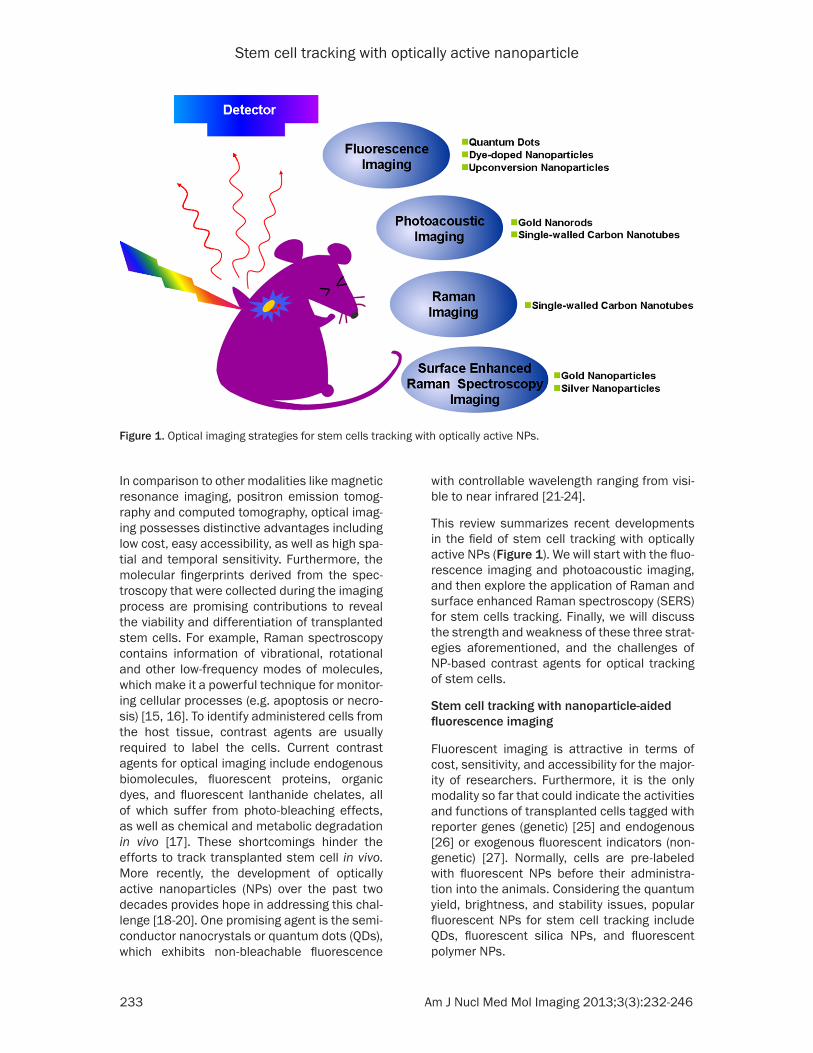
This review summarizes recent developments in the field of stem cell tracking with optically active NPs (Figure 1). We will start with the fluo-rescence imaging and photoacoustic imaging, and then explore the application of Raman and surface enhanced Raman spectroscopy (SERS) for stem cells tracking. Finally, we will discuss the strength and weakness of these three strat-egies aforementioned, and the challenges of NP-based contrast agents for optical tracking of stem cells.
Stem cell tracking with nanoparticle-aided fluorescence imaging
Fluorescent imaging is attractive in terms of cost, sensitivity, and accessibility for the major-ity of researchers. Furthermore, it is the only modality so far that could indicate the activities and functions of transplanted cells tagged with reporter genes (genetic) [25] and endogenous [26] or exogenous fluorescent indicators (non-genetic) [27]. Normally, cells are pre-labeled with fluorescent NPs before their administra-tion into the animals. Considering the quantum yield, brightness, and stability issues, popular fluorescent NPs for stem cell tracking include QDs, fluorescent silica NPs, and fluorescent polymer NPs.
Figure 1. Optical imaging strategies for stem cells tracking with optically active NPs.
Stem cell tracking with optically active nanoparticle
234 Am J Nucl Med Mol Imaging 2013;3(3):232-246
Quantum dots
QDs are highly fluorescent semiconductor NPs with high extinction coefficients, tunable emis-sions, sharp emission bandwidths, and good photostability [28, 29]. The tunable emission especially at the near infrared region (> ~800 nm) avoids the background signal of autofluo-rescence of the animal tissues (emissions are mainly at the visible region, ~300-550 nm). Good photostability allows QDs for the long-term tracking of stem cells.
Prior to the transplantation, stem cells have to be pre-labeled with QDs. There are at least six different ways to label cells including endocyto-sis through incubation, receptor-mediated uptake, lipid-based transduction, microinjec-tion, electroporation, and peptide-mediated delivery [30-32]. Passive incubation and pep-tide-mediated delivery are the most commonly used labeling methods. Rosen and colleagues
used passive incubation to load QDs into human mesenchymal stem cells (hMSCs), which was found more effective than electro-poration and receptor-mediated uptake [33]. They found that QDs aggregated around the nucleus when electroporation and receptor-mediated uptake were used and the inefficien-cy of labeling might be correlated with the impairment of cell membrane. Furthermore, they demonstrated that QDs labeled hMSCs could be identified in histological sections of canine ventricle, and the fluorescence signals were visible for at least 8 weeks following the injection [33].
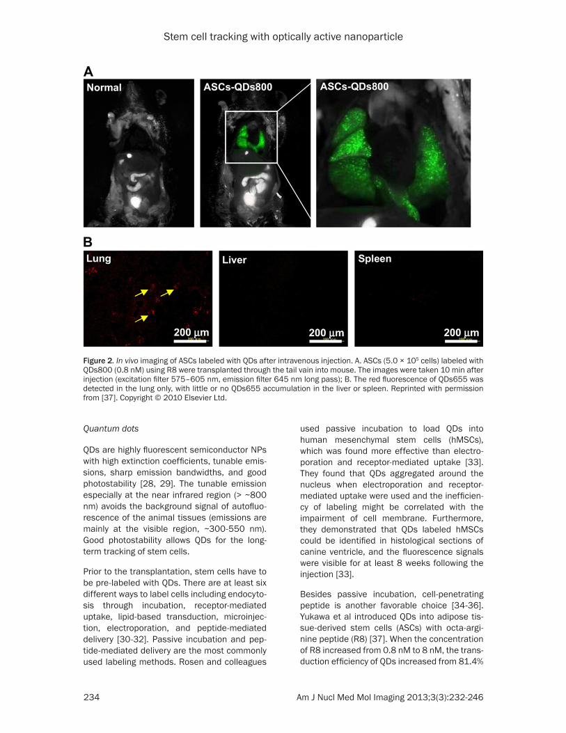
Besides passive incubation, cell-penetrating peptide is another favorable choice [34-36]. Yukawa et al introduced QDs into adipose tis-sue-derived stem cells (ASCs) with octa-argi-nine peptide (R8) [37]. When the concentration of R8 increased from 0.8 nM to 8 nM, the trans-duction efficiency of QDs increased from 81.4%
Figure 2. In vivo imaging of ASCs labeled with QDs after intravenous injection. A. ASCs (5.0 × 10⁵ cells) labeled with QDs800 (0.8 nM) using R8 were transplanted through the tail vain into mouse. The images were taken 10 min after injection (excitation filter 575–605 nm, emission filter 645 nm long pass); B. The red fluorescence of QDs655 was detected in the lung only, with little or no QDs655 accumulation in the liver or spleen. Reprinted with permission from [37]. Copyright © 2010 Elsevier Ltd.

Stem cell tracking with optically active nanoparticle
235 Am J Nucl Med Mol Imaging 2013;3(3):232-246
Table 1. Clinical trials related with major stem-cell-based cell therapies (Results from www.clinicaltrials.gov) Types of diseases Number of trialsCancer 3007Graft-versus-host disease 631Cardiac diseases 472Diabetes 101Bone repair 90Stroke 66
to 90.8% [37]. In their stem cell tracking assay, QDs labeled ASCs were visualized in situ with Maestro in vivo imaging system following trans-plantation through the tail vein of a mouse (Figure 2) and almost all ASCs were trapped in the lung [37]. Interestingly, if QDs labeled ACSs were mixed with heparin before the tail-vein-injection into the acute liver failure mice, ASCs were accumulated not only in the lung, but also in the liver. The accumulation rate of ASCs in liver increased to about 30%, which suggested heparin was effective for increasing the accu-mulation of transplanted ASCs in the liver [38]. To our knowledge, it is more likely that heparin increases the ability of transplanted ASCs to bypass the pulmonary circulation which is the first capillary system that the cells will encoun-ter after tail-vein-injection.
In contrast to the peptides that could improve the internalization of NPs by all types of cells, Lu et al recently developed a peptide which specifically targeted rhesus macaque embry-onic stem cells (RM-ESCs) [39]. The peptide was identified by phage display and contains a sequence of APWHLSSQYSRT. Peptide was covalently conjugated on the QDs surface via N-Hydroxysuccinimide (NHS) and ethyl(dimethylaminopropyl) carbodiimide (EDC) chemistry. The final conjugates should be a promising contrast agent specifically for imag-ing embryonic stem cells in vivo [39].
One major concern of QDs for stem cell labeling is their cytotoxicity [40, 41]. To address this issue, inert materials like silica have been used as the coating of QDs to decrease their cytotox-icity and to add extra functionalities. For exam-ple, silica coated QDs with cysteine (Cy) as cap-ping ligands showed lower cytotoxicity to hMSCs without compromising the quantum
yield [42]. When incubated with uncoated CdSe/ZnS-Cy at the concentration of 2.98 μM, the viability of hMSCs dropped down to 70%. In contrast, with the silica coated CdSe/ZnS-Cy at the same concentration, the cell viability was above 90% after 24 h incubation [42]. The con-focal imaging illustrated efficient labeling and advised that no particles were located on the cell membrane or inside the nucleus.
Dye-doped nanoparticles
Despite their unique optical properties, QDs are not clinically applicable because of their potential cytotoxicity generated from the leak-age of toxic metal ions [43]. As an alternative, researchers have designed biocompatible sili-ca and polymeric NPs containing fluorescent dyes [44, 45]. The biocompatible shell (e.g. polymer or silica) not only prevents organic dyes from oxidation or decomposition, but also enables the generation of strong fluorescence by concentrating the dyes.
Fluorescent silica NPs are mainly made through two approaches: sol-gel or reverse microemul-sion [46, 47]. One example is the fluorescent silica core-shell NPs, which were first named as Cornell dots or C-dots [48]. During the synthe-sis, organic dye molecules were covalently bound to a silica precursor to form adduct of the dye-rich core materials. Then silica sol-gel monomers were subsequently co-condensed with the core in specific order depending on the desired architecture to form a denser silica shell around the core [48]. C-dots possess enhanced brightness, photo-stability, biocom-patibility, and versatile surface functionalities. Recently, the commercial version of C-dots, C•spec® from Hybrid Silica Technologies (HST) has already been regulated by FDA for tumor imaging in a phase-I clinical trials, which con-firmed the safety of those silica-based NPs [49].
Besides C-dots, another type of silica NPs is cyanine dye-doped silica NPs (IRIS Dots), which were synthesized using a reverse microemul-sion method [50]. Briefly, spherical silica NPs containing fluorescent trimethine indocyanine dyes were prepared using a water-in-oil micro-emulsion method with diameter 50 nm. Entrapment of dye molecules in the silica matrix stabilized the photoemission over several hours of continuous irradiation [51]. IRIS Dots did not

Stem cell tracking with optically active nanoparticle
236 Am J Nucl Med Mol Imaging 2013;3(3):232-246
Figure 3. Detection of IRIS Dots uptake by hMSCs using confocal microscope and transmission electron microscope: A–C. hMSCs were pretreated for 48 h with 10 µg/mL ActD and then incubated with 20 µg/mL IRIS Dots for an ad-ditional 24 h. Both spontaneously detached and trypsinized cells were stained with either Annexin V–fluorescein isothiocyanate (A) or 2 μM calcein-AM (B, C) and then evaluated by confocal microscopy. The white scale bar repre-sents 10 μm. One representative apoptotic cell co-stained with Annexin V–fluorescein isothiocyanate (A), one repre-sentative apoptotic cell co-labeled with calcein-AM (B), and one representative live cell co-labeled with calcein-AM (C) are shown. D, E. hMSCs were pretreated for 48 h with 10 µg/mL ActD, incubated with 20 µg/mL IRIS Dots for an additional 24 h, and then analyzed by TEM. The black scale bar represents 0.5 μm. Reprinted with permission from [51]. Copyright © 2012 WILEY-VCH Verlag GmbH & Co. KGaA, Weinheim.
Stem cell tracking with optically active nanoparticle
237 Am J Nucl Med Mol Imaging 2013;3(3):232-246
affect the viability, proliferation and differentia-tion capability of hMSCs as well as C-dots [51]. More interestingly, IRIS Dots could allow the discrimination between live and early-stage apoptotic stem cells through the different sur-face distribution. Specifically, hMSCs were pre-treated with apoptosis-inducing agent actino-mycin D (ActD) to produce apoptotic cells. Then by incubating apoptotic and live hMSCs with IRIS Dots, they demonstrated that IRIS Dots were distributed in the cytoplasm of live cells (verified by stained with calcein-AM, Figure 3C), but on the outer cell surface of early apoptotic cells (stained with Annexin V-fluorescein iso-thiocyanate, Figure 3A and 3B) due to loss of active endocytosis.
Besides silica NPs, fluorescent polymeric NPs like polystyrene (PS) NPs are another popular choice likely due to their distribution by all the major biotechnology companies and the variety of functional groups including non-modified, sulfate-modified, aldehyde-modified, carboxyl-ate-modified or amine-modified surface [44, 52]. Recently, the uptake of the anionic PS NPs by hMSCs was investigated using spinning-disk confocal optical microscopy [53]. In this study, carboxyl-functionalized PS NPs were shown to be internalized mainly through the clathrin-mediated mechanism, which were more rapidly than the nonfunctionalized PS NPs.
Gold nanoparticles
With the rapid advancement of non-linear optics, fluorescence imaging with multiphoton excitation becomes a powerful technique for the high-resolution imaging. In this technique, noble metal NPs like gold NPs (Au NPs) were excited to a high energy state by two or more photons of red or near infrared (NIR) light simul-taneously. Farrer et al. demonstrated that mul-tiphoton-absorption-induced luminescence (MAIL) from Au NPs was generated efficiently with 800 nm laser and the luminescence spanned all the visible spectrum [54]. Compared with the traditional ultraviolet-visible (UV) excitation, NIR provided relatively higher depth of tissue penetration and minimized the interference of background fluorescence from the biological samples. In addition, Au NPs are generally considered biocompatible compared to other NPs [55], given that they have been used for human decorations for thousands of years. In another example, Nagesha et al. labeled mouse embryonic stem cells with Au
NPs and visualized them through MAIL. They expected the further application of this method for the investigation of the molecular machin-ery of endocytosis, post-internalization vesicle trafficking, lineage tracking, and cellular motili-ty assays [56]. Au NPs are also well known for their plasmonic properties as well as the appli-cations in photoacoustic imaging and SERS, which we will discuss later in other sections.
Upconversion nanoparticles
Upconversion (UC) is a process in which the sequential absorption of two or more photons leads to the emission of light at shorter wave-length. It is a non-linear optical process, which refers to anti-Stokes type emission. The most efficient UC materials are formed by solid-state materials doped with rare-earth ions [57]. In the nanoscale, NPs made of UC materials (so called, UCNPs) benefit from this unique proper-ties when they are utilized as contrast agents in molecular imaging. Imaging with UCNPs pro-vides higher sensitivity (lack of autofluores-cence background), less toxic components (in comparison to QDs), high penetration depths (excitation with NIR light), and good photosta-bility (no photobleaching) [58, 59].
A number of groups have labeled progenitor cells with UCNPs for both in vitro and in vivo fluorescence tracking [60-62]. For example, Wang et al labeled and tracked mouse MSCs (mMSCs) with UCNPs [63], which were conju-gated with R8 to facilitate cellular uptake. Little exocytosis of UCNPs from labeled mMSCs was found in the transwell assay after 10 days incu-bation, suggesting its potential for long-term cell tracking. Even after two weeks, the labeling with UCNPs did not show any influence over the survivability, proliferation, and differentiation of mMSCs. To determine the detection sensitivity of UCNP-labeled mMSCs in vivo, various amount of mMSCs (10-104) labeled with UCNPs were subcutaneously injected beneath the skin of a nude mouse. It was then imaged by a modi-fied Maestro in vivo imaging system with a 980 nm laser as excitation source. The sensitivity of fluorescence imaging with UCNPs was as few as 10 cells, nearly at the single cell level. By utilizing the UCNPs-labeling, they observed the migration of mMSCs from lung to liver in a nude mouse model with the whole body imaging by a modified Maestro in vivo imaging system using a 980 nm optical fiber-coupled laser as the excitation source [63].
Stem cell tracking with optically active nanoparticle
238 Am J Nucl Med Mol Imaging 2013;3(3):232-246
While unique, UCNPs have the following disad-vantages as contrast agents for fluorescence imaging. First of all, the upconversion efficien-cies have been relatively low (usually less than 1%) [64]. The excitation thresholds are quite high, and the investigated phosphors (generally fluorides) often presented poor chemical stabil-ity [65]. Secondly, the potential long-term toxic-ity of Ln3+ doped UCNPs is another significant concern [64]. A thorough and systematic inves-tigation is highly needed to reveal their biocom-patibility and biostability.
Stem cell tracking with nanoparticle-aided photoacoustic imaging
Photoacoustic (PA) imaging, also called opto-acoustic imaging is a new biomedical imaging
modality based on photoacoustic effect, in which the absorbed energy from the light is transformed into kinetic energy of the sample through energy exchange processes. It is a hybrid modality, combining the high-contrast and spectroscopic-based specificity of optical imaging with the high spatial resolution of ultra-sound imaging. In essence, a PA image is an ultrasound image in which contrast depends on the optical absorption of samples. Thus biologi-cal tissues with optical properties such as hemoglobin could be visualized with PA imaging.
As stem cells usually don’t have obvious optical properties, in PA imaging, they are usually labeled with biocompatible materials with opti-
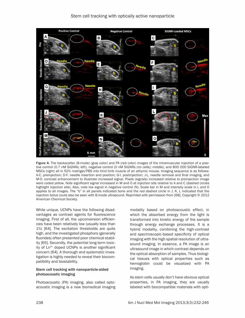
Figure 4. The backscatter (B-mode) (gray color) and PA (red color) images of the intramuscular injection of a posi-tive control (0.7 nM SiGNRs; left), negative control (0 nM SiGNRs (no cells); middle), and 800 000 SiGNR-labeled MSCs (right) all in 50% matrigel/PBS into hind limb muscle of an athymic mouse. Imaging sequence is as follows: A-C. preinjection; D-F. needle insertion and position; G-I. postinjection; J-L. needle removal and final imaging, and M-O. contrast enhancement to illustrate increased signal. Pixels (signals) increased relative to preinjection image were coded yellow. Note significant signal increased in M and O at injection site relative to A and C (dashed circles highlight injection site). Also, note low signal in negative control (N). Scale bar in M and intensity scale in L and O applies to all images. The “b” in all panels indicated bone and the red dashed circle in J, K, L indicated that the injection bolus could also be seen with B-mode ultrasound. Reprinted with permission from [68]. Copyright © 2012 American Chemical Society.

Stem cell tracking with optically active nanoparticle
239 Am J Nucl Med Mol Imaging 2013;3(3):232-246
cal properties such as Au NPs or Au nanorods (NRs). In a recent study, hMSCs were pre-labeled with 20 nm Au NPs before their incor-poration into PEGylated fibrin gel [66]. Then fibrin gel was injected intramuscularly in the lateral gastrocnemius of an anesthetized Lewis rat. The contrast brought by Au NPs allowed the researchers to visualize the in vivo differentia-tion and neovasularization of hMSCs using PA imaging. Au NRs, another attractive probe for PA imaging, have plasmon resonance absorp-tion and scattering in the NIR region [67]. Jokerst et al used silica coated Au NRs (SiGNRs) to label and image hMSCs [68]. They found that the silica coating could dramatically increase the cellular uptake of SiGNRs (5-fold) without any change to the viability and function of hMSCs. Figure 4 showed the process of intra-muscular implantation of SiGNR-labeled hMSCs to the hindlimb muscles of the mouse. PA imaging provides relative high spatial reso-lution (340 nm) and temporal resolution (0.2 s), which allows the real time monitoring of stem cell after transplantation.
Stem cell tracking with nanoparticle-aided Raman or surface enhanced Raman spectros-copy
The last technology we would like to discuss is Raman or surface enhanced Raman spectros-copy (SERS) based imaging. Raman scattering is the inelastic scattering of photons by molecu-lar bonds that utilize the fact that every chemi-cal bond in a molecule has a characteristic vibrational energy. This “molecular fingerprint” allows the nondestructive and label-free imag-ing of biological molecules such as DNA and protein in cells and tissue. In comparison to infrared (IR) and nuclear magnetic resonance (NMR) spectroscopy, Raman spectroscopy is not affected by the presence of water. However, Raman scattering from the natural molecules produces a very weak signal which is usually approximately 12-14 orders of magnitude weaker than fluorescence [69]. SERS could be used to further enhance the signal. Although Raman spectroscopy and SERS have been widely used from imaging to diagnoses [70-77], it is relatively a new direction to track stem cells with those techniques.
Stem cell tracking with nanoparticle-aided Ra-man imaging
Spontaneous Raman spectroscopy has been wildly used for monitoring the differentiation of
human embryonic stem cells (hESC) [78-80], adult stem cells [81], and neural stem cells [80] due to the spectral characteristics of different types of cells. Notingher et al observed a clear reduction in DNA (786 cm-1 Raman peaks) and RNA (813 cm-1 Raman peaks) during living murine embryonic stem (ES) cell differentiation (over 16 days of differentiation) [78]. Chan et al. demonstrated specific Raman spectra could distinguish undifferentiated hESC from hESC-derived cardiomyocytes and human fetal left ventricle cardiomyocytes [79]. These results lay the foundation for the development of single cell Raman spectroscopy as a systematic meth-od for sorting cardiomyocytes derived from reprogrammed, embryonic or adult stem cells for future cell-based heart therapies [79]. Spectral variations assigned to glycogen have also been reported for hESCs maintained under normal growth condition in vitro [82, 83]. In addition, Raman spectroscopy has been employed to monitor osteoblast differentiation and in vitro mineralization capacity of MSC and osteoprogentior cells [84, 85]. Cultured normal and abnormal stem cells including normal hESC, karyotypically abnormal hESC, normal and transformed hMSC can be identified by Raman spectroscopy. The changes of intensity peaks from phenylalanine (1005 cm-1), cyto-chrome C (1128 cm-1), protein, DNA/RNA, lipid in normal and abnormal stem cells show that Raman spectroscopy provides an alternative method allowing screening of cultured stem cells from abnormalities (abnormal and trans-formed stem cells) prior to cell transplantation [86].
As mentioned above, the fingerprint character-istics of Raman spectroscopy provides a way to distinguish stem cell types, differentiation pro-cesses, and abnormalism (chromosomal insta-bility, development of cell lines, in vitro replica-tive senescence and etc). Statistic methods such as principal component analysis and lin-ear discriminant analysis are usually required for the spectra analysis. However, due to the long acquisition time (normally several minutes for each Raman spectrum and hours for a sin-gle cell imaging), tracking the fate and the func-tion of stem cells in vivo based on its intrinsic Raman information remain significant challeng-es [82]. Alternatively, if the Raman signal of cer-tain probes within the cell is strong enough for fast imaging, tracking stem cell based on Raman signal could eventually be practical.

Stem cell tracking with optically active nanoparticle
240 Am J Nucl Med Mol Imaging 2013;3(3):232-246
Unlike fluorescent imaging, Raman imaging does not suffer from photobleaching and auto-fluorescence background interference once NIR excitation is used. Among various NP-based contrast agents, single-walled carbon nano-tubes (SWNTs) show an intense intrinsic Raman peak (G band at 1593 cm-1) produced by the strong electron photon coupling that causes efficient excitation of tangential vibration in the nanotubes quasi one-dimensional structure upon light exposure [87]. Strong and narrow signal of SWNTs not only enables fast mapping (with integration time of 0.1 s for each mapping point) but also provides easy differentiation from the tissue autofluoresence. So far SWNTs have been successfully used for specific tumor targeting in vivo within various tumor models [88-90] and whole-body, deep-tissue, small-animal imaging [91]. They are chemical and photo stable, and can be used for long-term tracking and imaging in biological systems [92]. Wang et al. found the protamine and PEG func-tionalized SWNTs (naturally accompany with Fe/Co metallic nanoparticles come from syn-thesis methods) were allowed to track hMSCs without interfering their proliferation and differ-entiation. Triple model Raman/MRI/PAT imag-ing of SWNT-labeled hMSCs was further dem-onstrated in living mice (Figure 5) [93]. Moreover, SWNTs with different isotope compo-
sitions (C12-SWNTs and C13-SWNTs) display well-shifted Raman G-band peaks (C12-SWNTs at around 1590 cm-1 and C13-SWNTs at around 1528 cm-1) [94]. This character enables them to be potentially used for multiplex tracking of different types of stem cells which are injected simultaneously.
Stem cell tracking with nanoparticle-aided surface enhanced Raman spectroscopy
Besides Raman spectroscopy, imaging with SERS has also gained significant interest over the last several years [95]. SERS is a phenom-enon that the Raman scattering from a mole-cule is enhanced by many orders of magnitude due to its proximity to a metal surface (usually gold and silver) with surface plasmon efficiently coupling the energy of the incoming laser light [96]. Normally, two different methodologies, direct detection (with metallic NPs as SERS substrates for label-free detection of the ana-lyte) and indirect detection (biotargeted research with SERS labels) are used for cell and biomedical research.
Similar to Raman spectroscopy, direct detec-tion of SERS provides vibration information of stem cell while enhancing the signal sensitivity. By labeling mESC with Au NPs, Sathuluri and
Figure 5. In vivo triple-modal imaging of SWNT-labeled hMSCs: unlabeled and SWNT-labeled hMSCs (10 nM of SWNTs during labeling) were subcutaneously injected into the back of a nude mouse before imaging. A. In vivo T2-weighted MR image. Arrows pointed to the sites where unlabeled (left) and SWNT-labeled (right) hMSCs were injected; B, C. In vivo Raman images of unlabeled (B) and SWNT-labeled (C) hMSCs; D, E. In vivo PAT images of unla-beled (D) and SWNT-labeled (E) hMSCs. The circles highlighted the locations were hMSCs were injected. PA signals in the rectangle highlighted in D were from a blood vessel crossing this area. Reprinted with permission from [93]. Copyright © 2012 WILEY-VCH Verlag GmbH & Co. KGaA, Weinheim.
Stem cell tracking with optically active nanoparticle
241 Am J Nucl Med Mol Imaging 2013;3(3):232-246
colleagues demonstrated that specific SERS fingerprint information of undifferentiated sin-gle cells, embryoid bodies and terminally differ-entiated cardiomyocytes has unique SERS fin-gerprints based upon nucleic acids, mitochondria, proteins, and cardiomyocytes adsorbed onto metal NPs. This information reflects the molecular changes accompanying with metabolic activities in each stage of dif-ferentiation [97].
By labeling cells with Raman probes (molecules with significant Raman activity) and biospecific antibodies, an indirect method to visualize the distribution of certain proteins within living cell can be engineered. The expressed proteins, including CD34, Sca-1, and SP-C in bronchioal-veolar stem cells (BASCs) in the murine lung were quantitatively compared based on multi-functional NPs, namely fluorescent-surface-enhanced Raman spectroscopic dots (F-SERS dots). Conjugated with BASC-specific markers, with the dual signal detection model, F-SERS dots provided multiple target analysis and clari-fied the effective stem cell specific markers and therapy strategies for disease [98]. In addi-tion to multiplex targeting, magnetic NP-based surface enhanced Raman spectroscopic dots (M-SERS Dots) could be used as a sorting sys-tem effectively isolate BASCs [99].
Perspectives and conclusion
With the growing demands of regenerative ther-apy, and the current lack of a sensitive and yet efficient method of tracking transplanted cells, there is an urgent need for the development of novel imaging technologies which allow the real-time and non-invasive monitoring of trans-planted stem cells for their survival, biodistribu-tion, migration, and differentiation. In compari-son to other imaging modalities like MRI, PET, and CT, optical imaging is cost-effective, time-efficient, and are thus more accessible for the majority of researchers during the pre-clinical studies with small animals. In this mini-review, we discussed three modalities of NP-aided optical imaging that have been widely used for stem cell imaging and tracking.
However, we should be aware that opportunity and risk always co-exist. Several concerns about using NPs for stem cell tracking should be addressed before they are routinely used in clinical treatments. Specifically, those challeng-
es include contrast agents transfer, signal dilu-tion, interference from tissue autofluorescence, and inability to gather information on cellular functions [14, 100].
In contrast agents (i.e. NPs) transfer, NPs leaked out from labeled cells due to normal cell processes (e.g. cell death and exocytosis) and were able to re-enter into adjacent cells over time. The possibility of NPs leakage, a common challenge in cell tracking studies, leads to pos-sible uptake of NPs by surrounding cells and thus introduces false positive results [101]. For example, stem cells were labeled with QDs in order to noninvasively track their distribution after being seeded on scaffolds and trans-planted into a nude rat that was critically sized femoral defects. As excepted, clear fluores-cence was observed at implantation sites; how-ever, signals were also observed at sites treat-ed with acellular QD-free scaffolds after surgery for 7-10 days [102]. Sarah et al demonstrated the transfer of QDs from labeled hMSCs to adjacent unlabeled cells in an in vitro co-cul-ture system. Their leakage was further exam-ined in vivo by transplantation of QDs-labeled hMSCs to an animal model of spinal cord injury, which indicated the deposition of QDs into the host cells after 1 week was probably due to their active excretion from labeled cells or the release from the dead cells [36]. These studies show that the leakage of NPs from labeled cells must be tested to avoid false positive results. Meanwhile, valid conclusion of cell localization cannot be drawn only from in vivo imaging data. It is also expected that modifications (e.g. cat-ionic lipids [32]) of NPs with some specific ligands which eliminate their leakage over a long period of time could provide significant potential in long term in vivo imaging.
Signal dilution, another issue for long-term in vivo stem cell tracking, is the signal loss result-ed from cell division and exocytosis. For exam-ple, Sarah et al found that the fluorescence of QDs labeled hMSCs fell progressively with cell divisions. After four subculturing passage, the fluorescence intensity decreased by more than 80% [36]. One approach is to genetically engi-neer the cell to produce NPs [103, 104], which would be similar to the expression of green fluo-rescence protein. However, it is still unclear whether genetic engineering of cells will have any long-term consequence on cell phenotype and function. Another approach is to enhance
Stem cell tracking with optically active nanoparticle
242 Am J Nucl Med Mol Imaging 2013;3(3):232-246
the signal of NPs which will still detectable after multiple cell divisions [105]. Single NP optical imaging technique holds the promise of in vivo stem cell tracking even when only one NP is left in one stem cell after cell divisions. The real time tracking of the transport and random walk of single NP (gold and silver) in zebrafish embry-os have been achieved by Xu’s group [106, 107].
Optical imaging modalities except PA imaging usually suffer from the background interfer-ence of autofluorescence of tissues [108]. NPs with tunable emission wavelength of light either generated from inherent properties (QDs) or encapsulated fluorescence probe (C-Dots) can avoid the autofluorescence background, which can also be reduced by UCNPs for fluorescence imaging and SWNTs for Raman imaging using NIR excitation light.
Although the location of stem cells post trans-plantation could be tracked in vivo with good temporal and spatial resolution, the functions (e.g. differentiation of stem cells) of the trans-planted cells are also important. In the clinical treatments, if the viability and function of trans-planted stem cells can be obtained simultane-ously, it will help the physician to make a deci-sion without the need for multiple biopsies. One potential approach is to design NP-aided sensors, which can respond to the secreted chemicals or expressed chemokines/cytokines during cell differentiation, the local pH changes during cell apoptosis and death. Another approach is using Raman and SERS imaging modalities to simultaneously track stem cells in vivo and record fingerprint spectrum of certain chemicals indicated different cellular condi-tions and functions.
Conflict of interest
The authors have declared that no competing interests exist.
Acknowledgements
This work was supported in part by Nanyang Technological University Start-Up Grant and Singapore MOE AcRF Tier 1 research fund (RG 64/12) to XCJ. JKYC received salary support from the Ministry of Health’s National Medical Research Council in Singapore (NMRC/CSA/042/2012).
Address correspondence to: Dr. Chenjie Xu, Division of BioEngineering, School of Chemical and Biomedical Engineering, Nanyang Technological University, Building N1.3, level B2, Room 06, 70 Nanyang Drive, Singapore 637457. Phone: 65-6513-8298; Fax: 65-6791-1761; E-mail: [email protected]
References
[1] Weissman IL. Stem cells: Units of develop-ment, units of regeneration, and units in evolu-tion. Cell 2000; 100: 157-168.
[2] Weissman IL. Translating stem and progenitor cell biology to the clinic: Barriers and opportu-nities. Science 2000; 287: 1442-1446.
[3] Singec I, Jandial R, Crain A, Nikkhah G and Snyder EY. The leading edge of stem cell thera-peutics. Annu Rev Med 2007; 58: 313-328.
[4] Pardal R, Clarke MF and Morrison SJ. Applying the principles of stem-cell biology to cancer. Nat Rev Cancer 2003; 3: 895-902.
[5] Pittenger MF and Martin BJ. Mesenchymal stem cells and their potential as cardiac thera-peutics. Circ Res 2004; 95: 9-20.
[6] Barry FP and Murphy JM. Mesenchymal stem cells: clinical applications and biological char-acterization. Int J Biochem Cell Biol 2004; 36: 568-584.
[7] Bianco P, Riminucci M, Gronthos S and Robey PG. Bone marrow stromal stem cells: Nature, biology, and potential applications. Stem Cells 2001; 19: 180-192.
[8] Caplan AI and Bruder SP. Mesenchymal stem cells: building blocks for molecular medicine in the 21st century. Trends Mol Med 2001; 7: 259-264.
[9] Prochymal - First stem cell drug approved. http://www.medicalnewstoday.com/arti-cles/245704.php 2012.
[10] www.clinicaltrials.gov 2013.[11] Halban PA, German MS, Kahn SE and Weir GC.
Current Status of Islet Cell Replacement and Regeneration Therapy. J Clin Endocr Metab 2010; 95: 1034-1043.
[12] Nguyen PK, Nag D and Wu JC. Methods to as-sess stem cell lineage, fate and function. Adv Drug Deliv Rev 2010; 62: 1175-1186.
[13] Pagani FD, DerSimonian H, Zawadzka A, Wet-zel K, Edge ASB, Jacoby DB, Dinsmore JH, Wright S, Aretz TH, Eisen HJ and Aaronson KD. Autologous skeletal myoblasts transplanted to ischemia-damaged myocardium in humans - Histological analysis of cell survival and differ-entiation. J Am Coll Cardiol 2003; 41: 879-888.
[14] Xu CJ, Mu LY, Roes I, Miranda-Nieves D, Nahrendorf M, Ankrum JA, Zhao WA and Karp JM. Nanoparticle-based monitoring of cell ther-apy. Nanotechnology 2011; 22: 494001.
Stem cell tracking with optically active nanoparticle
243 Am J Nucl Med Mol Imaging 2013;3(3):232-246
[15] Uzunbajakava N, Lenferink A, Kraan Y, Willek-ens B, Vrensen G, Greve J and Otto C. Nonreso-nant Raman imaging of protein distribution in single human cells. Biopolymers 2003; 72: 1-9.
[16] Kneipp J, Kneipp H, Wittig B and Kneipp K. Novel optical nanosensors for probing and im-aging live cells. Nanomed-Nanotechnol Biol Med 2010; 6: 214-226.
[17] Fujimoto JG and Farkas D. Biomedical optical imaging. USA: Oxford University Press, 2009.
[18] Solanki A, Kim JD and Lee KB. Nanotechnology for regenerative medicine: nanomaterials for stem cell imaging. Nanomedicine 2008; 3: 567-578.
[19] Villa C, Erratico S, Razini P, Fiori F, Rustichelli F, Torrente Y and Belicchi M. Stem Cell Tracking by Nanotechnologies. Int J Mol Sci 2010; 11: 1070-1081.
[20] Deb KD, Griffith M, De Muinck E and Rafat M. Nanotechnology in stem cells research: ad-vances and applications. Front Biosci 2012; 17: 1747-1760.
[21] Michalet X, Pinaud FF, Bentolila LA, Tsay JM, Doose S, Li JJ, Sundaresan G, Wu AM, Gambhir SS and Weiss S. Quantum dots for live cells, in vivo imaging, and diagnostics. Science 2005; 307: 538-544.
[22] Sharrna P, Brown S, Walter G, Santra S and Moudgil B. Nanoparticles for bioimaging. Adv Colloid Interface Sci 2006; 123: 471-485.
[23] Kim J, Piao Y and Hyeon T. Multifunctional nanostructured materials for multimodal imag-ing, and simultaneous imaging and therapy. Chem Soc Rev 2009; 38: 372-390.
[24] Hahn MA, Singh AK, Sharma P, Brown SC and Moudgil BM. Nanoparticles as contrast agents for in-vivo bioimaging: current status and fu-ture perspectives. Anal Bioanal Chem 2011; 399: 3-27.
[25] Lee DS. In Vivo Neuronal Cell Differentiation Imaging From Transplanted Stem Cells. Curr Med Imaging Rev 2012; 8: 278-283.
[26] Buschke DG, Squirrell JM, Fong JJ, Eliceiri KW and Ogle BM. Cell death, non-invasively as-sessed by intrinsic fluorescence intensity of NADH, is a predictive indicator of functional differentiation of embryonic stem cells. Biol Cell 2012; 104: 352-364.
[27] Zhao WA, Schafer S, Choi J, Yamanaka YJ, Lombardi ML, Bose S, Carlson AL, Phillips JA, Teo WS, Droujinine IA, Cui CH, Jain RK, Lam-merding J, Love JC, Lin CP, Sarkar D, Karnik R and Karp JM. Cell-surface sensors for real-time probing of cellular environments. Nat Nano-technol 2011; 6: 524-531.
[28] Alivisatos AP. Semiconductor clusters, nano-crystals, and quantum dots. Science 1996; 271: 933-937.
[29] Bruchez M, Moronne M, Gin P, Weiss S and Alivisatos AP. Semiconductor nanocrystals as fluorescent biological labels. Science 1998; 281: 2013-2016.
[30] Dubertret B, Skourides P, Norris DJ, Noireaux V, Brivanlou AH and Libchaber A. In vivo imag-ing of quantum dots encapsulated in phospho-lipid micelles. Science 2002; 298: 1759-1762.
[31] Chen FQ and Gerion D. Fluorescent CdSe/ZnS nanocrystal-peptide conjugates for long-term, nontoxic imaging and nuclear targeting in liv-ing cells. Nano Lett 2004; 4: 1827-1832.
[32] Voura EB, Jaiswal JK, Mattoussi H and Simon SM. Tracking metastatic tumor cell extravasa-tion with quantum dot nanocrystals and fluo-rescence emission-scanning microscopy. Nat Med 2004; 10: 993-998.
[33] Rosen AB, Kelly DJ, Schuldt AJT, Lu J, Potapova IA, Doronin SV, Robichaud KJ, Robinson RB, Rosen MR, Brink PR, Gaudette GR and Cohen IS. Finding fluorescent needles in the cardiac haystack: Tracking human mesenchymal stem cells labeled with quantum dots for quantita-tive in vivo three-dimensional fluorescence analysis. Stem Cells 2007; 25: 2128-2138.
[34] Lei Y, Tang H, Yao L, Yu R, Feng M and Zou B. Applications of mesenchymal stem cells la-beled with Tat peptide conjugated quantum dots to cell tracking in mouse body. Bioconju-gate Chem 2008; 19: 421-427.
[35] Chang JC, Hsu SH and Su HL. The regulation of the gap junction of human mesenchymal stem cells through the internalization of quantum dots. Biomaterials 2009; 30: 1937-1946.
[36] Ranjbarvaziri S, Kiani S, Akhlaghi A, Vosough A, Baharvand H and Aghdami N. Quantum dot labeling using positive charged peptides in hu-man hematopoetic and mesenchymal stem cells. Biomaterials 2011; 32: 5195-5205.
[37] Yukawa H, Kagami Y, Watanabe M, Oishi K, Mi-yamoto Y, Okamoto Y, Tokeshi M, Kaji N, Nogu-chi H, Ono K, Sawada M, Baba Y, Hamajima N and Hayashi S. Quantum dots labeling using octa-arginine peptides for imaging of adipose tissue-derived stem cells. Biomaterials 2010; 31: 4094-4103.
[38] Yukawa H, Watanabe M, Kaji N, Okamoto Y, Tokeshi M, Miyamoto Y, Noguchi H, Baba Y and Hayashi S. Monitoring transplanted adipose tissue-derived stem cells combined with hepa-rin in the liver by fluorescence imaging using quantum dots. Biomaterials 2012; 33: 2177-2186.
[39] Lu S, Xu X, Zhao WX, Wu WW, Yuan H, Shen HB, Zhou CH, Li S and Ma L. Targeting of embryonic stem cells by peptide-conjugated quantum dots. PloS One 2010; 5: e12705.
[40] Derfus AM, Chan WCW and Bhatia SN. Probing the cytotoxicity of semiconductor quantum dots. Nano Lett 2004; 4: 11-18.
Stem cell tracking with optically active nanoparticle
244 Am J Nucl Med Mol Imaging 2013;3(3):232-246
[41] Kirchner C, Liedl T, Kudera S, Pellegrino T, Ja-vier AM, Gaub HE, Stolzle S, Fertig N and Parak WJ. Cytotoxicity of colloidal CdSe and CdSe/ZnS nanoparticles. Nano Lett 2005; 5: 331-338.
[42] Lai CW, Wang YH, Chen YC, Hsieh CC, Uttam BP, Hsiao JK, Hsu CC and Chou PT. Homoge-nous, far-reaching tuning and highly emissive QD-silica core-shell nanocomposite synthe-sized via a delay photoactive procedure; their applications in two-photon imaging of human mesenchymal stem cells. J Mater Chem 2009; 19: 8314-8319.
[43] Chen N, He Y, Su YY, Li XM, Huang Q, Wang HF, Zhang XZ, Tai RZ and Fan CH. The cytotoxicity of cadmium-based quantum dots. Biomateri-als 2012; 33: 1238-1244.
[44] Wang F, Tan WB, Zhang Y, Fan XP and Wang MQ. Luminescent nanomaterials for biological labelling. Nanotechnology 2006; 17: R1-R13.
[45] Burns A, Ow H and Wiesner U. Fluorescent core-shell silica nanoparticles: towards “Lab on a Particle” architectures for nanobiotech-nology. Chem Soc Rev 2006; 35: 1028-1042.
[46] Vanblaaderen A and Vrij A. Synthesis and char-acterization of monodisperse colldidal organo-silica spheres. J Colloid Interface Sci 1993; 156: 1-18.
[47] Yamauchi H, Ishikawa T and Kondo S. Surface characterization of ultramicro spherical-parti-cles of silica prepared by w/o microemulsion method. Colloids Surf 1989; 37: 71-80.
[48] Ow H, Larson DR, Srivastava M, Baird BA, Webb WW and Wiesner U. Bright and stable core-shell fluorescent silica nanoparticles. Nano Lett 2005; 5: 113-117.
[49] Burns AA, Vider J, Ow H, Herz E, Penate-Medi-na O, Baumgart M, Larson SM, Wiesner U and Bradbury M. Fluorescent silica nanoparticles with efficient urinary excretion for nanomedi-cine. Nano Lett 2009; 9: 442-448.
[50] Alberto G, Miletto I, Viscardi G, Caputo G, Lat-terini L, Coluccia S and Martra G. Hybrid cya-nine-silica nanoparticles: Homogeneous pho-toemission behavior of entrapped fluorophores and consequent high brightness enhance-ment. J Phys Chem C 2009; 113: 21048-21053.
[51] Accomasso L, Rocchietti EC, Raimondo S, Cat-alano F, Alberto G, Giannitti A, Minieri V, Turi-netto V, Orlando L, Saviozzi S, Caputo G, Geuna S, Martra G and Giachino C. Fluorescent silica nanoparticles improve optical imaging of stem cells allowing direct discrimination between live and early-stage apoptotic cells. Small 2012; 8: 3192-3200.
[52] Brijmohan SB, Swier S, Weiss RA and Shaw MT. Synthesis and characterization of cross-linked sulfonated polystyrene nanoparticles. Ind Eng Chem Res 2005; 44: 8039-8045.
[53] Jiang XE, Musyanovych A, Rocker C, Landfester K, Mailander V and Nienhaus GU. Specific ef-fects of surface carboxyl groups on anionic polystyrene particles in their interactions with mesenchymal stem cells. Nanoscale 2011; 3: 2028-2035.
[54] Farrer RA, Butterfield FL, Chen VW and Four-kas JT. Highly efficient multiphoton-absorption-induced luminescence from gold nanoparti-cles. Nano Lett 2005; 5: 1139-1142.
[55] Thakor AS, Jokerst J, Zavaleta C, Massoud TF and Gambhir SS. Gold nanoparticles: A revival in precious metal administration to patients. Nano Lett 2011; 11: 4029-4036.
[56] Nagesha D, Laevsky GS, Lampton P, Banyal R, Warner C, DiMarzio C and Sridhar S. In vitro imaging of embryonic stem cells using multi-photon luminescence of gold nanoparticles. Int J Nanomed 2007; 2: 813-819.
[57] Wang F and Liu XG. Recent advances in the chemistry of lanthanide-doped upconversion nanocrystals. Chem Soc Rev 2009; 38: 976-989.
[58] Wang F, Banerjee D, Liu YS, Chen XY and Liu XG. Upconversion nanoparticles in biological labeling, imaging, and therapy. Analyst 2010; 135: 1839-1854.
[59] Haase M and Schafer H. Upconverting Nanoparticles. Angew Chem-Int Edit 2011; 50: 5808-5829.
[60] Liu Q, Sun Y, Yang TS, Feng W, Li CG and Li FY. Sub-10 nm hexagonal lanthanide-doped Na-LuF4 upconversion nanocrystals for sensitive bioimaging in vivo. J Am Chem Soc 2011; 133: 17122-17125.
[61] Nyk M, Kumar R, Ohulchanskyy TY, Bergey EJ and Prasad PN. High contrast in vitro and in vivo photoluminescence bioimaging using near infrared to near infrared up-conversion in TM3+ and Yb3+ doped fluoride nanophos-phors. Nano Lett 2008; 8: 3834-3838.
[62] Idris NM, Li ZQ, Ye L, Sim EK, Mahendran R, Ho PCL and Zhang Y. Tracking transplanted cells in live animal using upconversion fluorescent nanoparticles. Biomaterials 2009; 30: 5104-5113.
[63] Wang C, Cheng L, Xu H and Liu Z. Towards whole-body imaging at the single cell level us-ing ultra-sensitive stem cell labeling with oligo-arginine modified upconversion nanoparticles. Biomaterials 2012; 33: 4872-4881.
[64] Cheng L, Wang C and Liu Z. Upconversion nanoparticles and their composite nanostruc-tures for biomedical imaging and cancer thera-py. Nanoscale 2013; 5: 23-37.
[65] Wang M, Abbineni G, Clevenger A, Mao CB and Xu SK. Upconversion nanoparticles: synthesis, surface modification and biological applica-tions. Nanomed-Nanotechnol Biol Med 2011; 7: 710-729.

Stem cell tracking with optically active nanoparticle
245 Am J Nucl Med Mol Imaging 2013;3(3):232-246
[66] Nam SY, Ricles LM, Suggs LJ and Emelianov SY. In vivo ultrasound and photoacoustic moni-toring of mesenchymal stem cells labeled with gold nanotracers. PloS One 2012; 7: e37267.
[67] Tong L, Wei QS, Wei A and Cheng JX. Gold nanorods as contrast agents for biological im-aging: Optical properties, surface conjugation and photothermal effects. Photochem Photo-biol 2009; 85: 21-32.
[68] Jokerst JV, Thangaraj M, Kempen PJ, Sinclair R and Gambhir SS. Photoacoustic imaging of mesenchymal stem cells in living mice via sili-ca-coated gold nanorods. ACS Nano 2012; 6: 5920-5930.
[69] Le Ru EC, Schroeter LC and Etchegoin PG. Di-rect measurement of resonance Raman spec-tra and cross sections by a polarization differ-ence technique. Anal Chem 2012; 84: 5074-5079.
[70] Wachsmann-Hogiu S, Weeks T and Huser T. Chemical analysis in vivo and in vitro by Ra-man spectroscopy-from single cells to humans. Curr Opin Biotechnol 2009; 20: 63-73.
[71] Kneipp K, Kneipp H and Kneipp J. Surface-en-hanced Raman scattering in local optical fields of silver and gold nanoaggregatess - From sin-gle-molecule Raman spectroscopy to ultrasen-sitive probing in live cells. Accounts Chem Res 2006; 39: 443-450.
[72] Chan J, Fore S, Wachsman-Hogiu S and Huser T. Raman spectroscopy and microscopy of indi-vidual cells and cellular components. Laser Photon Rev 2008; 2: 325-349.
[73] Li MQ, Xu J, Romero-Gonzalez M, Banwart SA and Huang WE. Single cell Raman spectrosco-py for cell sorting and imaging. Curr Opin in Biotechnol 2012; 23: 56-63.
[74] Movasaghi Z, Rehman S and Rehman IU. Ra-man spectroscopy can detect and monitor can-cer at cellular level: Analysis of resistant and sensitive subtypes of testicular cancer cell lines. Appl Spectrosc Rev 2012; 47: 571-581.
[75] Vitol EA, Orynbayeva Z, Friedman G and Gogot-si Y. Nanoprobes for intracellular and single cell surface-enhanced Raman spectroscopy (SERS). J Raman Spectrosc 2012; 43: 817-827.
[76] Huh YS, Chung AJ and Erickson D. Surface en-hanced Raman spectroscopy and its applica-tion to molecular and cellular analysis. Micro-fluid and Nanofluid 2009; 6: 285-297.
[77] Qian XM and Nie SM. Single-molecule and sin-gle-nanoparticle SERS: from fundamental mechanisms to biomedical applications. Chem Soc Rev 2008; 37: 912-920.
[78] Notingher I, Bisson I, Bishop AE, Randle WL, Polak JM and Hench LL. In situ spectral moni-toring of mRNA translation in embryonic stem cells during differentiation in vitro. Anal Chem 2004; 76: 3185-3193.
[79] Chan JW, Lieu DK, Huser T and Li RA. Label-free separation of human embryonic stem cells and their cardiac derivatives using Ra-man spectroscopy. Anal Chem 2009; 81: 1324-1331.
[80] Chan JW and Lieu DK. Label-free biochemical characterization of stem cells using vibrational spectroscopy. J Biophotonics 2009; 2: 656-668.
[81] Downes A, Mouras R and Elfick A. Optical spec-troscopy for noninvasive monitoring of stem cell differentiation. J Biomed Biotechnol 2010; 101864: 1-10.
[82] Stewart S, Priore RJ, Nelson MP and Treado PJ. Raman imaging. Annu Rev Anal Chem 2012; 5: 337-360.
[83] Konorov SO, Schulze HG, Caron NJ, Piret JM, Blades MW and Turner RFB. Raman micro-spectroscopic evidence that dry-fixing pre-serves the temporal pattern of non-specific dif-ferentiation in live human embryonic stem cells. J Raman Spectrosc 2011; 42: 576-579.
[84] McManus LL, Burke GA, McCafferty MM, O’Hare P, Modreanu M, Boyd AR and Meenan BJ. Raman spectroscopic monitoring of the os-teogenic differentiation of human mesenchy-mal stem cells. Analyst 2011; 136: 2471-2481.
[85] Gentleman E, Swain RJ, Evans ND, Boonrung-siman S, Jell G, Ball MD, Shean TA, Oyen ML, Porter A and Stevens MM. Comparative materi-als differences revealed in engineered bone as a function of cell-specific differentiation. Nat Mater 2009; 8: 763-770.
[86] Harkness L, Novikov SM, Beermann J, Bozhev-olnyi SI and Kassem M. Identification of abnor-mal stem cells using Raman spectroscopy. Stem Cells Dev 2012; 21: 2152-2159.
[87] Jorio A, Saito R, Dresselhaus G and Dressel-haus MS. Determination of nanotubes proper-ties by Raman spectroscopy. Philos Trans R Soc A Math Phys Eng Sci 2004; 362: 2311-2336.
[88] Liu Z, Cai WB, He LN, Nakayama N, Chen K, Sun XM, Chen XY and Dai HJ. In vivo biodistri-bution and highly efficient tumour targeting of carbon nanotubes in mice. Nat Nanotechnol 2007; 2: 47-52.
[89] Bhirde AA, Patel V, Gavard J, Zhang GF, Sousa AA, Masedunskas A, Leapman RD, Weigert R, Gutkind JS and Rusling JF. Targeted killing of cancer cells in vivo and in vitro with EGF-direct-ed carbon nanotube-based drug delivery. ACS Nano 2009; 3: 307-316.
[90] McDevitt MR, Chattopadhyay D, Kappel BJ, Jaggi JS, Schiffman SR, Antczak C, Njardarson JT, Brentjens R and Scheinberg DA. Tumor tar-geting with antibody-functionalized, radiola-beled carbon nanotubes. J Nucl Med 2007; 48: 1180-1189.

Stem cell tracking with optically active nanoparticle
246 Am J Nucl Med Mol Imaging 2013;3(3):232-246
[91] Keren S, Zavaleta C, Cheng Z, de la Zerda A, Gheysens O and Gambhir SS. Noninvasive mo-lecular imaging of small living subjects using Raman spectroscopy. Proc Natl Acad Sci U S A 2008; 105: 5844-5849.
[92] Liu Z, Davis C, Cai WB, He L, Chen XY and Dai HJ. Circulation and long-term fate of function-alized, biocompatible single-walled carbon nanotubes in mice probed by Raman spectros-copy. Proc Natl Acad Sci U S A 2008; 105: 1410-1415.
[93] Wang C, Ma XX, Ye SQ, Cheng L, Yang K, Guo L, Li CH, Li YG and Liu Z. Protamine functional-ized single-walled carbon nanotubes for stem cell labeling and in vivo Raman/magnetic reso-nance/photoacoustic triple-modal imaging. Adv Funct Mater 2012; 22: 2363-2375.
[94] Liu ZA, Li XL, Tabakman SM, Jiang KL, Fan SS and Dai HJ. Multiplexed multicolor Raman im-aging of live cells with isotopically modified single walled carbon nanotubes. J Am Chem Soc 2008; 130: 13540-13541.
[95] Schlucker S. SERS microscopy: Nanoparticle probes and biomedical applications. ChemP-hysChem 2009; 10: 1344-1354.
[96] Moskovits M. Surface-enhanced spectroscopy. Rev Mod Phys 1985; 57: 783-826.
[97] Sathuluri RR, Yoshikawa H, Shimizu E, Saito M and Tamiya E. Gold nanoparticle-based sur-face-enhanced Raman scattering for noninva-sive molecular probing of embryonic stem cell differentiation. PloS One 2011; 6: e22802.
[98] Woo MA, Lee SM, Kim G, Baek J, Noh MS, Kim JE, Park SJ, Minai-Tehrani A, Park SC, Seo YT, Kim YK, Lee YS, Jeong DH and Cho MH. Multi-plex immunoassay using fluorescent-surface enhanced Raman spectroscopic dots for the detection of bronchioalveolar stem cells in mu-rine lung. Anal Chem 2009; 81: 1008-1015.
[99] Noh MS, Jun BH, Kim S, Kang H, Woo MA, Mi-nai-Tehrani A, Kim JE, Kim J, Park J, Lim HT, Park SC, Hyeon T, Kim YK, Jeong DH, Lee YS and Cho MH. Magnetic surface-enhanced Ra-man spectroscopic (M-SERS) dots for the iden-tification of bronchioalveolar stem cells in nor-mal and lung cancer mice. Biomaterials 2009; 30: 3915-3925.
[100] Janowski M, Bulte JW and Walczak P. Personal-ized nanomedicine advancements for stem cell tracking. Adv Drug Deliv Rev 2012; 64: 1488-1507.
[101] Pi QM, Zhang WJ, Zhou GD, Liu W and Cao YL. Degradation or excretion of quantum dots in mouse embryonic stem cells. BMC Biotechnol 2010; 10: 36.
[102] Dupont KM, Sharma K, Stevens HY, Boerckel JD, Garcia AJ and Guldberg RE. Human stem cell delivery for treatment of large segmental bone defects. Proc Natl Acad Sci U S A 2010; 107: 3305-3310.
[103] Bazylinski DA and Frankel RB. Magnetosome formation in prokaryotes. Nat Rev Microbiol 2004; 2: 217-230.
[104] Zurkiya O, Chan AW and Hu XP. MagA is suffi-cient for producing magnetic nanoparticles in mammalian cells, making it an MRI reporter. Magn Reson Med 2008; 59: 1225-1231.
[105] Xu CJ, Miranda-Nieves D, Ankrum JA, Matthie-sen ME, Phillips JA, Roes I, Wojtkiewicz GR, Juneja V, Kultima JR, Zhao WA, Vemula PK, Lin CP, Nahrendorf M and Karp JM. Tracking mes-enchymal stem cells with iron oxide nanopar-ticle loaded poly(lactide-co-glycolide) mic-roparticles. Nano Lett 2012; 12: 4131-4139.
[106] Lee KJ, Nallathamby PD, Browning LM, Osgood CJ and Xu XH. In vivo imaging of transport and biocompatibility of single silver nanoparticles in early development of zebrafish embryos. ACS Nano 2007; 1: 133-143.
[107] Browning LM, Lee KJ, Huang T, Nallathamby PD, Lowman JE and Xu XH. Random walk of single gold nanoparticles in zebrafish embryos leading to stochastic toxic effects on embry-onic developments. Nanoscale 2009; 1: 138-152.
[108] Laflamme MA and Murry CE. Regenerating the heart. Nat Biotechnol 2005; 23: 845-856.