review article dti measurements in multiple sclerosis...
TRANSCRIPT
Hindawi Publishing CorporationMultiple Sclerosis InternationalVolume 2013, Article ID 671730, 11 pageshttp://dx.doi.org/10.1155/2013/671730
Review ArticleDTI Measurements in Multiple Sclerosis: Evaluation ofBrain Damage and Clinical Implications
Emilia Sbardella, Francesca Tona, Nikolaos Petsas, and Patrizia Pantano
Department of Neurology and Psychiatry, Sapienza University, Viale dell’Universita 30, 00185 Rome, Italy
Correspondence should be addressed to Emilia Sbardella; [email protected]
Received 23 December 2012; Revised 20 February 2013; Accepted 5 March 2013
Academic Editor: Rob Bermel
Copyright © 2013 Emilia Sbardella et al.This is an open access article distributed under theCreativeCommonsAttribution License,which permits unrestricted use, distribution, and reproduction in any medium, provided the original work is properly cited.
Diffusion tensor imaging (DTI) is an effective means of quantifying parameters of demyelination and axonal loss. The applicationof DTI in Multiple Sclerosis (MS) has yielded noteworthy results. DTI abnormalities, which are already detectable in patientswith clinically isolated syndrome (CIS), become more pronounced as disease duration and neurological impairment increase.The assessment of the microstructural alterations of white and grey matter in MS may shed light on mechanisms responsible forirreversible disability accumulation. In this paper, we examine the DTI analysis methods, the results obtained in the various tissuesof the central nervous system, and correlations with clinical features and other MRI parameters. The adoption of DTI metrics toassess the outcome of prognostic measures may represent an extremely important step forward in the MS research field.
1. Introduction
In multiple sclerosis (MS) research, nonconventional mag-netic resonance imaging (MRI) techniques have demon-strated a high degree of specificity and sensitivity in detectingpathological tissue damage [1]. These techniques includediffusion-weighted imaging, which plays an important rolein highlighting brain microstructural damage not visiblewhen conventional sequences are used. Diffusion imagingprinciples are based on the measurement of motion of watermolecules within tissues [2]. Freewater usuallymoves equallyin all directions in an isotropic fashion; when, however,water is restricted inside or by tissues, preferential directionsare taken and movement consequently becomes anisotropic.Therefore, water mobility in the brain is markedly reducedin compact tissue, such as white matter (WM), is reduced toa lesser extent in the grey matter (GM), and is almost freein the cerebrospinal fluid (CSF). Pathological processes thatalter the normal brain structuremay affectwatermotion,witheffects on the resulting diffusion indexes.
Diffusion images can be acquired from a minimum ofthree gradient directions, which yield two different kinds ofsequences: diffusion-weighted imaging (DWI) and diffusiontensor imaging (DTI), respectively. The diffusion tensor is
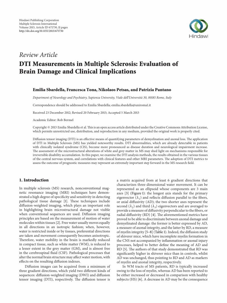
a matrix acquired from at least 6 gradient directions thatcharacterizes three-dimensional water movement. It can berepresented as an ellipsoid whose components are 3 mainaxes [3] (Figure 1): the longest axis stands for the primaryeigenvector (𝜆
1) and reflects diffusion parallel to the fibers,
or axial diffusivity (AD); the two shorter axes represent thesecond (𝜆
2) and third (𝜆
3) eigenvectors and are averaged to
provide ameasure of diffusivity perpendicular to the fibers, orradial diffusivity (RD) [4]. The aforementioned metrics haveproved to be able to discriminate between axonal damage anddemyelinated damage: the former is better expressed by AD,a measure of axonal integrity, and the latter by RD, a measureofmyelin integrity [5–8] (Table 1). Indeed, the diffusion studyof shiverer mice, which have incomplete myelin formation inthe CNS not accompanied by inflammation or axonal injuryprocesses, helped to better define the meaning of AD andRD [5]. The authors of that study demonstrated that RD wassignificantly higher in shiverer mice than in controls, whilstAD was unchanged, thus pointing to RD and AD as markersof myelin and axonal integrity, respectively.
In WM tracts of MS patients, RD is typically increasedowing to the loss of myelin, whereas AD has been reported tobe either increased or decreased in comparison with healthysubjects (HS) [6]. A decrease in AD may be the consequence

2 Multiple Sclerosis International
Table 1: Schematic description of the main DTI parameters.
Unit of measure Formula Object measured
FA Scalar value rangingbetween 0-1
√1
2
√(𝜆1− 𝜆2)2
+ (𝜆2− 𝜆3)2
+ (𝜆3− 𝜆1)2
√𝜆21+ 𝜆22+ 𝜆23
Fibers directionality/axonal loss
MD mm2/sec (𝜆1+ 𝜆2+ 𝜆3)/3 Amount of water diffusion/myelin loss
AD mm2/sec 𝜆1
Diffusivity parallel to the fibers/myelinand axonal content
RD mm2/sec (𝜆2+ 𝜆3)/2
Diffusivity perpendicular to thefibers/myelin content
FA: fractional anisotropy; MD: mean diffusivity; AD: axial diffusivity; RD: radial diffusivity; mm: millimeters; sec: second.
𝑥
𝑦
𝑧
𝜆1
𝜆2
𝜆3
Figure 1: Elliptical representation of a diffusion tensor with the 3main axes: the longest axis stands for the primary eigenvector (𝜆
1),
reflecting the diffusion parallel to the fibers; the two shorter axesrepresent the second (𝜆
2) and third (𝜆
3) eigenvectors, whose average
provides a measure of diffusivity perpendicular to the fibers.
of axonal loss, whereas an increase has been interpreted asan attempt made by a compensative mechanism to maintainfunctionality in the presence of WM damage [6].
TheDTImetrics usedmost, derived from amathematicalcombination of the three eigenvectors [7], are mean diffusiv-ity (MD), which measures overall water motion without anydirectionality, and fractional anisotropy (FA), which reflectsthe prevalence of diffusivity along one direction [8]. MD isa quantitative metric of water diffusion; the higher the MDvalue, the higher the diffusivity. FA is a scalar value rangingfrom 0 to 1 that is highest in compact WM tracts, decreasesin the GM, and approaches zero in the CSF [8] (Table 1).In early studies, anisotropy was correlated to axon densityand myelin content, while diffusivity was correlated aboveall to the amount of myelin [9]. However, both MD andFA have more recently been shown to be affected mainly bymyelin content [10] and, to a lesser extent, by axonal density[11]. Briefly, MD has so far been interpreted as an indexthat is primarily influenced by free space, which means that
processes such as vasogenic edema, axonal, and myelin lossincrease its value [12]. On the other hand, FA is believed tobe more sensitive to the detection of the integrity of WM, assignificant FA differences have been observed betweenmyeli-nated and nonmyelinated nerves [13–16]. Nonetheless, FA isnot very specific and does not distinguish between diseasescharacterized by a range of pathological processes, such asedema, inflammation, demyelination, and leukoaraiosis [17].
The idea of diffusion MRI was introduced in 1986 [2]and was subsequently applied to the study of a number ofneurological diseases, including MS [18]. Since it was firstapplied, methods of diffusion analyses have been steadilyimproved. Although region of interest (ROI) analysis wasadopted in early MS studies [12, 14, 18–21], this methodproved to have some drawbacks; that is, it is time-consuming,operator dependent, and subject to partial volume artifacts;furthermore, it does not provide a global assessment of tissuedamage [22]. Accordingly, histogram analyses were carriedout to evaluate the diffusion metrics in the whole brain[22–24]. Subsequently, a color was attributed to diffusiondirection along each of the three planes of space, thusproviding information on the direction of WM tracts [25].
Finally, in order to respond to a growing interest in theidentification of regional diffusion changes, a whole brainvoxel-based morphometry (VBM) approach [26, 27], usuallyapplied to T1-weighted images to study atrophy, was used fordiffusion images. However, owing to the limitations of thistechnique in terms of alignment inaccuracies and smoothingextent, VBM was partially superseded by tract-based spatialstatistics (TBSS), a fully automated, whole brain diffusionanalysis method [28].
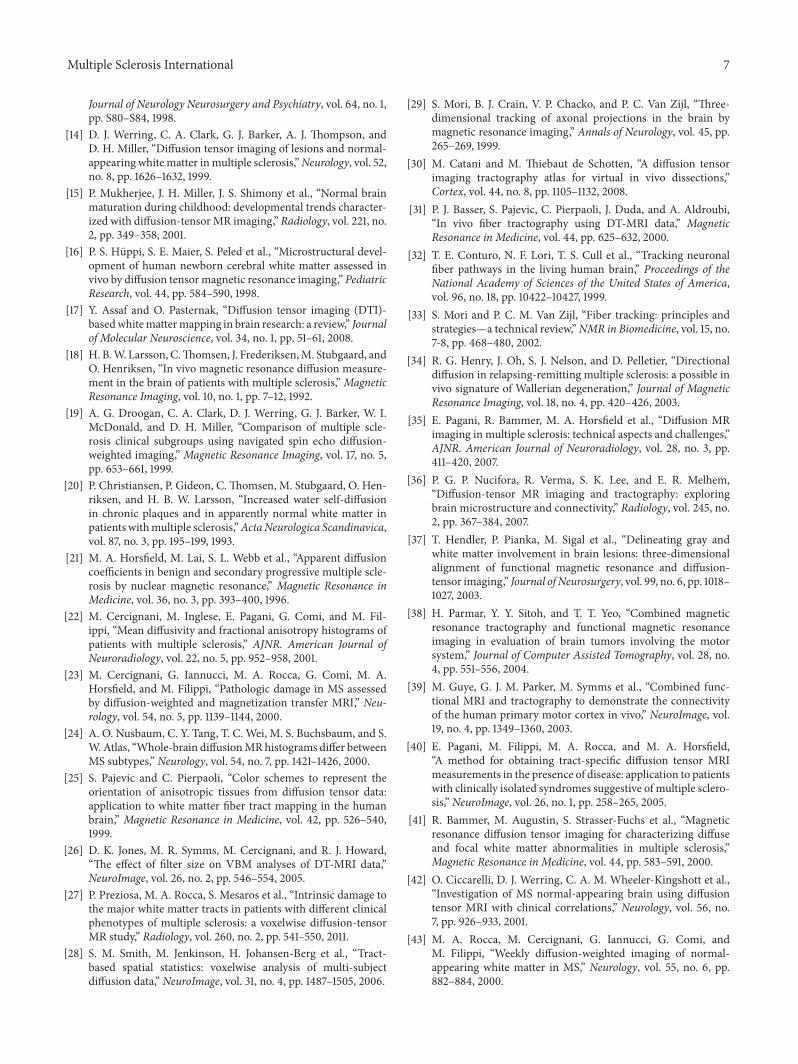
Another way of studying diffusion images is based on thereconstruction of large fiber bundles using three-dimensionaltractography [29–32] (Figure 2). This method delimits majortracts of WM in vivo: after the selection of one, or morethan one, seed ROI, nervous pathways are reconstructed bytracking along the principal direction of the fibers passingthrough the ROI [33]. This technique can be used to analyzethe displacement of fibers as well as to detect Walleriandegeneration [34]. It does, however, have some limitations,that is, difficulties to find the principal direction when thereare many crossing fibers, low signal-to-noise ratio, poorspatial resolution, nondetectable small fibers, and difficul-ties in case of distorted brain, all of which require an apriori knowledge of anatomical structures [35]. To partially

Multiple Sclerosis International 3
Figure 2: Three-dimensional tractography that reconstructs brain white matter bundles. The different colors represent different directionsof the fibers.
overcome these problems, the use of more sophisticatedmathematical models [36], constraints on the fiber tracking[37–39], and use of WM probability maps fromHS [40] havebeen proposed.
2. Diffusion Studies in Different MS Tissues
Since diffusion MRI was first introduced, many works haveapplied DWI, and particularly DTI, to study patients affectedby MS. Various methodological approaches of DTI analyseshave been used to characterize different types of tissuedamage. In this section, we describe the DTI results obtainedin different types of brain and spinal cord tissues.
2.1. Normal Appearing White Matter (NAWM). In MSpatients, widespread DTI abnormalities, consisting ofincreased MD and decreased FA, have been detected inNAWM [1, 12, 19, 23, 41–44]. A FA change gradient has alsobeen demonstrated, with lower values being observed closeto the plaques and higher values far from the plaques [44].Histological studies, which have detected demyelination andaxon transection even in WM outside the plaques [10, 45],suggest that DTI changes in NAWM may be ascribed toWallerian degeneration processes [46].
Abnormalities inNAWMhave been detected in almost allMS phenotypes, though the degree of damage varies accord-ing to the severity of the pathology. Indeed, although alteredNAWM is visible from the onset of clinical symptoms, evenat an early age, DTI-detectable damage becomes increasinglyevident as the disease worsens.
Disability is less pronounced and pathological tissuedamage is less severe in children withMS and in patients withclinically isolated syndrome (CIS) than in other phenotypes.In pediatric patients with CIS, at the very onset of disease, theNAWM diffusion metrics appear to be normal if comparedwith HS [47]. By contrast, increased RD and MD [27, 48, 49]and decreased FA and AD [50] values are found in mostWM tracts in pediatric patients with definite MS and in adultpatients with CIS compared with HS [47, 50–54]. Althoughagreement amongst researchers is not unanimous [27, 40, 55],these changes have been interpreted as a sign of early fiberloss in WM. Inconsistencies exist with regard to FA results,as some studies did not detect any changes. This may beexplained by the fact that axonal transaction may, despite
being present from disease onset [56], affect patients indifferent ways in the early stages and escape detection by DTIin the very early phases. By contrast, as the pathology pro-gresses, the structural damage becomes more pronounced.Indeed, changes in NAWM are less evident in CIS thanin patients with definite MS [27, 57]. Likewise, in patientswith benign MS, FA and AD have been shown to be higherthan those in relapsing remitting (RR) MS patients [27],although alterations in other DTI indexes, that is, MD andRD [58], have not been detected. This discrepancy points tothe existence of neuroprotective mechanisms that may pre-vent axonal injury [27, 59] and clinical worsening, despitethe inflammatory process. Moreover, DTI metrics haverevealed differences between benign and RRMS patients inthe topographical distribution of WM damage, which mightbe associated with the favorable clinical status in the formergroup [59].
Most [27], though not all [24, 60], studies, have detecteddifferences in NAWM diffusion metrics between RRMSpatients and HS. The lack of changes in RRMS observed insome studies is likely to be attributable either to the fact thatsubtle damage is not easily detected in the early phase of thedisease or to the poor sensitivity of the ROI methodologyadopted in those studies.
In comparison with CIS, RR, and benign MS, secondaryprogressive (SP) MS patients exhibit more pronounced WMdiffusion abnormalities [27, 61, 62]. The greater increase indiffusivity in SPMS than in other phenotypes may representa more advanced phase of the disease, presumably character-ized by a combination of axonal loss and tissue destructionprocesses with inflammatory events [62]. The high degreeof axonal degeneration in such patients is confirmed by thewidespread decrease in FA, not only within lesions but alsoin NAWM [27].
Although widespread diffusivity changes, consisting ofincreased MD, RD, and AD and decreased FA, have beendemonstrated in primary progressive (PP) MS if comparedwith HS [27, 63], NAWM is affected to a lesser degree inPPMS patients than in those with SPMS [27, 64], probablyowing to the more pronounced inflammation present in theSP than in the PP phenotype.
2.2. Normal Appearing GreyMatter (NAGM). Besides involv-ing the WM, MS also affects the GM, with microscopic

4 Multiple Sclerosis International
damage being detectable even in the absence of macroscopiclesions [65, 66]. Anatomical changes are usually visible in thedeep [67–69] and cortical GM [69, 70] in most MS pheno-types [64, 71–73]. However, some studies using ROI analysisdid not provide conclusive evidence regarding the presence ofNAGM abnormalities [60, 72], which suggests that differentanalysis methods might yield contrasting results.
The fact that no DTI changes have been detected in theNAGM of early onset patients [51] indicates that the GMmight be preserved at an early age. However, the patholog-ical involvement of NAGM was demonstrated when adultpatients with CIS were studied [54, 57], with increased MDand decreased FA being detected in such patients, though toa less severe degree than in RRMS patients [54, 57].
Adults with benign MS have also exhibited changes inDTI [19, 21, 74], consisting in particular of an increase in MD[75], which thus suggests that demyelination prevails overaxonal damage when the disease is clinically less severe.
Most recent works have detected abnormalities, consist-ing of both FA and MD [42, 76] changes, in the cortical orsubcortical NAGMof RRMSpatients comparedwith controls[27]. While MD usually increases, FA has been found toeither increase [42, 77] or decrease [76]. This discrepancymay be explained by the phase of GM inflammation at thetime of the analysis, with the prevailing activation in GM ofmicroglia or inflammatory processes leading, respectively, toan increased or decreased FA [78].Other authors have insteadreported no diffusion abnormalities in patients with earlyMSwhen compared with controls [1, 60, 77]. The reasons forthis discrepancy may be the use of ROIs, the improvementin processing steps, and the heterogeneity of the sample size.
SPMS patients display more pronounced GM diffusionabnormalities [27, 54, 64, 71], at both the deep and corticallevels [69], than other MS phenotypes, probably owing to theconcomitant presence of a high degree of inflammation anddegeneration in this group of patients.
2.3. Spinal Cord (SC). It has been demonstrated that NAWMand T2 lesions within the SC yield increased AD andRD and decreased FA if compared with normal tissues.These abnormalities have been correlated with the degree ofdemyelination [79], while FA has also been closely correlatedwith axonal density [9]. Given the high sensitivity displayedby RD to discriminate myelin content, a recent study pro-posed increased RD as a marker of increasing severity ofdemyelination [80].
When cervical cord was assessed in benign MS patients,few abnormalities, consisting of increasedMD, were detectedoutside focalmacroscopic lesions [81]. Similarly, patientswithearly onset MS exhibited a slight increase in the MD ofthe NAWM, which points to a mild degree of damage andexplains themore favorable clinical course in early onset thanin adult onset patients [82].
Conversely, reduced FA and increasedMDwere observedinNAWMand in surrounding demyelinating plaques [81, 83–86] in RRMS patients as well as in the NAWM of patientswithout any lesions within the whole SC [87]. This findingpoints to widespread pathological involvement of the spine,
regardless of the presence of T2 lesions. Subjects with aprogressive phenotype, that is, both PP and SP patients, alsodisplayed increased MD and decreased FA in the NAWM ofcervical SC [81, 88].
2.4. Lesion Tissue. FA within T2 lesions is usually decreasedandMD increased [12, 18–21, 23, 89]. An overlap betweenMDand FA maps and T2 lesion distribution has been demon-strated inmostMSphenotypes [27], with one exception beingPPMS patients, in whom a discrepancy between regionalWM diffusivity changes and T2-visible focal lesions has beenfound [69]. The absence of any overlap in PPMS between FAmaps and T2 lesions [27] as well as of any correlation betweendiffusion metrics and lesion volume [90] lends support tothe hypothesis that axonal damage and T2 lesions in thisphenotype are, unlike those in other phenotypes, partiallyindependent.
Pronounced FA and MD changes expressing both tis-sue damage and vasogenic edema have been detected ingadolinium-enhancing lesions (GEL) [24, 41, 89, 91, 92].However, FA is more sensitive to pathological damage thanMD [14, 62]. Indeed, in active lesions, FA values decreaseaccording to the severity of tissue disruption [41], whilst MDvalues may increase, decrease, or be similar to those detectedin chronic lesions [12, 19, 41, 89]. Acute demyelinating lesionsexhibiting restricted diffusion, which have been describedrecently, are thought to be caused by the presence of inflam-matory infiltrate or cytotoxic edema involving oligodendro-cytes [93].
T1 hypointense lesions (black holes) [94], characterizedby severe tissue injury, are associated with the most severediffusion alterations because of their pathological character-istics [1, 12, 19, 41, 62, 89].
Since lesions have also been detected in GM, diffusionparameters have been investigated in this structure as well[95]. FA in GM lesions has, unlike that inWM, been found toincrease.This finding may be due to the different histologicalcharacteristics of GM focal lesions, which present a higherlevel of activated microglia and greater loss of dendrites andaxons along with a lower degree of inflammation than WMlesions [78, 96, 97].
3. DTI Metrics and Clinical Disability
Nonconventional techniques may detect microstructuralchanges and correlate with the clinical impairment moreclosely than usual MRI measures (i.e., T2 lesions, GEL, andT1 hypointensities), thus partially overcoming the so-called“clinical-radiological paradox” [98].
Diffusion abnormalities are more pronounced in patientswith a long disease duration and severe neurological disability[54, 64, 71, 99], accurately reflecting the clinical condition.Attempts to correlate DTI metrics of the brain and SC withthe most widely used clinical disability scale, that is, theExpanded Disability Status Scale (EDSS) [100], have yieldedcontroversial results, with some studies finding a significantcorrelation [81, 101–104] not detected by others [6, 60]. Theweak correlation between the EDSS and diffusion parameters

Multiple Sclerosis International 5
may be due to several reasons: first of all, DTI measures,particularly when applied to NAWM, indicate pathologicallyaffected, though still functioning, fiber tracts [6]; secondly,the EDSS is a global clinical index that is affected above allby the motor system and might consequently not be severelyaltered in the early disease stage; thirdly, when diffusionchanges are evaluated in thewhole brain, a clinical correlationwith measures of disability is less likely to be found. Indeed,when diffusion was analyzed at the regional level, in specifictracts, more pronounced correlations with clinical featuresemerged; that is, the EDSS was correlated with DTI changesin motor tracts [105, 106] and with quantitative fiber tractog-raphy results [107].
Moreover, when correlations between regional DTI mea-sures and clinical scales assessing specific clinical featureswere investigated, the role of microstructural damage indisability became more evident [108, 109]. In particular,oculomotor function impairment was associated with a focalDTI alteration in small brainstem fiber pathways [3]. Inaddition, diffusion abnormalities in the optic nerves werealso significantly correlated with visual evoked potentialparameters, suggesting that DTI metrics may be used as asurrogatemeasure of axonal damage [110, 111]. Diffusionmea-sures in the optic nerve, optic radiation, and regional brainareas related to visual cortices were also associatedwith visualacuity [112, 113] and retinal nerve fiber layer thickness [113].
Regional diffusivity studies have also been used to findthe anatomical substrates underlying the impairment of otherfunctional systems. Tractography studies have demonstrateda correlation betweenMD and FA alterations in specificWMtracts, such as the corticospinal tract (CST) and the CorpusCallosum (CC), and motor disability [114–117]. Furthermore,cerebellarDTI abnormalities have been correlatedwith upperand lower limb disability [118]. Similarly, alteredDTI parame-ters along the cerebellar connections and supratentorial asso-ciativeWMbundleswere correlatedwith balance impairment[119].
DTI can also reveal tract injury responsible for cognitivedysfunction in MS patients [71, 72, 108, 120, 121]. Focalabnormalities, particularly in the CC, have been relatedto calculation, sequence learning, and memory [121–126].Moreover, several studies have detected correlations betweencognitive impairment and diffusion metrics abnormalitiesin the posterior thalamic radiations [121] as well as fronto-subcortical fiber tracts [127] and the thalamus [77].
FA abnormalities in specific NAWM tracts have evenbeen found to be significantly correlated with various cogni-tive abilities in pediatric patients in the early phase of disease[128].
Lastly, a strong correlation was found between DTImeasures in the SC and disability, thus corroborating the roleplayed by the pathological involvement of the spine in theclinical manifestations of the disease [86].
4. DTI Metrics and Nonconventional MRI
Diffusion studies have also been combined with other non-conventional MRI techniques, such as spectroscopic, magne-tization transfer ratio (MTR), and functional MRI.
When MRI spectroscopy was applied together with DTI,the results were inconsistent. One early work did not findany correlation between DTI metrics and N-acetyl aspartate(NAA) values [129], thus suggesting that chronic metabolicdysfunction contributes to axonal pathology in MS. Thisresult was not, however, confirmed in a more recent study,which reported correlations between diffusion metrics andNAA/Creatine ratios, thereby pointing to a link betweenmicrostructural andmetabolic alterations [130].This discrep-ancy is likely due to the fact that diffusionmeasures highlightchanges induced by structural axonal loss, whereas NAAchanges may be related to other, even transient, factors suchas the functionality of neurons [129].
A multiparametric study, based also on MTR MRI,detected metabolic and diffusivity changes not related toMTR measures in the CC of CIS patients [131]. The widelyreported lack of any correlation between diffusion metricsand MTR values [23, 44, 132, 133] suggests that these twomethods may be sensitive to different pathological processesand may provide independent measures of damage [132].
Lastly, DTI has been used to quantify brain damage infunctional MRI (fMRI) studies to investigate correlationsbetween structural damage and functional changes. Theresults of most task-related fMRI studies support the ideathat compensatory neuroplasticity is designed to maintainnormal function in the presence of widespread microstruc-tural damage. Indeed, altered DTI parameters have beenfound to correlate with an increase in fMRI activation duringvarious tasks in MS patients [134–136]. In addition, theultrastructural damage of specific WM tracts may affectcortical activity. For example, Lenzi et al. [136] found thatMD values in the body of CC correlated with activation ofthe ipsilateral motor cortex during handmovements, therebysuggesting that functional changes in this area are relatedto the loss of transcallosal inhibitory fibres in MS [136].More recent studies have correlated measures of anatomi-cal and functional connectivity: in MS, distinct functionalnetworks exhibit increases in functional connectivity despitewidespread diffusivity abnormalities within WM [137].
DTIwas used to investigate the relationship betweenWMdamage andGMatrophy inCIS.Henry et al. (2009)measureddiffusivity indexes in the thalamocortical tracts that connectWM lesions and the thalami. They found that both lesionsand DTI values in thalamocortical tracts correlated withatrophy, which points to a direct relationship between WMlesions and thalamic atrophy [49]. By contrast, DTI metricsalterations in GM were not related to concomitant brainatrophy progression [138, 139].
5. Future Directions
Given their marked sensitivity in detecting structural tissueabnormalities in MS, DTI metrics have been used to monitorstructural changes that occur during the course of thisdisease [73, 99, 140] in both WM and GM. The potential ofDTI parameters as prognostic markers of disease evolutionhas also been evaluated. Unfortunately, the results are notconclusive, probably because of differences in the methodsused to conduct these investigations.

6 Multiple Sclerosis International
Although abnormalities in RRMS patients were foundthroughout the brain, no longitudinal diffusion changes wereobserved in the follow-up [141], nor were any longitudinalFA changes observed in CIS patients when they were studiedagain after conversion to MS [139]. As these findings suggestthat WM damage occurs early but progresses slowly, theinformation yielded by a global diffusion assessment of nor-mal appearing brain tissuemay be of limited value as ameansof following disease progression, at least at disease onset.By contrast, serial diffusion MRI images in PPMS patientshave detected progressive NAWM changes, which proved tobe related to both lesion volume and the development ofclinical disability [142]. The role of DTI metrics as predictivemarkers has also been highlighted in other studies thathave evaluated various MS phenotypes. Although agreementon this question is not unanimous [48, 138], some authorshave demonstrated not only that altered regional NAWMmetrics are predictive of clinical impairment [46, 117], of thedevelopment of new T2 lesions and atrophy [43, 131], and ofthe future risk of MS development [143] but also that moresevere diffusivity brain abnormalities inWMandGMpredicthigher disability [78, 144]. DTI metrics also proved to bereliable as predictive markers of disease course when the SCwas evaluated in patients with cervical relapses. Indeed, alower RD in the lateral columns at baseline was associatedwith a better clinical outcome, with a greater decrease in RDbeing observed as patients improved clinically during follow-up [145].
Common guidelines are required for longitudinal DTIstudies to overcome the existing discrepancies in such studies.Further regional trials designed to detect more focused MRIabnormalities, which may be used to monitor structuralchanges over time and, consequently, to shed light on clinicalfeatures, are warranted. Indeed, in order to be consideredas an outcome measure in clinical trials, DTI parametersmust be sensitive to change over time and must be highlyreproducible and the sample size of the studied cohort mustbe appropriate to ensure the reliability of the results. Thesample size needs to be planned on the basis of the statisticalmethods to be adopted, of the precision of the informationrequired, and of the number of hypotheses to be tested, andin such a way as to take in account any missing values ordrop-out patients. One interesting study conducted a poweranalysis to calculate reasonable sample sizes for longitudinalDTI studies. In brief, the authors of that study found thatapproximately 40 participants per arm were required for 1- to2-year longitudinal DTI trials, though the number could varyaccording to the MS phenotypes and the anatomical regionbeing evaluated [146].
6. Conclusions
Diffusion abnormalities in theNAWMandNAGMhave beendemonstrated in all phenotypes of MS, with microstructuralchanges being detected in early disease stages, even in earlyonset MS. Nevertheless, diffusion metrics appear to differaccording to the MS phenotype and within different kinds oflesions. SinceMS phenotypes display different diffusivity pat-terns, which may be due to specific pathological substrates,
diffusion measures may represent useful markers of differ-ent MS subtypes. Moreover, when diffusion measures arecombined with other MRI findings, they may provide com-plementary information on different types of pathologicaldamage induced by MS. Lastly, given their high sensitivity indetecting structural tissue abnormalities, DTI measures havebeen proposed as prognosticmarkers of disease course and asa means of monitoring anatomical changes over time; furtherstudies are warranted in this field to achieve more consistentresults.
References
[1] M. Filippi, “Magnetic resonance imaging findings predictingsubsequent disease course in patients at presentation withclinically isolated syndromes suggestive of multiple sclerosis,”Neurological Sciences, vol. 22, no. 2, pp. S49–S51, 2001.
[2] D. Le Bihan, E. Breton, and D. Lallemand, “MR imaging ofintravoxel incoherent motions: application to diffusion andperfusion in neurologic disorders,” Radiology, vol. 161, no. 2, pp.401–407, 1986.
[3] R. J. Fox, “Picturing multiple sclerosis: conventional and diffu-sion tensor imaging,” Seminars in Neurology, vol. 28, no. 4, pp.453–466, 2008.
[4] P. J. Basser and C. Pierpaoli, “Microstructural and physiologicalfeatures of tissues elucidated by quantitative-diffusion-tensorMRI,” Journal of Magnetic Resonance. Series B, vol. 111, no. 3, pp.209–219, 1996.
[5] S. K. Song, S. W. Sun, M. J. Ramsbottom, C. Chang, J. Russell,and A. H. Cross, “Dysmyelination revealed through MRI asincreased radial (but unchanged axial) diffusion of water,”NeuroImage, vol. 17, no. 3, pp. 1429–1436, 2002.
[6] F. Fink, J. Klein,M. Lanz et al., “Comparison of diffusion tensor-based tractography and quantified brain atrophy for analyzingdemyelination and axonal loss inMS,” Journal of Neuroimaging,vol. 20, no. 4, pp. 334–344, 2010.
[7] D. Goldberg-Zimring, A. U. J. Mewes, M. Maddah, and S.K. Warfield, “Diffusion tensor magnetic resonance imaging inmultiple sclerosis,” Journal of Neuroimaging, vol. 15, no. 4, pp.68S–81S, 2005.
[8] C. Pierpaoli, P. Jezzard, P. J. Basser, A. Barnett, and G. Di Chiro,“Diffusion tensor MR imaging of the human brain,” Radiology,vol. 201, no. 3, pp. 637–648, 1996.
[9] J. P. Mottershead, K. Schmierer, M. Clemence et al., “High fieldMRI correlates ofmyelin content and axonal density inmultiplesclerosis: a post-mortem study of the spinal cord,” Journal ofNeurology, vol. 250, no. 11, pp. 1293–1301, 2003.
[10] J. Kolasinski, C. J. Stagg, S. A. Chance et al., “A combinedpost-mortem magnetic resonance imaging and quantitativehistological study of multiple sclerosis pathology,” Brain, vol.135, pp. 2938–2951, 2012.
[11] K. Schmierer, C. A. M. Wheeler-Kingshott, P. A. Boulby et al.,“Diffusion tensor imaging of post mortem multiple sclerosisbrain,” NeuroImage, vol. 35, no. 2, pp. 467–477, 2007.
[12] M. Filippi, G. Iannucci, M. Cercignani, M. A. Rocca, A. Pratesi,and G. Comi, “A quantitative study of water diffusion inmultiple sclerosis lesions and normal-appearing white matterusing echo-planar imaging,”Archives of Neurology, vol. 57, no. 7,pp. 1017–1021, 2000.
[13] M. A. Horsfield, H. B. W. Larsson, D. K. Jones, and A. Gass,“Diffusion magnetic resonance imaging in multiple sclerosis,”
Multiple Sclerosis International 7
Journal of Neurology Neurosurgery and Psychiatry, vol. 64, no. 1,pp. S80–S84, 1998.
[14] D. J. Werring, C. A. Clark, G. J. Barker, A. J. Thompson, andD. H. Miller, “Diffusion tensor imaging of lesions and normal-appearing whitematter inmultiple sclerosis,”Neurology, vol. 52,no. 8, pp. 1626–1632, 1999.
[15] P. Mukherjee, J. H. Miller, J. S. Shimony et al., “Normal brainmaturation during childhood: developmental trends character-ized with diffusion-tensorMR imaging,” Radiology, vol. 221, no.2, pp. 349–358, 2001.
[16] P. S. Huppi, S. E. Maier, S. Peled et al., “Microstructural devel-opment of human newborn cerebral white matter assessed invivo by diffusion tensormagnetic resonance imaging,” PediatricResearch, vol. 44, pp. 584–590, 1998.
[17] Y. Assaf and O. Pasternak, “Diffusion tensor imaging (DTI)-basedwhitemattermapping in brain research: a review,” Journalof Molecular Neuroscience, vol. 34, no. 1, pp. 51–61, 2008.
[18] H. B.W. Larsson, C.Thomsen, J. Frederiksen,M. Stubgaard, andO. Henriksen, “In vivo magnetic resonance diffusion measure-ment in the brain of patients with multiple sclerosis,”MagneticResonance Imaging, vol. 10, no. 1, pp. 7–12, 1992.
[19] A. G. Droogan, C. A. Clark, D. J. Werring, G. J. Barker, W. I.McDonald, and D. H. Miller, “Comparison of multiple scle-rosis clinical subgroups using navigated spin echo diffusion-weighted imaging,” Magnetic Resonance Imaging, vol. 17, no. 5,pp. 653–661, 1999.
[20] P. Christiansen, P. Gideon, C. Thomsen, M. Stubgaard, O. Hen-riksen, and H. B. W. Larsson, “Increased water self-diffusionin chronic plaques and in apparently normal white matter inpatients withmultiple sclerosis,”ActaNeurologica Scandinavica,vol. 87, no. 3, pp. 195–199, 1993.
[21] M. A. Horsfield, M. Lai, S. L. Webb et al., “Apparent diffusioncoefficients in benign and secondary progressive multiple scle-rosis by nuclear magnetic resonance,” Magnetic Resonance inMedicine, vol. 36, no. 3, pp. 393–400, 1996.
[22] M. Cercignani, M. Inglese, E. Pagani, G. Comi, and M. Fil-ippi, “Mean diffusivity and fractional anisotropy histograms ofpatients with multiple sclerosis,” AJNR. American Journal ofNeuroradiology, vol. 22, no. 5, pp. 952–958, 2001.
[23] M. Cercignani, G. Iannucci, M. A. Rocca, G. Comi, M. A.Horsfield, and M. Filippi, “Pathologic damage in MS assessedby diffusion-weighted and magnetization transfer MRI,” Neu-rology, vol. 54, no. 5, pp. 1139–1144, 2000.
[24] A. O. Nusbaum, C. Y. Tang, T. C. Wei, M. S. Buchsbaum, and S.W.Atlas, “Whole-brain diffusionMRhistograms differ betweenMS subtypes,” Neurology, vol. 54, no. 7, pp. 1421–1426, 2000.
[25] S. Pajevic and C. Pierpaoli, “Color schemes to represent theorientation of anisotropic tissues from diffusion tensor data:application to white matter fiber tract mapping in the humanbrain,” Magnetic Resonance in Medicine, vol. 42, pp. 526–540,1999.
[26] D. K. Jones, M. R. Symms, M. Cercignani, and R. J. Howard,“The effect of filter size on VBM analyses of DT-MRI data,”NeuroImage, vol. 26, no. 2, pp. 546–554, 2005.
[27] P. Preziosa, M. A. Rocca, S. Mesaros et al., “Intrinsic damage tothe major white matter tracts in patients with different clinicalphenotypes of multiple sclerosis: a voxelwise diffusion-tensorMR study,” Radiology, vol. 260, no. 2, pp. 541–550, 2011.
[28] S. M. Smith, M. Jenkinson, H. Johansen-Berg et al., “Tract-based spatial statistics: voxelwise analysis of multi-subjectdiffusion data,” NeuroImage, vol. 31, no. 4, pp. 1487–1505, 2006.
[29] S. Mori, B. J. Crain, V. P. Chacko, and P. C. Van Zijl, “Three-dimensional tracking of axonal projections in the brain bymagnetic resonance imaging,” Annals of Neurology, vol. 45, pp.265–269, 1999.
[30] M. Catani and M. Thiebaut de Schotten, “A diffusion tensorimaging tractography atlas for virtual in vivo dissections,”Cortex, vol. 44, no. 8, pp. 1105–1132, 2008.
[31] P. J. Basser, S. Pajevic, C. Pierpaoli, J. Duda, and A. Aldroubi,“In vivo fiber tractography using DT-MRI data,” MagneticResonance in Medicine, vol. 44, pp. 625–632, 2000.
[32] T. E. Conturo, N. F. Lori, T. S. Cull et al., “Tracking neuronalfiber pathways in the living human brain,” Proceedings of theNational Academy of Sciences of the United States of America,vol. 96, no. 18, pp. 10422–10427, 1999.
[33] S. Mori and P. C. M. Van Zijl, “Fiber tracking: principles andstrategies—a technical review,”NMR in Biomedicine, vol. 15, no.7-8, pp. 468–480, 2002.
[34] R. G. Henry, J. Oh, S. J. Nelson, and D. Pelletier, “Directionaldiffusion in relapsing-remitting multiple sclerosis: a possible invivo signature of Wallerian degeneration,” Journal of MagneticResonance Imaging, vol. 18, no. 4, pp. 420–426, 2003.
[35] E. Pagani, R. Bammer, M. A. Horsfield et al., “Diffusion MRimaging in multiple sclerosis: technical aspects and challenges,”AJNR. American Journal of Neuroradiology, vol. 28, no. 3, pp.411–420, 2007.
[36] P. G. P. Nucifora, R. Verma, S. K. Lee, and E. R. Melhem,“Diffusion-tensor MR imaging and tractography: exploringbrain microstructure and connectivity,” Radiology, vol. 245, no.2, pp. 367–384, 2007.
[37] T. Hendler, P. Pianka, M. Sigal et al., “Delineating gray andwhite matter involvement in brain lesions: three-dimensionalalignment of functional magnetic resonance and diffusion-tensor imaging,” Journal of Neurosurgery, vol. 99, no. 6, pp. 1018–1027, 2003.
[38] H. Parmar, Y. Y. Sitoh, and T. T. Yeo, “Combined magneticresonance tractography and functional magnetic resonanceimaging in evaluation of brain tumors involving the motorsystem,” Journal of Computer Assisted Tomography, vol. 28, no.4, pp. 551–556, 2004.
[39] M. Guye, G. J. M. Parker, M. Symms et al., “Combined func-tional MRI and tractography to demonstrate the connectivityof the human primary motor cortex in vivo,” NeuroImage, vol.19, no. 4, pp. 1349–1360, 2003.
[40] E. Pagani, M. Filippi, M. A. Rocca, and M. A. Horsfield,“A method for obtaining tract-specific diffusion tensor MRImeasurements in the presence of disease: application to patientswith clinically isolated syndromes suggestive of multiple sclero-sis,” NeuroImage, vol. 26, no. 1, pp. 258–265, 2005.
[41] R. Bammer, M. Augustin, S. Strasser-Fuchs et al., “Magneticresonance diffusion tensor imaging for characterizing diffuseand focal white matter abnormalities in multiple sclerosis,”Magnetic Resonance in Medicine, vol. 44, pp. 583–591, 2000.
[42] O. Ciccarelli, D. J. Werring, C. A. M. Wheeler-Kingshott et al.,“Investigation of MS normal-appearing brain using diffusiontensor MRI with clinical correlations,” Neurology, vol. 56, no.7, pp. 926–933, 2001.
[43] M. A. Rocca, M. Cercignani, G. Iannucci, G. Comi, andM. Filippi, “Weekly diffusion-weighted imaging of normal-appearing white matter in MS,” Neurology, vol. 55, no. 6, pp.882–884, 2000.

8 Multiple Sclerosis International
[44] A. C. Guo, V. L. Jewells, and J. M. Provenzale, “Analysis ofnormal-appearing white Matter in multiple sclerosis: compari-son of diffusion tensor MR imaging andmagnetization transferimaging,” AJNR. American Journal of Neuroradiology, vol. 22,no. 10, pp. 1893–1900, 2001.
[45] N. Evangelou, D. Konz,M.M. Esiri, S. Smith, J. Palace, and P.M.Matthews, “Regional axonal loss in the corpus callosum corre-lates with cerebral white matter lesion volume and distributionin multiple sclerosis,” Brain, vol. 123, no. 9, pp. 1845–1849, 2000.
[46] D. J. Werring, D. Brassat, A. G. Droogan et al., “The pathogen-esis of lesions and normal-appearing white matter changes inmultiple sclerosis. A serial diffusion MRI study,” Brain, vol. 123,no. 8, pp. 1667–1676, 2000.
[47] M. S.Vishwas, B. C.Healy, R. Pienaar,M. P.Gorman, P. E.Grant,and T. Chitnis, “Diffusion tensor analysis of pediatric multiplesclerosis and clinically isolated syndromes,” AJNR. AmericanJournal of Neuroradiology, vol. 34, no. 2, pp. 417–423, 2013.
[48] A. Gallo, M. Rovaris, R. Riva et al., “Diffusion-tensor magneticresonance imaging detects normal-appearing white matterdamage unrelated to short-term disease activity in patientsat the earliest clinical stage of multiple sclerosis,” Archives ofNeurology, vol. 62, no. 5, pp. 803–808, 2005.
[49] R. G. Henry, M. Shieh, B. Amirbekian, S. Chung, D. T. Okuda,and D. Pelletier, “Connecting white matter injury and thalamicatrophy in clinically isolated syndromes,” Journal of the Neuro-logical Sciences, vol. 282, no. 1-2, pp. 61–66, 2009.
[50] J. Tillema, J. Leach, and I. Pirko, “Non-lesional white mat-ter changes in pediatric multiple sclerosis and monophasicdemyelinating disorders,” Multiple Sclerosis, vol. 18, pp. 1754–1759, 2012.
[51] P. Tortorella, M. A. Rocca, D. M. Mezzapesa et al., “MRIquantification of gray and white matter damage in patients withearly-onsetmultiple sclerosis,” Journal of Neurology, vol. 253, no.7, pp. 903–907, 2006.
[52] M. S. Vishwas, T. Chitnis, R. Pienaar, B. C. Healy, and P. E.Grant, “Tract-based analysis of callosal, projection, and asso-ciation pathways in pediatric patients with multiple sclerosis: apreliminary study,” AJNR. American Journal of Neuroradiology,vol. 31, no. 1, pp. 121–128, 2010.
[53] E. Raz, M. Cercignani, E. Sbardella et al., “Clinically isolatedsyndrome suggestive of multiple sclerosis: voxelwise regionalinvestigation of white and gray matter,” Radiology, vol. 254, no.1, pp. 227–234, 2010.
[54] A. Pulizzi, M. Rovaris, E. Judica et al., “Determinants ofdisability in multiple sclerosis at various disease stages: a mul-tiparametric magnetic resonance study,” Archives of Neurology,vol. 64, no. 8, pp. 1163–1168, 2007.
[55] A. Bethune, V. Tipu, J. G. Sled et al., “Diffusion tensor imagingand cognitive speed in children with multiple sclerosis,” Journalof the Neurological Sciences, vol. 309, no. 1-2, pp. 68–74, 2011.
[56] T. Kuhlmann, G. Lingfeld, A. Bitsch, J. Schuchardt, and W.Bruck, “Acute axonal damage in multiple sclerosis is mostextensive in early disease stages and decreases over time,” Brain,vol. 125, no. 10, pp. 2202–2212, 2002.
[57] C. S. Yu, F. C. Lin, Y. Liu, Y. Duan, H. Lei, and K. C. Li,“Histogram analysis of diffusion measures in clinically isolatedsyndromes and relapsing-remitting multiple sclerosis,” Euro-pean Journal of Radiology, vol. 68, no. 2, pp. 328–334, 2008.
[58] B. Spano, M. Cercignani, B. Basile et al., “Multiparametric MRinvestigation of the motor pyramidal system in patients with?truly benign? multiple sclerosis,”Multiple Sclerosis, vol. 16, no.2, pp. 178–188, 2010.
[59] A. Ceccarelli, M. A. Rocca, E. Pagani et al., “The topographicaldistribution of tissue injury in benignMS: a 3TmultiparametricMRI study,” NeuroImage, vol. 39, no. 4, pp. 1499–1509, 2008.
[60] C. M. Griffin, D. T. Chard, O. Ciccarelli et al., “Diffusiontensor imaging in early relapsing—remittingmultiple sclerosis,”Multiple Sclerosis, vol. 7, no. 5, pp. 290–297, 2001.
[61] T. J. Braley, Y. H. Lee, S. Mohan, B. M. Segal, S. Berini, andA. Srinivasan, “Differences in diffusion tensor imaging-derivedmetrics in the corpus callosum of patients with multiple scle-rosis without and with gadolinium-enhancing cerebral lesions,”Journal of ComputerAssisted Tomography, vol. 36, no. 4, pp. 410–415, 2012.
[62] A. C. Scanderbeg, F. Tomaiuolo, U. Sabatini, U. Nocentini,M. G. Grasso, and C. Caltagirone, “Demyelinating plaques inrelapsing-remitting and secondary-progressive multiple sclero-sis: assessment with diffusion MR imaging,” AJNR. AmericanJournal of Neuroradiology, vol. 21, no. 5, pp. 862–868, 2000.
[63] P. E. Sijens, R. Irwan, J. H. Potze, J. P. Mostert, J. De Keyser,and M. Oudkerk, “Analysis of the human brain in primaryprogressive multiple sclerosis with mapping of the spatialdistributions using 1H MR spectroscopy and diffusion tensorimaging,” European Radiology, vol. 15, no. 8, pp. 1686–1693,2005.
[64] M. Rovaris, M. Bozzali, G. Iannucci et al., “Assessment ofnormal-appearing white and gray matter in patients with pri-mary progressivemultiple sclerosis: a diffusion-tensormagneticresonance imaging study,” Archives of Neurology, vol. 59, no. 9,pp. 1406–1412, 2002.
[65] A. Cifelli, M. Arridge, P. Jezzard,M.M. Esiri, J. Palace, and P.M.Matthews, “Thalamic neurodegeneration in multiple sclerosis,”Annals of Neurology, vol. 52, no. 5, pp. 650–653, 2002.
[66] A. Kutzelnigg and H. Lassmann, “Cortical lesions and brainatrophy inMS,” Journal of the Neurological Sciences, vol. 233, no.1-2, pp. 55–59, 2005.
[67] K. M. Hasan, C. Halphen, A. Kamali, F. M. Nelson, J. S.Wolinsky, and P. A. Narayana, “Caudate nuclei volume, diffu-sion tensor metrics, and T2 relaxation in healthy adults andrelapsing-remitting multiple sclerosis patients: implications forunderstanding gray matter degeneration,” Journal of MagneticResonance Imaging, vol. 29, no. 1, pp. 70–77, 2009.
[68] A. J. Fabiano, J. Sharma, B. Weinstock-Guttman et al., “Tha-lamic involvement in multiple sclerosis: a diffusion-weightedmagnetic resonance imaging study,” Journal of Neuroimaging,vol. 13, no. 4, pp. 307–314, 2003.
[69] A. Ceccarelli, M. A. Rocca, P. Valsasina et al., “A multiparamet-ric evaluation of regional brain damage in patients with primaryprogressive multiple sclerosis,” Human Brain Mapping, vol. 30,no. 9, pp. 3009–3019, 2009.
[70] F. Zhou, C. S. Zee, H. Gong, M. Shiroishi, and J. Li, “Differentialchanges in deep and cortical gray matters of patients withmultiple sclerosis: a quantitative magnetic resonance imagingstudy,” Journal of Computer Assisted Tomography, vol. 34, no. 3,pp. 431–436, 2010.
[71] M. Bozzali, M. Cercignani, M. P. Sormani, G. Comi, and M.Filippi, “Quantification of brain graymatter damage in differentMS phenotypes by use of diffusion tensor MR imaging,” AJNR.American Journal of Neuroradiology, vol. 23, no. 6, pp. 985–988,2002.
[72] H. Vrenken, P. J. W. Pouwels, J. J. G. Geurts et al., “Altered diffu-sion tensor in multiple sclerosis normal-appearing brain tissue:cortical diffusion changes seem related to clinical deterioration,”

Multiple Sclerosis International 9
Journal of Magnetic Resonance Imaging, vol. 23, no. 5, pp. 628–636, 2006.
[73] C. Oreja-Guevara, M. Rovaris, G. Iannucci et al., “Progressivegray matter damage in patients with relapsing-remitting multi-ple sclerosis: a longitudinal diffusion tensormagnetic resonanceimaging study,”Archives ofNeurology, vol. 62, no. 4, pp. 578–584,2005.
[74] M. Rovaris, G. Riccitelli, E. Judica et al., “Cognitive impairmentand structural brain damage in benign multiple sclerosis,”Neurology, vol. 71, no. 19, pp. 1521–1526, 2008.
[75] A. Ceccarelli, M. Filippi, M. Neema et al., “T2 hypointensity inthe deep gray matter of patients with benign multiple sclerosis,”Multiple Sclerosis, vol. 15, no. 6, pp. 678–686, 2009.
[76] A. Ceccarelli, M. A. Rocca, A. Falini et al., “Normal-appearingwhite and greymatter damage inMS: a volumetric and diffusiontensorMRI study at 3.0 Tesla,” Journal of Neurology, vol. 254, no.4, pp. 513–518, 2007.
[77] F. Tovar-Moll, I. E. Evangelou, A. W. Chiu et al., “Thalamicinvolvement and its impact on clinical disability in patients withmultiple sclerosis: a diffusion tensor imaging study at 3T,”AJNR.American Journal of Neuroradiology, vol. 30, no. 7, pp. 1380–1386, 2009.
[78] M. Calabrese, F. Rinaldi, D. Seppi et al., “Cortical diffusion-tensor imaging abnormalities in multiple sclerosis: a 3-yearlongitudinal study,” Radiology, vol. 261, no. 3, pp. 891–898, 2011.
[79] L. V. Zollinger, T. H. Kim, K. Hill, E. K. Jeong, and J. W.Rose, “Using diffusion tensor imaging and immunofluorescentassay to evaluate the pathology of multiple sclerosis,” Journal ofMagnetic Resonance Imaging, vol. 33, no. 3, pp. 557–564, 2011.
[80] E. C. Klawiter, R. E. Schmidt, K. Trinkaus et al., “Radialdiffusivity predicts demyelination in ex vivo multiple sclerosisspinal cords,” NeuroImage, vol. 55, no. 4, pp. 1454–1460, 2011.
[81] B. Benedetti, M. A. Rocca, M. Rovaris et al., “A diffusion tensorMRI study of cervical cord damage in benign and secondaryprogressive multiple sclerosis patients,” Journal of Neurology,Neurosurgery and Psychiatry, vol. 81, no. 1, pp. 26–30, 2010.
[82] D. M. Mezzapesa, M. A. Rocca, A. Falini et al., “A preliminarydiffusion tensor and magnetization transfer magnetic reso-nance imaging study of early-onset multiple sclerosis,” Archivesof Neurology, vol. 61, no. 3, pp. 366–368, 2004.
[83] L. C. H. Cruz Jr., R. C. Domingues, and E. L. Gasparetto,“Diffusion tensor imaging of the cervical spinal cord of patientswith relapsing-remising multiple sclerosis,” Arquivos de Neuro-Psiquiatria, vol. 67, no. 2 B, pp. 391–395, 2009.
[84] Y. Ohgiya, M. Oka, A. Hiwatashi et al., “Diffusion tensor MRimaging of the cervical spinal cord in patients with multiplesclerosis,” European Radiology, vol. 17, no. 10, pp. 2499–2504,2007.
[85] S. M. Hesseltine, M. Law, J. Babb et al., “Diffusion tensorimaging inmultiple sclerosis: assessment of regional differencesin the axial planewithin normal-appearing cervical spinal cord,”AJNR. American Journal of Neuroradiology, vol. 27, no. 6, pp.1189–1193, 2006.
[86] P. Valsasina, M. A. Rocca, F. Agosta et al., “Mean diffusivity andfractional anisotropy histogram analysis of the cervical cord inMS patients,” NeuroImage, vol. 26, no. 3, pp. 822–828, 2005.
[87] W. Van Hecke, G. Nagels, G. Emonds et al., “A diffusion tensorimaging group study of the spinal cord in multiple sclerosispatients with and without T2 spinal cord lesions,” Journal ofMagnetic Resonance Imaging, vol. 30, no. 1, pp. 25–34, 2009.
[88] F. Agosta, B. Benedetti, M. A. Rocca et al., “Quantificationof cervical cord pathology in primary progressive MS usingdiffusion tensor MRI,” Neurology, vol. 64, no. 4, pp. 631–635,2005.
[89] S. Roychowdhury, J. A. Maldjian, and R. I. Grossman, “Multiplesclerosis: comparison of trace apparent diffusion coefficientswith mr enhancement pattern of lesions,” AJNR. AmericanJournal of Neuroradiology, vol. 21, no. 5, pp. 869–874, 2000.
[90] M. A. Rocca, D. M. Mezzapesa, A. Falini et al., “Evidencefor axonal pathology and adaptive cortical reorganization inpatients at presentation with clinically isolated syndromessuggestive of multiple sclerosis,” NeuroImage, vol. 18, no. 4, pp.847–855, 2003.
[91] C. H. Sotak, “The role of diffusion tensor imaging in theevaluation of ischemic brain—a review,” NMR in Biomedicine,vol. 15, no. 7-8, pp. 561–569, 2002.
[92] F. Zelaya, N. Flood, J. B. Chalk et al., “An evaluation of the timedependence of the anisotropy of the water diffusion tensor inacute human ischemia,”Magnetic Resonance Imaging, vol. 17, no.3, pp. 331–348, 1999.
[93] K. E. Balashov and E. Lindzen, “Acute demyelinating lesionswith restricted diffusion inmultiple sclerosis,”Multiple Sclerosis,vol. 18, pp. 1745–1753, 2012.
[94] R. T. Naismith, J. Xu, N. T. Tutlam, K. Trinkaus, A. H. Cross,and S. K. Song, “Radial diffusivity in remote optic neuritisdiscriminates visual outcomes,” Neurology, vol. 74, no. 21, pp.1702–1710, 2010.
[95] D. Kidd, F. Barkhof, R. McConnell, P. R. Algra, I. V. Allen, andT. Revesz, “Cortical lesions inmultiple sclerosis,”Brain, vol. 122,no. 1, pp. 17–26, 1999.
[96] A. H. Poonawalla, K. M. Hasan, R. K. Gupta et al., “Diffusion-tensor MR imaging of cortical lesions in multiple sclerosis:initial findings,” Radiology, vol. 246, no. 3, pp. 880–886, 2008.
[97] B. D. Trapp, J. R. Wujek, G. A. Criste et al., “Evidence forsynaptic stripping by cortical microglia,” Glia, vol. 55, no. 4, pp.360–368, 2007.
[98] F. Barkhof, “The clinico-radiological paradox in multiple scle-rosis revisited,” Current Opinion in Neurology, vol. 15, no. 3, pp.239–245, 2002.
[99] M. Rovaris, A. Gallo, P. Valsasina et al., “Short-term accrual ofgraymatter pathology in patientswith progressivemultiple scle-rosis: an in vivo study using diffusion tensorMRI,”NeuroImage,vol. 24, no. 4, pp. 1139–1146, 2005.
[100] J. F. Kurtzke, “Rating neurologic impairment in multiple sclero-sis: an expanded disability status scale (EDSS),” Neurology, vol.33, no. 11, pp. 1444–1452, 1983.
[101] Y. Liu, Y. Duan, Y. He et al., “Whole brain white matter changesrevealed by multiple diffusion metrics in multiple sclerosis: aTBSS study,” European Journal of Radiology, vol. 81, no. 10, pp.2826–2832, 2012.
[102] A. Giorgio and N. De Stefano, “Cognition in multiple sclerosis:relevance of lesions, brain atrophy and proton MR spec-troscopy,” Neurological Sciences, vol. 31, no. 2, pp. S245–S248,2010.
[103] M. Onu, A. Roceanu, U. Sboto-Frankenstein et al., “Diffusionabnormality maps in demyelinating disease: correlations withclinical scores,” European Journal of Radiology, vol. 81, pp. e386–e391, 2012.
[104] O. Ciccarelli, C. A. Wheeler-Kingshott, M. A. McLean et al.,“Spinal cord spectroscopy and diffusion-based tractography toassess acute disability in multiple sclerosis,” Brain, vol. 130, no.8, pp. 2220–2231, 2007.

10 Multiple Sclerosis International
[105] D. S. Reich, S. A. Smith, K. M. Zackowski et al., “Multiparamet-ric magnetic resonance imaging analysis of the corticospinaltract in multiple sclerosis,” NeuroImage, vol. 38, no. 2, pp. 271–279, 2007.
[106] F. Lin, C. Yu, T. Jiang, K. Li, and P. Chan, “Diffusion tensortractography-based group mapping of the pyramidal tract inrelapsing-remitting multiple sclerosis patients,” AJNR. Ameri-can Journal of Neuroradiology, vol. 28, no. 2, pp. 278–282, 2007.
[107] B. Hu, B. Ye, Y. Yang et al., “Quantitative diffusion tensordeterministic and probabilistic fiber tractography in relapsing-remittingmultiple sclerosis,”European Journal of Radiology, vol.79, no. 1, pp. 101–107, 2011.
[108] M. Rovaris, G. Iannucci, M. Falautano et al., “Cognitive dys-function in patients with mildly disabling relapsing-remittingmultiple sclerosis: an exploratory study with diffusion tensorMR imaging,” Journal of the Neurological Sciences, vol. 195, no.2, pp. 103–109, 2002.
[109] J. Oh, R. G. Henry, C. Genain, S. J. Nelson, and D. Pelletier,“Mechanisms of normal appearing corpus callosum injuryrelated to pericallosal T1 lesions in multiple sclerosis usingdirectional diffusion tensor and 1H MRS imaging,” Journal ofNeurology, Neurosurgery and Psychiatry, vol. 75, no. 9, pp. 1281–1286, 2004.
[110] S. J. Hickman, C. A. M. Wheeler-Kingshott, S. J. Jones et al.,“Optic nerve diffusion measurement from diffusion-weightedimaging in optic neuritis,” AJNR. American Journal of Neurora-diology, vol. 26, no. 4, pp. 951–956, 2005.
[111] S. A. Trip, C. Wheeler-Kingshott, S. J. Jones et al., “Optic nervediffusion tensor imaging in optic neuritis,”NeuroImage, vol. 30,no. 2, pp. 498–505, 2006.
[112] S. C. Kolbe, M. Marriott, Av. Walt et al., “Diffusion tensorimaging correlates of visual impairment in multiple sclerosisand chronic optic neuritis,” Investigative Ophthalmology &Visual Science, vol. 53, no. 2, pp. 825–832, 2012.
[113] E.M. Frohman,M.G.Dwyer, T. Frohman et al., “Relationship ofoptic nerve and brain conventional and non-conventional MRImeasures and retinal nerve fiber layer thickness, as assessedby OCT and GDx: a pilot study,” Journal of the NeurologicalSciences, vol. 282, no. 1-2, pp. 96–105, 2009.
[114] M. Wilson, C. R. Tench, P. S. Morgan, and L. D. Blumhardt,“Pyramidal tract mapping by diffusion tensor magnetic res-onance imaging in multiple sclerosis: improving correlationswith disability,” Journal of Neurology Neurosurgery and Psychia-try, vol. 74, no. 2, pp. 203–207, 2003.
[115] X. Lin, C. R. Tench, P. S. Morgan, G. Niepel, and C. S.Constantinescu, “’Importance sampling’ inMS: use of diffusiontensor tractography to quantify pathology related to specificimpairment,” Journal of the Neurological Sciences, vol. 237, no.1-2, pp. 13–19, 2005.
[116] T. Sigal, M. Shmuel, D. Mark, H. Gil, and A. Anat, “Diffusiontensor imaging of corpus callosum integrity in multiple sclero-sis: correlation with disease variables,” Journal of Neuroimaging,vol. 22, pp. 33–37, 2012.
[117] K. C. Kern, J. Sarcona, M. Montag, B. S. Giesser, and N. L.Sicotte, “Corpus callosal diffusivity predicts motor impairmentin relapsing-remitting multiple sclerosis: a TBSS and tractogra-phy study,” NeuroImage, vol. 55, no. 3, pp. 1169–1177, 2011.
[118] V. M. Anderson, C. A. M. Wheeler-Kingshott, K. Abdel-Azizet al., “A comprehensive assessment of cerebellar damage inmultiple sclerosis using diffusion tractography and volumetricanalysis,”Multiple Sclerosis, vol. 17, no. 9, pp. 1079–1087, 2011.
[119] L. Prosperini, A. Kouleridou, N. Petsas et al., “The relationshipbetween infratentorial lesions, balance deficit and accidentalfalls in multiple sclerosis,” Journal of the Neurological Sciences,vol. 304, no. 1-2, pp. 55–60, 2011.
[120] R. A. Dineen, J. Vilisaar, J. Hlinka et al., “Disconnection asa mechanism for cognitive dysfunction in multiple sclerosis,”Brain, vol. 132, no. 1, pp. 239–249, 2009.
[121] H. J. Yu, C. Christodoulou, V. Bhise et al., “Multiple whitemattertract abnormalities underlie cognitive impairment in RRMS,”Neuroimage, vol. 59, no. 4, pp. 3713–3722, 2012.
[122] L. Bonzano, A. Tacchino, L. Roccatagliata, M. P. Sormani, G.L. Mancardi, and M. Bove, “Impairment in explicit visuomotorsequence learning is related to loss of microstructural integrityof the corpus callosum in multiple sclerosis patients withminimal disability,”NeuroImage, vol. 57, no. 2, pp. 495–501, 2011.
[123] S. Llufriu, Y. Blanco, E. Martinez-Heras et al., “Influence ofcorpus callosum damage on cognition and physical disabilityin multiple sclerosis: a multimodal study,” PLoS ONE, vol. 7, no.5, Article ID e37167, 2012.
[124] B. Audoin, M. Guye, F. Reuter et al., “Structure of WM bundlesconstituting the working memory system in early multiplesclerosis: a quantitative DTI tractography study,” NeuroImage,vol. 36, no. 4, pp. 1324–1330, 2007.
[125] S. D. Roosendaal, J. J. G. Geurts, H. Vrenken et al., “RegionalDTI differences in multiple sclerosis patients,”NeuroImage, vol.44, no. 4, pp. 1397–1403, 2009.
[126] X. Lin, C. R. Tench, P. S.Morgan, andC. S. Constantinescu, “Useof combined conventional and quantitative MRI to quantifypathology related to cognitive impairment inmultiple sclerosis,”Journal of Neurology, Neurosurgery and Psychiatry, vol. 79, no. 4,pp. 437–441, 2008.
[127] M.Roca, T. Torralva, F.Meli et al., “Cognitive deficits inmultiplesclerosis correlate with changes in fronto-subcortical tracts,”Multiple Sclerosis, vol. 14, no. 3, pp. 364–369, 2008.
[128] C. Till, A. Deotto, V. Tipu et al., “White matter integrity andmath performance in pediatric multiple sclerosis: a diffusiontensor imaging study,” Neuroreport, vol. 22, no. 18, pp. 1005–1009, 2011.
[129] S. Cader, H. Johansen-Berg, M. Wylezinska et al., “Discordantwhite matter N-acetylasparate and diffusion MRI measuressuggest that chronic metabolic dysfunction contributes toaxonal pathology inmultiple sclerosis,”NeuroImage, vol. 36, no.1, pp. 19–27, 2007.
[130] S. Hannoun, F. Durand-Dubief, C. Confavreux et al., “Diffusiontensor-MRI evidence for extra-axonal neuronal degeneration incaudate and thalamic nuclei of patients with multiple sclerosis,”AJNR. American Journal of Neuroradiology, vol. 33, no. 7, pp.1363–1368, 2012.
[131] J. P. Ranjeva, J. Pelletier, S. Confort-Gouny et al., “MRI/MRS ofcorpus callosum in patients with clinically isolated syndromesuggestive of multiple sclerosis,”Multiple Sclerosis, vol. 9, no. 6,pp. 554–565, 2003.
[132] M. C. G. Otaduy, D. Callegaro, L. A. Bacheschi, and C. C. Leite,“Correlation of magnetization transfer and diffusion magneticresonance imaging in multiple sclerosis,”Multiple Sclerosis, vol.12, no. 6, pp. 754–759, 2006.
[133] G. Iannucci, M. Rovaris, L. Giacomotti, G. Comi, and M.Filippi, “Correlation of multiple sclerosis measures derivedfrom T2-weighted, T1-weighted, magnetization transfer, anddiffusion tensor MR imaging,” AJNR. American Journal ofNeuroradiology, vol. 22, no. 8, pp. 1462–1467, 2001.

Multiple Sclerosis International 11
[134] M. A. Rocca, A. Falini, B. Colombo, G. Scotti, G. Comi, and M.Filippi, “Adaptive functional changes in the cerebral cortex ofpatients with nondisabling multiple sclerosis correlate with theextent of brain structural damage,” Annals of Neurology, vol. 51,no. 3, pp. 330–339, 2002.
[135] D. J. Werring, C. A. Clark, G. J. Parker, D. H. Miller, A.J. Thompson, and G. J. Barker, “A direct demonstration ofboth structure and function in the visual system: combiningdiffusion tensor imaging with functional magnetic resonanceimaging,” Neuroimage, vol. 9, no. 3, pp. 352–361, 1999.
[136] D. Lenzi, A. Conte, C. Mainero et al., “Effect of corpus callosumdamage on ipsilateral motor activation in patients withmultiplesclerosis: a functional and anatomical study,” Human BrainMapping, vol. 28, no. 7, pp. 636–644, 2007.
[137] D. J. Hawellek, J. F. Hipp, C. M. Lewis, M. Corbetta, and A. K.Engel, “Increased functional connectivity indicates the severityof cognitive impairment inmultiple sclerosis,”Proceedings of theNational Academy of Sciences of theUnited States of America, vol.108, pp. 19066–19071, 2011.
[138] M. Rovaris, E. Judica, A. Ceccarelli et al., “A 3-year diffusiontensorMRI study of greymatter damage progression during theearliest clinical stage of MS,” Journal of Neurology, vol. 255, no.8, pp. 1209–1214, 2008.
[139] E. Raz, M. Cercignani, E. Sbardella et al., “Gray- and white-matter changes 1 year after first clinical episode of multiplesclerosis: MR imaging,” Radiology, vol. 257, no. 2, pp. 448–454,2010.
[140] F. Agosta, M. Absinta, M. P. Sormani et al., “In vivo assessmentof cervical cord damage inMS patients: a longitudinal diffusiontensor MRI study,” Brain, vol. 130, no. 8, pp. 2211–2219, 2007.
[141] W. Rashid, A. Hadjiprocopis, G. Davies et al., “Longitudinalevaluation of clinically early relapsing-remitting multiple scle-rosis with diffusion tensor imaging,” Journal of Neurology, vol.255, no. 3, pp. 390–397, 2008.
[142] K. Schmierer, D. R. Altmann, N. Kassim et al., “Progres-sive change in primary progressive multiple sclerosis normal-appearing white matter: a serial diffusion magnetic resonanceimaging study,” Multiple Sclerosis, vol. 10, no. 2, pp. 182–187,2004.
[143] M. Bester, C. Heesen, S. Schippling et al., “Early anisotropychanges in the corpus callosum of patients with optic neuritis,”Neuroradiology, vol. 50, no. 7, pp. 549–557, 2008.
[144] M. Rovaris, E. Judica, A. Gallo et al., “Grey matter damagepredicts the evolution of primary progressive multiple sclerosisat 5 years,” Brain, vol. 129, no. 10, pp. 2628–2634, 2006.
[145] P. Freund, C. Wheeler-Kingshott, J. Jackson, D. Miller, A.Thompson, and O. Ciccarelli, “Recovery after spinal cordrelapse in multiple sclerosis is predicted by radial diffusivity,”Multiple Sclerosis, vol. 16, no. 10, pp. 1193–1202, 2010.
[146] D. M. Harrison, B. S. Caffo, N. Shiee et al., “Longitudinalchanges in diffusion tensor-based quantitative MRI in multiplesclerosis,” Neurology, vol. 76, no. 2, pp. 179–186, 2011.

Submit your manuscripts athttp://www.hindawi.com
Stem CellsInternational
Hindawi Publishing Corporationhttp://www.hindawi.com Volume 2014
Hindawi Publishing Corporationhttp://www.hindawi.com Volume 2014
MEDIATORSINFLAMMATION
of
Hindawi Publishing Corporationhttp://www.hindawi.com Volume 2014
Behavioural Neurology
EndocrinologyInternational Journal of
Hindawi Publishing Corporationhttp://www.hindawi.com Volume 2014
Hindawi Publishing Corporationhttp://www.hindawi.com Volume 2014
Disease Markers
Hindawi Publishing Corporationhttp://www.hindawi.com Volume 2014
BioMed Research International
OncologyJournal of
Hindawi Publishing Corporationhttp://www.hindawi.com Volume 2014
Hindawi Publishing Corporationhttp://www.hindawi.com Volume 2014
Oxidative Medicine and Cellular Longevity
Hindawi Publishing Corporationhttp://www.hindawi.com Volume 2014
PPAR Research
The Scientific World JournalHindawi Publishing Corporation http://www.hindawi.com Volume 2014
Immunology ResearchHindawi Publishing Corporationhttp://www.hindawi.com Volume 2014
Journal of
ObesityJournal of
Hindawi Publishing Corporationhttp://www.hindawi.com Volume 2014
Hindawi Publishing Corporationhttp://www.hindawi.com Volume 2014
Computational and Mathematical Methods in Medicine
OphthalmologyJournal of
Hindawi Publishing Corporationhttp://www.hindawi.com Volume 2014
Diabetes ResearchJournal of
Hindawi Publishing Corporationhttp://www.hindawi.com Volume 2014
Hindawi Publishing Corporationhttp://www.hindawi.com Volume 2014
Research and TreatmentAIDS
Hindawi Publishing Corporationhttp://www.hindawi.com Volume 2014
Gastroenterology Research and Practice
Hindawi Publishing Corporationhttp://www.hindawi.com Volume 2014
Parkinson’s Disease
Evidence-Based Complementary and Alternative Medicine
Volume 2014Hindawi Publishing Corporationhttp://www.hindawi.com