review article diagnosis & management of temporomandibular ... · the diagnostic process for...
TRANSCRIPT
Review Article
Dr.S.Venkateswaran, Dr.Saravanakumar, Dr.Annamalai, Dr.Divya, Dr.Yamini ABSTRACT
Introduction
Historically, dentistry has been geared primarily to the diagnosis and treatment of odontogenic pain (pulpal and periodontal). However dentists come across orofacial pain originating from various other structures such as sinuses, ear, muscles, bones, nerves(neuralgia), blood vessels(migraine headaches) and temporomandibular joint(TMJ), which may be referred to dental structures and vice versa. It can stem from simple causes to life-threatening diseases and it can cause great suffering for patients.
Apart from the odontogenic causes, temporomandibular joint disorders are one of the most common causes of orofacial pain1 .The temporomandibular joint has a close anatomical and functional association with the structures of the head, neck and face and any disturbance in the joints, can produce symptoms which may be referred to any of these structures. It can be from dental and non-dental source of origin including psycho-somatic disorders and therefore it is important that dentists, physicians, ophthalmologist, oto-laryngologist, surgeons, orthopedicians, psychologists and neurologists possess a fundamental knowledge of the TMJ disorders and consider it in the differential diagnosis of orofacial pain. .
Diagnosis & Management of Temporomandibular Joint Disorders-
What the Medical and Dental practitioners should know
Abstract
Temporomandibular disorders (TMD) are one of the most common causes of facial pain after
odontogenic origin. The temporomandibular disorders (TMD) are of multifactorial etiology and characterized by multiplicity of clinical signs and symptoms, making its diagnosis and management very difficult for the clinician. TMD should be considered in the differential diagnosis
of headache and orofacial pain in the absence of specific attributable organic cause. Scientific evidence shows that noninvasive methods are preferred in the management of TMD. These include occlusal, behavioral, physical and pharmacological treatment. Practitioners of medicine
and dentistry have the responsibility of diagnosing and managing people with TMD or refer them to an appropriate health care professional based on the nature and etiology of the problem.
Keywords – TMD, Temporomandibular disorders, headache, facial pain, diagnosis

The diagnostic process for temporomandibular disorders (TMD) is complicated by the multifactorial etiology and multiplicity of clinical signs and symptoms characterizing such disorders. However, proper diagnosis and management or referral of patients with these disorders as speedily as possible to the appropriate therapist is an important aspect of the quality of care provided by health care professionals.
The purpose of this article is to review the basic anatomy, function and disturbances of the temporomandibular joint, its diagnosis, differential diagnosis and clinical management.
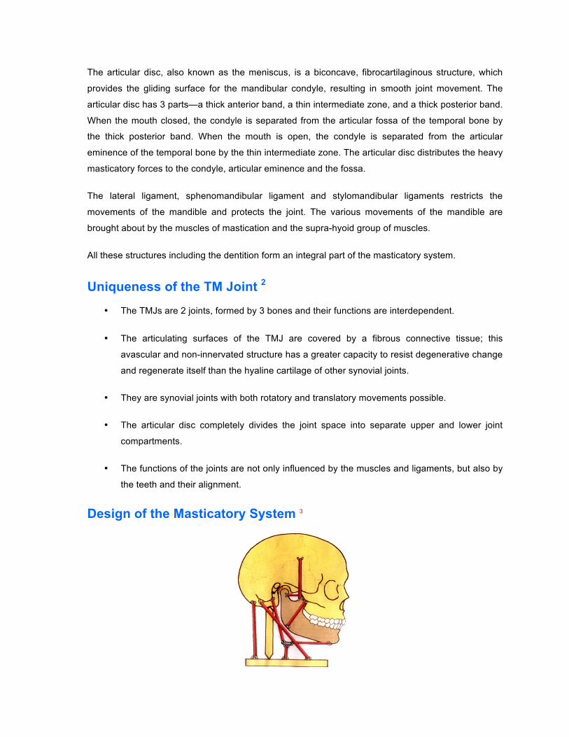
The Temporo-Mandibular Joint
The temporomandibular joint commonly called “TMJ” is an important joint that connects the mandible to the skull and regulates mandibular movement. It is one of the most complex joints in the body, performing multiple vital functions. It lets the mandible move up and down, side to side, forward and backward as a person does wondrous things as speaking, biting, chewing, swallowing, smiling, laughing, and frowning. It is a bicondylar joint in which the condyles, located at the two ends of the mandible, function at the same time.2 The joint Anatomy
The normal human skull possesses 2 temporo-mandibular joints (TMJs) that connect the skull to the
lower jaw bone (the mandible) so as to allow the mouth to open and close.
The TMJ is a gliding joint, formed by the condyle of the mandible and the squamous portion of the
temporal bone. The articular surface of the temporal bone consists of a convex articular eminence anteriorly and a concave articular fossa posterior. The articular surface of the mandible is formed
by the superior surface of the condyle. Articular surfaces of the mandible and temporal bone are
separated by an articular disc, which divides the joint cavity into 2 small spaces.
Articular Disc
The articular disc, also known as the meniscus, is a biconcave, fibrocartilaginous structure, which
provides the gliding surface for the mandibular condyle, resulting in smooth joint movement. The
articular disc has 3 parts—a thick anterior band, a thin intermediate zone, and a thick posterior band.
When the mouth closed, the condyle is separated from the articular fossa of the temporal bone by
the thick posterior band. When the mouth is open, the condyle is separated from the articular
eminence of the temporal bone by the thin intermediate zone. The articular disc distributes the heavy
masticatory forces to the condyle, articular eminence and the fossa.
The lateral ligament, sphenomandibular ligament and stylomandibular ligaments restricts the
movements of the mandible and protects the joint. The various movements of the mandible are
brought about by the muscles of mastication and the supra-hyoid group of muscles.
All these structures including the dentition form an integral part of the masticatory system.
Uniqueness of the TM Joint 2
• The TMJs are 2 joints, formed by 3 bones and their functions are interdependent.
• The articulating surfaces of the TMJ are covered by a fibrous connective tissue; this
avascular and non-innervated structure has a greater capacity to resist degenerative change
and regenerate itself than the hyaline cartilage of other synovial joints.
• They are synovial joints with both rotatory and translatory movements possible.
• The articular disc completely divides the joint space into separate upper and lower joint
compartments.
• The functions of the joints are not only influenced by the muscles and ligaments, but also by
the teeth and their alignment.
Design of the Masticatory System 3
The mandible is connected to the maxilla, cranium, clavicle, hyoid and spinal column through
trapezius, Sterno-cleido-mastoid, suprahyoid group of muscles including digastric muscle, muscles
of mastication and various ligaments. The design and functioning of the masticatory system is a
complex one and requires a balanced function of muscles, ligaments, bones and teeth. Each
movement is precisely co-ordinated, programmed and orchestrated.
Relationship to Adjacent Structures
All organs in the head-shoulder region are strongly networked via muscles and nerves by a complex
biological feed-back mechanism.
Organs in the head-shoulder region:
• Masticatory organ ( teeth, mandibular joints, chewing muscles)
• Organs of swallowing ( swallowing muscles)
• Alveolar organ, Periodontium (Mechano -sensors)
• Organ of speech ( speech muscles)
• Hearing organ ( ears)
• Organ of sight (eyes)
• Olfactory organ ( nose)
• Central nervous system ( brain, impulse network via sensors and nerves)
The temporo mandibular joint has a close anatomical and functional association with these
structures.
The Temporomandibular Joint Disorder In the past, many physicians addressed this condition as TMJ disease or TMJ syndrome. TMD was
also previously known under the eponymous title of Costen syndrome, after Dr. James Costen, an
oto-laryngologist, who elucidated many aspects of the syndrome as it relates to dental malocclusion.
Today, a much more comprehensive view of this condition exists, and the term temporomandibular
disorder (TMD) is the preferred term according to the American Academy of Orofacial Pain (AAOP)
which defines TMD as ” a collective term embracing a number of clinical problems that involve the
masticatory musculature, Temporomandibular joint and associated structures or both.”1
As a group these conditions have no common etiology or biological explanation and comprise of a
heterogeneous group of health problems whose signs and symptoms overlap.
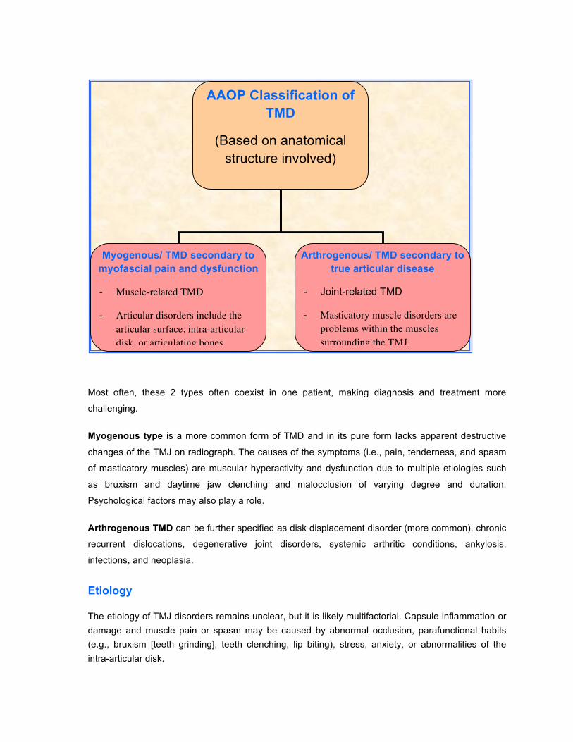
Most often, these 2 types often coexist in one patient, making diagnosis and treatment more
challenging.
Myogenous type is a more common form of TMD and in its pure form lacks apparent destructive
changes of the TMJ on radiograph. The causes of the symptoms (i.e., pain, tenderness, and spasm
of masticatory muscles) are muscular hyperactivity and dysfunction due to multiple etiologies such
as bruxism and daytime jaw clenching and malocclusion of varying degree and duration.
Psychological factors may also play a role.
Arthrogenous TMD can be further specified as disk displacement disorder (more common), chronic
recurrent dislocations, degenerative joint disorders, systemic arthritic conditions, ankylosis,
infections, and neoplasia.
Etiology
The etiology of TMJ disorders remains unclear, but it is likely multifactorial. Capsule inflammation or damage and muscle pain or spasm may be caused by abnormal occlusion, parafunctional habits (e.g., bruxism [teeth grinding], teeth clenching, lip biting), stress, anxiety, or abnormalities of the intra-articular disk.
AAOP Classification of TMD
(Based on anatomical structure involved)
Myogenous/ TMD secondary to myofascial pain and dysfunction
- Muscle-related TMD
- Articular disorders include the articular surface, intra-articular disk, or articulating bones.
Arthrogenous/ TMD secondary to true articular disease
- Joint-related TMD
- Masticatory muscle disorders are problems within the muscles surrounding the TMJ.
In recent years, many of the theories about the development of TMJ disorders have been questioned. Abnormal dental occlusion appears to be equally common in persons with and without TMJ symptoms and occlusal correction does not reliably improve the symptoms or signs of TMJ disorders. Parafunctional habits have been thought to cause TMJ microtrauma or masticatory muscle hyperactivity; however, these habits are also common in asymptomatic patients. Although parafunctional habits may play a role in initiating or perpetuating symptoms in some patients, the cause-and-effect relationship remains uncertain .4,5
There is some evidence to suggest that anxiety, stress, and other emotional disturbances may exacerbate TMJ disorders, especially in patients who experience chronic pain. As many as 75 percent of patients with TMJ disorders have a significant psychological abnormality. Recognition and treatment of concomitant mental illness is important in the overall approach to management of chronic pain, including pain caused by TMJ disorders.5
Genetic variations in the gene coding for catecholamine-O-methyltransferase (COMT), a gene that relates in to some aspects of pain sensitivity has also been implicated.6
The other classifications of Etiology of TMD are I. Dental and Non-Dental causes
II. Predisposing factors, Initiating factors, Perpetuating factors
Predisposing factors – factors that increase the risk of developing TMD. E.g.
• Systematic factors( degenerative, endocrine, infectious, metabolic, neoplastic,
neurological,vascular and rheumatological diseases),
• Psychological factors (Anxiety, depression)
• Structural factors (change in synovial fluid viscosity, increased intra-articular pressure)
• Genetic factors.
Initiating factors – factors that cause onset of the disorder. E.g. Trauma, parafunctional habits such
as bruxism, clenching
Perpetuating factors – factors that interfere with healing and complicate management of the
disorder. E.g. Mechanical and Muscular stress, metabolic problems.
Epidemiology 7 Sex Predilection
Temporomandibular disorder primarily affects women with a male-to-female ratio of 1:4. This could
possibly be due to the presence of estrogen receptors in the TM joint.
Age
Even though it is common in adults and adolescents, the highest incidence is among young adults;
especially women aged 20-40 years.
In epidemiologic studies, up to 75 percent of adults show at least one sign of joint dysfunction on
examination and as many as one third have at least one symptom. However, only 5 percent of adults
with TMJ symptoms require treatment and even fewer develop chronic or debilitating symptoms.
Symptoms of Temporo mandibular disorder 1,8,9
• Soreness of Muscles of mastication
• Attrition of teeth and sensitivity
• Hypermobility of teeth
• Pain in the joints
• Clicking in the joints
• Crepitation in the joints
• Difficulty in opening of mouth
• Open lock of the mandible
Apart from these symptoms, If a person suffers from any of the following symptoms without any
specific or organic cause, then the possibility of Tempo mandibular disorder could be considered.
• Headaches in the temple region
• Pain in the area of forehead and eyes
• Pain in the back of the head, possibly extending to the shoulders and neck
• Fullness in the ears
• Tinnitus
• Pressure on the eyes, sensitivity to light
• Dizzy spells, vertigo, nausea
• Lack of concentration

Temporomandibular disorders can cause these disturbances due to its anatomic proximity and
functional associations to these structures. The trigeminal nerve, which is one of the main nerve
supply to the TMJ and facial region, is a complex cranial nerve that can cause headaches when over
stimulated by the muscles surrounding it.
Diagnosis
Medical History
A comprehensive, chronological medical and dental history of the patient is essential for diagnosis of
the problem and to decide about further investigations and treatment plan.
Observation
• Forward head posture (this has been shown to displace the condyles posteriorly)
• Jaw malocclusion, abnormal dental wear, and poor dentition
• Visible clenching or spasm of the ipsilateral neck musculature
Physical Examination
Joint range of motion: Evaluation of jaw opening and closure as well as lateral deviation bilaterally. Normal range of motion for opening is 5 cm and lateral mandibular movement is 1 cm. Patients with TMD usually have reduced opening.
Palpation: The TMJ is best palpated laterally as a depression below the zygomatic arch and 1-2 cm anterior to the tragus. The posterior aspect of the joint is palpated through the external auditory canal. The joint should be palpated in both open and closed positions and also both laterally and posteriorly. While palpating, the examiner should feel for muscle spasm, muscle or joint tenderness, and joint sound. The muscles palpated as a part of complete TMJ examination are masseter, temporalis, medial pterygoid, lateral pterygoid, and sternocleidomastoid. In isolated myofascial pain and dysfunction, joint tenderness and joint click are usually absent.
Diagnostic Classification
An abbreviated version of the diagnostic classification system developed by the American Academy of Orofacial Pain is shown in Table 1 TMJ disorders are separated into two main categories based on the anatomic origin of the problem: articular disorders and masticatory muscle disorders. Accurate recognition of the origin of pain, either intra-articular or muscular, may help the dentst

recommend an initial therapy; however, it is not clear which noninvasive therapies work best and varies.
TABLE 1 Diagnostic Classification of TMJ Disorders 1
Articular disorders of the TMJ Masticatory muscle disorders
Ankylosis Local myalgia (unclassified) Congenital or developmental disorders Myofascial pain Aplasia, hyperplasia, or hypoplasia of the cranial bones or mandible
Myofibrotic contracture Neoplasia of the TMJ or associated structures Myositis Disk derangement disorders Myospasm Articular disk displacement with or without reduction Neoplasia
Fracture of the condylar process Inflammatory disorders like Synovitis, capsulitis, polyarthritides including the TMJ
Osteoarthritis TMJ dislocation
Differential Diagnosis 1
The differential diagnosis for orofacial pain is listed in Table 2. TMJ disorders can cause referred pain, particularly undifferentiated headache. Some studies have shown that as many as 55 percent of patients with chronic headache who were referred to a neurologist were found to have significant signs or symptoms of TMJ disorders. Educating patients on self-care techniques and referral for noninvasive treatment should be considered in patients with chronic undifferentiated headache or headache that is not responding to standard treatment.
TABLE 2 Differential Diagnosis of Orofacial Pain 1, 9
Condition Symptoms Signs
Dental pathology Tooth abscess Pain with chewing over affected
tooth Visible tooth decay; fluctuance along gum line;
pain with palpation over the tooth
Wisdom tooth eruption
Dull ache behind posterior molars
Tenderness to palpation over emerging tooth
Infection or inflammation
Herpes zoster and postherpetic neuralgia
Prodrome of pain followed by vesicular rash
Vesicular rash in dermatomal distribution, not crossing midline

Condition Symptoms Signs
Mastoiditis Fever; otalgia Postauricular erythema and swelling tenderness over mastoid process
Otitis externa Pruritus, pain, and tenderness of the external ear
Erythema and edema of external auditory canal
Otitis media Fever; malaise; otalgia Tympanic membrane dull, bulging, erythematous; loss of landmarks on tympanic membrane
Parotitis Fever; malaise; myalgia; pain over parotid gland
Tenderness and induration over parotid gland
Sialadenitis Pain and swelling of involved salivary gland
Tenderness, induration, and/or erythema of salivary gland; usually unilateral
Trigeminal neuralgia Paroxysmal, unilateral lancinating pains in trigeminal nerve distribution
Examination generally normal
Cluster Headache retro-orbital or sinus pain , Episode, last 6 to 8 weeks, long pain free
Rhinorrhea, nasal congestion and lacrimation from the involved side
Migraine Headache Unilateral dull, throbbing pain Unrelieved by a diagnostic block
Tumors Weight loss, History of cancer Unusual response to treatment, pain may become diffuse or paresthesia can occur
Diagnostic Testing
Diagnostic testing and radiologic imaging of the TMJ have uncertain usefulness and generally should only be used for the most severe or chronic symptoms. Local anesthetic nerve blocking can be helpful in differentiating whether orofacial pain originates from the TMJ capsule or from associated muscular structures. Sensory innervation of the TMJ is delivered primarily through the auriculotemporal branch of the third division of the trigeminal nerve. Patients who do not experience pain relief from diagnostic nerve blocking should be evaluated for other causes of orofacial pain..11
Laboratory Studies
If a systemic illness is suspected to be the cause of temporomandibular disorder (TMD), lab work is required. WBC count may be done if infection is suspected. Rheumatoid factor (RF), ESR, antinuclear antibody (ANA), and other specific antibodies should be checked if rheumatoid arthritis, temporal arteritis, or a connective tissue disorder is suspected. Uric acid should be checked for gout. Arthrocentesis may be required to demonstrate specific crystals.

Imaging Studies 12
Conventional radiography is the most utilized imaging study. It is simple, evaluates bony structures, and in most cases it is sufficient. It involves specific techniques and views such as modified Schuller views of each TMJ, both open mouth and closed mouth. Radiographic findings in TMJ depend on the etiology of TMD; in cases like rheumatoid arthritis, plain films show erosions, osteophytes, subchondral bony sclerosis, and condylar-glenoid fossa remodeling.
Dynamic high-resolution ultrasonography allows for visualization of the morphological elements and the functions of the TMJ, articular disk, mandibular condyle, and lateral pterygoid muscle.
CT scans can explore both bony structures and muscular soft tissues. Of interest, there is utility with cone beam computed tomography (CBCT). The patient is scanned with the mouth open and closed. Specifically, CBCT can aid in the diagnosis of osteoarthritis, rheumatoid arthritis, synovial chondromatosis, and neoplastic disorders.
Magnetic resonance image (MRI) should be used as the study of choice if an articular or meniscal pathology is suspected and an endoscopic or surgical procedure is contemplated, or in the case of traumatic TMD. erosion of right condyle and erosion of right condyle and Fig- CBCT and reconstructed image of condyles A a Shows normal condylar morphology B b shows erosion on condylar head Other Tests
Quantitative analysis of occlusal strain and stress can be helpful by using photoplastic phenomenon of some polymers. Results of strain analysis can help harmonize static and kinematic
A a b B

occlusal patterns by detecting and eliminating prematurities and interferences. Stress analysis helps to understand the temporomandibular mechanical relationship. Procedures
Diagnostic arthroscopy is an invasive diagnostic approach and should be used mainly in patients
suffering from internal TMJ derangements recalcitrant to conservative measures. MRI is suggested
to be obtained prior to arthroscopy.
Treatment 13,14,15
The signs and symptoms of TMD improve for most of the patients over time with or without treatment. As much as 50 percent of the patients improve from TMD symptoms in one year and almost 85 percent improve completely in three years. Interventions that change the anatomy of the joint, invade the integrity of the joint space, or manipulate the jaw have the potential to cause harm and have not been shown to improve symptoms. Therefore, self-care and noninvasive treatments are good options and should be attempted before invasive or permanent therapies, such as orthodontics or surgery, are recommended.
SELF-CARE
There is little evidence to suggest that any TMJ disorder treatment modality is superior to any other, although it is generally accepted that self-care and behavioral interventions should be encouraged for all patients, regardless of which therapies are considered. Providing a few simple exercises, behavioral instructions, and reassurance are important steps when treating the average patient with new or intermittent symptoms.
NONINVASIVE THERAPY 15,16
Many noninvasive therapies are commonly used for the treatment of TMJ disorders. The disciplines of medicine, dentistry (occlusal splint therapy, replacement of missing teeth, correcting malocclusion), physical therapy, and psychology can provide effective treatment in different clinical situations.
Pharmacotherapy
The indicated classes of pharmacologic agents include analgesics, anti-inflammatory agents,
corticosteroids, anxiolytics, muscle relaxants and antidepressants. Non-opiate analgesics are
effective for mild to moderate acute pain associated with TMD, and opioid narcotics are considered
for short-term use in only controlling acute severe pain. Additionally, tricyclic antidepressants appear
to be effective in the control of chronic orofacial pain of non-inflammatory origin, independent of their
effects on mood, with daily doses smaller than those typically used in the treatment of depression.17
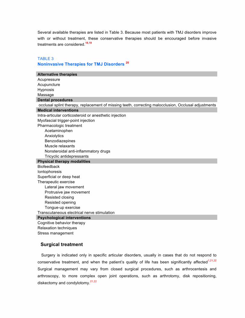
Several available therapies are listed in Table 3. Because most patients with TMJ disorders improve with or without treatment, these conservative therapies should be encouraged before invasive treatments are considered.18,19
TABLE 3 Noninvasive Therapies for TMJ Disorders 20
Alternative therapies Acupressure Acupuncture Hypnosis Massage Dental procedures occlusal splint therapy, replacement of missing teeth, correcting malocclusion, Occlusal adjustments Medical interventions Intra-articular corticosteroid or anesthetic injection Myofascial trigger-point injection Pharmacologic treatment Acetaminophen Anxiolytics Benzodiazepines Muscle relaxants Nonsteroidal anti-inflammatory drugs Tricyclic antidepressants Physical therapy modalities Biofeedback Iontophoresis Superficial or deep heat Therapeutic exercise Lateral jaw movement Protrusive jaw movement Resisted closing Resisted opening Tongue-up exercise Transcutaneous electrical nerve stimulation Psychological interventions Cognitive behavior therapy Relaxation techniques Stress management
Surgical treatment
Surgery is indicated only in specific articular disorders, usually in cases that do not respond to
conservative treatment, and when the patient’s quality of life has been significantly affected1,21,22
Surgical management may vary from closed surgical procedures, such as arthrocentesis and
arthroscopy, to more complex open joint operations, such as arthrotomy, disk repositioning,
diskectomy and condylotomy.21,22

Conclusion
Medical and dental practitioners should consider temporomandibular Joint disorders as a possible cause in the diagnosis of oro-facial pain including headaches, shoulder and neck pain, vertigo and associated pain, blurring of vision, disorders of hearing, nausea, vomiting, and disturbances in concentration, in the absence of any specific, attributable or organic cause.
While orofacial pain and headache secondary to jaw muscle function and dental structures ideally be managed by dentists, pain in the head and neck region unrelated to it should be should be referred to an appropriate medical care specialists for management.
References
1. Okeson JP, for the American Academy of Orofacial Pain. Orofacial Pain: Guidelines for Assessment, Diagnosis, and Management. Chicago, Ill.: Quintessence Pub, 1996.
2. Pertes RA, Gross SG. Functional anatomy and biomechanics of the temporomandibular joint. In: Pertes RA, Gross SG. Clinical Management of Temporomandibular Disorders and Orofacial Pain. Chicago, Ill.: Quintessence Pub, 1995:1–12.
3. Dawson PE., Functional occlusion-From TMJ to smile design: Mosby pub
4. Wadhwa L, Utreja A, Tewari A. A study of clinical signs and symptoms of temporomandibular dysfunction in subjects with normal occlusion, untreated, and treated malocclusions. Am J Orthod Dentofacial Orthop. 1993; 103:54-61.
5. McNamara JA Jr, Seligman DA, Okeson JP. Occlusion, orthodontic treatment, and temporomandibular disorders: a review. J Orofac Pain. 1995;9:73–90
6. Diatchenko, L., Slade, G.D., Nackley, A.G. et al. (2005) Genetic basis for individual variations in pain perception and the development of a chronic pain condition.. Human Molecular Genetics 14(1), 135-143
7. Dworkin SF, Huggins KH, LeResche L, Von Korff M, Howard J, Truelove E, et al. Epidemiology of signs and symptoms in temporomandibular disorders: clinical signs in cases and controls. J Am Dent Assoc. 1990; 120:273–81.
8. Hentschel K, Capobianco DJ, Dodick DW. Facial pain. Neurologist. 2005;11:244–9
9. Pertes RA, Bailey DR. General concepts of diagnosis and treatment. In: Pertes RA, Gross SG. Clinical Management of Temporomandibular Disorders and Orofacial Pain. Chicago, Ill.: Quintessence Pub, 1995:59–68
10. De Boever, J.A., Nilner, M., Orthlieb, J-D. and Steenks, M.H. (2007) Recommendations for examination, diagnosis, management of patients with temporomandibular disorders and orofacial pain by the general dental practitioner. European Academy of Craniomandibular Disorders.

11. DuPont JS Jr. Simplified anesthesia blocking of the temporomandibular joint. Gen Dent. 2004;52:318–20
12. American Academy of Oral and Maxillofacial Radiology. Radiographic imaging of the temporomandibular joint. Imaging of the TMJ, Oral Surg Oral Med Oral Pathol Oral Radiol Endod 83:609-18, (1997)
13. Mannheimer JS. Overview of physical therapy modalities and procedures. In: Pertes RA, Gross SG. Clinical Management of Temporomandibular Disorders and Orofacial Pain. Chicago, Ill.: Quintessence Pub, 1995:227–44
14. American Society of Temporomandibular Joint Surgeons. Guidelines for diagnosis and management of disorders involving the temporomandibular joint and related musculoskeletal structures. Cranio. 2003;21:68–76.
15. Al-Ani, M.Z., Davies, S.J., Gray, R.J.M. et al. (2004)Stabilisation splint therapy for temporomandibular pain dysfunction syndrome (Cochrane Review). Issue 1. John Wiley & Sons, Ltd.
16. Wassell RW, Adams N, Kelly PJ. The treatment of temporomandibular disorders with stabilizing splints in general dental practice: one-year follow-up. J Am Dent Assoc. 2006;137:1089–98.
17. Pettengill CA & Reisner-Keller L; The use of tricyclic antidepressants for the control of chronic orofacial pain. J Craniomand Pract 1997;15: 53-56.
18. Conti PC, dos Santos CN, Kogawa EM, de Castro Ferreira Conti AC, de Araujo Cdos R. The treatment of painful temporomandibular joint clicking with oral splints: a randomized clinical trial. J Am Dent Assoc. 2006;137:1108–14.
19. Truelove E, Huggins KH, Mancl L, Dworkin SF. The efficacy of traditional, low-cost and nonsplint therapies for temporomandibular disorder: a randomized controlled trial. J Am Dent Assoc. 2006;137:1099–107.
20. Shankland WE II. Temporomandibular disorders: standard treatment options. Gen Dent. 2004;52:349–55
21. Dolwick MF;The role of temporomandibular joint surgery in the treatment of patients with internal derangement. Oral Surg Oral Med Oral Pathol Oral Radiol Endod 1997;vol 83: 150-155.
22. McNeill C; Management of temporomandibular disorders: concept and controversies. J Prosth Dent 1997; vol 77: 510-522