review article compte rendu - publishpublish.uwo.ca/~dbetts/dean_h._betts/publications_files/koch et...
TRANSCRIPT

CVJ / VOL 50 / FEBRUARY 2009 155
Review Article Compte rendu
Current and future regenerative medicine — Principles, concepts, and therapeutic use of stem cell therapy and tissue engineering in equine medicine
Thomas G. Koch, Lise C. Berg, Dean H. Betts
Abstract — This paper provides a bird’s-eye perspective of the general principles of stem-cell therapy and tissue engineering; it relates comparative knowledge in this area to the current and future status of equine regenerative medicine.
The understanding of equine stem cell biology, biofactors, and scaffolds, and their potential therapeutic use in horses are rudimentary at present. Mesenchymal stem cell isolation has been proclaimed from several equine tissues in the past few years. Based on the criteria of the International Society for Cellular Therapy, most of these cells are more correctly referred to as multipotent mesenchymal stromal cells, unless there is proof that they exhibit the fundamental in vivo characteristics of pluripotency and the ability to self-renew. That said, these cells from various tissues hold great promise for therapeutic use in horses. The 3 components of tissue engineering — cells, biological factors, and biomaterials — are increasingly being applied in equine medicine, fuelled by better scaffolds and increased understanding of individual biofactors and cell sources.
The effectiveness of stem cell-based therapies and most tissue engineering concepts has not been demonstrated sufficiently in controlled clinical trials in equine patients to be regarded as evidence-based medicine. In the mean-time, the medical mantra “do no harm” should prevail, and the application of stem cell-based therapies in the horse should be done critically and cautiously, and treatment outcomes (good and bad) should be recorded and reported.
Stem cell and tissue engineering research in the horse has exciting comparative and equine specific perspectives that most likely will benefit the health of horses and humans. Controlled, well-designed studies are needed to move this new equine research field forward.
Résumé — Médecine régénérative actuelle et future — Principes, concepts et usage thérapeutique de la thérapie des cellules souches et de l’ingénierie tissulaire en médecine équine. Cet article fournit un survol des principes généraux de la thérapie des cellules souches et de l’ingénierie tissulaire; il établit un lien entre les connaissances comparées dans ce domaine et la situation actuelle et future de la médecine régénérative équine.
La compréhension de la biologie des cellules souches équines, des biofacteurs et des échafauds ainsi que de leur usage thérapeutique potentiel chez les chevaux sont rudimentaires à l’heure actuelle. L’isolement de la cellule souche mysenchymateuse a été proclamée pour plusieurs tissus équins au cours des dernières années. En se fondant sur les critères de l’International Society for Cellular Therapy, la plupart de ces cellules sont correctement appelées des cellules stromales mesenchymateuses à moins qu’il n’y ait preuve qu’elles présentent les caractéristiques fondamen-tales in vivo de la pluripotence et la capacité de s’auto-renouveler. Cela dit, ces cellules provenant de divers tissus s’annoncent très prometteuses pour l’usage thérapeutique chez les chevaux. L’application des 3 composantes de l’ingénierie tissulaire — les cellules, les facteurs biologiques et les biomatériaux — est de plus en plus utilisée en
Department of Biomedical Sciences, University of Guelph, Guelph, Ontario N1G 2W1 (Koch, Betts); Department of Basic Animal and Veterinary Sciences, Faculty of Life Sciences, University of Copenhagen, Grønnegårdsvej 7, 1870 Frederiksberg C, Denmark (Koch, Berg).Address all correspondence to Dr. Thomas G. Koch; e-mail: [email protected]. Koch completed a PhD program at the University of Guelph, funded by an international grant from the Danish Research Council. Additional operating funds are provided by the Equine Guelph Research Fund and its partners; E.P. Taylor Equine Research Fund, The Horsemen’s Benevolent and Protective Association of Ontario, Ontario Equestrian Federation, Ontario Harness Horse Association, Ontario Ministry of Agriculture and Food, Ontario Racing Commision, Ontario Veterinary College, University of Guelph.
156 CVJ / VOL 50 / FEBRUARY 2009
CO
MP
TE
RE
ND
U
IntroductionThe goal of this review is to provide practitioners, scientists, and other stakeholders in the equine industry with the latest knowledge of regenerative medicine and tissue engineering prin-ciples as a basis for their critical evaluation of current and future cell-based therapeutic modalities in the horse. Equine regenera-tive medicine is an exciting, but relatively new, research field. Current experimental and clinical use of equine stromal cells, scaffolds, and biofactor-based therapies are reviewed; references and examples related to human and laboratory animal models are included in an effort to elucidate comparative aspects and contrasts to equine cell-based therapies and tissue engineering.
What are stem cells?Stem cells are characterized by their ability to self-renew and to differentiate into multiple different cell types and tissues (1–3). Stem cells are generally considered as being embryonic or nonembryonic in origin. In this article, the use of nonem-bryonic equine stem cells mainly is reviewed, but references are made to human embryonic stem cell research in cases of principal interest. Stem cell concepts in general and in relation to equine regenerative medicine have been reviewed previously by the authors (4).
The nomenclature in the stem cell field is not consistent. The International Society for Cellular Therapy has recently reviewed the general term “mesenchymal stem cells” and has introduced the term “multipotent mesenchymal stromal cells” (5,6). Human multipotent mesenchymal stromal cells are plastic-adherent cells that express the surface markers CD73, CD90, and CD105; do not express CD45, CD34, CD14 [CD11b], CD79 [CD19], and HLA-DR; and have tri-lineage differentiation potential towards osteoblasts, adipocytes, and chondroblasts. The term mesenchymal stem cell should be reserved for cells that have shown in vivo long-term survival, with self-renewal capacity and tissue repopulation with multilineage differentiation (6). The purpose of redefining the mesenchymal nomenclature is primarily to enable more precise description of cell popula-tions, thereby allowing for a more valid comparison of research results from different investigators. Secondly, the term “stem” in mesenchymal stem cells might infer more biological and functional properties than the cells actually possess, which might
lead to unrealistic expectation of these cells, especially in the lay literature. The acronym MSC is therefore now nonspecific and used for both mesenchymal stem cells and multipotent mesen-chymal stromal cells. Unfortunately, the distinction between these 2 mesenchymal cell populations has not been universally accepted; therefore, in this review, the term mesenchymal stem cells has been maintained as reported by the investigators, although many of the studies were probably conducted on multipotent mesenchymal stromal cells.
Widespread clinical use of human stem cells is largely restricted to the use of hematopoietic stem cells derived from adult peripheral blood, adult bone marrow, or umbilical cord blood. These hematopoietic stem cells are used in either an autologous or an allogenic fashion for the treatment of leuke-mias, lymphomas, solid tumors, and nonmalignant disorders (7). For example, in patients suffering from leukemia, where the cancerous cells of the bone marrow are destroyed by radiation, chemotherapy, or both, leaving the patient severely immune-compromised and without the ability to produce white and red blood cells, hematopoietic stem cells are injected IV and “home” to the bone marrow, where they “engraft” and repopulate the marrow cavity in a functional manner, allowing the patient to produce noncancerous white and red blood cells. Human multipotent mesenchymal stromal cells or mesenchymal stem cells (MSCs) have not yet reached mainstream clinical practice, but the literature is full of reports on in vitro work and work in animal models in which these cells are used for cell-based therapies and as part of solid tissue reconstruction. Cell-based therapies, using MSCs, are increasingly being reported in equine medicine.
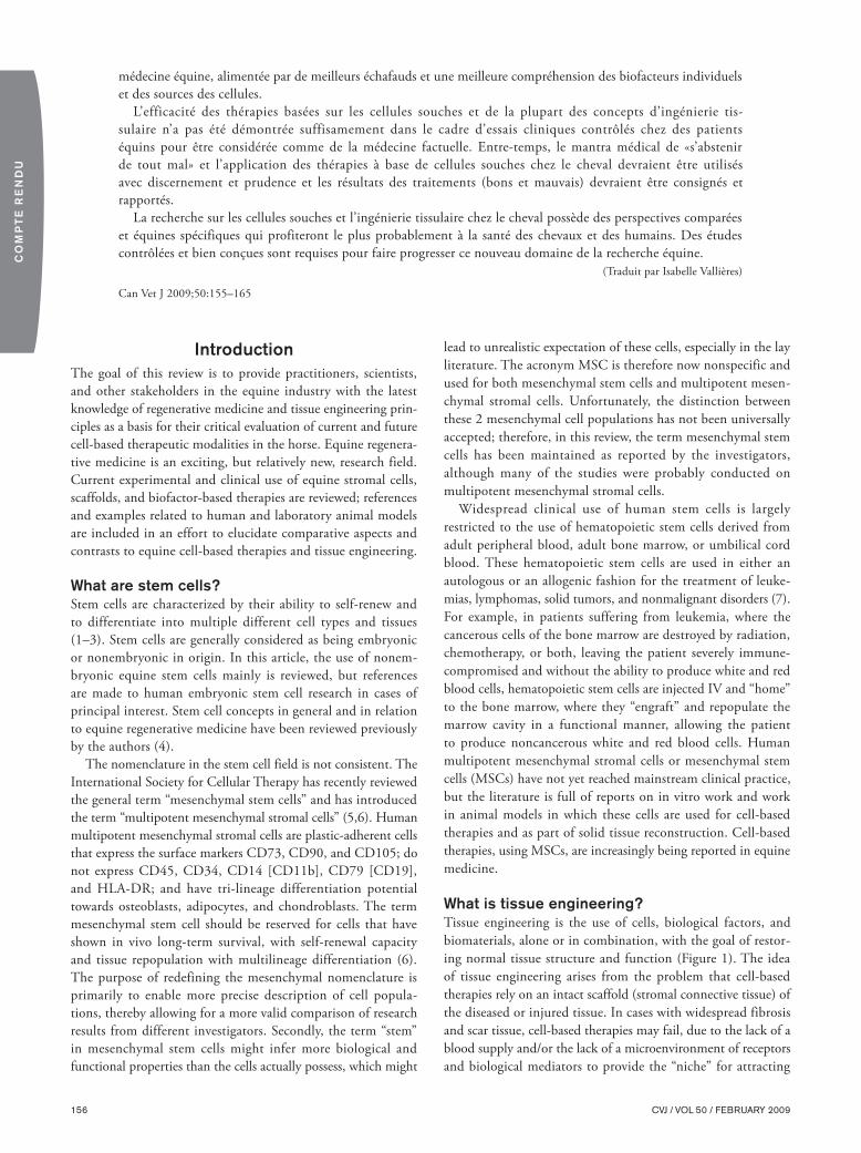
What is tissue engineering?Tissue engineering is the use of cells, biological factors, and biomaterials, alone or in combination, with the goal of restor-ing normal tissue structure and function (Figure 1). The idea of tissue engineering arises from the problem that cell-based therapies rely on an intact scaffold (stromal connective tissue) of the diseased or injured tissue. In cases with widespread fibrosis and scar tissue, cell-based therapies may fail, due to the lack of a blood supply and/or the lack of a microenvironment of receptors and biological mediators to provide the “niche” for attracting
médecine équine, alimentée par de meilleurs échafauds et une meilleure compréhension des biofacteurs individuels et des sources des cellules.
L’efficacité des thérapies basées sur les cellules souches et de la plupart des concepts d’ingénierie tis-sulaire n’a pas été démontrée suffisamement dans le cadre d’essais cliniques contrôlés chez des patients équins pour être considérée comme de la médecine factuelle. Entre-temps, le mantra médical de «s’abstenir de tout mal» et l’application des thérapies à base de cellules souches chez le cheval devraient être utilisés avec discernement et prudence et les résultats des traitements (bons et mauvais) devraient être consignés et rapportés.
La recherche sur les cellules souches et l’ingénierie tissulaire chez le cheval possède des perspectives comparées et équines spécifiques qui profiteront le plus probablement à la santé des chevaux et des humains. Des études contrôlées et bien conçues sont requises pour faire progresser ce nouveau domaine de la recherche équine.
(Traduit par Isabelle Vallières)
Can Vet J 2009;50:155–165
CVJ / VOL 50 / FEBRUARY 2009 157
RE
VIE
W A
RT
ICL
E
and supporting cell differentiation, proliferation, and function. Total joint replacement with a cell-based, in vitro, engineered joint that will completely integrate in vivo and provide the recipient with life-long function is an often cited tissue engi-neering goal, but so far it is far from reality, because multiple biological and technical barriers exist due to our incomplete understanding of the developmental biology of bone, cartilage, and soft tissues, as well as the difficulty of storing whole organs prior to transplantation without cell and tissue death occurring. However, improved repair, if not regeneration, of focal defects like traumatic cartilage injuries may result from tissue engineer-ing in the near future.
Literature reviewedMedline, The Commonwealth Animal Bureaux (CAB), Agricola PlusText, and the World Wide Web search engine Google were used to collect and review most of the references. The keywords used in the search of the databases were stem cells, mesenchy-mal, umbilical cord blood, umbilical cord matrix, Wharton’s jelly, immunogenicity, cell tracking, cryopreservation, safety, equine, lameness, arthritis, osteoarthritis, tendon, tendonitis, tissue engineering, biological scaffolds, gene-therapy, embryonic stem cells, stem cell stemness, stem cell plasticity and transdiffer-entiation. In addition, papers were identified from the reference lists of other papers, or through personal knowledge of reports or conference proceedings. Only peer-reviewed publications were considered and referenced for this review.
General and comparative considerations in stem cell-based therapiesStem cells can be injected as a cell suspension or as part of an injectable scaffold. Injection-based stem cell therapy is attrac-tive because of its minimal invasiveness, the relative ease of the
procedure, the ability of incorporated scaffolds to conform to normal anatomic form, and the reduced cost, morbidity, and decreased recovery time when compared with that of trans-plantation by open surgeries (8). Injection sites vary between systemic injection into peripheral or local vessels and direct injection into diseased tissues or body cavities. Currently, trans-fusion of hematopoietic stem cells is the only injectable stem cell treatment that has become mainstream in human medi-cine (9). However, experimental injection of MSCs in models of cardiac infarct, stroke, and meniscus regeneration have shown very promising results (10–17). In cartilage repair models, the compressive load exerted on the injected cells and scaffolds has proven to be a major challenge (8,18); an attractive option for cartilage and bone repair may be injectable scaffolds loaded with stem cells that, after injection, undergo a phase transition to gel form, but this approach requires better scaffold materials than those currently available (8).
Recently, the in vitro observation that MSCs may suppress T-cell function (19–22) and the positive clinical response to IV injection of MSCs in prevention or treatment of graft ver-sus host disease have prompted research into using MSCs for diseases of inflammatory or immunological origin (23–26). Based on the observations in nonequine species, the following equine diseases appear to lend themselves well for future stem cell research: equine herpes virus encephalomyelopathy due to the stroke-like nature of the condition; laminitis due to the vascular damage caused by microthrombi, mechanical factors, and ischemia; and equine recurrent uveitis due to its autoim-mune pathophysiologic component. The immune modulatory findings in human studies suggest a potential use of allogenic stem cells, and, in fact, a pilot project, using equine cells in the horse, has been reported recently (27). Although the latter study included only 2 horses and very short-term monitoring of 10 or 34 d, the apparent lack of cell-mediated immune response to allogenic cells injected into induced lesions of the superficial flexor tendon is encouraging. Larger controlled clinical studies with longer time outcome evaluations are needed to ensure the safety and efficacy of allogenic stem cell use in the horse.
Fundamental differences exist in the ways that MSCs are used clinically. Most equine and human clinical studies report on the use of bone marrow derived MSCs (BM-MSCs) that have been expanded in culture in vitro prior to in vivo use (27–32). However, there are reports where the mononuclear cell fraction without a cell selection step has been used in equine, human, and laboratory animal studies (33–36), and reports where techniques have been used to select or remove certain cell populations, based on surface markers prior to injection (37,38). Each of these different approaches have their pros and cons. Isolation and culture expansion of stem cells prior to use provide a percieved degree of quality control and an expectation that an observed treatment effect is caused by the stem cells. However, laboratory handling exerts selection pressure on the stem cells, and bone marrow stem cells from humans have been shown to have limited proliferative and/or functional potential after prolonged time in culture (39,40). Therefore, theorecti-cally, it is possible that the injected, culture-expanded, stem cells have different characteristics from those of most of the stem cells
Figure 1. The components of tissue engineering are cells, biological factors, and scaffolds as illustrated by the brown, yellow, and blue circles, respectively. These components can be used alone or in any possible combination. Determination of their mechanism of action and regulatory approval generally becomes increasingly complex and difficult with the number of components included. Figure by Koch and Berg.

158 CVJ / VOL 50 / FEBRUARY 2009
CO
MP
TE
RE
ND
U
in the original sample, which may lead to their reduced repair potential compared with that of nonexpanded stem cell popula-tions. However, such comparisons have not yet been performed experimentally. The direct use of cell-suspensions containing a mixture of nucleated cells (macrophages, neutrophils, mesen-chymal stem cells, etc.), such as the “mononuclear cell fraction” or “vascular-stromal fraction,” reduces the in vitro selection pressure on the cells and allows for harvest, isolation, and treat-ment during a single surgical procedure (34–36). Some concern with this approach is whether a critical number of stem cells are needed for inducing repair, in which case, do nonexpanded cell suspensions contain enough stem cells. Evaluating the role of stem cells in this “shotgun” approach is difficult and the role that other cells in the cell suspension play in tissue repair deserves investigation. Preliminary feasibility and safety studies in human patients with cardiac ischemia have been performed (34–36), and these studies indicated a possible beneficial effect of the procedure compared with baseline parameters measured prior to treatment. Randomized clinical trials with sufficient patient numbers are needed to determine the efficacy of using nucleated cell fractions. Oertel et al (38) selectively isolated liver progenitor cells (Dlk-1 positive cells) from fetal liver tissue, using immunomagnetic beads, and immediately transplanted them into mice, where they repopulated the liver. This approach is attractive, since selection of the desired cell type may allow for more targeted therapies. However, the technique requires that specific surface markers are known and available for the desired cell type, and that sufficient volumes of donor tissue or fluid are available to isolate required cell numbers. Therefore, culture expansion may still be required.
Currently, heterogenous populations of undifferentiated MSCs are used in the horse. Mesenchymal stem cells are clono-genic in nature, meaning that multiple discrete cell populations are isolated from a single bone marrow sample. Each of these cell populations has a common ancestral cell, the putative stem cell, and the different cell populations may vary with regard to reparative potential and lifespan (41). Pooling of the cells into 1 cell line for clinical use may not be optimal, since suitable cell populations may be diluted and/or inhibited by less efficient cell populations. The use of undifferentiated cells relies on the local tissue environment, the so-called “niche,” to guide the stem cells into the desired phenotype and function, which may be compro-mised, especially in diseased or injured tissues. Whether the use of stem cells that are partly differentiated in vitro towards the injured tissue type prior to in vivo application would optimize stem cell efficacy is not known at this time.
The optimal time of treatment and the optimal dose and route of administration of stem cells have not been determined for any stem cell, lesion, or animal species. In a rat model with induced cardiac infarct, the MSC engraftment, MSC survival, and cardiac function parameters were better in rats treated 1 wk after lesion induction than in rats receiving MSC treat-ment 1 h, 2 wk, and 1 mo after the injury (42). It is speculated that the better results at 1 wk were due to reduced inflamma-tion when compared with treatment after 1 h and to reduced scar tissue formation when compared with treatment after 2 wk and 1 mo. More research is needed to establish the most
favorable treatment regimes for specific stem cells and lesion types.
Current stem cell therapies in the horseIn the horse, only the therapeutic use of adult MSCs derived from bone marrow has been reported (27–30,33,43). The efficacy of these treatments is difficult to determine, since the use of control animals is rarely reported and often the stem cell treatment is combined with other biological factors, such as bone marrow supernatant, autologous serum, or platelet-rich plasma. In 2003, Smith et al (28) were the first to report on the reimplantation of culture-expanded autologous bone marrow-derived MSCs into a spontaneously occurring core lesion of the superficial digital flexor tendon. This case demonstrated the feasibility of using culture-expanded MSCs therapeutically, but, more importantly, no adverse reactions were noted at 10 d or 6 wk post-injection. Bone marrow-derived autologous MSCs are now offered commercially for treatment of ligament and tendon injuries, but data from controlled clinical trials on the efficacy of this treatment modality has lagged behind clinical use, due to technical difficulties in developing a good injury-induced model of core tendon lesions and the reluctance of horse own-ers to enroll valuable horses in a controlled study with placebo treatment groups.
In 2007, Pacini et al (29) reported that 9 out of 11 Italian racehorses with spontaneously occurring incomplete lesions of the superficial digital flexor tendon returned to racing and were still racing 2 y after treatment with bone marrow-derived culture-expanded MSCs. All 15 control horses reinjuried the tendon within 4 to 12 mo. Ultrasonographic evaluation revealed superior fiber alignment in the MSC-treated tendons compared with that in the tendons of the control horses. The significance of these observations is difficult to determine for a number of reasons: 1) The MSCs were resuspended in autologous serum for injection. 2) The horses were not randomly allocated to treatment and control groups. Instead horses were included in the treatment group based on availability of the MSC procedure and owner consent to the use of MSCs. The allocation of horses based on owner preference introduces the risk of owner-induced placebo effect, since owners of MSC-treated horses may adhere more stringently to the rehabilitation program. 3) The number of MSCs injected varied widely between horses (range 0.6 3 106 to 31.2 3 106) and the age of the horses also varied, with the MSC-treated horses being 2 to 15 years of age and the control horses being 4 to 8 years of age. 4) Histological examination and molecular analysis, which would have helped determine the quality of the repair tissue, were not possible due to the continuing performance of the horses.
Crovace et al (33) created core lesions in the superficial digital flexor tendon by injecting collagenase into 3 of 4 tendons in 3 horses. The lesions were then treated with either culture-expanded BM-MSCs suspended in fibrinogen, freshly isolated mononuclear cells from bone marrow aspirates suspended in fibrinogen, or a placebo treatment with an unknown substance. The purpose of this study was to evaluate whether the culture expansion of BM-MSCs provides an advantage over the use of freshly isolated mononuclear cells from bone marrow aspirates,
CVJ / VOL 50 / FEBRUARY 2009 159
RE
VIE
W A
RT
ICL
E
since avoidance of cellular culture expansion would allow for a 1-step procedure. However, the tendon lesions created by col-lagenase injection varied, the number of cells varied, and the volume of fibrinogen in which the cells were resuspended varied. Thus, the number of variables used makes comparisons between the treatment groups exceedingly difficult.
Recently, autologous and allogenic BM-MSCs were trans-fected with green fluorescent protein (GFP) prior to injection into surgically induced lesions of the superficial digital flexor tendon in 2 horses (27). This pilot study was not designed to evaluate treatment efficacy, but it warrants attention because it involved the injection BM-MSCs of allogenic origin, as well as the use of a cellular marker. Significant adverse reactions were not observed clinically during the 30-day study and on histologi-cal examination of the lesions, there was no increase in the num-ber of inflammatory cells surrounding the GFP-marked MSCs of allogenic versus autologous origin. Further studies are needed to evaluate whether the allogenic use of MSCs in the horse is safe and efficacious. This report on the use of cellular markers in in vivo equine MSC studies will greatly strengthen future claims of MSC treatment efficacy and especially the mechanism(s) by which MSCs exert their effect. However, the use of GFP is hampered by the need to 1) obtain tissue samples through tissue biopsies or at postmortem examination, and 2) determine ways of tracking the injected cell fraction in vivo.
The use of MSCs in equine cartilage repair is increasingly being studied. However, most work has been restricted to in vitro studies (44–50), and there are no reports on the feasibility, safety, and efficacy of MSCs in horses suffering from sponta-neously occuring cartilage injuries. Wilke et al (30) induced cartilage lesions in the femoropatellar joint of 6 horses and treated the defects with autologous fibrin alone or in combina-tion with culture-expanded BM-MSCs. They noted improved healing characteristics in the fibrin/BM-MSC-treated group, compared with the group treated with fibrin alone, at follow-up arthroscopic examination 30 d after treatment. This difference in treatment outcome was not sustained 8 mo after treatment, when no difference was observed between the 2 groups. More studies are needed to determine if the initial difference in treat-ment outcome was a genuine MSC-mediated effect. Under in vitro culture conditions, MSCs do not divide indefinitely and it is possible that the lack of sustained superior treatment effect in the fibrin/BM-MSC group was due to the death of injected MSCs or their becoming metabolically inactive with time (39,40,51,52).
Autologous cancellous bone is often used in equine patients with substantial bone loss in order to enhance bone repair by providing a scaffold of osteoprogenitor cells (some of which are MSCs) and various growth factors (53–56). However, the num-ber of osteoprogenitor cells in equine cancellous bone has been shown to vary between donor sites (57–59) and due to decreased potency of the MSCs, because of either an age-related decline in MSC number with increasing age or reduced metabolic function of MSC from aged individuals, as has been reported in human studies (40,51,52). The use of culture-expanded or otherwise purified and concentrated equine MSCs may therefore be desir-able in selected cases in order to obtain a sufficient number of
cells with appropriate osteogenic potential. A number of reports have evaluated the in vitro potential of MSCs from various equine tissue sources to differentiate towards the osteogenic cell lineages (44,46,49,60–63), but the in vivo use of culture-expanded or purified MSCs or of fractions of mononuclear cells in equine experimental or clinical cases for enhanced bone repair have not been reported. Mesenchymal stem cells and bone regeneration in veterinary medicine has been reviewed in detail elsewhere (54).
The retention of culture-expanded BM cells in repair tissue of the soft palate in a horse 14 d after surgical repair of a naturally occuring cleft palate has been reported (43). The cells were injected directly into the repaired tissue at the end of the surgical repair procedure. The regenerative or reparative significance of the injected cells is unknown, due to the short follow-up period and the lack of a control case.
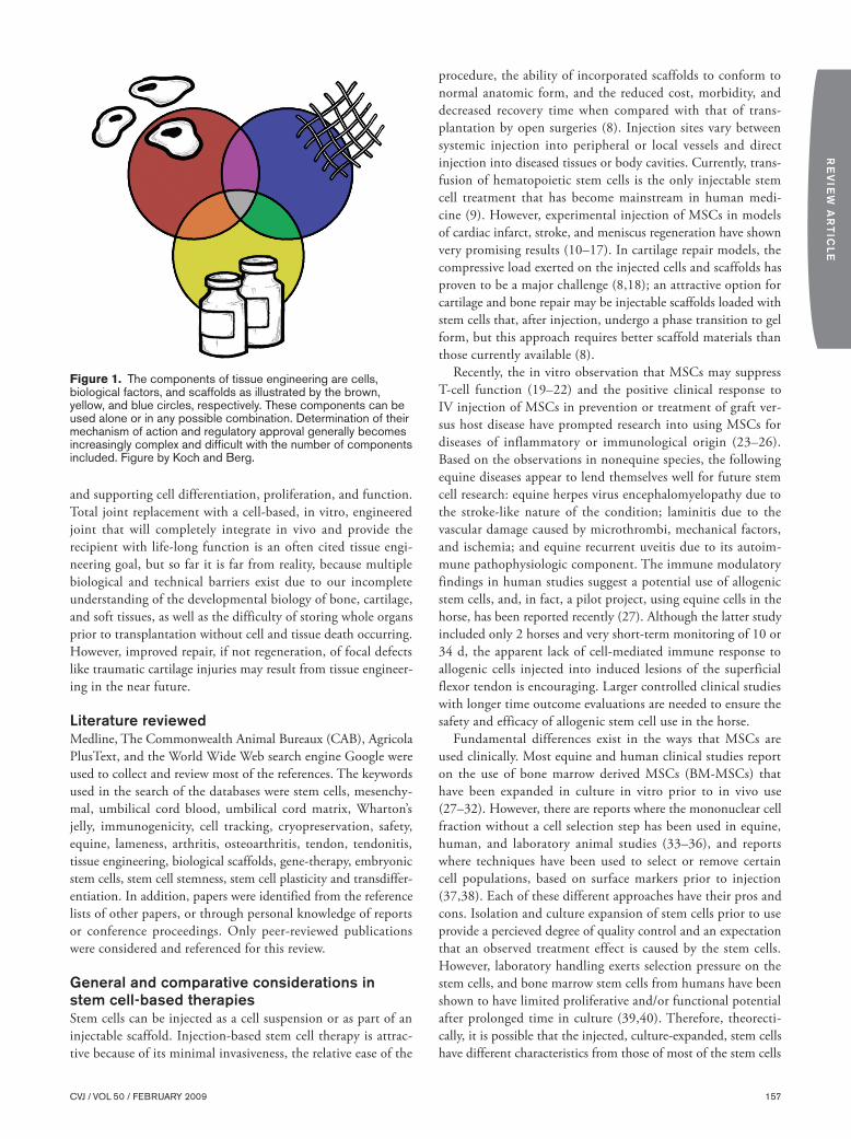
Scaffold-based therapies in generalScaffolds can be injectable, noninjectable, simple, complex, biological, or synthetic in nature. An important paradigm to have in mind when evaluating any scaffold-based therapy is the interaction between scaffold and tissue (Figure 2). The ideal scaf-fold has sufficient strength to protect cells from compression and shearing forces, while still having injury site anchoring potential and porosity to allow nutrient and differentiation factors to diffuse through it. The scaffold must also degrade at a rate that optimizes cellular growth and tissue regeneration. Such ideal scaffolds have not yet been designed. The optimal time point for evaluation of a scaffold-based treatment is also critical, and the best determination of treatment success can probably be made only after the “scaffold-tissue transition phase” has passed, which depends on the scaffold, cells, and tissue in question. In-depth
Figure 2. Ideally, any scaffold is degraded at a rate optimum for allowing complete tissue regeneration and ultimately replaced entirely by the regenerated tissue. Survival of the transplanted cells and successful tissue integration relies on diffusion of biological factors through the scaffold. The scaffold-tissue transition phase might be associated with decreased mechanical strength and function, leading to treatment failure if the scaffold degrades faster than the tissue can regenerate. On the other hand, a slowly degrading scaffold might impair and, potentially, prevent proper tissue healing. These concepts should be considered when evaluating scaffold-based studies and the time point chosen for evaluation of treatment success. Figure by Koch and Berg.

160 CVJ / VOL 50 / FEBRUARY 2009
CO
MP
TE
RE
ND
U
discussion of scaffolds in general and in relation to bone repair, cartilage repair, and repair of thick tissues has already been pub-lished and is beyond the scope of this paper (8,64–72). Instead, the characteristics and potential of scaffolds currently licensed for use in the horse will be reviewed.
Simple scaffold therapiesOne biological and 1 synthetic extracellular matrix scaffold are commercially available for use in the horse at this time.
Porcine urinary bladder matrix (UBM) is marketed as an acel-lular biological scaffold (Acell Vet; Acell, Columbia, Maryland, USA) for use in the horse. The product is available as powder, sheet, or granules; as a gel; and in disc form. Possible applica-tions according to the manufacturer include, but are not limited to, tendon and ligament injuries, hoof and hoof wall injuries, corneal ulcers, dental extractions, full and partial thickness wounds, burns, and post surgical skin closures. In a dog model, esophageal reconstruction was supported if the scaffold was covered with autologous muscle tissue (73). Porcine UBM scaf-fold alone resulted in intractable esophageal stricture, whereas using muscle alone resulted in similar results to using muscle combined with the scaffold. Full thickness experimentally induced myocardiac defects in dogs showed improved short-term healing 8 wk after coverage with porcine UBM-derived sheet scaffolds when compared with repair with a Dacron patch (74). Degradation products from porcine UBM-based scaffolds have shown antibacterial activity in vitro against Staphylococcus aureus and Escherichia coli (75). To the authors’ knowledge, there are no reports on using porcine UBM for ligament or tendon repair in horses or any other species, although a scientific review article conveys the impression that this treatment approach is rooted in evidence-based medicine (76). Scientifically validated studies are needed before porcine UBM-based scaffolds in horses can be recommended.
Synthetic extracellular matrix scaffolds (EquitrX; Sentrex Animal Care, Salt Lake City, Utah, USA) have recently become available to the equine practitioner for the treatment of skin wounds. The scaffolds are hydrogel films made of chemically cross-linked glycosaminoglycans (77). No reports are available on the product’s efficacy in the horse. In a pig model of skin wounds, enhanced healing was observed on days 3 and 5, but on day 7, no statistical difference was noted compared with that in the control group (78).
Complex scaffold therapiesReplacing whole joints with artificial joints has benefited thou-sands of human patients and the longevity of these implants continues to increase with improved patient selection, improved surgical technique, and better implants (79–82). The number of young human patients with very active lifestyles receiving joint replacements is increasing and the long-term performance of established and new techniques and materials remains to be seen in these patients. Development of techniques that can replace the diseased joint in a more biological way, allowing life-long treatment success, is being investigated. Such complex tissue engineering is ambitious and requires a multidisciplinary approach. Current target tissues of complex tissue engineering
include oral and craniofacial tissues; bone; joints and articular cartilage; tendons and ligaments, including their critical inter-faces with bone; and heart valves (83). Some of the general challenges include contaminated repair sites (oral cavity); scaf-fold designs; cell-surface interactions, promoting desirable cells and discouraging undesirable cells to adhere to the injury site; growth-factor delivery (which, when, and how much); in vivo tools for clinical assessment; and scale-up of cell and scaffold production to accommodate repair of large defects.
There are no reports on the use of engineered complex tis-sues in the horse. In sheep, engineered osteochondral-like plugs for use in mosaic arthroplasty have been investigated (84). These studies used autologous chondrocytes, not stem cells, loaded on to a polycalciumphosphate scaffold. This biphasic construct was cultured in vitro prior to placement in induced defects in the stifle. Improved healing was noted histologically when compared with that in the sham operated contralateral joint. However, the compressive and shear strength of these constructs is below that of native osteochondral plugs (85). Compressive and shearing forces are higher in the horse than in the sheep, so compression- and shearing-induced injuries might be of significance if the technique were to be transferred to the horse in its current form. However, mosaic arthroplasty, using autologous or allogenic osteochoindral plugs, is currently used in selected equine patients and continues to be investigated in experimental models (Figure 3) (86). The correct cells combined with the correct scaffold may improve the outcome of mosaic arthroplasty in the future.
Figure 3. Autologous osteochondral grafts are currently used in mosaic arthroplasty for selected focal cartilage defects in the horse. Incongruency between graft and native cartilage, as well as reduced biomechanical properties of the graft compared with native cartilage and bone, are the main limitations of the technique today. In the future, tissue engineering and stem cell-based therapies may help to negate these limitations. The asterisk (*) marks 1 of several autologous osteochondral grafts placed in the medial femoral condyle following harvest from the medial trochlear ridge of the same stifle joint. Image: Courtesy of Dr. Mark Hurtig, University of Guelph.
CVJ / VOL 50 / FEBRUARY 2009 161
RE
VIE
W A
RT
ICL
E
Biofactor treatments and gene-therapyRecombinant human bone morphogenetic protein-2 (rhBMP-2) and platelet-derived growth factor-BB (rhPDGF-BB) are licensed for use in human medicine to enhance lumbar interbody fusion and orofacial reconstruction (87–91). These 2 biofactors exem-plify the increasing use of biofactors in human medicine. Biofactors are used indirectly in equine medicine, as discussed under stem cell-based therapies, in cases where stem cells are combined with platelet rich plasma, bone marrow supernatant, or autologous serum. No purified biofactors are licensed for use in the horse.
In vitro studies on the effect of platelet rich plasma (PRP) on equine ligament and tendon explants have demonstrated increased transcription (upregulation) of anabolic genes and increased secretion of proteins, including cartilage oligomeric matrix protein (COMP), which is a protein believed to play an organizational role in tendon and ligament matrices (92,93). Smith et al (93) also evaluated acellular bone marrow superna-tant, which had a better effect than PRP on protein and COMP secretion. Most recently, acelluar bone marrow supernatant was shown to have an anabolic effect on tendon matrix products that was superior to that of platelet poor plasma, plasma, or blood (94). Insulin-like growth factor-1 has been shown to improve cellular and molecular healing parameters in an in vivo equine collagenase-induced tendonitis model (95). However, the mechanical strength of the tendon tissue did not vary between the treatment and control groups. The dominating molecular family in tendon regeneration is the bone morphogenetic protein (BMP) family, where growth differentiation factor 5 (GDF5), GDF6, GDF7, and the signaling mediators (Smads), in particular Smad8, appear to be of major importance for tendon healing (96–98). Regenerative treatment approaches to equine tendon healing have recently been reviewed (99). In vivo studies are needed to evaluate efficacy and safety before the use of these biological products becomes part of routine clinical medicine in horses.
Gene therapy in the horse is reviewed briefly here, since gene therapy may be a way of enhancing the performance of regen-erative therapies in the future through specific in vivo biofactor release at the injury site. The rationale is that a virus vector is able to invade the endogenous cell type of interest and alter its function by integrating an exogenous gene into its genome, thus enabling the cells to overexpress an existing gene or express a gene novel to that cell type. Subsequently, the cells will tran-scribe the implanted gene material into mRNA and translate it into functional protein. Gene therapy for various biofactors has mainly been investigated in vitro and experimentally in horses for the purpose of arresting osteoarthritis and/or improving joint cartilage healing, but gene therapy has yet to become a widespread clinical practice (100–112). Encouraging short-term results have been obtained in equine arthritis models by over-expression of interleukin-1 receptor antagonist protein (IL-1Ra) and insulin-like growth factor-1 (IGF-1), but long-term results have been less favorable. However, the technique continues to improve and more hyaline-like cartilage was present in equine cartilage defects treated with chondrocytes transfected to over-express IGF-1 than in defects treated with native chondrocytes
(100); the expression of IGF-1 decreased over time in this study. Multiple factors, including the target cell type, may affect the longevity of gene expression after transfection. Equine bone marrow-derived MSCs transfected by using recombinant and modified adenoviruses have shown greater permissiveness and sustained expression of transgenes than equine chondrocytes and synovioytes (112). Fundamentally, genes can be delivered to the target cell in 1 of 2 ways: 1) The target cell can be cultured in vitro, transfected with the gene(s) of interest, and then deliv-ered to the patient, a process known as ex vivo gene transfer and used by Goodrich et al (100) in the above cited study; 2) virus vectors containing the gene(s) of interest can be injected directly into the patient where the they seek out the target cells, invade the cells, and insert the gene into the cellular genome, so-called in vivo gene transfer. In vivo gene transfer is appealing due to its decreased cost, decreased laboratory involvement, and ease of administration, but concerns have been raised against this direct use of viral vectors after adverse effects were reported in human patients where unwanted immunoreactions were trig-gered (113,114). Morisset et al (102), in an equine model of induced chondral defects, have investigated in vivo gene transfer as follows. Adenovirus vectors containing the genes coding for IL-1Ra and IGF-1 were injected intraarticularly after a cartilage defect had been created. No macroscopic or histological differ-ences were noted between the treatment, control, and placebo groups after 16 wk. However, biochemical assays revealed increased type II collagen and proteoglycan content in the early repair tissue phase of the treatment group compared with that in the other groups.
A variation of the technique, called in vivo electroporation, is the direct intramuscular injection of naked DNA in the form of plasmids. It has been reported in the horse as a potential treat-ment for chronic laminitis (115): Injection of the gene coding for growth hormone releasing hormone (GHRH) resulted in weight gain, increased erythrocyte production, and increased IGF-1 levels in 6 nonlaminitic horses. Two horses suffering from chronic laminitis gained weight and showed an improved lameness score 6 mo after treatment. This plasmid-based therapy technique is attractive, since it avoids the use of a virus-vector.
Long-term storage of stem cells and engineered tissuesCryopreservation and long-term storage of stem cells will greatly enhance the applicability of stem cell-based therapies. Embryonic stem cells have proven to be difficult to cryopreserve; this remains an unsolved problem, although new techniques show promising results (116,117). The long-term cryotoler-ance of equine bone marrow-derived MSCs is unknown. In a human study, the in vivo engraftment capacity of BM-MSCs in patients with hematopoietic disease was not affected after 2.0–7.8 y of cryopreservation (118). Human cord blood-derived hematopoietic stem cells showed normal in vitro functions after 5 y of cryopreservation (119). The frequency of isolating viable mesenchymal-like stem cells from frozen mononuclear cell frac-tions of human umbilical cord blood was significantly lower than that from fresh, nonfrozen mononuclear cell fractions, so stem cell isolation and expansion is recommended prior to

162 CVJ / VOL 50 / FEBRUARY 2009
CO
MP
TE
RE
ND
U
cryopreservation (120). In conclusion, the upper time limit for cryopreservation of stem cells is currently unknown.
Cryopreservation of engineered complex tissues is experi-mental, but it might provide an “off-the-shelf ” product in the future. Vitrification is the most promising cryotechnique for this purpose at present (83).
Safety considerations of stem cell-based therapiesThe safety, both short-term and, in particular, long-term, of stem cell technologies is largely unknown. In the horse, no controlled safety studies have been performed and no central regulatory body is overlooking the current liberal and empiri-cal use of these cells in horses. To date, there have not been any reports of significant adverse reactions. However, this could be due to reporting bias.
Use of the cells and technologies presented here in the horse is likely to continue and expand in the near future. The establish-ment of a safety or adverse effect body, where unexpected clinical outcomes could be reported, is encouraged at this time. Until clinical efficacy has been proven, such an institution would, at least, be able to assess whether these procedures “do no harm.” Safety concerns to consider include, but are not restricted to, aberrant cell development and tissue or vehicle contamination with infectious agents or foreign biological and nonbiological substances used in the laboratory processing of the stem cells.
A requirement for authenticity of any embryonic stem cell is its capacity to form teratomas in immune suppressed mice. The extent to which adult and neonatal stem cells can form such teratomas, or other undesired tissue types, is less well studied, but this possibility should be considered if and when stem cell-based therapies are being used (121).
Transmission of infectious diseases is of special concern if allogenous cells are being used. Obviously, an animal with clini-cal or hematological signs of systemic disease is unlikely to be used as a stem cell donor for allogenic transplantation, but the possibility of adult stem cells harboring agents of latent chronic diseases has not been studied to the authors’ knowledge. The risk of infection associated with xenotransplant techniques have been studied. Studies evaluating the risk of viral transmission, in particular porcine endogenous retrovirus, from pig livers used for extracorporeal liver perfusion, showed that immune com-promised mice did become infected, but immune-competent baboons did not (122,123). Potent immunosuppresive drugs are not commonly used in equine medicine, but the risk of immuno suppression secondary to disease should be considered before instituting allogenic-based therapies. Overwhelming cell death of the injected cells could potentially impair tissue repair or, in more severe cases, trigger a significant inflamma-tory response.
The horse as an animal model for stem cell and tissue engineering studiesThe horse is already established as an animal model for focal car-tilage injuries and osteoarthritis (100,124). Advantages of horse joint models compared with those of other animals are their sheer size, which allows for easy manipulation and exploration,
and their cartilage thickness and composition, which most closely resemble those of human articular cartilage among the current animal models (124). In addition, the subchondral bone plate of equine bone is significantly thicker than that of rabbit and sheep bone, which allows for more reproducible cartilage defects without inadverent penetration of the subchondral bone plate, a potential for treatment bias (100). The main disadvantages are cost and heterogenic genetic composition, which require the use of larger animal numbers than when inbred animals are used. The horse has also been advocated as an animal model of tendon and ligament injuries, since many of the spontaneous injuries seen in horses are similar to those seen in human athletes (125). Other equine tissues and diseases, such as wounds and various hypoxic ischemic injuries, seem like straightforward candidates for equine stem cell research. The use of the horse as an animal model for stem cell therapies is not without problems at this time, especially due to a lack of equine-specific molecular markers needed to precisely character-ize the cell populations. These short-term limitations are likely to be overcome with time, as more research is done in the horse, thus positioning the horse as a good long-term animal model for stem cell therapies.
It has been argued that indisputable proof of stem cells as curative of an animal disorder/disease may provide the “tipping-point” for the public debate surrounding human stem cell research (126). The rationale is that if stem cell therapy can cure a naturally occurring animal disease or injury (not artificial induced injuries in small laboratory animals), who would then be against human stem cell research and therapies?
The understanding of equine stem cell biology, biofactors, and scaffolds, and their potential therapeutic uses in horses is rudimentary at present. Stem cell research in the horse has excit-ing comparative and equine specific perspectives that most likely will benefit the health of horses. Controlled, well-designed stud-ies are needed to move this new equine research field forward. The application of stem cell-based therapies in the horse should be done critically and cautiously, and treatment outcomes (good and bad) should be recorded and reported.
Authors’ contributionsDr. Koch originated the idea of this review article, conceptual-ized it, and wrote most of the manuscript. Drs. Berg and Betts participated in the conceptualization and critical review of the manuscript. Drs. Berg and Koch drew the figures. CVJ
References 1. Thomson JA, Itskovitz-Eldor J, Shapiro SS, et al. Embryonic
stem cell lines derived from human blastocysts. Science 1998;282: 1145–1147.
2. Odorico JS, Kaufman DS, Thomson JA. Multilineage differen-tiation from human embryonic stem cell lines. Stem Cells 2001;19: 193–204.
3. Pittenger MF, Mackay AM, Beck SC, et al. Multilineage potential of adult human mesenchymal stem cells. Science 1999;284:143–147.
4. Koch TG, Berg LC, Betts DH. Concepts for the clinical use of stem cells in equine medicine. Can Vet J 2008;49:1009–1017.
5. Dominici M, Le Blanc K, Mueller I, et al. Minimal criteria for defining multipotent mesenchymal stromal cells. The International Society for Cellular Therapy Position Statement. Cytotherapy 2006;8: 315–317.
CVJ / VOL 50 / FEBRUARY 2009 163
RE
VIE
W A
RT
ICL
E
6. Horwitz EM, Le Blanc K, Dominici M, et al. Clarification of the nomenclature for MSC: The International Society for Cellular Therapy Position Statement. Cytotherapy 2005;7:393–395.
7. Gratwohl A, Baldomero H, Frauendorfer K, Rocha V, Apperley J, Niederwieser D. The EBMT activity survey 2006 on hematopoietic stem cell transplantation: Focus on the use of cord blood products. Bone Marrow Transplant 2008;41:687–705.
8. Elisseeff J. Injectable cartilage tissue engineering. Expert Opin Biol Ther 2004;4:1849–1859.
9. Copelan EA. Hematopoietic stem-cell transplantation. N Engl J Med 2006;354:1813–1826.
10. Charwat S, Gyongyosi M, Lang I, et al. Role of adult bone marrow stem cells in the repair of ischemic myocardium: Current state of the art. Exp Hematol 2008;36:672–680.
11. Ballard VL, Edelberg JM. Stem cells for cardiovascular repair — The challenges of the aging heart. J Mol Cell Cardiol 2008. In press.
12. Izuta Y, Ochi M, Adachi N, Deie M, Yamasaki T, Shinomiya R. Meniscal repair using bone marrow-derived mesenchymal stem cells: Experimental study using green fluorescent protein transgenic rats. Knee 2005;12:217–223.
13. Angele P, Johnstone B, Kujat R, et al. Stem cell based tissue engineer-ing for meniscus repair. J Biomed Mater Res A 2008;85:445–455.
14. Murphy JM, Fink DJ, Hunziker EB, Barry FP. Stem cell therapy in a caprine model of osteoarthritis. Arthritis Rheum 2003;48: 3464–3474.
15. Kondziolka D, Wechsler L. Stroke repair with cell transplantation: Neuronal cells, neuroprogenitor cells, and stem cells. Neurosurg Focus 2008;24:E13.
16. Kalluri HS, Dempsey RJ. Growth factors, stem cells, and stroke. Neurosurg Focus 2008;24:E14.
17. Guzman R, Choi R, Gera A, De Los Angeles A, Andres RH, Steinberg GK. Intravascular cell replacement therapy for stroke. Neurosurgi Focus 2008;24:E15.
18. Caplan AI, Dennis JE. Mesenchymal stem cells as trophic mediators. J Cell Biochem 2006;98:1076–1084.
19. Sato K, Ozaki K, Oh I, et al. Nitric oxide plays a critical role in sup-pression of T-cell proliferation by mesenchymal stem cells. Blood 2007;109:228–234.
20. Ren G, Zhang L, Zhao X, et al. Mesenchymal stem cell-mediated immunosuppression occurs via concerted action of chemokines and nitric oxide. Cell Stem Cell 2008;2:141–150.
21. Le Blanc K, Ringden O. Immunomodulation by mesenchymal stem cells and clinical experience. J Intern Med 2007;262:509–525.
22. Keating A. How do mesenchymal stromal cells suppress T cells? Cell Stem Cell 2008;2:106–108.
23. Tisato V, Naresh K, Girdlestone J, Navarrete C, Dazzi F. Mesenchymal stem cells of cord blood origin are effective at preventing but not treating graft-versus-host disease. Leukemia 2007;21:1992–1999.
24. Aksu AE, Horibe E, Sacks J, et al. Co-infusion of donor bone mar-row with host mesenchymal stem cells treats GVHD and promotes vascularized skin allograft survival in rats. Clin Immunol 2008. In press.
25. Tian Y, Deng YB, Huang YJ, Wang Y. Bone marrow-derived mes-enchymal stem cells decrease acute graft-versus-host disease after allogeneic hematopoietic stem cells transplantation. Immunol Invest 2008;37:29–42.
26. Bacigalupo A. Management of acute graft-versus-host disease. Br J Haematol 2007;137:87–98.
27. Guest DJ, Smith MR, Allen WR. Monitoring the fate of autologous and allogeneic mesenchymal progenitor cells injected into the super-ficial digital flexor tendon of horses: Preliminary study. Equine Vet J 2008;40:178–181.
28. Smith RK, Korda M, Blunn GW, Goodship AE. Isolation and implan-tation of autologous equine mesenchymal stem cells from bone marrow into the superficial digital flexor tendon as a potential novel treatment. Equine Vet J 2003;35:99–102.
29. Pacini S, Spinabella S, Trombi L, et al. Suspension of bone marrow-derived undifferentiated mesenchymal stromal cells for repair of superficial digital flexor tendon in race horses. Tissue Eng 2007;13: 2949–2955.
30. Wilke MM, Nydam DV, Nixon AJ. Enhanced early chondrogenesis in articular defects following arthroscopic mesenchymal stem cell implantation in an equine model. J Orthop Res 2007;25:913–925.
31. Abdallah BM, Kassem M. Human mesenchymal stem cells: From basic biology to clinical applications. Gene Ther 2008;15:109–116.
32. Brooke G, Cook M, Blair C, et al. Therapeutic applications of mes-enchymal stromal cells. Semin Cell Dev Biol 2007;18:846–858.
33. Crovace A, Lacitignola L, De Siena R, Rossi G, Francioso E. Cell therapy for tendon repair in horses: An experimental study. Vet Res Commun 2007;31 Suppl 1:281–283.
34. Blatt A, Cotter G, Leitman M, et al. Intracoronary administration of autologous bone marrow mononuclear cells after induction of short ischemia is safe and may improve hibernation and ischemia in patients with ischemic cardiomyopathy. Am Heart J 2005;150: 986.e1–986.e7.
35. Beeres SL, Lamb HJ, Roes SD, et al. Effect of intramyocardial bone marrow cell injection on diastolic function in patients with chronic myocardial ischemia. J Magn Reson Imaging 2008;27:992–997.
36. Bartsch T, Brehm M, Zeus T, Strauer BE. Autologous mononuclear stem cell transplantation in patients with peripheral occlusive arterial disease. J Cardiovasc Nurs 2006;21:430–432.
37. Schafer R, Wiskirchen J, Guo K, et al. Aptamer-based isolation and subsequent imaging of mesenchymal stem cells in ischemic myocard by magnetic resonance imaging. Rofo 2007;179:1009–1015.
38. Oertel M, Menthena A, Chen YQ, Teisner B, Jensen CH, Shafritz DA. Purification of fetal liver stem/progenitor cells containing all the repopulation potential for normal adult rat liver. Gastroenterology 2008;134:823–832.
39. Kern S, Eichler H, Stoeve J, Kluter H, Bieback K. Comparative analysis of mesenchymal stem cells from bone marrow, umbilical cord blood, or adipose tissue. Stem Cells 2006;24:1294–1301.
40. Sethe S, Scutt A, Stolzing A. Aging of mesenchymal stem cells. Ageing Res Rev 2006;5:91–116.
41. Kassem M, Kristiansen M, Abdallah BM. Mesenchymal stem cells: Cell biology and potential use in therapy. Basic Clin Pharmacol Toxicol 2004;95:209–214.
42. Hu X, Wang J, Chen J, et al. Optimal temporal delivery of bone mar-row mesenchymal stem cells in rats with myocardial infarction. Eur J Cardiothorac Surg 2007;31:438–443.
43. Carstanjen B, Desbois C, Hekmati M, Behr L. Successful engraft-ment of cultured autologous mesenchymal stem cells in a surgically repaired soft palate defect in an adult horse. Can J Vet Res 2006; 70:143–147.
44. Koch TG, Heerkens T, Thomsen PD, Betts DH. Isolation of mesen-chymal stem cells from equine umbilical cord blood. BMC Biotechnol 2007;7:26.
45. Reed SA, Johnson SE. Equine umbilical cord blood contains a popula-tion of stem cells that express Oct4 and differentiate into mesodermal and endodermal cell types. J Cell Physiol 2008;215:329–336.
46. Giovannini S, Brehm W, Mainil-Varlet P, Nesic D. Multilineage dif-ferentiation potential of equine blood-derived fibroblast-like cells. Differentiation 2008;76:118–129.
47. Stewart AA, Byron CR, Pondenis H, Stewart MC. Effect of fibroblast growth factor-2 on equine mesenchymal stem cell monolayer expan-sion and chondrogenesis. Am J Vet Res 2007;68:941–945.
48. Kisiday JD, Kopesky PW, Evans CH, Grodzinsky AJ, McIlwraith CW, Frisbie DD. Evaluation of adult equine bone marrow- and adipose-derived progenitor cell chondrogenesis in hydrogel cultures. J Orthop Res 2008;26:322–331.
49. Koerner J, Nesic D, Romero JD, Brehm W, Mainil-Varlet P, Grogan SP. Equine peripheral blood-derived progenitors in com-parison to bone marrow-derived mesenchymal stem cells. Stem Cells 2006;24:1613–1619.
50. Fortier LA, Nixon AJ, Williams J, Cable CS. Isolation and chondro-cytic differentiation of equine bone marrow-derived mesenchymal stem cells. Am J Vet Res 1998;59:1182–1187.
51. Stolzing A, Jones E, McGonagle D, Scutt A. Age-related changes in human bone marrow-derived mesenchymal stem cells: Consequences for cell therapies. Mech Ageing Dev 2008;129:163–173.
52. Bonyadi M, Waldman SD, Liu D, Aubin JE, Grynpas MD, Stanford WL. Mesenchymal progenitor self-renewal deficiency leads to age-dependent osteoporosis in Sca-1/Ly-6A null mice. Proc Natl Acad Sci USA 2003;100:5840–5845.
53. Yoo JU, Johnstone B. The role of osteochondral progenitor cells in fracture repair. Clin Orthop Relat Res 1998;355 Suppl:S73–81.
54. Kraus KH, Kirker-Head C. Mesenchymal stem cells and bone regen-eration. Vet Surg 2006;35:232–242.
55. O’Rielly JL, Bertone AL, Genovese RL. Treatment of a chronic com-minuted fracture of the fibula in a horse. J Am Vet Med Assoc 1998; 212:396–398.

164 CVJ / VOL 50 / FEBRUARY 2009
CO
MP
TE
RE
ND
U
56. Lescun TB, Morisset SM, Fugaro MN, Blevins WE. Facilitated anky-losis of the distal interphalangeal joint in a foal. Aust Vet J 2004;82: 282–285.
57. Harriss FK, Galuppo LD, Decock HE, McDuffee LA, Macdonald MH. Evaluation of a technique for collection of cancellous bone graft from the proximal humerus in horses. Vet Surg 2004;33:293–300.
58. McDuffee LA, Anderson GI. In vitro comparison of equine cancel-lous bone graft donor sites and tibial periosteum as sources of viable osteoprogenitors. Vet Surg 2003;32:455–463.
59. McDuffee LA, Anderson GI, Wright GM, Ryan DA. In vitro hetero-geneity of osteogenic cell populations at various equine skeletal sites. Can J Vet Res 2006;70:277–284.
60. Hoynowski SM, Fry MM, Gardner BM, et al. Characterization and differentiation of equine umbilical cord-derived matrix cells. Biochem Biophys Res Commun 2007;362:347–353.
61. Vidal MA, Kilroy GE, Lopez MJ, Johnson JR, Moore RM, Gimble JM. Characterization of equine adipose tissue-derived stromal cells: Adipogenic and osteogenic capacity and comparison with bone marrow- derived mesenchymal stromal cells. Vet Surg 2007;36:613–622.
62. Vidal MA, Kilroy GE, Johnson JR, Lopez MJ, Moore RM, Gimble JM. Cell growth characteristics and differentiation frequency of adherent equine bone marrow-derived mesenchymal stromal cells: Adipogenic and osteogenic capacity. Vet Surg 2006;35:601–610.
63. Arnhold SJ, Goletz I, Klein H, et al. Isolation and characterization of bone marrow-derived equine mesenchymal stem cells. Am J Vet Res 2007;68:1095–1105.
64. Engel E, Michiardi A, Navarro M, Lacroix D, Planell JA. Nanotechnology in regenerative medicine: The materials side. Trends Biotechnol 2008;26:39–47.
65. Arumuganathar S, Jayasinghe SN. Living scaffolds (specialized and unspecialized) for regenerative and therapeutic medicine. Biomacromolecules 2008;9:759–766.
66. Hutmacher DW, Cool S. Concepts of scaffold-based tissue engineering — the rationale to use solid free-form fabrication techniques. J Cell Mol Med 2007;11:654–669.
67. Hutmacher DW, Schantz JT, Lam CX, Tan KC, Lim TC. State of the art and future directions of scaffold-based bone engineering from a biomaterials perspective. J Tissue Eng Regen Med 2007;1:245–260.
68. Bonfield W. Designing porous scaffolds for tissue engineering. Philos Transact A Math Phys Eng Sci 2006;364:227–232.
69. Raghunath J, Rollo J, Sales KM, Butler PE, Seifalian AM. Biomaterials and scaffold design: Key to tissue-engineering cartilage. Biotechnol Appl Biochem 2007;46:73–84.
70. Ko HC, Milthorpe BK, McFarland CD. Engineering thick tissues — the vascularisation problem. Eur Cell Mater 2007;14:18–19.
71. Lynn AK, Brooks RA, Bonfield W, Rushton N. Repair of defects in articular joints. Prospects for material-based solutions in tissue engi-neering. J Bone Joint Surg Br 2004;86:1093–1099.
72. Tognana E, Borrione A, De Luca C, Pavesio A. Hyalograft C: hyaluronan- based scaffolds in tissue-engineered cartilage. Cells Tissues Organs 2007;186:97–103.
73. Badylak SF, Vorp DA, Spievack AR, et al. Esophageal reconstruction with ECM and muscle tissue in a dog model. J Surg Res 2005;128: 87–97.
74. Badylak SF, Kochupura PV, Cohen IS, et al. The use of extracellular matrix as an inductive scaffold for the partial replacement of functional myocardium. Cell Transplant 2006;15 Suppl 1:S29–40.
75. Brennan EP, Reing J, Chew D, Myers-Irvin JM, Young EJ, Badylak SF. Antibacterial activity within degradation products of biological scaffolds composed of extracellular matrix. Tissue Eng 2006;12: 2949–2955.
76. Badylak SF. Extracellular matrix as a scaffold for tissue engineering in veterinary medicine: Applications to soft tissue healing. Clin Tech Equine Pract 2004;3:173–181.
77. Shu XZ, Liu Y, Palumbo F, Prestwich GD. Disulfide-crosslinked hyaluronan-gelatin hydrogel films: A covalent mimic of the extracellular matrix for in vitro cell growth. Biomaterials 2003;24:3825–3834.
78. Kirker KR, Luo Y, Morris SE, Shelby J, Prestwich GD. Glycosaminoglycan hydrogels as supplemental wound dressings for donor sites. J Burn Care Rehabil 2004;25:276–286.
79. Eingartner C. Current trends in total hip arthroplasty. Orthop Traumatol Rehabil 2007;9:8–14.
80. Breusch SJ, Lukoschek M, Thomsen M, Mau H, Ewerbeck V, Aldinger PR. Ten-year results of uncemented hip stems for failed
intertrochanteric osteotomy. Arch Orthop Trauma Surg 2005;125: 304–309.
81. Parsch D, Jung AW, Thomsen M, Ewerbeck V, Aldinger PR. Good survival of uncemented tapered stems for failed intertrochanteric osteotomy: A mean 16 year follow-up study in 45 patients. Arch Orthop Trauma Surg 2007. In press.
82. Eingartner C, Heigele T, Dieter J, Winter E, Weise K. Long-term results with the BiCONTACT system — aspects to investigate and to learn from. Int Orthop 2003;27 Suppl 1:S11–15.
83. Mikos AG, Herring SW, Ochareon P, et al. Engineering complex tis-sues. Tissue Eng 2006;12:3307–3339.
84. Pilliar RM, Kandel RA, Grynpas MD, Zalzal P, Hurtig M. Osteochon-dral defect repair using a novel tissue engineering approach: Sheep model study. Technol Health Care 2007;15:47–56.
85. Kandel RA, Grynpas M, Pilliar R, et al. Repair of osteochondral defects with biphasic cartilage-calcium polyphosphate constructs in a sheep model. Biomaterials 2006;27:4120–4131.
86. Bodo G, Hangody L, Modis L, Hurtig M. Autologous osteo-chondral grafting (mosaic arthroplasty) for treatment of subchon-dral cystic lesions in the equine stifle and fetlock joints. Vet Surg 2004;33:588–596.
87. Huang KY, Yan JJ, Hsieh CC, Chang MS, Lin RM. The in vivo bio-logical effects of intradiscal recombinant human bone morphogenetic protein-2 on the injured intervertebral disc: An animal experiment. Spine 2007;32:1174–1180.
88. Hamilton DK, Jones-Quaidoo SM, Sansur C, Shaffrey CI, Oskouian R, Jane JA, Sr. Outcomes of bone morphogenetic protein-2 in mature adults: Posterolateral non-instrument-assisted lumbar decompression and fusion. Surg Neurol 2008;69:457–461.
89. DiPaola CP, Molinari RW. Posterior lumbar interbody fusion. J Am Acad Orthop Surg 2008;16:130–139.
90. Hollinger JO, Hart CE, Hirsch SN, Lynch S, Friedlaender GE. Recombinant human platelet-derived growth factor: Biology and clinical applications. J Bone Joint Surg Am 2008;90 Suppl 1:48–54.
91. Rocchietta I, Dellavia C, Nevins M, Simion M. Bone regenerated via rhPDGF-bB and a deproteinized bovine bone matrix: Backscattered electron microscopic element analysis. Int J Periodontics Restorative Dent 2007;27:539–545.
92. Schnabel LV, Mohammed HO, Miller BJ, et al. Platelet rich plasma (PRP) enhances anabolic gene expression patterns in flexor digitorum superficialis tendons. J Orthop Res 2007;25:230–240.
93. Smith JJ, Ross MW, Smith RK. Anabolic effects of acellular bone mar-row, platelet rich plasma, and serum on equine suspensory ligament fibroblasts in vitro. Vet Comp Orthop Traumatol 2006;19:43–47.
94. Schnabel LV, Sonea HO, Jacobson MS, Fortier LA. Effects of platelet rich plasma and acellular bone marrow on gene expression patterns and DNA content of equine suspensory ligament explant cultures. Equine Vet J 2008;40:260–265.
95. Dahlgren LA, van der Meulen MC, Bertram JE, Starrak GS, Nixon AJ. Insulin-like growth factor-I improves cellular and molecular aspects of healing in a collagenase-induced model of flexor tendinitis. J Orthop Res 2002;20:910–919.
96. Hoffmann A, Pelled G, Turgeman G, et al. Neotendon formation induced by manipulation of the Smad8 signalling pathway in mesen-chymal stem cells. J Clin Invest 2006;116:940–952.
97. Towler DA, Gelberman RH. The alchemy of tendon repair: A primer for the (S)mad scientist. J Clin Invest 2006;116:863–866.
98. Aslan H, Kimelman-Bleich N, Pelled G, Gazit D. Molecular targets for tendon neoformation. J Clin Invest 2008;118:439–444.
99. Fortier LA, Smith RK. Regenerative medicine for tendinous and ligamentous injuries of sport horses. Vet Clin North Am Equine Pract 2008;24:191–201.
100. Goodrich LR, Hidaka C, Robbins PD, Evans CH, Nixon AJ. Genetic modification of chondrocytes with insulin-like growth factor-1 enhances cartilage healing in an equine model. J Bone Joint Surg Br 2007;89:672–685.
101. Frisbie DD, McIlwraith CW. Gene therapy: Future therapies in osteoarthritis. Vet Clin North Am Equine Pract 2001;17:233–243.
102. Morisset S, Frisbie DD, Robbins PD, Nixon AJ, McIlwraith CW. IL-1ra/IGF-1 gene therapy modulates repair of microfractured chon-dral defects. Clin Orthop Relat Res 2007;462:221–228.
103. Goodrich LR, Brower-Toland BD, Warnick L, Robbins PD, Evans CH, Nixon AJ. Direct adenovirus-mediated IGF-I gene transduction of synovium induces persisting synovial fluid IGF-I ligand elevations. Gene Ther 2006;13:1253–1262.
CVJ / VOL 50 / FEBRUARY 2009 165
RE
VIE
W A
RT
ICL
E
104. Nixon AJ, Brower-Toland BD, Bent SJ, et al. Insulinlike growth factor-I gene therapy applications for cartilage repair. Clin Orthop Relat Res 2000;379 Suppl: S201–213.
105. Frisbie DD, McIlwraith CW. Evaluation of gene therapy as a treatment for equine traumatic arthritis and osteoarthritis. Clin Orthop Relat Res 2000;379 Suppl: S273–287.
106. Brower-Toland BD, Saxer RA, Goodrich LR, et al. Direct adenovirus-mediated insulin-like growth factor I gene transfer enhances transplant chondrocyte function. Hum Gene Ther 2001;12:117–129.
107. Saxer RA, Bent SJ, Brower-Toland BD, et al. Gene mediated insulin-like growth factor-I delivery to the synovium. J Orthop Res 2001;19: 759–767.
108. Frisbie DD, Ghivizzani SC, Robbins PD, Evans CH, McIlwraith CW. Treatment of experimental equine osteoarthritis by in vivo delivery of the equine interleukin-1 receptor antagonist gene. Gene Ther 2002; 9:12–20.
109. Nixon AJ, Haupt JL, Frisbie DD, et al. Gene-mediated restoration of cartilage matrix by combination insulin-like growth factor-I/ interleukin-1 receptor antagonist therapy. Gene Ther 2005;12: 177–186.
110. Haupt JL, Frisbie DD, McIlwraith CW, et al. Dual transduction of insulin-like growth factor-I and interleukin-1 receptor antagonist protein controls cartilage degradation in an osteoarthritic culture model. J Orthop Res 2005;23:118–126.
111. Strauss EJ, Goodrich LR, Chen CT, Hidaka C, Nixon AJ. Biochemical and biomechanical properties of lesion and adjacent articular cartilage after chondral defect repair in an equine model. Am J Sports Med 2005;33:1647–1653.
112. Ishihara A, Zachos TA, Bartlett JS, Bertone AL. Evaluation of permis-siveness and cytotoxic effects in equine chondrocytes, synovial cells, and stem cells in response to infection with adenovirus 5 vectors for gene delivery. Am J Vet Res 2006;67:1145–1155.
113. Zaiss AK, Muruve DA. Immunity to adeno-associated virus vectors in animals and humans: A continued challenge. Gene Ther 2008; 15:808–816.
114. Bauer G, Dao MA, Case SS, et al. In vivo biosafety model to assess the risk of adverse events from retroviral and lentiviral vectors. Mol Ther 2008;16:1308–1315.
115. Brown PA, Bodles-Brakhop A, Draghia-Akli R. Plasmid growth hor-mone releasing hormone therapy in healthy and laminitis-afflicted horses-evaluation and pilot study. J Gene Med 2008;10:564–574.
116. Yang PF, Hua TC, Tsung HC, Cheng QK, Cao YL. Effective cryo-preservation of human embryonic stem cells by programmed freezing. Eng Med Biol Soc 2005;1:482–485.
117. Heng BC, Ye CP, Liu H, Toh WS, Rufaihah AJ, Cao T. Kinetics of cell death of frozen-thawed human embryonic stem cell colonies is reversibly slowed down by exposure to low temperature. Zygote 2006;14:341–348.
118. Attarian H, Feng Z, Buckner CD, MacLeod B, Rowley SD. Long-term cryopreservation of bone marrow for autologous transplantation. Bone Marrow Transplant 1996;17:425–430.
119. Goodwin HS, Grunzinger LM, Regan DM, et al. Long term cryostor-age of UC blood units: Ability of the integral segment to confirm both identity and hematopoietic potential. Cytotherapy 2003;5:80–86.
120. Kogler G, Radke TF, Lefort A, et al. Cytokine production and hematopoiesis supporting activity of cord blood-derived unrestricted somatic stem cells. Exp Hematol 2005;33:573–583.
121. Poh KK, Sperry E, Young RG, Freyman T, Barringhaus KG, Thompson CA. Repeated direct endomyocardial transplantation of allogeneic mesenchymal stem cells: Safety of a high dose, “off-the-shelf,” cellular cardiomyoplasty strategy. Int J Cardiol 2007;117:360–364.
122. van der Laan LJ, Lockey C, Griffeth BC, et al. Infection by porcine endogenous retrovirus after islet xenotransplantation in SCID mice. Nature 2000;407:90–94.
123. Nishitai R, Ikai I, Shiotani T, et al. Absence of PERV infection in baboons after transgenic porcine liver perfusion. J Surg Res 2005;124: 45–51.
124. Frisbie DD, Cross MW, McIlwraith CW. A comparative study of articular cartilage thickness in the stifle of animal species used in human pre-clinical studies compared to articular cartilage thickness in the human knee. Vet Comp Orthop Traumatol 2006;19:142–146.
125. Smith RK, Webbon PM. Harnessing the stem cell for the treatment of tendon injuries: Heralding a new dawn? Br J Sports Med 2005;39: 582–584.
126. Fiester A, Scholer H, Caplan A. Stem cell therapies: Time to talk to the animals. Cloning Stem Cells 2004;6:3–4.