review article chinese herbs interfering with cancer...
TRANSCRIPT
Review ArticleChinese Herbs Interfering withCancer Reprogramming Metabolism
Zhangfeng Zhong,1 William W. Qiang,1,2 Wen Tan,3 Haotian Zhang,1 Shengpeng Wang,1
Chunming Wang,1 Wenan Qiang,4 and Yitao Wang1
1 Institute of Chinese Medical Sciences, State Key Laboratory of Quality Research in Chinese Medicine,University of Macau, Macau2Yale University, New Haven, CT 06511, USA3School of Pharmacy, Lanzhou University, Lanzhou, Gansu 730000, China4Division of Reproductive Science in Medicine, Department of Obstetrics and Gynecology,Feinberg School of Medicine at Northwestern University, Chicago, IL 60611, USA
Correspondence should be addressed to Wenan Qiang; [email protected] and Yitao Wang; um [email protected]
Received 6 December 2015; Accepted 3 February 2016
Academic Editor: Kuang C. Lai
Copyright © 2016 Zhangfeng Zhong et al. This is an open access article distributed under the Creative Commons AttributionLicense, which permits unrestricted use, distribution, and reproduction in any medium, provided the original work is properlycited.
Emerging evidence promotes a reassessment of metabolic reprogramming regulation in cancer research. Although there exists along history of Chinese herbs applied in cancer treatment, few reports have addressed the effects of Chinese herbal componentson metabolic reprogramming, which is a central cancer hallmark involved in the slowing or prevention of chemoresistance incancer cells. In this review, we have focused on four core elements altered by metabolic reprogramming in cancer cells. Theseinclude glucose transport, glycolysis, mitochondrial oxidative phosphorylation, and fatty acid synthesis. With this focus, we havesummarized recent advances in metabolic reprogramming of cancer cells in response to specific Chinese herbal components. Wepropose that exploring Chinese herbal interference in cancer metabolic reprogramming might identify new therapeutic targets forcancer and more ways in which to approach metabolism-related diseases.
1. Introduction
Since the 1920s, cancer cell metabolism has been studied inthe context of the so-called Warburg effect. This metabolicfeature of neoplastic cells involvesmany complex biochemicalreactions andmultiple interrelated signaling pathways, whichhave yet to be fully elucidated. The Warburg effect is anubiquitous event in the malignant proliferation of cancercells, distinguishing them from normal cells, and recently agrowing number of novel findings promoted a reassessmentof metabolic reprogramming regulation in cancer research[1]. For example, recent work suggests that the Warburgeffect regulates chemosensitivity in Taxol-resistant humanbreast cancer cells [2]. Other studies have shown that it influ-ences estrogen-related receptor expression [3]. In autophagy-senescent fibroblasts that promote the growth and metastasisof human breast cancer cells, the Warburg effect participates
in the crosstalk between fibroblasts and cancer cells. Theprecise relationship between the two cell types is governedby mitochondrial metabolism [4].
Many reviews and research studies show that systemicmetabolic disorders intersect with cancer development, andmetabolomics profiling for prognostic and predictive mark-ers has identified shared markers associated with bothconditions [5–9]. Recent work with several tumor typeshas documented the synergistic effect of combining 2-deoxy-D-glucose (2-DG) and berberine, two tumor cell-toxiccompounds, to target glycolysis and oxidative phosphory-lation (OXPHOS) simultaneously [10]. In nuclear magneticresonance- (NMR-) based metabonomics research, signif-icant differences for prognosis have been documented foracute myeloid leukemia (AML) patients with “favorable” vis-a-vis “intermediate” statuses in the cytogenetic determinants
Hindawi Publishing CorporationEvidence-Based Complementary and Alternative MedicineVolume 2016, Article ID 9282813, 10 pageshttp://dx.doi.org/10.1155/2016/9282813
2 Evidence-Based Complementary and Alternative Medicine
governing glycolysis, the tricarboxylic acid (TCA) cycle, andfatty acid synthesis [11].
The metabolic phenotype of cancer cells provides a greatdeal of valuable information about treatment design andprognosis. This information overrides genetic and individualdiversity as well as pathologic diversity of specific cancer celltypes. For example, in human colorectal cancer, the presenceor absence of significantly altered metabolites correlates withrecurrence and survival rates after surgery and chemotherapy[12].
Large numbers of studies identifying these correlativeeffects suggest that exogenous metabolic regulation has goodtherapeutic potential for cancer treatment. Across manypublished studies, this has been shown to be particularly truewhen related to targeting the uptake and the utilization ofglucose, the abnormally enhanced glycolysis, altered mito-chondrial oxidative phosphorylation (OXPHOS), and dereg-ulated fatty acid synthesis. Meanwhile, other reprogrammingevents have been shown to occur in parallel with the onesidentified above, including altered glycine consumption andactivated mitochondrial glycine biosynthetic pathway [13].Finally, many studies have focused on the systems biologyand metabolic transformation of cancer cells, where thekey enzymes in glycolysis, the TCA cycle, and the pentosephosphate pathways (PPP) were investigated as correlatedmetabolic changes occurring within the context of the War-burg effect [14].
In Chinese medicine, treatment strategies have empha-sized promoting blood circulation, supporting health andenergy while strengthening body resistance, heat clearance,and systemic detoxification, resolving phlegm production,promoting T cell cytotoxic functions, dispersing edema, andrelieving pain. In addition, many natural products derivedfrom Chinese herbs have been shown to have therapeuticpotential toward the prevention of cancer initiation andprogression [12, 15]. One example is the Chinese botanicalagent berberine, which inhibits mitochondrial complex I andinteracts with the adenine nucleotide translocator in cancercells [16]. The powdered fruiting bodies of Pleurotus eryngii(DC. ex Fr.) Quel have been shown to significantly inhibitthe proliferation of several cancer cell lines (A549, BGC-823, HepG2, and HGC-27) and to have immunopotentiationactivity in RAW 264.7 cells [17]. 1,2,3,4,6-Penta-O-galloyl-beta-D-glucose, which is a polyphenolic compound isolatedfrom Rhus chinensis Mill, regulates a series of metabolicgenes in glycolysis, pyruvate metabolism, gluconeogenesis,and tyrosine metabolism in the breast cancer cell line, MDA-MB-231 [18]. These compounds are isolated examples of anextended body of work, with biochemical agents that havebeen used in China for up to 3,000 years that recently haveshown targeted inhibition of cancer metabolic reprogram-ming, by which tumor initiation, progression, and spreadmight be addressed.
In this review, we have focused on glucose, glycolysis,mitochondrial OXPHOS, and fatty acid synthesis and haveidentified recent successful studies using Chinese herbs thattarget and inhibit cancer cell growth.
2. Glucose Transport
Abnormal glucose metabolism is closely linked with cancercell metabolism. In normal cells, glucose uptake sustains thetwo main metabolic processes that produce ATP as an endproduct, which are glycolysis and mitochondrial OXPHOS[19–24]. There have been few studies on glucose uptake bycancer cells; instead, the vast majority of work has focusedon the activity of glucose transporters (GLUTs). In recentyears, another family of glucose transporters, called sodium-dependent glucose transporters (SGLTs), was studied fortheir observed ability to slow tumor cell growth. Somecomponents derived from Chinese herbs exert direct GLUTinhibition. Notably, GLUT expression is significantly differ-ent in cancer cells, in relation to their normal counterparts.This property of cancer cells makes them vulnerable toGLUT inhibitors, which reduce glucose consumption [25–28]. Tetrandrine, a bisbenzylisoquinoline alkaloid isolatedfrom Stephania tetrandra S Moore, reduces glucose uptakein cancer cells, and this energetic impairment induces apop-tosis [29]. Finally, chlorogenic acid, a phenolic secondarymetabolite isolated from many Chinese herbs (Eucommiaulmoides Oliv. and Lonicera japonica Thunb. among others),has significant effects on glucose transport in cancer cells,through activating the AMPK signaling pathway [30].
3. Glycolysis
Cancer cells require high levels of glycolytic intermediates tosupport their biosynthetic requirements [64]. Many promis-ing anticancer agents, currently under study, are compoundsthat inhibit aerobic glycolysis [65–67]. Some of these agentsalso target autophagy in cancer cells, suggesting that theymight be used for combinatorial therapies [68].
Some bioactive compounds derived from Chinese herbsare now known to regulate glycolysis [67]. For exam-ple, prosapogenin A, isolated from Veratrum, regulates theexpression of glycolysis-related genes to induce apoptosis inhuman cervical carcinoma (HeLa), hepatocellular carcinoma(HepG2), and breast adenocarcinoma (MCF-7) cells [45].Other Chinese herbal components target different glycolyticintermediates.
Epigallocatechin, for example, is one component derivedfrom the Chinese herb Spatholobus suberectus that targetsglycolytic lactate production by inhibiting the expression oractivity of lactate dehydrogenase A (LDH-A), an importantenzyme checkpoint in glycolysis that catalyzes the intercon-version of pyruvate and L-lactate. Its activity has been shownto induce apoptosis and suppress breast cancer growth invivo, as upregulated LDH-A facilitates glycolysis and reducestumor dependency on oxygen [69]. LDH-A promotes tumorinitiation, progression, and metastasis, and it is a prognosticfactor for poor survival [70]. Inhibiting LDH-A activitysignificantly reduces cell proliferation and tumor size, andit induces elevated intracellular oxidative stress, resulting inapoptosis. Silencing LDH-A also contributes to suppressingtumorigenicity in breast cancer cells [71]. There is alsoevidence that the LDH-B isozyme also participates in tumordevelopment and is regulated by oncogenic transcription
Evidence-Based Complementary and Alternative Medicine 3
factors mammalian target of rapamycin (mTOR) and signaltransducer and activator of transcription 3 (STAT3) [72].
Oleanolic acid, another Chinese herbal derivative, sup-presses aerobic glycolysis via the PK-M2/PK-M1 switch thataccompanies mTOR/c-Myc/heterogeneous nuclear ribonu-cleoprotein (hnRNP) signaling regulation [39]. Pyruvatekinase isoenzyme type M2 (PK-M2), like LDH-A, is a criticalenzyme in glycolysis, and its underlying mechanisms havebeen also explored extensively in the context of tumorcell physiology [39, 73]. In hepatocellular carcinoma cells,targeting PK-M2 shows therapeutic potential, as it regulatesepithelial-mesenchymal transition and migration [74]. Ofinterest, PK-M2 activity is augmented by hypoxia-induciblefactor 1𝛼- (HIF-1𝛼-) mediated transcription activation inmTOR-hyperactive cancer cells [75].
Finally, neoalbaconol, an isolate of the fruiting body ofAlbatrellus confluens, induces energy depletion in cancer cellsby inhibiting the phosphatidylinositol 3-kinase (PI3K)/HK2pathway and reduces glucose consumption and ATP gen-eration [38]. Hexokinase 2 (HK2) is also a key metabolicregulator implied in many tumor types.
4. Mitochondrial OxidativePhosphorylation (OXPHOS)
Mitochondrial OXPHOS is abnormal in cancer cells, andmany studies suggest that it may underlie tumor initiation,growth, andmetastasis of cancer cells.However, until recentlyit has been unclear (1) whether these abnormalities arethe results of mitochondrial dysfunction and (2) whethermitochondrial functional suppression would inhibit tumorcell growth [76–79]. Many current studies use natural originderivatives to target mitochondrial OXPHOS in tumors andto study the exerted synergistic effects these compounds havein the presence of other chemotherapeutic agents [80]. Forexample, berberine, as mentioned previously, synergisticallyenhances the suppression of cancer cell proliferation throughATP depletion, when it is combined with 2-deoxyglucose (2-DG) [10].
Other natural origin derivatives may act alone or useother mechanisms to achieve similar outcomes. Chryso-phanol, an anthraquinone derivative, induces necrotic tumorcell death in Hep3B hepatoma cells by decreasing ATPlevels [32]. Shikonin, a major bioactive component iso-lated from Lithospermum erythrorhizon, induces apoptosisthrough reactive oxygen species (ROS) deregulation andOXPHOS uncoupling [48]. Of interest, shikonin seems toaccumulate preferentially in the mitochondria, and its induc-tion of ROS species results in the collapse intracellular redoxbalance [49]. A botanical extract obtained from the leaves ofthe tropical Papaya plant Carica papaya exerts a significantcytotoxic effect on hypoxic cancer cells, by way of hypoxia-inducible factor (HIF) inhibition [81]. HIF transcriptionfactors regulate glucose and lipid metabolism, by mediatingthe switch fromoxidative phosphorylation to glycolysis (HIF-1) and coordinating 𝛽-oxidation of fatty acids to maintaincell survival (HIF-2) [82]. More importantly, they regulatecell response to hypoxic environments and adapt cancercell metabolism to promote cell survival when faced with
the unreliable oxygen supply of the tumor microenviron-ment. HIF has become an attractive target for cancer therapyas HIF regulation is correlated with glucose metabolism andcell survival inmany cancer cells, includingHepG2 hepatomacells [81, 83–85]. Finally, vitamin C is cytotoxic in HIF-positive cancer cells, as HIF inhibition by Carica papayaextract contributes synergistically in these cells with vitaminC to reduce ATP regeneration [86].
5. Fatty Acid Synthesis
Because they depend upon glycolysis for energy, cancercells generally contain more glycolytic metabolites than theirnormal cell counterparts. This surfeit of nucleotide andprotein precursors combined with lipids produced in fattyacid synthesis act to promote tumor initiation, proliferation,and spread [87]. Fatty acid synthesis providesmacromoleculesupport for tumor cell proliferation and is activated abovecontrol cell levels in many tumor types [88, 89]. Many com-ponents of Chinese herbs have fatty acid synthase (FASN)inhibitory activity [90, 91]. In the human hepatoma cell lineHepG2, botanical compound quercetin induces apoptosisthrough inhibiting the activity and expression of intracellularFASN [46]. Osthole, isolated from Cnidium monnieri (L.)Cusson, inhibits FASN, and by this activity it induces apop-tosis in human epidermal growth factor receptor 2- (HER2-)overexpressing breast cells [44]. Oridonin, a diterpenoidisolated from Rabdosia rubescens, exerts antitumor effects byinhibiting fatty acid synthesis in two human colorectal cancercell lines, SW480 and SW620 [42].
The studies cited above are selected from a large numberof recent reports which identify FASN inhibitors, fromChinese herbal components, as being effective agents formetabolic conditions that do not target the propagation ofneoplasia directly, but through their ability to promote bloodcirculation, prevent blood stasis and pathological coagu-lation, support systemic energy demands, and strengthenresistance to microbes and clear toxic materials [92].
6. Discussion
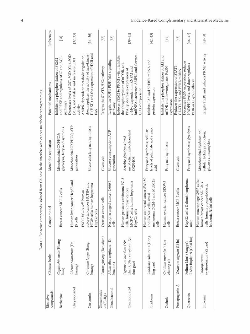
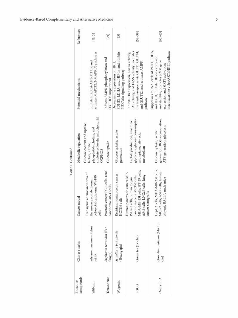
6.1. Chinese Herbs Interfere with Cancer Cell Metabolismthrough Many Pathways and Targets. We have shown pre-viously that berberine, an alkaloid isolated from Chineseherbs, interferes with cancer cell metabolism through manydifferent pathways and targets, including but not limitedto glycolysis (PKM2, PFKP) and fatty acid synthesis (ACC,ACL) [31, 93, 94]. Other herbal derivatives also interfere withcancer cell metabolism in different ways, and their activitiesare summarized in Table 1. When compared to conventionalapproaches (surgery, radiation, and chemotherapeutic drugagents), agents like berberine are more tolerable and lesstoxic than these listed procedures whose use is the standardof care for cancer in Western medicine. Recent studies ofChinese medicine-affiliated compounds, examined throughthe lens of modern medical practices, have shown that theyare sustainable and progressive modes of antitumor activity.These findings should be interpreted against a large bodyof data (centuries of use of these agents) showing that theirsystemic cytotoxicity is low or absent [15].
4 Evidence-Based Complementary and Alternative Medicine
Table1:Bioactivec
ompo
unds
isolated
from
Chineseh
erbs
interfe
rewith
cancer
metabolicreprogramming.
Bioactive
compo
unds
Chineseh
erbs
Cancer
mod
elMetabolicregulation
Potentialm
echanism
sRe
ferences
Berberine
Coptischinensis
(Huang
lian)
Breastcancer
MCF
-7cells
Mito
chon
drialO
XPHOS;
glycolysis;
fatty
acid
synthesis
Inhibitsthep
hospho
rylatio
nof
PKM2
andPF
KPandregu
latesA
CCandAC
Lpathways
[31]
Chrysoph
anol
Rheum
palm
atum
(Da
huang)
Hum
anliver
cancer
Hep3B
and
J5cells
Mito
chon
drialO
XPHOS;AT
Pgeneratio
n
Decreases
levelsof
GST,SOD(Cu),SOD
(Mn),and
catalase
andincreasesL
DH
activ
ity[32,33]
Curcum
inCu
rcum
alonga(Jiang
huang)
ESCC
EC109cell;hu
man
colorectalcancer
HCT
116and
HT2
9cells;hum
anhepatoma
HepG2cells
Glycolysis
;fattyacid
synthesis
AMPK
-dependent
metabolicregu
latio
n;do
wnregulates
thea
ctivity
ofhexokinase
II(H
KII)andthee
xpressionof
HKI
Iand
FAS
[34–
36]
Ginseno
side
20(S)-Rg
3Pa
naxginseng(Ren
shen)
Ovaria
ncancer
cells
Glycolysis
Targetsthe
STAT
3/HK2
pathway
[37]
Neoalbacono
lAlbatre
llusconflu
ens(Di
huajun
)Nasop
haryngealcancerC
666-1
cells
Glucose
consum
ption;
ATP
generatio
nTargetsthe
PDK1-PI3K/
Akt
signalin
gpathway
[38]
Oleanolicacid
Ligustr
umlucid
um(N
vzhen);Olea
europaea
(Qi
dunguo)
Hum
anprostatecarcinom
aPC-
3cells;hum
anbreastcancer
MCF
-7cells;hum
anhepatoma
HepG2cells
Aerobicg
lycolysis;lipid
metabolism
;mito
chon
drial
OXP
HOS
Indu
cesP
KM2to
PKM1switch,inhibits
thep
hospho
rylatio
nof
mTO
R,and
PPAR𝛾
,decreases
expressio
nof
c-Myc-dependent
hnRN
PA1and
hnRN
PA1,activ
ates
AMPK
,and
elevates
COX-
2expressio
n
[39–
41]
Orid
onin
Rabdosiarubescens(Don
glin
gcao)
Hum
ancolorectalcancer
SW480
andSW
620cells;uveal
melanom
aOCM
-1andMUM2B
cells
Fatty
acid
synthesis
;cellular
levelsof
palm
itateandste
aric
acid
InhibitsFA
SandSR
EBP1
mRN
Aand
proteinexpressio
n[42,43]
Osth
ole
Cnidium
monnieri(She
chuang
zi)
Hum
anovariancancer
SKOV3
cells
Fatty
acid
synthesis
Inhibitsthep
hospho
rylatio
nof
Akt
and
mTO
Randdo
wnregulates
FASN
expressio
n[44]
Prosapogenin
AVeratru
mnigrum
(Lilu)
Breastcancer
MCF
-7cells
Glycolysis
Redu
cesthe
expressio
nof
STAT
3,GLU
T1,H
K,andPF
KLmRN
A[45]
Quercetin
Foliu
mMori(Sang
ye);
RadixBu
pleuri(C
haih
u)HepG2cells;D
alton’s
lymph
oma
mice
Fatty
acid
synthesis
;glycolysis
Decreases
FASN
expressio
n,redu
ces
NADPH
levels,
anddo
wnregulates
PI3K
-AKT
-p53
pathway
[46,47]
Shikon
inLithosperm
umerythrorhizon(Zicao)
Hum
anmacroph
ageU
937cells;
human
breastcancer
SK-BR-3
cells;hum
anprom
yeloblastic
leuk
emiaHL6
0cells
Mito
chon
driald
ysfunctio
n;cellu
larlactateprod
uctio
n;glucosec
onsumption
TargetsT
rxR1
andinhibitsPK
M2activ
ity[48–50]
Evidence-Based Complementary and Alternative Medicine 5
Table1:Con
tinued.
Bioactive
compo
unds
Chineseh
erbs
Cancer
mod
elMetabolicregulation
Potentialm
echanism
sRe
ferences
Silib
inin
Silyb
ummarianu
m(Shu
ifeiji)
Transgenicadenocarcino
mao
fthem
ouse
prostate;hum
ancolorectalcarcinom
aSW480
cells
Glucose
contentand
uptake;
lactate,citrate,
phosph
atidylcholine,and
cholesterollevels;mito
chon
drial
OXP
HOS
InhibitsPIK3
CA-AKT
-MTO
Rand
activ
ates
MAP2
K1/2-M
APK
1/3pathways
[51,52]
Tetrandrine
Stephaniatetra
ndra
(Fen
fang
ji)Prostatic
cancer
PC-3
cells;renal
carcinom
a786-O
cells
Glucose
uptake
Indu
cesA
MPK
phosph
orylationand
OXP
HOSim
pairm
ent
[29]
Wogon
inScutellariabaica
lensis
(Huang
qin)
Resistant
human
coloncancer
HCT
116cells
Glucose
uptake;lactate
generatio
n
Decreases
thee
xpressionof
HKI
I,PD
HK1
,LDHA,and
HIF-1𝛼andinhibits
PI3K
/Akt
signalin
gpathway
[53]
EGCG
Green
tea(
Lvcha)
Hum
anpancreaticcancer
MIA
PaCa
-2cells;hum
antong
uecarcinom
acells;
MCF
-7cells;
MDA-
MB-231cells;
HT-29
cells;
A549cells;LNCa
Pcells;lun
gcancer
xeno
graft
s
Lactatep
rodu
ction,
anaerobic
glycolysis;
glucosec
onsumption
andup
take;fattyacid
metabolism
InhibitsHK2
expressio
n,LD
HAactiv
ity,
FASactiv
ity,and
FASN
activ
ity;m
ediates
theinsulin-respo
nsev
iaGLU
T1,G
LUT4
;andGLU
T12,andactiv
ates
AMPK
pathway
[54–
59]
Oroxylin
AOroxylum
indicum
(Muhu
die)
HepG2cells;M
DA-
MB-231cells;
MCF
-7cells;A
549cells;fem
ale
athymicBA
LB/cnu
demice
Glucose
uptake;lactate
generatio
n;RO
Saccumulation;
ATPgeneratio
n;glycolysis
Supp
resses
mRN
Alevelsof
PDK1
,LDHA,
andHKII;inh
ibits
HIF-1𝛼expressio
nandsta
bility;prom
otes
SOD2gene
expressio
nandSIRT
3activ
ation;
inactiv
ates
thec
-Src/A
KT/H
KIIpathway
[60–
63]
6 Evidence-Based Complementary and Alternative Medicine
HKPFKPPFKL
PK-M2PHGDH
LDH
FASNACCACL
SREBP
Chinese herbsGlycolysis Fatty acid synthesis
ATP levelHIF-1𝛼PDHKPDH
IDHGDHGLSAMPK
Mitochondrial oxidative phosphorylation
Glucose transport
GLUTs
PRA EGCG
BER
BER
CHRY
CUR
CUR
CUR
Rg3
OLA
OLA ORDOSTPRA
QCTSHK
TET WGN
WGN
EGCG ORX
EGCGEGCGORX
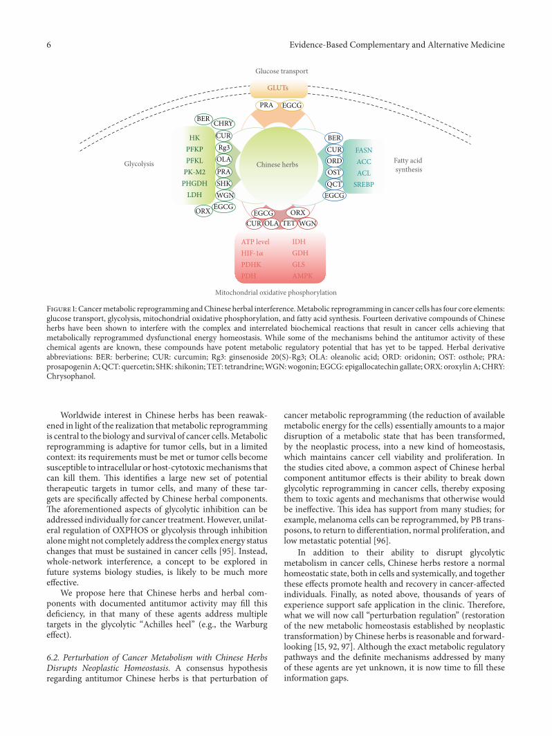
Figure 1: Cancermetabolic reprogramming andChinese herbal interference.Metabolic reprogramming in cancer cells has four core elements:glucose transport, glycolysis, mitochondrial oxidative phosphorylation, and fatty acid synthesis. Fourteen derivative compounds of Chineseherbs have been shown to interfere with the complex and interrelated biochemical reactions that result in cancer cells achieving thatmetabolically reprogrammed dysfunctional energy homeostasis. While some of the mechanisms behind the antitumor activity of thesechemical agents are known, these compounds have potent metabolic regulatory potential that has yet to be tapped. Herbal derivativeabbreviations: BER: berberine; CUR: curcumin; Rg3: ginsenoside 20(S)-Rg3; OLA: oleanolic acid; ORD: oridonin; OST: osthole; PRA:prosapogeninA;QCT: quercetin; SHK: shikonin; TET: tetrandrine;WGN:wogonin; EGCG: epigallocatechin gallate;ORX: oroxylinA;CHRY:Chrysophanol.
Worldwide interest in Chinese herbs has been reawak-ened in light of the realization thatmetabolic reprogrammingis central to the biology and survival of cancer cells.Metabolicreprogramming is adaptive for tumor cells, but in a limitedcontext: its requirements must be met or tumor cells becomesusceptible to intracellular or host-cytotoxicmechanisms thatcan kill them. This identifies a large new set of potentialtherapeutic targets in tumor cells, and many of these tar-gets are specifically affected by Chinese herbal components.The aforementioned aspects of glycolytic inhibition can beaddressed individually for cancer treatment. However, unilat-eral regulation of OXPHOS or glycolysis through inhibitionalonemight not completely address the complex energy statuschanges that must be sustained in cancer cells [95]. Instead,whole-network interference, a concept to be explored infuture systems biology studies, is likely to be much moreeffective.
We propose here that Chinese herbs and herbal com-ponents with documented antitumor activity may fill thisdeficiency, in that many of these agents address multipletargets in the glycolytic “Achilles heel” (e.g., the Warburgeffect).
6.2. Perturbation of Cancer Metabolism with Chinese HerbsDisrupts Neoplastic Homeostasis. A consensus hypothesisregarding antitumor Chinese herbs is that perturbation of
cancer metabolic reprogramming (the reduction of availablemetabolic energy for the cells) essentially amounts to a majordisruption of a metabolic state that has been transformed,by the neoplastic process, into a new kind of homeostasis,which maintains cancer cell viability and proliferation. Inthe studies cited above, a common aspect of Chinese herbalcomponent antitumor effects is their ability to break downglycolytic reprogramming in cancer cells, thereby exposingthem to toxic agents and mechanisms that otherwise wouldbe ineffective. This idea has support from many studies; forexample, melanoma cells can be reprogrammed, by PB trans-posons, to return to differentiation, normal proliferation, andlow metastatic potential [96].
In addition to their ability to disrupt glycolyticmetabolism in cancer cells, Chinese herbs restore a normalhomeostatic state, both in cells and systemically, and togetherthese effects promote health and recovery in cancer-affectedindividuals. Finally, as noted above, thousands of years ofexperience support safe application in the clinic. Therefore,what we will now call “perturbation regulation” (restorationof the new metabolic homeostasis established by neoplastictransformation) by Chinese herbs is reasonable and forward-looking [15, 92, 97]. Although the exact metabolic regulatorypathways and the definite mechanisms addressed by manyof these agents are yet unknown, it is now time to fill theseinformation gaps.
Evidence-Based Complementary and Alternative Medicine 7
6.3. ChineseHerbs andHerbal Derivatives Are PromisingTher-apeutics for Cancer and Other Metabolism-Related Diseases.Targeting glycolytic tumor reprogramming may be useful incombination with current treatments of cancer, for example,chemotherapy, by making lower doses of toxic agents moreeffective. Another promising use for Chinese herbs wouldbe in metabolic syndrome disorders, including diabetes,wherein their ability to perturb pathological homeostaticmechanisms (by perturbation, returning to normal intra-cellular and systemic metabolism) would offer a gentle andeffective treatment protocol for these diseases, whose basicpathophysiology involves harmfulmetabolic reprogrammingthat cannot be disrupted by current treatment regimens.
7. Conclusion
In this review, we propose that Chinese herbs and herbalcomponents with identified antitumor efficacy should bestudied in detail, toward the identification of multiple newtargets and pathways that are expressed in “neoplastic home-ostasis.”We have summarized several of the core changes thatoccur in metabolically reprogrammed cells and used these toillustrate where recent and older work have already given us anewdirection (perturbation of the aforementionedneoplastichomeostasis). This new direction provides a novel contextfor the identification of targets and pathways that exposetumor cells to the toxicity of Chinese herbs. As noted above,many compounds derived from Chinese herbs are known tohave potent metabolic regulatory potential, as indicated inFigure 1.
Conflict of Interests
The authors declared that they have no competing interests.
Authors’ Contribution
Mr. Zhangfeng Zhong and Mr. William W. Qiang preparedthe paper for final submission, and Dr. Wen Tan participatedin the early drafting process. Mr. Haotian Zhang and Mr.Shengpeng Wang revised the paper and provided valuablefeedback. Professor Yitao Wang, Dr. Chunming Wang andDr. Wenan Qiang conceived the paper, and Dr. Wenan Qiangorganized the paper revision process for final submission.All authors read and approved the paper. Zhangfeng Zhong,William W. Qiang, and Wen Tan contributed equally to thiswork.
Acknowledgments
This study was supported by the Macao Science andTechnology Development Fund (077/2011/A3 and048/2013/A2), the research Fund of University of Macau(UL016/09Y4/CMS/WYT01/ICMS and MYRG208 (Y3-L4)-ICMS11-WYT), and the Fundamental Research Funds forthe Central Universities (lzujbky-2014-148). Wenan Qiangwas supported by the Baskes Foundation and Robert H.Lurie Comprehensive Cancer Center at the Northwestern
University. The authors thank Dr. Virginia Scofield from theUniversity of Texas Rio Grande Valley, Edinburg, for hercritical suggestions and her proofreading of the paper. Wealso thank Stacy Ann Kujawa at Northwestern University forher proofreading of the paper.
References
[1] I. Nakano, “Therapeutic potential of targeting glucosemetabolism in glioma stem cells,” Expert Opinion onTherapeutic Targets, vol. 18, no. 11, pp. 1233–1236, 2014.
[2] M. Zhou, Y. Zhao, Y. Ding et al., “Warburg effect in chemosen-sitivity: targeting lactate dehydrogenase-A re-sensitizes Taxol-resistant cancer cells to Taxol,” Molecular Cancer, vol. 9, article33, 2010.
[3] Q. Cai, T. Lin, S. Kamarajugadda, and J. Lu, “Regulation ofglycolysis and theWarburg effect by estrogen-related receptors,”Oncogene, vol. 32, no. 16, pp. 2079–2086, 2013.
[4] C. Capparelli, C. Guido, D. Whitaker-Menezes et al.,“Autophagy and senescence in cancer-associated fibroblastsmetabolically supports tumor growth and metastasis, viaglycolysis and ketone production,” Cell Cycle, vol. 11, no. 12, pp.2285–2302, 2012.
[5] J. Yu, J. Shen, T. T. Sun, X. Zhang, andN.Wong, “Obesity, insulinresistance, NASH and hepatocellular carcinoma,” Seminars inCancer Biology, vol. 23, no. 6, pp. 483–491, 2013.
[6] Y. Guo, X. Wang, L. Qiu et al., “Probing gender-specific lipidmetabolites and diagnostic biomarkers for lung cancer usingFourier transform ion cyclotron resonance mass spectrometry,”Clinica Chimica Acta, vol. 414, pp. 135–141, 2012.
[7] G. Shen, Y. Chen, J. Sun et al., “Time-course changes in potentialbiomarkers detected using a metabonomic approach in Walker256 tumor-bearing rats,” Journal of Proteome Research, vol. 10,no. 4, pp. 1953–1961, 2011.
[8] Y. Zhang, Z. Liu, X. Yu et al., “The association betweenmetabolic abnormality and endometrial cancer: a large case-control study in China,”Gynecologic Oncology, vol. 117, no. 1, pp.41–46, 2010.
[9] A.-H. Zhang, H. Sun, S. Qiu, and X.-J. Wang, “Metabolomics innoninvasive breast cancer,” Clinica Chimica Acta, vol. 424, pp.3–7, 2013.
[10] L.-X. Fan, C.-M. Liu, A.-H. Gao, Y.-B. Zhou, and J. Li, “Berber-ine combined with 2-deoxy-d-glucose synergistically enhancescancer cell proliferation inhibition via energy depletion andunfolded protein response disruption,” Biochimica et BiophysicaActa (BBA)—General Subjects, vol. 1830, no. 11, pp. 5175–5183,2013.
[11] Y. Wang, L. Zhang, W.-L. Chen et al., “Rapid diagnosis andprognosis of de novo acutemyeloid leukemia by serummetabo-nomic analysis,” Journal of Proteome Research, vol. 12, no. 10, pp.4393–4401, 2013.
[12] Y.-C. Li, S.-M. He, Z.-X. He et al., “Plumbagin inducesapoptotic and autophagic cell death through inhibition of thePI3K/Akt/mTOR pathway in human non-small cell lung cancercells,” Cancer Letters, vol. 344, no. 2, pp. 239–259, 2014.
[13] M. Jain, R. Nilsson, S. Sharma et al., “Metabolite profilingidentifies a key role for glycine in rapid cancer cell proliferation,”Science, vol. 336, no. 6084, pp. 1040–1044, 2012.
[14] A. P. Drabovich, M. P. Pavlou, A. Dimitromanolakis, andE. P. Diamandis, “Quantitative analysis of energy metabolicpathways in MCF-7 breast cancer cells by selected reaction
8 Evidence-Based Complementary and Alternative Medicine
monitoring assay,”Molecular andCellular Proteomics, vol. 11, no.8, pp. 422–434, 2012.
[15] W. L. W. Hsiao and L. Liu, “The role of traditional Chineseherbal medicines in cancer therapy—from TCM theory tomechanistic insights,” Planta Medica, vol. 76, no. 11, pp. 1118–1131, 2010.
[16] C. V. Diogo, N. G. Machado, I. A. Barbosa, T. L. Serafim, A.Burgeiro, and P. J. Oliveira, “Berberine as a promising safe anti-cancer agent—is there a role for mitochondria?” Current DrugTargets, vol. 12, no. 6, pp. 850–859, 2011.
[17] A. M. Mariga, F. Pei, W.-J. Yang et al., “Immunopotentiation ofPleurotus eryngii (DC. ex Fr.) Quel,” Journal of Ethnopharmacol-ogy, vol. 153, no. 3, pp. 604–614, 2014.
[18] W. S. Yu, S.-J. Jeong, J.-H. Kim et al., “The genome-wide expres-sion profile of 1,2,3,4,6-penta-O-galloyl-𝛽-D-glucose-treatedMDA-MB-231 breast cancer cells: molecular target on cancermetabolism,”Molecules andCells, vol. 32, no. 2, pp. 123–132, 2011.
[19] S. E. Elf and J. Chen, “Targeting glucose metabolism in patientswith cancer,” Cancer, vol. 120, no. 6, pp. 774–780, 2014.
[20] S. O. Nam, F. Yotsumoto, K. Miyata, N. Shirasu, S. Miyamoto,and M. Kuroki, “Possible therapeutic targets among themolecules involved in the warburg effect in tumor cells,”Anticancer Research, vol. 33, no. 7, pp. 2855–2860, 2013.
[21] A. Annibaldi and C.Widmann, “Glucose metabolism in cancercells,” Current Opinion in Clinical Nutrition andMetabolic Care,vol. 13, no. 4, pp. 466–470, 2010.
[22] R. K. Reyes, T. Motiwala, and S. T. Jacob, “Regulation ofglucose metabolism in hepatocarcinogenesis by microRNAs,”Gene Expression, vol. 16, no. 2, pp. 85–92, 2014.
[23] R. B. Hamanaka and N. S. Chandel, “Targeting glucosemetabolism for cancer therapy,” Journal of ExperimentalMedicine, vol. 209, no. 2, pp. 211–215, 2012.
[24] E. Madan, R. Gogna, M. Bhatt, U. Pati, P. Kuppusamy, and A.A. Mahdi, “Regulation of glucose metabolism by p53: emergingnew roles for the tumor suppressor,” Oncotarget, vol. 2, no. 12,pp. 948–957, 2011.
[25] Y.-Z. Li, S.-L. Li, X. Li et al., “Expression of endogenous hypoxiamarkers in vulvar squamous cell carcinoma,” Asian PacificJournal of Cancer Prevention, vol. 13, no. 8, pp. 3675–3680, 2012.
[26] L. Szablewski, “Expression of glucose transporters in cancers,”Biochimica et Biophysica Acta (BBA)—Reviews on Cancer, vol.1835, no. 2, pp. 164–169, 2013.
[27] K. Adekola, S. T. Rosen, and M. Shanmugam, “Glucose trans-porters in cancer metabolism,” Current Opinion in Oncology,vol. 24, no. 6, pp. 650–654, 2012.
[28] F. Nualart, M. Los Angeles Garcia, R. A. Medina, and G. I.Owen, “Glucose transporters in sex steroid hormone relatedcancer,” Current Vascular Pharmacology, vol. 7, no. 4, pp. 534–548, 2009.
[29] W. Qiu, M. Su, F. Xie et al., “Tetrandrine blocks autophagic fluxand induces apoptosis via energetic impairment in cancer cells,”Cell Death and Disease, vol. 5, no. 3, Article ID e1123, 2014.
[30] S. Meng, J. Cao, Q. Feng, J. Peng, and Y. Hu, “Roles ofchlorogenic acid on regulating glucose and lipids metabolism:a review,” Evidence-Based Complementary and AlternativeMedicine, vol. 2013, Article ID 801457, 11 pages, 2013.
[31] W. Tan, N. Li, R. Tan et al., “Berberine interfered with breastcancer cells metabolism, balancing energy homeostasis,” Anti-Cancer Agents in Medicinal Chemistry, vol. 15, no. 1, pp. 66–78,2014.
[32] C.-H. Ni, P.-Y. Chen, H.-F. Lu et al., “Chrysophanol-inducednecrotic-like cell death through an impaired mitochondrialATP synthesis in Hep3B human liver cancer cells,” Archives ofPharmacal Research, vol. 35, no. 5, pp. 887–895, 2012.
[33] C.-C. Lu, J.-S. Yang, A.-C. Huang et al., “Chrysophanol inducesnecrosis through the production of ROS and alteration of ATPlevels in J5 human liver cancer cells,” Molecular Nutrition andFood Research, vol. 54, no. 7, pp. 967–976, 2010.
[34] F. J. Zhang, H. S. Zhang, Y. Liu, and Y. H. Huang, “Curcumininhibits Ec109 cell growth via an AMPK-mediated metabolicswitch,” Life Sciences, vol. 134, pp. 49–55, 2015.
[35] K.Wang,H. Fan, Q. Chen et al., “Curcumin inhibits aerobic gly-colysis and induces mitochondrial-mediated apoptosis throughhexokinase II in human colorectal cancer cells in vitro,” Anti-Cancer Drugs, vol. 26, no. 1, pp. 15–24, 2014.
[36] H. Fan, W. Tian, and X. Ma, “Curcumin induces apoptosisof HepG2 cells via inhibiting fatty acid synthase,” TargetedOncology, vol. 9, no. 3, pp. 279–286, 2014.
[37] J. Li, T. Liu, L. Zhao et al., “Ginsenoside 20(S)-Rg3 inhibitsthe Warburg effect through STAT3 pathways in ovarian cancercells,” International Journal of Oncology, vol. 46, no. 2, pp. 775–781, 2015.
[38] Q. Deng, X. Yu, L. Xiao et al., “Neoalbaconol induces energydepletion and multiple cell death in cancer cells by targetingPDK1-PI3-K/Akt signaling pathway,” Cell Death & Disease, vol.4, no. 9, article e804, 2013.
[39] J. Liu, N. Wu, L. Ma et al., “Oleanolic acid suppresses aerobicglycolysis in cancer cells by switching pyruvate kinase type Misoforms,” PLoS ONE, vol. 9, no. 3, Article ID e91606, 2014.
[40] J. Liu, L. Zheng, N.Wu et al., “Oleanolic acid induces metabolicadaptation in cancer cells by activating the AMP-activatedprotein kinase pathway,” Journal of Agricultural and FoodChemistry, vol. 62, no. 24, pp. 5528–5537, 2014.
[41] X. Wang, H. Bai, X. Zhang et al., “Inhibitory effect of oleanolicacid on hepatocellular carcinoma via ERK-p53-mediated cellcycle arrest and mitochondrial-dependent apoptosis,” Carcino-genesis, vol. 34, no. 6, pp. 1323–1330, 2013.
[42] H.-Y. Kwan, Z. Yang, W.-F. Fong, Y.-M. Hu, Z.-L. Yu, andW.-L.W.Hsiao, “The anticancer effect of oridonin is mediated by fattyacid synthase suppression in human colorectal cancer cells,”Journal of Gastroenterology, vol. 48, no. 2, pp. 182–192, 2013.
[43] Z. Gu, X. Wang, R. Qi et al., “Oridonin induces apoptosis inuveal melanoma cells by upregulation of Bim and downreg-ulation of Fatty Acid Synthase,” Biochemical and BiophysicalResearch Communications, vol. 457, no. 2, pp. 187–193, 2015.
[44] V. C.-H. Lin, C.-H. Chou, Y.-C. Lin et al., “Osthole suppressesfatty acid synthase expression in HER2-overexpressing breastcancer cells through modulating Akt/mTOR pathway,” Journalof Agricultural and Food Chemistry, vol. 58, no. 8, pp. 4786–4793, 2010.
[45] T.-X. Wang, Z.-Q. Zhang, Y. Cong, X.-Y. Shi, Y.-H. Liu, and F.-L. Zhao, “Prosapogenin A induces apoptosis in human cancercells in vitro via inhibition of the STAT3 signaling pathway andglycolysis,” Oncology Letters, vol. 6, no. 5, pp. 1323–1328, 2013.
[46] P. Zhao, J.-M. Mao, S.-Y. Zhang, Z.-Q. Zhou, Y. Tan, and Y.Zhang, “Quercetin induces HepG2 cell apoptosis by inhibitingfatty acid biosynthesis,” Oncology Letters, vol. 8, no. 2, pp. 765–769, 2014.
[47] A. K. Maurya and M. Vinayak, “Quercetin Regresses Dalton’sLymphoma Growth via Suppression of PI3K/AKT SignalingLeading to Upregulation of p53 and Decrease in EnergyMetabolism,” Nutrition and Cancer, 2015.
Evidence-Based Complementary and Alternative Medicine 9
[48] B. Wiench, T. Eichhorn, M. Paulsen, and T. Efferth, “Shikonindirectly targets mitochondria and causes mitochondrial dys-function in cancer cells,” Evidence-Based Complementary andAlternative Medicine, vol. 2012, Article ID 726025, 15 pages,2012.
[49] D. Duan, B. Zhang, J. Yao, Y. Liu, and J. Fang, “Shikonin tar-gets cytosolic thioredoxin reductase to induce ROS-mediatedapoptosis in human promyelocytic leukemia HL-60 cells,” FreeRadical Biology and Medicine, vol. 70, pp. 182–193, 2014.
[50] J. Chen, J. Xie, Z. Jiang, B.Wang, Y.Wang, and X. Hu, “Shikoninand its analogs inhibit cancer cell glycolysis by targeting tumorpyruvate kinase-M2,” Oncogene, vol. 30, no. 42, pp. 4297–4306,2011.
[51] K. Raina, N. J. Serkova, and R. Agarwal, “Silibinin feeding altersthe metabolic profile in TRAMP prostatic tumors: 1H-NMRS-based metabolomics study,” Cancer Research, vol. 69, no. 9, pp.3731–3735, 2009.
[52] K. Raina, C.Agarwal, R.Wadhwa,N. J. Serkova, andR.Agarwal,“Energy deprivation by silibinin in colorectal cancer cells: adouble-edged sword targeting both apoptotic and autophagicmachineries,” Autophagy, vol. 9, no. 5, pp. 697–713, 2013.
[53] H. Wang, L. Zhao, L.-T. Zhu et al., “Wogonin reverses hypoxiaresistance of human colon cancer HCT116 cells via downregula-tion of HIF-1𝛼 and glycolysis, by inhibiting PI3K/Akt signalingpathway,” Molecular Carcinogenesis, vol. 53, supplement 1, pp.E107–E118, 2014.
[54] Q.-Y. Lu, L. Zhang, J. K. Yee, V.-L.Go, andW.-N. Lee, “Metabolicconsequences of LDHA inhibition by epigallocatechin gal-late and oxamate in MIA PaCa-2 pancreatic cancer cells,”Metabolomics, vol. 11, no. 1, pp. 1–80, 2015.
[55] F. Gao, M. Li, W.-B. Liu et al., “Epigallocatechin gallate inhibitshuman tongue carcinoma cells via HK2-mediated glycolysis,”Oncology Reports, vol. 33, no. 3, pp. 1533–1539, 2015.
[56] L.Moreira, I. Araujo, T. Costa et al., “Quercetin and epigallocat-echin gallate inhibit glucose uptake and metabolism by breastcancer cells by an estrogen receptor-independent mechanism,”Experimental Cell Research, vol. 319, no. 12, pp. 1784–1795, 2013.
[57] J.-T. Hwang, J. Ha, I.-J. Park et al., “Apoptotic effect of EGCGin HT-29 colon cancer cells via AMPK signal pathway,” CancerLetters, vol. 247, no. 1-2, pp. 115–121, 2007.
[58] J. Relat, A. Blancafort, G. Oliveras et al., “Different fatty acidmetabolism effects of (-)-epigallocatechin-3-gallate and C75 inadenocarcinoma lung cancer,” BMC Cancer, vol. 12, article 280,2012.
[59] K. Brusselmans, E. De Schrijver, W. Heyns, G. Verhoeven, andJ. V. Swinnen, “Epigallocatechin-3-gallate is a potent naturalinhibitor of fatty acid synthase in intact cells and selectivelyinduces apoptosis in prostate cancer cells,” International Journalof Cancer, vol. 106, no. 6, pp. 856–862, 2003.
[60] Q. Dai, Q. Yin, L. Wei et al., “Oroxylin A regulates glucosemetabolism in response to hypoxic stress with the involvementof Hypoxia-inducible factor-1 in human hepatoma HepG2cells,”Molecular Carcinogenesis, 2015.
[61] L. Wei, Y. Zhou, C. Qiao et al., “Oroxylin A inhibits glycolysis-dependent proliferation of human breast cancer via promotingSIRT3-mediated SOD2 transcription and HIF1𝛼 destabiliza-tion,”Cell Death andDisease, vol. 6, no. 4, Article ID e1714, 2015.
[62] L. Wei, Y. Zhou, Q. Dai et al., “Oroxylin A induces dissociationof hexokinase II from the mitochondria and inhibits glycolysisby SIRT3-mediated deacetylation of cyclophilin D in breastcarcinoma,”Cell Death&Disease, vol. 4, no. 4, article e601, 2013.
[63] L. Wei, Q. Dai, Y. Zhou et al., “Oroxylin A sensitizes non-smallcell lung cancer cells to anoikis via glucose-deprivation-likemechanisms: C-Src and hexokinase II,”Biochimica et BiophysicaActa (BBA)—General Subjects, vol. 1830, no. 6, pp. 3835–3845,2013.
[64] S. Y. Lunt and M. G. Vander Heiden, “Aerobic glycolysis: meet-ing the metabolic requirements of cell proliferation,” AnnualReview of Cell and Developmental Biology, vol. 27, pp. 441–464,2011.
[65] S. Cardaci, E. Desideri, and M. R. Ciriolo, “Targeting aerobicglycolysis: 3-bromopyruvate as a promising anticancer drug,”Journal of Bioenergetics and Biomembranes, vol. 44, no. 1, pp.17–29, 2012.
[66] S. Ganapathy-Kanniappan, M. Vali, R. Kunjithapatham et al.,“3-Bromopyruvate: a new targeted antiglycolytic agent and apromise for cancer therapy,” Current Pharmaceutical Biotech-nology, vol. 11, no. 5, pp. 510–517, 2010.
[67] Q. Zhang, J. Pan, P. E. North et al., “Aerosolized 3-bromopyruvate inhibits lung tumorigenesis without causingliver toxicity,” Cancer Prevention Research, vol. 5, no. 5, pp.717–725, 2012.
[68] S. Pavlides, I. Vera, R. Gandara et al., “Warburg meetsautophagy: cancer-associated fibroblasts accelerate tumorgrowth and metastasis via oxidative stress, mitophagy, andaerobic glycolysis,” Antioxidants and Redox Signaling, vol. 16,no. 11, pp. 1264–1284, 2012.
[69] Z. Wang, D. Wang, S. Han et al., “Bioactivity-guided identi-fication and cell signaling technology to delineate the lactatedehydrogenase A inhibition effects of Spatholobus suberectus onbreast cancer,” PLoS ONE, vol. 8, no. 2, Article ID e56631, 2013.
[70] P. Miao, S. Sheng, X. Sun, J. Liu, and G. Huang, “LactatedehydrogenaseA in cancer: a promising target for diagnosis andtherapy,” IUBMB Life, vol. 65, no. 11, pp. 904–910, 2013.
[71] Z.-Y. Wang, T. Y. Loo, J.-G. Shen et al., “LDH-A silencingsuppresses breast cancer tumorigenicity through induction ofoxidative stress mediated mitochondrial pathway apoptosis,”Breast Cancer Research and Treatment, vol. 131, no. 3, pp. 791–800, 2012.
[72] X. Zha, F. Wang, Y. Wang et al., “Lactate dehydrogenase B iscritical for hyperactivemTOR-mediated tumorigenesis,”CancerResearch, vol. 71, no. 1, pp. 13–18, 2011.
[73] L. Huang, Z. Yu, T. Zhang, X. Zhao, and G. Huang, “HSP40interacts with pyruvate kinase M2 and regulates glycolysis andcell proliferation in tumor cells,” PLoS ONE, vol. 9, no. 3, ArticleID e92949, 2014.
[74] F.-T. Fan, C.-S. Shen, L. Tao et al., “PKM2 regulates hepato-cellular carcinoma cell epithelial-mesenchymal transition andmigration upon EGFR activation,” Asian Pacific Journal ofCancer Prevention, vol. 15, no. 5, pp. 1961–1970, 2014.
[75] Q. Sun, X. Chen, J. Ma et al., “Mammalian target of rapamycinup-regulation of pyruvate kinase isoenzyme type M2 is criticalfor aerobic glycolysis and tumor growth,” Proceedings of theNational Academy of Sciences of the United States of America,vol. 108, no. 10, pp. 4129–4134, 2011.
[76] M. Huttemann, I. Lee, A. Pecinova, P. Pecina, K. Przyklenk,and J. W. Doan, “Regulation of oxidative phosphorylation, themitochondrial membrane potential, and their role in humandisease,” Journal of Bioenergetics and Biomembranes, vol. 40, no.5, pp. 445–456, 2008.
[77] S. Dimauro and P. Rustin, “A critical approach to the therapy ofmitochondrial respiratory chain and oxidative phosphorylation
10 Evidence-Based Complementary and Alternative Medicine
diseases,” Biochimica et Biophysica Acta, vol. 1792, no. 12, pp.1159–1167, 2009.
[78] D.Whitaker-Menezes, U. E.Martinez-Outschoorn, N. Flomen-berg et al., “Hyperactivation of oxidative mitochondrialmetabolism in epithelial cancer cells in situ: visualizing thetherapeutic effects ofmetformin in tumor tissue,”Cell Cycle, vol.10, no. 23, pp. 4047–4064, 2011.
[79] K.M.Owens,M. Kulawiec,M.M.Desouki, A. Vanniarajan, andK. K. Singh, “Impaired OXPHOS complex III in breast cancer,”PLoS ONE, vol. 6, no. 8, Article ID e23846, 2011.
[80] U. Elia and E. Flescher, “Combined chemotherapy or biotherapywith jasmonates: targeting energy metabolism for cancer treat-ment,” Current Pharmaceutical Biotechnology, vol. 14, no. 3, pp.331–341, 2013.
[81] S.-Y. Liew, E. J. Stanbridge, K. Yusoff, and N. Shafee,“Hypoxia affects cellular responses to plant extracts,” Journal ofEthnopharmacology, vol. 144, no. 2, pp. 453–456, 2012.
[82] W. Zeng, P. Liu, W. Pan, S. R. Singh, and Y. Wei, “Hypoxia andhypoxia inducible factors in tumormetabolism,”Cancer Letters,vol. 356, no. 2, pp. 263–267, 2015.
[83] C.-M. Tang and J. Yu, “Hypoxia-inducible factor-1 as a thera-peutic target in cancer,” Journal of Gastroenterology and Hepa-tology, vol. 28, no. 3, pp. 401–405, 2013.
[84] R. P. Baumann, P. G. Penketh, H. A. Seow, K. Shyam, and A.C. Sartorelli, “Generation of oxygen deficiency in cell cultureusing a two-enzyme system to evaluate agents targeting hypoxictumor cells,” Radiation Research, vol. 170, no. 5, pp. 651–660,2008.
[85] Z. Yu, X. Zhao, Y. Ge et al., “A regulatory feedback loop betweenHIF-1𝛼 and PIM2 in HepG2 cells,” PLoS ONE, vol. 9, no. 2,Article ID e88301, 2014.
[86] W. Tian, Y. Wang, Y. Xu et al., “The hypoxia-inducible factorrenders cancer cells more sensitive to vitamin C-inducedtoxicity,”The Journal of Biological Chemistry, vol. 289, no. 6, pp.3339–3351, 2014.
[87] Y. Zhang, Y. Du, W. Le, K. Wang, N. Kieffer, and J. Zhang,“Redox control of the survival of healthy and diseased cells,”Antioxidants & Redox Signaling, vol. 15, no. 11, pp. 2867–2908,2011.
[88] R. Lupu and J. A. Menendez, “Pharmacological inhibitorsof Fatty Acid Synthase (FASN)—catalyzed endogenous fattyacid biogenesis: a new family of anti-cancer agents?” CurrentPharmaceutical Biotechnology, vol. 7, no. 6, pp. 483–494, 2006.
[89] S. Biswas, J. Lunec, andK. Bartlett, “Non-glucosemetabolism incancer cells—is it all in the fat?”Cancer andMetastasis Reviews,vol. 31, no. 3-4, pp. 689–698, 2012.
[90] W.-H. Zhao, C.-Y. Zhao, L.-F. Gao et al., “The novel inhibitoryeffect of pangdahai on fatty acid synthase,” IUBMB Life, vol. 60,no. 3, pp. 185–194, 2008.
[91] W.-X. Tian, L.-C. Li, X.-D.Wu, and C.-C. Chen, “Weight reduc-tion by Chinese medicinal herbs may be related to inhibition offatty acid synthase,” Life Sciences, vol. 74, no. 19, pp. 2389–2399,2004.
[92] C.-S. Cheng, Z. Wang, and J. Chen, “Targeting FASN in breastcancer and the discovery of promising inhibitors from naturalproducts derived from traditional Chinesemedicine,” Evidence-Based Complementary and Alternative Medicine, vol. 2014,Article ID 232946, 16 pages, 2014.
[93] W. Tan, J. Lu, M. Huang et al., “Anti-cancer natural productsisolated from Chinese medicinal herbs,” Chinese Medicine, vol.6, no. 1, article 27, 2011.
[94] W. Tan, Y. Li, M. Chen, and Y.Wang, “Berberine hydrochloride:anticancer activity and nanoparticulate delivery system,” Inter-national Journal of Nanomedicine, vol. 6, pp. 1773–1777, 2011.
[95] S. Rodrıguez-Enrıquez, J. C. Gallardo-Perez, A. Marın-Hernandez et al., “Oxidative phosphorylation as a target toarrest malignant neoplasias,” Current Medicinal Chemistry, vol.18, no. 21, pp. 3156–3167, 2011.
[96] J. Yin and X. Bi, “Generation and characterization of virus-freereprogrammed melanoma cells by the piggyBac transposon,”Journal of Cancer Research and Clinical Oncology, vol. 139, no.9, pp. 1591–1599, 2013.
[97] J. Yu, S. A. Nag, and R. Zhang, “Advances in translationalpharmacological investigations in identifying and validatingmolecular targets of natural product anticancer agents,”CurrentCancer Drug Targets, vol. 13, no. 5, pp. 596–609, 2013.

Submit your manuscripts athttp://www.hindawi.com
Stem CellsInternational
Hindawi Publishing Corporationhttp://www.hindawi.com Volume 2014
Hindawi Publishing Corporationhttp://www.hindawi.com Volume 2014
MEDIATORSINFLAMMATION
of
Hindawi Publishing Corporationhttp://www.hindawi.com Volume 2014
Behavioural Neurology
EndocrinologyInternational Journal of
Hindawi Publishing Corporationhttp://www.hindawi.com Volume 2014
Hindawi Publishing Corporationhttp://www.hindawi.com Volume 2014
Disease Markers
Hindawi Publishing Corporationhttp://www.hindawi.com Volume 2014
BioMed Research International
OncologyJournal of
Hindawi Publishing Corporationhttp://www.hindawi.com Volume 2014
Hindawi Publishing Corporationhttp://www.hindawi.com Volume 2014
Oxidative Medicine and Cellular Longevity
Hindawi Publishing Corporationhttp://www.hindawi.com Volume 2014
PPAR Research
The Scientific World JournalHindawi Publishing Corporation http://www.hindawi.com Volume 2014
Immunology ResearchHindawi Publishing Corporationhttp://www.hindawi.com Volume 2014
Journal of
ObesityJournal of
Hindawi Publishing Corporationhttp://www.hindawi.com Volume 2014
Hindawi Publishing Corporationhttp://www.hindawi.com Volume 2014
Computational and Mathematical Methods in Medicine
OphthalmologyJournal of
Hindawi Publishing Corporationhttp://www.hindawi.com Volume 2014
Diabetes ResearchJournal of
Hindawi Publishing Corporationhttp://www.hindawi.com Volume 2014
Hindawi Publishing Corporationhttp://www.hindawi.com Volume 2014
Research and TreatmentAIDS
Hindawi Publishing Corporationhttp://www.hindawi.com Volume 2014
Gastroenterology Research and Practice
Hindawi Publishing Corporationhttp://www.hindawi.com Volume 2014
Parkinson’s Disease
Evidence-Based Complementary and Alternative Medicine
Volume 2014Hindawi Publishing Corporationhttp://www.hindawi.com