review article brain reorganization following intervention
TRANSCRIPT
Hindawi Publishing CorporationNeural PlasticityVolume 2013, Article ID 356275, 8 pageshttp://dx.doi.org/10.1155/2013/356275
Review ArticleBrain Reorganization following Intervention in Children withCongenital Hemiplegia: A Systematic Review
E. Inguaggiato,1,2 G. Sgandurra,2 S. Perazza,2,3 A. Guzzetta,2 and G. Cioni2,4
1 Scuola Superiore Sant’Anna, Piazza Martiri della Liberta, I-56127 Pisa, Italy2 Department of Developmental Neuroscience, IRCCS Stella Maris Scientific Institute, Via dei Giacinti 2,Calambrone, I-56128 Pisa, Italy
3 Physical and Rehabilitation Medicine, University of Rome Tor Vergata, I-00173 Rome, Italy4Department of Clinical and Experimental Medicine, University of Pisa, I-56126 Pisa, Italy
Correspondence should be addressed to A. Guzzetta; [email protected]
Received 26 July 2013; Revised 29 October 2013; Accepted 30 October 2013
Academic Editor: Alessandro Sale
Copyright © 2013 E. Inguaggiato et al. This is an open access article distributed under the Creative Commons Attribution License,which permits unrestricted use, distribution, and reproduction in any medium, provided the original work is properly cited.
Noninvasive rehabilitation strategies for children with unilateral cerebral palsy are routinely used to improve handmotor function,activity, and participation. Nevertheless, the studies exploring their effects on brain structure and function are very scarce. Recently,structural neuroplasticity was demonstrated in adult poststroke patients, in response to neurorehabilitation. Our purpose is toreview current evidence on the effects of noninvasive intervention strategies on brain structure or function, in children withunilateral cerebral palsy. The main literature databases were searched up to October 2013. We included studies where the effectsof upper limb training were evaluated at neurofunctional and/or neurostructural levels. Only seven studies met our selectioncriteria; selected studies were case series, six using the intervention of the constraint-induced movement therapy (CIMT) andone used virtual reality therapy (VR). CIMT and VR seem to produce measurable neuroplastic changes in sensorimotor cortexassociated with enhancement of motor skills in the affected limb. However, the level of evidence is limited, due to methodologicalweaknesses and small sample sizes of available studies. Well-designed and larger experimental studies, in particular RCTs, areneeded to strengthen the generalizability of the findings and to better understand the mechanism of intervention-related brainplasticity in children with brain injury.
1. Introduction
Unilateral cerebral palsy (U-CP) is the most common typeof cerebral palsy (CP), with an incidence of 1 in 1000 live-births [1]. Typically, the upper limb (UL) is more involvedthan the lower, with impairments of spasticity, sensation, andreduced strength. Effective use of the arm and hand to reach,grasp, release, and manipulate objects is often compromised.Children with hemiplegia usually have the intellectual capac-ity to attend regular school; however, impaired arm functionrestricts their participation in educational, leisure, and latervocational roles [2].
U-CP can result from a wide variety of brain lesions,with respect to the timing of insults (acquired during thepre-, peri- or postnatal period), and the type of structuralpathology (brain malformations, periventricular lesions, and
corticosubcortical lesions) [3]. U-CP often leads to delays inmotor development or deconditioning of the affected limb,as individuals are inclined to functional compensation withthe intact limb rather than attempting to use the involvedlimb [4]; this may result in suppression of developmentof cortical representation of the affected limb, and it mayfurther inhibit its functional use [5, 6]. When the lesionoccurs at an early stage of development, either during theintrauterine life or soon after birth, the mechanisms ofplastic (re-)organization of the sensory motor system can bedifferent from those observed at later stages of development[7]. Primary motor control of the hemiplegic upper limbcan be eventually maintained within the spared tissue of theaffected hemisphere (ipsilesional reorganization), or it can bereorganized within the unaffected hemisphere, as a result ofthe complete withdraw of the crossing fibers from the affected

2 Neural Plasticity
hemisphere and the survival of the fast-conducting ipsilateralmotor projections from the unaffected one (contralesionalreorganization) [8]. The type of reorganization can be influ-enced by the size and site of damage, but it appears stronglyinfluenced also by the experience following damage, that is,by the complex interaction between residual motor outputfrom the affected hemisphere and somatosensory feedbackfrom the affected limb [9].
In general terms, adaptive plasticity of the central nervoussystem (CNS) refers to functional and structural changesin the brain, which are advantageous to offset or improvefunctions; the term denotes several capacities including theability to adapt to changes in the environment and to storeinformation in memory associated with learning [10]. Thereis abundant evidence that the structure of certain braincircuits can change in response to environmental stimuli [11].Recently, structural neuroplasticity has been demonstratedin response to neurorehabilitation intervention in adultpoststroke patients. Gauthier et al. [12] have shown in strokepatients treated with CIMT a significant increase in graymatter volume in several regions, including bilateral primarysensory and motor areas, both hippocampi, and anteriorsupplementary motor area contralateral to the motor deficit[12].
In children with U-CP, several types of interventionhave been used to improve abilities of the affected limb(e.g., neurodevelopmental treatment, neuromuscular electri-cal stimulation, constraint-induced movement therapy, etc.).Compared to adult poststroke research, a relatively smallnumber of studies investigated the effects of rehabilitation onbrain reorganization. The purpose of this study has been toevaluate current evidence on brain reorganization in childrenwith U-CP following noninvasive intervention strategies.
2. Methods
Articles were identified through comprehensive searches ofcomputerized bibliographic databases: PubMed, MedLine(1973 to October 2013), CINAHL (Cumulative Index to Nurs-ing and Allied Health Literature) (up from 1994 to October2013), Web of Science (1992 to October 2013), and ERIC (pre-1966 to October 2013). We also searched for reviews on thistopic on the Cochrane Central Register of Controlled Trials,with no result.
The search explored Medical Subject Headings (MeSH)terms and text words:
(1) “cerebral palsy” or “hemiplegia”,(2) “child” or “adolescent” or “infant”,(3) “therapy” or “training” or “intervention”,(4) “MRI” or “fMRI” or “EEG” or “TMS” or “PET” or
“MEG” or “reorganization”.
Selection Criteria. To be included in this systematic review,studies had to meet the following criteria.
(1) Participants were diagnosed with U-CP.(2) Interventions to improve outcome were noninvasive
and did not include drugs.
(3) Outcomes included functional activities and evidenceof brain reorganization through neurophysiologicalexperiments, carried out before and after the inter-vention.
Studies were excluded if they
(1) reported only clinical measures as outcomes;(2) were case reports;(3) were not published in English.
The initial search yield was reviewed by only one revieweron the basis of title and abstract. All the studies emergedfrom the search focused on upper limb (UL) intervention.The search strategy allowed to identify 12 articles thatmet ourinclusion/exclusion criteria. The full-text articles were exam-ined by 3 reviewers, and the eligibility for study inclusionwas assessed independently; in case of mismatched opinionbetween the 3 reviewers, the eligibility of the study wasdiscussed together and consensuswas reached. Following oursearch in the different databases, only 5 eligible studies wereidentified while two additional ones were selected withintheir reference lists. The final analysis included 7 studies. Thegeneral purpose of the studies was to evaluate the effects ofnoninvasive rehabilitation strategies on brain reorganizationand on functional improvement of affected upper limb (UL)in unilateral cerebral palsy. In Figure 1, flow chart describesstudy selection and reasons for exclusion.
3. Results
3.1. StudyDesigns andParticipants. Selected studieswere caseseries; no controlled studies were found.
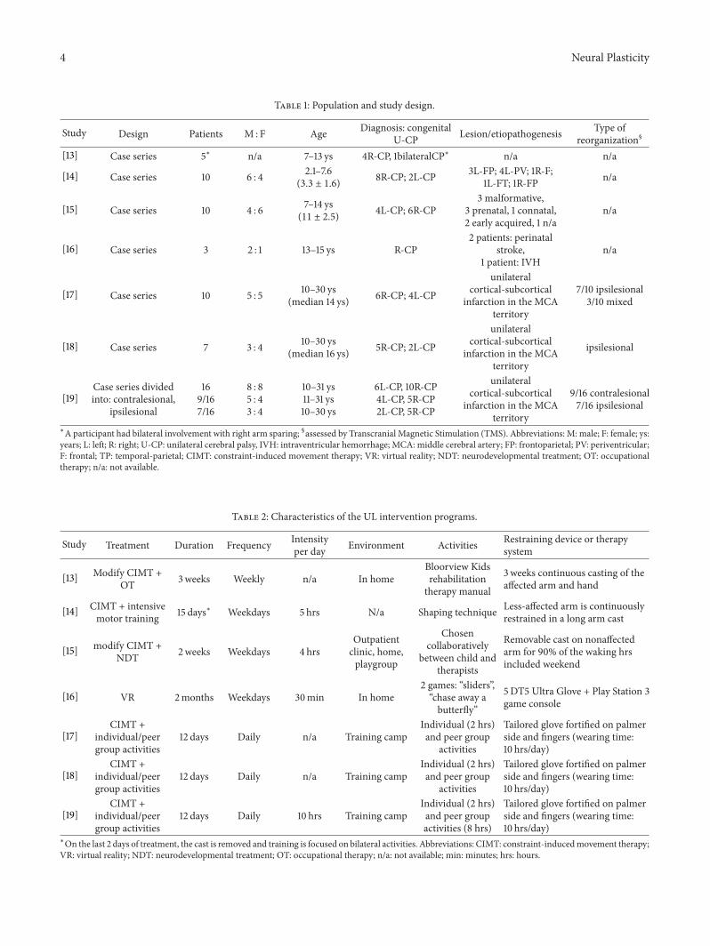
We found seven trials specifically targeted on childrenwith unilateral cerebral palsy; only in one case a participanthad bilateral impairment with right arm sparing [13].The agerange of participants was between 2.1 and 7.6 years in onestudy [14], between 7 and 14 years in two studies [13, 15],between 13 and 15 years in another study [16], and between10 and 30 years in the others [17–19]. Some studies wereperformed by the same research group and some subjects par-ticipated in more than one study [17–19]. Table 1 summarizesthe characteristics of the population for each study.
3.2. Type of Interventions. The most frequently proposedintervention, in six of the seven studies, was the constraint-induced movement therapy (CIMT); CIMT was used inassociation with neurodevelopmental treatment (NDT) [15],in association with occupational therapy (OT) [13], in associ-ation with intensive motor training [14] or during a trainingcamp in associationwith individual and peer groups activities[17–19]. Standard CIMT for children involves a restraintworn on the non-affected upper limb for 90% of wakinghours and 6hours/day of intensive intervention using shapingtechniques and massed practice typically over a 2-weekperiod [20]. The standard CIMT model has been adapted tobe less intensive (<6 hours/day) due to concerns that youngchildren are unable to participate in such intensive therapyregimen [21]. The remaining study employed an innovative

Neural Plasticity 3
7 studies were excluded:
Articles screened by title or abstract
n = 356
Articles identified according to our inclusion/exclusion criteria
n = 12
Articles excluded due to inappropriate population, outcome measures,
purpose, etc.n = 345
Full-text articles were examined by 3 reviewers independently
7 studies were included in final analysis
Two articles were selected within reference lists of eligible studies
[Junger et al.; 2007, Walther et al.; 2009]
Articles identified on Medline n = 147
Articles identified on CINAHL n = 29
Articles identified on web of science n = 30
Articles identifiedon ERIC n = 0
Articles identifiedon PubMed n = 150
2 = absent intervention; 1 = review;1 = absent neurophysiological
outcomes; 1 = used botulinum toxin;2 = case report
Figure 1: Flow chart of search strategy and selection process.
treatment strategy: virtual reality (VR) [16], a virtual envi-ronment system that uses new technologies to make thepatient perceptions similar to those coming from real-lifeactivities. In none of the studies children received botulinuminjections or upper limb surgery for the affected upper limbin the 6 months prior to intervention. Table 2 describes thecharacteristics of each UL intervention.
3.3. Outcome Measures. Selected studies were case series;no controlled studies were found. Studies aimed to evaluatethe effects of noninvasive intervention on (i) functionalityof UL, through scales and/or questionnaires and (ii) brainreorganization, through neuroimaging and neurophysiologi-cal techniques (i.e., MRI, fMRI, TMS, MEG). In three of thestudies [17–19], the different patterns of corticospinal reorga-nization (ipsilesional versus contralesional) were determinedby using TMS. Outcome measures were applied both beforeand after the intervention; in a few studies, assessments werealso recorded during followup [15, 18].
3.3.1. Upper Limb Function. The effects of noninvasive inter-vention on the functionality of the hemiplegic upper limbwere monitored using different functional measures, scales,or questionnaires. The type of clinical assessments varied
among studies; we therefore grouped the functional motoroutcomes in 3 categories, according to the dimensions ofthe International Classification of Functioning. Disabilityand Health (ICF-CY): (a) body functions and structures,(b) activities, and (c) participation [22]. (Table 1s sum-marizes clinical assessment and corresponding results; seeTable 1s in the Supplementary Material available online athttp://dx.doi.org/10.1155/2013/356275). Most of the studiesinvestigated functional motor outcomes according to at leastone of the dimensions of the International Classificationof Functioning, Disability and Health (ICF). The most fre-quently used outcomes measures were WMFT [17–19] in 3/7papers and P-MAL [13, 14, 18] in 3/7 papers.
3.3.2. Brain Reorganization. To evaluate the effects of theintervention on brain reorganization, 6 studies used func-tional magnetic resonance imaging (fMRI) [13, 15–19]. AllMRI experiments were performed on 1.5T scanner, butthe fMRI procedure and the tasks performed during theexamination were different among studies. Half of the sixstudies used, as fMRI task, active and passive movementsof the paretic and the nonparetic hands, while the otherhalf only performed an active task on the paretic hand. Onestudy combined fMRI with Transcranial Magnetic Stimu-lation (TMS) [18] and another combined fMRi, TMS, and
4 Neural Plasticity
Table 1: Population and study design.
Study Design Patients M : F Age Diagnosis: congenitalU-CP Lesion/etiopathogenesis Type of
reorganization§
[13] Case series 5∗ n/a 7–13 ys 4R-CP, 1bilateralCP∗ n/a n/a
[14] Case series 10 6 : 4 2.1–7.6(3.3 ± 1.6) 8R-CP; 2L-CP 3L-FP; 4L-PV; 1R-F;
1L-FT; 1R-FP n/a
[15] Case series 10 4 : 6 7–14 ys(11 ± 2.5) 4L-CP; 6R-CP
3 malformative,3 prenatal, 1 connatal,2 early acquired, 1 n/a
n/a
[16] Case series 3 2 : 1 13–15 ys R-CP2 patients: perinatal
stroke,1 patient: IVH
n/a
[17] Case series 10 5 : 5 10–30 ys(median 14 ys) 6R-CP; 4L-CP
unilateralcortical-subcortical
infarction in the MCAterritory
7/10 ipsilesional3/10 mixed
[18] Case series 7 3 : 4 10–30 ys(median 16 ys) 5R-CP; 2L-CP
unilateralcortical-subcortical
infarction in the MCAterritory
ipsilesional
[19]Case series dividedinto: contralesional,
ipsilesional
169/167/16
8 : 85 : 43 : 4
10–31 ys11–31 ys10–30 ys
6L-CP, 10R-CP4L-CP, 5R-CP2L-CP, 5R-CP
unilateralcortical-subcortical
infarction in the MCAterritory
9/16 contralesional7/16 ipsilesional
∗A participant had bilateral involvement with right arm sparing; §assessed by Transcranial Magnetic Stimulation (TMS). Abbreviations: M: male; F: female; ys:years; L: left; R: right; U-CP: unilateral cerebral palsy, IVH: intraventricular hemorrhage; MCA: middle cerebral artery; FP: frontoparietal; PV: periventricular;F: frontal; TP: temporal-parietal; CIMT: constraint-induced movement therapy; VR: virtual reality; NDT: neurodevelopmental treatment; OT: occupationaltherapy; n/a: not available.
Table 2: Characteristics of the UL intervention programs.
Study Treatment Duration Frequency Intensityper day Environment Activities Restraining device or therapy
system
[13] Modify CIMT +OT 3weeks Weekly n/a In home
Bloorview Kidsrehabilitationtherapy manual
3 weeks continuous casting of theaffected arm and hand
[14] CIMT + intensivemotor training 15 days∗ Weekdays 5 hrs N/a Shaping technique Less-affected arm is continuously
restrained in a long arm cast
[15] modify CIMT +NDT 2weeks Weekdays 4 hrs
Outpatientclinic, home,playgroup
Chosencollaboratively
between child andtherapists
Removable cast on nonaffectedarm for 90% of the waking hrsincluded weekend
[16] VR 2months Weekdays 30min In home2 games: “sliders”,“chase away abutterfly”
5DT5 Ultra Glove + Play Station 3game console
[17]CIMT +
individual/peergroup activities
12 days Daily n/a Training campIndividual (2 hrs)and peer group
activities
Tailored glove fortified on palmerside and fingers (wearing time:10 hrs/day)
[18]CIMT +
individual/peergroup activities
12 days Daily n/a Training campIndividual (2 hrs)and peer group
activities
Tailored glove fortified on palmerside and fingers (wearing time:10 hrs/day)
[19]CIMT +
individual/peergroup activities
12 days Daily 10 hrs Training campIndividual (2 hrs)and peer groupactivities (8 hrs)
Tailored glove fortified on palmerside and fingers (wearing time:10 hrs/day)
∗On the last 2 days of treatment, the cast is removed and training is focused on bilateral activities. Abbreviations: CIMT: constraint-inducedmovement therapy;VR: virtual reality; NDT: neurodevelopmental treatment; OT: occupational therapy; n/a: not available; min: minutes; hrs: hours.
Neural Plasticity 5
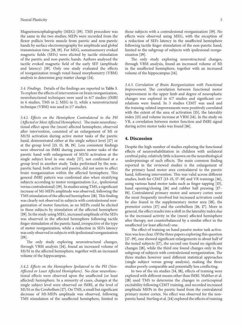
Magnetoencephalography (MEG) [19]. TMS procedure wasthe same in the two studies; MEPs were recorded from theflexor pollicis brevis muscle from paretic and non-paretichands by surface electromyography for amplitude and globaltransmission time [18, 19]. For MEG, somatosensory evokedmagnetic fields (SEFs) were elicited by tactile stimulationof the paretic and non-paretic hands. Authors analyzed thetactile evoked magnetic field of the early SEF (amplitudeand latency) [19]. Only one study evaluated the effectsof reorganization trough voxel-based morphometry (VBM)analysis to determine gray matter change [14].
3.4. Findings. Details of the findings are reported in Table 3.To explore the effects of intervention on brain reorganization,neurofunctional techniques were used in 6/7 studies (fMRIin 6 studies, TMS in 2, MEG in 1), while a neurostructuraltechnique (VBM) was used in 1/7 studies.
3.4.1. Effects on the Hemisphere Contralateral to the PH(Affected orMost AffectedHemisphere). Themain neurofunc-tional effect upon the (most) affected hemisphere, observedafter intervention, consisted of an enlargement of M1 orM1/S1 activation during active motor tasks of the paretichand, demonstrated either at the single subject level [17] orat the group level [13, 15, 18, 19]. Less consistent findingswere observed on fMRI during passive motor tasks of theparetic hand with enlargement of M1/S1 activation at thesingle subject level in one study [17], not confirmed at agroup level in another study. Tasks performed by the non-paretic hand, both active and passive, did not seem to affectbrain reorganization within the affected hemisphere. Thisgeneral fMRI pattern was confirmed also when stratifyingsubjects according to motor reorganization (i.e., ipsilesionalversus contralesional) [19]. In studies usingTMS, a significantincrease of M1-MEPs amplitude was observed, following theTMS stimulation of the affected hemisphere [18].This findingwas clearly not observed in subjects with contralesional reor-ganization of motor function, as no MEPs could be elicitedin these subjects by stimulation of the affected hemisphere[19]. In the study usingMEG, increased amplitude of the SEFswas observed in the affected hemisphere following tactilefinger stimulation of the paretic hand, irrespective of the typeof motor reorganization, while a reduction in SEFs latencywas only observed in subjects with ipsilesional reorganization[19].
The only study exploring neurostructural changes,through VBM analysis [14], found an increased volume ofM1/S1 in the affected hemisphere, together with an increasedvolume of the hippocampus.
3.4.2. Effects on the Hemisphere Ipsilateral to the PH (Non-Affected or Least Affected Hemisphere). No clear neurofunc-tional effects were observed upon the unaffected (or leastaffected) hemisphere. In a minority of cases, changes at thesingle subject level were observed on fMRI, at the level ofM1/S1 or the Cerebellum [17]. OnTMS, a small but significantdecrease of M1-MEPs amplitude was observed, followingTMS stimulation of the unaffected hemisphere, limited to
those subjects with a contralesional reorganization [19]. Noeffects were observed using MEG, with the exception ofa reduction of SEFs latency in the unaffected hemispherefollowing tactile finger stimulation of the non-paretic hand,limited to the subgroup of subjects with ipsilesional reorga-nization [19].
The only study exploring neurostructural changes,through VBM analysis, found an increased volume of M1in the unaffected hemisphere, together with an increasedvolume of the hippocampus [14].
3.4.3. Correlation of Brain Reorganization with FunctionalImprovement. The correlation between functional motorimprovement in the upper limb and degree of neuroplasticchanges was explored in 4/7 studies and significant cor-relations were found. In 3 studies CIMT was used andthe training-related improvements were positively correlatedwith the extent of the area of activation [15], the lateralityindex [13] and volume increase at VBM [14]. In the study onVR, a correlation between motor function and fMRI signalduring active motor tasks was found [16].
4. Discussion
Despite the high number of studies exploring the functionaleffects of neurorehabilitation in children with unilateralcerebral palsy, relatively little is known on the neurobiologicalunderpinnings of such effects. The main common findingreported in the reviewed studies is the enlargement ofthe primary hand motor area contralateral to the paretichand, following intervention. This was valid across differentstudies, both for CIMT [13–15, 17–19] and VR trainings [16],using various hand motor tasks such as finger tapping [15],hand opening/closing [16] and rubber ball pressing [17–19]. Contralateral primary motor and sensory cortex werethe most frequently involved but increased activation couldbe also found in the supplementary motor area [18], thepremotor cortex [17] and the cerebellum [16, 17]. More ingeneral, the effect results into a shift in the laterality index dueto the increased activity in the (most) affected hemisphereafter therapy, not counterbalanced by a similar effect in theunaffected (or least affected) one.
The effect of training on hand passive motor task activa-tion was less clear. Of the three papers exploring this question[17–19], one showed significant enlargements in about half ofthe tested subjects [17], the second one found no significantchanges [18], while the third one found changes only in thesubgroup of subjects with contralesional reorganization. Thethree studies however used different statistical approaches(single subject versus group analysis), making the threestudies poorly comparable and potentially less conflicting.
In two of the six studies [14, 18], effects of training wereexplored with different means other than fMRI.Walther et al.[18] used TMS to determine the changes in corticospinalexcitability following CIMT training, and recorded increasedamplitude MEPs in the paretic hand from the contralateralprimary motor cortex. No effect was observed for the non-paretic hand. Sterling et al. [14] explored the effects of training

6 Neural Plasticity
Table 3: Neuroimaging and neurophysiological outcome measures and results.
CIMTFunctional magnetic resonanceimaging PH N-PH Notes
Active movements fMRI task
Four-finger/wrist extension/flexion[13]
2/4 LI shift to contralateralhemisphere, 2/4 reduced LI(group stat; 𝑛 = 4)
—
Finger tapping [15] 6/7 (M1c) ↑ area of activation2/7 (M1c) ↑ signal (1–3%) —
3/10 were excluded due toartifacts (2/10) orclaustrophobia.fMRI task was tested on 5controls who showed M1cactivation.
Rubber ball press [17] 1/3 (M1S1c + M1S1i + CBMi +PMC) ↑ area of activation
4/10 M1S1c ↑ area of activation1/10 M1S1i ↑ area of activation
3/10 were excluded for PH taskdue to movement artifacts.
Rubber ball press [18] (M1S1c + SMA) ↑ area ofactivation (group stat; 𝑛 = 5) No changes (group stat; 𝑛 = 5) 2/7 were excluded for inability
to perform the task.
Rubber ball press [19]Ipsilesional group:(M1S1c + SMA) ↑ area ofactivation (group stat; 𝑛 = 5)
No changes (group stat; 𝑛 = 5) 2/7 were excluded for inabilityto perform the task.
Contralesional group(M1S1c + CBMi/c) ↑ area ofactivation(M1i) ↓ activation(group stat; 𝑛 = 6)
No changes (group stat; 𝑛 = 6) 3/9 were excluded formovement artefacts.
Passive movements
Flexion/extension at themetacarpophalangeal of fingersII–V of the patient’s hand [17]
4/8 (M1S1c) ↑ area of activation1/8 (M1S1i) ↑ area of activation2/8 (IHF) ↑ area of activation1/8 (CMBi) ↑ area of activation4/8 no changes observed
2/10 M1S1c ↑ area of activation1/10 IHF ↑ area of activation
2/10 were excluded for PH taskdue to movement artifacts.
Flexion/extension at themetacarpophalangeal of fingersII–V of the patient’s hand [18]
No changes (group stat; 𝑛 = 7) No changes (group stat; 𝑛 = 7)
Flexion/extension at themetacarpophalangeal of fingersII–V of the patient’s hand [19]
Ipsilesional group: no changes(group stat; 𝑛 = 7)
Ipsilesional group: no changes(group stat; 𝑛 = 7)
Contralesional groupParietal operculumc + M2S2c ↓activation(group stat; 𝑛 = 9)
Contralesional groupM1S1c ↓ activation(group stat; 𝑛 = 9)
Voxel-based morphometry Posttreatment—pretreatment Pretreatment—baseline Notes
VBM [14] (M1S1c + M1i + Hippocampi) ↑volume (group stat; 𝑛 = 10)
No changes (group stat;𝑛 = 10)
Transcranial magnetic stimulation PH N-PH Notes
TMS [18] (M1-MEPs) ↑ amplitude(group stat; 𝑛 = 7)
No changes(group stat; 𝑛 = 7)
TMS [19] amplitude Ipsilesional group:(M1-MEPs) ↑ amplitude(group stat; 𝑛 = 7)
Ipsilesional group:No changes(group stat; 𝑛 = 7)
Contralesional group:(M1-MEPs) ↓ amplitude(group stat; 𝑛 = 9)
Contralesional group:(M1-MEPs) ↓ amplitude(group stat; 𝑛 = 9)
TMS [19] conduction time
Ipsilesional group:No changes (group stat; 𝑛 = 7)Contralesional group:No changes (group stat; 𝑛 = 9)
Ipsilesional group:No changes (group stat; 𝑛 = 7)Contralesional group:No changes (group stat; 𝑛 = 9)

Neural Plasticity 7
Table 3: Continued.
CIMTMagnetoencephalography PH N-PH Notes
MEG [19] latency
Ipsilesional group:↓ early-SEF latency (group stat;𝑛 = 7)Contralesional group:No changes in early-SEF latency(group stat; 𝑛 = 8)
Ipsilesional group:↓ early-SEF latency (groupstat; 𝑛 = 7)Contralesional group:No changes in early-SEFlatency.(group stat; 𝑛 = 8)
1/9 was excluded due to strongmagnetic artefacts.
MEG [19] amplitude
Ipsilesional group:↑ early-SEF amplitude (groupstat; 𝑛 = 7)Contralesional group:↑ early SEF amplitude(group stat; 𝑛 = 8)
Ipsilesional group:≈early-SEF amplitude (groupstat; 𝑛 = 7)Contralesional group:No changes SEF amplitude(group stat; 𝑛 = 8)
1/9 was excluded due to strongmagnetic artefacts.
VRFunctional magnetic resonanceimaging PH N-PH Notes
Active movements fMRI task
Hand open/close [16] 2/3 (M1c) ↑ area of activation2/3 (CBM) ↑ area of activation — Training dose was variable in
the 3 cases.Abbreviations: PH: paretic hand; N-PH: nonparetic hand; fMRI: functional magnetic resonance imaging; VBM: voxel-based morphometry; TMS: transcranialmagnetic stimulation, MEG: magnetoencephalography; M1: primary motor cortex; FP: frontoparietal; M1S1: primary sensory motor cortex, M2S2c secondarysensory motor cortex c/i: indicate contralateral/ipsilateral, CBM: cerebellum, IHF interhemispheric fissure (including cingulate motor area supplementarymotor area), PMC: premotor cortex; LI: lateral index; LI is calculated [(contralateral − ipsilateral)/(contralateral + ipsilateral)]; SMA: supplementary motorarea, MEPs: motor evoked potentials; SEF: somatosensory evoked potentials.
on a structural level by using VBM analysis. This is alsothe only paper with an actual control condition consistingof a same-length interval pretraining used to explore brainchanges unrelated to intervention. It is of interest that whileno changes were observed from baseline to pretreatment, asignificant volume increase was observed at a group levelposttreatment in the primary motor cortex bilaterally, in thecontralateral primary sensory area and in both hippocampi.
Not surprisingly, a key factor influencing treatment-related brain neuroplasticity appeared to be the type ofreorganization of the corticospinal tract (i.e., ipsilesionalor contralesional). Type of reorganization was taken intoaccount in 3/7 studies; these studies came from the sameresearch group, with the most recent one [19] confirming andexpanding the results of the previous two [17, 18]. Althoughthe overall small figures do not allow for definite conclusions,there appears to be enough evidence supporting the existenceof two types of treatment-related neuroplasticity with themain hallmark of an increase in M1 excitability in subjectswith ipsilesional reorganization and of a decrease in M1excitability in subjects with contralesional reorganization.
Positive effects of training on handmotor function, in theselected studies, were almost invariably reported, althoughthe outcome measures used were very heterogeneous. Whencorrelating functional improvements with the amount ofplastic brain reorganization, significant results were generallyobserved after intervention, including enlarged area of M1S1activation in fMRI, increased M1-MEPs amplitude fromstimulation of the affected hemisphere, and increased M1S1
brain volumes on VBM. However, data are too scatteredand heterogeneous to allow for definite conclusions on thepossible correlations between neurobiological changes andfunctional improvements.
Themain limitation of the findings of this review is relatedto the number and type of papers found in our systematicsearch. Studies included in this review consist of quasi-experimental or descriptive pre-post designs. Their level ofevidence, based upon a modified Sackett score [23] adaptedto include PEDro ratings, is between 2b for CIMT studiesand 5 for VR. It is of great interest that none of the studiesselectedwas a randomized controlled study. Although severalRCTs have been performed comparing different trainings inchildren with unilateral cerebral palsy, some ethical prob-lems might have hindered the possibility of testing controlsubjects with relatively invasive techniques such as TMS.Nevertheless, since MRI, MEG, and EEG techniques, whenused without sedation, can be considered noninvasive, thereis no obvious reason why RCTs have not yet been performedusing these methods. Lack of RCTs might be more simplyjustified by this field of research being relatively new and thistype of study design being more complex.
In summary, noninvasive rehabilitation strategies seem toproduce measurable neuroplastic changes in sensory motorcortex associated with enhancement of motor skills in theaffected limb. This conclusion is however largely restricteddue to the strong limitations of the reviewed studies, themostrelevant of which concerns their methodological characteris-tics. It is also important to underline that the selected studies

8 Neural Plasticity
only investigated the effects of two types of intervention,namely, CIMT and VR, making therefore our conclusionsnot applicable to other approaches. For the same reason,this review cannot provide any contribution to the definitionof the type of intervention that should be recommendedin children with U-CP. Well-designed experimental studieswith larger sample sizes should be carried out to strengthenthe generalizability of these preliminary findings. Moreover,for further studies it would be important to investigatethe clinical outcomes according to the dimensions of ICFwith the best measures created for children with hemiplegiaconsidering psychometric properties. More researches, andin particular RCT studies, are needed to better understand themechanism of brain plasticity in children with brain injuryand to inform and fine-tune current or novel rehabilitationstrategies in children with cerebral palsy.
Conflict of Interests
The authors declare that there is no conflict of interests.
References
[1] http://www.cerebralpalsysource.com/.[2] L. Sakzewski, J. Ziviani, and R. Boyd, “Systematic review and
meta-analysis of therapeutic management of upper-limb dys-function in childrenwith congenital hemiplegia,”Pediatrics, vol.123, no. 6, pp. e1111–e1122, 2009.
[3] M. Staudt,W.Grodd, C.Gerloff,M. Erb, J. Stitz, and I. Krageloh-Mann, “Two types of ipsilateral reorganization in congenitalhemiparesis: a TMS and fMRI study,” Brain, vol. 125, no. 10, pp.2222–2237, 2002.
[4] J. Held, “Recovery of function after brain damage: theoreticalimplications for therapeutic intervention,” inMovement Science:Foundations for Physical Therapy in Rehabilitation, J. H. Carrand R. B. Shepherd, Eds., pp. 189–211, Aspen, Oxford, UK, 2ndedition, 2000.
[5] P. Cicinelli, R. Traversa, and P. M. Rossini, “Post-stroke reor-ganization of brain motor output to the hand: a 2–4 monthfollow-up with focal magnetic transcranial stimulation,” Elec-troencephalography and Clinical Neurophysiology, vol. 105, no.6, pp. 438–450, 1997.
[6] J. Liepert, H. Bauder, W. H. R. Miltner, E. Taub, and C. Weiller,“Treatment-induced cortical reorganization after stroke inhumans,” Stroke, vol. 31, no. 6, pp. 1210–1216, 2000.
[7] J. A. Eyre, “Corticospinal tract development and its plasticityafter perinatal injury,” Neuroscience and Biobehavioral Reviews,vol. 31, no. 8, pp. 1136–1149, 2007.
[8] M. Staudt, C. Gerloff, W. Grodd, H. Holthausen, G. Nie-mann, and I. Krageloh-Mann, “Reorganization in congenitalhemiparesis acquired at different gestational ages,” Annals ofNeurology, vol. 56, no. 6, pp. 854–863, 2004.
[9] A. Guzzetta, P. Bonanni, L. Biagi et al., “Reorganisation ofthe somatosensory system after early brain damage,” ClinicalNeurophysiology, vol. 118, no. 5, pp. 1110–1121, 2007.
[10] M.V. Johnston, “Clinical disorders of brain plasticity,”Brain andDevelopment, vol. 26, no. 2, pp. 73–80, 2004.
[11] R. Chen, L. G. Cohen, and M. Hallett, “Nervous systemreorganization following injury,”Neuroscience, vol. 111, no. 4, pp.761–773, 2002.
[12] L. V. Gauthier, E. Taub, C. Perkins, M. Ortmann, V. W. Mark,and G. Uswatte, “Remodeling the brain: plastic structural brainchanges produced by different motor therapies after stroke,”Stroke, vol. 39, no. 5, pp. 1520–1525, 2008.
[13] T. L. Sutcliffe, W. J. Logan, and D. L. Fehlings, “Pedi-atric constraint-induced movement therapy is associated withincreased contralateral cortical activity on functional magneticresonance imaging,” Journal of Child Neurology, vol. 24, no. 10,pp. 1230–1235, 2009.
[14] C. Sterling, E. Taub, D. Davis et al., “Structural neuroplasticchange after constraint-induced movement therapy in childrenwith cerebral palsy,” Pediatrics, vol. 131, no. 5, pp. e1664–e1669,2013.
[15] S. M. Cope, X. C. Liu, M. D. Verber, C. Cayo, S. Rao, andJ. C. Tassone, “Upper limb function and brain reorganizationafter constraint-induced movement therapy in children withhemiplegia,” Developmental Neurorehabilitation, vol. 13, no. 1,pp. 19–30, 2010.
[16] M. R. Golomb, B. C. McDonald, S. J. Warden et al., “In-homevirtual reality videogame telerehabilitation in adolescents withhemiplegic cerebral palsy,” Archives of Physical Medicine andRehabilitation, vol. 91, no. 1, pp. 1.e1–8.e1, 2010.
[17] H. Juenger, M. Linder-Lucht, M. Walther, S. Berweck, V.Mall, and M. Staudt, “Cortical neuromodulation by constraint-induced movement therapy in congenital hemiparesis: an fMRIstudy,” Neuropediatrics, vol. 38, no. 3, pp. 130–136, 2007.
[18] M. Walther, H. Juenger, N. Kuhnke et al., “Motor cortexplasticity in ischemic perinatal stroke: a transcranial magneticstimulation and functionalMRI study,” Pediatric Neurology, vol.41, no. 3, pp. 171–178, 2009.
[19] H. Juenger, N. Kuhnke, C. Braun et al., “Two types of exercise-induced neuroplasticity in congenital hemiparesis: a tran-scranial magnetic stimulation, functional MRI, and magne-toencephalography study,” Developmental Medicine and ChildNeurology, vol. 55, no. 10, pp. 941–951, 2013.
[20] E. Taub, A. Griffin, J. Nick, K. Gammons, G. Uswatte, and C.R. Law, “Pediatric CI therapy for stroke-induced hemiparesis inyoung children,”Developmental Neurorehabilitation, vol. 10, no.1, pp. 3–18, 2007.
[21] A. Eliasson, L. Krumlinde-Sundholm, K. Shaw, and C. Wang,“Effects of oncstraint-induced movement therapy in youngchildren with hemiplegic cerebral palsy: an adapted model,”Developmental Medicine and Child Neurology, vol. 47, no. 4, pp.266–275, 2005.
[22] M. Leonardi and A. Martinuzzi, “ICF and ICF-CY for an inno-vative holistic approach to persons with chronic conditions,”Disability and Rehabilitation, vol. 31, supplement 1, pp. S83–S87,2009.
[23] D. L. Sackett,W. S. Richardson,W.Rosenberg, andR. B.Haynes,Eds., Evidence-BasedMedicine: How to Practice and Teach EBM,Churchill Livingstone, New York, NY, USA, 2nd edition, 2000.

Submit your manuscripts athttp://www.hindawi.com
Neurology Research International
Hindawi Publishing Corporationhttp://www.hindawi.com Volume 2014
Alzheimer’s DiseaseHindawi Publishing Corporationhttp://www.hindawi.com Volume 2014
International Journal of
ScientificaHindawi Publishing Corporationhttp://www.hindawi.com Volume 2014
Hindawi Publishing Corporationhttp://www.hindawi.com Volume 2014
BioMed Research International
Hindawi Publishing Corporationhttp://www.hindawi.com Volume 2014
Research and TreatmentSchizophrenia
The Scientific World JournalHindawi Publishing Corporation http://www.hindawi.com Volume 2014
Hindawi Publishing Corporationhttp://www.hindawi.com Volume 2014
Neural Plasticity
Hindawi Publishing Corporationhttp://www.hindawi.com Volume 2014
Parkinson’s Disease
Hindawi Publishing Corporationhttp://www.hindawi.com Volume 2014
Research and TreatmentAutism
Sleep DisordersHindawi Publishing Corporationhttp://www.hindawi.com Volume 2014
Hindawi Publishing Corporationhttp://www.hindawi.com Volume 2014
Neuroscience Journal
Epilepsy Research and TreatmentHindawi Publishing Corporationhttp://www.hindawi.com Volume 2014
Hindawi Publishing Corporationhttp://www.hindawi.com Volume 2014
Psychiatry Journal
Hindawi Publishing Corporationhttp://www.hindawi.com Volume 2014
Computational and Mathematical Methods in Medicine
Depression Research and TreatmentHindawi Publishing Corporationhttp://www.hindawi.com Volume 2014
Hindawi Publishing Corporationhttp://www.hindawi.com Volume 2014
Brain ScienceInternational Journal of
StrokeResearch and TreatmentHindawi Publishing Corporationhttp://www.hindawi.com Volume 2014
Neurodegenerative Diseases
Hindawi Publishing Corporationhttp://www.hindawi.com Volume 2014
Journal of
Cardiovascular Psychiatry and NeurologyHindawi Publishing Corporationhttp://www.hindawi.com Volume 2014