review article are onconeural antibodies a clinical
TRANSCRIPT
Hindawi Publishing CorporationMediators of InflammationVolume 2013, Article ID 172986, 9 pageshttp://dx.doi.org/10.1155/2013/172986
Review ArticleAre Onconeural Antibodies a Clinical Phenomenology inParaneoplastic Limbic Encephalitis?
Hongliang Zhang,1,2 Chunkui Zhou,1,3 Limin Wu,1,4 Fengming Ni,5 Jie Zhu,1,2 and Tao Jin1
1 Department of Neurology, The First Bethune Hospital of Jilin University, Jilin University, Xinmin Street 71, Changchun 130021, China2Department of Neurobiology, Care Sciences and Society, Karolinska Institute, Novum, Plan 5, 141 86 Stockholm, Sweden3Department of Neurology, The Second Part of the First Hospital, Jilin University, Lequn Street, Changchun 130021, China4Neuroprotection Research Laboratory, Massachusetts General Hospital, Harvard Medical School, Charlestown, MA 02129, USA5Department of Radiotherapy, The First Bethune Hospital of Jilin University, Xinmin Street 71, Changchun 130021, China
Correspondence should be addressed to Tao Jin; taotao [email protected]
Received 21 March 2013; Revised 31 May 2013; Accepted 4 July 2013
Academic Editor: Jessica Teeling
Copyright © 2013 Hongliang Zhang et al. This is an open access article distributed under the Creative Commons AttributionLicense, which permits unrestricted use, distribution, and reproduction in any medium, provided the original work is properlycited.
Paraneoplastic neurological syndromes (PNSs) occur in patients with cancer and can cause clinical symptoms and signs ofdysfunction of the nervous system that are not due to a local effect of the tumor or itsmetastases.Most of these clinical syndromes inadults are associated with lung cancer, especially small cell lung cancer (SCLC), lymphoma, and gynecological tumors.The findingof highly specific antibodies directed against onconeural antigens has revolutionized the diagnosis and promoted the understandingof these syndromes and led to the current hypothesis of an autoimmune pathophysiology. Accumulating data strongly suggesteddirect pathogenicity of these antibodies. The field of PNS has expanded rapidly in the past few years with the discovery of limbicencephalitis associated with glutamic acid decarboxylase (GAD) 65, the voltage (VGKC-gated potassium channel) complex, themethyl (N-NMDA-D-aspartate), alpha-amino-3-hydroxy-5-methyl-4-isoxazolepropionic acid (AMPA), and gamma aminobutyricacid (GABA) (B) receptors, and so forth. Despite this, the clinical spectrum of these diseases has not yet been fully investigated.Theclinical importance of these conditions lies in their frequent response to immunotherapies and, less commonly, their associationwith distinctive tumors. This review provides an overview on the pathogenesis and diagnosis of PNS, with emphasis on the role ofantibodies in limbic encephalitis.
1. An Overview of ParaneoplasticNeurological Syndromes
The idea that neural cells can be the target of autoimmuneresponses mediated by antibodies is still not well recognizedin the medical community [1]. Paraneoplastic neurologicalsyndromes (PNSs) are rare dysfunctions of the nervous sys-tem in patients with cancer, which are not due to a localeffect of the tumor or its metastases. Most of these clinicallydefined syndromes in adults are associated with lung cancer,especially small cell lung cancer (SCLC), lymphoma, or gyne-cological tumors. Antibodies directed against onconeuralantigens are frequently detected in patients with PNS. So far,these antibodies have been thought to be the only markers
of the disease and not to play a role in the pathophysiolo-gy. However, the recent description of antibodies directedagainst membrane receptors or ion channels and playing apathogenic role has challenged this concept. In case of anti-bodies targeting intracellular onconeural antigens, patientsalmost always harbor a tumor; some tumors might be foundseveral years after the onset of neurological symptoms.However, it is not the case in the patients with antibodies tar-geting surface antigens (ion channels, receptors, or receptorassociated proteins).
The reported incidence of PNS varies greatly since mostestimates are from referral centers and not from population-based studies [2]. Paraneoplastic sensory neuropathy is prob-ably the most common (3–7 per 1000 cancer diagnoses),
2 Mediators of Inflammation
followed closely by paraneoplastic encephalitis (3 per 1000)and cerebellar degeneration (2 per 1000) [3]. A rough classi-fication of PNS is illustrated in Table 1 [4].
2. Limbic Encephalitis: An IncreasinglyRecognized Entity Belonging to PNS
The limbic system of brain comprises hippocampus, amyg-dala, hypothalamus, corpus mamillare, fornix, and gyruscinguli (the Papez circuit) and is responsible for cognition,affect, and autonomic regulation. Limbic encephalitis wasdescribed for the first time by Brierley and colleagues in 1960[5]. It is characterized by subacute onset (from days to severalmonths) of short-termmemory loss, disorientation, seizures,confusion, behavioral disturbance, psychiatric symptoms,and altered consciousness suggestive of involvement of thelimbic system [6]. Less frequently, patients can have delu-sional thoughts and paranoid ideation [7], and some patientsmay have hyponatremia.
In the last decades, limbic encephalitis has been exten-sively investigated. According to the current knowledge, alltypes of limbic encephalitis fall into one of two main cat-egories, infectious or autoimmune etiology. Infectious lim-bic encephalitis is caused by direct invasion of the brainby infectious agents, usually viruses, whereas autoimmunelimbic encephalitis is caused by the individual’s autoimmunereaction against itself. The current review will center onautoimmune limbic encephalitis and its clinical character-istics. Of note is that although the etiology was historicallyconsidered paraneoplastic, limbic encephalitis may also arisefrom nonparaneoplastic mechanisms, that is, autoimmuneprocesses independent of malignancy. The clinical presen-tations are quite similar in the two groups. Prodromal flu-like symptoms may point to a nonparaneoplastic etiology,whereas smoking and weight loss suggest a paraneoplasticetiology [8].Thedifficulty in differentiating the two categoriesstems from the fact that in 60% to 70% of paraneoplasticcases, neurological symptoms precede the detection of thetumor [9, 10].
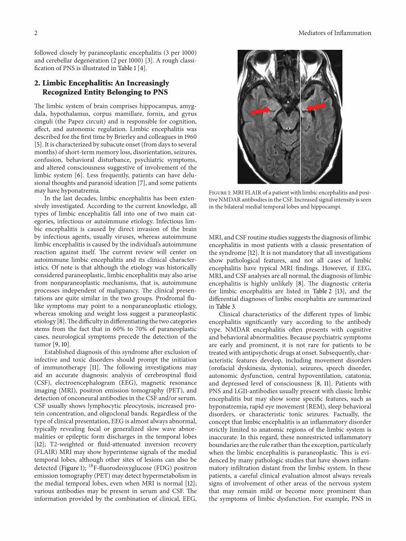
Established diagnosis of this syndrome after exclusion ofinfective and toxic disorders should prompt the initiationof immunotherapy [11]. The following investigations mayaid an accurate diagnosis: analysis of cerebrospinal fluid(CSF), electroencephalogram (EEG), magnetic resonanceimaging (MRI), positron emission tomography (PET), anddetection of onconeural antibodies in the CSF and/or serum.CSF usually shows lymphocytic pleocytosis, increased pro-tein concentration, and oligoclonal bands. Regardless of thetype of clinical presentation, EEG is almost always abnormal,typically revealing focal or generalized slow wave abnor-malities or epileptic form discharges in the temporal lobes[12]; T2-weighted or fluid-attenuated inversion recovery(FLAIR) MRI may show hyperintense signals of the medialtemporal lobes, although other sites of lesions can also bedetected (Figure 1); 18F-fluorodeoxyglucose (FDG) positronemission tomography (PET) may detect hypermetabolism inthe medial temporal lobes, even when MRI is normal [12];various antibodies may be present in serum and CSF. Theinformation provided by the combination of clinical, EEG,
Figure 1: MRI FLAIR of a patient with limbic encephalitis and posi-tiveNMDARantibodies in theCSF. Increased signal intensity is seenin the bilateral medial temporal lobes and hippocampi.
MRI, andCSF routine studies suggests the diagnosis of limbicencephalitis in most patients with a classic presentation ofthe syndrome [12]. It is not mandatory that all investigationsshow pathological features, and not all cases of limbicencephalitis have typical MRI findings. However, if EEG,MRI, and CSF analyses are all normal, the diagnosis of limbicencephalitis is highly unlikely [8]. The diagnostic criteriafor limbic encephalitis are listed in Table 2 [13], and thedifferential diagnoses of limbic encephalitis are summarizedin Table 3.
Clinical characteristics of the different types of limbicencephalitis significantly vary according to the antibodytype. NMDAR encephalitis often presents with cognitiveand behavioral abnormalities. Because psychiatric symptomsare early and prominent, it is not rare for patients to betreated with antipsychotic drugs at onset. Subsequently, char-acteristic features develop, including movement disorders(orofacial dyskinesia, dystonia), seizures, speech disorder,autonomic dysfunction, central hypoventilation, catatonia,and depressed level of consciousness [8, 11]. Patients withPNS and LGI1-antibodies usually present with classic limbicencephalitis but may show some specific features, such ashyponatremia, rapid eye movement (REM), sleep behavioraldisorders, or characteristic tonic seizures. Factually, theconcept that limbic encephalitis is an inflammatory disorderstrictly limited to anatomic regions of the limbic system isinaccurate. In this regard, these nonrestricted inflammatoryboundaries are the rule rather than the exception, particularlywhen the limbic encephalitis is paraneoplastic. This is evi-denced by many pathologic studies that have shown inflam-matory infiltration distant from the limbic system. In thesepatients, a careful clinical evaluation almost always revealssigns of involvement of other areas of the nervous systemthat may remain mild or become more prominent thanthe symptoms of limbic dysfunction. For example, PNS in

Mediators of Inflammation 3
Table 1: Classification of paraneoplastic neurological syndromes.
Central nervous systemLimbic encephalitisEncephalomyelitisBrainstem encephalitisStiff-person syndromeOpsoclonus-myoclonusSubacute cerebellar degenerationParaneoplastic visual syndromes
Cancer-associated retinopathyMelanoma-associated retinopathyParaneoplastic optic neuropathy
Motor neuron syndromesSubacute motor neuronopathyOther motor neuron syndromes
Peripheral nervous systemAcute sensorimotor neuropathySubacute sensory neuronopathyChronic sensorimotor neuropathySubacute autonomic neuropathyParaneoplastic peripheral nerve vasculitis
Neuromuscular junction and muscleMyasthenia gravisLambert-Eaton syndromePolymyositis/dermatomyositisAcute necrotizing myopathyCachectic myopathyNeuromyotonia
many patients with anti-Hu antibodies may start as limbicencephalitis that often evolves to encephalomyelitis withdorsal root ganglionitis.
3. Tumors That Are Associated withLimbic Encephalitis
In PNS, 50% to 80% of the patients present with neuro-logical symptoms of PNS prior to diagnosis of tumors [15].The associated tumors in PNS are a lung cancer in 50–60% of patients, usually SCLC (40–55%), and the associ-ated tumor is a testicular germ cell tumor in 20% of patients.Other associated tumors include breast cancer, thymoma,Hodgkin’s lymphoma, and teratomas [2]. In paraneoplasticlimbic encephalitis, the most common tumors and corre-sponding antibodies are SCLC (anti-Hu, anti-CRMP5, andanti-amphiphysin), testicular cancer (anti-Ma2), thymoma(anti-CRMP5), and breast cancer (anti-amphiphysin) [16]. Inmen younger than the age of 50 years with anti-Ma2 antibod-ies, limbic encephalitis is almost always associated with tes-ticular germ cell tumors, which however can be microscopicand difficult to detect.
As one of the classical PNS, limbic encephalitis can bediagnosed within less than 5 years before cancer is detected
[14]. Removal of the tumor is critical for neurologic improve-ment or stabilization of symptoms in PNS. Therefore, tumorshould be screened in patients with limbic encephalitis.
4. Antibodies Commonly Detected inLimbic Encephalitis
Tumor immunologists introduced the term “onconeural”antibodies to designate antibodies that target antigens presentin neuroectodermal tissues and tumors [17]. These antibod-ies are unambiguously demonstrated by standardized tests,associated with limited subsets of tumors, and are presentin several PNS types [1]. Since the 1980s, various onconeuralantibodies have been discovered,which can serve as biomark-ers for classic paraneoplastic syndromes [18]. Classical limbicencephalitides with temporal lobe seizures are associatedwith onconeural antibodies directed against the intracellularantigens. Onconeural antibodies are found in about 60%of the patients with paraneoplastic limbic encephalitis. Themost frequent related antibodies are anti-Hu, anti-Ma2 (withor without Ma1), anti-amphiphysin, and anti-CRMP5. Themajority of patients with anti-Hu antibodies have symptomsalso suggestive of the dysfunction of areas of the nervoussystem outside the limbic system.
In recent years, the spectrum of chronic inflammatorybrain diseases characterized by the presence of antigen-specific antibodies in serum and CSF has greatly expanded.Many patients with paraneoplastic limbic encephalitis pre-viously characterized as “seronegative” have in fact anti-bodies against cell surface antigens. Recent studies indi-cated that most cases previously considered “seronega-tive” have, in fact, antibodies against surface antigens [19].More and more cases such as glutamic acid decarboxylase(GAD) 65 antibody encephalitis [20], the voltage-gatedpotassium channel (VGKC) complex antibody encephalitis[21] (including LGI1 and Caspr2 antibodies), N-methyl-D-aspartate receptor (NMDAR) antibody encephalitis [22],alpha-amino-3-hydroxy-5-methyl-4-isoxazolepropionic acidreceptor (AMPAR) [23], and gamma aminobutyric acidreceptor GABA(B) antibody encephalitis [24] are recog-nized. In a few years, the number of onconeural antibodiesdescribed in PNS has increased dramatically. Antibodies tothe components of VGKCs, NMDARs, AMPARs, GABA(B),mGluR5 receptor, and glycine receptors (GlyRs) can beidentified in patients and are associated with various clinicalpresentations, such as limbic encephalitis and complex anddiffuse encephalopathies [23, 25, 26]. These diseases can beassociated with tumors, but some of them are nonparaneo-plastic, and antibody assays can help with the diagnosis. Theidentification of these new antibodies (cell surface antigenassociated) has allowed recognition of a syndrome with clini-cal and radiological features indistinguishable from “classiclimbic encephalitis.” The course of the newly identifiedsyndromes tends to be less severe and it is often possibleto achieve complete recovery with prompt immunomodula-tory treatment. The most representative condition is LGI1-encephalitis, previously known as limbic encephalitis withVGKC complex antibodies [27, 28].
4 Mediators of Inflammation
Table 2: Diagnostic criteria of paraneoplastic limbic encephalitis.
Criteria by Gultekin et al. [13]Pathological demonstration of limbic encephalitis, or all 4 of the following.(1) Symptoms of short-term memory loss, seizures, or psychiatric symptoms suggesting involvement of the limbic system(2) <4 yr between the onset of neurological symptoms and the cancer diagnosis(3) Exclusion of metastasis, infection, metabolic and nutritional deficits, stroke, and side-effects of therapy that may cause limbicencephalopathy(4) At least one of the following:
(a) CSF with inflammatory findings(b) MRI FLAIR or T2 unilateral or bilateral temporal lobe hyperintensities(c) EEG with epileptic or slow activity focally involving the temporal lobes
Criteria by the Paraneoplastic Neurological Syndrome Euronetwork [14]All 4 of the following items are met.(i) Subacute onset (days or up to 12wk) of seizures, short-term memory loss, confusion, and psychiatric symptoms(ii) Neuropathologic or radiologic evidence (MRI, SPECT, PET) of involvement of the limbic system(iii) Exclusion of other possible etiologies of limbic dysfunction(iv) Demonstration of a cancer within 5 yr of the diagnosis of neurologic symptoms or the development of classic symptoms oflimbic dysfunction in association with a well-characterized paraneoplastic antibody (Hu, Ma2, CRMP5, amphiphysin, Ri)
These antibodies are directed against two categoriesof antigens: (1) intracellular antigens (Hu, Ma2, CRMP5,amphiphysin, etc.) and (2) cell surface antigens (the VGKCcomplex, NMDAR, AMPARs, GABABRs, mGluR5 receptor,GlyRs, etc.). Whereas the disorders related to the first cat-egory of antibodies are associated with cancer (lung, testis,etc.), prominent brain infiltrates of cytotoxic T cells, andlimited response to treatment, the disorders related to the sec-ond category of antibodies are associated less frequently withcancer (thymoma, teratoma), seem to be antibody mediated,and respond significantly better to immunotherapy. Thesetwo antibodies have in common the association with idio-pathic or paraneoplastic limbic encephalitis [23, 24]. Sevenout of 15 (47%) patients with limbic encephalitis associatedwithGABA (B) receptor antibodies had an underlying tumor,usually an SCLC [24]. In limbic encephalitis associated withAMPAR antibodies, the frequency of cancer was 64%, withSCLC being the most common type, followed by thymomaand breast cancer [23].These patients have a better prognosisthan those with antibodies against intracellular proteins [29,30]. Table 4 summarizes the common antibodies againstonconeural antigens detected in PNS and their potentiallyassociated tumors.
5. Do Antibodies Play a Pathogenic Role inLimbic Encephalitis?
A cancer-stimulated immune response that cross-reacts withneural tissue—onconeural immunity—is considered theprincipal pathologic mechanism for PNS [31]. Some cancercells express proteins that are normally restricted to the ner-vous system. For example, when serum from a patient withlimbic encephalitis was incubated with the patient’s cancercells and with a rat’s brain tissue, antibody fixation to thesame Ma proteins on both neurons and cancer cells couldbe observed [31]. Pathological examination of the nervoussystem showed loss of neurons in affected areas of the nervous
systemwith inflammatory infiltration by CD4+ T helper cellsand B cells in the perivascular spaces and cytotoxic CD8+ Tcells in the interstitial spaces [32–34]. Examination of CSFfrequently demonstrates pleocytosis, intrathecal synthesis ofIgG, and oligoclonal bands, supporting an inflammatory orimmune-mediated etiology.
The discovery of paraneoplastic antineuronal antibodiesresulted in the general belief that these are immune-mediateddisorders triggered by onconeural antigens expressed bytumor cells. Despite the clear clinical evidence that many ofthe syndromes described earlier are antibodymediated, thereis lack of direct evidence showing that these antibodies arepathogenic in PNS. Support for a pathogenic role of antibod-ies comes from the fact that the target paraneoplastic antigensare expressed both in the tumors and in the affected regions ofthe nervous system. Furthermore, the size of tumors is usuallysmall and they are heavily infiltrated with inflammatorycells. Interestingly, spontaneous remissions of carcinomamayoccur at the time of neurological presentation [35, 36]. Onestudy even foundmore limited disease distribution and betteroncologic outcome in SCLC patients with paraneoplasticantibodies [37].
There are studies on the effects of the serum or CSFIgG antibodies on the neuronal function in cultured cells[22, 23, 38] or on brain slices, but the transfer of clinicalor electrophysiological evidence of disease to experimentalanimals by either systemic or intrathecal injection has notyet been reported, with the exception of mGluR1-Ab inparaneoplastic cerebellar degeneration [39] and reports ofGAD-65 or amphiphysin antibodies [40, 41]. In some PNSs,circumstantial evidence suggests that T-cell-mediated mech-anisms play a major pathogenic role [42]. It has been sug-gested that the most important determinant of the under-lying immunopathogenesis and responsiveness to immuno-suppression is the antibody type and level of the affectedindividual, which may determine the response to treatment[1, 18, 43].
Mediators of Inflammation 5
Table 3: Differential diagnoses of limbic encephalitis.
Infectious disordersHerpes simplex virus encephalitisNeurosyphilisProgressive multifocal leukoencephalopathyRabiesCreutzfeldt-Jakob disease
Metabolic disordersMetabolic encephalopathy (uremic, hepatic, Cushingsyndrome, etc.)Wernicke-Korsakoff syndromeHashimoto’s encephalopathy
Systemic autoimmune disordersSjogren syndromeSystemic lupus erythematosusAntiphospholipid syndrome
MalignanciesLymphomaGliomaGliomatosis cerebri
Degenerative disordersAlzheimer’s diseaseLewy-body dementiaFrontotemporal dementia
OthersStroke with posterior cerebral artery involvementCentral nervous system vasculitisTemporal lobe epilepsyNonconvulsive status epilepticusTransient global amnesiaAcute demyelinating encephalomyelitisPosterior reversible encephalopathy syndromeIntoxication (alcohol, lithium, etc.)Alcohol withdrawal syndromePsychiatric disorder
Specifically, striking differences have been found betweendisorderswith antibodies against intracellular antigens versusthose to neural surface antigens. Disorders with antibodiesto intracellular antigens are considered poorly responsive toimmunotherapy [18, 20] and may be mediated by cytotoxicT cells [18, 34]. On the other hand, disorders associated withantibodies against cell surface antigens, such as the VGKC-complex or NMDAR, often respond well to treatment [20,44].
Some laboratory evidence supports the role of pathogenicB-cell responses in limbic encephalitis. NMDAR antibodiesfrom patients have been shown to decrease the numbers of
NMDAR in postsynaptic dendrites of cultured hippocam-pal neurons. One study suggested that anti-Hu antibodiesinduced apoptosis when applied to cultures of neuroblastomaor mesenteric cells [45]. There is also evidence, however,pointing to that paraneoplastic limbic encephalitis may beT-cell mediated, as Hu-specific T cells have been found inthe blood and CSF [46], and there are cytotoxic infiltrates ofT cells in the brain and tumor of the patients with anti-Huantibodies-associated encephalomyelitis [47].
A pathogenic role could only be proven for those parane-oplastic antibodies that are directed against easily accessibleantigens located on the cell surface. In these disorders,indirect lines of evidence support the view that the cellularimmune responses against these antigens are responsible forthe neurological damage [46, 48, 49]. The relative contribu-tion of the cellular and humoral immunity to the clinicaland pathological manifestations has not been displayed. Theparaneoplastic antibodies may, in these cases, be surrogatemarkers for T-cell activation [50]. Elevated CD8/CD3 ratiosin diseases were associated with antibodies to intracellu-lar antigens and suggested a cytotoxic T-cell-driven path-omechanism. In diseases with antibodies to surface anti-gens, this finding supports a B-cell-related pathomechanism,with evidence of a complement-mediated pathogenesis inpatients with VGKC-complex antibodies. Interestingly, thisimmunopathogenic dichotomy parallels other autoimmunedisorders such as polymyositis and dermatomyositis, whichhave a predominant T-cell- and antibody-mediated patho-genesis, respectively [51]. These observations may contributeto a rational choice in immunotherapies for these disorders[52]. A totally different mechanism seems at work in para-neoplastic cerebellar degeneration in Hodgkin’s lymphomabecause the target antigens of the associated anti-Tr and anti-mGluR1 antibodies are not expressed in Hodgkin’s tumor tis-sue [53]. Dysregulation of the immune response in Hodgkin’slymphoma and an etiologic role for viral infections have beenpostulated in this disorder [53].
Thus far, it is still unclear whether antibody-mediatedPNS, for example, VGKC complex antibody-associated lim-bic encephalitis, is driven by serum or intrathecal antibodies.The absolute concentrations of antibodies against a certainonconeural antigen are usually higher in serum than in theCSF. Moreover, antibodies are not always detectable in theCSF. Ideally, both serum and CSF samples should be sentfor antibody testing, but their relative utility in followup ofpatients is under debate. Intrathecal synthesis of IgG andoligoclonal bands can help pointing to an immune-mediateddisorder before the results of specific antibodies can beobtained, but the oligoclonal bands are not always presentat onset or even thereafter, and whether their presenceis evidence for ongoing pathology or merely a secondaryepiphenomenon is not yet clear. The intrathecal synthesis ofantibodies can actually be assessed by the calculation of theamount of specific antibodies in the CSF relative to the totalCSF IgG and by comparison with similar calculations in theserum.The ratio represents intrathecal synthesis and is oftenhigher in some PNS. In favor of a role for systemic ratherthan intrathecal antibodies, animal experiments have shownthat certain regions of the brain, that is, the hippocampus

6 Mediators of Inflammation
Table 4: The common antibodies detected in PNS and their associated tumors.
Antibodies PNS Associated tumorsAntibodies against intracellular antigens
Anti-Hu sensory neuronopathy, LE, BSE,encephalomyelitis SCLC
Anti-Yo SCD Gynecological cancerAnti-Ri Opsoclonus-myoclonus, BSE Breast cancerAnti-Ma2 BSE, LE Testis cancer, SCLC, breast cancer
Anti-CRMP5 SCD, chorea, myelitis, LE, sensoryneuronopathy, optic neuritis SCLC, thymoma
Anti-amphiphysin SPS, myelitis, SCD, sensory neuronopathy SCLC, breast cancer
Anti-GAD-65 SPS, myelitis SCLC, breast cancerAnti-SOX-1 LEMS SCLC
Antibodies against cell surface onconeural antigensAnti-VGCC SPS, LEMS SCLCAnti-VGKC complex LE SCLC, thymomaAnti-NMDA receptor LE TeratomaAnti-AMPA receptor LE SCLC, breast cancer, thymomaAnti-AQP-4 NMO spectrum disorders SCLC, breast cancer, thymomaAnti-GABA-B receptor LE SCLCAnti-CAR Retinopathy SCLC, melanoma, gynecological cancerAnti-contactin-associated protein 2 Morvan syndrome ThymomaAnti-AchR/MuSK/RyR/Titin MG ThymomaAMPA: amino-3-hydroxyl-5-methyl-4-isoxazole-propionate; AQP-4: aquaporin 4; CAR: cancer-associated retinopathy; CRMP5: collapsin response mediatorprotein 5; GABA-B: gamma-aminobutyric acid B; GAD-65: glutamic acid decarboxylase 65; LE: limbic encephalitis; LEMS: Lambert-Eaton myasthenicsyndrome; MG: myasthenia gravis; NMDA: N-methyl-D-aspartate; NMO: neuromyelitis optica; SCD: subacute cerebellar degeneration; SCLC: small cell lungcancer; SPS: stiff-person syndrome; VGCC: voltage-gated calcium channel; VGKC: voltage-gated potassium channel.
and the hypothalamus, seem to be particularly vulnerable,and it is notable that limbic encephalitis with VGKC-complex(LGI1 and Caspr2) antibodies and anti-NMDAR encephalitisusually start with symptoms originating from the temporallobe cortex, even though the target antigens are presentmuchmore widely in the CNS. The former usually affects hip-pocampus, amygdala, and anterior temporal cortex, whereasthe latter usually affects hippocampus, cerebral cortex, basalganglia, and thalamus [54]. Until recently, only 50% ofpatients with limbic encephalitis and SCLC were foundantibody positive, usually harboring anti-Hu antibodies or,less frequently, other onconeural antibodies [29].
Immunopathological analysis of various antibody-asso-ciated limbic encephalitis may help elucidate the underly-ing immunopathogenic mechanisms, whereas unfortunatelythere has been a lack of laboratory data [52]. Why is limbicencephalitis reversible in patients with NMDAR antibodiesthat are in frequent association with ovarian teratoma [44,55]? Furthermore, how does one classify those patients withGAD-65 antibodies that are not paraneoplastic in origin,who suffer from limbic encephalitis or chronic temporal lobeepilepsy [20]?
An important issue is that a positive report for any well-characterized onconeural antibody has to be assessed accord-ing to the clinical setting. All these antibodies, particularlythose associated with SCLC, can be found in the patients with
cancer without PNS [56]. Therefore, one should still rule outother potential causes of the neurological syndrome that isbeing evaluated. Up to 16% of patients with SCLC withoutPNS have low titers of Hu antibodies, whereas in the patientswith PNS and Hu antibodies, the titers are substantiallyhigher [37].
6. Treatments of Limbic Encephalitis
The basic principles of paraneoplastic limbic encephalitistherapy are resection of the tumor or oncological treatment[10]. When a patient with tumor is found in associationwith a possible paraneoplastic disorder, removal of thetumor is critical for neurologic improvement or stabilizationof symptoms. Antibodies against onconeural antigens aresensitive and should prompt an extensive tumor screeningin antibody-positive patients. In the patients with limbicencephalitis associated with ion channel/receptor antibod-ies, immunosuppressive or immunomodulatory treatmentis promising to improve the disease. Limbic encephalitideswith antibodies against intracellular onconeural antigens donot normally respond to immunosuppressive treatment; onlytumor therapymay stabilize the syndrome. Treatment of PNSstill remains difficult. Anti-Hu-antibody-positive patients donot normally respond to immunosuppressive treatment. Theonly therapy that stabilizes these patients is perhaps the
Mediators of Inflammation 7
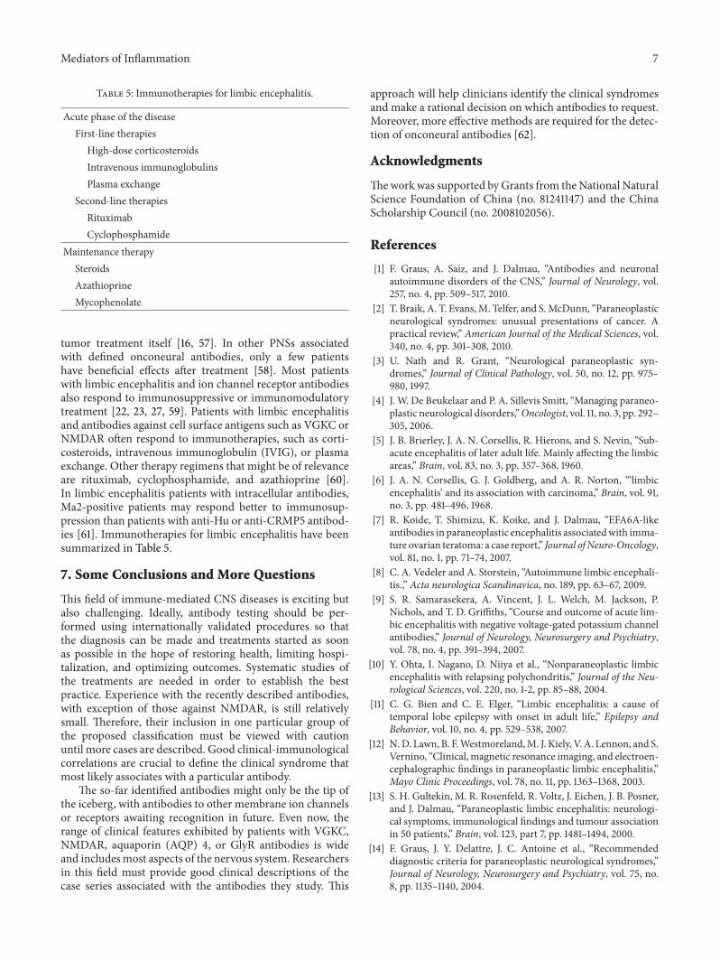
Table 5: Immunotherapies for limbic encephalitis.
Acute phase of the diseaseFirst-line therapies
High-dose corticosteroidsIntravenous immunoglobulinsPlasma exchange
Second-line therapiesRituximabCyclophosphamide
Maintenance therapySteroidsAzathioprineMycophenolate
tumor treatment itself [16, 57]. In other PNSs associatedwith defined onconeural antibodies, only a few patientshave beneficial effects after treatment [58]. Most patientswith limbic encephalitis and ion channel receptor antibodiesalso respond to immunosuppressive or immunomodulatorytreatment [22, 23, 27, 59]. Patients with limbic encephalitisand antibodies against cell surface antigens such as VGKC orNMDAR often respond to immunotherapies, such as corti-costeroids, intravenous immunoglobulin (IVIG), or plasmaexchange. Other therapy regimens that might be of relevanceare rituximab, cyclophosphamide, and azathioprine [60].In limbic encephalitis patients with intracellular antibodies,Ma2-positive patients may respond better to immunosup-pression than patients with anti-Hu or anti-CRMP5 antibod-ies [61]. Immunotherapies for limbic encephalitis have beensummarized in Table 5.
7. Some Conclusions and More Questions
This field of immune-mediated CNS diseases is exciting butalso challenging. Ideally, antibody testing should be per-formed using internationally validated procedures so thatthe diagnosis can be made and treatments started as soonas possible in the hope of restoring health, limiting hospi-talization, and optimizing outcomes. Systematic studies ofthe treatments are needed in order to establish the bestpractice. Experience with the recently described antibodies,with exception of those against NMDAR, is still relativelysmall. Therefore, their inclusion in one particular group ofthe proposed classification must be viewed with cautionuntil more cases are described. Good clinical-immunologicalcorrelations are crucial to define the clinical syndrome thatmost likely associates with a particular antibody.
The so-far identified antibodies might only be the tip ofthe iceberg, with antibodies to other membrane ion channelsor receptors awaiting recognition in future. Even now, therange of clinical features exhibited by patients with VGKC,NMDAR, aquaporin (AQP) 4, or GlyR antibodies is wideand includesmost aspects of the nervous system. Researchersin this field must provide good clinical descriptions of thecase series associated with the antibodies they study. This
approach will help clinicians identify the clinical syndromesand make a rational decision on which antibodies to request.Moreover, more effective methods are required for the detec-tion of onconeural antibodies [62].
Acknowledgments
Thework was supported by Grants from theNational NaturalScience Foundation of China (no. 81241147) and the ChinaScholarship Council (no. 2008102056).
References
[1] F. Graus, A. Saiz, and J. Dalmau, “Antibodies and neuronalautoimmune disorders of the CNS,” Journal of Neurology, vol.257, no. 4, pp. 509–517, 2010.
[2] T. Braik, A. T. Evans,M. Telfer, and S.McDunn, “Paraneoplasticneurological syndromes: unusual presentations of cancer. Apractical review,” American Journal of the Medical Sciences, vol.340, no. 4, pp. 301–308, 2010.
[3] U. Nath and R. Grant, “Neurological paraneoplastic syn-dromes,” Journal of Clinical Pathology, vol. 50, no. 12, pp. 975–980, 1997.
[4] J. W. De Beukelaar and P. A. Sillevis Smitt, “Managing paraneo-plastic neurological disorders,”Oncologist, vol. 11, no. 3, pp. 292–305, 2006.
[5] J. B. Brierley, J. A. N. Corsellis, R. Hierons, and S. Nevin, “Sub-acute encephalitis of later adult life. Mainly affecting the limbicareas,” Brain, vol. 83, no. 3, pp. 357–368, 1960.
[6] J. A. N. Corsellis, G. J. Goldberg, and A. R. Norton, “‘limbicencephalitis’ and its association with carcinoma,” Brain, vol. 91,no. 3, pp. 481–496, 1968.
[7] R. Koide, T. Shimizu, K. Koike, and J. Dalmau, “EFA6A-likeantibodies in paraneoplastic encephalitis associatedwith imma-ture ovarian teratoma: a case report,” Journal ofNeuro-Oncology,vol. 81, no. 1, pp. 71–74, 2007.
[8] C. A. Vedeler and A. Storstein, “Autoimmune limbic encephali-tis.,” Acta neurologica Scandinavica, no. 189, pp. 63–67, 2009.
[9] S. R. Samarasekera, A. Vincent, J. L. Welch, M. Jackson, P.Nichols, and T. D. Griffiths, “Course and outcome of acute lim-bic encephalitis with negative voltage-gated potassium channelantibodies,” Journal of Neurology, Neurosurgery and Psychiatry,vol. 78, no. 4, pp. 391–394, 2007.
[10] Y. Ohta, I. Nagano, D. Niiya et al., “Nonparaneoplastic limbicencephalitis with relapsing polychondritis,” Journal of the Neu-rological Sciences, vol. 220, no. 1-2, pp. 85–88, 2004.
[11] C. G. Bien and C. E. Elger, “Limbic encephalitis: a cause oftemporal lobe epilepsy with onset in adult life,” Epilepsy andBehavior, vol. 10, no. 4, pp. 529–538, 2007.
[12] N.D. Lawn, B. F.Westmoreland,M. J. Kiely, V. A. Lennon, and S.Vernino, “Clinical, magnetic resonance imaging, and electroen-cephalographic findings in paraneoplastic limbic encephalitis,”Mayo Clinic Proceedings, vol. 78, no. 11, pp. 1363–1368, 2003.
[13] S. H. Gultekin, M. R. Rosenfeld, R. Voltz, J. Eichen, J. B. Posner,and J. Dalmau, “Paraneoplastic limbic encephalitis: neurologi-cal symptoms, immunological findings and tumour associationin 50 patients,” Brain, vol. 123, part 7, pp. 1481–1494, 2000.
[14] F. Graus, J. Y. Delattre, J. C. Antoine et al., “Recommendeddiagnostic criteria for paraneoplastic neurological syndromes,”Journal of Neurology, Neurosurgery and Psychiatry, vol. 75, no.8, pp. 1135–1140, 2004.

8 Mediators of Inflammation
[15] F. Graus and J. Dalmau, “Paraneoplastic neurological syn-dromes: diagnosis and treatment,” Current Opinion in Neurol-ogy, vol. 20, no. 6, pp. 732–737, 2007.
[16] F. Graus, F. Keime-Guibert, R. Rene et al., “Anti-Hu-associatedparaneoplastic encephalomyelitis: analysis of 200 patients,”Brain, vol. 124, no. 6, pp. 1138–1148, 2001.
[17] R. C. Seeger, P. M. Zeltzer, and S. A. Rayner, “Onco-neuralantigen: a new neural differentiation antigen expressed byneuroblastoma, oat cell carcinoma, Wilms’ tumor, and sarcomacells,” Journal of Immunology, vol. 122, no. 4, pp. 1548–1555, 1979.
[18] J. Dalmau and M. R. Rosenfeld, “Paraneoplastic syndromes ofthe CNS,”The Lancet Neurology, vol. 7, no. 4, pp. 327–340, 2008.
[19] F. Graus, A. Saiz, M. Lai et al., “Neuronal surface antigen anti-bodies in limbic encephalitis: clinical-immunologic associa-tions,” Neurology, vol. 71, no. 12, pp. 930–936, 2008.
[20] M. P. Malter, C. Helmstaedter, H. Urbach, A. Vincent, and C. G.Bien, “Antibodies to glutamic acid decarboxylase define a formof limbic encephalitis,” Annals of Neurology, vol. 67, no. 4, pp.470–478, 2010.
[21] S. R. Irani, S. Alexander, P. Waters et al., “Antibodies to Kv1potassium channel-complex proteins leucine-rich, glioma inac-tivated 1 protein and contactin-associated protein-2 in limbicencephalitis,Morvan’s syndrome and acquired neuromyotonia,”Brain, vol. 133, no. 9, pp. 2734–2748, 2010.
[22] J. Dalmau, A. J. Gleichman, E. G. Hughes et al., “Anti-NMDA-receptor encephalitis: case series and analysis of the effects ofantibodies,” The Lancet Neurology, vol. 7, no. 12, pp. 1091–1098,2008.
[23] M. Lai, E. G. Hughes, X. Peng et al., “AMPA receptor antibodiesin limbic encephalitis alter synaptic receptor location,” Annalsof Neurology, vol. 65, no. 4, pp. 424–434, 2009.
[24] E. Lancaster, M. Lai, X. Peng et al., “Antibodies to the GABABreceptor in limbic encephalitis with seizures: case series andcharacterisation of the antigen,” The Lancet Neurology, vol. 9,no. 1, pp. 67–76, 2010.
[25] A. Boronat, L. Sabater, A. Saiz, J. Dalmau, and F. Graus,“GABAB receptor antibodies in limbic encephalitis and anti-GAD-associated neurologic disorders,” Neurology, vol. 76, no.9, pp. 795–800, 2011.
[26] E. Lancaster, E.Martinez-Hernandez,M. J. Titulaer et al., “Anti-bodies to metabotropic glutamate receptor 5 in the Opheliasyndrome,” Neurology, vol. 77, no. 18, pp. 1698–1701, 2011.
[27] A. Vincent, C. Buckley, J. M. Schott et al., “Potassium channelantibody-associated encephalopathy: a potentially immuno-therapy-responsive form of limbic encephalitis,” Brain, vol. 127,no. 3, pp. 701–712, 2004.
[28] M. Lai, M. G. M. Huijbers, E. Lancaster et al., “Investigation ofLGI1 as the antigen in limbic encephalitis previously attributedto potassium channels: a case series,”The Lancet Neurology, vol.9, no. 8, pp. 776–785, 2010.
[29] S. Alamowitch, F. Graus, M. Uchuya, R. Rene, E. Bescansa, andJ. Y. Delattre, “Limbic encephalitis and small cell lung cancer.Clinical and immunological features,” Brain, vol. 120, no. 6, pp.923–928, 1997.
[30] L. Bataller, K. A. Kleopa, G. F. Wu, J. E. Rossi, M. R. Rosenfeld,and J. Dalmau, “Autoimmune limbic encephalitis in 39 patients:Immunophenotypes and outcomes,” Journal of Neurology, Neu-rosurgery and Psychiatry, vol. 78, no. 4, pp. 381–385, 2007.
[31] L. Bataller and J. Dalmau, “Paraneoplastic neurologic syn-dromes,” Neurologic Clinics, vol. 21, no. 1, pp. 221–247, 2003.
[32] D. Denny-Brown, “Primary sensory neuropathy with muscularchanges associated with carcinoma,” Journal of Neurology,Neurosurgery & Psychiatry, vol. 11, no. 2, pp. 73–87, 1948.
[33] W. C. Jean, J. Dalmau, A. Ho, and J. B. Posner, “Analysis ofthe IgG subclass distribution and inflammatory infiltrates inpatients with anti-Hu-associated paraneoplastic encephalomy-elitis,” Neurology, vol. 44, no. 1, pp. 140–147, 1994.
[34] F. Bernal, F. Graus, A. Pifarre, A. Saiz, B. Benyahia, and T.Ribalta, “Immunohistochemical analysis of anti-Hu-associatedparaneoplastic encephalomyelitis,” Acta Neuropathologica, vol.103, no. 5, pp. 509–515, 2002.
[35] R. B. Darnell and L. M. DeAngelis, “Regression of small-cell lung carcinoma in patients with paraneoplastic neuronalantibodies,”The Lancet, vol. 341, no. 8836, pp. 21–22, 1993.
[36] T. Byrne, W. P. Mason, J. B. Posner, and J. Dalmau, “Spon-taneous neurological improvement in anti-Hu associatedencephalomyelitis,” Journal of Neurology Neurosurgery and Psy-chiatry, vol. 62, no. 3, pp. 276–278, 1997.
[37] F. Graus, J. Dalmau, R. Rene et al., “Anti-Hu antibodies inpatients with small-cell lung cancer: association with completeresponse to therapy and improved survival,” Journal of ClinicalOncology, vol. 15, no. 8, pp. 2866–2872, 1997.
[38] E. G. Hughes, X. Peng, A. J. Gleichman et al., “Cellular andsynaptic mechanisms of anti-NMDA receptor encephalitis,”Journal of Neuroscience, vol. 30, no. 17, pp. 5866–5875, 2010.
[39] P. Sillevis Smitt, A. Kinoshita, B. De Leeuw et al., “Paraneoplas-tic cerebellar ataxia due to autoantibodies against a glutamatereceptor,” New England Journal of Medicine, vol. 342, no. 1, pp.21–27, 2000.
[40] C. Geis, A. Weishaupt, S. Hallermann et al., “Stiff personsyndrome-associated autoantibodies to amphiphysin mediatereduced GABAergic inhibition,” Brain, vol. 133, no. 11, pp. 3166–3180, 2010.
[41] M. Manto, M. Laute, M. Aguera, V. Rogemond, M. Pandolfo,and J. Honnorat, “Effects of anti-glutamic acid decarboxylaseantibodies associated with neurological diseases,” Annals ofNeurology, vol. 61, no. 6, pp. 544–551, 2007.
[42] J. Dalmau, H. S. Gultekin, and J. B. Posner, “Paraneoplastic neu-rologic syndromes: pathogenesis and physiopathology,” BrainPathology, vol. 9, no. 2, pp. 275–284, 1999.
[43] A. Vincent, S. R. Irani, and B. Lang, “The growing recog-nition of immunotherapy-responsive seizure disorders withautoantibodies to specific neuronal proteins,” Current Opinionin Neurology, vol. 23, no. 2, pp. 144–150, 2010.
[44] J. Dalmau, E. Lancaster, E. Martinez-Hernandez, M. R. Rosen-feld, and R. Balice-Gordon, “Clinical experience and laboratoryinvestigations in patients with anti-NMDAR encephalitis,” TheLancet Neurology, vol. 10, no. 1, pp. 63–74, 2011.
[45] R. DeGiorgio,M. Bovara, G. Barbara et al., “Anti-HuD-inducedneuronal apoptosis underlying paraneoplastic gut dysmotility,”Gastroenterology, vol. 125, no. 1, pp. 70–79, 2003.
[46] M. L. Albert, L. M. Austin, and R. B. Darnell, “Detection andtreatment of activated T cells in the cerebrospinal fluid ofpatients with paraneoplastic cerebellar degeneration,” Annals ofNeurology, vol. 47, no. 1, pp. 9–17, 2000.
[47] R. Voltz, J. Dalmau, J. B. Posner, and M. R. Rosenfeld, “T-cell receptor analysis in anti-Hu associated paraneoplasticencephalomyelitis,”Neurology, vol. 51, no. 4, pp. 1146–1150, 1998.
[48] B. Benyahia, R. Liblau, H. Merle-Beral et al., “Cell-mediatedautoimmunity in paraneoplastic neurological syndromes withanti-Hu antibodies,”Annals of Neurology, vol. 45, no. 2, pp. 162–167, 1999.
Mediators of Inflammation 9
[49] M. Tanaka, K. Tanaka, S. Tokiguchi, K. Shinozawa, and S. Tsuji,“Cytotoxic T cells against a peptide of Yo protein in patientswithparaneoplastic cerebellar degeneration and anti-Yo antibody,”Journal of the Neurological Sciences, vol. 168, no. 1, pp. 28–31,1999.
[50] Z. Yu, T. J. Kryzer, G. E. Griesmann et al., “CRMP-5 neuronalautoantibody: marker of lung cancer and thymoma-relatedautoimmunity,” Annals of Neurology, vol. 49, no. 2, pp. 146–154,2001.
[51] K. Arahata and A. G. Engel, “Monoclonal antibody analysisof mononuclear cells in myopathies. I: Quantitation of subsetsaccording to diagnosis and sites of accumulation and demon-stration and counts of muscle fibers invaded by T cells,” Annalsof Neurology, vol. 16, no. 2, pp. 193–208, 1984.
[52] C. G. Bien, A. Vincent, M. H. Barnett et al., “Immunopathologyof autoantibody-associated encephalitides: clues for pathogen-esis,” Brain, vol. 135, part 5, pp. 1622–1638, 2012.
[53] F. Bernal, S. Shams’Ili, I. Rojas et al., “Anti-Tr antibodies asmarkers of paraneoplastic cerebellar degeneration and Hod-gkin’s disease,” Neurology, vol. 60, no. 2, pp. 230–234, 2003.
[54] P. Demaerel, W. Van Dessel, W. Van Paesschen, R. Vanden-berghe, K. Van Laere, and J. Linn, “Autoimmune-mediatedencephalitis,” Neuroradiology, vol. 53, no. 11, pp. 837–851, 2011.
[55] T. Iizuka, S. Yoshii, S. Kan et al., “Reversible brain atrophy inanti-NMDA receptor encephalitis: a long-term observationalstudy.,” Journal of neurology, vol. 257, no. 10, pp. 1686–1691, 2010.
[56] S. E. Monstad, A. Knudsen, H. B. Salvesen, J. H. Aarseth, and C.A. Vedeler, “Onconeural antibodies in sera from patients withvarious types of tumours.,” Cancer Immunology, Immunother-apy, vol. 58, no. 11, pp. 1795–1800, 2009.
[57] F. Graus, F. Vega, J. Y. Delattre et al., “Plasmapheresis andantineoplastic treatment in CNS paraneoplastic syndromeswith antineuronal autoantibodies,”Neurology, vol. 42, no. 3, part1, pp. 536–540, 1992.
[58] F. Blaes, M. Strittmatter, S. Merkelbach et al., “Intravenousimmunoglobulins in the therapy of paraneoplastic neurologicaldisorders,” Journal of Neurology, vol. 246, no. 4, pp. 299–303,1999.
[59] B. M. Ances, R. Vitaliani, R. A. Taylor et al., “Treatment-responsive limbic encephalitis identified by neuropil antibodies:MRI and PET correlates,” Brain, vol. 128, part 8, pp. 1764–1777,2005.
[60] H. Ishiura, S. Matsuda, M. Higashihara et al., “Response of anti-nmda receptor encephalitis without tumor to immunotherapyincluding rituximab,” Neurology, vol. 71, no. 23, pp. 1921–1923,2008.
[61] J. Dalmau, F. Graus, A. Villarejo et al., “Clinical analysis of anti-Ma2-associated encephalitis,” Brain, vol. 127, no. 8, pp. 1831–1844, 2004.
[62] A. Storstein, S. E. Monstad, M. Haugen et al., “Onconeural anti-bodies: improved detection and clinical correlations,” Journal ofNeuroimmunology, vol. 232, no. 1-2, pp. 166–170, 2011.

Submit your manuscripts athttp://www.hindawi.com
Stem CellsInternational
Hindawi Publishing Corporationhttp://www.hindawi.com Volume 2014
Hindawi Publishing Corporationhttp://www.hindawi.com Volume 2014
MEDIATORSINFLAMMATION
of
Hindawi Publishing Corporationhttp://www.hindawi.com Volume 2014
Behavioural Neurology
EndocrinologyInternational Journal of
Hindawi Publishing Corporationhttp://www.hindawi.com Volume 2014
Hindawi Publishing Corporationhttp://www.hindawi.com Volume 2014
Disease Markers
Hindawi Publishing Corporationhttp://www.hindawi.com Volume 2014
BioMed Research International
OncologyJournal of
Hindawi Publishing Corporationhttp://www.hindawi.com Volume 2014
Hindawi Publishing Corporationhttp://www.hindawi.com Volume 2014
Oxidative Medicine and Cellular Longevity
Hindawi Publishing Corporationhttp://www.hindawi.com Volume 2014
PPAR Research
The Scientific World JournalHindawi Publishing Corporation http://www.hindawi.com Volume 2014
Immunology ResearchHindawi Publishing Corporationhttp://www.hindawi.com Volume 2014
Journal of
ObesityJournal of
Hindawi Publishing Corporationhttp://www.hindawi.com Volume 2014
Hindawi Publishing Corporationhttp://www.hindawi.com Volume 2014
Computational and Mathematical Methods in Medicine
OphthalmologyJournal of
Hindawi Publishing Corporationhttp://www.hindawi.com Volume 2014
Diabetes ResearchJournal of
Hindawi Publishing Corporationhttp://www.hindawi.com Volume 2014
Hindawi Publishing Corporationhttp://www.hindawi.com Volume 2014
Research and TreatmentAIDS
Hindawi Publishing Corporationhttp://www.hindawi.com Volume 2014
Gastroenterology Research and Practice
Hindawi Publishing Corporationhttp://www.hindawi.com Volume 2014
Parkinson’s Disease
Evidence-Based Complementary and Alternative Medicine
Volume 2014Hindawi Publishing Corporationhttp://www.hindawi.com