review article adipo-myokines: two sides of the same coin...
TRANSCRIPT

Hindawi Publishing CorporationMediators of InflammationVolume 2013, Article ID 320724, 16 pageshttp://dx.doi.org/10.1155/2013/320724
Review ArticleAdipo-Myokines: Two Sides of the Same Coin—Mediators ofInflammation and Mediators of Exercise
Silja Raschke and Jürgen Eckel
German Diabetes Center, Paul-Langerhans-Group of Integrative Physiology, Auf ’m Hennekamp 65, 40225 Dusseldorf, Germany
Correspondence should be addressed to Jurgen Eckel; [email protected]
Received 26 February 2013; Revised 29 April 2013; Accepted 7 May 2013
Academic Editor: Daniel Konrad
Copyright © 2013 S. Raschke and J. Eckel. This is an open access article distributed under the Creative Commons AttributionLicense, which permits unrestricted use, distribution, and reproduction in any medium, provided the original work is properlycited.
This review summarizes the current literature regarding the most discussed contraction-regulated moykines like IL-6, IL-15, irisin,BDNF, ANGPTL4, FGF21, myonectin and MCP-1. It is suggested that the term myokine is restricted to proteins secreted fromskeletal muscle cells, excluding proteins that are secreted by other cell types in skeletal muscle tissue and excluding proteins whichare only described on the mRNA level. Interestingly, many of the contraction-regulated myokines described in the literature areadditionally known to be secreted by adipocytes. We termed these proteins adipo-myokines. Within this review, we try to elaborateon the question why pro-inflammatory adipokines on the one hand are upregulated in the obese state, and have beneficial effectsafter exercise on the other hand. Both, adipokines and myokines do have autocrine effects within their corresponding tissues. Inaddition, they are involved in an endocrine crosstalkwith other tissues. Depending on the extent and the kinetics of adipo-myokinesin serum, these molecules seem to have a beneficial or an adverse effect on the target tissue.
1. Skeletal Muscle and Adipose Tissue asEndocrine Organs
In line with the acceptance of adipose tissue as an endocrineorgan [1–3], path-breaking work during the last decadedemonstrated that skeletal muscle is an active endocrineorgan releasing myokines, which might in part be respon-sible for the beneficial effect of exercise [4–6]. Thesemyokines are described to communicate with cells in anautocrine/paracrinemanner, locally within themuscles, or inan endocrine fashion to distant tissues.
Obesity in a combinationwith a lack of exercise is a strongrisk factor to develop metabolic diseases and type 2 diabetes.Physical inactivity causes the accumulation of visceral fat andthe health consequences of both are related to systemic low-grade inflammation [7, 8]. Adipocytes from obese patientsare characterized by altered endocrine function, leading toincreased secretion of proinflammatory adipokines, such asTNF𝛼, chemerin,MCP-1, dipeptidyl peptidase 4 (DPP4), andothers [9–14]. Thus, the dysregulation of adipokine secretionis related to metabolic diseases. The activation of inflamma-tory pathways leads to insulin resistance in peripheral tissues
such as skeletal muscle and adipose tissue itself, constitutingan early defect in the pathogenesis of type 2 diabetes [15].Different research strategies revealed the complexity of theadipocyte secretome, and to date more than 600 potentiallysecretory proteins were identified [2].
In addition, it is well accepted that contracting skeletalmuscle secretes enhanced levels of myokines which have abeneficial endocrine effect on other organs, presenting noveltargets for the treatment of metabolic diseases and type 2diabetes [16].
2. Identification of Contraction-RegulatedMyokines
It is well accepted that physical activity exertsmultiple benefi-cial effects on the prevention of chronic diseases, both due toan improved energy balance and due to effects independentof obesity. It is assumed that contraction-regulated myokinesplay a pivotal role in the communication betweenmuscle andother tissues such as adipose tissue, liver, and pancreatic cells[16–18].

2 Mediators of Inflammation
Table 1: Contraction-regulated myokines. A search of original articles in pubMed was performed for all myokines described to identifycontraction regulation of a myokine on the level of enhanced muscle mRNA expression and enhanced serum level. In addition, studiesdescribing basal secretion of the indicated myokine frommyotubes (in vitro studies) are given.The search terms used were “skeletal muscle,”“myokine,” “exercise,” “secretion,” and the indicated myokine. Reference lists of identified articles were also used to search for further papers.
Myokine Secreted by cells Enhanced muscle mRNAlevel after exercise
Enhanced serumlevel after exercise
ANGPTL4 ✓ [157] ✓ [32] ✓ [75]BDNF n.d. [19] ✓ [19] ✓ [63, 64]FGF21 ✓ [79] — ✓ [83]#
FSTL1 ✓ [112, 113] ✓ [31] ✓ [112]IL-6 ✓ [24] ✓ [158] ✓ [35]IL-7 ✓ [159] ✓ [159] —IL-8 ✓ [140] ✓ [51, 70, 160, 161] —
IL-15 n.d. [162–164] ✓ [48, 52, 55]× [47]
✓ [49–51]× [52, 165]
Irisin ✓ [55] ✓ [55, 57]× [56]
LIF ✓ [166] ✓ [166, 167] —MCP-1 ✓ [68, 140] ✓ [70, 72] ✓ [32, 69]
Myonectin ✓ [86, 87] ✓ [86] ✓ [86][88]#
Myostatin ✓ [147] ✓ [168–172]# ✓ [173]#
PAI-1 ✓ [31] ✓ [31]PEDF ✓ [31] ✓ [31]VEGF ✓ [24] ✓ [174] ✓ [51]✓: secretion, enhanced muscle mRNA level, or serum level of myokines have been shown in indicated publications. ×: contraction regulation of myokine hasnot been shown. #Myokine serum levels are described to be decreased after exercise, n.d.: not detected in supernatants of myotubes.
Research of the last decade revealed that severalmyokines are regulated by contraction, like angiopoietin-like4 (ANGPTL4), brain-derived neurotrophic factor (BDNF),fibroblast growth factor (FGF) 21, follistatin-like 1 (FSTL1),interleukin (IL)-6, IL-7, IL-15, irisin, leukemia inhibitoryfactor (LIF), myonectin, myostatin, and vascular endothelialgrowth factor (VEGF) (for references see Table 1). Forsome of these reported myokines, the description as amyokine is based on mRNA data of skeletal muscle biopsies.For example, Matthews et al. [19] report increased BNDFmRNA level in human contracting skeletal muscle biopsies.Although the authors could prove enhanced serum levelsafter exercise in humans and increased BDNF protein levelafter electrical pulse stimulation of C2C12 cells, BDNF basalsecretion could not be detected in the media from skeletalmuscle cells in vitro [19]. Secretion is the critical characteristicof a myokine and it is preferable to restrict the term myokineto those proteins that are released by skeletal muscle cellsthemselves.
Nevertheless, the term myokine has also been employedto describe a protein that is synthesized by skeletal muscletissue, rather than by the skeletal muscle cell. The initialcharacterization of a candidate myokine is frequently thedetection of the gene expression in skeletal muscle tissue bymRNA expression or immunodetection of protein lysates.One dilemma in only determining gene expression or proteinlevel in skeletal muscle biopsies is that aside from skeletal
muscle fibres, skeletal muscle contains extended layers ofconnective tissues, capillaries, and nerve cells among others.Thus satellite cells, endothelial cells, fibroblasts, and motorneurons are included in the analysis. Gene expression mustbe followed by the detection of the encoded protein in skeletalmuscle fibers. Additional immunostaining of the skeletalmuscle tissue sections shows that the protein productionis indeed intramyocellular. When the expression is firstidentified in skeletal muscle tissue, the validation of a proteinas a myokine has to include that secretion from skeletalmuscle cells is demonstrated. In practice, this will generallyreflect selective release from skeletal muscle cells in vitroeither by the use of primary human or animal skeletal musclecells or from clonal cell lines. Equally, proteins that have beenidentified in skeletal muscle cells needs to be verified forthe native tissue. We recommend that the term myokine isused for a protein that is synthesized and secreted by skeletalmuscle cells.
The identification of a protein as a contraction-regulatedmyokine represents an additional critical step in the analysis.Repeated biopsy sampling from one muscle is necessary toinvestigatemuscular adaptation to different forms of exercise.The adaptation is thought to be the result of cumulativeeffects of transient changes in gene expression in responseto single exercise bouts. Nevertheless, it was shown thatmultiple fine needle biopsies obtained from the same muscleregion can per se influence the expression of marker genes
Mediators of Inflammation 3
induced by an acute bout of resistance exercise [20]. Thus,repeated biopsies have to be taken carefully in regard toavoiding an inflammatory response in the tissue. In thecase that contraction regulation of a protein is first identi-fied in muscle biopsies on the mRNA level, it is essentialto determine whether the enhanced mRNA expression istranslated to enhanced protein level. An additional elegantapproach is to induce contraction of human skeletal musclecells or clonal cell lines by electrical pulse stimulation [21–24]. The potentially contraction-regulated myokine can beanalyzed on the mRNA and protein level. Most importantly,enhanced secretion can be determined in the supernatants byimmunodetection.
3. Secretome of Muscle Cells
To gain a broader view, recent efforts have focused onexploring the complete secretome of skeletal muscle byproteomic studies. New technological advances, like arraystudies and proteomic analysis, made the analysis of thequalitative and quantitative analysis of the secretome ofskeletalmuscle possible.These approaches extended the list ofdescribedmyokines rapidly. Chan et al. andHenningsen et al.have investigated altered regulation of secretome componentsat different time points of muscle differentiation of murineC2C12 cells by a quantitative proteomics approach [25–27],while Yoon et al. have studied the regulation of myokinesecretion by rat skeletal muscle cells after insulin stimulation[28] and TNF𝛼 treatment [29]. Recently, Hittel et al. haveexplored the secretome from cultured myotubes derivedfrom extremely obese compared with healthy nonobesewomen [30]. All these studies found hundreds of secretedproteins from skeletal muscle, some regulated by insulinor TNF𝛼, others during differentiation. A drawback of allthese studies is the use of noncontracting cells althoughcontraction is a major characteristic of skeletal muscle acti-vating intracellular signalling pathways, changing the secre-tory profile, inducing metabolic adaption and the changeof its plasticity. To overcome this problem, Norheim et al.combined the proteomic analysis of the secretome of humanmyotubes with mRNA expression data of muscle biopsiesin response to strength training. Using this approach theauthors identified 15 novel contraction-regulated myokines[31]. Recently, Catoire et al. described the effect of enduranceexercise on gene expression in exercising and nonexercisinghuman muscle by one-legged cycling [32]. Noticeably, acuteexercise also caused substantial gene expression changesin nonexercising leg [32]. This effect might be mediatedby changes in circulating factors such as free fatty acids,adrenalin, and lactate, but might also support the myokineconcept.
Nevertheless, all these studies indicate that skeletal mus-cle cells are, like adipocytes, major secretory cells. Clusteringthese skeletal muscle-derived proteins according to theirpostulated function revealed that these myokines can besorted to several groups, including myokines contributingto energy metabolism, angiogenesis, blood vessel regulation,and myogenesis [33].
4. IL-6: The Best Characterized Myokine
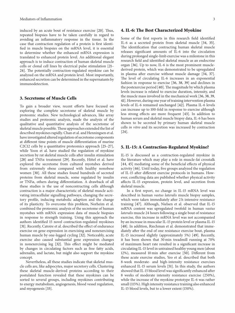
Some of the first reports in this research field identifiedIL-6 as a secreted protein from skeletal muscle [34, 35].The identification that contracting human skeletal musclereleases significant amounts of IL-6 into the circulationduring prolonged single-limb exercise was a milestone in thisresearch field and identified skeletal muscle as an endocrineorgan [36]. Up to now, IL-6 is the most prominent muscle-derived protein, which was demonstrated to be upregulatedin plasma after exercise without muscle damage [34, 37].The level of circulating IL-6 increases in an exponentialfashion in response to exercise [36, 38, 39] and declines inthe postexercise period [40].Themagnitude bywhich plasmalevels increase is related to exercise duration, intensity, andthe muscle mass involved in the mechanical work [36, 38, 39,41]. However, during one year of training intervention plasmalevels of IL-6 remained unchanged [42]. Plasma IL-6 levelscan increase up to 100-fold in response to exercise althoughless strong effects are more frequent [43]. In addition tohuman serum and skeletal muscle biopsy data, IL-6 has beenshown to be secreted by primary human skeletal musclecells in vitro and its secretion was increased by contraction[24].
5. IL-15: A Contraction-Regulated Myokine?
IL-15 is discussed as a contraction-regulated myokine inthe literature which may play a role in muscle-fat crosstalk[44, 45] mediating some of the beneficial effects of physicalactivity [46]. Until today, five groups analyzed the regulationof IL-15 after different exercise protocols in humans. How-ever, conflicting data are published whether physical activityaffects IL-15 expression, protein level, and secretion fromskeletal muscle.
In a first report, no change in IL-15 mRNA level wasdescribed in human vastus lateralis muscle biopsy samples,which were taken immediately after 2 h intensive resistancetraining [47]. Although, Nielsen et al. observed that IL-15mRNA content was upregulated twofold in human vastuslateralismuscle 24 hours following a single bout of resistanceexercise, this increase in mRNA level was not accompaniedby an increase in muscular IL-15 protein level or plasma IL-15[48]. In addition, Riechman et al. demonstrated that imme-diately after the end of one resistance exercise bout, plasmaIL-15 increased slightly (approximately 5%) [49]. Recently,it has been shown that 30min treadmill running at 70%of maximum heart rate resulted in a significant increase incirculating IL-15 level in untrained healthy youngmen (about12%), measured 10min after exercise [50]. Different fromthese acute exercise studies, Yeo et al. described that both8-week moderate- and high-intensity resistance exercisesenhanced IL-15 serum levels [51]. In this study, the authorsshowed that IL-15 blood level was significantly enhanced after8 weeks of moderate intensity resistance exercise (250%),while the increase of the myokine prototype IL-6 was rathersmall (115%).High intensity resistance training also enhancedIL-15 blood levels, but to a lower extent (150%).

4 Mediators of Inflammation
In addition, training studies were performed in mice andrats. It has been shown that IL-15 mRNA expression in soleusand gastrocnemiusmuscle is increased after 8-week treadmillrunning training in rats, while plasma IL-15 level was notchanged [52]. Yang et al. observed about 1.7-fold increasein IL-15 mRNA expression after three weeks of free wheelrunning in mice but did not analyze protein or serum level.
Nevertheless, while the observed increase in plasma IL-15levels in humans is rather small (5–12%) after acute exerciseand not depending on the mode of exercise, particularlymoderate intensity resistance exercise had a significant effecton IL-15 blood levels. However, to the best of our knowledge,secretion of IL-15 from muscle cells has not been describedyet, and it has not been shown that the observations on mus-cle mRNA level are translated to meaningful contributions toIL-15 serum levels.
6. Irisin: A Novel Myokine
Just recently, a novel identified myokine has drawn theattention as a novel preventive and therapeutic target totreat obesity and metabolic diseases like type 2 diabetes.Bostrom et al. observed that overexpression of PGC1𝛼 inmice muscle as well as exercise induces the expression ofthe FNDC5 (fibronectin type III domain containing protein5) gene, a gene which has scarcely been studied before.FNDC5 is described as a protein containing a signal pep-tide, fibronectin type III repeats, and hydropathy analysisrevealed a hydrophobic region, which is likely to encode atransmembrane domain. Previous studies linked the geneto differentiation of myoblasts and neurones [53, 54], andit has been suggested that FNDC5 is located in the matrixof peroxisomes [53]. Bostrom et al. described that the C-terminal tail of the protein is located in the cytoplasm,whereas the extracellular N-terminal part is supposed tobe cleaved and released as novel messenger molecule calledirisin [55]. Mice subjected to three weeks of free wheelrunning showed enhanced muscle mRNA expression andelevated irisin plasma concentrations (65%). In addition, tenweeks of supervised endurance exercise training revealed atwofold increase in circulating irisin levels compared to thenonexercised state in a cohort of older subjects [55]. Boththe mice and human study analyzed the long-term effectof exercise on irisin plasma levels. It is not described ifenhanced serum levels of FNDC5 after muscle contractionare dependent on enhanced gene expression or dependent onenhanced cleavage of the membrane protein.
However, using gene-chip probe sets Timmons et al.observed no effect on FNDC5 mRNA level neither after6 weeks of intense endurance cycling in younger subjectsnor after supervised resistance training [56]. Timmons et al.demonstrated that FNDC5 induction in muscle occurredonly in highly active elderly subjects compared to sedentarycontrols (1.3fold), whichwere aminority of analyzed subjects.They failed to confirm FNDC5 gene expression by aerobicexercise in younger subjects. Huh et al. observed minoreffects on irisin plasma levels after 1 week of exercise (increaseof about 18%) and no effect after prolonged training over
8 weeks [57]. Up to now, there is only one study showinga robust activation of FNDC5 after exercise in humansmeasured by RT-PCR in muscle biopsies [24], however,limited to a very small number of subjects.
Although Bostrom et al. described the discovery of thenovel myokine irsin, the authors showed that the release ofirisin exclusively in HEK 293 cells transfected with a vectorexpressing FNDC5 followed immunodetection of culturemedia protein. It has not been confirmed thatmuscle FNDC5mRNA is translated to protein in primary or clonal skeletalmuscle cells, and, most importantly, it has not been shownthat irisin is secreted from skeletal muscle cells. Furthermore,to demonstrate the secretion of irisin from HEK293 cellsand to analyze murine and human serum samples, theauthors used an antibody, which most likely cannot detectthe cleaved irisin, since the antibody used is directed againstthe C-terminal part of the protein (Abcam 149–178, C-terminal) [58]. Taken together, future studies should addressFNDC5/irisin precise expression and cleavage mechanism toclarify the controversy of current literature.
7. BDNF: Released from the Muscle orthe Brain?
BDNF belongs to the family of neurotrophins (NT), whichincludes nerve growth factor, BDNF, NT-3, NT-4/5, and NT-6. These proteins are produced as large precursor proteinsthat are then cleaved to form themature neurotrophic protein(reviewed in [59]). In the literature BDNF is discussedas a contraction-regulated myokine [6]. During myogenicdifferentiation, the expression of BDNF is drastically reducedand is hardly detectable in adult rat skeletal myofibers[60]. By reverse transcription PCR, in situ hybridization,and immunofluorescence, it was shown that BDNF is notexpressed at significant levels within mature myofibers [60].In situ hybridisation analysis revealed that in adult rat musclethe constitutive expression of muscular BDNF is confined tothe myofibres. Satellite cells, Schwann cells, endothelial cells,fibroblasts, or axons did not appear to contribute to BDNFproduction in normal muscle [61]. In complementary cellculture experiments, it has been shown that levels of BDNFcorrelate with the population of satellite cells [60]. BDNF isrequired for early phases of myogenic differentiation, whichis delayed in the absence of BDNF [62]. Nevertheless, BDNFprotein level was determined in lysates of rat L6 cells [60] andmurine C2C12 cells [19].
Serum BDNF levels increased after a graded cycling exer-cise test in humans (30%) [63] and at the point of exhaustionat the end of a ramp test (about 25%) [64]. Matthews et al.reported enhanced BDNF mRNA and protein expression inhuman skeletal muscle and after bicycle exercise [19]. Cellculture experiments using murine C2C12 cells stimulatedby electrical pulse stimulation confirmed that contractileactivity enhanced BNDF mRNA and protein level. AlthoughMatthews et al. report increased serum levels after exercisein humans, BDNF basal secretion by C2C12 that underwentcontraction has not been proven and overexpression of BDNFinmouse skeletal muscle did not lead to differences in plasma
Mediators of Inflammation 5
BDNF [19], leading the authors to the conclusion that BNDFexerts its action locally and is not released into the circulation.Matthews et al. reported an autocrine effect of BNDF sincetreatment of skeletal muscle cells with recombinant BDNFresulted in enhanced phosphorylation of AMP-activatedprotein kinase (AMPK) and ACC in rat L6 cells which leadsto enhanced fatty acid oxidation [19].
In mice, treadmill exercise induced an increase in BDNFmRNA expression in the hippocampus and cortex (three-to fivefold) [65]. In humans, a BDNF release from thebrain was observed at rest and increased two- to threefoldduring exercise. Both at rest and during exercise, the braincontributed 70–80% of circulating BDNF [65]. These resultssuggest that the brain is a major, but not the sole contributorto circulating BDNF after exercise.
8. MCP-1
MCP-1 is a chemokine and member of the small induciblecytokine family. It plays a crucial role in the recruitment ofmonocytes and T lymphocytes into tissues [66, 67]. MCP-1 was detected in supernatants of C2C12 cells [68]. In mice,plasma IL-6 levels were markedly increased 3 h followingmaximumprogressive swimming, whileMCP-1 plasma levelswere not altered by exercise [69]. However, a single bout ofintense resistance exercise increased MCP-1 mRNA expres-sion in muscle biopsy samples obtained from vastus lateralismuscle about 35-fold after two hours. In comparison, IL-6 mRNA expression, the myokine prototype, was enhancedabout 400-fold [70]. One bout of moderate-intensity cycleexercise increased MCP-1 mRNA levels in vastus lateralismuscle biopsy samples after 40min [71]. One-legged cyclingof male subjects induced a significant change in MCP-1mRNA levels in the exercising leg and enhanced MCP-1plasma levels after exercise and after 3 hours of recovery [72].In addition, increased MCP-1 mRNA expression in skeletalmuscle was reported in elderly individuals following onebout of resistance exercise [73] and in young men after arepeated eccentric exercise bout [74]. Immunohistochem-istry analysis of muscle biopsies colocalized MCP-1 withresidentmacrophage and satellite cell populations, suggestingthat alterations in cytokine signalling between these cellpopulations may play a role in muscle adaptation to exercise[74].
9. ANGPTL4: Regulated by Free Fatty Acids
ANGPTL4 represents a prominent long chain fatty acid-responsive gene in human myotubes. Kersten et al. reportedthat plasma ANGPTL4 levels in humans increased signif-icantly in response to long-term fasting, chronic caloricrestriction, and endurance training. All these states are char-acterized by enhanced circulating FFA [75]. Fasting plasmaANGPTL4 levels of healthy, untrained male volunteersincreased during endurance exercise at 50% VO2 max for 2 hand especially during subsequent recovery. Importantly, theincrease in plasma ANGPTL4 was abolished when subjectswere given oral glucose, which induces insulin release and
thereby suppresses plasma FFA levels [75]. While ANGPTL4is below the detection limit in supernatants of differentiatedC2C12 cells, long-term treatment of human myotubes (48 h)with the PPAR𝛿-specific activator GW501516 results in theaccumulation of ANGPTL4 in the supernatant [76]. In addi-tion, incubation of human primary myocytes with oleic acidand linoleic acid enhanced ANGPTL4 mRNA expression.Nevertheless, this effect was not only observed in primaryhuman myocytes, but also in FAO hepatoma cells and mouseintestinal MSIE cells [75].
Catoire et al. have shown in a human one-legged exercisestudy that target genes of PPAR transcription factors includ-ingANGPTL4were induced equally in exercising and nonex-ercising muscle [77]. Although PPAR𝛿 is known to be acti-vated by high-intensity exercise [78], Catoire et al. have con-cluded that the increase of plasma free fatty acid levels due toacute exercise activates PPARs and therefore ANGPTL4 [77].
Long-term changes in plasma ANGPTL4 levels are mostlikely mediated by changes in plasma free fatty acids, whichraise ANGPTL4 gene expression in target tissues. Neverthe-less, ANGPTL4 is ubiquitously expressed in human tissuesand highest expression levels were found in liver, followedby adipose tissue, thyroid, brain, small intestine, and lessin skeletal muscle [75]. Thus, skeletal muscle might not bethe only tissue which is responsible for enhanced ANGPTL4plasma levels in states of increased FFA levels like endurancetraining. Furthermore, ANGPTL4 stimulates adipose tissuelipolysis, leading to elevation of plasma free fatty acid levels.Kersten et al. speculated that both mechanisms operateas a positive feedback loop. Free fatty acids raises plasmaANGPTL4 and ANGPTL4 raises plasma free fatty acids bythe stimulation of adipose tissue lipolysis [75].
10. FGF21
FGF21 is a member of the fibroblast growth factor superfamily, a large family of proteins involved in cell proliferation,growth, and differentiation. The first evidence that FGF21 isan Akt-regulated myokine was published by Izumiya et al.[79]. FGF21 protein expression and secretion are upregulatedby insulin and inhibited by PI3-kinase inhibitor in culturedC2C12 myocytes [79]. Skeletal muscle mRNA level andplasma level are induced by hyperinsulinemia studied inyoung healthy men during a hyperinsulinemic-euglycemicclamp [80]. In line with this observation, circulating FGF21is elevated in impaired glucose tolerance and type 2 diabetespatients and correlates with muscle and hepatic insulinresistance [81].
Interestingly, an acute bout of treadmill exercise didnot change FGF21 serum levels in sedentary young women.However, after two weeks of exercising there was a 1.6-foldincrease in serum FGF21 [82]. In contrast, twelve-weekexercise program combining aerobic and resistance exercise,five times per week, reduced FGF21 plasma levels innondiabetic, obese women (from 230.2 ± 135.9 versus 102.6 ±117.8 pg/mL) [83]. Nevertheless, nothing is known about theacute effect of contraction on the expression, protein level,and secretion of FGF21 from skeletal muscle cells.
6 Mediators of Inflammation
11. Myonectin
Myonectin belongs to the C1q/TNF-related protein family(C1QTNF isoform 5) and shows a sequence homology withadiponectin in the shared C1q domain, the signature thatdefines this protein family [84]. Before myonectin wasdescribed as a myokine, the protein was reported to beexpressed in the retinal pigment epithelium, andmutations inthis gene caused abnormal high molecular weight aggregateformation, which results in late-onset retinal macular degen-eration in humans [85].
Myonectin is supposed to be myokine due to the detec-tion of the myonectin transcript in mice skeletal muscle, withsignificantly lower expression in other tissues, immunoblotdetection of myonectin in mouse skeletal muscle lysates [86],and L6 supernatants [87] aswell as induced expression duringdifferentiation of mouse C2C12 cells [86].
Currently, one human and one mice exercise stud-ies report divergent results regarding the regulation ofmyonectin by contraction. Lim et al. reported that a 10-week exercise training program in younger and older groupsof healthy women decreased significantly myonectin serumlevels, while training increased VO2 max, mitochondrialDNA density in skeletal muscle, and plasma adiponectinlevels significantly [88].
On the other hand, free wheel running for two weeksincreased myonectin expression in soleus und plantarismuscle of mice and circulating serum levels, suggesting apotential role of myonectin in exercise-induced physiology[86]. Recombinant myonectin induced the phosphorylationof AMPK, leading to increased cell surface recruitment ofGLUT4, enhanced glucose uptake, and stimulated fatty acidoxidation [87]. Thus, enhanced myonectin secretion inducedby contraction could activate signaling pathways providingenhanced energy demands during contraction.
Most intriguingly, Seldin et al. reported that recombinantmyonectin promotes fatty acid uptake in mouse adipocytesand rat hepatocytes in vitro by enhancing CD36, FATP1,and Fabp4 mRNA expressions, which are known to playimportant roles in fatty acid uptake. In addition, recombinantmyonectin had no effect on adipose tissue lipolysis [86].A question that should be addressed by future studies iswhy physical activity activates the secretion of a myokinethat induces fatty acid uptake by adipose tissue. Enhancedmyonectin serum levels after exercise would therefore leadto an endocrine signal that would deplete energy sourcesin the blood which is needed by the exercising muscle. Onthe other hand, long-lasting increase in myonectin serumlevels could increase fatty acid uptake in adipose tissue andtherefore improve fat metabolism and lipid handling.
12. Adipo-Myokines
In a recently published review, Pedersen and Febbraio sug-gest that skeletal muscle might mediate some of the well-established protective effects of exercise via the secretion ofmyokines that counteract the harmful effects of proinflam-matory adipokines [6].
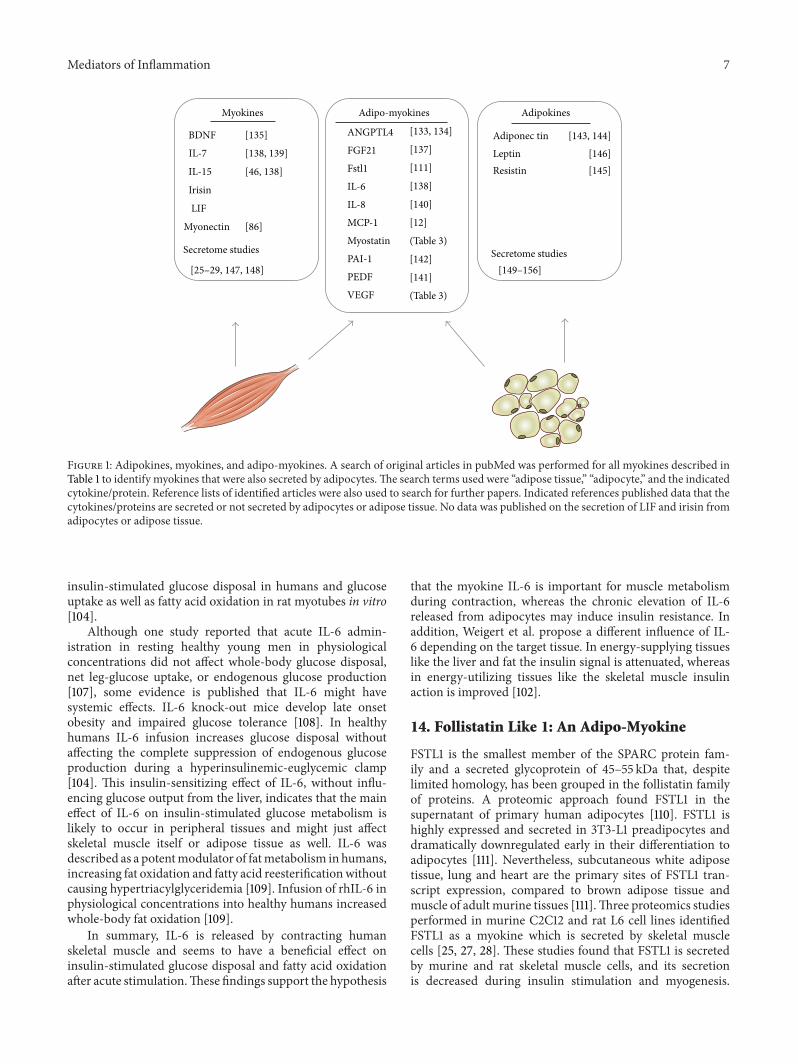
Table 1 summarizes themost prominentmyokines, whichare described to be contraction regulated. For more than halfof the described myokines, ten out of seventeen, secretionby adipocytes has also been described (Figure 1). We termedthese cytokines adipo-myokines. How should a cytokineexert on the one hand inflammatory signalling in the obesestate and have beneficial effects after exercise? Is it likely thata bidirectional communication between fat and muscle cellstakes place? Just recently, Christiansen et al. reported thatacute exercise increases circulating inflammatory markers inoverweight and obese compared with lean subjects [89].Whyshould inflammatory markers increase after exercise?
13. IL-6: The Prototype Adipo-Myokine
IL-6 seems to be a good example for an adipo-myokine thatis released by both tissues and has the potential to act onboth tissues. As described before, the level of circulating IL-6 increases after an acute bout of exercise in an exponentialfashion [36, 38, 39] and declines in the postexercise period[40].
In addition, the quantitative release from adipose tis-sue correlates positively with increased body fat content,which results in systemic elevation of IL-6 plasma levels[90]. It is overexpressed in human fat cells from insulin-resistant subjects [91], increased in the plasma of obesepatients [92, 93], and associated with type 2 diabetes [90,94], while it was described to be decreased after bariatricsurgery [95]. IL-6 expression was known to be activatedby proinflammatory IKK𝛽/NF𝜅B signalling pathway whichis thought to contribute to the development of obesity-induced insulin resistance [96, 97]. In addition, it has beenshown to inhibit insulin-signalling pathways in the liver[98, 99] and adipocytes [91]. In vitro experiments revealedthat IL-6 induced insulin resistance in hepatocytes [99],adipocytes [91], and in skeletal muscle cells after treatmentwith high doses [100]. Incubation of the rat L6myotubes with200 ng/mL recombinant IL-6 induced insulin resistance onthe level of diminished Akt phosphorylation after 96 h [101]and in primary human myotubes after 48 h [100].
Since exercise is thought to increase insulin sensitivity,the observation that IL-6 is also increased after exerciseseemed quite paradoxical. Interestingly, for skeletal musclecells, in vitro studies showed that a rather brief challengeof minutes to few hours with recombinant IL-6 had a pos-itive autocrine effect on skeletal muscle cells. RecombinantIL-6 enhanced insulin-stimulated Akt phosphorylation inprimary human myotubes (20 ng/mL) [102, 103] and ratL6 myotubes (about 200 ng/mL) [101]. In addition, basaland insulin-stimulated glucose uptake and translocation ofGLUT4 to the plasma membrane were enhanced after 5–120min in L6 myotubes (1–100 ng/mL) [104]. Furthermore,IL-6 rapidly and markedly increased AMPK and increasedfatty acid oxidation [104, 105]. Interestingly, the regulation ofintracellular signalling mechanisms, mediating IL-6 expres-sion, differs from the classical proinflammatory pathway. IL-6 expression in contracting muscle is regulated by c-Junterminal kinase (JNK)/activator protein-1 [106] and increases
Mediators of Inflammation 7
Myokines Adipokines
Adiponec tin [143, 144]Leptin [146]Resistin [145]
BDNF [135]
IL-7 [138, 139]
IL-15 [46, 138]
Irisin
LIF
Myonectin [86]
[25–29, 147, 148]
Secretome studies Secretome studies
ANGPTL4 [133, 134]
FGF21 [137]
Fstl1 [111]
IL-6 [138]
IL-8 [140]
MCP-1 [12]
Myostatin (Table 3)
PAI-1 [142]PEDF [141]VEGF (Table 3)
[149–156]
Adipo-myokines
Figure 1: Adipokines, myokines, and adipo-myokines. A search of original articles in pubMed was performed for all myokines described inTable 1 to identify myokines that were also secreted by adipocytes.The search terms used were “adipose tissue,” “adipocyte,” and the indicatedcytokine/protein. Reference lists of identified articles were also used to search for further papers. Indicated references published data that thecytokines/proteins are secreted or not secreted by adipocytes or adipose tissue. No data was published on the secretion of LIF and irisin fromadipocytes or adipose tissue.
insulin-stimulated glucose disposal in humans and glucoseuptake as well as fatty acid oxidation in rat myotubes in vitro[104].
Although one study reported that acute IL-6 admin-istration in resting healthy young men in physiologicalconcentrations did not affect whole-body glucose disposal,net leg-glucose uptake, or endogenous glucose production[107], some evidence is published that IL-6 might havesystemic effects. IL-6 knock-out mice develop late onsetobesity and impaired glucose tolerance [108]. In healthyhumans IL-6 infusion increases glucose disposal withoutaffecting the complete suppression of endogenous glucoseproduction during a hyperinsulinemic-euglycemic clamp[104]. This insulin-sensitizing effect of IL-6, without influ-encing glucose output from the liver, indicates that the maineffect of IL-6 on insulin-stimulated glucose metabolism islikely to occur in peripheral tissues and might just affectskeletal muscle itself or adipose tissue as well. IL-6 wasdescribed as a potentmodulator of fatmetabolism in humans,increasing fat oxidation and fatty acid reesterificationwithoutcausing hypertriacylglyceridemia [109]. Infusion of rhIL-6 inphysiological concentrations into healthy humans increasedwhole-body fat oxidation [109].
In summary, IL-6 is released by contracting humanskeletal muscle and seems to have a beneficial effect oninsulin-stimulated glucose disposal and fatty acid oxidationafter acute stimulation.These findings support the hypothesis
that the myokine IL-6 is important for muscle metabolismduring contraction, whereas the chronic elevation of IL-6released from adipocytes may induce insulin resistance. Inaddition, Weigert et al. propose a different influence of IL-6 depending on the target tissue. In energy-supplying tissueslike the liver and fat the insulin signal is attenuated, whereasin energy-utilizing tissues like the skeletal muscle insulinaction is improved [102].
14. Follistatin Like 1: An Adipo-Myokine
FSTL1 is the smallest member of the SPARC protein fam-ily and a secreted glycoprotein of 45–55 kDa that, despitelimited homology, has been grouped in the follistatin familyof proteins. A proteomic approach found FSTL1 in thesupernatant of primary human adipocytes [110]. FSTL1 ishighly expressed and secreted in 3T3-L1 preadipocytes anddramatically downregulated early in their differentiation toadipocytes [111]. Nevertheless, subcutaneous white adiposetissue, lung and heart are the primary sites of FSTL1 tran-script expression, compared to brown adipose tissue andmuscle of adult murine tissues [111].Three proteomics studiesperformed in murine C2C12 and rat L6 cell lines identifiedFSTL1 as a myokine which is secreted by skeletal musclecells [25, 27, 28]. These studies found that FSTL1 is secretedby murine and rat skeletal muscle cells, and its secretionis decreased during insulin stimulation and myogenesis.
8 Mediators of Inflammation
In addition, Gorgens et al. recently showed that FSTL1 isalso secreted by primary human skeletal muscle cells [112].Data have shown that FSTL1 is secreted into the mediaby cultured C2C12 skeletal muscle cells, and it can directlyact on endothelial cell-signalling pathways that promotefunction and survival. FSTL1 overexpression in endothelialcells was found to enhance endothelial cell differentiationand migration and diminish endothelial apoptosis [113].Thus, FSTL1 might be a myokine that mediates some of thewell-established protective effects of exercise that counteractthe harmful effects of proinflammatory adipokines on thevasculature. In addition, treatment of neonatal rat ventricualcardiomyocyteswith recombinant FSTL1 induced a time- anddose-dependent increase in AMPK and ACC phosphoryla-tion [114]. Given that FSTL1 mRNA expression was increasedafter strength training [31] and after an acute bout of cyclingat 70% VO2 max [112], it might be speculated that exerciseactivates FSTL1 expression and secretion in skeletal muscle,which might act in an autocrine and/or endocrine mannerand activate muscular and/or adipogenic AMPK. However,the biological role of adipocyte-derived and muscle-derivedFSTL1 has still to be defined.
15. Leptin: An Adipokine rather thana Myokine
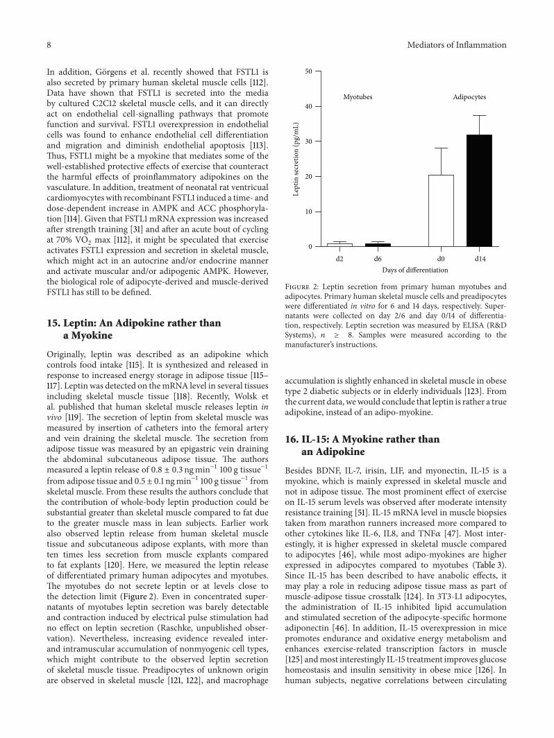
Originally, leptin was described as an adipokine whichcontrols food intake [115]. It is synthesized and released inresponse to increased energy storage in adipose tissue [115–117]. Leptin was detected on themRNA level in several tissuesincluding skeletal muscle tissue [118]. Recently, Wolsk etal. published that human skeletal muscle releases leptin invivo [119]. The secretion of leptin from skeletal muscle wasmeasured by insertion of catheters into the femoral arteryand vein draining the skeletal muscle. The secretion fromadipose tissue was measured by an epigastric vein drainingthe abdominal subcutaneous adipose tissue. The authorsmeasured a leptin release of 0.8 ± 0.3 ngmin−1 100 g tissue−1from adipose tissue and 0.5 ± 0.1 ngmin−1 100 g tissue−1 fromskeletal muscle. From these results the authors conclude thatthe contribution of whole-body leptin production could besubstantial greater than skeletal muscle compared to fat dueto the greater muscle mass in lean subjects. Earlier workalso observed leptin release from human skeletal muscletissue and subcutaneous adipose explants, with more thanten times less secretion from muscle explants comparedto fat explants [120]. Here, we measured the leptin releaseof differentiated primary human adipocytes and myotubes.The myotubes do not secrete leptin or at levels close tothe detection limit (Figure 2). Even in concentrated super-natants of myotubes leptin secretion was barely detectableand contraction induced by electrical pulse stimulation hadno effect on leptin secretion (Raschke, unpublished obser-vation). Nevertheless, increasing evidence revealed inter-and intramuscular accumulation of nonmyogenic cell types,which might contribute to the observed leptin secretionof skeletal muscle tissue. Preadipocytes of unknown originare observed in skeletal muscle [121, 122], and macrophage
d2 d6 d0 d140
10
20
30
40
50
Myotubes Adipocytes
Days of differentiation
Lept
in se
cret
ion
(pg/
mL)
Figure 2: Leptin secretion from primary human myotubes andadipocytes. Primary human skeletal muscle cells and preadipocyteswere differentiated in vitro for 6 and 14 days, respectively. Super-natants were collected on day 2/6 and day 0/14 of differentia-tion, respectively. Leptin secretion was measured by ELISA (R&DSystems), 𝑛 ≥ 8. Samples were measured according to themanufacturer’s instructions.
accumulation is slightly enhanced in skeletal muscle in obesetype 2 diabetic subjects or in elderly individuals [123]. Fromthe current data, wewould conclude that leptin is rather a trueadipokine, instead of an adipo-myokine.
16. IL-15: A Myokine rather thanan Adipokine
Besides BDNF, IL-7, irisin, LIF, and myonectin, IL-15 is amyokine, which is mainly expressed in skeletal muscle andnot in adipose tissue. The most prominent effect of exerciseon IL-15 serum levels was observed after moderate intensityresistance training [51]. IL-15 mRNA level in muscle biopsiestaken from marathon runners increased more compared toother cytokines like IL-6, IL8, and TNF𝛼 [47]. Most inter-estingly, it is higher expressed in skeletal muscle comparedto adipocytes [46], while most adipo-myokines are higherexpressed in adipocytes compared to myotubes (Table 3).Since IL-15 has been described to have anabolic effects, itmay play a role in reducing adipose tissue mass as part ofmuscle-adipose tissue crosstalk [124]. In 3T3-L1 adipocytes,the administration of IL-15 inhibited lipid accumulationand stimulated secretion of the adipocyte-specific hormoneadiponectin [46]. In addition, IL-15 overexpression in micepromotes endurance and oxidative energy metabolism andenhances exercise-related transcription factors in muscle[125] andmost interestingly IL-15 treatment improves glucosehomeostasis and insulin sensitivity in obese mice [126]. Inhuman subjects, negative correlations between circulating

Mediators of Inflammation 9
Table 2: Overview of selected adipo-myokines which are associated with obesity and insulin resistance.
Adipo-Myokine Associated with obesity Associated with insulinresistance/T2D
Associated with improved glucosemetabolism
IL-6✓
Plasma IL-6 is positively related to fatmass [175], elevated in type 2 diabetics[90, 94]
✓
IL-6 promotes insulin resistance[91, 98, 99]
✓
Insulin-sensitizing effect on skeletalmuscle [102, 104] increases wholebody fat oxidation [109]
IL-7
?Increased mRNA level in omentaladipose tissue [139] although miceoverexpressing IL-7 have reducedadipose tissue mass [177]
n.d. n.d.
IL-8✓
Higher expression in visceral adiposetissue in type 2 diabetics and insulinresistant subjects [91, 178]
✓
IL-8 plasma levels correlate withmeasures of insulin resistance[97, 179]
n.d.
MCP-1✓
SerumMCP-1 is increased in obesity[129]
✓
Promotes insulin resistance [12, 129] n.d.
PEDF✓
PEDF serum levels increased inobesity [180, 181]
✓
PEDF serum levels associated withinsulin resistance [180–183], PEDFpromotes insulin resistance [141]
n.d.
✓: association has been shown in indicated publications; ✓: contradictory data published; n.d.: not described.
Table 3: Concentrations of various factors in conditioned medium from primary human adipocytes and primary humanmyotubes. Primaryhuman skeletal muscle cells were differentiated for 5 days and primary human preadipocytes were differentiated for 14 days in vitro to maturecells. During the last 24 h cells were incubated with serum-free medium to obtain conditioned medium. Concentrations of secreted factorsfrom cells within this conditioned medium were analyzed by enzyme-linked immunosorbent assay. Data are means ± SEM, 𝑛 ≥ 3.
Secreted factor Concentration in adipocyte-conditionedmedium (ng/mL)
Concentration in skeletal muscle-conditionedmedium (ng/mL)
Chemerin 2.12 ± 0.3 [141] 0.006 ± 0.001DPP4 2.19 ± 1.4 [11] 0.69 ± 0.18IL-6 0.03 ± 0.002 [141] 0.02 ± 0.003IL-8 0.15 ± 0.04 0.07 ± 0.01MCP-1 0.35 ± 0.06 0.33 ± 0.08Myostatin 12.64 ± 4.44 3.44 ± 1.64PEDF 45.7 ± 0.82 [141] 5.4 ± 0.86VEGF 0.33 ± 0.09 [141] 0.05 ± 0.03
IL-15 levels and both total and abdominal fat have beendemonstrated [127]. Since both IL-15 and physical exercisehave positive effects on body composition, IL-15 is discussedas a contraction-regulated myokine in the literature whichmay play a role in muscle-fat cross-talk [44, 45] mediatingsome of the beneficial effects of physical activity. Yang et al.published just recently a direct link between treadmill exer-cise of high-fat diet rats, enhanced expression of IL-15 inmuscle, and increased IL-15 receptor alpha expression inadipose tissue [52].
17. Other Adipo-Myokines
MCP-1 is one of these adipo-myokines. Before MCP-1 wascharacterized as a myokine, it was described to be produced
in isolated adipocytes, associated with adiposity and reducedafter weight loss in morbid obese subjects [128]. It is overex-pressed in obese rodents [129, 130] and reaches significantlyhigher plasma levels in diabetic patients [131]. In addition,MCP-1-induced macrophage infiltration in adipose tissueleads to a chronic state of low-grade inflammation [132],which is linked to insulin resistance. In vitro data demonstratethat this factor has the ability to induce insulin resistance inadipocytes and skeletal muscle cells [12].
Nevertheless, while IL-6, IL-7, IL-8, MCP-1, and pigmentendothelial derived factor (PEDF) are associated with obe-sity and insulin resistance, these proteins are contraction-regulated myokines (Table 1), and only for IL-6 a beneficialeffect has been described (Table 2). The description of a ben-eficial effect for thesemyokines is lacking and is an interesting

10 Mediators of Inflammation
open question for future studies (Table 2).Myostatin is a well-known myokine, but our group recently identified myostatinas an adipokine [2].
18. Conclusion
Taken together, one protein can be a myokine as well as anadipokine, indeed two sides of the same coin. In healthy,normal weight subjects skeletal muscle is the largest tissue inthe human body, accounting for 40–50% of total human bodymass, while body fat accounts for 20–35%. In obese subjectsthe percentage of total body fat increases to 40–60% resultingin an increased secretion of proinflammatory adipokines,while the percentage of proteins secreted from skeletal mus-cle during a sedentary lifestyle is decreased. However, formany adipo-myokines the local tissue concentration may bedivergent from the serum level, and substantial differencesbetween auto- and endocrine effects of these molecules needto be considered.
As Paracelsus (1493–1541) already coined the famousphrase; “dosis sola facit venenum,” “Only the dose makes thepoison”.Thismight also be true for adipo-myokines. Findingssupport the hypothesis that the myokines are essential formuscle metabolism during contraction, whereas the chronicelevation of adipokines released from adipocytes may induceadverse effects, even leading to insulin resistance.
Abbreviations
AMPK: AMP-activated protein kinaseANGPTL4: Angiopoietin like 4BDNF: Brain-derived neurotrophic factorDPP4: Dipeptidyl peptidase 4FGF: Fibroblast growth factorFNDC5: Fibronectin type III domain containing
protein 5FSTL1: Follistatin-like 1IL: InterleukinLIF: Leukemia inhibitory factorMCP-1: Monocyte chemoattractant protein 1NT: NeurotrophinsPEDF: Pigment endothelial derived factorTNF𝛼: Tumor necrosis factor 𝛼VEGF: Vascular endothelial growth factor.
Conflict of Interests
The authors have no conflict of interests in relation to thecontents of this paper.
Acknowledgments
This review article is based on discussions originating fromthe PhD thesis of SR. The authors would like to acknowledgeDr. Kristin Eckardt and Sven W. Gorgens for providing dataon the secretion of myostatin and IL-8 from primary cells(data shown in Table 3). This work was supported by theMinisterium fur Wissenschaft und Forschung des Landes
Nordrhein-Westfalen (Ministry of Science and Research ofthe State of North Rhine-Westphalia), the Bundesminis-terium fur Gesundheit (Federal Ministry of Health). Thisstudy was supported in part by a grant from the GermanFederal Ministry of Education and Research (BMBF) to theGerman Center for Diabetes Research (DZD e.V.)).
References
[1] M. Adamczak and A. Wiecek, “The adipose tissue as anendocrine organ,” Seminars in Nephrology, vol. 33, pp. 2–13,2013.
[2] S. Lehr, S. Hartwig, and H. Sell, “Adipokines: a treasuretrove for the discovery of biomarkers for metabolic disorders,”Proteomics. Clinical Applications, vol. 6, pp. 91–101, 2012.
[3] N. Ouchi, J. L. Parker, J. J. Lugus, and K. Walsh, “Adipokines ininflammation andmetabolic disease,”Nature Reviews Immunol-ogy, vol. 11, no. 2, pp. 85–97, 2011.
[4] B. K. Pedersen, “The diseasome of physical inactivity- and therole of myokines in muscle-fat cross talk,” Journal of Physiology,vol. 587, no. 23, pp. 5559–5568, 2009.
[5] B. K. Pedersen, “Exercise-induced myokines and their role inchronic diseases,” Brain, Behavior, and Immunity, vol. 25, no. 5,pp. 811–816, 2011.
[6] B. K. Pedersen and M. A. Febbraio, “Muscles, exercise andobesity: skeletal muscle as a secretory organ,” Nature ReviewsEndocrinology, vol. 8, pp. 457–465, 2012.
[7] C. Handschin and B. M. Spiegelman, “The role of exercise andPGC1𝛼 in inflammation and chronic disease,” Nature, vol. 454,no. 7203, pp. 463–469, 2008.
[8] J. S. Yudkin, “Inflammation, obesity, and the metabolic syn-drome,” Hormone and Metabolic Research, vol. 39, no. 10, pp.707–709, 2007.
[9] P. Arner, “Regional differences in protein production by humanadipose tissue,” Biochemical Society Transactions, vol. 29, no. 2,pp. 72–75, 2001.
[10] P. Trayhurn and J. H. Beattie, “Physiological role of adiposetissue: white adipose tissue as an endocrine and secretoryorgan,” Proceedings of the Nutrition Society, vol. 60, no. 3, pp.329–339, 2001.
[11] D. Lamers, S. Famulla, N. Wronkowitz et al., “Dipeptidylpeptidase 4 is a novel adipokine potentially linking obesity tothe metabolic syndrome,” Diabetes, vol. 60, pp. 1917–1925, 2011.
[12] H. Sell, D. Dietze-Schroeder, U. Kaiser, and J. Eckel, “Monocytechemotactic protein-1 is a potential player in the negative cross-talk between adipose tissue and skeletal muscle,” Endocrinology,vol. 147, no. 5, pp. 2458–2467, 2006.
[13] H. Sell, J. Eckel, and D. Dietze-Schroeder, “Pathways leading tomuscle insulin resistance—themuscle-fat connection,”Archivesof Physiology and Biochemistry, vol. 112, no. 2, pp. 105–113, 2006.
[14] H. Sell, A. Divoux, C. Poitou et al., “Chemerin correlates withmarkers for fatty liver in morbidly obese patients and stronglydecreases after weight loss induced by bariatric surgery,” Journalof Clinical Endocrinology and Metabolism, vol. 95, no. 6, pp.2892–2896, 2010.
[15] R. A. DeFronzo and D. Tripathy, “Skeletal muscle insulinresistance is the primary defect in type 2 diabetes,” DiabetesCare, vol. 32, supplement 2, pp. S157–S163, 2009.
[16] B. K. Pedersen, “Muscles and their myokines,” Journal ofExperimental Biology, vol. 214, no. 2, pp. 337–346, 2011.

Mediators of Inflammation 11
[17] H. Ellingsgaard, I. Hauselmann, B. Schuler et al., “Interleukin-6enhances insulin secretion by increasing glucagon-like peptide-1 secretion from L cells and alpha cells,”NatureMedicine, vol. 17,pp. 1481–1489, 2011.
[18] B. K. Pedersen and M. A. Febbraio, “Muscle as an endocrineorgan: focus on muscle-derived interleukin-6,” PhysiologicalReviews, vol. 88, no. 4, pp. 1379–1406, 2008.
[19] V. B. Matthews, M. B. Astrom, M. H. S. Chan et al., “Brain-derived neurotrophic factor is produced by skeletal musclecells in response to contraction and enhances fat oxidation viaactivation of AMP-activated protein kinase,” Diabetologia, vol.52, no. 7, pp. 1409–1418, 2009.
[20] B. Friedmann-Bette, F. R. Schwartz, H. Eckhardt, R. Billeter,G. Bonaterra, and R. Kinscherf, “Similar changes of geneexpression in human skeletal muscle after resistance exerciseandmultiple fine needle biopsies,” Journal of Applied Physiology,vol. 112, pp. 289–295, 2012.
[21] N. Nikolic, S. S. Bakke, E. T. Kase et al., “Electrical pulsestimulation of cultured human skeletal muscle cells as an invitro model of exercise,” PLoS ONE, vol. 7, Article ID e33203,2012.
[22] T. Nedachi, H. Fujita, and M. Kanzaki, “Contractile C2C12myotube model for studying exercise-inducible responses inskeletal muscle,” American Journal of Physiology, vol. 295, no.5, pp. E1191–E1204, 2008.
[23] H. Fujita, T. Nedachi, and M. Kanzaki, “Accelerated de novosarcomere assembly by electric pulse stimulation in C2C12myotubes,” Experimental Cell Research, vol. 313, no. 9, pp. 1853–1865, 2007.
[24] S. Lambernd, A. Taube, A. Schober et al., “Contractile activityof human skeletal muscle cells prevents insulin resistance byinhibiting pro-inflammatory signalling pathways,” Diabetolo-gia, vol. 55, pp. 1128–1113, 2012.
[25] X. C. Y. Chan, J. C. McDermott, and K. W. M. Siu, “Identifica-tion of secreted proteins during skeletal muscle development,”Journal of Proteome Research, vol. 6, no. 2, pp. 698–710, 2007.
[26] C. Y. Chan, J. C.McDermott, and K.W. Siu, “Secretome analysisof skeletal myogenesis using SILAC and shotgun proteomics,”International Journal of Proteomics, vol. 2011, Article ID 329467,13 pages, 2011.
[27] J. Henningsen, K. T. G. Rigbolt, B. Blagoev, B. K. Pedersen, andI. Kratchmarova, “Dynamics of the skeletal muscle secretomeduring myoblast differentiation,” Molecular and Cellular Pro-teomics, vol. 9, no. 11, pp. 2482–2496, 2010.
[28] J. H. Yoon, K. Yea, J. Kim et al., “Comparative proteomic analysisof the insulin-induced L6 myotube secretome,” Proteomics, vol.9, no. 1, pp. 51–60, 2009.
[29] J. H. Yoon, P. Song, J. H. Jang et al., “Proteomic analysis oftumor necrosis factor-alpha (TNF-alpha)-induced L6 myotubesecretome reveals novel TNF-alpha-dependent myokines indiabetic skeletal muscle,” Journal of Proteome Research, vol. 10,pp. 5315–5325, 2011.
[30] D. S. Hittel, Y. Hathout, and E. P. Hoffman, “Proteomicsand systems biology in exercise and sport sciences research,”Exercise and Sport Sciences Reviews, vol. 35, no. 1, pp. 5–11, 2007.
[31] F. Norheim, T. Raastad, B. Thiede, A. C. Rustan, C. A. Drevon,and F. Haugen, “Proteomic identification of secreted proteinsfrom human skeletal muscle cells and expression in response tostrength training,” American Journal of Physiology, vol. 301, pp.E1013–E1021, 2011.
[32] M. Catoire, M.Mensink,M. V. Boekschoten et al., “Pronouncedeffects of acute endurance exercise on gene expression in resting
and exercising human skeletalmuscle,”PLoSONE, vol. 7, ArticleID e51066, 2012.
[33] J. H. Yoon, J. Kim, P. Song, T. G. Lee, P. G. Suh, and S. H.Ryu, “Secretomics for skeletal muscle cells: a discovery of novelregulators?” Advances in Biological Regulation, vol. 52, pp. 340–350, 2012.
[34] K.Ostrowski, T. Rohde, S. Asp, P. Schjerling, andB. K. Pedersen,“Pro- and anti-inflammatory cytokine balance in strenuousexercise in humans,” Journal of Physiology, vol. 515, no. 1, pp.287–291, 1999.
[35] D. A. Papanicolaou, J. S. Petrides, C. Tsigos et al., “Exercisestimulates interleukin-6 secretion: inhibition by glucocorti-coids and correlation with catecholamines,” American Journalof Physiology, vol. 271, no. 3, pp. E601–E605, 1996.
[36] A. Steensberg, G. Van Hall, T. Osada, M. Sacchetti, B. Saltin,and B. K. Pedersen, “Production of interleukin-6 in contractinghuman skeletal muscles can account for the exercise-inducedincrease in plasma interleukin-6,” Journal of Physiology, vol. 529,no. 1, pp. 237–242, 2000.
[37] J. L. Croisier, G. Camus, I. Venneman et al., “Effects oftraining on exercise-induced muscle damage and interleukin 6production,”Muscle & Nerve, vol. 22, pp. 208–212, 1999.
[38] D. C. Nieman, S. L. Nehlsen-Cannarella, O. R. Fagoaga et al.,“Influence of mode and carbohydrate on the cytokine responseto heavy exertion,”Medicine and Science in Sports and Exercise,vol. 30, no. 5, pp. 671–678, 1998.
[39] R. L. Starkie, M. J. Arkinstall, I. Koukoulas, J. A. Hawley, andM.A. Febbraio, “Carbohydrate ingestion attenuates the increasein plasma interleukin-6, but not skeletal muscle interleukin-6mRNA, during exercise in humans,” Journal of Physiology, vol.533, no. 2, pp. 585–591, 2001.
[40] A. Steensberg, M. A. Febbraio, T. Osada et al., “Interleukin-6production in contracting human skeletal muscle is influencedby pre-exercise muscle glycogen content,” Journal of Physiology,vol. 537, no. 2, pp. 633–639, 2001.
[41] M. A. Febbraio and B. K. Pedersen, “Muscle-derivedinterleukin-6: mechanisms for activation and possiblebiological roles,” The FASEB Journal, vol. 16, no. 11, pp.1335–1347, 2002.
[42] M. H. Rokling-Andersen, J. E. Reseland, M. B. Veierød et al.,“Effects of long-term exercise and diet intervention on plasmaadipokine concentrations,” American Journal of Clinical Nutri-tion, vol. 86, no. 5, pp. 1293–1301, 2007.
[43] C. P. Fischer, “Interleukin-6 in acute exercise and training: whatis the biological relevance?”Exercise immunology review, vol. 12,pp. 6–33, 2006.
[44] A. R. Nielsen and B. K. Pedersen, “The biological roles ofexercise-induced cytokines: IL-6, IL-8, and IL-15,” AppliedPhysiology, Nutrition andMetabolism, vol. 32, no. 5, pp. 833–839,2007.
[45] J. M. Argiles, J. Lopez-Soriano, V. Almendro, S. Busquets, andF. J. Lopez-Soriano, “Cross-talk between skeletal muscle andadipose tissue: a linkwith obesity?”Medicinal Research Reviews,vol. 25, no. 1, pp. 49–65, 2005.
[46] L. S. Quinn, L. Strait-Bodey, B. G. Anderson, J. M. Argiles,and P. J. Havel, “Interleukin-15 stimulates adiponectin secretionby 3T3-L1 adipocytes: evidence for a skeletal muscle-to-fatsignaling pathway,” Cell Biology International, vol. 29, no. 6, pp.449–457, 2005.
[47] D. C. Nieman, J. M. Davis, V. A. Brown et al., “Influenceof carbohydrate ingestion on immune changes after 2 h of

12 Mediators of Inflammation
intensive resistance training,” Journal of Applied Physiology, vol.96, no. 4, pp. 1292–1298, 2004.
[48] A. R. Nielsen, R. Mounier, P. Plomgaard et al., “Expression ofinterleukin-15 in human skeletal muscle—effect of exercise andmuscle fibre type composition,” Journal of Physiology, vol. 584,no. 1, pp. 305–312, 2007.
[49] S. E. Riechman, G. Balasekaran, S. M. Roth, and R. E. Fer-rell, “Association of interleukin-15 protein and interleukin-15 receptor genetic variation with resistance exercise trainingresponses,” Journal of Applied Physiology, vol. 97, no. 6, pp. 2214–2219, 2004.
[50] Y. Tamura, K. Watanabe, T. Kantani, J. Hayashi, N. Ishida,and M. Kaneki, “Upregulation of circulating IL-15 by treadmillrunning in healthy individuals: is IL-15 an endocrine mediatorof the beneficial effects of endurance exercise?” EndocrineJournal, vol. 58, no. 3, pp. 211–215, 2011.
[51] N. H. Yeo, J.Woo, K. O. Shin, J. Y. Park, and S. Kang, “The effectsof different exercise intensity on myokine and angiogenesisfactors,” Journal of Sports Medicine and Physical Fitness, vol. 52,pp. 448–454, 2012.
[52] H. Yang, J. Chang, W. Chen et al., “Treadmill exercise promotesinterleukin 15 expression in skeletal muscle and interleukin 15receptor alpha expression in adipose tissue of high-fat diet rats,”Endocrine, vol. 43, no. 3, pp. 579–585, 2012.
[53] A. Ferrer-Martınez, P. Ruiz-Lozano, and K. R. Chien, “MousePeP: a novel peroxisomal protein linked to myoblast differenti-ation and development,”Developmental Dynamics, vol. 224, no.2, pp. 154–167, 2002.
[54] M. S. Hashemi, K. Ghaedi, A. Salamian et al., “Fndc5 knock-down significantly decreased neural differentiation rate ofmouse embryonic stem cells,” Neuroscience, vol. 231, pp. 296–304, 2013.
[55] P. Bostrom, J. Wu, M. P. Jedrychowski et al., “A PGC1-alpha-dependent myokine that drives brown-fat-like development ofwhite fat and thermogenesis,” Nature, vol. 481, pp. 463–468,2012.
[56] J. A. Timmons, K. Baar, P. K. Davidsen, and P. J. Atherton, “Isirisin a human exercise gene?” Nature, vol. 488, pp. 9–10, 2012.
[57] J. Y. Huh, G. Panagiotou, V. Mougios et al., “FNDC5 and irisinin humans: I. Predictors of circulating concentrations in serumand plasma and II. mRNA expression and circulating concen-trations in response to weight loss and exercise,” Metabolism,vol. 61, no. 12, pp. 1725–1738, 2012.
[58] A. Roca-Rivada, C. Castelao, L. L. Senin et al., “FNDC5/Irisinis not only a myokine but also an adipokine,” PLOS ONE, vol. 8,Article ID e60563, 2013.
[59] G. Chevrel, R. Hohlfeld, and M. Sendtner, “The role of neu-rotrophins in muscle under physiological and pathologicalconditions,”Muscle & Nerve, vol. 33, no. 4, pp. 462–476, 2006.
[60] K. Mousavi and B. J. Jasmin, “BDNF is expressed in skeletalmuscle satellite cells and inhibits myogenic differentiation,”Journal of Neuroscience, vol. 26, no. 21, pp. 5739–5749, 2006.
[61] R. S. B. Liem, N. Brouwer, and J. C. V. M. Copray, “Ultrastruc-tural localisation of intramuscular expression of BDNF mRNAby silver-gold intensified non-radioactive in situ hybridisation,”Histochemistry andCell Biology, vol. 116, no. 6, pp. 545–551, 2001.
[62] C. Clow and B. J. Jasmin, “Brain-derived neurotrophic factorregulates satellite cell differentiation and skeltal muscle regen-eration,” Molecular Biology of the Cell, vol. 21, no. 13, pp. 2182–2190, 2010.
[63] L. T. Ferris, J. S. Williams, and C. L. Shen, “The effect of acuteexercise on serum brain-derived neurotrophic factor levels andcognitive function,”Medicine and Science in Sports and Exercise,vol. 39, no. 4, pp. 728–734, 2007.
[64] S. Rojas Vega, H. K. Struder, B. VeraWahrmann, A. Schmidt,W.Bloch, andW.Hollmann, “Acute BDNF and cortisol response tolow intensity exercise and following ramp incremental exerciseto exhaustion in humans,” Brain Research, vol. 1121, no. 1, pp.59–65, 2006.
[65] P. Rasmussen, P. Brassard,H. Adser et al., “Evidence for a releaseof brain-derived neurotrophic factor from the brain duringexercise,”Experimental Physiology, vol. 94, no. 10, pp. 1062–1069,2009.
[66] S. Qin, G. LaRosa, J. J. Campbell et al., “Expression of mono-cyte chemoattractant protein-1 and interleukin-8 receptors onsubsets of T cells: correlation with transendothelial chemotacticpotential,” European Journal of Immunology, vol. 26, no. 3, pp.640–647, 1996.
[67] M. Baggiolini, B. Dewald, and B. Moser, “Interleukin-8 andrelated chemotactic cytokines—CXC and CC chemokines,”Advances in Immunology, vol. 55, pp. 97–179, 1994.
[68] J. Henningsen, B. K. Pedersen, and I. Kratchmarova, “Quantita-tive analysis of the secretion of the MCP family of chemokinesby muscle cells,”Molecular BioSystems, vol. 7, no. 2, pp. 311–321,2011.
[69] L. S. Cleto, A. F. Oleto, L. P. Sousa et al., “Plasma cytokineresponse, lipid peroxidation and NF-𝜅B activation in skeletalmuscle following maximumprogressive swimming,” BrazilianJournal of Medical and Biological Research, vol. 44, no. 6, pp.546–552, 2011.
[70] L. Vella, M. K. Caldow, A. E. Larsen et al., “Resistance exerciseincreases NF-kappaB activity in human skeletal muscle,” Amer-ican Journal of Physiology, vol. 302, pp. R667–R673, 2012.
[71] P. Tantiwong, K. Shanmugasundaram, A.Monroy et al., “NF-𝜅Bactivity inmuscle from obese and type 2 diabetic subjects underbasal and exercise-stimulated conditions,” American Journal ofPhysiology, vol. 299, no. 5, pp. E794–E801, 2010.
[72] M. Catoire, M. Mensink, P. Schrauwen, and S. Kersten, “Effectsof acute endurance exercise on gene expression in humanskeletalmuscle: search for putativemyokines,”Diabetologia, vol.55, article S598, 2012.
[73] J. L. Mathers, M. M. Farnfield, A. P. Garnham, M. K. Caldow,D. Cameron-Smith, and J. M. Peake, “Early inflammatory andmyogenic responses to resistance exercise in the elderly,”Muscle& Nerve, vol. 46, pp. 407–412, 2012.
[74] M. J. Hubal, T. C. Chen, P. D. Thompson, and P. M. Clarkson,“Inflammatory gene changes associated with the repeated-bouteffect,” American Journal of Physiology, vol. 294, no. 5, pp.R1628–R1637, 2008.
[75] S. Kersten, L. Lichtenstein, E. Steenbergen et al., “Caloricrestriction and exercise increase plasma ANGPTL4 levels inhumans via elevated free fatty acids,” Arteriosclerosis, Thrombo-sis, and Vascular Biology, vol. 29, no. 6, pp. 969–974, 2009.
[76] H. Staiger, C. Haas, J. Machann et al., “Muscle-derivedangiopoietin-like protein 4 is induced by fatty acids via per-oxisome proliferator-activated receptor (PPAR)-𝛿 and is ofmetabolic relevance in humans,”Diabetes, vol. 58, no. 3, pp. 579–589, 2009.
[77] M. Catoire, M.Mensink,M. V. Boekschoten et al., “Pronouncedeffects of acute endurance exercise on gene expression in restingand exercising human skeletalmuscle,”PLoSONE, vol. 7, ArticleID e51066, 2012.

Mediators of Inflammation 13
[78] R. Barres, J. Yan, B. Egan et al., “Acute exercise remodels pro-moter methylation in human skeletal muscle,” Cell Metabolism,vol. 15, pp. 405–411, 2012.
[79] Y. Izumiya, H. A. Bina, N. Ouchi, Y. Akasaki, A. Kharitonenkov,and K. Walsh, “FGF21 is an Akt-regulated myokine,” FEBSLetters, vol. 582, no. 27, pp. 3805–3810, 2008.
[80] P. Hojman, M. Pedersen, A. R. Nielsen et al., “Fibroblastgrowth factor-21 is induced in human skeletal muscles byhyperinsulinemia,”Diabetes, vol. 58, no. 12, pp. 2797–2801, 2009.
[81] A. O. Chavez, M. Molina-Carrion, M. A. Abdul-Ghani, F.Folli, R. A. DeFronzo, and D. Tripathy, “Circulating fibroblastgrowth factor-21 is elevated in impaired glucose tolerance andtype 2 diabetes and correlates with muscle and hepatic insulinresistance,” Diabetes Care, vol. 32, no. 8, pp. 1542–1546, 2009.
[82] D. Cuevas-Ramos, P. Almeda-Valdes, C. E. Meza-Arana et al.,“Exercise increases serum fibroblast growth factor 21 (FGF21)levels,” PLOS ONE, vol. 7, Article ID e38022, 2012.
[83] S. J. Yang,H.C.Hong,H. Y. Choi et al., “Effects of a three-monthcombined exercise programme on fibroblast growth factor 21and fetuin-A levels and arterial stiffness in obese women,”Clinical Endocrinology, vol. 75, pp. 464–469, 2011.
[84] U. Kishore and K. B. M. Reid, “C1q: structure, function, andreceptors,” Immunopharmacology, vol. 49, no. 1-2, pp. 159–170,2000.
[85] C. Hayward, X. Shu, A. V. Cideciyan et al., “Mutation in a short-chain collagen gene, CTRP5, results in extracellular depositformation in late-onset retinal degenaration: a geneticmodel forage-related macular degeneration,” Human Molecular Genetics,vol. 12, no. 20, pp. 2657–2667, 2003.
[86] M. M. Seldin, J. M. Peterson, M. S. Byerly, Z. Wei, andG. W. Wong, “Myonectin (CTRP15), a novel myokine thatlinks skeletal muscle to systemic lipid homeostasis,” Journal ofBiological Chemistry, vol. 287, pp. 11968–11980, 2012.
[87] S. Y. Park, J. H. Choi, H. S. Ryu et al., “C1q tumor necrosisfactor 𝛼-related protein isoform 5 is increased in mitochondrialDNA-depleted myocytes and activates AMP-activated proteinkinase,” Journal of Biological Chemistry, vol. 284, no. 41, pp.27780–27789, 2009.
[88] S. Lim, S. H. Choi, B. K. Koo et al., “Effects of aerobic exercisetraining on C1q tumor necrosis factor alpha-related proteinisoform 5 (myonectin): association with insulin resistance andmitochondrial DNA density in women,” Journal of ClinicalEndocrinology & Metabolism, vol. 97, pp. E88–E93, 2012.
[89] T. Christiansen, J. M. Bruun, S. K. Paulsen et al., “Acute exerciseincreases circulating inflammatory markers in overweight andobese comparedwith lean subjects,”European Journal of AppliedPhysiology. In press.
[90] B. Vozarova, C.Weyer, K. Hanson, P. A. Tataranni, C. Bogardus,and R. E. Pratley, “Circulating interleukin-6 in relation to adi-posity, insulin action, and insulin secretion,” Obesity Research,vol. 9, no. 7, pp. 414–417, 2001.
[91] V. Rotter, I. Nagaev, andU. Smith, “Interleukin-6 (IL-6) inducesinsulin resistance in 3T3-L1 adipocytes and is, like IL-8 andtumor necrosis factor-alpha, overexpressed in human fat cellsfrom insulin-resistant subjects,” Journal of Biological Chemistry,vol. 278, no. 46, pp. 45777–45784, 2003.
[92] S. J. Piva, E. Tatsch, and J. A. De Carvalho, “Assessment ofinflammatory and oxidative biomarkers in obesity and theirassociations with body mass index,” Inflammation, vol. 36, no.1, pp. 226–231, 2012.
[93] I. Stelzer, S. Zelzer, R. B. Raggam et al., “Link between leptinand interleukin-6 levels in the initial phase of obesity related
inflammation,” Translational Research, vol. 159, pp. 118–124,2012.
[94] J. P. Bastard, M. Maachi, J. T. Van Nhieu et al., “Adipose tissueIL-6 content correlates with resistance to insulin activation ofglucose uptake both in vivo and in vitro,” Journal of ClinicalEndocrinology and Metabolism, vol. 87, no. 5, pp. 2084–2089,2002.
[95] F. Illan-Gomez, M. Gonzalvez-Ortega, I. Orea-Soler et al.,“Obesity and inflammation: change in adiponectin, C-reactiveprotein, tumour necrosis factor-alpha and interleukin-6 afterbariatric surgery,” Obesity Surgery, vol. 22, pp. 950–955, 2012.
[96] J. M. Bruun, A. S. Lihn, C. Verdich et al., “Regulation ofadiponectin by adipose tissue-derived cytokines: in vivo and invitro investigations in humans,”American Journal of Physiology,vol. 285, no. 3, pp. E527–E533, 2003.
[97] J. M. Bruun, C. Verdich, S. Toubro, A. Astrup, and B. Richelsen,“Association between measures of insulin sensitivity and circu-lating levels of interleukin-8, interleukin-6 and tumor necrosisfactor-𝛼. Effect of weight loss in obese men,” European Journalof Endocrinology, vol. 148, no. 5, pp. 535–542, 2003.
[98] P. J. Klover, T. A. Zimmers, L. G. Koniaris, and R. A. Mooney,“Chronic exposure to interleukin-6 causes hepatic insulinresistance inmice,”Diabetes, vol. 52, no. 11, pp. 2784–2789, 2003.
[99] J. J. Senn, P. J. Klover, I. A. Nowak, and R. A. Mooney,“Interleukin-6 induces cellular insulin resistance in hepato-cytes,” Diabetes, vol. 51, no. 12, pp. 3391–3399, 2002.
[100] D. Dietze, S. Ramrath, O. Ritzeler, N. Tennagels, H. Hauner,and J. Eckel, “Inhibitor 𝜅B kinase is involved in the paracrinecrosstalk between human fat and muscle cells,” InternationalJournal of Obesity, vol. 28, no. 8, pp. 985–992, 2004.
[101] B. Seyoum, A. Fite, and A. B. Abou-Samra, “Effects of 3T3adipocytes on interleukin-6 expression and insulin signaling inL6 skeletal muscle cells,” Biochemical and Biophysical ResearchCommunications, vol. 410, no. 1, pp. 13–18, 2011.
[102] C.Weigert, A.M.Hennige, K. Brodbeck,H.U.Haring, andE.D.Schleicher, “Interleukin-6 acts as insulin sensitizer on glycogensynthesis in human skeletal muscle cells by phosphorylation ofSer473 of Akt,” American Journal of Physiology, vol. 289, no. 2,pp. E251–E257, 2005.
[103] C. Weigert, A. M. Hennige, R. Lehmann et al., “Direct cross-talk of interleukin-6 and insulin signal transduction via insulinreceptor substrate-1 in skeletal muscle cells,” Journal of Biologi-cal Chemistry, vol. 281, no. 11, pp. 7060–7067, 2006.
[104] A. L. Carey, G. R. Steinberg, S. L. Macaulay et al., “Interleukin-6 increases insulin-stimulated glucose disposal in humans andglucose uptake and fatty acid oxidation in vitro via AMP-activated protein kinase,” Diabetes, vol. 55, no. 10, pp. 2688–2697, 2006.
[105] E. Wolsk, H. Mygind, T. S. Grøndahl, B. K. Pedersen, and G.Van Hall, “IL-6 selectively stimulates fat metabolism in humanskeletal muscle,” American Journal of Physiology, vol. 299, no. 5,pp. E832–E840, 2010.
[106] M. Whitham, M. H. Chan, M. Pal et al., “Contraction-inducedinterleukin-6 gene transcription in skeletal muscle is regulatedby c-Jun terminal kinase/activator protein-1,” Journal of Biolog-ical Chemistry, vol. 287, pp. 10771–10779, 2012.
[107] A. Steensberg, C. P. Fischer, M. Sacchetti et al., “Acuteinterleukin-6 administration does not impair muscle glucoseuptake or whole-body glucose disposal in healthy humans,”Journal of Physiology, vol. 548, no. 2, pp. 631–638, 2003.

14 Mediators of Inflammation
[108] V. Wallenius, K. Wallenius, B. Ahren et al., “Interleukin-6-deficient mice develop mature-onset obesity,” Nature Medicine,vol. 8, no. 1, pp. 75–79, 2002.
[109] G. Van Hall, A. Steensberg, M. Sacchetti et al., “Interleukin-6 stimulates lipolysis and fat oxidation in humans,” Journal ofClinical Endocrinology and Metabolism, vol. 88, no. 7, pp. 3005–3010, 2003.
[110] S. Lehr, S. Hartwig, D. Lamers et al., “Identification andvalidation of novel adipokines released from primary humanadipocytes,” Molecular & Cellular Proteomics, vol. 11, p. M111,2012.
[111] Y. Wu, S. Zhou, and C. M. Smas, “Downregulated expressionof the secreted glycoprotein follistatin-like 1 (Fstl1) is a robusthallmark of preadipocyte to adipocyte conversion,”Mechanismsof Development, vol. 127, no. 3-4, pp. 183–202, 2010.
[112] S. W. Gorgens, S. Raschke, K. B. Holven, J. Jensen, K. Eckardt,and J. Eckel, “Regulation of follistatin-like protein 1 expressionand secretion in primary human skeletal muscle cells,” Archivesof Physiology and Biochemistry, vol. 119, no. 2, pp. 75–80, 2013.
[113] N. Ouchi, Y. Oshima, K. Ohashi et al., “Follistatin-like 1, asecreted muscle protein, promotes endothelial cell functionand revascularization in ischemic tissue through a nitric-oxide synthase-dependent mechanism,” Journal of BiologicalChemistry, vol. 283, no. 47, pp. 32802–32811, 2008.
[114] M. Shimano, N. Ouchi, K. Nakamura et al., “Cardiac myocytefollistatin-like 1 functions to attenuate hypertrophy followingpressure overload,” Proceedings of the National Academy ofSciences of the United States of America, vol. 108, pp. E899–E906,2011.
[115] Y. Zhang, R. Proenca, M. Maffei, M. Barone, L. Leopold, and J.M. Friedman, “Positional cloning of the mouse obese gene andits human homologue,” Nature, vol. 372, no. 6505, pp. 425–432,1994.
[116] M. A. Pelleymounter, M. J. Cullen, M. B. Baker et al., “Effectsof the obese gene product on body weight regulation in ob/obmice,” Science, vol. 269, no. 5223, pp. 540–543, 1995.
[117] L. A. Campfield, F. J. Smith, Y. Guisez, R. Devos, and P. Burn,“Recombinant mouse OB protein: evidence for a peripheralsignal linking adiposity and central neural networks,” Science,vol. 269, no. 5223, pp. 546–549, 1995.
[118] J. Wang, R. Liu, M. Hawkins, N. Barzilial, and L. Rossetti, “Anutrient-sensing pathway regulates leptin gene expression inmuscle and fat,” Nature, vol. 393, no. 6686, pp. 684–688, 1998.
[119] E. Wolsk, H. Mygind, T. S. Grondahl, B. K. Pedersen, and H. G.van, “Human skeletal muscle releases leptin in vivo,” Cytokine,vol. 60, pp. 667–673, 2012.
[120] M. Lappas, K. Yee, M. Permezel, and G. E. Rice, “Releaseand regulation of leptin, resistin and adiponectin from humanplacenta, fetal membranes, and maternal adipose tissue andskeletal muscle from normal and gestational diabetes mellitus-complicated pregnancies,” Journal of Endocrinology, vol. 186, no.3, pp. 457–465, 2005.
[121] J. D. Starkey, M. Yamamoto, S. Yamamoto, and D. J. Goldhamer,“Skeletal muscle satellite cells are committed to myogenesisand do not spontaneously adopt nonmyogenic fates,” Journal ofHistochemistry and Cytochemistry, vol. 59, no. 1, pp. 33–46, 2011.
[122] A. P. Russell, M. Crisan, B. Leger et al., “Brown adipocyteprogenitor population is modified in obese and diabetic skeletalmuscle,” International Journal of Obesity, vol. 36, pp. 155–158,2012.
[123] C. S. Tam, L. M. Sparks, D. L. Johannsen, J. D. Covington, T.S. Church, and E. Ravussin, “Lowmacrophage accumulation in
skeletal muscle of obese type 2 diabetics and elderly subjects,”Obesity, vol. 20, pp. 1530–1533, 2012.
[124] N. Carbo, J. Lopez-Soriano, P. Costelli et al., “Interleukin-15mediates reciprocal regulation of adipose and muscle mass: apotential role in body weight control,” Biochimica et BiophysicaActa, vol. 1526, no. 1, pp. 17–24, 2001.
[125] L. S. Quinn, B. G. Anderson, J. D. Conner, and T. Wolden-Hanson, “IL-15 overexpression promotes endurance, oxidativeenergymetabolism, andmuscle PPARdelta, SIRT1, PGC-1alpha,and PGC-1beta expression in male mice,” Endocrinology, vol.154, no. 1, pp. 232–245, 2013.
[126] N. G. . Barra, M. V. Chew, A. C. Holloway, and A. A.Ashkar, “Interleukin-15 treatment improves glucose homeosta-sis and insulin sensitivity in obese mice,” Diabetes, Obesity andMetabolism, vol. 14, pp. 190–193, 2012.
[127] A. R. Nielsen, P. Hojman, C. Erikstrup et al., “Associationbetween interleukin-15 and obesity: interleukin-15 as a potentialregulator of fat mass,” Journal of Clinical Endocrinology andMetabolism, vol. 93, no. 11, pp. 4486–4493, 2008.
[128] T. Christiansen, B. Richelsen, and J. M. Bruun, “Monocytechemoattractant protein-1 is produced in isolated adipocytes,associated with adiposity and reduced after weight loss inmorbid obese subjects,” International Journal of Obesity, vol. 29,no. 1, pp. 146–150, 2005.
[129] P. Sartipy and D. J. Loskutoff, “Monocyte chemoattractantprotein 1 in obesity and insulin resistance,” Proceedings of theNational Academy of Sciences of the United States of America,vol. 100, no. 12, pp. 7265–7270, 2003.
[130] K. Takahashi, S. Mizuarai, H. Araki et al., “Adiposity elevatesplasma MCP-1 levels leading to the increased CD11b-positivemonocytes in mice,” Journal of Biological Chemistry, vol. 278,no. 47, pp. 46654–46660, 2003.
[131] S. Nomura, A. Shouzu, S. Omoto, M. Nishikawa, and S.Fukuhara, “Significance of chemokines and activated plateletsin patients with diabetes,” Clinical and Experimental Immunol-ogy, vol. 121, no. 3, pp. 437–443, 2000.
[132] J. M. Bruun, A. S. Lihn, S. B. Pedersen, and B. Richelsen,“Monocyte chemoattractant protein-1 release is higher in vis-ceral than subcutaneous human adipose tissue (AT): implica-tion of macrophages resident in the AT,” Journal of ClinicalEndocrinology and Metabolism, vol. 90, no. 4, pp. 2282–2289,2005.
[133] N. E. Gray, L. N. Lam, K. Yang, A. Y. Zhou, S. Koliwad,and J. C. Wang, “Angiopoietin-like 4 (Angptl4) protein isa physiological mediator of intracellular lipolysis in murineadipocytes,” Journal of Biological Chemistry, vol. 287, pp. 8444–8456, 2012.
[134] T. Yamada, N. Ozaki, Y. Kato, Y. Miura, and Y. Oiso, “Insulindownregulates angiopoietin-like protein 4 mRNA in 3T3-L1adipocytes,” Biochemical and Biophysical Research Communica-tions, vol. 347, no. 4, pp. 1138–1144, 2006.
[135] E. Golden, A. Emiliano, S. Maudsley et al., “Circulating brain-derived neurotrophic factor and indices of metabolic andcardiovascular health: data from the baltimore longitudinalstudy of aging,” PLoS ONE, vol. 5, no. 4, Article ID e10099, 2010.
[136] F. Sornelli, M. Fiore, G. N. Chaldakov, and L. Aloe, “Adiposetissue-derived nerve growth factor and brain-derived neu-rotrophic factor: results from experimental stress and diabetes,”General Physiology and Biophysics, vol. 28, pp. 179–183, 2009.
[137] F. M. Fisher, S. Kleiner, N. Douris et al., “FGF21 regulatesPGC-1alpha and browning of white adipose tissues in adaptive

Mediators of Inflammation 15
thermogenesis,” Genes & Development, vol. 26, pp. 271–281,2012.
[138] K. Meijer, M. de Vries, S. Al-Lahham et al., “Human primaryadipocytes exhibit immune cell function: adipocytes primeinflammation independent of macrophages,” PLoS ONE, vol. 6,no. 3, Article ID e17154, 2011.
[139] E. Maury, K. Ehala-Aleksejev, Y. Guiot, R. Detry, A. Van-denhooft, and S. M. Brichard, “Adipokines oversecreted byomental adipose tissue in human obesity,” American Journal ofPhysiology, vol. 293, no. 3, pp. E656–E665, 2007.
[140] H. Sell, K. Eckardt, A. Taube et al., “Skeletalmuscle insulin resis-tance induced by adipocyte-conditioned medium: underlyingmechanisms and reversibility,” American Journal of Physiology,vol. 294, no. 6, pp. E1070–E1077, 2008.
[141] S. Famulla, D. Lamers, S. Hartwig et al., “Pigment epithelium-derived factor (PEDF) is one of the most abundant proteinssecreted by human adipocytes and induces insulin resistanceand inflammatory signaling in muscle and fat cells,” Interna-tional Journal of Obesity, vol. 35, no. 6, pp. 762–772, 2011.
[142] S. M. Lobo, B. M. Quinto, L. Oyama et al., “TNF-alphamodulates statin effects on secretion and expression of MCP-1, PAI-1 and adiponectin in 3T3-L1 differentiated adipocytes,”Cytokine, vol. 60, pp. 150–156, 2012.
[143] E. Hu, P. Liang, and B. M. Spiegelman, “AdipoQ is a noveladipose-specific gene dysregulated in obesity,” Journal of Bio-logical Chemistry, vol. 271, no. 18, pp. 10697–10703, 1996.
[144] P. E. Scherer, S. Williams, M. Fogliano, G. Baldini, and H.F. Lodish, “A novel serum protein similar to C1q, producedexclusively in adipocytes,” Journal of Biological Chemistry, vol.270, no. 45, pp. 26746–26749, 1995.
[145] K. H. Kim, K. Lee, Y. S. Moon, and H. S. Sul, “A cysteine-rich adipose tissue-specific secretory factor inhibits adipocytedifferentiation,” Journal of Biological Chemistry, vol. 276, no. 14,pp. 11252–11256, 2001.
[146] L. J. Hardie, N. Guilhot, and P. Trayhurn, “Regulation of leptinproduction in culturedmature white adipocytes,”Hormone andMetabolic Research, vol. 28, no. 12, pp. 685–689, 1996.
[147] D. S. Hittel, J. R. Berggren, J. Shearer, K. Boyle, and J. A.Houmard, “Increased secretion and expression of myostatin inskeletal muscle from extremely obese women,”Diabetes, vol. 58,no. 1, pp. 30–38, 2009.
[148] M. C. Le Bihan, A. Bigot, and S. S. Jensen, “In-depth analysisof the secretome identifies three major independent secretorypathways in differentiating human myoblasts,” Journal of Pro-teomics, vol. 77, pp. 344–356, 2012.
[149] P. Wang, E. Mariman, J. Keijer et al., “Profiling of the secretedproteins during 3T3-L1 adipocyte differentiation leads to theidentification of novel adipokines,” Cellular and Molecular LifeSciences, vol. 61, no. 18, pp. 2405–2417, 2004.
[150] X. Chen, S. W. Cushman, L. K. Pannell, and S. Hess, “Quanti-tative proteomic analysis of the secretory proteins from rat adi-pose cells using a 2D liquid chromatography-MS/MSapproach,”Journal of Proteome Research, vol. 4, no. 2, pp. 570–577, 2005.
[151] G. Alvarez-Llamas, E. Szalowska, M. P. de Vries et al., “Char-acterization of the human visceral adipose tissue secretome,”Molecular and Cellular Proteomics, vol. 6, no. 4, pp. 589–600,2007.
[152] S. Zvonic, M. Lefevre, G. Kilroy et al., “Secretome of primarycultures of human adipose-derived stem cells: modulation ofserpins by adipogenesis,”Molecular andCellular Proteomics, vol.6, no. 1, pp. 18–28, 2007.
[153] H. Molina, Y. Yang, T. Ruch et al., “Temporal profiling of theadipocyte proteome during differentiation using a five-plexSILAC based strategy,” Journal of Proteome Research, vol. 8, no.1, pp. 48–58, 2009.
[154] J. Kim, Y. S. Choi, S. Lim et al., “Comparative analysis of thesecretory proteome of human adipose stromal vascular fractioncells during adipogenesis,” Proteomics, vol. 10, no. 3, pp. 394–405, 2010.
[155] A. Rosenow, T. N. Arrey, F. G. Bouwman et al., “Identification ofnovel human adipocyte secreted proteins by using SGBS cells,”Journal of Proteome Research, vol. 9, no. 10, pp. 5389–5401, 2010.
[156] J. Zhong, S. A. Krawczyk, R. Chaerkady et al., “Temporalprofiling of the secretome during adipogenesis in humans,”Journal of Proteome Research, vol. 9, no. 10, pp. 5228–5238, 2010.
[157] H. Staiger, C. Haas, J. Machann et al., “Muscle-derivedangiopoietin-like protein 4 is induced by fatty acids via per-oxisome proliferator-activated receptor (PPAR)-𝛿 and is ofmetabolic relevance in humans,”Diabetes, vol. 58, no. 3, pp. 579–589, 2009.
[158] C. Keller, A. Steensberg, H. Pilegaard et al., “Transcriptionalactivation of the IL-6 gene in human contracting skeletalmuscle: influence of muscle glycogen content,” The FASEBJournal, vol. 15, no. 14, pp. 2748–2750, 2001.
[159] F. Haugen, F. Norheim, H. Lian et al., “IL-7 is expressed andsecreted by human skeletal muscle cells,” American Journal ofPhysiology, vol. 298, no. 4, pp. C807–C816, 2010.
[160] D. C. Nieman, J. M. Davis, D. A. Henson et al., “Carbohy-drate ingestion influences skeletal muscle cytokine mRNA andplasma cytokine levels after a 3-h run,” Journal of AppliedPhysiology, vol. 94, no. 5, pp. 1917–1925, 2003.
[161] K.Ostrowski, T. Rohde, S. Asp, P. Schjerling, andB. K. Pedersen,“Pro- and anti-inflammatory cytokine balance in strenuousexercise in humans,” Journal of Physiology, vol. 515, no. 1, pp.287–291, 1999.
[162] T. Sugiura,M.Harigai, Y. Kawaguchi et al., “Increased IL-15 pro-duction of muscle cells in polymyositis and dermatomyositis,”International Immunology, vol. 14, no. 8, pp. 917–924, 2002.
[163] P. L. Lollini, G. Palmieri, G. C. De et al., “Expression of inter-leukin 15 (IL-15) in human rhabdomyosarcoma, osteosarcomaand Ewing’s sarcoma,” International Journal of Cancer, vol. 71,pp. 732–736, 1997.
[164] T. Stegall and K. A. Krolick, “Myocytes respond to bothinterleukin-4 and interferon-𝛾: cytokine responsiveness withthe potential to influence the severity and course of experimen-tal myasthenia gravis,” Clinical Immunology, vol. 94, no. 2, pp.133–139, 2000.
[165] A. R. Nielsen, R. Mounier, P. Plomgaard et al., “Expression ofinterleukin-15 in human skeletal muscle—effect of exercise andmuscle fibre type composition,” Journal of Physiology, vol. 584,no. 1, pp. 305–312, 2007.
[166] C. Broholm, M. J. Laye, C. Brandt et al., “LIF is a contraction-induced myokine stimulating human myocyte proliferation,”Journal of Applied Physiology, vol. 111, no. 1, pp. 251–259, 2011.
[167] C. Broholm, O. H. Mortensen, S. Nielsen et al., “Exerciseinduces expression of leukaemia inhibitory factor in humanskeletal muscle,” Journal of Physiology, vol. 586, no. 8, pp. 2195–2201, 2008.
[168] J. L. Ruas, J. P. White, R. R. Rao et al., “A PGC-1alphaisoform induced by resistance training regulates skeletal musclehypertrophy,” Cell, vol. 151, pp. 1319–1331, 2012.

16 Mediators of Inflammation
[169] G. C. Laurentino, C. Ugrinowitsch, H. Roschel et al., “Strengthtraining with blood flow restriction diminishes myostatin geneexpression,”Medicine & Science in Sports & Exercise, vol. 44, pp.406–412, 2012.
[170] E. Louis, U. Raue, Y. Yang, B. Jemiolo, and S. Trappe, “Timecourse of proteolytic, cytokine, and myostatin gene expressionafter acute exercise in human skeletalmuscle,” Journal of AppliedPhysiology, vol. 103, no. 5, pp. 1744–1751, 2007.
[171] S. M. Roth, G. F. Martel, R. E. Ferrell, E. J. Metter, B. F. Hurley,and M. A. Rogers, “Myostatin gene expression is reducedin humans with heavy-resistance strength training: a briefcommunication,” Experimental Biology and Medicine, vol. 228,no. 6, pp. 706–709, 2003.
[172] H. Mascher, J. Tannerstedt, T. Brink-Elfegoun, B. Ekblom, T.Gustafsson, and E. Blomstrand, “Repeated resistance exercisetraining induces different changes in mRNA expression ofMAFbx and MuRF-1 in human skeletal muscle,” AmericanJournal of Physiology, vol. 294, no. 1, pp. E43–E51, 2008.
[173] A. Saremi, R. Gharakhanloo, S. Sharghi, M. R. Gharaati, B.Larijani, and K. Omidfar, “Effects of oral creatine and resistancetraining on serum myostatin and GASP-1,” Molecular andCellular Endocrinology, vol. 317, no. 1-2, pp. 25–30, 2010.
[174] B. Hoier, N. Nordsborg, and S. Andersen, “Pro- and anti-angiogenic factors in human skeletal muscle in response toacute exercise and training,” Journal of Physiology, vol. 590, pp.595–606, 2012.
[175] A. L. Carey, C. R. Bruce, M. Sacchetti et al., “Interleukin-6 andtumor necrosis factor-𝛼 are not increased in patients with Type2 diabetes: evidence that plasma interleukin-6 is related to fatmass and not insulin responsiveness,” Diabetologia, vol. 47, no.6, pp. 1029–1037, 2004.
[176] A. L. Carey, G. R. Steinberg, S. L. Macaulay et al., “Interleukin-6 increases insulin-stimulated glucose disposal in humans andglucose uptake and fatty acid oxidation in vitro via AMP-activated protein kinase,” Diabetes, vol. 55, no. 10, pp. 2688–2697, 2006.
[177] S. Lucas, S. Taront, C. Magnan et al., “Interleukin-7 regulatesadipose tissue mass and insulin sensitivity in high-fat diet-fed mice through lymphocyte-dependent and independentmechanisms,” PLOS ONE, vol. 7, Article ID e40351, 2012.
[178] K. Samaras, N. K. Botelho, D. J. Chisholm, and R. V. Lord,“Subcutaneous and visceral adipose tissue gene expression ofserum adipokines that predict type 2 diabetes,” Obesity, vol. 18,no. 5, pp. 884–889, 2010.
[179] C. S. Kim, H. S. Park, T. Kawada et al., “Circulating levelsof MCP-1 and IL-8 are elevated in human obese subjectsand associated with obesity-related parameters,” InternationalJournal of Obesity, vol. 30, no. 9, pp. 1347–1355, 2006.
[180] A. Jenkins, S. X. Zhang, A. Gosmanova et al., “Increased serumpigment epithelium derived factor levels in Type 2 diabetespatients,” Diabetes Research and Clinical Practice, vol. 82, no. 1,pp. e5–e7, 2008.
[181] M. Sabater, J. M. Moreno-Navarrete, F. J. Ortega et al., “Circu-lating pigment epithelium-derived factor levels are associatedwith insulin resistance and decrease after weight loss,” Journal ofClinical Endocrinology andMetabolism, vol. 95, no. 10, pp. 4720–4728, 2010.
[182] S. I. Yamagishi, H. Adachi, A. Abe et al., “Elevated serumlevels of pigment epithelium-derived factor in the metabolicsyndrome,” Journal of Clinical Endocrinology and Metabolism,vol. 91, no. 6, pp. 2447–2450, 2006.
[183] A. K. Gattu, A. L. Birkenfeld, F. Jornayvaz et al., “Insulin resis-tance is associated with elevated serum pigment epithelium-derived factor (PEDF) levels in morbidly obese patients,” ActaDiabetologica, vol. 49, no. 1, pp. 161–169, 2012.

Submit your manuscripts athttp://www.hindawi.com
Stem CellsInternational
Hindawi Publishing Corporationhttp://www.hindawi.com Volume 2014
Hindawi Publishing Corporationhttp://www.hindawi.com Volume 2014
MEDIATORSINFLAMMATION
of
Hindawi Publishing Corporationhttp://www.hindawi.com Volume 2014
Behavioural Neurology
EndocrinologyInternational Journal of
Hindawi Publishing Corporationhttp://www.hindawi.com Volume 2014
Hindawi Publishing Corporationhttp://www.hindawi.com Volume 2014
Disease Markers
Hindawi Publishing Corporationhttp://www.hindawi.com Volume 2014
BioMed Research International
OncologyJournal of
Hindawi Publishing Corporationhttp://www.hindawi.com Volume 2014
Hindawi Publishing Corporationhttp://www.hindawi.com Volume 2014
Oxidative Medicine and Cellular Longevity
Hindawi Publishing Corporationhttp://www.hindawi.com Volume 2014
PPAR Research
The Scientific World JournalHindawi Publishing Corporation http://www.hindawi.com Volume 2014
Immunology ResearchHindawi Publishing Corporationhttp://www.hindawi.com Volume 2014
Journal of
ObesityJournal of
Hindawi Publishing Corporationhttp://www.hindawi.com Volume 2014
Hindawi Publishing Corporationhttp://www.hindawi.com Volume 2014
Computational and Mathematical Methods in Medicine
OphthalmologyJournal of
Hindawi Publishing Corporationhttp://www.hindawi.com Volume 2014
Diabetes ResearchJournal of
Hindawi Publishing Corporationhttp://www.hindawi.com Volume 2014
Hindawi Publishing Corporationhttp://www.hindawi.com Volume 2014
Research and TreatmentAIDS
Hindawi Publishing Corporationhttp://www.hindawi.com Volume 2014
Gastroenterology Research and Practice
Hindawi Publishing Corporationhttp://www.hindawi.com Volume 2014
Parkinson’s Disease
Evidence-Based Complementary and Alternative Medicine
Volume 2014Hindawi Publishing Corporationhttp://www.hindawi.com