review anti-infective potential of natural products: how ... · journal of ethnopharmacology 106...
TRANSCRIPT
Journal of Ethnopharmacology 106 (2006) 290–302
Review
Anti-infective potential of natural products: How to developa stronger in vitro ‘proof-of-concept’
Paul Cos a,∗, Arnold J. Vlietinck b, Dirk Vanden Berghe a, Louis Maes a,c
a Laboratory for Microbiology, Parasitology and Hygiene (LMPH), Faculty of Pharmaceutical, Biomedical and Veterinary Sciences,University of Antwerp, Universiteitsplein 1, 2610 Antwerp, Belgium
b Laboratory of Pharmacognosy and Phytochemistry, Faculty of Pharmaceutical, Biomedical and Veterinary Sciences,University of Antwerp, Universiteitsplein 1, 2610 Antwerp, Belgium
c Institute of Tropical Medicine, Nationalestraat 155, B-2000 Antwerp, Belgium
Received 6 February 2006; received in revised form 1 April 2006; accepted 3 April 2006Available online 18 April 2006
Abstract
Natural products, either as pure compounds or as standardized plant extracts, provide unlimited opportunities for new drug leads because of the
unmatched availability of chemical diversity. To secure this, a number of pivotal quality standards need to be set at the level of extract processingand primary evaluation in pharmacological screening models. This review provides a number of recommendations that will help to define a moresound ‘proof-of-concept’ for antibacterial, antifungal, antiviral and antiparasitic potential in natural products. An integrated panel of pathogens isproposed for antimicrobial profiling, using accessible standard in vitro experimental procedures, endpoint parameters and efficacy criteria. Primaryrequirements include: (1) use of reference strains or fully characterized clinical isolates, (2) in vitro models on the whole organism and if possiblecell-based, (3) evaluation of selectivity by parallel cytotoxicity testing and/or integrated profiling against unrelated micro-organisms, (4) adequatelybroad dose range, enabling dose–response curves, (5) stringent endpoint criteria with IC50-values generally below 100 �g/ml for extracts and below25 �M for pure compounds, (6) proper preparation, storage and in-test processing of extracts, (7) inclusion of appropriate controls in each in vitrotest replicate (blanks, infected and reference controls) and (8) follow-up of in vitro activity (‘hit’-status) in matching animal models (‘lead’-status).© 2006 Elsevier Ireland Ltd. All rights reserved.Keywords: Natural products; Antibacterial; Antifungal; Antiviral; Antiparasitic; Screening
Contents
1. Introduction . . . . . . . . . . . . . . . . . . . . . . . . . . . . . . . . . . . . . . . . . . . . . . . . . . . . . . . . . . . . . . . . . . . . . . . . . . . . . . . . . . . . . . . . . . . . . . . . . . . . . . . . . . . . 2912. Selection of plants . . . . . . . . . . . . . . . . . . . . . . . . . . . . . . . . . . . . . . . . . . . . . . . . . . . . . . . . . . . . . . . . . . . . . . . . . . . . . . . . . . . . . . . . . . . . . . . . . . . . . . . 2923. Extraction scheme . . . . . . . . . . . . . . . . . . . . . . . . . . . . . . . . . . . . . . . . . . . . . . . . . . . . . . . . . . . . . . . . . . . . . . . . . . . . . . . . . . . . . . . . . . . . . . . . . . . . . . . 2924. Selection of the appropriate bioassay . . . . . . . . . . . . . . . . . . . . . . . . . . . . . . . . . . . . . . . . . . . . . . . . . . . . . . . . . . . . . . . . . . . . . . . . . . . . . . . . . . . . . . 292
4.1. Subcellular target approach . . . . . . . . . . . . . . . . . . . . . . . . . . . . . . . . . . . . . . . . . . . . . . . . . . . . . . . . . . . . . . . . . . . . . . . . . . . . . . . . . . . . . . . . 2934.2. Conventional “whole-cell” test systems . . . . . . . . . . . . . . . . . . . . . . . . . . . . . . . . . . . . . . . . . . . . . . . . . . . . . . . . . . . . . . . . . . . . . . . . . . . . . 294
5. Compound handling and storage . . . . . . . . . . . . . . . . . . . . . . . . . . . . . . . . . . . . . . . . . . . . . . . . . . . . . . . . . . . . . . . . . . . . . . . . . . . . . . . . . . . . . . . . . . 2946. Integrated in vitro screening for selective activity . . . . . . . . . . . . . . . . . . . . . . . . . . . . . . . . . . . . . . . . . . . . . . . . . . . . . . . . . . . . . . . . . . . . . . . . . . . 294
Abbreviations: ATCC, American type culture collection; BSL, biosafety level; CFU, colony forming unit; CPE, cytopathic effect; EPTT, endpoint titrationtechnique; EPS, extracellular polymeric substance; HMI, hirumi-9 medium; HSV, Herpes simplex virus; MBC, minimal bactericidal concentration; MH, Mueller-Hinton; MIC, minimal inhibitory concentration; MRSA, methicillin-resistant Staphylococcus aureus; NBT, nitro blue tetrazolium; NCCLS, national committee forclinical laboratory standards; PES, phenazine ethosulphate; SAB, Sabouraud; SI, selectivity index; TCID50, 50% tissue culture infective dose; TSA, tryptic soy agar;TSB, tryptic soy broth; VRE, vancomycin-resistant Enterococci; WHO, World Health Organization
∗ Corresponding author. Tel.: +32 3 820 25 43; fax: +32 3 820 25 44.E-mail address: [email protected] (P. Cos).
0378-8741/$ – see front matter © 2006 Elsevier Ireland Ltd. All rights reserved.doi:10.1016/j.jep.2006.04.003

P. Cos et al. / Journal of Ethnopharmacology 106 (2006) 290–302 291
7. Antibacterial and antifungal assays . . . . . . . . . . . . . . . . . . . . . . . . . . . . . . . . . . . . . . . . . . . . . . . . . . . . . . . . . . . . . . . . . . . . . . . . . . . . . . . . . . . . . . . . 2957.1. Overview of standard assays . . . . . . . . . . . . . . . . . . . . . . . . . . . . . . . . . . . . . . . . . . . . . . . . . . . . . . . . . . . . . . . . . . . . . . . . . . . . . . . . . . . . . . . 295
7.1.1. Agar-diffusion methods . . . . . . . . . . . . . . . . . . . . . . . . . . . . . . . . . . . . . . . . . . . . . . . . . . . . . . . . . . . . . . . . . . . . . . . . . . . . . . . . . . . . 2957.1.2. Dilution methods . . . . . . . . . . . . . . . . . . . . . . . . . . . . . . . . . . . . . . . . . . . . . . . . . . . . . . . . . . . . . . . . . . . . . . . . . . . . . . . . . . . . . . . . . 2967.1.3. Bio-autographic methods . . . . . . . . . . . . . . . . . . . . . . . . . . . . . . . . . . . . . . . . . . . . . . . . . . . . . . . . . . . . . . . . . . . . . . . . . . . . . . . . . . 296
7.2. Specific recommendations on antibacterial and antifungal screening . . . . . . . . . . . . . . . . . . . . . . . . . . . . . . . . . . . . . . . . . . . . . . . . . . . . 2967.2.1. Panel of test organisms . . . . . . . . . . . . . . . . . . . . . . . . . . . . . . . . . . . . . . . . . . . . . . . . . . . . . . . . . . . . . . . . . . . . . . . . . . . . . . . . . . . . 2967.2.2. Growth medium . . . . . . . . . . . . . . . . . . . . . . . . . . . . . . . . . . . . . . . . . . . . . . . . . . . . . . . . . . . . . . . . . . . . . . . . . . . . . . . . . . . . . . . . . . 2977.2.3. Inoculum. . . . . . . . . . . . . . . . . . . . . . . . . . . . . . . . . . . . . . . . . . . . . . . . . . . . . . . . . . . . . . . . . . . . . . . . . . . . . . . . . . . . . . . . . . . . . . . . . 297
8. Antiviral assays . . . . . . . . . . . . . . . . . . . . . . . . . . . . . . . . . . . . . . . . . . . . . . . . . . . . . . . . . . . . . . . . . . . . . . . . . . . . . . . . . . . . . . . . . . . . . . . . . . . . . . . . . 2988.1. Overview of antiviral assays . . . . . . . . . . . . . . . . . . . . . . . . . . . . . . . . . . . . . . . . . . . . . . . . . . . . . . . . . . . . . . . . . . . . . . . . . . . . . . . . . . . . . . . 2988.2. Specific recommendations on antiviral assays . . . . . . . . . . . . . . . . . . . . . . . . . . . . . . . . . . . . . . . . . . . . . . . . . . . . . . . . . . . . . . . . . . . . . . . . 298
8.2.1. Panel of viruses . . . . . . . . . . . . . . . . . . . . . . . . . . . . . . . . . . . . . . . . . . . . . . . . . . . . . . . . . . . . . . . . . . . . . . . . . . . . . . . . . . . . . . . . . . . 2988.2.2. Endpoints . . . . . . . . . . . . . . . . . . . . . . . . . . . . . . . . . . . . . . . . . . . . . . . . . . . . . . . . . . . . . . . . . . . . . . . . . . . . . . . . . . . . . . . . . . . . . . . . 298
9. Antiparasitic assays . . . . . . . . . . . . . . . . . . . . . . . . . . . . . . . . . . . . . . . . . . . . . . . . . . . . . . . . . . . . . . . . . . . . . . . . . . . . . . . . . . . . . . . . . . . . . . . . . . . . . 2999.1. Overview of antiparasitic assays . . . . . . . . . . . . . . . . . . . . . . . . . . . . . . . . . . . . . . . . . . . . . . . . . . . . . . . . . . . . . . . . . . . . . . . . . . . . . . . . . . . . 2999.2. Description of antiparasitic bio-assays . . . . . . . . . . . . . . . . . . . . . . . . . . . . . . . . . . . . . . . . . . . . . . . . . . . . . . . . . . . . . . . . . . . . . . . . . . . . . . 299
9.2.1. In vitro model for Leishmania . . . . . . . . . . . . . . . . . . . . . . . . . . . . . . . . . . . . . . . . . . . . . . . . . . . . . . . . . . . . . . . . . . . . . . . . . . . . . . 2999.2.2. In vitro model for African trypanosomes . . . . . . . . . . . . . . . . . . . . . . . . . . . . . . . . . . . . . . . . . . . . . . . . . . . . . . . . . . . . . . . . . . . . 2999.2.3. In vitro model for Chagas disease . . . . . . . . . . . . . . . . . . . . . . . . . . . . . . . . . . . . . . . . . . . . . . . . . . . . . . . . . . . . . . . . . . . . . . . . . . . 2999.2.4. In vitro model for malaria . . . . . . . . . . . . . . . . . . . . . . . . . . . . . . . . . . . . . . . . . . . . . . . . . . . . . . . . . . . . . . . . . . . . . . . . . . . . . . . . . . 299
10. Cytotoxicity assays . . . . . . . . . . . . . . . . . . . . . . . . . . . . . . . . . . . . . . . . . . . . . . . . . . . . . . . . . . . . . . . . . . . . . . . . . . . . . . . . . . . . . . . . . . . . . . . . . . . . . 30011. Test validation and reference compounds . . . . . . . . . . . . . . . . . . . . . . . . . . . . . . . . . . . . . . . . . . . . . . . . . . . . . . . . . . . . . . . . . . . . . . . . . . . . . . . . . 30012. Criteria for ‘activity’ . . . . . . . . . . . . . . . . . . . . . . . . . . . . . . . . . . . . . . . . . . . . . . . . . . . . . . . . . . . . . . . . . . . . . . . . . . . . . . . . . . . . . . . . . . . . . . . . . . . 30013. Rules of thumb for defining anti-infective potential in natural products . . . . . . . . . . . . . . . . . . . . . . . . . . . . . . . . . . . . . . . . . . . . . . . . . . . . . . 300
Acknowledgements . . . . . . . . . . . . . . . . . . . . . . . . . . . . . . . . . . . . . . . . . . . . . . . . . . . . . . . . . . . . . . . . . . . . . . . . . . . . . . . . . . . . . . . . . . . . . . . . . . . . 301References . . . . . . . . . . . . . . . . . . . . . . . . . . . . . . . . . . . . . . . . . . . . . . . . . . . . . . . . . . . . . . . . . . . . . . . . . . . . . . . . . . . . . . . . . . . . . . . . . . . . . . . . . . . . 301
1
adlo(mbnt(apipi(msipoo
. Introduction
Infectious diseases caused by bacteria, fungi, viruses and par-sites are still a major threat to public health, despite the tremen-ous progress in human medicine. Their impact is particularlyarge in developing countries due to the relative unavailabilityf medicines and the emergence of widespread drug resistanceOkeke et al., 2005). Research on new antimicrobial substancesust therefore be continued and all possible strategies should
e explored. Besides small molecules from medicinal chemistry,atural products are still major sources of innovative therapeu-ic agents for various conditions, including infectious diseasesClardy and Walsh, 2004). Only a minute portion of the avail-ble diversity among fungi, marine fauna and flora, bacteria andlants has yet been explored and ample opportunities lie theoret-cally ahead. Current research on natural molecules and productsrimarily focuses on plants since they can be sourced more eas-ly and be selected on the basis of their ethno-medicinal useVerpoorte et al., 2005). However, the chemical complexity ofany natural products and the lack of assurance of a renewable
upply have created a diminishing interest by the pharmaceuticalndustry, which in turn endorses the pivotal role of academia andublic organisations in the protracted exploration and evaluationf natural products. Use of ethnopharmacological knowledge is
macology include either the isolation and characterisation ofbioactive molecules in the extract and the problem of “re-isolation” of known bioactive compounds or the standardizationof plant extracts. In addition, fractionation of extracts frequentlyleads to a reduction or loss of biological activity by compoundbreak-down or loss of additive or synergistic effects betweenanalogue constituents.
Validation and selection of primary screening assays are piv-otal to guarantee sound selection of extracts or molecules withrelevant pharmacological action and worthy following-up. Pri-mary bioassays are generally designed for rapid screening oflarge numbers of products or extracts. They are simple, easyto implement and produce results quickly and preferably atlow cost. Compounds or extracts with a specific activity at anon-toxic dose, so-called “hits”, then need further evaluationin secondary or specialized in vitro bioassays and in animalmodels to define “lead” status. Advanced assessment of kineticand toxicological properties will ultimately define full ‘proof-of-concept’ and ‘development-candidate’ status (Verkman, 2004).A recent review of the literature (Rios and Recio, 2005) revealedthat still too many articles on natural products claim so-called“exciting” antimicrobial activities, despite major flaws in usedmethodologies. Most frequent are the lack of sound criteria foractivity, the omission of appropriate in-test controls, the inclu-
ne attractive way to reduce empiricism and enhance the proba- sion of unrealistically high assay dosages and the nature of thebioassay itself (selection of target organism, endpoints, etc.).
qwg
In an effort to provide some guidance on how to improveuality of screening against infectious organisms, this articleill focus on primary in vitro antiviral, antibacterial, antifun-al and antiparasitic bioassays. Within the established need
bility of success in new drug-finding efforts (Patwardhan, 2005;Cordell and Colvard, 2005).
Any effort to identify pharmacological action entails theaccess to both robust bioassays and targeted collections of com-pounds and extracts for testing. Specific hurdles for ethnophar-
292 P. Cos et al. / Journal of Ethnopharmacology 106 (2006) 290–302
to improve “good bioassay practices”, recommendations forcompound sourcing and processing, selection of appropriateorganisms in bioassays, selection of reference controls, endpointparameters and criteria for efficacy are proposed. Knowing thatthe perfect single approach does not exist, it is up to the scientistto define the set of criteria that will yield the greatest chances toattain a robust “proof-of-concept”.
2. Selection of plants
Four standard approaches are available for selecting plants:(1) random selection followed by chemical screening, (2) ran-dom selection followed by antimicrobial assays, (3) follow-upof antimicrobial activity reports and (4) follow-up of ethnomed-ical or traditional uses of plants against infectious diseases(Fabricant and Farnsworth, 2001). The first, so-called phyto-chemical approach searches for classes of secondary metabo-lites containing various antimicrobial substances (e.g. alkaloids,isothiocyanates, e.a.). This approach is still very popular indeveloping countries because the tests are easy to perform; how-ever, false-positive tests often render results difficult to interpret.In the second approach, all available plant parts are collected,irrespective of prior knowledge and experience. This method-ology is expensive and laborious and depends heavily on thepanel of test pathogens and the ‘activity’ criteria used. Thethird approach exploits the vast resource of published reports onattnnu(hiiefiUwtwa
3
cigoteeeoa
vents such as methanol, ethanol or ethyl-acetate are used. Forextraction of more lipophilic compounds, dichloromethane ora mixture of dichloromethane/methanol 1:1 are used. In someinstances, extraction in hexane is used to remove chlorophyll.Important to consider in the ethnomedical approach is the needto prepare the extract as described by the traditional healer inorder to mimic as closely as possible the traditional ‘herbal’drug.
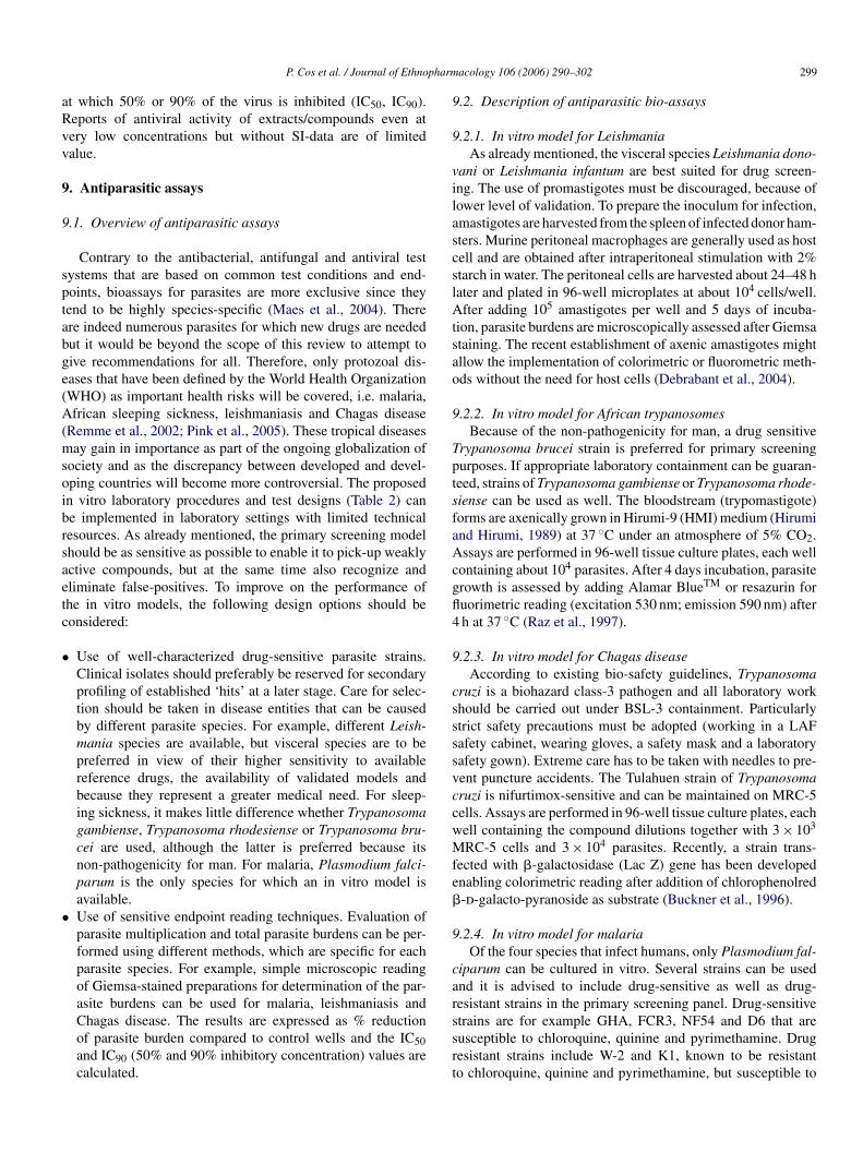
To detect active substances present in very small quanti-ties in the extracts, a concentration step is usually requiredand is based on evaporation of the solvent in vacuo. It isadvised to extract and evaporate at low temperature not todestroy any thermolabile constituent. Unfortunately, this con-centration step often results in precipitation or co-precipitationthereby hampering proper performance and interpretation ofthe bioassay. Introducing pH differences may further enhanceseparation of acid, neutral and basic constituents. Of the manyextraction schemes that have been published, the method pro-posed by Mitscher et al. (1972) and later adapted by Ieven etal. (1979) can be considered as a practical standard since itoffers a logical, low-cost, feasible and highly performing start-ing approach (Fig. 1). In some instances and if logistics permit,a ‘primary’ fractionation of the total extract can be carried outprior to testing to separate polar from less-polar constituents bysequential use of solvents from high to low polarity (VandenBerghe and Vlietinck, 1991). This permits better discriminationbi‘spbpac5c1
4
ppcoiaarlelelos
ntimicrobial activities. However, critical evaluation of some-imes contradictory test results is warranted and prior confirma-ion of the published results remains prerequisite. In the eth-omedical approach, oral or written information on the medici-al use of a plant forms the basis for selection and focused eval-ation. Information from organized traditional medical systemsAyurveda, Unani, Kampo and traditional Chinese medicine),erbalism, folklore and shamanism can be acquired from var-ous sources, such as books, herbals, review articles (usuallynvolving surveys of medicinal plants by geographic region orthnic culture), notes placed on voucher herbarium specimens,eld work and computer databases, such as NAPRALERT andSDA-Duke. A fifth, non-systematic approach is serendipity,here plant selection is based on ethnomedical use, but where
he recovered bioactivity is new or unexpected. Among others,ell known examples are the anticancer compounds vinblastine
nd taxol (Clardy and Walsh, 2004).
. Extraction scheme
Irrespective of the adopted plant collection strategy, a criti-al step is the processing of the plant material that will be usedn the panel of screens. Appropriate measures must be taken touarantee that potential active constituents are not lost, alteredr destroyed during the preparation of the extract. Being awarehat individual constituents may require specific processing, forxample at the later stage of bio-guided fractionation, a genericxtraction scheme is best indicated for the preparation of crudextracts. Plant extracts are prepared by maceration or percolationf fresh green plants or dried powdered plant material in waternd/or organic solvents. For hydrophilic compounds, polar sol-
etween fractions that exhibit aspecific activity or cytotoxic-ty and fractions that show selective antimicrobial activity. Thisprimary’ fractionation scheme may also contain dereplicationteps to avoid re-isolation of known compounds; for exam-le, acidic polysaccharides and tannins frequently produce aroad, non-selective activity against several micro-organisms,articularly viruses. It may therefore be appropriate to usecidic polysaccharide-free and/or tannin-free extracts, whichan easily be obtained respectively by precipitation after adding0% ethanol and/or chromatography on a polyamide 6 or 8olumn, using water and methanol as eluents (Cordell et al.,993).
. Selection of the appropriate bioassay
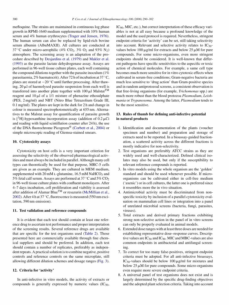
Different screening approaches are available to identify therimary pharmacological activity in chemical and/or naturalroducts. The screening option will largely depend on the spe-ific nature of the disease being targeted and on the availabilityf practical and biologically validated laboratory models. Asllustrated in Fig. 2, four levels of screening can be identifiednd the most rewarding strategy is to opt for models that remains close as possible to the final target, i.e. the patient. In thatespect, some compromises will have to be made for throughput,abour-intensiveness, costs and compound requirements. When-ver possible, activities discovered at one particular screeningevel should be confirmed using a model in the next highervaluation level. For example, results obtained in a subcellu-ar (enzymatic) screen should be confirmed against the wholerganism. A good in vitro activity against the whole organismhould then be linked to a confirmation test in an animal model.

P. Cos et al. / Journal of Ethnopharmacology 106 (2006) 290–302 293
Fig. 1. Standard scheme for preparation of plant extracts for biological screening (Mitscher scheme adapted by Ieven et al., 1979).
For most infectious diseases, this can easily be achieved sincevalidated in vitro and in vivo laboratory models using the wholeorganism are available.
4.1. Subcellular target approach
A theoretical first step in drug discovery is to identify andvalidate specific molecular targets. With the expanding knowl-edge of microbial biochemistry and genetics, it has now becomepossible to identify important microbial enzymes, receptors andprocesses at the molecular level that might be suitable tar-gets for drug action. Compounds are identified that block thefunctioning of these targets and hence would inhibit microbialgrowth. For the listed microbial diseases, several targets haveindeed been identified and new ones are still being discovered.
Advanced genomic and proteomic research may enable a morerapid progression towards 3D-structural information and createpossibilities for computer (in silico) drug design and/or screen-ing (Kitchen et al., 2004). The initial “active” compounds (‘hits’)may result from high-throughput screening of large and diverseproduct libraries, or from a more rational approach if struc-tural information on the target is available. On large-scale drugscreening platforms, enzymatic screens are attractive because ofease of implementation and minimal compound requirements;however, their overall poor validation status has contributed tothe failure of delivering initial expectations (Sams-Dodd, 2005).An opposite trend is even being noted where target validationis positioned after whole-organism screens (Hart, 2005). Sincenatural products and extracts are generally complex mixtures ofquite diverse molecules, target-oriented screens are perceived
n (an
Fig. 2. General approaches i ti-infective) drug screening.
294 P. Cos et al. / Journal of Ethnopharmacology 106 (2006) 290–302
inappropriate and will therefore not be considered here as a validscreening option.
4.2. Conventional “whole-cell” test systems
Conventional screening systems on the whole organismhave considerable advantages over the target-oriented options(Table 1): they involve all targets and take bioavailability phe-nomena into account. All too frequently, compounds that arepotent inhibitors of enzymes in vitro ultimately fail to confirmagainst the whole organism because, among other reasons, theydo not pass the cell membrane.
In vitro models using the whole organism are the ‘golden’standard and should be used whenever possible. Most micro-organisms can easily be cultivated either in cell-free medium orin cell cultures. Important to note is that any drug action on themicro-organism should be discriminated from aspecific cell tox-icity, necessitating the inclusion of a parallel evaluation in hostcell lines (cytotoxicity evaluation) and/or by inclusion of othermicrobial screens, covering a panel of bacteria, fungi, parasitesand viruses. Most recommendations and examples given in thisarticle refer to the standard 96-well microplate format.
Animal models in laboratory rodents are indispensable forconfirmation of in vitro active drug candidates. Because of ani-mal welfare considerations, high cost and labour-intensiveness,tmipwtS
TA
I
W
I
5. Compound handling and storage
The most frequently used solvents to make up test compoundsolutions include dimethyl sulfoxide (DMSO), methanol andethanol. The latter two, however, have the disadvantage of rapidevaporation whereby the stated concentration of stock solutionscannot be maintained. Solutions in 100% DMSO have becomethe standard and are prepared at adequate strength (“masterplates” at 20 mM or �g/ml for pure compounds and extracts,respectively). Added advantages of stock solutions in 100%DMSO are: (1) elimination of microbial contamination, therebyreducing the need for sterilisation by autoclaving or other stren-uous methods, which affect the quality of the test sample and (2)good compatibility with test automation and integrated screen-ing platforms, assuring for example good solubility during theserial dilution procedures. It is important to note that DMSOis potentially toxic for cells and many micro-organisms, andto avoid later interference in the biological test systems, thein-test concentration of DMSO should not exceed 1%. In prac-tical terms, this entails the need for inclusion of an intermediatedilution step in water since minimum dispensing volumes formost standard pipettes are 5–10 �l. Because of the variabilityof individual compounds, there are no general storage condi-tions that guarantee sample integrity (Verkman, 2004). A prac-tical recommendation for storage of compounds or extracts iseither without solvent for long term storage or in 100% DMSOah
6
seifsiobaiata
iota(mMttao
he number of animal tests is generally kept to an absolute mini-um. In these models, a more profound pharmacological picture
s obtained, as pharmacokinetic, metabolic and toxicologicalhenomena are taken into account. Since animal models are notithin the scope of this article, referral for protocols is made to
he book: Handbook of Animal Models of Infection (Zak andande, 1999).
able 1dvantages and disadvantages of the different drug screening approaches
n vitro screeningapproach
Advantages Disadvantages
hole organism vs.subcellular target
• Known and unknowntargets are considered
• No link to themode-of-action
• More relevant biologicaltest conditions
• Existence ofmembrane barriers
• Indicated forempirical/random screeningof ‘unknown’ chemicals andnatural products
• Stringent endpointfor weakly activecompounds
• Difficult to run inhigh-throughput(HTS) mode
ntegrated screeningvs. single-model
• Rapid profiling for relatedand unrelated antimicrobialactivities
• More complex,labour-intensive,expensive
• ‘Selectivity’ criterion moreimportant than ‘potency’criterion
• Need for integratedexpertise
• Higher compoundrequirements• More sophisticateddata management
t −20 ◦C with minimal exposure to freeze–thaw cycles andumidity.
. Integrated in vitro screening for selective activity
Too often, natural products are tested only against a singlepecies or class of micro-organisms, mostly selected based onthnomedical use data. When the product exhibits high activity,t is then identified as a potential ‘hit’. However, this ‘narrowocus’ strategy overlooks the incidence of false-positives anduffers from the lack of discrimination from aspecific cell toxic-ty. This can easily be solved by inclusion of a parallel evaluationn host cell lines (cytotoxicity evaluation) and/or other micro-ial screens on bacteria, fungi, parasites and viruses (Maes etl., 2004). In antiviral screening, this selectivity is intrinsicallyncluded and reflected in the selectivity index (SI). Total extractsnd derived fractions exhibiting strong non-selective action inhe panel of in vitro screens can only be properly evaluated innimal models.
An integrated screening concept for anti-infective activitys described in detail and should be regarded as a practicalperational example rather than a strict set of rigid instruc-ions. Besides antibacterial, antifungal, antiviral and antipar-sitic ‘whole organism’ assays, cytotoxicity on MRC-5 cellshuman lung fibroblasts) is performed in parallel. Althoughany other cell types can be used for cytotoxicity evaluation,RC-5 cells were selected because of their sensitivity and recep-
iveness for several viruses and parasites. Standardization acrosshe different bioassays maximizes efficiency, minimizes cost andllows easy and reproducible data acquisition. It includes the usef:

P. Cos et al. / Journal of Ethnopharmacology 106 (2006) 290–302 295
Fig. 3. Templates for primary integrated in vitro screening (common 96-well microplate format for all screens, separate plates are used for replicate testing).
1. Standard (20 mM or 20 mg/ml) stock solutions in 100%DMSO, stored at −20 ◦C in 96-well deep-well stacker plates.Since DMSO is highly hygroscopic, tight sealing is required.
2. Fixed concentrations in all screens (using 2- or 4-fold serialdilutions) with 100–150 �M or �g/ml as the highest in-testconcentration. Higher concentrations generally result in amarkedly increased incidence of false-positives.
3. Standard lay-out of 96-well microplates to facilitate plateproduction and to minimize human errors during the bioas-say screen. The proposed plate layouts (Fig. 3), althoughnot restrictive and to be modified depending on the specificobjectives of the test, always includes the presence of nega-tive, positive and reference controls.
4. Spectrophotometric reading of endpoints, whenever possi-ble. It is advisable for compounds with known molecularweight to express activity/inhibition parameters in molarconcentrations. For natural products or compound mixtureswhere the exact molecular weight is not known, concentra-tions are expressed in �g/ml.
5. Standard templates (spreadsheet), allowing rapid result pro-cessing and reporting. Endpoints include IC50, IC90, MICand MBC-values.
7. Antibacterial and antifungal assays
7.1. Overview of standard assays
pv
with them. Several methods for detecting activity are available,but since they are not equally sensitive or not based upon thesame principle, results will be profoundly influenced by themethod. The general problems inherent to antimicrobial screen-ing of plant extracts have already been discussed by severalauthors (Vanden Berghe and Vlietinck, 1991; Cole, 1994; Rioset al., 1988; Hadacek and Greger, 2000), so focus here willmainly be on the correct implementation and interpretation ofthe diverse laboratory models.
The antibacterial and antifungal test methods are classi-fied into three main groups, i.e. diffusion, dilution and bio-autographic methods. A fourth and upcoming test method is theconductimetric assay, detecting microbial growth as a change inthe electrical conductivity or impedance of the growth medium(Sawai et al., 2002). The latter method is not discussed here. Itshould be emphasized that many research groups have modifiedthese methods for specific samples, such as essential oils andnon-polar extracts and these small modifications make it almostimpossible to directly compare results. It is therefore a ‘must’to include at least one, but preferentially several reference com-pounds in each assay.
7.1.1. Agar-diffusion methodsIn the diffusion technique, a reservoir containing the test com-
pound at a known concentration is brought into contact with aninoculated medium and the diameter of the clear zone aroundtiib
Antimicrobial activity of natural extracts and pure com-ounds can be detected by observing the growth response ofarious micro-organisms to samples that are placed in contact
he reservoir (inhibition diameter) is measured at the end of thencubation period. In order to enhance the detection limit, thenoculated system is kept at lower temperature for several hoursefore incubation to favour compound diffusion over microbial

296 P. Cos et al. / Journal of Ethnopharmacology 106 (2006) 290–302
growth, thereby increasing the inhibition diameter. Differenttypes of reservoirs can be used, such as filter paper discs, stain-less steel cylinders placed on the surface and holes punched inthe medium. The hole-punch method is the only suitable dif-fusion technique for aqueous extracts, because interference byparticulate matter is much less than with other types of reser-voirs. To ensure that the sample does not leak under the agarlayer, fixed agar is left on the bottom of the hole (Cole, 1994).The small sample requirements and the possibility to test upto six extracts per plate against a single micro-organism arespecific advantages (Hadacek and Greger, 2000). The diffusionmethod is not appropriate for testing non-polar samples or sam-ples that do not easily diffuse into agar. In general, the relativeantimicrobial potency of different samples may not always becompared, mainly because of differences in physical properties,such as solubility, volatility and diffusion characteristics in agar.Furthermore, agar-diffusion methods are difficult to run on high-capacity screening platforms.
7.1.2. Dilution methodsIn the dilution methods, test compounds are mixed with a
suitable medium that has previously been inoculated with thetest organism. It can be carried out in liquid as well as in solidmedia and growth of the micro-organism can be measured ina number of ways. In the agar-dilution method, the MinimalInhibitory Concentration (MIC) is defined as the lowest concen-tomo4iamacbmciaeeumvttclnSaFiap
redox indicators or turbidimetric endpoints, dose–responseeffects allow calculation of IC50- and IC90-values, which arethe concentrations required to produce 50 and 90% growthinhibition.
7.1.3. Bio-autographic methodsBio-autography localizes antimicrobial activity on a chro-
matogram using three approaches: (a) direct bio-autography,where the micro-organism grows directly on the thin-layer-chromatographic (TLC) plate, (b) contact bio-autography, wherethe antimicrobial compounds are transferred from the TLC plateto an inoculated agar plate through direct contact and (c) agar-overlay bio-autography, where a seeded agar medium is applieddirectly onto the TLC plate (Hamburger and Cordell, 1987;Rahalison et al., 1991). Despite high sensitivity, its applica-bility is limited to micro-organisms that easily grow on TLCplates. Other problems are the need for complete removal ofresidual low volatile solvents, such as n-BuOH, trifluoroaceticacid and ammonia and the transfer of the active compounds fromthe stationary phase into the agar layer by diffusion. Becausebio-autography allows localizing antimicrobial activities of anextract on the chromatogram, it supports a quick search for newantimicrobial agents through bioassay-guided isolation. How-ever, this technique is not directly applicable in current highcapacity screening designs.
7a
7
prmnaImtdaaascfiowbclhomoTa
ration able to inhibit any visible microbial growth. In liquid-r broth-dilution methods, turbidity and redox-indicators areost frequently used. Turbidity can be estimated visually or
btained more accurately by measuring the optical density at05 nm. However, test samples that are not fully soluble maynterfere with turbidity readings, emphasizing the need for a neg-tive control or sterility control, i.e. extract dissolved in blankedium without micro-organisms. The liquid-dilution method
lso allows determination whether a compound or extract has aidal or static action at a particular concentration. The minimalactericidal or fungicidal concentration (MBC or MFC) is deter-ined by plating-out samples of completely inhibited dilution
ultures and assessing growth (static) or no-growth (cidal) afterncubation. At present, the redox indicators MTT and resazurinre frequently used to quantify bacterial (Eloff, 1998; Gabrielsont al., 2002) and fungal growth (Jahn et al., 1995; Pelloux-Prayert al., 1998). Resazurin has the advantage not to precipitatepon reduction, allowing direct reading. Easy and reproducibleeasurements can be obtained with a microplate-reader, but
isual reading may also be used in cases where spectropho-ometry is not available. Another assay exploits the principlehat only living cells convert fluorescein-diacetate to fluores-ein, producing a yellowish-green fluorescescence under UVight (Chand et al., 1994). However, it requires a more sig-ificant investment in equipment and validation is not easy:abouraud liquid medium can quench up to 95% of fluorescence,nd sodium phosphate buffers hydrolyse fluorescein-diacetate.luorescent constituents present in crude plant extracts may also
nterfere (Clarke et al., 2001). In general, dilution methods areppropriate for assaying polar and non-polar extracts or com-ounds for determination of MIC and MBC/MFC-values. Using
.2. Specific recommendations on antibacterial andntifungal screening
.2.1. Panel of test organismsThe choice of test organisms depends on the specific pur-
ose of the investigation. In a primary screening, drug-sensitiveeference strains are preferably used and should represent com-on pathogenic species of different classes. Various combi-
ations are possible, but the panel should at least consist ofGram-positive and a Gram-negative bacterium (Table 2).
t has been well-established that Gram-positive bacteria areuch more sensitive to drug action than Gram-negative bac-
eria, which is reflected by a higher number of random ‘hits’uring a screening campaign. Extracts with prominent activitygainst Gram-positive cocci should preferentially also be testedgainst methicillin-resistant Staphylococcus aureus (MRSA)nd vancomycin-resistant Enterococci (VRE), as they repre-ent the greatest current medical need. ATCC strains are well-haracterized and very popular for that purpose, but clinicaleld isolates may also be used if fully characterized by antibi-gram. Another challenging new area in the microbiologicalorld is biofilms (Mah and O’Toole, 2001). Although manyacteria grow in a free-living, ‘planktonic’ state, it is quiteommon for them to adhere to surfaces by producing extracel-ular polymeric substances (EPS), e.g. biofilms. Due to theirigher resistance against antimicrobial agents, an interestingption in antibacterial research is to include a bacterial biofilmodel (e.g. Staphylococcus aureus ATCC6538). A small set
f reference fungi is used for primary screening and includesrichophyton mentagrophytes and Epidermophyton floccosums representatives of the dermatophytes. As opportunistic

P. Cos et al. / Journal of Ethnopharmacology 106 (2006) 290–302 297
Table 2Proposed panel of test organisms for primary antibacterial, antifungal, antiviral and antiparasitic in vitro screening
Screen Species BSLa Culture CO2b Reference drug
Bacteriology (Gram-positive, Gram-negative, Acid-fast)- Gram-positive cocci Staphylococcus
aureus2 Axenic in
Mueller-HintonNo A broad selection of
antibiotics is available (e.g.penicillin, ampicillin,norfloxacin, doxycyclin,kanamycin)
- Spore-formingGram-positive rods
Bacillus cereus orBacillus subtilis
- Enterobacteriaceae –non-encapsulated
Escherichia coli
- Enterobacteriaceae –encapsulated
Klebsiellapneumoniae
- Non-Enterobacteriaceae Pseudomonasaeruginosa
- Acid-fast bacteria Mycobacterium spp. 2 or 3 Axenic or cellular No e.g. Rifampicin
Mycology (yeasts and fungi)- Yeasts Candida albicans 2c Axenic in Sabouraud No Several broad-spectrum
antifungals are available (e.g.amphotericin B, miconazole,ketoconazole, flucytosine)
- Dermatophytes Trichophytonmentagrophytes,Epidermophytonfloccosum
- Opportunistic filamentousfungi
Aspergillus niger,Fusarium solani
Virology- Single-stranded RNA,naked
Coxsackievirus 2 Cellular on VERO orother receptive cell
Yes List of available referencedrugs is limited. Acyclovir isused for Herpes virus.
- Double-stranded DNA,enveloped
Herpes simplex virus
- Double-stranded DNA,naked
Adenovirus
Parasitology- Malaria Plasmodium
falciparum3* Cellular (human
RBC)
d e.g. Chloroquine, artemether
- Leishmaniasis Leishmania donovani 3* Cellular(macrophages)
Yes e.g. Amphotericin B, sodiumstibogluconate
- Sleeping sickness Trypanosoma brucei 2 Axenic HMI-medium Yes e.g. Suramin, melarsoprol,pentamidine
- Chagas disease Trypanosoma cruzi 3 Cellular (MRC-5) Yes Nifurtimox, benznidazole
a BSL: biosafety level according to the European directive 2000/54/EC. Micro-organisms classified in BSL 3* may present a limited risk of infection for workersbecause they are not normally infectious by the airborne route.
b 5% CO2 incubator.c Fusarium solani is an opportunistic pathogen.d Micro-aerophilic atmosphere (4% CO2, 3% O2, and 93% N2).
filamentous fungi, Aspergillus niger and Fusarium solani arelisted.
7.2.2. Growth mediumMueller-Hinton (MH) agar or broth and tryptic soy agar or
broth (TSA or TSB) are general growth media for bacteria,while Sabouraud (SAB) agar or broth is used for fungi. Growthof fastidious micro-organisms, such as Streptococcus pneumo-niae and Legionella pneumophila, may require more complexmedia, enrichment of the incubation atmosphere with 5% CO2and/or extension of the incubation time. Slight differences inthe composition of the growth medium can greatly affect theantibacterial activity of a compound. For example, addition of
sheep blood to Mueller-Hinton medium increases the MIC offlavomycin from 0.12 to 256 mg/l (Butaye et al., 2000). Con-sequently, a definite choice of growth medium is essential tocompare different antibacterial compounds or extracts. Mueller-Hinton medium allows good growth of most non-fastidiousbacteria and is generally low in antagonists. It also meets therequirements of the NCCLS standard and is recommended as ref-erence medium for agar- and broth-dilution tests (Anon., 2000,2003).
7.2.3. InoculumThe level of infection, i.e. inoculum concentration can have
a profound influence on the antibacterial and antifungal potency

298 P. Cos et al. / Journal of Ethnopharmacology 106 (2006) 290–302
of a sample, endorsing the need for standardization of inocu-lates (Anon., 2003). In dilution methods, an inoculum of about105 CFU/ml is adequate for most bacterial species while foryeasts and fungi between 103 and 104 CFU/ml is sufficient(Hadacek and Greger, 2000). A too low inoculum size (e.g.102 CFU/ml) will create many false-positives, while a too highinoculum size (e.g. 107 CFU/ml) will hamper endpoint readingand increase the chances for false-negatives. Bacterial or yeastinoculates can be prepared from overnight cultures or from exist-ing biofreeze stocks. It is recommended to collect from culturesduring the logarithmic growth phase and always to take fouror five colonies of a pure culture on agar to avoid selecting anatypical variant (Anon., 2003).
8. Antiviral assays
8.1. Overview of antiviral assays
Evaluation of antiviral activity in vitro includes antiviralefficacy and cell toxicity. Because different viruses grow in dif-ferent cell systems, it is virtually impossible to design a singleantiviral test that could be applied for all viruses. In addition,specific methodologies for assessment of antiviral agents mayvary greatly from one laboratory to another, making direct com-parisons of test results difficult. Various cell-based assays havebeen successfully applied for the antiviral or virucidal evalua-tervceicacpotp
8
8
asptwmpmCroa
Table 3In vitro antiviral screening optionsa
Test Comment Application
Plaque inhibition assay Only for viruses which formplaques in suitable cellsystems
A-S
Titration of a limited numberof viruses in the presence of anon-toxic dose of the testsubstance
Plaque reduction assay Only for viruses which formplaques in suitable cellsystems
V-S; V-M
Titration of residual virusinfectivity after extracellularaction of test substance(s).Cytotoxicity should beeliminated, e.g. by dilution,filtration before the titration
Inhibition ofvirus-inducedcytopathic effect (CPE)
For viruses that induce CPE,but not readily form plaquesin cell cultures
A-S; A-M
Determination ofvirus-induced CPE inmonolayers, infected with alimited dose of virus andtreated with a non-toxic doseof the test substance
Virus yield reductionassay
Determination of the virusyield in tissue cultures,infected with a given amountof virus and treated with anon-toxic dose of the testsubstance
A-S; A-M
Virus titration is carried outafter virus multiplication bythe plaque test (PT) or the50% tissue culture dose endpoint test (TCD50)
Endpoint titrationtechnique (EPTT)
Determination of virus titerreduction in the presence of2-fold dilutions of testcompound
A-S; A-M
a Determination of the viral infectivity in cultured cells during virus multipli-cation in the presence of a single compound (A-S) or a mixture of compounds,e.g. plant extracts (A-M) or after extracellular incubation with a single compound(V-S) or a mixture of compounds (V-M).
sackievirus represents the Picornaviridae, which includes rhi-noviruses and the enteroviruses. Titers up to 105 TCID50/mlor higher are obtained, which markedly increases the sensi-tivity of the test system. The TCID50 or 50% Tissue CultureInfective Dose is defined as that dilution of virus required toinfect 50% of inoculated cell cultures. Finally, for most of theseviruses suitable animal models are available, enabling in vivoconfirmation.
8.2.2. EndpointsAn intrinsic component of the antiviral testing is the deter-
mination of a selectivity index (SI) towards the supporting hostcell. The SI refers to the ratio of the maximum drug concen-tration causing either 50% or 90% inhibition of growth of nor-mal cells (CC50, CC90) and the minimum drug concentration
ion of single substances or mixtures of compounds, e.g. plantxtracts (Table 3) (Vlietinck and Vanden Berghe, 1991). Antivi-al agents interfere with one or more dynamic processes duringirus biosynthesis and are consequently candidates for clini-al use, whereas virucidal substances inactivate virus infectivityxtracellularly and are rather candidates as biocides exhibit-ng a broad spectrum of germicidal activities. The methodsommonly used for evaluation of in vitro antiviral activityre based on the different abilities of viruses to replicate inultured cells. Some cause cytopathic effects (CPE) or formlaques. Others are capable of producing specialized functionsr cell transformation. Virus replication can also be moni-ored by detection of viral products, i.e. viral DNA, RNA orolypeptides.
.2. Specific recommendations on antiviral assays
.2.1. Panel of virusesThere is a great variety of viruses that can be selected for
ntiviral screening. The panel presented here has not beenelected based on medical or clinical relevance but rather forractical feasibility in standard BSL-II laboratory infrastruc-ure, easy culture on one cell type (VERO, MRC-5, e.a.)ith formation of extensive CPE (Table 2). Their ability toultiply in the same cell culture allows an objective com-
arison of antiviral activities while toxicity testing can beinimized. Moreover, virus multiplication causes extensivePE within about 1 week of infection, facilitating endpoint
eading. The proposed virus battery includes HSV and aden-virus as representatives of DNA viruses and coxsackieviruss prototype of the RNA virus group. More specifically, cox-
P. Cos et al. / Journal of Ethnopharmacology 106 (2006) 290–302 299
at which 50% or 90% of the virus is inhibited (IC50, IC90).Reports of antiviral activity of extracts/compounds even atvery low concentrations but without SI-data are of limitedvalue.
9. Antiparasitic assays
9.1. Overview of antiparasitic assays
Contrary to the antibacterial, antifungal and antiviral testsystems that are based on common test conditions and end-points, bioassays for parasites are more exclusive since theytend to be highly species-specific (Maes et al., 2004). Thereare indeed numerous parasites for which new drugs are neededbut it would be beyond the scope of this review to attempt togive recommendations for all. Therefore, only protozoal dis-eases that have been defined by the World Health Organization(WHO) as important health risks will be covered, i.e. malaria,African sleeping sickness, leishmaniasis and Chagas disease(Remme et al., 2002; Pink et al., 2005). These tropical diseasesmay gain in importance as part of the ongoing globalization ofsociety and as the discrepancy between developed and devel-oping countries will become more controversial. The proposedin vitro laboratory procedures and test designs (Table 2) canbe implemented in laboratory settings with limited technicalresources. As already mentioned, the primary screening modelsaetc
•
•
9.2. Description of antiparasitic bio-assays
9.2.1. In vitro model for LeishmaniaAs already mentioned, the visceral species Leishmania dono-
vani or Leishmania infantum are best suited for drug screen-ing. The use of promastigotes must be discouraged, because oflower level of validation. To prepare the inoculum for infection,amastigotes are harvested from the spleen of infected donor ham-sters. Murine peritoneal macrophages are generally used as hostcell and are obtained after intraperitoneal stimulation with 2%starch in water. The peritoneal cells are harvested about 24–48 hlater and plated in 96-well microplates at about 104 cells/well.After adding 105 amastigotes per well and 5 days of incuba-tion, parasite burdens are microscopically assessed after Giemsastaining. The recent establishment of axenic amastigotes mightallow the implementation of colorimetric or fluorometric meth-ods without the need for host cells (Debrabant et al., 2004).
9.2.2. In vitro model for African trypanosomesBecause of the non-pathogenicity for man, a drug sensitive
Trypanosoma brucei strain is preferred for primary screeningpurposes. If appropriate laboratory containment can be guaran-teed, strains of Trypanosoma gambiense or Trypanosoma rhode-siense can be used as well. The bloodstream (trypomastigote)forms are axenically grown in Hirumi-9 (HMI) medium (Hirumiand Hirumi, 1989) at 37 ◦C under an atmosphere of 5% CO .Acgfl4
9
cssssvccwMfe�
9
carssrt
hould be as sensitive as possible to enable it to pick-up weaklyctive compounds, but at the same time also recognize andliminate false-positives. To improve on the performance ofhe in vitro models, the following design options should beonsidered:
Use of well-characterized drug-sensitive parasite strains.Clinical isolates should preferably be reserved for secondaryprofiling of established ‘hits’ at a later stage. Care for selec-tion should be taken in disease entities that can be causedby different parasite species. For example, different Leish-mania species are available, but visceral species are to bepreferred in view of their higher sensitivity to availablereference drugs, the availability of validated models andbecause they represent a greater medical need. For sleep-ing sickness, it makes little difference whether Trypanosomagambiense, Trypanosoma rhodesiense or Trypanosoma bru-cei are used, although the latter is preferred because itsnon-pathogenicity for man. For malaria, Plasmodium falci-parum is the only species for which an in vitro model isavailable.Use of sensitive endpoint reading techniques. Evaluation ofparasite multiplication and total parasite burdens can be per-formed using different methods, which are specific for eachparasite species. For example, simple microscopic readingof Giemsa-stained preparations for determination of the par-asite burdens can be used for malaria, leishmaniasis andChagas disease. The results are expressed as % reductionof parasite burden compared to control wells and the IC50and IC90 (50% and 90% inhibitory concentration) values arecalculated.
2ssays are performed in 96-well tissue culture plates, each well
ontaining about 104 parasites. After 4 days incubation, parasiterowth is assessed by adding Alamar BlueTM or resazurin foruorimetric reading (excitation 530 nm; emission 590 nm) afterh at 37 ◦C (Raz et al., 1997).
.2.3. In vitro model for Chagas diseaseAccording to existing bio-safety guidelines, Trypanosoma
ruzi is a biohazard class-3 pathogen and all laboratory workhould be carried out under BSL-3 containment. Particularlytrict safety precautions must be adopted (working in a LAFafety cabinet, wearing gloves, a safety mask and a laboratoryafety gown). Extreme care has to be taken with needles to pre-ent puncture accidents. The Tulahuen strain of Trypanosomaruzi is nifurtimox-sensitive and can be maintained on MRC-5ells. Assays are performed in 96-well tissue culture plates, eachell containing the compound dilutions together with 3 × 10³
RC-5 cells and 3 × 10⁴ parasites. Recently, a strain trans-ected with �-galactosidase (Lac Z) gene has been developednabling colorimetric reading after addition of chlorophenolred-d-galacto-pyranoside as substrate (Buckner et al., 1996).
.2.4. In vitro model for malariaOf the four species that infect humans, only Plasmodium fal-
iparum can be cultured in vitro. Several strains can be usednd it is advised to include drug-sensitive as well as drug-esistant strains in the primary screening panel. Drug-sensitivetrains are for example GHA, FCR3, NF54 and D6 that areusceptible to chloroquine, quinine and pyrimethamine. Drugesistant strains include W-2 and K1, known to be resistanto chloroquine, quinine and pyrimethamine, but susceptible to
300 P. Cos et al. / Journal of Ethnopharmacology 106 (2006) 290–302
mefloquine. The strains are maintained in continuous log phasegrowth in RPMI-1640 medium supplemented with 10% humanserum and 4% human erythrocytes (Trager and Jensen, 1976).The human serum can also be replaced by lipid-rich bovineserum albumin (AlbuMAXII). All cultures are conducted at37 ◦C under micro-aerophilic (4% CO2, 3% O2 and 93% N2)atmosphere. The screening assay is an adaptation of the pro-cedure described by Desjardins et al. (1979) and Makler et al.(1993) as the parasite lactate dehydrogenase assay. Assays areperformed in 96-well tissue culture plates, each well containingthe compound dilutions together with the parasite inoculum (1%parasitaemia, 2% haematocrit). After 72 h of incubation at 37 ◦C,plates are stored at −20 ◦C until further processing. After thaw-ing, 20 �l of haemolysed parasite suspension from each well istransferred into another plate together with 100 �l MalstatTM
reagent and 10 �l of a 1/1 mixture of phenazine ethosulphate(PES, 2 mg/ml) and NBT (Nitro Blue Tetrazolium Grade III,0.1 mg/ml). The plates are kept in the dark for 2 h and change incolour is measured spectrophotometrically at 655 nm. Alterna-tives to the Malstat assay for quantification of parasite growthis [3H]-hypoxanthine incorporation assay (addition of 0.2 �Ciand reading with liquid scintillation counter after 24 h), the useof the DNA fluorochrome Picogreen® (Corbett et al., 2004) orsimple microscopic reading of Giemsa-stained smears.
10. Cytotoxicity assays
aitas5i4a2t
1
eotpisdca
1
c
IC90, MIC, etc.), but correct interpretation of these efficacy vari-ables is not at all easy because a profound knowledge of themodel and the used protocol is required. Nevertheless, stringentendpoint criteria for ‘activity’ can be set, still taking selectivityinto account. Relevant and selective activity relates to IC50-values below 100 �g/ml for extracts and below 25 �M for purecompounds. For some micro-organisms, even more stringentendpoints should be considered. It is well-known that differ-ent pathogens have specific sensitivities to the aspecific or toxicaction of chemical molecules. For example, a same cell typebecomes much more sensitive for in vitro cytotoxic effects whencultivated in serum-free conditions; Gram-negative bacteria aremuch less sensitive to ‘drug action’ than Gram-positive speciesand in random antiprotozoal screens, a consistent observation isthat free-living organisms (for example, Trichomonas spp.) aremuch more robust than the blood protozoa Plasmodium, Leish-mania or Trypanosoma. Among the latter, Plasmodium tends tobe the most sensitive.
13. Rules of thumb for defining anti-infective potentialin natural products
1. Identification and documentation of the plants (voucherspecimen and number) and preparation and storage ofextracts need to be reported. In a bioassay-guided fraction-ation, a scattered activity across the different fractions is
Cytotoxicity on host cells is a very important criterion forssessing the selectivity of the observed pharmacological activ-ties and must always be included in parallel. Although many cellypes can theoretically be used for that purpose, MRC-5 cellsre given as an example. They are cultured in MEM medium,upplemented with 20 mM l-glutamine, 16.5 mM NaHCO3 and% fetal calf serum. Assays are performed at 37 ◦C and 5% CO2n 96-well tissue culture plates with confluent monolayers. After–7 days incubation, cell proliferation and viability is assessedfter addition of Alamar BlueTM or resazurin (McMillian et al.,002). After 4 h at 37 ◦C, fluorescence is measured (550 nm exci-ation, 590 nm emission).
1. Test validation and reference compounds
It is evident that each test should contain at least one refer-nce drug to ascertain test performance and proper interpretationf the screening results. Several reference drugs are availablehat are specific for the test organisms used (Table 2). Thoseresented here are commercially available through fine chem-cal suppliers and should be preferred. In addition, each testhould contain a number of replicates, preferably as indepen-ent repeats. A practical solution is to combine negative, positiveontrols and reference controls on the same microplate, stillllowing different dilution schemes and dosage ranges (Fig. 3).
2. Criteria for ‘activity’
In anti-infective in vitro models, the activity of extracts orompounds is generally expressed by numeric values (IC50,
mostly indicative for non-selectivity.2. Test organisms are preferably ATCC strains as they are
widely used and well-characterized. Defined clinical iso-lates may also be used, but only if the susceptibility torelevant reference compounds is reported.
3. In vitro models using the whole organism are the ‘golden’standard and should be used whenever possible. If micro-organisms can be cultivated either in cell-free medium(‘axenic’) or in cell cultures, the latter one is preferred sinceit resembles more the in vivo situation.
4. Antimicrobial activity must be discriminated from non-specific toxicity by inclusion of a parallel cytotoxicity eval-uation on mammalian cell lines or integration into a panelof unrelated microbial screens (bacteria, fungi, parasites,viruses).
5. Total extracts and derived primary fractions exhibitingstrong non-selective action in the panel of in vitro screenscan only be properly evaluated in animal models.
6. Extended dose ranges with at least three doses are needed forestablishing representative dose–response curves. Descrip-tive values are IC50 and IC90. MIC and MBC-values are alsocommon endpoints in antibacterial and antifungal screen-ing.
7. To correct for too many false-positives, stringent endpointcriteria must be adopted. For all anti-infective bioassays,IC50-values should be below 100 �g/ml for mixtures andbelow 25 �M for pure compounds. Some micro-organismseven require more severe endpoint criteria.
8. A universal panel of test organisms does not exist and islargely determined by the specific drug-finding objectivesand the adopted plant selection criteria. Taking into account

P. Cos et al. / Journal of Ethnopharmacology 106 (2006) 290–302 301
the sometimes broad traditional ethnomedical use, a mini-mum panel of test organisms is recommended (Table 2).
9. Inclusion of appropriate controls in each test replicate(blank-, infected- and reference controls) is necessary. Foreach test organism, one or more commercially available ref-erence compounds can be considered (Table 2).
10. Differences in composition of the growth medium cangreatly affect the potency of a compound. Mueller-Hintonis recommended for antibacterial and Sabouraud for anti-fungal testing in agar and/or broth dilution tests. For fastid-ious bacteria, supplementation with growth factors or othermedia are allowed.
11. The infective dose can have a profound impact on the testresults. For most bacteria, 105 CFU/ml is adequate, whilefor yeasts and fungi about 103 and 104 CFU/ml is satisfac-tory. For viruses and parasites, optimal infective doses arespecies-specific.
12. For antibacterial and antifungal activities, follow-up ofbioassay-guided fractionation can be performed with anassay of choice. Whenever possible, final reporting of activeextracts or pure compounds should be done by applyingthe broth-dilution method. For essential oils, addition ofsolvents or emulsifiers may be necessary, but their final con-centrations should be limited and reported.
13. The phenomenon of ‘additive’ or ‘synergistic’ effects inmixtures or extracts frequently causes loss-of-activity dur-
A
titg
R
A
A
B
B
C
C
Clarke, J.M., Gillings, M.R., Altavilla, N., Beattie, A.J., 2001. Potential prob-lems with fluorescein diacetate assays of cell viability when testing naturalproducts for antimicrobial activity. Journal of Microbiological Methods46, 261–267.
Cole, M.D., 1994. Key antifungal, antibacterial and anti-insect assays—acritical review. Biochemical Systematics and Ecology 22, 837–856.
Corbett, Y., Herrera, L., Gonzalez, J., Cubilla, L., Capson, T.L., Coley, P.D.,Kursar, T.A., Romero, L.I., Ortega-Barria, E., 2004. A novel DNA-basedmicrofluorimetric method to evaluate antimalarial drug activity. AmericanJournal of Tropical Medicine and Hygiene 70, 119–124.
Cordell, G.A., Colvard, M.D., 2005. Some thoughts on the future ofethnopharmacology. Journal of Ethnopharmacology 100, 5–14.
Cordell, G.A., Kinghorn, A.D., Pezzuto, J.M., 1993. Separation, structureelucidation, and bioassay of cytotoxic natural products. In: Colegate, S.M.,Molyneux, R.J. (Eds.), Bioactive Natural Products—Detection, Isolationand Structural Determination. CRC Press, London, pp. 195–220 (Chapter10).
Debrabant, A., Joshi, M.B., Pimenta, P.F.P., Dwyer, D.M., 2004. Generationof Leishmania donovani axenic amastigotes: their growth and biologicalcharacteristics. International Journal of Parasitology 34, 205–217.
Desjardins, R.E., Canfield, C.J., Haynes, J.D., Chulay, J.D., 1979. Quantitativeassessment of antimalarial activity in vitro by a semiautomated microdi-lution technique. Antimicrobial Agents and Chemotherapy 16, 710–718.
Eloff, J.N., 1998. A sensitive and quick microplate method to determinethe minimal inhibitory concentration of plant extracts for bacteria. PlantaMedica 64, 711–713.
Fabricant, D.S., Farnsworth, N.R., 2001. The value of plants used in tra-ditional medicine for drug discovery. Environmental Health Perspectives109, 69–75.
Gabrielson, J., Hart, M., Jarelov, A., Kuhn, I., McKenzie, D., Mollby, R.,2002. Evaluation of redox indicators and the use of digital scanners and
H
H
H
H
I
J
K
M
M
M
M
ing bio-guided fractionation efforts and hence precludes theidentification or characterization of the relevant fraction thatcould be retained for further evaluation. In that case, furtherstudies should focus on the defined crude extract.
cknowledgements
Paul Cos is a postdoctoral researcher of the Fund for Scien-ific Research (FWO-Flanders, Belgium). The integrated screen-ng concept for tropical parasitic diseases was developed withhe support of UNICEF/UNDP/World Bank/WHO special pro-ramme for research and training in tropical diseases.
eferences
non., 2000. Determination of minimum inhibitory concentrations (MICs) ofantibacterial agents by agar dilution. Clinical Microbiology and Infection6, 509–515.
non., 2003. Determination of minimum inhibitory concentrations (MICs) ofantibacterial agents by broth dilution. Clinical Microbiology and Infection9, 1–7.
uckner, F.S., Verlinde, C.L., La Flamme, A.C., Van Voorhis, W.C., 1996.Efficient technique for screening drugs for activity against Trypanosomacruzi using parasites expressing beta-galactosidase. Antimicrobial Agentsand Chemotherapy 40, 2592–2597.
utaye, P., Devriese, L.A., Haesebrouck, F., 2000. Influence of differ-ent medium components on the in vitro activity of the growth-promoting antibiotic flavomycin against enterococci. Journal of Antimi-crobial Chemotherapy 46, 713–716.
hand, S., Lusunzi, I., Veal, D.A., Williams, L.R., Karuso, P., 1994. Rapidscreening of the antimicrobial activity of extracts and natural products.Journal of Antibiotics 47, 1295–1304.
lardy, J., Walsh, C., 2004. Lessons from natural molecules. Nature 432,829–837.
spectrophotometer for quantification of microbial growth in microplates.Journal of Microbiological Methods 50, 63–73.
adacek, F., Greger, H., 2000. Testing of antifungal natural products: method-ologies, comparability of results and assay choice. Phytochemical Anal-ysis 11, 137–147.
amburger, M.O., Cordell, G.A., 1987. A direct bioautographic TLC assayfor compounds possessing antibacterial activity. Journal of Natural Prod-ucts 50, 19–22.
art, C.P., 2005. Finding the target after screening the phenotype. DrugDiscovery Today 10, 513–519.
irumi, H., Hirumi, K., 1989. Continuous cultivation of Trypanosoma bruceiblood stream forms in a medium containing a low concentration of serumprotein without feeder cell layers. Journal of Parasitology 75, 985–989.
even, M., Vanden Berghe, D.A., Mertens, F., Vlietinck, A., Lammens, E.,1979. Screening of higher plants for biological activities I. Antimicrobialactivity. Planta Medica 36, 311–321.
ahn, B., Martin, E., Stueben, A., Bhakdi, S., 1995. Susceptibilitytesting of Candida albicans and Aspergillus species by a sim-ple microtitre menadione-augmented 3-(4,5-dimethyl-2-thiazolyl)-2,5-diphenyl-2H-tetrazolium bromide assay. Journal of Clinical Microbiology33, 661–667.
itchen, D.B., Decomez, H., Furr, J.R., Bajorath, J., 2004. Docking andscoring in virtual screening for drug discovery: methods and applications.Nature Reviews Drug Discovery 3, 935–949.
aes, L., Vanden Berghe, D., Germonprez, N., Quirijnen, L., Cos, P., DeKimpe, N., Van Puyvelde, L., 2004. In vitro and in vivo activities ofa triterpenoid saponin extract (PX-6518) from the plant Maesa bal-ansae against visceral Leishmania species. Antimicrobial Agents andChemotherapy 48, 130–136.
ah, T.F.C., O’Toole, G.A., 2001. Mechanisms of biofilm resistance toantimicrobial agents. Trends in Microbiology 9, 34–39.
akler, M.T., Ries, J.M., Williams, J.A., Bancroft, J.E., Piper, R.C., Gibbins,B.L., Hinrichs, D.J., 1993. Parasite lactate dehydrogenase as an assay forPlasmodium falciparum drug sensitivity. American Journal of TropicalMedicine and Hygiene 48, 739–741.
cMillian, M.K., Li, L., Parker, J.B., Patel, L., Zhong, Z., Gunnett,J.W., Powers, W.J., Johnson, M.D., 2002. An improved resazurin-based

302 P. Cos et al. / Journal of Ethnopharmacology 106 (2006) 290–302
cytotoxicity assay for hepatic cells. Cell Biology and Toxicology 18,157–173.
Mitscher, L.A., Beal, J.L., Bathala, M.S., Wu, W.N., White, R., Leu, R.P.,1972. Antimicrobial agents from higher plants—1. Introduction, rationale,and methodology. Lloydia 35, 157–176.
Okeke, I.N., Laxmaninarayan, R., Bhutta, Z.A., Duse, A.G., Jenkins, P.,O’Brien, T.F., Pablos-Mendez, A., Klugman, K.P., 2005. Antimicrobialresistance in developing countries. Part 1: recent trends and current sta-tus. Lancet Infectious Diseases 5, 481–493.
Patwardhan, B., 2005. Ethnopharmacology and drug discovery. Journal ofEthnopharmacology 100, 50–52.
Pelloux-Prayer, A.L., Priem, B., Joseleau, J.P., 1998. Kinetic evaluation ofconidial germination of Botrytis cinerea by a spectrofluorometric method.Mycology Research 102, 320–322.
Pink, R., Hudson, A., Mouries, M.A., Bendig, M., 2005. Opportunities andchallenges in antiparasitic drug discovery. Nature Reviews Drug Discov-ery 4, 727–740.
Rahalison, L., Hamburger, M., Hostettmann, K., Monod, M., Frenk, E., 1991.A bioautographic agar overlay method for the detection of antifungalcompounds from higher plants. Phytochemical Analysis 2, 199–203.
Raz, B., Iten, M., Grether-Buhler, Y., Kaminsky, R., Brun, R., 1997. TheAlamar Blue assay to determine drug sensitivity of African trypanosomes(T.b. rhodensiense and T.b. gambiense) in vitro. Acta Tropica 68, 139–147.
Remme, J.H., Blas, E., Chitsulo, L., Desjeux, P.M., et al., 2002. Strategicemphases for tropical diseases research: a TDR perspective. Trends inMicrobiology 10, 435–440.
Rios, J.L., Recio, M.C., 2005. Medicinal plants and antimicrobial activity.Journal of Ethnopharmacology 100, 80–84.
Rios, J.L., Recio, M.C., Villar, A., 1988. Screening methods for natural prod-ucts with antimicrobial activity: a review of the literature. Journal ofEthnopharmacology 23, 127–149.
Sams-Dodd, F., 2005. Target-based drug discovery: is something wrong? DrugDiscovery Today: Targets 10, 139–147.
Sawai, J., Doi, R., Maekawa, Y., Yoshikawa, T., Kojima, H., 2002. Indi-rect conductimetric assay of antibacterial activities. Journal of IndustrialMicrobiology & Technology 29, 296–298.
Trager, W., Jensen, J.B., 1976. Human malaria parasites in continuous culture.Science 193, 673–675.
Vanden Berghe, D.A., Vlietinck, A.J., 1991. Screening for antibacterial andantiviral agents. In: Hostettmann, K. (Ed.), Methods in Plant Biochem-istry, vol. 6, Assays for Bioactivity. Academic Press, London, pp. 47–59(Chapter 3).
Verkman, A.S., 2004. Drug discovery in academia. American Journal ofPhysiology—Cell Physiology 286, 465–474.
Verpoorte, R., Choi, Y.H., Kim, H.K., 2005. Ethnopharmacology and systemsbiology: a perfect holistic match. Journal of Ethnopharmacology 100,53–56.
Vlietinck, A.J., Vanden Berghe, D.A., 1991. Can ethnopharmacology con-tribute to the development of antiviral drugs? Journal of Ethnopharma-cology 32, 141–153.
Zak, O., Sande, M.A., 1999. Handbook of Animal Models of Infection. Aca-demic Press, London.