revealing the full biosphere structure and versatile
TRANSCRIPT

RESEARCH Open Access
Revealing the full biosphere structure andversatile metabolic functions in the deepestocean sediment of the Challenger DeepPing Chen1†, Hui Zhou1,2†, Yanyan Huang3,4†, Zhe Xie5†, Mengjie Zhang1,2†, Yuli Wei5, Jia Li1,2, Yuewei Ma3,Min Luo5, Wenmian Ding3, Junwei Cao5, Tao Jiang1,2, Peng Nan3*, Jiasong Fang5* and Xuan Li1,2*
* Correspondence: [email protected]; [email protected]; [email protected]†Ping Chen, Hui Zhou, YanyanHuang, Zhe Xie and Mengjie Zhangcontributed equally to this work.3Ministry of Education KeyLaboratory for Biodiversity Scienceand Ecological Engineering, Schoolof Life Sciences, Fudan University,Shanghai, China5Shanghai Engineering ResearchCenter of Hadal Science andTechnology, College of MarineSciences, Shanghai OceanUniversity, Shanghai, China1CAS-Key Laboratory of SyntheticBiology, CAS Center for Excellencein Molecular Plant Sciences, Instituteof Plant Physiology and Ecology,Chinese Academy of Sciences,Shanghai, ChinaFull list of author information isavailable at the end of the article
Abstract
Background: The full biosphere structure and functional exploration of the microbialcommunities of the Challenger Deep of the Mariana Trench, the deepest knownhadal zone on Earth, lag far behind that of other marine realms.
Results: We adopt a deep metagenomics approach to investigate the microbiomein the sediment of Challenger Deep, Mariana Trench. We construct 178metagenome-assembled genomes (MAGs) representing 26 phyla, 16 of which arereported from hadal sediment for the first time. Based on the MAGs, we find themicrobial community functions are marked by enrichment and prevalence ofmixotrophy and facultative anaerobic metabolism. The microeukaryotic community isfound to be dominated by six fungal groups that are characterized for the first timein hadal sediment to possess the assimilatory and dissimilatory nitrate/sulfatereduction, and hydrogen sulfide oxidation pathways. By metaviromic analysis, wereveal novel hadal Caudovirales clades, distinctive virus-host interactions, andspecialized auxiliary metabolic genes for modulating hosts’ nitrogen/sulfurmetabolism. The hadal microbiome is further investigated by large-scale cultivationthat cataloged 1070 bacterial and 19 fungal isolates from the Challenger Deepsediment, many of which are found to be new species specialized in the hadalhabitat.
Conclusion: Our hadal MAGs and isolates increase the diversity of the ChallengerDeep sediment microbial genomes and isolates present in the public. The deepmetagenomics approach fills the knowledge gaps in structure and diversity of thehadal microbiome, and provides novel insight into the ecology and metabolism ofeukaryotic and viral components in the deepest biosphere on earth.
Keywords: Mariana Trench, Challenger Deep, Hadal sediment, Metagenomics,Versatile metabolism, Metavirome, Large-scale cultivation
BackgroundThe hadal trench represents the deepest habitat for living organisms on the surface of
the earth and accounts for a significant portion of the global benthic area [1]. In
© The Author(s). 2021 Open Access This article is licensed under a Creative Commons Attribution 4.0 International License, whichpermits use, sharing, adaptation, distribution and reproduction in any medium or format, as long as you give appropriate credit tothe original author(s) and the source, provide a link to the Creative Commons licence, and indicate if changes were made. Theimages or other third party material in this article are included in the article's Creative Commons licence, unless indicated otherwisein a credit line to the material. If material is not included in the article's Creative Commons licence and your intended use is notpermitted by statutory regulation or exceeds the permitted use, you will need to obtain permission directly from the copyrightholder. To view a copy of this licence, visit http://creativecommons.org/licenses/by/4.0/. The Creative Commons Public DomainDedication waiver (http://creativecommons.org/publicdomain/zero/1.0/) applies to the data made available in this article, unlessotherwise stated in a credit line to the data.
Chen et al. Genome Biology (2021) 22:207 https://doi.org/10.1186/s13059-021-02408-w
addition to elevated hydrostatic pressure (60–110 MPa), the trench environments are
characterized as near-freezing temperatures, total darkness, poor nutrient availability,
and isolation in topography [2]. Despite the harsh conditions, abundant microorgan-
isms and metabolic activities were found to exist in both the hadal water columns and
hadal sediments [3–5]. Hadal trenches were proposed to comprise specialized biodiver-
sity related to their geographic isolation and unique environment characteristics [2].
The microbial abundance in hadal trenches was related to the availability of sediment-
ary organic matter, which reached the deep hadal environments by sinking via the fun-
neling effect and by occasional landslides induced by deep ocean earthquakes [4–6].
A great deal of effort has been focused on the Mariana Trench system in the Western
Pacific ocean where two tectonic plates, the Philippine Sea plate and the Pacific plate,
collide [7, 8]. It contains the deepest habitat known on earth, the Challenger Deep, ~
11,000 m below the ocean surface [9]. Characterization of the microbial species in the
hadal habitat began in the 1950s using culture-based techniques [10–12]. Recent
technological advances using 16S ribosomal RNA (rRNA) gene profiling expanded
studies on ocean microbes by circumventing the limitation of culture dependency. In-
vestigation on microbiome from abyssal water to bottom sediment in the Mariana
Trench via the 16 s rRNA gene profiling found that Proteobacteria, Bacteroidetes, Acti-
nobacteria, Gemmatimonadetes, Thaumarchaeota, and Planctomycetes were dominant
in the Mariana Trench habitats [4, 13, 14]. In a comparison of microbes between Mari-
ana and Kermadec trench habitats, they comprised cosmopolitan taxa with different
abundances, in addition to some autochthonous microbes associated with unique and
rare OTUs [15, 16].
However, our understanding of the hadal microbial diversity and association of envir-
onmental factors has largely relied on the technology of 16 s rRNA gene profiling, and
the studies of limited number of cultured microbes. It was suggested that genetic and
phenotypic details of trench microbial communities would ultimately require whole
metagenome studies, but not just 16S rRNA gene analyses [16]. Despite the advance in
sequencing technology, few high-throughput metagenomics studies on the microbial
communities in the Challenger Deep sediment habitat have been conducted. Know-
ledge gaps remain about its biosphere structure, and details are sparse on the metabolic
functions of different microbial components that drive the biogeochemical processes in
the deepest habitat. Furthermore, currently little is known about the microeukaryotic
and viral components of the hadal sediment biosphere, and essential questions about
their community functions and ecological importance remain unanswered.
In the current study, a twofold strategy was adopted to investigate the microbial com-
munity structure and metabolic functions in the Challenger Deep sediment, the deepest
habitat on the earth. First, a deep metagenomics approach was designed and employed,
by preparing extensive metagenomic libraries for Illumina sequencing, to achieve un-
precedented coverage depth on its metagenome. This strategy is necessary to capture
microbes of low abundances and unravel the full community structure of the hadal bio-
sphere, underpinning its eukaryotic and viral components. It enabled us to reconstruct
the largest dataset of metagenomic assembled genomes (MAGs) and to identify the ver-
satile metabolic functions of the hadal microbiome in great detail. Second, large-scale
cultivation was conducted to isolate microorganisms from the Challenger Deep sedi-
ment biosphere, using twenty-four different types of media in combination with
Chen et al. Genome Biology (2021) 22:207 Page 2 of 28

different culture conditions. We obtained more than 2000 microbial isolates and cata-
loged 1070 bacteria and 19 fungi by 16S rRNA gene or nuclear ribosomal internal tran-
scribed spacer (ITS) tag sequencing. These microbial isolates became valuable
resources for further study of adaptive mechanisms in extreme habitat.
Results and discussionHadal sediment geochemistry and deep metagenomic sequencing
Sediment samples were collected using two deep-sea hadal landers from the seafloor
(depth of 10840 meters) at the Challenger Deep (142° 21.7806′ E, 11° 25.8493′ N). The
sediment samples were dissected into three depth segments, i.e., the surficial segment
MT-1 (sediment depth 0–5 cm), the mid-segment MT-2 (5–10 cm), and the deep seg-
ment MT-3 (10–14 cm). Geochemical measurements on the samples were taken either
onboard the ship ZhangJian or inland laboratories using preserved samples. The con-
tents of total organic carbon (TOC) and total nitrogen (TN) in the sediment were mea-
sured between 0.49 and 0.55 (wt%) and between 0.05 and 0.06 (wt%), respectively
(Additional file 1: Table S1). They are within the previously reported values for the
sediment of the Challenger Deep [13]. The values of δ13C (− 21.41 to − 21.53‰) and
δ15N (5.42 to 6.69‰) were within the ranges of the commonly observed values for mar-
ine organic matter [17]. This agrees with the results of recent studies that marine algae
were the dominant source of sedimentary organic matter in the southern Mariana
Trench [18, 19]. For nutrient ions, while the porewater SO42− concentrations were con-
stant throughout the three segments, those of NH4+, PO4
3−, and NO2− were trending
upward with increasing sediment depth (Additional file 2: Table S2). The NO3− con-
centrations, on the other hand, decreased with depth. The dissolved major and trace el-
ements remained relatively homogenous with little variations among the three
segments (Additional file 3: Table S3 and S4).
To explore the full microbiome structure in the Challenger Deep sediment, we per-
formed deep metagenomic sequencing on the sediment samples which was designed
with enhanced sensitivity, to capture the genetic contents of microbes with low abun-
dances. Note that we took steps to minimize the impact of sample temperature increase
by swiftly processing and freezing sediment samples, preserving the big-picture charac-
teristics of the microbiome for metagenomic analysis. Three or more independent Illu-
mina libraries were generated for each segment, and on average 22.6 Gb sequence data
were obtained from each library, generating a total of 248.65 Gb raw sequence data
(Additional file 5: Table S5). Metagenomic co-assembly was carried out using all the
clean data, which resulted in a metagenome of 6.65 Gb in total length with ~ 6.21 mil-
lion contigs and an N50 of ~ 1.14 kb.
To reveal the compositions of the hadal sediment communities, taxonomic profiling
analysis was performed on the metagenomics sequences using the kaiju program and
the NCBI-nr library [20, 21]. The relative sequence abundance for bacteria and archaea
in the sediment accounted for 82.07% and 6.23%, respectively, whereas that for micro-
eukaryotes and viruses was 0.69% and 0.12%, neither of which has been reported for
the Challenger Deep sediment before. Different from previous studies based on rRNA
gene PCR-tag sequencing, our approach estimated the sequence abundance values via
the same workflow, generating scores directly comparable within the community scope.
Chen et al. Genome Biology (2021) 22:207 Page 3 of 28
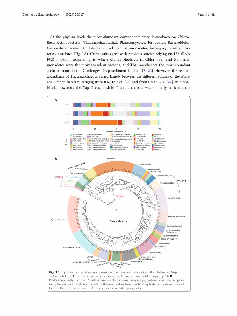
At the phylum level, the most abundant components were Proteobacteria, Chloro-
flexi, Actinobacteria, Thaumarchaeotathat, Planctomycetes, Firmicutes, Bacteroidetes,
Gemmatimonadetes, Acidobacteria, and Gemmatimonadetes, belonging to either bac-
teria or archaea (Fig. 1A). Our results agree with previous studies relying on 16S rRNA
PCR-amplicon sequencing, in which Alphaproteobacteria, Chloroflexi, and Gemmati-
monadetes were the most abundant bacteria, and Thaumarchaeota the most abundant
archaea found in the Challenger Deep sediment habitat [16, 22]. However, the relative
abundance of Thaumarchaeota varied largely between the different studies of the Mari-
ana Trench habitats, ranging from 0.67 to 67% [23] and from 0.5 to 40% [22]. In a non-
Mariana system, the Yap Trench, while Thaumarchaeota was similarly enriched, the
Fig. 1 Composition and phylogenetic diversity of the microbial community in the Challenger Deepsediment habitat. A The relative sequence abundance of dominant microbial groups (top 30). BPhylogenetic analysis of the 178 MAGs based on 43 conserved single-copy, protein-coding marker genesusing the maximum likelihood algorithm. Bootstrap values based on 1000 replications are shown for eachbranch. The scale bar represents 0.1 amino acid substitution per position
Chen et al. Genome Biology (2021) 22:207 Page 4 of 28

abundance of Chloroflexi, Actinobacteria, Planctomycetes, and Gemmatimonadetes in
its sediment microbiome was significantly lower than that of Mariana Trench sediment
habitats [24]. We found that the overall relative abundance of archaea decreased with
sediment depth, similar to findings in a previous study [13]. However, the relative
abundance of both the microeukaryotes and marine viruses increased with sediment
depth. The most abundant eukaryotes were Opisthokonta (Fungi), Alveolata, Strameno-
piles, and Rhodophyta. Note the top eukaryotic phyla, Ascomycota and Basidiomycota,
ranked the 16th and 17th overall in sequence abundance (Fig. 1A). We found MT-1
and MT-2 had consistent microbial compositions compared to those of MT-3, suggest-
ing a geochemical boundary separated them at a 10-cm depth that was previously
shown to limit oxygen access in hadal sediment [25]. Albeit in a relatively low abun-
dance, the eukaryotic and viral components were unraveled for the first time in the
Challenger Deep sediment, and as integral parts of the hadal biosphere, their ecological
significance was further addressed below.
Phylogenetic diversity of the hadal sediment microbiome
Metagenomic binning on the assembled metagenome and filtering resulted in 178 qual-
ity draft genomes, i.e., MAGs (metagenome-assembled genomes) (Additional file 6:
Table S6). These MAGs were at or above the medium-quality standards for draft gen-
ome, which required > 50% completeness and < 10% contamination (“Materials and
method”) [26]. Furthermore, among them, 101 draft genomes were > 70% complete-
ness, and 22 were > 90% completeness. The MAGs represented the most diverse meta-
genome reconstructed from the hadal sediment biosphere, comprising members from
26 phyla or candidate phyla. Sixteen of them had MAGs found from the Changer Deep
sediment for the first time, i.e., Gemmatimonadetes (8), Verrucomicrobia (8), Nitrospi-
nae (5), Elusimicrobia (1), Ignavibacteriae (1), Calditrichaeota (4), candidate phyla in-
cluding Zixibacteria (4), Ca. Hydrogenedentes (2), Ca. Dependentiae (1), Ca.
Diapherotrites (1), Ca. Doudnabacteria (2), Ca. Latescibacteria (2), Ca. Omnitrophica
(3), Ca. Marinimicrobia (2), and unclassified bacteria including Bacterium SM23_31 (2),
Ca. Sumeriaeota (2) and a newly defined phylum UBP7_A (1) [27]. In comparison, pre-
vious studies reported eleven and thirty MAGs that were co-assembled by combing
water and sediment microbial sequences from the Mariana Trench and were affiliated
with three and twelve phyla, respectively [28, 29]. Besides, using the single-cell sequen-
cing approach, twelve single-cell amplified genomes (SAGs) were generated for Parcu-
bacteria from the Mariana Trench sediment [30]. Note that these earlier works had
fewer MAGs despite co-assembly by mixing sample data from multiple sources. Thus,
the deep metagenomics approach has significantly enhanced the coverage and sensitiv-
ity of the hadal microbiome.
To illustrate the taxonomic diversity of the hadal sediment microbes, a phylogenetic
tree for the 178 MAGs was constructed (Fig. 1B), using 43 conserved single-copy,
protein-coding marker genes [31, 32]. A substantial number of uncultured microbial
lineages were uncovered and classified with the phylogenetic analysis. The main bacter-
ial groups included Alphaproteobacteria (22 MAGs), Gammaproteobacteria (19),
Chloroflexi (19), Planctomycetota (14), Bacteroidota (10), Actinobacteriota (10), Gem-
matimonadetes (8), Verrucomicrobia (6), Nitrospinae (4), as well as Elusimicrobia (1),
Chen et al. Genome Biology (2021) 22:207 Page 5 of 28
Ignavibacteriae (1), Nitrospirae (1), and Calditrichaeota (4). Notably, bin.97 formed a
monophyletic clade in the phylogenetic tree and was distant from the PVC groups that
include Planctomycetes (Fig. 1B). It was closely related to Planctomycetales bacterium
4484_113 based on analysis using GTDB-Tk [33], for which the Average Nucleotide
Identity (ANI) to bacterium 4484_113 stood at 63.86%. Bacterium 4484_113 was origin-
ally defined as unclassified Planctomycetes, but a recent study separated it from Planc-
tomycetes to form a new phylum UBP7_A [27]. Thus, our study uncovered a likely
second member of UBP7_A from the Challenger Deep sediment habitat.
The main archaea groups included Ca. Woesearchaeota (3 MAGs), Thaumarchaeota
(4), and Ca. Diapherotrites (1) (Fig. 1B). Note that Ca. Diapherotrites, represented by
bin.150, was found for the first time in the Challenger Deep sediment. Intriguingly,
bin.150 was situated as an outgroup to both Thaumarchaeota (TACK group) and Woe-
searchaeota, and had a closer relationship with bacteria in the phylogenetic tree
(Fig. 1B). Based on analysis using GTDB-Tk, bin.150 was close to the unclassified Dia-
pherotrites archaeon UBA493 that is affiliated to DPANN group. Its ANI to UBA493,
the closest relative, was 72.19%. We placed bin.150 with additional members of Ca.
Diapherotrites for phylogenetic analysis and found bin.150 formed a long-branch that
represents a specialized Diapherotrites clade in the Challenger Deep sediment (Add-
itional file 17: Fig. S1). A previous study reported that as a member of Ca. Diaphero-
trites, Ca. Iainarchaeum acquired anabolic genes from bacteria via horizontal gene
transfer [34]. The same reason may explain why bin.150 had a close relationship with
bacteria in our phylogenetic results. So by reconstructing the largest metagenome of
the hadal sediment biosphere, we recovered representative genomes of all major pro-
karyotic lineages previously identified by 16S rRNA gene amplicon-based surveys for
the Challenger Deep sediment habitat [35], providing valuable references for us to fur-
ther look into details of hadal microbiome regarding genetic diversity, metabolic func-
tions, and symbiotic relationship. While our analyses were based on constructed
MAGs, we acknowledged the missing pathway components that are likely attributed in
part by the incomplete assembly, and the many assembled sequences of unknown mo-
lecular functions that are knowledge gaps to be bridged with new means [36].
Versatile metabolic function of the hadal sediment microbiome
Heterotrophy vs. autotrophy
To investigate the metabolic potential of each component, the 178 MAGs reconstructed
from the Challenger Deep sediment microbiome were assigned with metabolic functions
based on KEGG annotation. To determine microbes’ lifestyle, the MAGs were first inves-
tigated for heterotrophic potential and were found to contain genes involved in various
pathways for degradation of carbohydrates (all MAGs), hydrocarbons (34 MAGs affiliated
with 9 phyla), and aromatic compounds (146 MAGs affiliated with 24 phyla) (Fig. 2 and
Additional file 7: Table S7). It is likely that sinking particulates from the upper ocean or
terrestrial inputs, partly due to the funneling effect and earthquake-inducing landslides,
were the source of the organic matter in the deepest habitat [37].
On the other hand, we found 69 out of 178 MAGs (39%; 16 phyla) contained path-
ways for inorganic carbon fixation. Six different CO2 fixation mechanisms were found
to involve various microbes (Fig. 2). Among them, 3-hydroxypropionate bi-cycle (3-HP)
Chen et al. Genome Biology (2021) 22:207 Page 6 of 28

was the most prevalent in 43 MAGs, compared to the Calvin-Benson-Bassham Cycle
(CBB or Calvin cycle) (11 MAGs), Wood-Ljungdahl pathway (WL pathway) (6 MAGs),
the reverse TCA cycle (rTCA) (2 MAGs), the 3 hydroxypropionate-4 hydroxybutyrate
cycle (3-HP/4-HB cycle) (6 MAGs), and methanogenesis (16 MAGs). Previously, 3-HP
was found in Chloroflexaceae, Alpha- and Gammaproteobacteria, and SAR202 [38, 39].
In our study, the 43 MAGs detected to have the 3-HP pathway were taxonomically
Fig. 2 Heat-map presentation of genomic features and metabolic potential for the 178 MAGs (with thetaxonomic assignment) reconstructed for the Challenger Deep sediment microbiome. Key genes involvedin carbohydrate degradation (CAYzme), CO2 fixation, aerobic respiration, anaerobic respiration, andchemolithotrophy are illustrated (refer to Additional file 7: Table S7 for details). Abbreviations: GH,glycosidases or glycosyl hydrolases; PL, polysaccharide lyases; CE, carbohydrate esterases; GT, glycosyltransferases;AA, auxiliary activities; CBM, carbohydrate-binding modules; WL, Wood-Ljungdahl pathway; CBB, Calvin-Benson-Bessham cycle; rTCA, reverse tricarboxylic acid cycle; 3-HP: 3-hydroxypropionate bi-cycle; 3-HP/4-HB cycle,3-hydroxypropionate/4-hydroxybuty rate cycle
Chen et al. Genome Biology (2021) 22:207 Page 7 of 28
assigned to eight phyla (31%) (Fig. 2 and Additional file 7: Table S7). They include the
unclassified Chloroflexi bacterium RBG_16_64_32, Alpha-, Delta-, and Gammaproteo-
bacteria, Nitrospinae, Acidobacteria, Actinobacteria, Calditrichaeota, Ca. Hydrogene-
dentes, and Gemmatimonadetes.
For other CO2 fixation pathways, the key enzymes in the Calvin Cycle, ribulose-
bisphosphate carboxylase (rbcS), and phosphoribulokinase (prkB) were detected in five
phyla (Fig. 2 and Additional file 7: Table S7). The rTCA cycle biomarker Acl was present
in one MAG for Nitrospira and one MAG for Ca. Woesearchaeota. Further, rTCA cycle
was the only CO2 fixation pathway employed by Nitrospira and Ca. Woesearchaeota.
While nitrite-oxidizing Nitrospira was previously known to use rTCA cycle for CO2 fix-
ation [40], this is the first reported case that Ca. Woesearchaeota may be capable of rTCA
reaction. 3-HP/4-HB cycle was the most energy-efficient aerobic autotrophic pathway for
CO2 fixation [41]. The biomarker enoyl-CoA hydratase/3-hydroxyacyl-CoA dehydrogen-
ase involved in 3-HP/4-HB or DC/4-HB cycle was found in three bacterial groups, Nitros-
pinae, Acidobacteria, and Planctomycetes, and one archaea, Thaumarchaeota. The WL
pathway biomarkers, i.e., acetyl-CoA decarbonylase/synthase complex subunit delta
(cdhD), acetyl-CoA synthase (acsB), anaerobic carbon monoxide dehydrogenase (cooS and
cooF), and 5-methyltetrahydrofolate corrinoid/iron-sulfur protein methyltransferase (acsE)
were found in Bacteroidetes, Alphaproteobacteria, Ca. Sumerlaeota, Chloroflexi, Gamma-
proteobacteria, and Planctomycetes. Moreover, we found 16 MAGs associated with 7
phyla, i.e., Proteobacteria, Bacteroidetes, Chloroflexi, Gemmatimonadetes, Nitrospinae,
Planctomycetes, and Thaumarchaeota, contained the Coenzyme M biosynthesis and
methanogenesis genes (Fig. 2 and Additional file 7: Table S7), indicating the hadal micro-
organisms had the potential to produce methane from fixing CO2 in the hadal zone.
Overall, among the 178 MAGs reconstructed for the hadal sediment microbiome, 69
MAGs (~ 39%) were affiliated with 16 phyla and mixotrophic based on their capacity of
inorganic carbon fixation (Fig. 2 and Additional file 7: Table S7). The enrichment and
significant taxonomic expansion of mixotrophic microbes we revealed for the deepest
hadal microbiome may indicate an adaptive niche that mixotrophy confers to microbes
living in an oligotrophic habitat like the Challenger Deep sediment.
Aerobic vs anaerobic respiration
We investigated the hadal microbiome for its potential to carry out aerobic or anaerobic
respiration. In total, 155 MAGs (~ 87%) were found to contain aerobic respiratory genes,
such as Cytochrome c oxidases (Cox/Cyd/Qox/cco/Cyo). These MAGs are associated
with 24 phyla, such as Acidobacteria, Bacteroidetes, Gemmatimonadetes, and Proteobac-
teria (Fig. 2 and Additional file 7: Table S7). Therefore, a large majority of the microbes in
the hadal habitat can potentially use oxygen as an electron acceptor for energy generation.
On the other hand, anaerobic respiration pathways were also found to be widely dis-
tributed, for which nitrate/nitrite or sulfate was the alternative electron acceptors. Dis-
similatory nitrate reduction to ammonia pathway (DNRA), denitrification pathway, and
sulfate reduction pathways were searched and found in a total of 145 MAGs (81%)
(Fig. 2 and Additional file 7: Table S7). These MAGs covered 21 of the 26 phyla we
identified in the hadal microbiome. Specifically, the genes for DNRA (nirB and nirD as
markers), denitrification pathway (nirK and norC), assimilatory sulfate reduction
Chen et al. Genome Biology (2021) 22:207 Page 8 of 28

pathway (sat, aprAB, and dsrAB), and dissimilatory sulfate reduction pathway (aprA-
and aprB) were present in 64 MAGs (15 phyla), 77 MAGs (16 phyla), 135 MAGs (20
phyla), and 67 MAGs (15 phyla), respectively (Fig. 2 and Additional file 7: Table S7).
Previous studies based on 16S rRNA gene sequencing analysis suggested that dissimila-
tory nitrate reduction and denitrification were the dominant reactions, whereas micro-
bial sulfate reduction was negligible in the Challenger Deep sediment [13]. However,
our analysis shows that the broad distribution of both assimilatory sulfate reduction
and dissimilatory sulfate reduction points to the equal importance of sulfate reduction
pathways for anaerobic respiration, if not otherwise more important. In addition, for
microbes capable of anaerobic respiration, many were found to have the potential for
different types of fermentation for degradation of organic matters (Additional file 17:
Analysis of fermentation; Additional file 7: Table S7).
The enrichment of anaerobic or anaerobic respiration metabolism in microbial com-
munity was apparently driven by the local habitat conditions. Examples of the deep pet-
roleum seep sediments and Guaymas Basin hydrothermal sediments illustrated MAGs
enriched for anaerobic respiration in the presence of hydrocarbons, i.e., alkanes and
aromatic compounds, or depleted oxygen levels in sediments [31, 42]. We further in-
vestigated the MAGs for the potential to carry out both aerobic and anaerobic respir-
ation and found 136 MAGs out of 178 (~ 76%), affiliated with 20 phyla (Fig. 2 and
Additional file 7: Table S7), were capable of facultative anaerobic metabolism. Com-
pared to the microbial communities in deep petroleum seeps or Guaymas Basin hydro-
thermal habitat, the high proportion of facultative anaerobes in the Challenger Deep
sediment, reflects the microbial metabolic versatility and the ability to adapt by the
hadal microbes to endemic habitats or environmental disturbance.
Chemolithotrophy
Microbes are known to acquire their energy for growth and CO2 fixation from the oxi-
dation of inorganic compounds, such as hydrogen (H2), hydrogen sulfide (H2S), ammo-
nia (NH3), carbon monoxide (CO), and metals [43]. The nitrification gene markers
were found in 53 MAGs (30%, 17 phyla) of the Challenger Deep sediment microbiome
(Fig. 2 and Additional file 7: Table S7). The key enzyme in nitrite oxidation, nitrite oxi-
doreductase/nitrate reductase was present in 35 MAGs (20%; 13 phyla), suggesting that
nitrification is an important approach for energy production for a substantial propor-
tion of microbes in the hadal sediment biosphere. Other genes involved in nitrification
pathways, PmoA-amoA, PmoB-amoB, and PmoC-amoC, were detected in 4 MAGs asso-
ciated with 3 phyla (Additional file 7: Table S7). Hydroxylamine dehydrogenase gene
(hao) was detected in 21 MAGs (12%; 12 phyla). Interestingly, the gene, hao, was not
found in bathypelagic microbes capable of nitrification [44], reflecting the difference in
microbial mediators between the bathypelagic and hadal zones in similar biogeochem-
ical process.
The genes for CO oxidation, sulfide oxidation, H2 oxidation, and iron oxidation were
also detected in the Challenger Deep sediment microbiome. Carbon monoxide de-
hydrogenase (CODH; cox gene) was found in 92 MAGs (52%; 15 phyla) (Fig. 2 and
Additional file 7: Table S7), indicating its broad distribution, and the importance of
CO-oxidation as an energy source in the hadal habitat. Sulfide oxidation genes such as
Chen et al. Genome Biology (2021) 22:207 Page 9 of 28

sqr and fccB were found in 33 MAGs (19%; 8 phyla), i.e., Acidobacteria, Actinobacteria,
Bacteroidetes, Chloroflexi, Gemmatimonadetes, Planctomycetes, Thaumarchaeota, and
Proteobacteria. For iron oxidation, we detected cyc2 gene (encoding Cytochrome c) in
one MAG associated with Gemmatimonadetes, suggesting that Fe2+ can serve as an al-
ternative electron donor for Gemmatimonadetes.
Microeukaryotic community and predominant fungal groups
Microbial eukaryotes in hadal sediment were the least studied and remained largely un-
known. Our analysis found that Opisthokonta (Fungi) were the predominant eukary-
otes in the Challenger Deep sediment, accounting for 87.42% of the total eukaryotic
sequences. Ascomycota (41.73%), Basidiomycota (26.82%), and Mucoromycota (12.64%)
were the top phyla, followed by Chlorophyta (6.61%), Rhodophyta (2.44%), Bacillario-
phyta (2.31%), Chytridiomycota (2.21%), Zoopagomycota (1.91%), Apicomplexa (0.96%),
etc. (Fig. 3A). Our results revealed the presence of Stramenopiles, e.g., Blastocladiomy-
cota, and Alveolata, e.g., Apicomplexa, two members of the super-group SAR (i.e., Stra-
menopiles, Alveolata, and Rhizaria), in the hadal sediment, albeit at low abundances.
Rhizaria was, however, notably absent. Previous studies showed that the abundance of
Alveolata and Stramenopiles decreased with depth in the water column of the Mariana
Trench, whereas Opisthokonta had an inverse trend [45]. Our study expanded the find-
ings into the trench sediment, illustrating the co-existence pattern of Alveolata, Strame-
nopiles, and Opisthokonta.
As the dominant eukaryotic group, fungi in the Challenger Deep sediment biosphere
comprised six phyla, i.e., Zoopagomycota, Mucoromycota, Ascomycota, Basidiomycota,
Chytridiomycota, and Blastocladiomycota, which can be further classified into twenty-
seven classes (Fig. 3B). Fungi were known to be distributed globally in deep-sea water,
including that in the Mariana Trench, as Basidiomycota and Ascomycota were the pri-
mary components of the deep-sea fungal community [46, 47]. Basidiomycota and Usti-
laginomycetes were previously detected in deep-sea sediment using a cloning library
method [48]. The capability of living in anoxia conditions may be crucial for fungi to
adapt to the hadal sediment environments, as Ascomycetes, Basidiomycetes, and Chy-
tridiomycetes were reportedly capable of fermentation and anaerobic growth in the
deep ocean [49].
Metabolic functions of the fungal community in the hadal sediment biosphere
The details of gene contents and metabolic functions of the fungal community in both
the water column and deep-sea sediment remained largely unknown, as early studies
were based on 18S rRNA gene/ ITS amplicon sequencing [45, 47]. In the current work,
we explored the metabolic potential of the fungal groups using the assembled
metagenome.
Carbon metabolism
We investigated the potential of the hadal fungi to metabolize carbohydrates and pep-
tides in the hadal sediment by looking into genes for carbohydrate-active enzymes
(CAZYmes) and peptidases. In total, we detected 262 CAZYmes (88 family) and 703
peptidases (77 family) (Fig. 3B and Additional file 8: Table S8). Members of CAZyme
Chen et al. Genome Biology (2021) 22:207 Page 10 of 28

included 79 GHs (glycosidases or glycosyl hydrolases), 8 PLs (polysaccharide lyases), 51
CEs (carbohydrate esterases), 83 GTs (glycosyltransferases), 14 AAs (auxiliary activities;
associated with polysaccharide and lignin degradation), and 27 CBM (carbohydrate-
binding modules). The major sources of CAZymes were Eurotiomycetes (number of
genes n = 20), Agaricomycetes (n = 16), Sordariomycetes (n = 38), and Malasseziomy-
cetes (n = 30). Particularly, key CAZyme genes encoding xylanase (GH10) were found
in Agaricomycetes, and those encoding cellulase (GH5) were found in Agaricomycetes,
Chytridiomycetes, and Microbotryomycetes (Additional file 8: Table S8).
Peptidase genes were abundant in Endogonomycetes (n = 124), Agaricomycetes (n =
84), Sordariomycetes (n = 70), and Malasseziomycetes (n = 67) (Fig. 3B and Additional
file 8: Table S8). Six peptidase groups, i.e., Aspartic (A), Cysteine (C), Metallo (M),
Mixed (P), Serine (S), and Threonine (T) peptidases, were found in the fungal genomes.
Among them, the most abundant was Metallo peptidase, consisting of 230 members
that belong to 30 families, followed by Cysteine (n = 225; 21 families), Serine (n = 168;
Fig. 3 Composition of microeukaryotic community, and metabolic functions of the dominant fungal groupsin the Challenger Deep sediment habitat. A The relative sequence abundance of different eukaryoticgroups within total eukaryotes. B Phylogenetic relationship of major fungal groups identified in the hadalhabitat, and the profiles of a carbohydrate-active enzyme family (CAZymes) and peptidase family genes.The numbers of genes detected are denoted by shade intensity. Abbreviations: GH, glycosidases or glycosylhydrolases; PL, polysaccharide lyases; CE, carbohydrate esterases; GT, glycosyltransferases; AA, auxiliaryactivities; CBM, carbohydrate-binding modules. C Metabolic potentials of carbon, nitrogen, sulfur, and ironmetabolism shown for the six dominant fungal groups. The presence of genes within the metabolicpathways is denoted for each group by the fan areas with color for the corresponding phylum. Genesymbols and metabolites are labeled with the KEGG designation (refer to Additional file 1: Table S8for details).
Chen et al. Genome Biology (2021) 22:207 Page 11 of 28

19 families), Threonine (n = 43; 3 families), and Aspartic peptidases (n = 14; 3 families)
(Fig. 3B and Additional file 8: Table S8). So, the hadal fungi have broad potentials in
organic carbon cycling, capable of degrading a variety of carbohydrate and peptide
substrates.
Nitrogen metabolism
. The hadal sediment fungi were found to possess the complete pathways for dissimila-
tory nitrate reduction and assimilatory nitrate reduction, and the partial pathway for ni-
trogen denitrification, but lacked the ability for nitrification or anammox (Fig. 3C). The
key enzymes for nitrate reduction, namely NarGHI/NapAB and NirBD for dissimilatory
pathway, and NR/NasAB and NIT-6 for assimilatory pathway, were found in several
fungal groups, indicating dissimilatory nitrate reduction is an important function for
the hadal fungi. Ammonia in hadal water could be generated from decomposition of ni-
trogenous organic matter [4]. However, it can be limited in hadal sediment. Therefore,
the nitrate reduction by the sediment fungi can be an important source of ammonia for
hadal sediment microbes.
The partial pathway for nitrogen denitrification was also found in the hadal sediment
fungi, missing the NosZ gene that coverts nitrous oxide to nitrogen. Downstream of the
NarGHI/NapAB genes (shared with the dissimilatory nitrate reduction pathway), the
NirK and NorBC genes completed the pathway to covert nitrite to nitric oxide, and ni-
tric oxide to nitrous oxide, which may be released into the hadal sediment environment
(Fig. 3C). However, unlike the bacterial community, the fungal community appeared
lacking the capability of nitrification, as some key enzymes were generally missing in
nitrification and anammox pathways (Fig. 3C). Notably, denitrification pathway en-
zymes were found in Fusarium, Penicillium, and Aspergillus from the Challenger Deep
sediment (Additional file 8: Table S8). Some Fusarium species in the deep-sea oxygen-
limiting environments were reportedly capable of denitrification [50]. In other low-
oxygen habitats, such as anaerobic marine sediment, salt-tolerant Penicillium and As-
pergillus species were also identified to carry out the denitrification process [50]. These
data illustrate the potential of the hadal fungi in nitrogen metabolism, possibly having a
significant role in the ecological process in the hadal environment.
Sulfur metabolism
Sulfate reduction is one of the main anaerobic respiratory pathways that many mi-
crobes living in anaerobic conditions depend on. The hadal fungi in the Challenger
Deep sediment were found to possess the complete pathways for assimilatory sulfate
reduction, dissimilatory sulfate reduction, and sulfide oxidation (Fig. 3C). They were
also found to contain partial pathway for the SOX system, but were incomplete for oxi-
dation of thiosulfate. The data suggest that hadal sediment fungi—like hadal bacteria,
could take part in sedimentary sulfur cycle, which has not been reported for fungal
communities in deep-sea sediment.
The possession of complete sulfate reduction enzymes indicated the hadal fungi had
the potential of using sulfate reduction for energy production when living under anaer-
obic conditions (Fig. 3C). A high copy number of genes for sulfate reduction enzymes,
like Sat, CysC, CysH, and CysJ, were detected in the hadal sediment fungi (Additional
Chen et al. Genome Biology (2021) 22:207 Page 12 of 28

file 8: Table S8). The sulfate adenylyltransferase gene (sat) for catalyzing the bidirec-
tional reactions between APS and sulfate was widely distributed in members of diffe-
rent phyla, e.g., Ascomycota (4 classes), Basidiomycota (3 classes), Chytridiomycota
(2 classes), and Mucoromycota (4 classes).
In the sulfide oxidation pathway, oxidation of sulfide to sulfite was catalyzed by
DsrA/B, whose genes were detected in several classes of Basidiomycota and Ascomy-
cota, like Malasseziomycetes and Sordariomycetes. Subsequently, sulfite was oxidized
to APS by adenylylsulfate reductases. Dothideomycetes (belonging to Ascomycota) con-
tained Apr-A gene for sulfite oxidization to APS. Agaricomycetes (belonging to Basidio-
mycota) had the adenylylsulfate reductase gene (glutathione), denoted as APR, for the
same function. Finally, APS was oxidized to sulfate by the sat-coding enzymes, which
was found in Ascomycota, Basidiomycota, Chytridiomycota, and Mucoromycota. The
genes encoding taurine dioxygenase (tauD) for converting taurine to sulfite were found
in Basidiomycota and Ascomycota, like Agaricomycetes, Dothideomycetes, and Eurotio-
mycetes. The genes encoding alkanesulfonate monooxygenase (ssuD) for converting
alkanesulfonate to sulfite were found in Leotiomycetes (belonging to Ascomycota).
These enzymes would produce sulfite from an organic sulfur substrate, which in turn
would be fed into the sulfur metabolic pathways.
The hadal sediment fungi were also found to possess sox genes, indicating their po-
tential for thiosulfate/sulfide oxidization. For example, Endogonomycetes of Mucoro-
mycota contained soxC encoding sulfane dehydrogenase subunits for oxidizing
thiosulfate to sulfate. Malasseziomycetes of Basidiomycota and Sordariomycetes of As-
comycota were found to have SQOR (eukaryotic sulfide quinone oxidoreductase) genes
that are involved in the first step of hydrogen sulfide metabolism to produce sulfane
sulfur metabolites. A previous study reported that fungi could feed sulfate to sulfate-
reducing bacteria (SRB) [50]. Interestingly, we also found SRB, e.g., Desulfobacterales
and Desulfuromonadales (within the class Deltaproteobacteria), and Nitrospirae (class)
in the Challenger Deep sediment, which corroborates the evidence for the existence of
sulfide oxidation reactions and the roles that fungi that play in sulfur cycling in the
hadal sediment habitat. In addition, the hydrogen sulfide metabolisms possessed by the
hadal sediment fungi would counter the accumulation of sulfide (H2S, HS−, and S2−)
that was generated by SRB in the anaerobic environment. Thus, the discovery of the
critical metabolic genes in sulfur metabolism implicated the important role of hadal
fungal community in sulfur cycling and energy transformation in the trench
environments.
Metavirome in the Challenger Deep sediment habitat
The existence and composition of viral community in the hadal sediment habitat
were an open question remaining to be answered. Our deep metagenomics ap-
proach offered a new opportunity to look into the viral components for the dee-
pest biosphere. Viral sequence reads and their taxonomic affiliations were
determined using Kaiju by mapping to the viral references from NCBI-nr database.
A total of fifteen major viral families were identified in the Challenger Deep sedi-
ment (Fig. 4A). dsDNA viruses were found most frequently, which were mainly af-
filiated with the order Caudovirales, also known as the tailed bacteriophages [52].
Chen et al. Genome Biology (2021) 22:207 Page 13 of 28

The predominant family was Myoviridae, accounting for 24.62% of the total viral
sequences, followed by Siphoviridae (19.70%) and Podoviridae (9.40%). Notably, we
detected a relatively high abundance of giant viruses in the Challenger Deep sedi-
ment, such as Mimiviridae (4.16%) and Phycodnaviridae (1.06%) (Fig. 4A, red star).
Giant viruses were often missed or underestimated in early studies due to the tech-
nique used to capture viral particles via filtering [53]. Our approach is indifferent
to the viral components of different particle sizes.
To investigate the diversity and evolution among the members of Caudovirales
found in the Challenger Deep sediment, phylogenetic analysis was performed on
the viral contigs, using a marker gene coding the terminase large subunit (TerL)
[54]. Interestingly, for the two families, Myoviridae and Siphoviridae, there were
separate branche(s) forming within each of them (Fig. 4B). They were tentatively
named MT clade I and MT clade II for Myoviridae, and MT clade III for Siphovir-
idae. The new diversity of Caudovirales in the Challenger Deep sediment may re-
flect the differentiation of viral niche to microbial hosts living in the hadal habitat
of high-hydrostatic pressure and oligotrophy.
Fig. 4 Diversity of metavirome, virus-host association, and viral genome annotation. A The relativeabundance of dominant virus groups within metavirome. B Phylogenetic analysis of Caudovirales based onTerL using the maximum likelihood algorithm. Reference viral sequences from NCBI are colored in black.Scale bar, one amino acid substitution per site. The same tree with detailed labeling is provided in Fig. S3(Additional file 17: Fig. S3). C Visualization of the virus-host association network. Diamonds and circlesdenote marine viruses and microbial hosts, respectively. Association is summarized between a virus familyand a microbe class, represented by a linked grey line. D Annotation of viral genomes. COG annotation wasperformed using eggNOG-mapper [51]. AMGs related to carbohydrate metabolism are illustrated in theinner panel
Chen et al. Genome Biology (2021) 22:207 Page 14 of 28

Virus-host association network
To infer virus and microbial host interactions, we took the use of a combination
of different methods as described [55]. Possible virus-host interactions were sum-
marized for each viral family. As a result, members from 131 bacteria and archaea
classes were associated with 20 viral families (Fig. 4C and Additional file 8: Table
S9). The association network revealed many one-to-many relationships between vi-
ruses and hosts, and vice versa. Siphoviridae was connected with the largest num-
ber of hosts that belong to 95 classes. Notably, many of the hosts were
uncultured microbes that were only inferred from their presence in metagenome
sequences.
Unlike in epipelagic and mesopelagic ocean waters, where the most frequent
hosts were Cyanobacteria and Alphaproteobacteria (mainly SAR11 [52]), the most
frequent hosts in the Challenger Deep sediment were Firmicutes (mainly Bacilli)
and Bacteroidetes, followed by Euryarchaeota (including uncultured marine group
II/III and Diaforarchaea), unclassified Chloroflexi, and Alphaproteobacteria. A pre-
vious study reported that the dsDNA virus T7virus (belonging to Podoviridae)
could infect Deltaproteobacteria, Gammaproteobacteria, Alphaproteobacteria, Firmi-
cutes, and Cyanobacteria [55]. Our results indicated that the T7virus from the
Challenger Deep sediment was also associated with an archaeal host, e.g., the Dia-
forarchaea group of Euryarchaeota.
Functional viromics
To investigate the gene contents and functions of the hadal sediment viruses, we anno-
tated their contigs using the Clusters of Orthologous Groups (COGs) database [56].
While a large proportion of their ORFs had unknown functions, high abundance genes
were found in the COG categories of “replication, recombination and repair,” “tran-
scription,” “signal transduction mechanism,” “cell wall/membrane/envelope biogenesis,”
etc. (Fig. 4D). Besides the genes for basic viral functions, a special group of virus-
encoded genes can modulate the activities of the hosts upon infection. These so-called
auxiliary metabolic genes (AMGs) were a means for viruses to manipulate host metab-
olism, like sulfur and nitrogen cycling.
We identified 40 putative AMGs from the Challenger Deep sediment viruses,
having roles in carbon, sulfur, or nitrogen metabolism (Additional file 10: Table
S10). Among them, auxiliary carbohydrate metabolic genes were the most frequent.
By searching against the CAZymes database, 45 carbohydrate metabolism-related
genes were identified, which included 31 GHs (glycosidases or glycosyl hydrolases),
six PLs (polysaccharide lyases), five GTs (glycosyltransferases), two CBM (carbohy-
drate-binding modules), and one AA (auxiliary activities). The presence of frequent
auxiliary carbohydrate metabolic genes provided evidence that the hadal sediment
viruses may heavily manipulate carbohydrate metabolism of hosts, especially pro-
moting carbohydrate degradation on hosts in an oligotrophic habitat. AMGs for
carbohydrate metabolism were also the largest group in other marine virome data-
set, like the Tara Oceans Viromes [55], which concentrated, however, on different
pathways, like galactose metabolism and glycosyltransferases, reflecting the differen-
tiated viral niche to hosts in dramatically different habitats.
Chen et al. Genome Biology (2021) 22:207 Page 15 of 28

For nitrogen metabolism, NirK that encodes nitrite reductase [57] was found in the hadal
viruses, suggesting possible roles to enhance host nitrite reduction pathway and release of ni-
tric oxide into hadal sediment. For sulfur cycling, the CysN/NodQ gene encoding ATP sul-
phurylase [58] that reduces sulfate to produce 3'-phosphoadenosine-5'- phosphosulfate
(PAPS), the first step of assimilatory sulfate reduction, was found in the hadal viruses. Not-
ably, both NirK and CysN/NodQ are found for the first time among AMGs for ocean viruses.
Large-scale isolation of microbes from the Challenger Deep sediment habitat
To catalog and characterize microbes in the hadal sediment habitat, an intensive effort
was made for the isolation of microbes from the Challenger Deep sediment. We
adopted a protocol previously developed for the isolation of environmental microorgan-
isms, which used serial dilutions of samples to allow the growth of “disadvantaged” mi-
crobes that otherwise would not be isolated [59]. To broaden the diversity of microbial
isolates, we employed 24 different media and combined them with various culture con-
ditions (Additional file 11: Table S11). Notably, the experimental conditions would en-
able facultative anaerobes but not strict anaerobes. As a result, more than two
thousand individual isolates were obtained, for which 1089 were completed for 16S
rRNA gene (for prokaryotes) or ITS amplicon (for eukaryotes) sequencing (Additional
file 12: Table S12 and S13).
Bacterial isolates
The taxonomy of 1070 bacterial isolates was assigned based on 16S rRNA gene se-
quences using SILVA database (release 123) [60]. The bacterial isolates belonged to
four phyla, i.e., Proteobacteria, Bacteroidetes, Actinobacteria, and Firmicutes, which
were further categorized into 7 classes, 18 orders, 25 families, and 40 genera (Add-
itional file 17: Fig. S2 and Additional file 12: Table S12). They matched the microbes
represented by sequences in the metagenome. Halomonas, Pseudoalteromonas, and
Idiomarina were the top genus, accounting for 32.77%, 22.23%, and 9.92% of the iso-
lates, respectively (Fig. 5A). Nineteen bacterial isolates from five classes were candidates
for new species, which had a 16S rRNA gene sequence with ≤ 97% identity to their
closest references (Additional file 14: Table S14) [61].
We characterized representative isolates from each of the four phyla, i.e., MTFD_053
(Zunongwangia sp.), MTFD_039 (Microbacterium sp.), MTFD_075 (Halomonas sp.),
MTFD_0511 (Salipiger sp.), MTFD_0323 (Pseudoalteromonas sp), and MTFD_035
(Paenibacillus sp.), which were visualized after incubation at elevated pressure (100
MPa) for 7 days. Some morphologic changes were observed with MTFD_039, MTFD_
075, MTFD_0323, and MTFD_0511, when compared between their cells cultured at 0.1
and 100MPa (Fig. 5B). Cases were found that sister cells were connected after cell divi-
sions under culture at elevated pressure. These isolates exemplified the small fraction
of culturable microbes in contrast to the community background in the Challenger
Deep sediment illustrated by the MAG data, which complemented the lack of taxo-
nomic details from the metagenomic analysis. The results suggested that they were
piezotolerant, which are likely derived from the microbes that descended from the
water column, contributing to the diversity and metabolic functions of the hadal sedi-
ment microbiome.
Chen et al. Genome Biology (2021) 22:207 Page 16 of 28

Microeukaryotic isolates
Nineteen microbial eukaryotes (fungi) were isolated from the Challenger Deep sedi-
ment (Additional file 13: Table S13 and Additional file 17: Fig. S4). Their taxonomy
was assigned based on their ITS sequences using the NCBI ITS database. They
belonged to nine fungal genera, namely Aspergillus, Penicillium, Acremonium, Micro-
ascus, Exophiala, Cladosporium, Gymnoascus, Purpureocillium, and Aureobasidium.
They matched the eukaryotic microbes represented by sequences in the metagenome.
Representative isolates from each genus were further characterized after incubation
at 0.1 and 100MPa for 14 days. MT19_18 (Aureobasidium sp.) tended to form long
pseudomycelia under 100MPa (Fig. 5C). Some elongated cells contained multiple nu-
clei but did not have a diaphragm separating them. The filamentous fungi, i.e., MT20_2
(Exophiala sp.), MT20_5 (Aspergillus sp.), MT20_3 (Penicillium sp.), MT19-20 (Ony-
genaceae sp.), MT19_19 (Gymnoascus sp.), MT19_17 (Cladosporium sp.), MT20_1
(Purpureocillium sp.), and MT20_8 (Microascus sp.), have some morphologic variations
induced by elevated pressure, such as hyphal swelling (MT19_19) and swollen conidia
(MT20_3) (Fig. 5C). Conidiophore appeared normal for MT19_17 under 100MPa com-
pared to that under 0.1MPa. MT20_1 (Purpureocillium sp.), MT19_19 (Gymnoascus
Fig. 5 Bacterial and fungal isolates from the Challenger Deep sediment habitat. A Phylogeny andpercentage of bacterial and fungal isolates (Additional file 1: Table S12 and S13). B Visualization ofrepresentative bacterial isolates from four different phyla. Cells were cultured under 0.1 and 100 MPa,respectively. Scale bars, 10 μM; DIC, images taken with a DIC microscope; DAPI, cells stained with DAPI andobserved with an epifluorescence microscope. C Visualization of representative fungal isolates from ninegenera. Scale bars, 30 μM; CW, cells stained with Calcofluor White and observed with an epifluorescencemicroscope. The phylogeny of fungal isolates is shown to the right. Red arrows indicate swollen hypha(MT19_19) or swollen conidia (MT20_3)
Chen et al. Genome Biology (2021) 22:207 Page 17 of 28

sp.), and MT20_8 (Microascus sp.) may represent new species specialized in the Chal-
lenger Deep habitat, whose ITS sequences diverged from their closest known species
(Additional file 13: Table S13).
ConclusionsWe adopted a twofold strategy to investigate the microbial community structure and
functions in the Challenger Deep sediment biosphere. The deep metagenomics ap-
proach reconstructed 178 MAGs from the deepest habitat, and revealed the full bio-
sphere structure and novel biodiversity, particularly enabling and extending our study
to include microeukaryotic and viral components. The largest MAG set reconstructed
for the hadal microbiome established versatile community functions marked by unex-
pected enrichment and prevalence of mixotrophy and facultative anaerobic metabolism.
On the other hand, fungi as heterotrophs had broad metabolic potentials in carbon, ni-
trogen, and sulfur metabolism, particularly the capability of dissimilatory nitrate reduc-
tion and sulfate reduction that had not been reported for deep ocean microeukaryotes.
These findings implicated the possible roles of hadal fungi in the biogeochemical pro-
cesses of the hadal trench environment. The viral components evolved with new diver-
sities and carried AMGs important for modulating hosts’ metabolic functions, which
illustrated the differentiation of viral niche to microbial hosts in the hadal habitat.
The large-scale cultivation approach obtained more than 2000 isolates from the Chal-
lenger Deep sediment, and cataloged 1070 bacteria and 19 fungi by 16S rRNA gene or
ITS amplicon sequencing. Many of them were likely new species specialized in the
Challenger Deep habitat and showed morphological variations under elevated pressure
(100MPa). These isolates represent a small fraction of the diversity in the Challenger
Deep sediment habitat based on the deep metagenomic data. They would serve as
model organisms and provide new opportunities to study and understand the physi-
ology of piezotolerant microbes.
Materials and methodsSample collection and geochemical measurements
The one intact sediment core was obtained from the seafloor in the Challenger Deep,
the Mariana Trench (142° 21.7806' E, 11° 25.8493′ N, 10,840 m) during the cruise from
December 2016 to January 2017 onboard the ship ZhangJian. The sediment core was
collected using a box corer (with a base area of 400 cm2 and a height of 25 cm) attached
to the hadal lander-II, developed by Shanghai Engineering Research Center of Hadal
Science and Technology, Shanghai Ocean University. After the lander reached the sea-
floor, the sampling chamber was slowly driven into the subseafloor until it reached
around 21 cm below the sediment surface. A lid was then released to seal the box corer,
before the lander was recovered. Once the lander surfaced and box corer was recovered
and examined for integrity, only one intact sediment core with well-preserved sediment
stratification was used in this study. The sediment core was immediately subsampled
by inserting polycarbonate tubes to preserve the segments. The samples were either
frozen at − 80 °C before stored at − 20 °C or stored at 4 °C for culturing and isolation
of microbes. Note, the temperature of the recovered core was not obtained. For
Chen et al. Genome Biology (2021) 22:207 Page 18 of 28

reference, the recorded environment temperature at the time was 2–4 °C at the ocean
floor, and 27–28 °C at the ocean surface.
Total organic carbon (TOC), total nitrogen (TN), and carbon and nitrogen isotopic
compositions of particulate organic matter were measured by high-temperature com-
bustion on a Vario Pyro Cube elemental analyzer connected to an Isoprime 100 con-
tinuous flow isotope ratio mass spectrometer. All samples were pre-treated with 10%
HCl to remove inorganic carbon. Carbon and nitrogen isotope ratios are expressed in
the delta notation (δ13C and δ15N) relative to V-PDB and atmospheric nitrogen. The
average standard deviation of each measurement, determined by replicate analyses of
the same sample, was ± 0.02% for TOC, ± 0.006% for TN, ± 0.2‰ for δ13C, and ±
0.3‰ for δ15N.
For the measurement of major and minor elements, approximately 40 mg of dry
sediment powder was weighed into a Teflon beaker and dissolved in super-pure
HF (0.2 ml), HNO3 (0.8 ml), and HCl (0.1 mL). The beaker was then sealed and
heated on a hot plate at 185 °C for 36 h. After cooling, the solution was evaporated
at 120 °C to dryness. The residue was re-dissolved by adding 2 ml HNO3 (super-
pure) and 3 ml of deionized water at 135 °C in an airtight beaker for 8 h. The final
solution was diluted to 50 ml with 3% HNO3. Blanks, duplicate samples, and sev-
eral certified reference materials (GSR-1, OU-6, 1633-a, GXR-2, GXR-5) were also
prepared using the same procedure. Major and minor elements were determined
using a Thermo-Fisher iCAP6300 ICP-OES and a Perkin-Elmer ELAN 6000 ICP-
MS, respectively. The analytical precision was 5% for major and 10% for minor
elements.
Measurements of NO2−, NO3
−, NH4+, and PO4
3− were performed using a QuAAtro
autoanalyzer (Seal Analytical) with a detection limit of 1 μM and a precision of 2%. Sul-
fate (SO42−) was measured by a Dionex ICS-5000+ ion chromatograph with a detection
limit of 10 μM and a precision of 2%.
Nucleic acid extraction and metagenomic sequencing
DNA was extracted from 10 g of sediment sample for each experiment replicate, using
the MoBio PowerSoil DNA Isolation kit (MO BIO Laboratories, USA) according to the
manufacturer’s protocol. DNA concentrations of the extract were measured with a
Qubit fluorometer. For each library construction, about 100 ng DNA was fragmented
with Covaris S2 (Covaris, USA) and was used to construct metagenomic DNA library
with NEXTflex™ DNA Sequencing Kit compatible with the Biomek® FXp (Bio Scientific,
USA). Notably, PCR amplification was limited to 12 cycles for each Illumina library.
The quality of DNA library was examined by Agilent Bioanalyzer 2100 (Agilent, USA)
with DNA 12000 Kit. Paired-end Illumina sequencing (2 × 150 bp) was performed for
each metagenomic library on Hiseq Xten instruments (Illumina).
Metagenomic assembly, mapping, and binning
Raw sequence data were cleaned by removing low-quality sequence reads, artificial se-
quence reads, and contaminated sequences with the following steps: (1) Reads with
average quality score less than Q20 and a length < 30 bp, or with adapter sequences,
were removed with trimmomatic (version 0.38) [62]; (2) reads with contaminated
Chen et al. Genome Biology (2021) 22:207 Page 19 of 28

sequences, like plasmid sequences and human sequences, were removed with bowtie2
(v2.3.4.1) [63]; (3) duplicated reads generated by PCR amplification were removed for
metagenomic assembly using fastuniq [64]. The metagenome was assembled with clean
reads using MEGAHIT (v1.1.3) [65] with the following parameters: --k-list 21, 29, 39,
59, 79, 99, 119, 127, 139. Read coverage for contigs was determined by mapping
sequencing reads to contigs using bowtie2 with default parameters.
Metagenomic binning and refinement were performed on contigs using a combin-
ation of different tools, including CONCOCT [66], Metabat [67], DAS Tool (v1.0) [68],
and metaWRAP [69]. Briefly, initial binning was conducted with CONCOCT and
Metabat based on tetranucleotide frequencies of contigs and coverage depth as covari-
ance, using a contig length cutoff of 1 or 1.5 kb, respectively, for which 310 and 400
bins were recovered. Results from the two different binning tools were then combined
and merged using the DAS Tool. The resulting bins were refined and consolidated into
the final bin sets using metaWRAP’s Bin_refinement module with options: -c 50 -x 10.
As a result, 418 bins were recovered across the entire quality spectrum. The quality of
the binning results was evaluated by estimating the completeness and contamination
scores using CheckM (v1.0.5) lineage_wf tool [32]. A cutoff of 50% completeness and
10% contamination was used to filter and obtain quality genomes (MAGs).
Taxonomic classification of sequences and determination of relative sequence
abundance
The method for taxonomic assignment of sequence reads and contigs was adapted
from the previous work using Kaiju [20]. Briefly, the NCBI-nr database compiled in a
GORG-Tropics format was downloaded, which included reference sequences from ar-
chaea, bacteria, viruses, and microbial eukaryotes. The assignment of the sequence
reads or contigs was conducted using the NCBI-nr database and Kaiju (v1.7.0) [70] in
Greedy-5 mode with the default options. Sequences were assigned with the taxonomic
ID and functional annotations based on mapped references. Their taxonomic lineage
information was then obtained using Kaiju’s tool “addTaxonNames.” For sequence
reads assigned with taxonomic ID, the relative sequence abundance of a phylum, class,
order, etc. was estimated by summarizing the total number of assigned sequence reads
for a given category and dividing it with the total number of assigned reads.
Phylogenetic analysis and taxonomy assignments of the metagenome-assembled
genomes (MAGs)
The sequences of 43 conserved proteins from previous work [32] were retrieved
from the metagenome-assembled genomes (MAGs), and multi-aligned using the
MUSCLE program (v3.8.31) [71]. The alignments were trimmed to remove gaps
and poorly aligned regions using TrimAL with the options -gt 0.95 -cons 50 [72].
The “cleaned” alignment datasets for the 43 conserved proteins were then
concatenated and were subsequently used for constructing a phylogenetic tree
using the maximum likelihood algorithm. The RAxML program (v8.1.24) [73] was
employed with the options “-f a -n boot -m PROTGAMMALG -c 4 -e 0.001 -#
1000”. To visualize the phylogenetic tree topology, the Newick files were processed
using iTOL online tool (v4) [74].
Chen et al. Genome Biology (2021) 22:207 Page 20 of 28

Initial taxonomy assignments of MAGs were provided from the evaluation of MAGs
with CheckM, which were often short with taxonomic information for the order, fam-
ily, or genus. The MAGs were further classified using CAT and GTDB-Tk programs
[33, 75]. CAT assigns the taxonomy of MAGs based on the BLAST results of the nr
database using the lowest common ancestor (LCA) algorithm [75]. On the other hand,
GTDB-Tk provides taxonomic classifications with the new rank-normalized GTDB tax-
onomy, by using two criteria, relative evolutionary divergence (RED) and average nu-
cleotide identity (ANI) for establishing taxonomic ranks [27, 33]. The taxonomy
assignment for MAGs was determined to the deepest taxonomic levels by combining
the results from both CAT and GTDB-Tk analyses. A few inconsistent assignments be-
tween the two programs were manually resolved based on information of their nearest
reference neighbors.
Annotation of contigs and metabolic pathway analysis
Genes were predicted for contigs / metagenome-assembled genomes (MAGs) using
prodigal (v2.6.2) with -p meta parameters [76]. Putative genes were then annotated with
KEGG Automatic Annotation Server (KAAS) using the GHOSTX tool with the “cus-
tom genome dataset” and “BBH” options, by uploading their predicted amino acid se-
quences to KAAS [77]. Furthermore, putative genes were annotated using blastp
program with default options against several different databases, like hmmer, the cu-
rated database of Anaerobic Hydrocarbon Degradation Genes (“AnHyDeg”), and MER-
OPS database [42]. To identify genes encoding carbohydrate degradation enzymes, the
dbcan tool was used to search the Carbohydrate-Active enzymes (CAZYmes) database
with an e-value cutoff of 1e−5 [78]. To reconstruct metabolic pathways for a draft gen-
ome (MAG), its predicted genes were annotated and pathway results were summarized
using the KEGG server. To illustrate metabolic activity for certain pathway(s) within a
taxonomic unit (e.g., phylum, class, order) or a phylogenetic cluster, all genomes within
the specific taxonomic unit/cluster were summarized, based on their taxonomic or
phylogenetic assignments.
Identification of viral contigs and phylogenic analysis
Viral contigs in the metagenome assembly were identified using Kaiju (v1.7.0). For the
phylogenic analysis of Caudovirales, the TerL gene [79] was identified for each contig
using blastp with a threshold e-value of 10−5, minimum identity of 50%, and minimum
coverage of 30%. Identified TerL gene sequences were retrieved from contigs and
aligned using the Mafft program (version 7.407) [80]. The alignments were trimmed to
remove gaps and ambiguously aligned regions before the phylogenetic tree was con-
structed using the Fasttree program [81], and manually formatted and visualized using
iTOL.
Virus-host association and viral function analysis
Viruses and their possible hosts from the hadal sediment environment were inferred
using the previously developed methods [55]. Briefly, virus-host association was pre-
dicted using three different means: (1) Virus contig-host genome similarity analysis: All
identified viral contigs from the sediment of the Challenger Deep were compared to
Chen et al. Genome Biology (2021) 22:207 Page 21 of 28

the archaeal and bacterial contigs in the same metagenome, using blastn with a thresh-
old of 50 for bit score and 0.001 for E-value. (2) Virus contig-host CRISPR spacer
match analysis: CRISPR spacers were predicted for all microbial contigs using
MetaCRT [82]. Association between a virus and its hosts was predicted when a host
CRISPR spacer was found to match a virus contig. (3) Nucleotide composition com-
parison between virus contigs and host genomes. The tetranucleotide-frequency vectors
and mean absolute error between the vectors for each virus-host pair were computed.
A viral contig was assigned to the closest microbial host whose genome having the low-
est mean absolute error (d) to the viral contig if d < 0.0015.
For functional viromics analysis, COG annotation of viral contigs was performed
using eggNOG-mapper (http://eggnog-mapper.embl.de/) with a threshold of 10− 5 Viral
ORFs annotated as the CAZymes family genes in eggNOG-mapper annotation results
were considered as auxiliary metabolic genes (AMG) related to carbohydrate metabol-
ism [83]. Further, additional viral ORFs related to sulfur and nitrogen metabolism
(determined by KEGG kos) in the eggNOG-mapper annotation results were also con-
sidered as AMGs.
Large-scale cultivation and identification of the hadal sediment microbes
The cultivation and isolation of the microbes from the Challenger Deep sediment were
adopted from previous studies as described [59, 84]. To reduce their exposure to oxy-
gen, the sediment sub-samples were sealed in airtight bags with residue air removed be-
fore they were stored at 4 °C. A series of dilutions, e.g., 10−1, 10−2, 10−3, 10−4, and 10−5,
were made to the sediment samples in 96-well plates using various culture media (Add-
itional file 11: Table S11). The high ratio dilutions, e.g., 10−3, 10−4, 10−5, were then
plated out in 90-mm plates with solid culture media. The dilution and inoculation were
performed under aerobic conditions in the laboratory. The plates were incubated under
various conditions, i.e., 4 °C or 28 °C, aerobic or anaerobic, with or without antibiotic,
etc., for up to 1 month. For anaerobic culture, the agar media plates were first treated
with AnaeroPack (Mitsubishi Gas Chemical Company, Inc., Tokyo) to remove oxygen.
For large-scale isolation, a combination of 24 different media (modified from nine base-
types) with various culture conditions were used for cultivation (Additional file 11:
Table S11). Single colonies emerging at different time points were picked to grow in li-
quid media for cryopreservation and further analysis. Notably some single colonies
emerging at late stages were collected from the highest dilution plates, which would
have been overlaid by faster-growing colonies in the low-dilution plates.
The identity of the single colonies was analyzed by 16S rRNA gene (for prokaryotes)
or ITS gene (for eukaryotes) sequencing. The primer sets, 8F/805R (8F-5′AGAGTTT-
GATCCTGGCTCAG; 805R-5′GACTACCAGGGTATCTAATC) (targeting V1-V4
regions) and 8F/1492R (8F-5′AGAGTTTGATCMTGGCTCAG; 1492R-5′
GGTTACCTTGTTACGACTT) (targeting V1-V9 regions) were used for 16S rRNA
gene tag PCR amplification and sequencing [85, 86]. They generated the PCR-amplified
tag with length of 805 and 1492 bp, respectively. The primer set, ITS-F/ITS-R
(ITS-F-5’GGAAGTAAAAGTCGTAACAAGG; ITS-R-5’TCCTCCGCTTATTGATAT
GC) were used for ITS gene tag PCR amplification and sequencing. It generated
the PCR-amplified tag with length of 578 bp. Identification of the sequenced single
Chen et al. Genome Biology (2021) 22:207 Page 22 of 28

colony was conducted by searching 16S ribosomal RNA sequence database, and
Internal transcribed spacer region (ITS) database at NCBI (https://www.ncbi.nlm.nih.gov/)
using blastn.
Culture of microbes under elevated hydrostatic pressure and microscopic imaging
Single colony was picked from solid media to inoculate 2-ml liquid culture media in a
sterile 15-ml glass tube and was incubated on a rotary shaker (180 rpm) at 28 °C over-
night for bacteria or for 3 days for fungi to produce seed broth. For single-cell mi-
crobes, cultures were diluted to O.D.600 = ~ 0.01 in appropriate media. For
filamentous fungi, mycelia in the seed broth were fragmented with sterilized scissors (if
necessary) before the cultures were diluted (1/15~1/60 depending on fungal biomass)
in the same media. The seeded cultures were transferred to 2-ml sterile plastic syringes
before they were placed inside pressure vessels (Model: FY2016108; Feiyu Petroleum
Technology Development Co., Ltd., Nantong, China). Hydrostatic pressure of 100MPa
was applied at room temperature to bacterial cultures for 7 days, or to fungal cultures
for 14 days, respectively. After high-pressure incubation, the syringes were taken out of
pressure vessels and visually examined for physical integrity. The cultures were re-
plated on solid media to test viability for growth or were examined by microscopy.
For microscopic imaging of bacteria and fungi, an epifluorescence microscope (Nikon
Ecllipse 80i) and a digital camera (Nikon DS-Ri1; software: NIS-Elements F Ver4.30.01)
attached to the microscope were used. For bacterial culture, cells were harvested by
centrifugation. Pellets were washed twice with PBS buffer and stained with DAPI (4',6-
diamidino-2-phenylindole) for 10 min in the darkness [87]. Stained cellular suspension
(2.5 μL) was spread onto a class microscope slide and covered with a cover slide with
excess liquid removed. Slides were imaged at × 100 magnification (oil immersion len)
for Differential Interference Contrast (DIC) micrograph and epifluorescent micrograph
(excitation wavelength 300–380 nm), respectively. For fungal culture, 10 μL fungal cul-
ture was mixed with 10 μL Calcofluor White solution (1 mg/mL) on a glass slide for 3
min at room temperature [88]. Fungal hyphae with or without Calcofluor staining were
imaged at × 40 magnification for DIC micrograph and epifluorescent micrograph,
respectively.
Supplementary InformationThe online version contains supplementary material available at https://doi.org/10.1186/s13059-021-02408-w.
Additional file 1: Table S1. Geochemical characterization of the sediment samples from Challenger Deep.
Additional file 2: Table S2. Concentrations of nutrient ions NO3-, NO2
-, PO4-, NH4
+, and SO42- in porewater of the
samples.
Additional file 3: Table S3. Dissolved major elements in the sediment samples.
Additional file 4: Table S4. Dissolved trace elements in the sediment samples.
Additional file 5: Table S5. Deep metagenomic sequencing on the Challenger Deep sediment samples andassembly.
Additional file 6: Table S6. Taxonomic assignment and characteristics of the 178 MAGs.
Additional file 7: Table S7. Functional analysis of archaeal and bacterial MAGs. The key genes of each metabolicpathways for MAG annotation are listed on the top. Presence/absence of genes are listed as: Presence: 1 (green);Absence: a (no color). The module completeness of each metabolic pathway is the percentage of the encoded keygenes in the corresponding pathway (e.g. module completeness value of 25 means the MAG contained one of thefour key genes in the pathway).
Chen et al. Genome Biology (2021) 22:207 Page 23 of 28

Additional file 8: Table S8. The metabolic characteristics of the six major the Challenger Deep Fungi groups,and detailed information of the carbohydrate-active enzyme family (CAZymes) and peptidase family genes de-tected in major fungal groups.
Additional file 9: Table S9. List of host prediction for the Challenger Deep virome. For each prediction, the typeof signal (blastn, CRISPR, tetranucleotide composition), the host sequence used for the prediction alongside itsaffiliation, and the strength of the prediction (length of the blastn match, number of mismatches in the CRISPRspacer, and distance between viral and host tetranucleotide frequencies vectors) are indicated.
Additional file 10: Table S10. List of eggNOG-mapper annotations for the Challenger Deep virome.
Additional file 11: Table S11. Twenty-four different media modified from several base-type that favored thegrowth of different bacteria or fungi.
Additional file 12: Table S12. The taxonomic assignment of the 1070 bacterial isolates. Their taxonomy wasassigned based on 16S rRNA gene sequences using NCBI rRNA/ITS databases.
Additional file 13: Table S13. Characterization of nineteen sequenced Fungi.
Additional file 14: Table S14. Nineteen bacterial isolates are considered as new species, which have 16S rRNAgene sequences with <=97% identity to closest reference.
Additional file 15: Table S15. Composition of microbial communities with other Mariana trench sites.
Additional file 16: Table S16. The relative abundance comparison of dominant microbial groups in MarianaTrench sediment with other hadal sediment samples.
Additional file 17: Supplementary Figs. S1-S4. Fig. S1. Phylogenetic analyses of bin.150 and some otherCandidatus Diapherotrites archaeon reference genomes downloaded from the NCBI database. A FastTreeapproximate maximum likelihood phylogenetic tree was built using 15 conserved ribosomal protein. Fig. S2.Phylogenetic analyses of 1070 bacterial isolates from the hadal sediment based on the 16S rRNA sequence withthe FastTree. The color bar represents the order level. Fig. S3. Phylogenetic analysis of Caudovirales based on TerLusing the maximum likelihood algorithm. Reference viral sequences from NCBI are colored in black. Scale bar, oneamino acid substitution per site. Fig. S4. Culture of fungi under elevated hydrostatic pressure. A) High-pressure cul-tivation system. B) Nineteen fungi isolated from the Challenger Deep sediment, which was tested to be viable togrow on plates after subjecting to high hydrostatic pressure (100 MPa) for fourteen days. Isolate names are listed atthe bottom.
Additional file 18. Analysis of MAGs on potential for different types of fermentation.
Additional file 19. Review history.
AcknowledgementsWe thank the reviewers for their constructive comments which have greatly assisted us in improving this manuscript.
Review historyThe review history is available as Additional file 19.
Peer review informationAnahita Bishop was the primary editor of this article and managed its editorial process and peer review incollaboration with the rest of the editorial team.
Authors’ contributionsX.L. conceived the project and wrote the manuscript. J.F. and P.N. advised on the study and directed experiments forbacterial isolation and high-hydrostatic cultivation. P.C., H.Z., and J.L. conducted metagenomics experiments, analyzeddata, and prepared the manuscript. Y.H., M.Z., Z.X., T.J., and W.D. performed bacterial cultivation and isolation. Y.W., J.C.,and M.L. conducted geochemistry experiments and data collection. All author(s) read and approved the finalmanuscript.
FundingThis work was supported in part by grants from the National Key R&D Program of China (2018YFC0310600,2018YFA0900700), the National Natural Science Foundation of China (31771412, 91951210, 31972881, 41773069), andBiological Resources Programme from Chinese Academy of Sciences (KFJ-BRP-009).
Availability of data and materialsThe metagenomic sequence data have been deposited in the NCBI Sequence Read Archive (SRA) under accessionPRJNA723166 [89]. They are also available from the BioMed Big Data Center (https://www.biosino.org/node/) underaccession number OEP002357. The 16s rRNA/ITS tag sequences have been deposited to NCBI GenBank (Accessionnumbers listed in Tables S12 and S13).
Declarations
Ethics approval and consent to participateNot applicable.
Consent for publicationNot applicable.
Chen et al. Genome Biology (2021) 22:207 Page 24 of 28

Competing interestsThe authors declare that they have no competing interests.
Author details1CAS-Key Laboratory of Synthetic Biology, CAS Center for Excellence in Molecular Plant Sciences, Institute of PlantPhysiology and Ecology, Chinese Academy of Sciences, Shanghai, China. 2University of Chinese Academy of Sciences,Beijing, China. 3Ministry of Education Key Laboratory for Biodiversity Science and Ecological Engineering, School of LifeSciences, Fudan University, Shanghai, China. 4Chinese Ancient Books reservation and Conservation Institute, FudanUniversity, Shanghai, China. 5Shanghai Engineering Research Center of Hadal Science and Technology, College ofMarine Sciences, Shanghai Ocean University, Shanghai, China.
Received: 11 November 2020 Accepted: 10 June 2021
References1. Jamieson AJ, Fujii T, Mayor DJ, Solan M, Priede IG. Hadal trenches: the ecology of the deepest places on Earth. Trends
Ecol Evol. 2010;25(3):190–7. https://doi.org/10.1016/j.tree.2009.09.009.2. Jamieson A. The hadal zone: life in the deepest oceans: Cambridge University Press; 2015. https://doi.org/10.1017/
CBO9781139061384.3. Lauro FM, Bartlett DH. Prokaryotic lifestyles in deep sea habitats. Extremophiles. 2008;12(1):15–25. https://doi.org/10.1
007/s00792-006-0059-5.4. Nunoura T, Takaki Y, Hirai M, Shimamura S, Makabe A, Koide O, et al. Hadal biosphere: insight into the microbial
ecosystem in the deepest ocean on Earth. Proc Natl Acad Sci. 2015;112(11):E1230–6. https://doi.org/10.1073/pnas.1421816112.
5. Ichino MC, Clark MR, Drazen JC, Jamieson A, Jones DO, Martin AP, et al. The distribution of benthic biomass in hadaltrenches: a modelling approach to investigate the effect of vertical and lateral organic matter transport to the seafloor.Deep Sea Research Part I: Oceanographic Research Papers. 2015;100:21–33. https://doi.org/10.1016/j.dsr.2015.01.010.
6. Liu R, Wang L, Wei Y, Fang J. The hadal biosphere: recent insights and new directions. Deep Sea Research Part II: TopicalStudies in Oceanography. 2018;155:11–8. https://doi.org/10.1016/j.dsr2.2017.04.015.
7. Hsui AT, Youngquist S. A dynamic model of the curvature of the Mariana Trench. Nature. 1985;318(6045):455–7. https://doi.org/10.1038/318455a0.
8. Ohara Y, Reagan MK, Fujikura K, Watanabe H, Michibayashi K, Ishii T, et al. A serpentinite-hosted ecosystem in theSouthern Mariana Forearc. Proc Natl Acad Sci. 2012;109(8):2831–5. https://doi.org/10.1073/pnas.1112005109.
9. Nakanishi M, Hashimoto J. A precise bathymetric map of the world’s deepest seafloor, Challenger Deep in the MarianaTrench. Marine Geophys Res. 2011;32(4):455–63. https://doi.org/10.1007/s11001-011-9134-0.
10. Bartlett DH. Microbial life in the trenches. Marine Technol Soc J. 2009;43(5):128–31. https://doi.org/10.4031/MTSJ.43.5.5.11. Kato C, Li L, Nogi Y, Nakamura Y, Tamaoka J, Horikoshi K. Extremely barophilic bacteria isolated from the Mariana
Trench, Challenger Deep, at a depth of 11,000 meters. Appl Environ Microbiol. 1998;64(4):1510–3. https://doi.org/10.1128/AEM.64.4.1510-1513.1998.
12. Kusube M, Kyaw TS, Tanikawa K, Chastain RA, Hardy KM, Cameron J, Bartlett DH: Colwellia marinimaniae sp. nov., ahyperpiezophilic species isolated from an amphipod within the Challenger Deep, Mariana Trench. Int J Syst EvolMicrobiol 2017, 67:824-831, 4, doi: https://doi.org/10.1099/ijsem.0.001671.
13. Nunoura T, Nishizawa M, Hirai M, Shimamura S, Harnvoravongchai P, Koide O, et al. Microbial diversity in sedimentsfrom the bottom of the Challenger Deep, the Mariana Trench. Microbes Environ. 2018;33(2):186–94. https://doi.org/10.1264/jsme2.ME17194.
14. Liu J, Zheng Y, Lin H, Wang X, Li M, Liu Y, et al. Proliferation of hydrocarbon-degrading microbes at the bottom of theMariana Trench. Microbiome. 2019;7(1):47. https://doi.org/10.1186/s40168-019-0652-3.
15. Peoples LM, Donaldson S, Osuntokun O, Xia Q, Nelson A, Blanton J, et al. Vertically distinct microbial communities in theMariana and Kermadec trenches. Plos one. 2018;13(4):e0195102. https://doi.org/10.1371/journal.pone.0195102.
16. Peoples LM, Grammatopoulou E, Pombrol M, Xu X, Osuntokun O, Blanton JM, et al. Microbial community diversitywithin sediments from two geographically separated hadal trenches. Fron Microbiol. 2019;10:347. https://doi.org/10.3389/fmicb.2019.00347.
17. Graham M, Eaves M, Farmer J, Dobson J, Fallick A. A study of carbon and nitrogen stable isotope and elemental ratiosas potential indicators of source and fate of organic matter in sediments of the Forth Estuary, Scotland. EstuarineCoastal Shelf Sci. 2001;52(3):375–80. https://doi.org/10.1006/ecss.2000.0742.
18. Luo M, Gieskes J, Chen L, Shi X, Chen D. Provenances, distribution, and accumulation of organic matter in the southernMariana Trench rim and slope: implication for carbon cycle and burial in hadal trenches. Marine Geol. 2017;386:98–106.https://doi.org/10.1016/j.margeo.2017.02.012.
19. Guan H, Chen L, Luo M, Liu L, Mao S, Ge H, et al. Composition and origin of lipid biomarkers in the surface sedimentsfrom the southern Challenger Deep, Mariana Trench. Geosci Front. 2019;10(1):351–60. https://doi.org/10.1016/j.gsf.2018.01.004.
20. Menzel P, Ng KL, Krogh A. Fast and sensitive taxonomic classification for metagenomics with Kaiju. Nat Commun. 2016;7:1–9.
21. Hiraoka S, Okazaki Y, Anda M, Toyoda A, Nakano S-I, Iwasaki W. Metaepigenomic analysis reveals the unexploreddiversity of DNA methylation in an environmental prokaryotic community. Nat Commun. 2019;10:159.
22. Cui G, Li J, Gao Z, Wang Y. Spatial variations of microbial communities in abyssal and hadal sediments across theChallenger Deep. PeerJ. 2019;7:e6961. https://doi.org/10.7717/peerj.6961.
23. Hiraoka S, Hirai M, Matsui Y, Makabe A, Minegishi H, Tsuda M, et al. Microbial community and geochemical analyses oftrans-trench sediments for understanding the roles of hadal environments. ISME J. 2020;14(3):740–56. https://doi.org/10.1038/s41396-019-0564-z.
Chen et al. Genome Biology (2021) 22:207 Page 25 of 28

24. Xinxu, Zhang, Wei, Xu, Yang, Liu, Mingwei, Cai, Zhuhua, Luo. Metagenomics reveals microbial diversity and metabolicpotentials of seawater and surface sediment from a hadal biosphere at the Yap Trench. Front Microbiol. 2018;9:2402.https://doi.org/10.3389/fmicb.2018.02402.
25. Wenzhöfer F, Oguri K, Middelboe M, Turnewitsch R, Toyofuku T, Kitazato H, Glud RN: Benthic carbon mineralization inhadal trenches: assessment by in situ O2 microprofile measurements. Deep Sea Research Part I: OceanographicResearch Papers 2016.
26. Bowers RM, Kyrpides NC, Stepanauska R, Harmon-Smith M, Doud D, Reddy TBK, et al. Minimum information about asingle amplified genome (MISAG) and a metagenome-assembled genome (MIMAG) of bacteria and archaea. NatBiotechnol. 2017;35(8):725–31. https://doi.org/10.1038/nbt.3893.
27. Parks DH, Chuvochina M, Waite DW, Rinke C, Skarshewski A, Chaumeil P-A, et al. A standardized bacterial taxonomybased on genome phylogeny substantially revises the tree of life. Nat Biotechnol. 2018;36(10):996–1004. https://doi.org/10.1038/nbt.4229.
28. Gao ZM, Huang JM, Cui GJ, Li WL, Li J, Wei ZF, et al. In situ meta-omic insights into the community compositions andecological roles of hadal microbes in the Mariana Trench. Environ Microbiol. 2019;21(11):4092–108. https://doi.org/10.1111/1462-2920.14759.
29. Huang J-M, Wang Y. Genomic differences within the phylum Marinimicrobia: from waters to sediments in the MarianaTrench. Marine Genomics. 2019;50:100699. https://doi.org/10.1016/j.margen.2019.100699.
30. León-Zayas R, Peoples L, Biddle JF, Podell S, Novotny M, Cameron J, et al. The metabolic potential of the single cellgenomes obtained from the Challenger Deep, Mariana Trench within the candidate superphylum P arcubacteria (OD 1).Environ Microbiol. 2017;19(7):2769–84. https://doi.org/10.1111/1462-2920.13789.
31. Dong X, Greening C, Rattray JE, Chakraborty A, Chuvochina M, Mayumi D, et al. Metabolic potential of unculturedbacteria and archaea associated with petroleum seepage in deep-sea sediments. Nat Commun. 2019;10:1–12.
32. Parks DH, Imelfort M, Skennerton CT, Hugenholtz P, Tyson GW. CheckM: assessing the quality of microbial genomesrecovered from isolates, single cells, and metagenomes. Genome Res. 2015;25(7):1043–55. https://doi.org/10.1101/gr.186072.114.
33. Chaumeil PA, Mussig AJ, Hugenholtz P, Parks DH. “GTDB-Tk: a toolkit to classify genomes with the Genome TaxonomyDatabase.” Oxford: Bioinformatics. 2019;36(6):1925–7.https://doi.org/10.1093/bioinformatics/btz848.
34. Youssef NH, Rinke C, Stepanauskas R, Farag I, Woyke T, Elshahed MS. Insights into the metabolism, lifestyle and putativeevolutionary history of the novel archaeal phylum ‘Diapherotrites’. ISME J. 2015;9(2):447–60. https://doi.org/10.1038/ismej.2014.141.
35. Nunoura T, Nishizawa M, Hirai M, Shimamura S, Harnvoravongchai P, Koide O, Morono Y, Fukui T, Inagaki F, Miyazaki J:Microbial diversity in sediments from the bottom of the Challenger Deep, the Mariana Trench. Microbes Environ. 2018;33(2):ME17194. https://doi.org/10.1264/jsme2.ME17194.
36. Vanni C, Schechter MS, Acinas SG, Barberán A, Buttigieg PL, Casamayor EO, Delmont TO, Duarte CM, Eren AM, Finn RD,Kottmann R, Mitchell A, Sanchez P, Siren K, Steinegger M, Glöckner FO, Fernandez-Guerra A. bioRxiv. 2020;6(30):180448.https://doi.org/10.1101/2020.06.30.180448.
37. Jover LF, Effler TC, Buchan A, Wilhelm SW, Weitz JS. The elemental composition of virus particles: implications formarine biogeochemical cycles. Nat Rev Microbiol. 2014;12(7):519–28. https://doi.org/10.1038/nrmicro3289.
38. Zarzycki J, Brecht V, Muller M, Fuchs G. Identifying the missing steps of the autotrophic 3-hydroxypropionate CO2fixation cycle in Chloroflexus aurantiacus. Proc Natl Acad Sci. 2009;106(50):21317–22. https://doi.org/10.1073/pnas.0908356106.
39. Mehrshad M, Rodriguez-Valera F, Amoozegar MA, López-García P, Ghai R. The enigmatic SAR202 cluster up close:shedding light on a globally distributed dark ocean lineage involved in sulfur cycling. ISME J. 2018;12(3):655–68.https://doi.org/10.1038/s41396-017-0009-5.
40. Lucker S, Wagner M, Maixner F, Pelletier E, Koch H, Vacherie B, et al. A Nitrospira metagenome illuminates thephysiology and evolution of globally important nitrite-oxidizing bacteria. Proc Natl Acad Sci U S A. 2010;107(30):13479–84. https://doi.org/10.1073/pnas.1003860107.
41. Konneke M, Schubert DM, Brown PC, Hugler M, Standfest S, Schwander T, et al. Ammonia-oxidizing archaea use themost energy-efficient aerobic pathway for CO2 fixation. Proc Natl Acad Sci USA. 2014;111(22):8239–44. https://doi.org/10.1073/pnas.1402028111.
42. Dombrowski N, Teske AP, Baker BJ. Expansive microbial metabolic versatility and biodiversity in dynamic Guaymas Basinhydrothermal sediments. Nat Commun. 2018;9(1):4999. https://doi.org/10.1038/s41467-018-07418-0.
43. Dar R, Bandh SA, Shafi S, Shameem N. Bacterial diversity of the rock-water interface in freshwater ecosystem. In:Freshwater Microbiology: Elsevier; 2019. p. 73–104. https://doi.org/10.1016/B978-0-12-817495-1.00002-5.
44. Acinas SG, Sánchez P, Salazar G, et al. Deep ocean metagenomes provide insight into the metabolic architecture ofbathypelagic microbial communities. Commun Biol. 2021;4:604. https://doi.org/10.1038/s42003-021-02112-2.
45. Xu Z, Wang M, Wu W, Li Y, Liu Q, Han Y, et al. Vertical distribution of microbial eukaryotes from surface to the hadalzone of the Mariana Trench. Front Microbiol. 2018;9:2023. https://doi.org/10.3389/fmicb.2018.02023.
46. Pernice MC, Giner CR, Logares R, Perera-Bel J, Acinas SG, Duarte CM, et al. Large variability of bathypelagic microbialeukaryotic communities across the world’s oceans. ISME J. 2016;10(4):945–58. https://doi.org/10.1038/ismej.2015.170.
47. Wang Z-P, Liu Z-Z, Wang Y-L, Bi W-H, Liu L, Wang H-Y, et al. Fungal community analysis in seawater of the MarianaTrench as estimated by Illumina HiSeq. RSC Adv. 2019;9(12):6956–64. https://doi.org/10.1039/C8RA10142F.
48. Edgcomb VP, Beaudoin D, Gast R, Biddle JF, Teske A. Marine subsurface eukaryotes: the fungal majority. EnvironMicrobiol. 2011;13(1):172–83. https://doi.org/10.1111/j.1462-2920.2010.02318.x.
49. Jebaraj CS, Raghukumar C, Behnke A, Stoeck T. Fungal diversity in oxygen-depleted regions of the Arabian Sea revealedby targeted environmental sequencing combined with cultivation. FEMS Microbiol Ecol. 2010;71(3):399–412. https://doi.org/10.1111/j.1574-6941.2009.00804.x.
50. Sohlberg E, Bomberg M, Miettinen H, Nyyssönen M, Salavirta H, Vikman M, et al. Revealing the unexplored fungalcommunities in deep groundwater of crystalline bedrock fracture zones in Olkiluoto, Finland. Front Microbiol. 2015;6:573.
51. Powell S, Szklarczyk D, Trachana K, Roth A, Kuhn M, Muller J, et al. eggNOG v3. 0: orthologous groups covering 1133organisms at 41 different taxonomic ranges. Nucleic Acids Res. 2012;40(D1):D284–9. https://doi.org/10.1093/nar/gkr1060.
Chen et al. Genome Biology (2021) 22:207 Page 26 of 28

52. López-Pérez M, Haro-Moreno JM, Gonzalez-Serrano R, Parras-Moltó M, Rodriguez-Valera F. Genome diversity of marinephages recovered from Mediterranean metagenomes: Size matters. Plos Gen. 2017;13(9):e1007018. https://doi.org/10.1371/journal.pgen.1007018.
53. Bäckström D, Yutin N, Jørgensen SL, Dharamshi J, Homa F, Zaremba-Niedwiedzka K, et al. Virus genomes from deep seasediments expand the ocean megavirome and support independent origins of viral gigantism. MBio. 2019;10:e02497–18.
54. Jin M, Guo X, Zhang R, Qu W, Gao B, Zeng R. Diversities and potential biogeochemical impacts of mangrove soil viruses.Microbiome. 2019;7(1):58. https://doi.org/10.1186/s40168-019-0675-9.
55. Roux S, Brum JR, Dutilh BE, Sunagawa S, Duhaime MB, Loy A, et al. Ecogenomics and potential biogeochemical impactsof globally abundant ocean viruses. Nature. 2016;537(7622):689–93. https://doi.org/10.1038/nature19366.
56. Galperin MY, Makarova KS, Wolf YI, Koonin EV. Expanded microbial genome coverage and improved protein familyannotation in the COG database. Nucleic Acids Res. 2015;43(D1):D261–9. https://doi.org/10.1093/nar/gku1223.
57. Cantera JJL, Stein LY. Molecular diversity of nitrite reductase genes (nirK) in nitrifying bacteria. Environ Microbiol. 2007;9(3):765–76. https://doi.org/10.1111/j.1462-2920.2006.01198.x.
58. Margus T, Remm M, Tenson T. Phylogenetic distribution of translational GTPases in bacteria. BMC Genomics. 2007;8(1):15. https://doi.org/10.1186/1471-2164-8-15.
59. Button D, Schut F, Quang P, Martin R, Robertson BR. Viability and isolation of marine bacteria by dilution culture: theory,procedures, and initial results. Appl Environ Microbiol. 1993;59(3):881–91. https://doi.org/10.1128/aem.59.3.881-891.1993.
60. Quast C, Pruesse E, Yilmaz P, Gerken J, Schweer T, Yarza P, et al. The SILVA ribosomal RNA gene database project:improved data processing and web-based tools. Nucleic Acids Res. 2012;41(D1):D590–6. https://doi.org/10.1093/nar/gks1219.
61. Stackebrandt E, GOEBEL BM. Taxonomic note: a place for DNA-DNA reassociation and 16S rRNA sequence analysis inthe present species definition in bacteriology. Int J Syst Evol Microbiol. 1994;44(4):846–9. https://doi.org/10.1099/00207713-44-4-846.
62. Bolger AM, Lohse M, Usadel B. Trimmomatic: a flexible trimmer for Illumina sequence data. Bioinformatics. 2014;30(15):2114–20. https://doi.org/10.1093/bioinformatics/btu170.
63. Langmead B, Salzberg SL. Fast gapped-read alignment with Bowtie 2. Nat Methods. 2012;9(4):357–9. https://doi.org/10.1038/nmeth.1923.
64. Xu H, Luo X, Qian J, Pang X, Song J, Qian G, et al. FastUniq: a fast de novo duplicates removal tool for paired shortreads. Plos one. 2012;7(12):e52249. https://doi.org/10.1371/journal.pone.0052249.
65. Li D, Luo R, Liu C-M, Leung C-M, Ting H-F, Sadakane K, et al. MEGAHIT v1. 0: a fast and scalable metagenome assemblerdriven by advanced methodologies and community practices. Methods. 2016;102:3–11. https://doi.org/10.1016/j.ymeth.2016.02.020.
66. Alneberg J, Bjarnason BS, De Bruijn I, Schirmer M, Quick J, Ijaz UZ, et al. Binning metagenomic contigs by coverage andcomposition. Nat Methods. 2014;11(11):1144–6. https://doi.org/10.1038/nmeth.3103.
67. Kang D, Froula J, Egan R, Wang Z. MetaBAT: metagenome binning based on abundance and tetranucleotide frequence;2014.
68. Sieber CM, Probst AJ, Sharrar A, Thomas BC, Hess M, Tringe SG, et al. Recovery of genomes from metagenomes via adereplication, aggregation and scoring strategy. Nat Microbiol. 2018;3(7):836–43. https://doi.org/10.1038/s41564-018-0171-1.
69. Uritskiy GV, DiRuggiero J, Taylor J. MetaWRAP—a flexible pipeline for genome-resolved metagenomic data analysis.Microbiome. 2018;6:1–13.
70. Menzel P, Ng KL, Krogh A. Fast and sensitive taxonomic classification for metagenomics with Kaiju. Nat Commun. 2016;7(1):11257. https://doi.org/10.1038/ncomms11257.
71. Edgar RC. MUSCLE: multiple sequence alignment with high accuracy and high throughput. Nucleic Acids Res. 2004;32(5):1792–7. https://doi.org/10.1093/nar/gkh340.
72. Capella-Gutiérrez S, Silla-Martínez JM, Gabaldón T. trimAl: a tool for automated alignment trimming in large-scalephylogenetic analyses. Bioinformatics. 2009;25(15):1972–3. https://doi.org/10.1093/bioinformatics/btp348.
73. Stamatakis A. RAxML version 8: a tool for phylogenetic analysis and post-analysis of large phylogenies. Bioinformatics.2014;30(9):1312–3. https://doi.org/10.1093/bioinformatics/btu033.
74. Letunic I, Bork P. Interactive Tree Of Life (iTOL): an online tool for phylogenetic tree display and annotation.Bioinformatics. 2006;23:127–8.
75. von Meijenfeldt FB, Arkhipova K, Cambuy DD, Coutinho FH, Dutilh BE. Robust taxonomic classification of unchartedmicrobial sequences and bins with CAT and BAT. Genome Biol. 2019;20(1):217. https://doi.org/10.1186/s13059-019-1817-x.
76. Hyatt D, Chen G-L, LoCascio PF, Land ML, Larimer FW, Hauser LJ. Prodigal: prokaryotic gene recognition and translationinitiation site identification. BMC Bioinformatics. 2010;11(1):119. https://doi.org/10.1186/1471-2105-11-119.
77. Moriya Y, Itoh M, Okuda S, Yoshizawa AC, Kanehisa M. KAAS: an automatic genome annotation and pathwayreconstruction server. Nucleic Acids Res. 2007;35(Web Server):W182–5. https://doi.org/10.1093/nar/gkm321.
78. Yin Y, Mao X, Yang J, Chen X, Mao F, Xu Y. dbCAN: a web resource for automated carbohydrate-active enzymeannotation. Nucleic acids Res. 2012;40(W1):W445–51. https://doi.org/10.1093/nar/gks479.
79. Adriaenssens EM, Cowan DA. Using signature genes as tools to assess environmental viral ecology and diversity. ApplEnviron Microbiol. 2014;80(15):4470–80. https://doi.org/10.1128/AEM.00878-14.
80. Katoh K, Toh H. Parallelization of the MAFFT multiple sequence alignment program. Bioinformatics. 2010;26(15):1899–900. https://doi.org/10.1093/bioinformatics/btq224.
81. Price MN, Dehal PS, Arkin AP. FastTree 2–approximately maximum-likelihood trees for large alignments. Plos one. 2010;5(3):e9490. https://doi.org/10.1371/journal.pone.0009490.
82. Bland C, Ramsey TL, Sabree F, Lowe M, Brown K, Kyrpides NC, et al. CRISPR recognition tool (CRT): a tool for automaticdetection of clustered regularly interspaced palindromic repeats. BMC Bioinformatics. 2007;8(1):209. https://doi.org/10.1186/1471-2105-8-209.
83. Levasseur A, Drula E, Lombard V, Coutinho PM, Henrissat B. Expansion of the enzymatic repertoire of the CAZy databaseto integrate auxiliary redox enzymes. Biotechnol Biofuels. 2013;6(1):41. https://doi.org/10.1186/1754-6834-6-41.
84. Rappé MS, Connon SA, Vergin KL, Giovannoni SJ. Cultivation of the ubiquitous SAR11 marine bacterioplankton clade.Nature. 2002;418(6898):630–3. https://doi.org/10.1038/nature00917.
Chen et al. Genome Biology (2021) 22:207 Page 27 of 28

85. Toju H, Tanabe AS, Yamamoto S, Sato H. High-coverage ITS primers for the DNA-based identification of ascomycetesand basidiomycetes in environmental samples. Plos one. 2012;7(7):e40863. https://doi.org/10.1371/journal.pone.0040863.
86. Song T, Chen M, Chai W, Zhang Z, Lian X-Y. New bioactive pyrrospirones C− I from a marine-derived fungus Penicilliumsp. ZZ380. Tetrahedron. 2018;74(8):884–91. https://doi.org/10.1016/j.tet.2018.01.015.
87. Terashima M, Kamagata Y, Kato S. Rapid enrichment and isolation of polyphosphate-accumulating organisms through4’6-diamidino-2-phenylindole (DAPI) staining with fluorescence-activated cell sorting (FACS). Front Microbiol. 2020;11.https://doi.org/10.3389/fmicb.2020.00793.
88. Hageage GJ, Harrington BJ. Use of Calcofluor White in clinical mycology. Labmedicine. 1984;15:109–12.89. Chen P, Zhou H, Huang YY, Xie Z, Zhang MJ, Wei YL, Li J, Ma YW, Luo M, Ding WM, Cao JW, Jiang T, Nan P, Fang JS, Li
X. Mariana Trench Sediment Metagenome. NCBI BioProject PRJNA723166. https://www.ncbi.nlm.nih.gov/bioproject/PRJNA723166. Accessed 06 June 2021.
Publisher’s NoteSpringer Nature remains neutral with regard to jurisdictional claims in published maps and institutional affiliations.
Chen et al. Genome Biology (2021) 22:207 Page 28 of 28